Review

Contribution of Toxin–Antitoxin Systems to Adherent-Invasive E. coli Pathogenesis

by
Paula Bustamante
1,*,
María Núria Ramos-Corominas
2 and
Margarita Martinez-Medina
2
1
Molecular and Cellular Microbiology Laboratory, Instituto de Ciencias Biomédicas, Facultad de Ciencias de la Salud, Universidad Autónoma de Chile, Santiago 8910060, Chile
2
Microbiology of Intestinal Diseases, Biology Department, Universitat de Girona, 17003 Girona, Spain
*
Author to whom correspondence should be addressed.
Microorganisms 2024, 12(6), 1158; https://doi.org/10.3390/microorganisms12061158
Submission received: 10 May 2024 / Revised: 24 May 2024 / Accepted: 5 June 2024 / Published: 6 June 2024
(This article belongs to the Special Issue Pathogenic Mechanisms of Bacterial Infections)

Abstract

:
Pathobionts have been implicated in various chronic diseases, including Crohn’s disease (CD), a multifactorial chronic inflammatory condition that primarily affects the gastrointestinal tract, causing inflammation and damage to the digestive system. While the exact cause of CD remains unclear, adherent-invasive Escherichia coli (AIEC) strains have emerged as key contributors to its pathogenesis. AIEC are characterized by their ability to adhere to and invade intestinal epithelial cells and survive and replicate inside macrophages. However, the mechanisms underlying the virulence and persistence of AIEC within their host remain the subject of intensive research. Toxin–antitoxin systems (TAs) play a potential role in AIEC pathogenesis and may be therapeutic targets. These systems generally consist of two components: a toxin harmful to the cell and an antitoxin that neutralizes the toxin’s effects. They contribute to bacterial survival in adverse conditions and regulate bacterial growth and behavior, affecting various cellular processes in bacterial pathogens. This review focuses on the current information available to determine the roles of TAs in the pathogenicity of AIEC. Their contribution to the AIEC stress response, biofilm formation, phage inhibition, the maintenance of mobile genetic elements, and host lifestyles is discussed.

1. Introduction

The discovery of novel targets for antimicrobial agents is essential to fight against bacterial pathogens that cause diverse pathologies, including chronic diseases. Toxin–antitoxin systems (TAs) are present in nearly all bacterial and archaeal strains, and they have emerged as potential virulence factors, as they not only affect pathogenicity but are also related to biofilm formation and persistence. In addition, they play a role in the stabilization of mobile genetic elements (MGEs) and stress response [1,2]. As a result, components of TAs have been recognized as promising therapeutic targets [3]. They were initially found to promote plasmid maintenance by selectively eliminating daughter cells that do not inherit a plasmid copy during cell division (i.e., PSK systems) [4], and their plasmid maintenance function has been well established. Subsequently, TA modules were found to be highly abundant in the chromosomes of almost all free-living bacteria, and despite their abundance and prevalence, their biological roles have remained poorly defined and even controversial [5].
Canonical TA modules consist of two genes in an operon: a stable toxin whose overexpression affects bacterial growth and a usually unstable antitoxin that neutralizes the toxin’s action. Based on the antitoxin mechanism, six major types of TAs are recognized. While almost all known TA toxins are proteins, antitoxins can be small RNAs that repress toxin protein expression by interacting with the toxin mRNA (type I) or sequester the toxin by direct binding (type III). Antitoxins can also be proteins that directly bind and inhibit the toxin (type II), function as a toxin antagonist (type IV), cleave the toxin mRNA (type V), or promote the degradation of the toxin serving as a ClpXP protease adaptor (type VI). However, there are examples of newly discovered TAs classified as type VII and VIII, in which the antitoxin chemically modifies the toxin post-translationally to neutralize it [6] or the toxin is a small RNA whose activity is masked by the antitoxin anti-sense binding [7], respectively. On the other hand, most toxins are enzymes that interfere with translation, although they can affect a wide variety of cellular processes such as DNA replication, cell division, and membrane stability [2].
Stresses to which TAs respond when they are upregulated and/or the toxin is activated are also stresses that are encountered by pathogens in different niches inside their hosts, such as nutrient starvation, bile salts, acidic pH, and oxidative stress [8,9,10]. Hence, TAs could modulate bacterial physiology and, consequently, play a crucial role in bacterial virulence and pathogenesis.

2. Crohn’s Disease and Adherent-Invasive Escherichia coli

Crohn’s disease (CD) is a subtype of inflammatory bowel disease (IBD) characterized by a severe and recurrent chronic immune-mediated granulomatous inflammation, which can affect any region of the gastrointestinal tract and cause diarrhea, intestinal bleeding, abdominal pain, anemia, and weight loss [11]. As a global disease, CD (and IBD in general) has increased its incidence worldwide in the 21st century, particularly in the Western hemisphere [12].
The etiology of CD has not been elucidated, although it is known to be a multifactorial process driven by an aberrant immune response to gut bacteria in a genetically susceptible host [13]. Microbial factors have proven to be indispensable for the onset of this disease. IBD patients share microbial patterns such as reduced microbial diversity, a decreased relative abundance of Firmicutes, and increased Proteobacteria [14]. Among the Proteobacteria, adherent-invasive E. coli (AIEC) are frequently isolated from CD patients [15,16], suggesting their role in disease development. Indeed, evidence was recently found of AIEC’s causal role in intestinal inflammation [17].
AIEC are a clonally diverse E. coli pathotype that genetically clusters with extraintestinal pathogenic E. coli (ExPEC) [18], but they can penetrate the mucin layer, adhere to and invade intestinal epithelial cells (IECs), translocate to the intestinal epithelium, and colonize macrophages [19,20,21,22]. While they lack the known virulence factors and invasive determinants of other E. coli pathotypes, their pathogenic mechanisms are not fully understood, and most of their virulence genes are not AIEC-specific [23,24]. The prototypes for AIEC are the E. coli LF82 [25] and NRG857c [18] strains, which are included in most studies analyzing E. coli strains associated with CD.
AIEC rely on metabolic adaptations to outcompete the native microbiota and successfully colonize the intestinal mucosa [23,24] and can modulate the immune responses to persist in the gut [26]. The pathogenic potential of AIEC manifests under certain host conditions, e.g., AIEC upregulates L-serine metabolism pathways in the inflamed gut to adapt to the inflammatory microenvironment [27]. Moreover, exposure to bile salts upregulates the expression of metabolic genes such as eut and pdu, allowing AIEC to metabolize 1,2-propanediol (pdu operon) and ethanolamine (eut operon) [23,28]. Consequently, the pdu and eut operons, which are both enriched in AIEC genomes [24], represent a metabolic adaptation that may foster AIEC blooms in the gut. In addition, the ability of AIEC to secrete mucinolytic enzymes such as Vat promotes mucosal invasion [21]. The adhesion to and invasion of IECs is mediated by the bind of FimH adhesin of type I pili to the Carcinoembryonic Antigen-related Cell Adhesion Molecules 6 (CEACAM6) receptor, which indeed is upregulated in CD patients [29]. On the other hand, some AIEC strains have FimH adhesin variants that more efficiently bind IECs [30]. OmpC [31], ChiA [32], and the flagella [33] are also important for AIEC to adhere to IECs.
An outstanding feature of AIEC is their ability to survive inside macrophages [20], and genes such as hrtA [34], dsbA [35], ibeA [36], and hfq [37] are important for AIEC intramacrophage fitness, along with the SOS and stringent responses [38], the capacity to form biofilm-like structures within phagolysosomes to avoid lysis [39], and the ability to switch to a non-replicative state after phagocytosis, with a fraction of the population becoming persister cells [38].

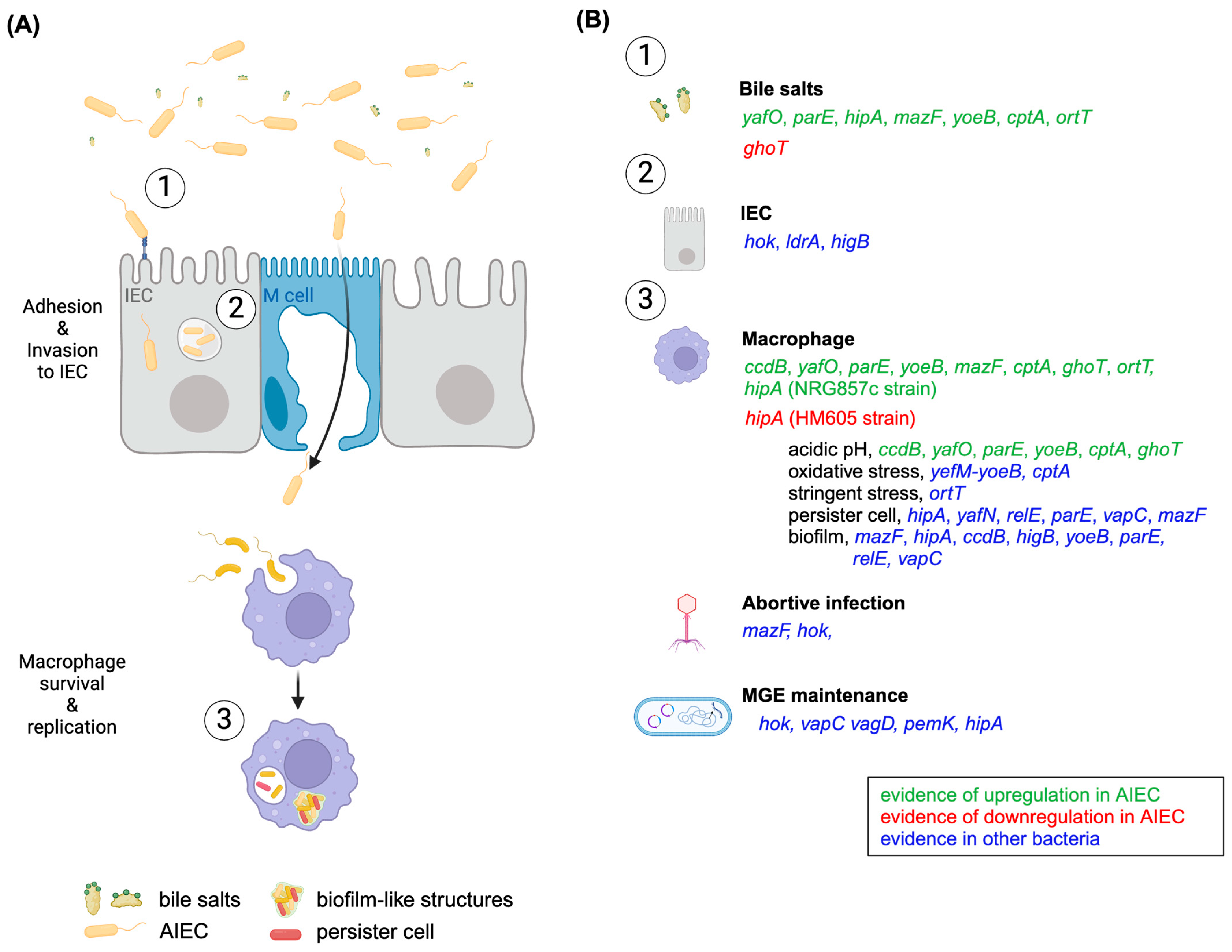
3. Putative TA Roles in AIEC’s Pathogenesis

The array of TAs in AIEC became known some time ago [9] with the revelation that the NRG857c chromosome contains a minimum of 33 TAs belonging to types I, II, IV, and V. Some of these TA toxin genes exhibited in vitro responsiveness to bile salts and acid stress, as well as to conditions found within macrophages [9]. A recently updated online TA database, TADB 3.0 [40], reports 16 and 20 TAs in LF82 and NRG857c genomes, respectively, in both the chromosome and extrachromosomal plasmids (Table 1 and Table 2). Discrepancies in NRG857c between both sources are due to non-annotated genes that are not contained in TADB 3.0, as well as novel genes (type VIII SdsR-RyeA) or plasmid systems not considered in the first report [9]. Nevertheless, these findings suggest that AIEC possess a responsive arsenal of TAs, which are intriguing bacterial factors whose exploration can deepen our comprehension of AIEC’s pathogenesis and their involvement in the chronicity of CD.
TAs reportedly control a broad range of cell transition states in response to various environmental stresses and are involved in different biological processes. However, there are four currently accepted bona fide TA roles in bacterial cell physiology, including growth diminution during stress, phage inhibition, MGE maintenance, and biofilm formation [2]. All of them may play a role in AIEC’s pathogenesis and are discussed here.

3.1. Roles in Stress Response

Although the physiological role of TAs as stress response modules is questioned [5], there are several reports of TAs playing important roles in pathogenic bacteria activity in response to various environmental stress, with not all TAs responding to the same stress [8,10,41].
The presence of bile salts is a host signal encountered by enteric bacteria as they travel through the gastrointestinal tract, and enteric pathogens utilize bile as a signal to modulate virulence factor expression [42]. Accordingly, in the presence of bile salts, AIEC induce long polar fimbriae expression to allow the bacteria to interact with Peyer’s patches and M cells [43]; they undergo metabolic adaptations [23], and some of their chromosomal TA toxins are upregulated [9]. Homologs of toxin genes yafO, parE, hipA, mazF, yoeB, cptA, and ortT, are upregulated in response to bile salt treatment in both NRG857c and another phylogenetically distant AIEC strain, HM605 [9,44].
In addition to bile salts, AIEC encounter diverse stressful conditions during host infection, as the intracellular environment of macrophages is a great threat to survival. In this setting, AIEC must face acid stress and oxidative stress, toxic metal cations, and antimicrobial peptides. Under in vitro acidic conditions and intramacrophage conditions, AIEC respond, inducing expression of an arsenal of toxin genes, such as ccdB, yafO, parE, yoeB, mazF, cptA, ghoT, and ortT [9]. As TA genes can respond to different stress conditions, activation of different toxin genes could be triggered by diverse intramacrophage stresses, collectively contributing to AIEC survival. Indeed, toxin genes upregulated within the macrophage, such as mazF, cptA, and ortT, do not necessarily respond to acid stress in vitro [9], which suggests that they could be responsive to different stress in the intramacrophage environment.
CcdB toxin targets DNA gyrase, affecting bacterial DNA replication [45]; it belongs to the CcdAB system, which is well known for ensuring F-plasmid maintenance [46]. Its chromosomal counterpart in pathogenic E. coli O157:H7 has been found to contribute to antibiotic tolerance [47]. C-terminal residues W99, G100, and I101 are important for CcdB toxicity but not for its regulatory function as a transcriptional regulator of its own operon (as part of the TA complex with its cognate antitoxin CcdA) [48]. AIEC NRG857c harbors a CcdB W99D variant, identical to its homolog from the ExPEC CFT073 strain [9], but it is unknown if these are bona fide TA toxins. However, in Salmonella, a CcdB W99R variant is known to be non-toxic but is still expressed in vitro and within the host [41]. Similarly, in AIEC NRG857c, the ccdB gene is upregulated in bile salt and acidic stress conditions, as well as within macrophages [9]. As suggested for Salmonella [41], these CcdB toxins may be diverging and losing properties compared to their functional homologs, highlighting that their contributions to bacterial pathogenicity should be tested.
YafNO, ParDE, and YefM/YoeB all belong to the TA relBE family, a diverse family whose members are classified by similarities in secondary and/or tertiary structures [49], although they may have different properties and respond differentially to environmental stresses [49,50]. In AIEC, the exposure to bile salt and acidic stress conditions upregulates the yafO, parE, and yoeB homologs [9].
YafO is a ribosome-dependent mRNA interferase that inhibits protein synthesis [51]. E. coli yafO toxin gene is induced by several stressful conditions, including antibiotic treatment, amino acid starvation, and glucose starvation [50]. yafO is also upregulated by the SOS response [52], which is crucial for AIEC survival inside the macrophage [38]. Consequently, yafO is highly upregulated in response to bile salts and stresses inside the macrophage [9].
ParE is a DNA gyrase inhibitor that blocks DNA replication [53]. Ectopic expression of parDE from conjugative IncI and IncF plasmids found in E. coli and Salmonella species was observed to promote biofilm formation in E. coli [54]. NRG857c parE genes are encoded downstream of higA genes, which appear to be their cognate antitoxins. The HigBA system, also a member of the relBE family, is involved in tolerance to bile salts in the Gram-positive bacteria Weissella cibaria. When bacteria were exposed to bile salts, HigBA was activated, and persister cells were formed to escape the stress, improving viability [55]. In addition, HigBA was shown to be a growth regulator during DNA damage stress in Caulobacter crescentus [56]. Although they remain to be characterized, parE/higA TA pairs could be bona fide TAs and play a role as stress response elements in AIEC.
YoeB is a ribosome-dependent mRNA interferase involved in the stress response of different pathogens. In E. coli, YoeB is activated during thermal stress without eliciting growth arrest [57]. In the Gram-positive Streptococcus pneumoniae and the aquatic Edwardsiella piscicida pathogens, a yefM-yoeB deletion reduces the response to oxidative stress [58,59], while in the ExPEC isolate CFT073, yefM-yoeB enhances bladder colonization [8]. During their intramacrophage lifestyle, AIEC must face acidic and oxidative stresses to survive; in consequence, yoeB expression is induced under acidic stress and is one of the most expressed toxin genes inside macrophages [9].
Altogether, antecedents of the NRG857c and HM605 strains and other pathogens suggest that relBE members probably affect AIEC’s response to the diverse stressful conditions found during host infection.
Another relBE member corresponds to the MqsRA system, which influences E. coli during bile acid stress [60] and whose antitoxin MqsA is considered a regulator of other cell regulators [61]. However, the role of MqsRA in stress response has been questioned [62]. Wang et al. [61] proposed that MqsA regulates the general stress response through the direct transcriptional repression of the stationary phase sigma factor RpoS, reducing metabolism through mRNA decay and activating type V toxin GhoT. Homologous MqsRA was not identified in NRG857c or LF82; indeed, ghoT, a target regulated by MqsA, was downregulated in response to bile salts [9]. Conversely, the GhoT-related orphan toxin gene, ortT-1, was highly upregulated in these conditions and in different AIEC strains [9]. In E. coli, OrtT was found to be important for maintaining cell fitness during stringent stress, diminishing both growth and metabolism [63]. As AIEC rely on their stringent response to survive inside the macrophage, OrtT toxin, as well as other TA toxins, may be important to triggering survival strategies in this intracellular environment, as discussed below (see Section 3.5.2).
In E. coli, the type IV CptAB system comprises a membrane-associating toxin, CptA, that inhibits cell division by interfering with the polymerization of cytoskeletal proteins [64]. However, homologs in Serratia sp. strain ATCC 39006 [65] and Shewanella oneidensis [66] are not part of a bona fide TA system, although the genes conserve synteny and a CptA homolog still might interact with multiple cell division proteins. The homologous antitoxin CptB in S. oneidensis is required for normal growth and contributes to stress tolerance [66]. In AIEC, the cptAB system identified in silico (Table 1 and Table 2, [9]) is identical to the one characterized by Masuda et al. [64], and a homologous cptA toxin gene is slightly upregulated under bile salt and acidic stress, as well as inside macrophages [9]. On the contrary, in Acinetobacter baumannii, cptAB genes are downregulated under oxidative and antibiotic stress [67]. This emphasizes the variability within TA systems and that each system must be studied considering its natural genetic context to decipher its contribution to bacterial physiology.
Different TAs may independently provide significant advantages to AIEC within specific host environments where they must deal with diverse stresses. However, although the role of TAs in stress response has been questioned [5,62], it is important to characterize them in their native strain’s background and biological context, especially in pathogens like AIEC. Here, it is relevant to consider AIEC’s special features, which contrast with those of other E. coli strains, to uncover the real contribution of TAs to stress response, physiology, and pathogenicity.

3.2. Roles in Biofilm Formation

Biofilms consist of organized bacterial communities embedded within polysaccharide polymers, providing protection against antibiotics and evasion of host innate immunity; they can exist both extracellularly and intracellularly. Interestingly, biofilm formation can be a strategy to sustain intracellular bacterial populations inside host cells, contributing to persistence [39]. Moreover, microbial biofilms are often linked to chronic diseases like IBD [68]. Several studies indicate that TAs may influence biofilm development, although the exact mechanisms remain unclear.
AIEC are known biofilm producers [39,69] and possess TAs that can be involved in biofilm formation. In particular, AIEC possess homologs of TA mazEF, hipBA, ccdAB, higBA, yefM-yoeB, and other parE toxins (Table 1 and Table 2) that are reportedly involved in biofilm formation in other bacteria.
MazF, the toxin component of TA MazEF, is a ribosome-independent sequence-specific endoribonuclease [70]. The deletion of mazEF was observed to reduce biofilm formation in E. coli MC4100relA+, and it was suggested that TA-mediated cell death was important for optimal biofilm formation [71]. In contrast to its role in E. coli, mazF is proposed to inhibit biofilm formation and promote biofilm antibiotic tolerance in Staphylococcus aureus [72]. Moreover, the deletion of mazEF along with four more TAs (relBE, chpB, yefM-yoeB, dinJ-yafQ) influenced biofilm formation in a temporal manner in E. coli MG1655 (less biofilm formation at 8 h and more biofilm formation at 24 h). Deleting these five TA systems promotes the expression of yjgK, an uncharacterized protein that represses fimbria genes in E. coli MG1655 [73]. On the other hand, the deletion of each of these five toxins independently (mazF, relE, chpB, yoeB, and yafQ) increased early biofilm formation while overexpression of the toxins repressed biofilm formation in E. coli BW25113, suggesting the role of the antitoxins in the regulation [73].
RelE is a ribosome-dependent codon-specific endoribonuclease [74], and deletion of relBE also reduced biofilm formation in E. coli K12 [75]. Similar to the role in E. coli, deletion mutants of relBE systems formed significantly less biofilm than the wild-type strain in Vibrio cholerae, and deletion mutants of relBE and yefM-yoeB also decreased the biofilm formation in Streptococcus pneumoniae [58,76]. It has been suggested that the RelBE family influences the entire process of biofilm development in V. cholerae because different relBE deletion mutants decreased biofilm formation at different stages of biofilm development [76]. However, mazF, relE, or double deletion mutants had no effect on biofilm formation in Streptococcus mutants [77]. Moreover, yefM-yoeB inhibited biofilm formation in Edwardsiella piscicida [59], and overexpression of toxins resembling RelE and VapC in Burkholderia cenocepacia showed a positive effect on biofilm formation [78]. In addition, ectopic expression of parDE TA promoted biofilm formation in E. coli [54], and deletion of parDE in Caulobacter crescentus increased biofilm formation [79].
Homologs of mazEF, relBE, and yefM-yoeB are encoded by AIEC, and we can speculate on their similar roles in biofilm formation. In AIEC, these systems are induced in the presence of bile salts [9], a condition that also increases the transcription of genes involved in biofilm formation [23]. Notably, enteric pathogens like Salmonella and Shigella form a biofilm in the presence of bile salts [42,80], which might favor gut colonization.
higAB was found to play no role in biofilm formation in E. coli. In Pseudomonas aeruginosa, HigB toxin from the higAB system reduces biofilm formation by reducing the intracellular levels of c-di-GMP, which, in turn, induces motility [81,82]. In addition, the deletion of higAB and higB influences biofilm formation in Edwardsiella piscicida [83].
HipA is a serine/threonine kinase that phosphorylates elongation factor thermal unstable (EFTu) and inhibits protein synthesis [84]. hipBA is reported to be involved in the production of eDNA, an important structural component of biofilms in E. coli [85]. In line with this, transcriptional silencing of hipBA and ccdAB significantly reduced biofilm formation in the probiotic strain E. coli Nissle 1917 [86]. Homologs of hipA and ccdB are induced in AIEC both in the presence of bile salts and inside macrophages [9], conditions where biofilm formation is important to successfully colonize and persist.
As noted, several TAs could influence the development of biofilm formation in AIEC. Given the significance of biofilm formation in AIEC pathogenicity, exploring the involvement of TAs in this process represents a novel and clinically relevant area of research.

3.3. Role as Phage Inhibition Systems

Bacteriophages reside within the gut environment and form a major part of the gut microbiota. They selectively infect bacterial strains and naturally aid the maintenance of the gut microbiota and its composition [87]. There is growing evidence that TAs play critical roles in protecting bacteria against bacteriophages, and these systems are thought to mediate abortive infection, wherein the host cell dies in response to phage infection [88].
AIEC are susceptible to phage infection and possess TAs as part of their genomic repertoire, which may be involved in defending against phage predation [9]. Type II mazEF system and type I hok/sok system, which are included in the AIEC TA repertoire, were reported to participate in phage defense in E. coli. mazEF was described to mediate cell death as a phage P1 defense mechanism [89]. However, the role of mazEF remains uncertain because these results could not be replicated [90]. Moreover, the hok/sok system from the R1 plasmid was found to protect against the T4 phage in E. coli K12, although the mechanism is still not clear [91].
Prophages are not unusual on AIEC genomes. While some defective prophages are present on NRG857c [18], LF82 encodes five prophages considered to be complete and functional [25], and homologs have been identified in contigs from E. coli isolated from CD patients [92]. However, phage resistance assays performed with E. coli isolated from CD patients revealed that sensitivity or resistance to some tested phages was not necessarily related to the presence or absence of a particular prophage in a genome [92], which suggests a putative role for TAs in mediating abortive infection in AIEC. On the other hand, it has been hypothesized that the survival of LF82 in macrophages is partly due to its ability to control the induction level of its most active prophage [93]. Notably, inside macrophages, AIEC express an arsenal of TA toxin genes [9], and we can speculate on the role of TAs in controlling prophage induction levels in these conditions by a mechanism similar to abortive infection. Overall, TA’s activation could contribute to the survival and persistence of AIEC within the gut environment and macrophage by providing phage defense mechanisms.
In recent years, phage therapy has regained attention as a therapeutic approach to combat infectious diseases [94]. Bacteriophages that target AIEC were found to reduce DSS-induced colitis symptoms in CEABAC10 transgenic LF82-colonized mice and significantly reduce the number of AIEC in feces and in the adherent microbiota of intestinal sections [95]. Therefore, phages targeting AIEC strains are a promising new treatment for IBD, and elucidating whether their TAs mediate abortive infections is crucial for the design of efficient bacteriophage therapies.

3.4. Roles in MGEs Maintenance

Although TAs are nearly ubiquitous within bacterial genomes, individual TAs exhibit restricted gene synteny, and they are commonly part of the accessory genome. Indeed, TAs were originally discovered on plasmids and associated with the PSK of plasmid-free cells [4], leading to plasmid maintenance in the bacterial population. Nowadays, it is recognized that, regardless of their location in plasmids or chromosomes, TAs may influence the maintenance of genetic elements to which they are physically linked, such as genomic islands (GIs) [96,97,98,99]. This maintenance role may influence the host-adapted lifestyle and evolution of important pathogens [100].
Plasmids play a critical role in enabling bacteria to adapt to specific environments and stresses, as they often carry genes that confer resistance to antibiotics and/or genes associated with pathogenicity [101,102]. AIEC LF82 and NRG857c harbor different extrachromosomal plasmids, whose contribution to AIEC evolution and pathogenicity remains to be investigated. Nevertheless, plasmids seem to be ubiquitous in AIEC, as sequencing data from AIEC clinical isolates retrieved plasmid contigs similar to sequences of plasmids from pathogenic bacteria, e.g., UPEC and Salmonella [92].
AIEC LF82 contains a plasmid of size 108,379 bp (plLF82), which may be acquired via horizontal gene transfer from Yersinia or Salmonella [25]. plLF82-homologous sequences in CD-associated E. coli have also been identified [92]. A different plasmid is carried by AIEC NRG857c (pO83_CORR, 147,060 bp), resembling an antimicrobial multi-resistance plasmid [18]. According to the database TADB 3.0 [40], plLF82 is devoid of TAs, while pO83_CORR possibly carries at least three putative TAs belonging to type I hok-sok and type II VagCD and VapBC families (Table 2).
The hok/sok locus is well known for its plasmid stabilization function, but it also affects growth control and may complement the existing or defective SOS mechanism [103]. Besides being chromosomally encoded, VapBC and VagCD TA systems are abundant on plasmids from different bacterial pathogens [104,105], where they might participate as plasmid maintenance modules. VapC and VagD toxins belong to the PIN-domain family of proteins and inhibit translation through the cleavage of RNAs [106]. On Shigella, a large virulence plasmid, pINV, critical for virulence, relies on a member of the VapBC family (MvpAT) to ensure its retention inside the host [107]. In consequence, type I hok/sok and VapBC (VagCD) in pO83_CORR may function similarly to their plasmidial homologs in other bacteria, which would enhance the ability of AIEC NRG857c to establish infections and propagate the antibiotic resistance elements carried on pO83_CORR, thereby contributing to its pathogenicity.
Besides plasmids, GIs are mobile elements known to contribute to bacterial fitness and could encode genes involved in pathogenicity. AIEC carry different GIs not exclusive to the pathotype. For instance, 9 large GIs were identified in the LF82 genome [25] and 35 GIs on NRG857c, which are also highly conserved in LF82 [18], suggesting that they may encode traits relevant to the AIEC pathotype.
HipBA and PemKI type II TAs were identified on chromosomal MGEs in AIEC (Table 1 and Table 2). HipBA (hipBA-1, TA21 at [9]) is encoded close to a variable region upstream of an F9 fimbrial operon. This hipBA locus is conserved between non-pathogenic and pathogenic bacteria; in addition, a complete F9 operon is only present in pathogenic E. coli [9,108]. In UPEC, F9 fimbrial expression is regulated by H-NS and temperature; it plays a role in biofilm formation [108,109], and it provides a fitness advantage during inflammatory conditions in a mouse model [110]. The regulation and contribution of the F9 operon to AIEC pathogenicity are unknown, along with the contribution of the hipBA locus. For instance, in AIEC NRG857c, hipA-1 is upregulated under in vitro stress conditions and those found inside macrophages; on the other hand, it is completely switched off inside macrophages in the HM605 strain [9]. Differential expression regulation between AIEC strains and TAs highlights the diversity of both the AIEC pathotype and TAs, and it underscores the importance of studying the biological roles of TAs in their specific genetic contexts.
PemKI was originally considered responsible for the stable maintenance of plasmid R100 [111] but is now known to have different roles in several plasmids and bacteria [112,113]. In AIEC NRG857c, the pemKI locus (mazEF-1, TA22 at [9]) is encoded within an MGE containing the genes of a phosphoenolpyruvate-dependent sugar phosphotransferase system [9]. PemKI is known to be plasmid-encoded, its chromosomal counterpart being the MazEF system (also known as ChpBA) [114]. However, several pemKI loci have been found on plasmids and chromosomes from different bacteria [115]. These recall the mobile nature of TAs, which can jump from extrachromosomal elements to the chromosome or within the same chromosome. However, the functionality of these systems must be tested. Of note, Janczak et al. [116] reported that the location of a pemKI locus in the bacterial chromosome results in the loss of its toxicity. Thus, it is important to test whether AIEC’s mobile TAs are bona fide systems and how they contribute to bacterial pathogenicity.

3.5. Role in the Host Lifestyle

AIEC’s main pathophysiological features are the adhesion to and invasion of IECs and replication inside macrophages without inducing cell death [15,19,20]. Although some genetic factors were shown to be important for these intracellular lifestyles, much remains to be revealed. TAs were reported to be involved in bacterial pathogenicity, including intracellular survival [41,117]. Indeed, different TAs control the Salmonella lifestyle inside eukaryotic cells [41]. For AIEC, distinct microenvironments found inside different eukaryotic cells could trigger disparate arsenals of TAs. In consequence, AIEC could rely on their TA repertoire to ensure their intracellular survival, which is critical for the progression of the infection, as is discussed here.

3.5.1. Intra-IECs Lifestyle

To date, both type I and II TAs have been reported to influence bacterial intracellular survival within epithelial cells, though only a few studies have been performed on epithelial cells of intestinal origin. Within IECs, LF82 is in vacuoles or free in the host cell cytoplasm [19]; however, besides some transcriptomic studies of AIEC during in vitro infection [118], its physiological status inside IECs remains unknown. Further research on the role TAs might play in IECs’ intracellular survival is necessary.
The AIEC array of TAs previously reported to be involved in intracellular eukaryotic lifestyles in other bacteria includes hok, ldrA, and higB toxins (Table 1 and Table 2).
Several studies performed on the well-known intracellular pathogen Salmonella report that TAs control bacteria lifestyle inside eukaryotic cells. A proteomic and expression analysis confirmed that intracellular S. Typhimurium produces functional toxins encoded by type I (Hok, LdrA, and TisB) and type II (T2ST, T4ST, T5ST and VapC2) TAs in fibroblasts. Deletion mutants of hok-sokST, ldrA-rdlAST, tisB-istRST, ta4ST, and vapBC2ST showed reduced intracellular survival inside fibroblasts [41]. Notably, only the vapBC2ST deletion mutant showed reduced intracellular survival inside HeLa epithelial cells [41]. Furthermore, the deletion mutant of hha and tomB4 from type II TAs showed reduced invasion ability across HCT116 colon carcinoma cells compared to the wild-type strain in Salmonella enterica [119]. The downregulation of the master regulator hilA and the reduced expression of Salmonella Pathogenicity Island-1 (SPI-1) genes in the mutant strain is proposed to cause the decreased invasion ability [119]. Similarly, Song et al. reported the activation of type II TA PA1030/PA1029 (not yet characterized), PA1878/PA1879 (VapBC homolog), and PA4674.1/PA4674 (denoted as HigBA) during the P. aeruginosa infection of A549 lung epithelial cells. Deletion mutants of these TAs showed no difference in the adhesion ability to A549 epithelial cells compared to the wild-type strain, whereas the higB mutant strain showed reduced invasion ability [120]. Other studies performed using a model of the primary human upper airway tissue indicated that deletion mutants of type II TA toxAvapA, vapBC-1, vapXD had lower intracellular survival levels over the 8 days of co-culture compared to the wild-type strain in non-typeable Haemophilus influenzae [121,122].
On the other hand, TA disruption was also found to increase intracellular survival. Disruption of the fit (fast intracellular trafficker) locus (fitAfitB) TA caused an accelerated replication rate of Neisseria gonorrhoeae within the A431 epithelial carcinoma cell line and T84 colonic carcinoma cell line and a quick transit through the polarized T84 epithelial monolayer compared to the wild-type strain [123].
The role of TAs in intracellular survival has been demonstrated in several bacterial species, though it is very limited in E. coli. Deciphering the role TAs may play in intracellular survival and replication within IEC could lead to the design of new therapies for CD patients colonized by AIEC.

3.5.2. Intra-Macrophage Lifestyle

The hallmark of AIEC is their capacity to survive longer and replicate within macrophages [20]. In this intracellular environment, LF82 remains in mature phagolysosomes [35] and forms biofilm-like structures [39] to confront several stresses. As we have noted, the stress response is key to the survival of AIEC in macrophages, and some genes have been identified supporting AIEC survival and replication in this niche, including htrA, dsbA, ibeA, and hfq [34,35,36,37], as well as the SOS and stringent responses [38].
The role of TAs in the survival of bacteria inside macrophages has been studied almost exclusively in Salmonella Typhimurium. S. Typhimurium strain 12023s cells enter a non-growing state upon entry to host macrophages through a mechanism dependent on type II TAs [124]. Fourteen type II TA operons were upregulated within 30 min of Salmonella phagocytosis, and although their deletion did not impair intracellular replication rates, the deletion of these TA genes reduced the proportion of nonreplicating bacteria in infected macrophages [124]. TacT, a novel GCN5-related N-acetyltransferase (GNAT)-like toxin, was responsible for promoting the Salmonella non-growing state through acetylation of tRNA [125]. The participation of different GNAT toxins in the macrophage survival of clinical invasive strains of S. Typhimurium and Enteritidis was later revealed [126]. In addition to GNAT toxins, Rhs toxins were shown to comprise functional type II TAs and to affect the proliferation of Salmonella during macrophage infection [127].
On the other hand, in Enterococcus faecalis, the type I ef0409-ef0408 system was shown to be involved in the infection process and survival inside host cells. A Δef0408 antitoxin mutant exhibited a hypervirulence phenotype in the infection model Galleria mellonella and in macrophages, as the mutant survived better than the wild-type [128]. Michaux et al. [128] hypothesized that free toxin ef0409 might have contributed to the hypervirulent phenotype and that ef0408 sRNA could be acting as a sensor and suppressor of ef0409 toxin activity to control growth and virulence. They proposed an equilibrium between favorable colonization (by repressing virulence) and pathogenicity according to the host environment, and sRNA such as that from type I TA could act as a key regulator in the transition from a commensal relationship to virulence [128,129].
As in Salmonella [124], an arsenal of TAs is upregulated in AIEC after phagocytosis [9]. However, GCN5 toxins have not been identified in AIEC, and currently, no Rhs toxin is characterized in this pathotype, meaning different toxins should be involved in the growth regulation of AIEC within macrophages. Among them, ccdB, yafO, hipA, parE, yoeB, mazF, cptA, ghoT, and ortT are putative candidates as they are highly upregulated after AIEC phagocytosis [9].
TAs may participate in different processes important for AIEC intramacrophage survival. For instance, they could serve as stress response modules (See Section 3.1), in the formation of intracellular biofilm-like structures (see Section 3.2), and as phage inhibition systems (see Section 3.3). However, forming persister cells is also a strategy of AIEC to avoid macrophage killing and favor survival [38], and the participation of TAs in this process is discussed in the following subsection.

3.6. Roles in Persister Cell Formation

Bacterial persistence corresponds to a reversible phenotypic state in which a small subpopulation of bacteria remains non-replicative, which allows them to survive deadly stress conditions such as antibiotic treatments. In consequence, persister cells hinder the treatment of bacterial infections and chronic diseases [130] like CD. For instance, AIEC rely on their biofilm and persister cell formation to acquire the maximum protection against macrophage attack or antibiotics [131], which could allow them to establish a long-term survival niche within phagocytic cells in CD patients, making treatment more challenging.
Persister cells are produced spontaneously or stress-induced; however, the molecular mechanisms underlying their formation remain elusive. Activations of stringent response through (p)ppGpp [132,133], SOS response [134], and TAs [124,125] are some of the molecular mechanisms linked to persister cell formation. Nevertheless, the participation of TAs is controversial [5,135,136,137], in part because some experiments were conducted in experimental conditions not relevant to TA activation. Of note, certain TA mutants displayed altered levels of persisters in a specific biological context but not when they were studied in in vitro laboratory conditions.
Remarkably, the first gene associated with persistence corresponded to a TA toxin, hipA [138]. In E. coli HipA corresponds to a serine/threonine-protein kinase that phosphorylates glutamyl-tRNA-synthetase, leading to the accumulation of uncharged tRNA-Glu in the cell and the consequent activation of stringent response [139]. A variant HipA7 (G22S and D291A) is non-toxic and associated with a high-persistence phenotype in E. coli [132]. Similarly, a homologous HipA was associated with persistence in Caulobacter crescentus [140].
TAs other than hipA have been shown to contribute to persistence in several bacterial species, for example, yafQ/dinJT, tisAB/istR, hokB/sokB, and mqsRA in E. coli [141,142,143,144]; tacT toxin, relBE, parDE, higBA, and vapBC in Salmonella [124,125]; and smuATR in S. mutans [145]. Recently, Ma et al. described the role of the MazEF system in S. aureus chronic infection [72]. They examined MazEF in virulence using a murine model and found that mazF increases antibiotic tolerance and allows bacteria to transition from acute to chronic infection [72].
AIEC encode homologs of TAs previously reported to be involved in persister cell formation. While homologs of tacT, yafQ/dinJT, and mqsRA were not identified, hok/sok, relBE, higBA, vapBC, mazEF, and hipA genes are present on AIEC genomes (Table 1 and Table 2). Of them, mazF and hipA were shown to be upregulated inside macrophages [9], where AIEC persister cells increase [38]. Curiously, hipA homologs were highly upregulated in NRG857c under conditions found inside the macrophage but completely repressed in HM605 [9]. An AIEC HipA homolog lacks the described mutations of the high-persister HipA7 variant but instead shares some amino acid variants with its phylogenetically close ExPEC CFT073 [9].
Undoubtedly, further research on the effect TAs might have on AIEC persistence will be meaningful, with special attention paid to the contribution of hipA and its variants.

4. Conclusions

Bacteria are constantly evolving to improve their fitness and pathogenicity, so discovering new targets for antimicrobial strategies is vital in the fight against bacterial pathogens involved in infectious and chronic diseases. Conversely, understanding the mechanisms and virulence factors contributing to different pathogenesis stages may enable the rational and successful development of new treatments for those diseases.
For AIEC, potential therapeutic strategies include targeting bacterial colonization of gut mucosa using phage therapy, bacteriocins, and anti-adhesion molecules, as well as genetically engineered microbes as biosensors or delivery vehicles to potentially deliver therapeutics to disease sites [146]. However, given the special lifestyle of AIEC, which differs from that of other E. coli pathotypes, strategies to target intracellular bacteria should also be considered.
TAs have arisen as novel therapeutic targets [3] partly because there are no eukaryotic homologs, and TAs produce toxins that are not secreted but instead act only within the producing cell, disabling their microbial host from the inside. As we have reviewed here, TAs can have a role in different stages of AIEC pathogenicity, for instance, stress response, biofilm formation, phage inhibition, MGE maintenance, and persister cell formation; all are also important for AIEC intracellular lifestyles (Figure 1). Therefore, understanding the contribution of TAs to AIEC physiology and pathogenicity is meaningful.
In a previous study, the expression of an AIEC toxin array was determined in different stress conditions, such as bile salts, acidic pH, and inside macrophages [9]. However, we lack functional studies assessing the role of the toxins since the transcription of a TA does not indicate activity [147]. Moreover, since genetic context and growth conditions will undoubtedly affect the possible biological role of a TA, the contribution of TAs to AIEC pathogenicity should be verified in light of their special growth characteristics, which are different from those of laboratory E. coli strains and other E. coli pathotypes, in which most TAs were previously characterized.
Undoubtedly, although more research is needed, TAs have the potential to contribute to AIEC pathogenicity, with roles in different stages. As AIEC face changing stress conditions inside the host, each encoded TA could be activated and contribute to their fitness in different ways.

Author Contributions

Conceptualization, P.B.; writing—original draft preparation, P.B., M.N.R.-C. and M.M.-M.; writing—review and editing, P.B., M.N.R.-C. and M.M.-M.; funding acquisition, P.B. and M.M.-M. All authors have read and agreed to the published version of the manuscript.

Funding

This research was funded by Agencia Nacional de Investigación y Desarrollo (ANID), grants PAI77190004 (P.B.) and Fondecyt 11230634 (P.B.); grant PID2021-126699NB-I00/MICIU/AEI/10.13039/501100011033 and by FEDER, UE (M.M.-M.).

Data Availability Statement

No new data were created or analyzed in this study. Data sharing is not applicable to this article.

Conflicts of Interest

The authors declare no conflicts of interest.

References

  1. Gerdes, K.; Christensen, S.K.; Løbner-Olesen, A. Prokaryotic Toxin–Antitoxin Stress Response Loci. Nat. Rev. Microbiol. 2005, 3, 371–382. [Google Scholar] [CrossRef]
  2. Jurėnas, D.; Fraikin, N.; Goormaghtigh, F.; Van Melderen, L. Biology and Evolution of Bacterial Toxin–Antitoxin Systems. Nat. Rev. Microbiol. 2022, 20, 335–350. [Google Scholar] [CrossRef]
  3. Równicki, M.; Lasek, R.; Trylska, J.; Bartosik, D. Targeting Type II Toxin–Antitoxin Systems as Antibacterial Strategies. Toxins 2020, 12, 568. [Google Scholar] [CrossRef]
  4. Gerdes, K.; Rasmussen, P.B.; Molin, S. Unique Type of Plasmid Maintenance Function: Postsegregational Killing of Plasmid-Free Cells. Proc. Natl. Acad. Sci. USA 1986, 83, 3116–3120. [Google Scholar] [CrossRef]
  5. Fraikin, N.; Goormaghtigh, F.; Van Melderen, L. Type II Toxin-Antitoxin Systems: Evolution and Revolutions. J. Bacteriol. 2020, 202, e00763-19. [Google Scholar] [CrossRef]
  6. Wang, X.; Yao, J.; Sun, Y.-C.; Wood, T.K. Type VII Toxin/Antitoxin Classification System for Antitoxins That Enzymatically Neutralize Toxins. Trends Microbiol. 2021, 29, 388–393. [Google Scholar] [CrossRef]
  7. Choi, J.S.; Kim, W.; Suk, S.; Park, H.; Bak, G.; Yoon, J.; Lee, Y. The Small RNA, SdsR, Acts as a Novel Type of Toxin in Escherichia coli. RNA Biol. 2018, 15, 1319–1335. [Google Scholar] [CrossRef]
  8. Norton, J.P.; Mulvey, M.A. Toxin-Antitoxin Systems Are Important for Niche-Specific Colonization and Stress Resistance of Uropathogenic Escherichia coli. PLoS Pathog. 2012, 8, e1002954. [Google Scholar] [CrossRef]
  9. Bustamante, P.; Vidal, R. Repertoire and Diversity of Toxin—Antitoxin Systems of Crohn’s Disease-Associated Adherent-Invasive Escherichia coli. New Insight of T His Emergent E. coli Pathotype. Front. Microbiol. 2020, 11, 807. [Google Scholar] [CrossRef]
  10. Lobato-Márquez, D.; Díaz-Orejas, R.; García-del Portillo, F. Toxin-Antitoxins and Bacterial Virulence. FEMS Microbiol. Rev. 2016, 40, 592–609. [Google Scholar] [CrossRef]
  11. Abraham, C.; Cho, J.H. Inflammatory Bowel Disease. N. Engl. J. Med. 2009, 361, 2066–2078. [Google Scholar] [CrossRef] [PubMed]
  12. Ng, S.C.; Shi, H.Y.; Hamidi, N.; Underwood, F.E.; Tang, W.; Benchimol, E.I.; Panaccione, R.; Ghosh, S.; Wu, J.C.Y.; Chan, F.K.L.; et al. Worldwide Incidence and Prevalence of Inflammatory Bowel Disease in the 21st Century: A Systematic Review of Population-Based Studies. Lancet 2017, 390, 2769–2778. [Google Scholar] [CrossRef] [PubMed]
  13. Sartor, R.B. Mechanisms of Disease: Pathogenesis of Crohn’s Disease and Ulcerative Colitis. Nat. Clin. Pract. Gastroenterol. Hepatol. 2006, 3, 390–407. [Google Scholar] [CrossRef] [PubMed]
  14. Qiu, P.; Ishimoto, T.; Fu, L.; Zhang, J.; Zhang, Z.; Liu, Y. The Gut Microbiota in Inflammatory Bowel Disease. Front. Cell. Infect. Microbiol. 2022, 12, 73992. [Google Scholar] [CrossRef] [PubMed]
  15. Darfeuille-Michaud, A.; Boudeau, J.; Bulois, P.; Neut, C.; Glasser, A.-L.; Barnich, N.; Bringer, M.-A.; Swidsinski, A.; Beaugerie, L.; Colombel, J.-F. High Prevalence of Adherent-Invasive Escherichia coli Associated with Ileal Mucosa in Crohn’s Disease. Gastroenterology 2004, 127, 412–421. [Google Scholar] [CrossRef] [PubMed]
  16. López-Siles, M.; Camprubí-Font, C.; Gómez del Pulgar, E.M.; Sabat Mir, M.; Busquets, D.; Sanz, Y.; Martinez-Medina, M. Prevalence, Abundance, and Virulence of Adherent-Invasive Escherichia coli in Ulcerative Colitis, Colorectal Cancer, and Coeliac Disease. Front. Immunol. 2022, 13, 748839. [Google Scholar] [CrossRef] [PubMed]
  17. Kittana, H.; Gomes-Neto, J.C.; Heck, K.; Juritsch, A.F.; Sughroue, J.; Xian, Y.; Mantz, S.; Segura Muñoz, R.R.; Cody, L.A.; Schmaltz, R.J.; et al. Evidence for a Causal Role for Escherichia coli Strains Identified as Adherent-Invasive (AIEC) in Intestinal Inflammation. mSphere 2023, 8, e0047822. [Google Scholar] [CrossRef] [PubMed]
  18. Nash, J.H.; Villegas, A.; Kropinski, A.M.; Aguilar-Valenzuela, R.; Konczy, P.; Mascarenhas, M.; Ziebell, K.; Torres, A.G.; Karmali, M.A.; Coombes, B.K. Genome Sequence of Adherent-Invasive Escherichia coli and Comparative Genomic Analysis with Other E. coli Pathotypes. BMC Genom. 2010, 11, 667. [Google Scholar] [CrossRef] [PubMed]
  19. Boudeau, J.; Glasser, A.-L.; Masseret, E.; Joly, B.; Darfeuille-Michaud, A. Invasive Ability of an Escherichia coli Strain Isolated from the Ileal Mucosa of a Patient with Crohn’s Disease. Infect. Immun. 1999, 67, 4499–4509. [Google Scholar] [CrossRef] [PubMed]
  20. Glasser, A.-L.; Boudeau, J.; Barnich, N.; Perruchot, M.-H.; Colombel, J.-F.; Darfeuille-Michaud, A. Adherent Invasive Escherichia coli Strains from Patients with Crohn’s Disease Survive and Replicate within Macrophages without Inducing Host Cell Death. Infect. Immun. 2001, 69, 5529–5537. [Google Scholar] [CrossRef]
  21. Gibold, L.; Garenaux, E.; Dalmasso, G.; Gallucci, C.; Cia, D.; Mottet-Auselo, B.; Faïs, T.; Darfeuille-Michaud, A.; Nguyen, H.T.T.; Barnich, N.; et al. The Vat-AIEC Protease Promotes Crossing of the Intestinal Mucus Layer by Crohn’s Disease-Associated Escherichia coli. Cell Microbiol. 2016, 18, 617–631. [Google Scholar] [CrossRef] [PubMed]
  22. Vazeille, E.; Chassaing, B.; Buisson, A.; Dubois, A.; de Vallée, A.; Billard, E.; Neut, C.; Bommelaer, G.; Colombel, J.-F.; Barnich, N.; et al. GipA Factor Supports Colonization of Peyer’s Patches by Crohn’s Disease-Associated Escherichia coli. Inflamm. Bowel. Dis. 2016, 22, 68–81. [Google Scholar] [CrossRef] [PubMed]
  23. Delmas, J.; Gibold, L.; Faïs, T.; Batista, S.; Leremboure, M.; Sinel, C.; Vazeille, E.; Cattoir, V.; Buisson, A.; Barnich, N.; et al. Metabolic Adaptation of Adherent-Invasive Escherichia coli to Exposure to Bile Salts. Sci. Rep. 2019, 9, 2175. [Google Scholar] [CrossRef] [PubMed]
  24. Dogan, B.; Suzuki, H.; Herlekar, D.; Sartor, B.R.B.; Campbell, B.J.; Roberts, C.L.; Stewart, K.; Scherl, E.J.; Araz, Y.; Bitar, P.P.; et al. Inflammation-Associated Adherent-Invasive Escherichia coli Are Enriched in Pathways for Use of Propanediol and Iron and M-Cell Translocation. Inflamm. Bowel. Dis. 2014, 20, 1919–1932. [Google Scholar] [CrossRef] [PubMed]
  25. Miquel, S.; Peyretaillade, E.; Claret, L.; de Vallée, A.; Dossat, C.; Vacherie, B.; Zineb, E.H.; Segurens, B.; Barbe, V.; Sauvanet, P.; et al. Complete Genome Sequence of Crohn’s Disease-Associated Adherent-Invasive E. coli Strain LF82. PLoS ONE 2010, 5, e12714. [Google Scholar] [CrossRef] [PubMed]
  26. Viladomiu, M.; Metz, M.L.; Lima, S.F.; Jin, W.B.; Chou, L.; Guo, C.J.; Diehl, G.E.; Simpson, K.W.; Scherl, E.J.; Longman, R.S. Adherent-Invasive E. coli Metabolism of Propanediol in Crohn’s Disease Regulates Phagocytes to Drive Intestinal Inflammation. Cell Host Microbe 2021, 29, 607–619.e8. [Google Scholar] [CrossRef] [PubMed]
  27. Kitamoto, S.; Alteri, C.J.; Rodrigues, M.; Nagao-Kitamoto, H.; Sugihara, K.; Himpsl, S.D.; Bazzi, M.; Miyoshi, M.; Nishioka, T.; Hayashi, A.; et al. Dietary L-Serine Confers a Competitive Fitness Advantage to Enterobacteriaceae in the Inflamed Gut. Nat. Microbiol. 2019, 5, 116–125. [Google Scholar] [CrossRef] [PubMed]
  28. Fornelos, N.; Franzosa, E.A.; Bishai, J.; Annand, J.W.; Oka, A.; Lloyd-Price, J.; Arthur, T.D.; Garner, A.; Avila-Pacheco, J.; Haiser, H.J.; et al. Growth Effects of N-Acylethanolamines on Gut Bacteria Reflect Altered Bacterial Abundances in Inflammatory Bowel Disease. Nat. Microbiol. 2020, 5, 486–497. [Google Scholar] [CrossRef] [PubMed]
  29. Barnich, N.; Carvalho, F.A.; Glasser, A.L.; Darcha, C.; Jantscheff, P.; Allez, M.; Peeters, H.; Bommelaer, G.; Desreumaux, P.; Colombel, J.F.; et al. CEACAM6 Acts as a Receptor for Adherent-Invasive E. coli, Supporting Ileal Mucosa Colonization in Crohn Disease. J. Clin. Investig. 2007, 117, 1566–1574. [Google Scholar] [CrossRef]
  30. Dreux, N.; Denizot, J.; Martinez-Medina, M.; Mellmann, A.; Billig, M.; Kisiela, D.; Chattopadhyay, S.; Sokurenko, E.; Neut, C.; Gower-Rousseau, C.; et al. Point Mutations in FimH Adhesin of Crohn’s Disease-Associated Adherent-Invasive Escherichia coli Enhance Intestinal Inflammatory Response. PLoS Pathog. 2013, 9, e1003141. [Google Scholar] [CrossRef]
  31. Rolhion, N.; Carvalho, F.A.; Darfeuille-Michaud, A. OmpC and the ΣE Regulatory Pathway Are Involved in Adhesion and Invasion of the Crohn’s Disease-Associated Escherichia coli Strain LF82. Mol. Microbiol. 2007, 63, 1684–1700. [Google Scholar] [CrossRef] [PubMed]
  32. Low, D.; Tran, H.T.; Lee, I.A.; Dreux, N.; Kamba, A.; Reinecker, H.C.; Darfeuille-Michaud, A.; Barnich, N.; Mizoguchi, E. Chitin-Binding Domains of Escherichia coli ChiA Mediate Interactions with Intestinal Epithelial Cells in Mice with Colitis. Gastroenterology 2013, 145, 602–612.e9. [Google Scholar] [CrossRef]
  33. Sevrin, G.; Massier, S.; Chassaing, B.; Agus, A.; Delmas, J.; Denizot, J.; Billard, E.; Barnich, N. Adaptation of Adherent-Invasive E. coli to Gut Environment: Impact on Flagellum Expression and Bacterial Colonization Ability. Gut. Microbes 2020, 11, 364–380. [Google Scholar] [CrossRef] [PubMed]
  34. Bringer, M.-A.; Barnich, N.; Glasser, A.-L.; Bardot, O.; Darfeuille-Michaud, A. HtrA Stress Protein Is Involved in Intramacrophagic Replication of Adherent and Invasive Escherichia coli Strain LF82 Isolated from a Patient with Crohn’s Disease. Infect. Immun. 2005, 73, 712–721. [Google Scholar] [CrossRef] [PubMed]
  35. Bringer, M.A.; Rolhion, N.; Glasser, A.L.; Darfeuille-Michaud, A. The Oxidoreductase DsbA Plays a Key Role in the Ability of the Crohn’s Disease-Associated Adherent-Invasive Escherichia coli Strain LF82 to Resist Macrophage Killing. J. Bacteriol. 2007, 189, 4860–4871. [Google Scholar] [CrossRef]
  36. Cieza, R.J.; Hu, J.; Ross, B.N.; Sbrana, E.; Torres, A.G. The IbeA Invasin of Adherent-Invasive Escherichia coli Mediates Interaction with Intestinal Epithelia and Macrophages. Infect. Immun. 2015, 83, 1904–1918. [Google Scholar] [CrossRef] [PubMed]
  37. Simonsen, K.T.; Nielsen, G.; Bjerrum, J.V.; Kruse, T.; Kallipolitis, B.H.; Møller-Jensen, J. A Role for the RNA Chaperone Hfq in Controlling Adherent-Invasive Escherichia coli Colonization and Virulence. PLoS ONE 2011, 6, e16387. [Google Scholar] [CrossRef] [PubMed]
  38. Demarre, G.; Prudent, V.; Schenk, H.; Rousseau, E.; Bringer, M.A.; Barnich, N.; Van Nhieu, G.T.; Rimsky, S.; De Monte, S.; Espéli, O. The Crohn’s Disease-Associated Escherichia coli Strain LF82 Relies on SOS and Stringent Responses to Survive, Multiply and Tolerate Antibiotics within Macrophages. PLoS Pathog 2019, 15, e1008123. [Google Scholar] [CrossRef] [PubMed]
  39. Prudent, V.; Demarre, G.; Vazeille, E.; Wery, M.; Quenech’Du, N.; Ravet, A.; Dauverd-Girault, J.; van Dijk, E.; Bringer, M.A.; Descrimes, M.; et al. The Crohn’s Disease-Related Bacterial Strain LF82 Assembles Biofilm-like Communities to Protect Itself from Phagolysosomal Attack. Commun. Biol. 2021, 4, 627. [Google Scholar] [CrossRef]
  40. Guan, J.; Chen, Y.; Goh, Y.-X.; Wang, M.; Tai, C.; Deng, Z.; Song, J.; Ou, H.-Y. TADB 3.0: An Updated Database of Bacterial Toxin-Antitoxin Loci and Associated Mobile Genetic Elements. Nucleic Acids Res. 2024, 52, D784–D790. [Google Scholar] [CrossRef]
  41. Lobato-Márquez, D.; Moreno-Córdoba, I.; Figueroa, V.; Díaz-Orejas, R.; García-del Portillo, F. Distinct Type I and Type II Toxin-Antitoxin Modules Control Salmonella Lifestyle inside Eukaryotic Cells. Sci. Rep. 2015, 5, 9374. [Google Scholar] [CrossRef]
  42. Sistrunk, J.R.; Nickerson, K.P.; Chanin, R.B.; Rasko, D.A.; Faherty, C.S. Survival of the Fittest: How Bacterial Pathogens Utilize Bile To Enhance Infection. Clin. Microbiol. Rev. 2016, 29, 819–836. [Google Scholar] [CrossRef]
  43. Chassaing, B.; Etienne-Mesmin, L.; Bonnet, R.; Darfeuille-Michaud, A. Bile Salts Induce Long Polar Fimbriae Expression Favouring Crohn’s Disease-Associated Adherent-Invasive Escherichia coli Interaction with Peyer’s Patches. Environ. Microbiol. 2013, 15, 355–371. [Google Scholar] [CrossRef]
  44. Clarke, D.J.; Chaudhuri, R.R.; Martin, H.M.; Campbell, B.J.; Rhodes, J.M.; Constantinidou, C.; Pallen, M.J.; Loman, N.J.; Cunningham, A.F.; Browning, D.F.; et al. Complete Genome Sequence of the Crohn’s Disease-Associated Adherent-Invasive Escherichia coli Strain HM605. J. Bacteriol. 2011, 193, 4540. [Google Scholar] [CrossRef]
  45. Bernard, P.; Kézdy, K.E.; Van Melderen, L.; Steyaert, J.; Wyns, L.; Pato, M.L.; Higgins, P.N.; Couturier, M. The F Plasmid CcdB Protein Induces Efficient ATP-Dependent DNA Cleavage by Gyrase. J. Mol. Biol. 1993, 234, 534–541. [Google Scholar] [CrossRef]
  46. Ogura, T.; Hiraga, S. Mini-F Plasmid Genes That Couple Host Cell Division to Plasmid Proliferation. Proc. Natl. Acad. Sci. USA 1983, 80, 4784–4788. [Google Scholar] [CrossRef]
  47. Gupta, K.; Tripathi, A.; Sahu, A.; Varadarajan, R. Contribution of the Chromosomal CcdAB Operon to Bacterial Drug Tolerance. J. Bacteriol. 2017, 199, e00397-17. [Google Scholar] [CrossRef]
  48. Bahassi, E.M.; Salmon, M.A.; van Melderen, L.; Bernard, P.; Couturier, M. F Plasmid CcdB Killer Protein CcdB Gene Mutants Coding for Non-cytotoxic Proteins Which Retain Their Regulatory Functions. Mol. Microbiol. 1995, 15, 1031–1037. [Google Scholar] [CrossRef]
  49. Gerdes, K. Type II Toxin-Antitoxins Loci: The RelBE Family. In Prokaryotic Toxin-Antitoxins; Springer: Berlin/Heidelberg, Germany, 2013; pp. 69–92. [Google Scholar]
  50. Christensen-Dalsgaard, M.; Jørgensen, M.G.; Gerdes, K. Three New RelE-Homologous MRNA Interferases of Escherichia coli Differentially Induced by Environmental Stresses. Mol. Microbiol. 2010, 75, 333–348. [Google Scholar] [CrossRef]
  51. Zhang, Y.; Yamaguchi, Y.; Inouye, M. Characterization of YafO, an Escherichia coli Toxin. J. Biol. Chem. 2009, 284, 25522–25531. [Google Scholar] [CrossRef]
  52. Singletary, L.A.; Gibson, J.L.; Tanner, E.J.; McKenzie, G.J.; Lee, P.L.; Gonzalez, C.; Rosenberg, S.M. An SOS-Regulated Type 2 Toxin-Antitoxin System. J. Bacteriol. 2009, 191, 7456–7465. [Google Scholar] [CrossRef]
  53. Jiang, Y.; Pogliano, J.; Helinski, D.R.; Konieczny, I. ParE Toxin Encoded by the Broad-Host-Range Plasmid RK2 Is an Inhibitor of Escherichia coli Gyrase. Mol. Microbiol. 2002, 44, 971–979. [Google Scholar] [CrossRef]
  54. Kamruzzaman, M.; Iredell, J. A ParDE-Family Toxin Antitoxin System in Major Resistance Plasmids of Enterobacteriaceae Confers Antibiotic and Heat Tolerance. Sci. Rep. 2019, 9, 9872. [Google Scholar] [CrossRef]
  55. Cai, T.; Zhao, Q.; Xiang, W.; Zhu, L.; Rao, Y.; Tang, J. HigBA Toxin–Antitoxin System of Weissella Cibaria Is Involved in Response to the Bile Salt Stress. J. Sci. Food Agric. 2022, 102, 6749–6756. [Google Scholar] [CrossRef]
  56. Kirkpatrick, C.L.; Martins, D.; Redder, P.; Frandi, A.; Mignolet, J.; Chapalay, J.B.; Chambon, M.; Turcatti, G.; Viollier, P.H. Growth Control Switch by a DNA-Damage-Inducible Toxin–Antitoxin System in Caulobacter Crescentus. Nat. Microbiol. 2016, 1, 16008. [Google Scholar] [CrossRef]
  57. Janssen, B.D.; Garza-Sánchez, F.; Hayes, C.S. YoeB Toxin Is Activated during Thermal Stress. Microbiologyopen 2015, 4, 682–697. [Google Scholar] [CrossRef]
  58. Chan, W.; Domenech, M.; Moreno-Córdoba, I.; Navarro-Martínez, V.; Nieto, C.; Moscoso, M.; García, E.; Espinosa, M. The Streptococcus Pneumoniae YefM-YoeB and RelBE Toxin-Antitoxin Operons Participate in Oxidative Stress and Biofilm Formation. Toxins 2018, 10, 378. [Google Scholar] [CrossRef]
  59. Ma, D.; Gu, H.; Shi, Y.; Huang, H.; Sun, D.; Hu, Y. Edwardsiella Piscicida YefM-YoeB: A Type II Toxin-Antitoxin System That Is Related to Antibiotic Resistance, Biofilm Formation, Serum Survival, and Host Infection. Front. Microbiol. 2021, 12, 646299. [Google Scholar] [CrossRef]
  60. Kwan, B.W.; Lord, D.M.; Peti, W.; Page, R.; Benedik, M.J.; Wood, T.K. The MqsR/MqsA Toxin/Antitoxin System Protects E Scherichia Coli during Bile Acid Stress. Environ. Microbiol. 2015, 17, 3168–3181. [Google Scholar] [CrossRef]
  61. Wang, X.; Kim, Y.; Hong, S.H.; Ma, Q.; Brown, B.L.; Pu, M.; Tarone, A.M.; Benedik, M.J.; Peti, W.; Page, R.; et al. Antitoxin MqsA Helps Mediate the Bacterial General Stress Response. Nat. Chem. Biol. 2011, 7, 359–366. [Google Scholar] [CrossRef]
  62. Fraikin, N.; Rousseau, C.J.; Goeders, N.; Van Melderen, L. Reassessing the Role of the Type II MqsRA Toxin-Antitoxin System in Stress Response and Biofilm Formation: MqsA Is Transcriptionally Uncoupled from MqsR. mBio 2019, 10, e02678-19. [Google Scholar] [CrossRef] [PubMed]
  63. Islam, S.; Benedik, M.J.; Wood, T.K. Orphan Toxin OrtT (YdcX) of Escherichia coli Reduces Growth during the Stringent Response. Toxins 2015, 7, 299–321. [Google Scholar] [CrossRef] [PubMed]
  64. Masuda, H.; Tan, Q.; Awano, N.; Yamaguchi, Y.; Inouye, M. A Novel Membrane-Bound Toxin for Cell Division, CptA (YgfX), Inhibits Polymerization of Cytoskeleton Proteins, FtsZ and MreB, in Escherichia coli. FEMS Microbiol. Lett. 2012, 328, 174–181. [Google Scholar] [CrossRef]
  65. McNeil, M.B.; Iglesias-Cans, M.C.; Clulow, J.S.; Fineran, P.C. YgfX (CptA) Is a Multimeric Membrane Protein That Interacts with the Succinate Dehydrogenase Assembly Factor SdhE (YgfY). Microbiology 2013, 159, 1352–1365. [Google Scholar] [CrossRef] [PubMed]
  66. Zhang, M.-X.; Zheng, K.-L.; Tang, A.-G.; Hu, X.-X.; Guo, X.-X.; Wu, C.; Cheng, Y.-Y. YgfY Contributes to Stress Tolerance in Shewanella Oneidensis Neither as an Antitoxin Nor as a Flavinylation Factor of Succinate Dehydrogenase. Microorganisms 2021, 9, 2316. [Google Scholar] [CrossRef] [PubMed]
  67. ElBanna, S.A.; Moneib, N.A.; Aziz, R.K.; Samir, R. Genomics-Guided Identification of a Conserved CptBA-like Toxin-Antitoxin System in Acinetobacter Baumannii. J. Adv. Res. 2021, 30, 159–170. [Google Scholar] [CrossRef] [PubMed]
  68. Vestby, L.K.; Grønseth, T.; Simm, R.; Nesse, L.L. Bacterial Biofilm and Its Role in the Pathogenesis of Disease. Antibiotics 2020, 9, 59. [Google Scholar] [CrossRef] [PubMed]
  69. Martinez-Medina, M.; Naves, P.; Blanco, J.; Aldeguer, X.; Blanco, J.E.; Blanco, M.; Ponte, C.; Soriano, F.; Darfeuille-Michaud, A.; Garcia-Gil, L.J. Biofilm Formation as a Novel Phenotypic Feature of Adherent-Invasive Escherichia coli(AIEC). BMC Microbiol. 2009, 9, 202. [Google Scholar] [CrossRef]
  70. Zhang, Y.; Zhang, J.; Hoeflich, K.P.; Ikura, M.; Qing, G.; Inouye, M. MazF Cleaves Cellular MRNAs Specifically at ACA to Block Protein Synthesis in Escherichia coli. Mol. Cell. 2003, 12, 913–923. [Google Scholar] [CrossRef]
  71. Kolodkin-Gal, I.; Verdiger, R.; Shlosberg-Fedida, A.; Engelberg-Kulka, H. A Differential Effect of E. Coli Toxin-Antitoxin Systems on Cell Death in Liquid Media and Biofilm Formation. PLoS ONE 2009, 4, e6785. [Google Scholar] [CrossRef]
  72. Ma, D.; Mandell, J.B.; Donegan, N.P.; Cheung, A.L.; Ma, W.; Rothenberger, S.; Shanks, R.M.Q.; Richardson, A.R.; Urish, K.L. The Toxin-Antitoxin MazEF Drives Staphylococcus Aureus Biofilm Formation, Antibiotic Tolerance, and Chronic Infection. mBio 2019, 10, e01658-19. [Google Scholar] [CrossRef] [PubMed]
  73. Kim, Y.; Wang, X.; Ma, Q.; Zhang, X.-S.; Wood, T.K. Toxin-Antitoxin Systems in Escherichia coli Influence Biofilm Formation through YjgK (TabA) and Fimbriae. J. Bacteriol. 2009, 191, 1258–1267. [Google Scholar] [CrossRef] [PubMed]
  74. Pedersen, K.; Zavialov, A.V.; Pavlov, M.Y.; Elf, J.; Gerdes, K.; Ehrenberg, M. The Bacterial Toxin RelE Displays Codon-Specific Cleavage of MRNAs in the Ribosomal A Site. Cell 2003, 112, 131–140. [Google Scholar] [CrossRef]
  75. Kasari, V.; Kurg, K.; Margus, T.; Tenson, T.; Kaldalu, N. The Escherichia coli MqsR and YgiT Genes Encode a New Toxin-Antitoxin Pair. J. Bacteriol. 2010, 192, 2908–2919. [Google Scholar] [CrossRef] [PubMed]
  76. Wang, Y.; Wang, H.; Hay, A.J.; Zhong, Z.; Zhu, J.; Kan, B. Functional RelBE-Family Toxin-Antitoxin Pairs Affect Biofilm Maturation and Intestine Colonization in Vibrio Cholerae. PLoS ONE 2015, 10, e0135696. [Google Scholar] [CrossRef] [PubMed]
  77. Lemos, J.A.C.; Brown, T.A.; Abranches, J.; Burne, R.A. Characteristics of Streptococcus Mutans Strains Lacking the MazEF and RelBE Toxinâ-Antitoxin Modules. FEMS Microbiol. Lett. 2005, 253, 251–257. [Google Scholar] [CrossRef] [PubMed]
  78. Van Acker, H.; Sass, A.; Dhondt, I.; Nelis, H.J.; Coenye, T. Involvement of Toxin-Antitoxin Modules in Burkholderia Cenocepacia Biofilm Persistence. Pathog. Dis. 2014, 71, 326–335. [Google Scholar] [CrossRef] [PubMed]
  79. Berne, C.; Zappa, S.; Brun, Y. V EDNA-Stimulated Cell Dispersion from Caulobacter Crescentus Biofilms upon Oxygen Limitation Is Dependent on a Toxin–Antitoxin System. eLife 2023, 12, e80808. [Google Scholar] [CrossRef] [PubMed]
  80. Nickerson, K.P.; Chanin, R.B.; Sistrunk, J.R.; Rasko, D.A.; Fink, P.J.; Barry, E.M.; Nataro, J.P.; Faherty, C.S. Analysis of Shigella Flexneri Resistance, Biofilm Formation, and Transcriptional Profile in Response to Bile Salts. Infect. Immun. 2017, 85. [Google Scholar] [CrossRef]
  81. Wood, T.L.; Wood, T.K. The HigB/HigA Toxin/Antitoxin System of Pseudomonas Aeruginosa Influences the Virulence Factors Pyochelin, Pyocyanin, and Biofilm Formation. Microbiologyopen 2016, 5, 499–511. [Google Scholar] [CrossRef]
  82. Zhang, Y.; Xia, B.; Li, M.; Shi, J.; Long, Y.; Jin, Y.; Bai, F.; Cheng, Z.; Jin, S.; Wu, W. HigB Reciprocally Controls Biofilm Formation and the Expression of Type III Secretion System Genes through Influencing the Intracellular C-Di-GMP Level in Pseudomonas Aeruginosa. Toxins 2018, 10, 424. [Google Scholar] [CrossRef] [PubMed]
  83. Xie, J.; Zhao, Q.; Huang, H.; Fang, Z.; Hu, Y. Edwardsiella Piscicida HigB: A Type II Toxin That Is Essential to Oxidative Resistance, Biofilm Formation, Serum Survival, Intracellular Propagation, and Host Infection. Aquaculture 2021, 535, 736382. [Google Scholar] [CrossRef]
  84. Hansen, S.; Vulić, M.; Min, J.; Yen, T.-J.; Schumacher, M.A.; Brennan, R.G.; Lewis, K. Regulation of the Escherichia coli HipBA Toxin-Antitoxin System by Proteolysis. PLoS ONE 2012, 7, e39185. [Google Scholar] [CrossRef] [PubMed]
  85. Zhao, J.; Wang, Q.; Li, M.; Heijstra, B.D.; Wang, S.; Liang, Q.; Qi, Q. Escherichia coli Toxin Gene HipA Affects Biofilm Formation and DNA Release. Microbiology 2013, 159, 633–640. [Google Scholar] [CrossRef] [PubMed]
  86. Xu, J.; Xia, K.; Li, P.; Qian, C.; Li, Y.; Liang, X. Functional Investigation of the Chromosomal CcdAB and HipAB Operon in Escherichia coli Nissle 1917. Appl. Microbiol. Biotechnol. 2020, 104, 6731–6747. [Google Scholar] [CrossRef]
  87. Sutton, T.D.S.; Hill, C. Gut Bacteriophage: Current Understanding and Challenges. Front. Endocrinol. 2019, 10, 784. [Google Scholar] [CrossRef] [PubMed]
  88. LeRoux, M.; Laub, M.T. Toxin-Antitoxin Systems as Phage Defense Elements. Annu. Rev. Microbiol. 2022, 76, 21–43. [Google Scholar] [CrossRef]
  89. Hazan, R.; Engelberg-Kulka, H. Escherichia coli MazEF-Mediated Cell Death as a Defense Mechanism That Inhibits the Spread of Phage P1. Mol. Genet. Genom. 2004, 272, 227–234. [Google Scholar] [CrossRef] [PubMed]
  90. Guegler, C.K.; Laub, M.T. Shutoff of Host Transcription Triggers a Toxin-Antitoxin System to Cleave Phage RNA and Abort Infection. Mol. Cell 2021, 81, 2361–2373.e9. [Google Scholar] [CrossRef]
  91. Pecota, D.C.; Wood, T.K. Exclusion of T4 Phage by the Hok/Sok Killer Locus from Plasmid R1. J. Bacteriol. 1996, 178, 2044–2050. [Google Scholar] [CrossRef]
  92. Rakitina, D.V.; Manolov, A.I.; Kanygina, A.V.; Garushyants, S.K.; Baikova, J.P.; Alexeev, D.G.; Ladygina, V.G.; Kostryukova, E.S.; Larin, A.K.; Semashko, T.A.; et al. Genome Analysis of E. coli Isolated from Crohn’s Disease Patients. BMC Genom. 2017, 18, 544. [Google Scholar] [CrossRef] [PubMed]
  93. Misson, P.; Bruder, E.; Cornuault, J.K.; De Paepe, M.; Nicolas, P.; Demarre, G.; Lakisic, G.; Petit, M.-A.; Espeli, O.; Lecointe, F. Phage Production Is Blocked in the Adherent-Invasive Escherichia coli LF82 upon Macrophage Infection. PLoS Pathog. 2023, 19, e1011127. [Google Scholar] [CrossRef] [PubMed]
  94. Gutiérrez, B.; Domingo-Calap, P. Phage Therapy in Gastrointestinal Diseases. Microorganisms 2020, 8, 1420. [Google Scholar] [CrossRef] [PubMed]
  95. Galtier, M.; De Sordi, L.; Sivignon, A.; de Vallée, A.; Maura, D.; Neut, C.; Rahmouni, O.; Wannerberger, K.; Darfeuille-Michaud, A.; Desreumaux, P.; et al. Bacteriophages Targeting Adherent Invasive Escherichia coli Strains as a Promising New Treatment for Crohn’s Disease. J. Crohns Colitis 2017, 11, jjw224. [Google Scholar] [CrossRef] [PubMed]
  96. Zhao, Y.; Wang, W.; Yao, J.; Wang, X.; Liu, D.; Wang, P. The HipAB Toxin-Antitoxin System Stabilizes a Composite Genomic Island in Shewanella Putrefaciens CN-32. Front. Microbiol. 2022, 13, 858857. [Google Scholar] [CrossRef]
  97. Yao, X.; Chen, T.; Shen, X.; Zhao, Y.; Wang, M.; Rao, X.; Yin, S.; Wang, J.; Gong, Y.; Lu, S.; et al. The Chromosomal SezAT Toxin-Antitoxin System Promotes the Maintenance of the SsPI-1 Pathogenicity Island in Epidemic Streptococcus Suis. Mol. Microbiol. 2015, 98, 243–257. [Google Scholar] [CrossRef] [PubMed]
  98. Huguet, K.T.; Gonnet, M.; Doublet, B.; Cloeckaert, A. A Toxin Antitoxin System Promotes the Maintenance of the IncA/C-Mobilizable Salmonella Genomic Island 1. Sci. Rep. 2016, 6, 32285. [Google Scholar] [CrossRef] [PubMed]
  99. Wozniak, R.A.F.; Waldor, M.K. A Toxin-Antitoxin System Promotes the Maintenance of an Integrative Conjugative Element. PLoS Genet. 2009, 5, e1000439. [Google Scholar] [CrossRef] [PubMed]
  100. McVicker, G.; Tang, C.M. Deletion of Toxin–Antitoxin Systems in the Evolution of Shigella Sonnei as a Host-Adapted Pathogen. Nat. Microbiol. 2016, 2, 16204. [Google Scholar] [CrossRef]
  101. Mathers, A.J.; Peirano, G.; Pitout, J.D.D. The Role of Epidemic Resistance Plasmids and International High-Risk Clones in the Spread of Multidrug-Resistant Enterobacteriaceae. Clin. Microbiol. Rev. 2015, 28, 565–591. [Google Scholar] [CrossRef]
  102. Pilla, G.; Tang, C.M. Going around in Circles: Virulence Plasmids in Enteric Pathogens. Nat. Rev. Microbiol. 2018, 16, 484–495. [Google Scholar] [CrossRef] [PubMed]
  103. Chukwudi, C.U.; Good, L. The Role of the Hok/Sok Locus in Bacterial Response to Stressful Growth Conditions. Microb. Pathog. 2015, 79, 70–79. [Google Scholar] [CrossRef] [PubMed]
  104. Duprilot, M.; Decre, D.; Genel, N.; Drieux, L.; Sougakoff, W.; Arlet, G. Diversity and Functionality of Plasmid-Borne VagCD Toxin-Antitoxin Systems of Klebsiella Pneumoniae. J. Antimicrob. Chemother. 2017, 72, 1320–1326. [Google Scholar] [CrossRef] [PubMed]
  105. Bustamante, P.; Iredell, J.R. Carriage of Type II Toxin-Antitoxin Systems by the Growing Group of IncX Plasmids. Plasmid 2017, 91, 19–27. [Google Scholar] [CrossRef] [PubMed]
  106. Arcus, V.L.; Mckenzie, J.L.; Robson, J.; Cook, G.M. The PIN-Domain Ribonucleases and the Prokaryotic VapBC Toxin-Antitoxin Array. Protein. Eng. Des. Sel. 2011, 24, 33–40. [Google Scholar] [CrossRef] [PubMed]
  107. Sayeed, S.; Brendler, T.; Davis, M.; Reaves, L.; Austin, S. Surprising Dependence on Postsegregational Killing of Host Cells for Maintenance of the Large Virulence Plasmid of Shigella Flexneri. J. Bacteriol. 2005, 187, 2768–2773. [Google Scholar] [CrossRef] [PubMed]
  108. Wurpel, D.J.; Beatson, S.A.; Totsika, M.; Petty, N.K.; Schembri, M.A. Chaperone-Usher Fimbriae of Escherichia coli. PLoS ONE 2013, 8, e52835. [Google Scholar] [CrossRef] [PubMed]
  109. Ulett, G.C.; Mabbett, A.N.; Fung, K.C.; Webb, R.I.; Schembri, M.A. The Role of F9 Fimbriae of Uropathogenic Escherichia coli in Biofilm Formation. Microbiology 2007, 153, 2321–2331. [Google Scholar] [CrossRef] [PubMed]
  110. Conover, M.S.; Ruer, S.; Taganna, J.; Kalas, V.; De Greve, H.; Pinkner, J.S.; Dodson, K.W.; Remaut, H.; Hultgren, S.J. Inflammation-Induced Adhesin-Receptor Interaction Provides a Fitness Advantage to Uropathogenic E. coli during Chronic Infection. Cell Host Microbe 2016, 20, 482–492. [Google Scholar] [CrossRef]
  111. Tsuchimoto, S.; Ohtsubo, H.; Ohtsubo, E. Two Genes, PemK and PemI, Responsible for Stable Maintenance of Resistance Plasmid R100. J. Bacteriol. 1988, 170, 1461–1466. [Google Scholar] [CrossRef]
  112. Bukowski, M.; Lyzen, R.; Helbin, W.M.; Bonar, E.; Szalewska-Palasz, A.; Wegrzyn, G.; Dubin, G.; Dubin, A.; Wladyka, B. A Regulatory Role for Staphylococcus Aureus Toxin–Antitoxin System PemIKSa. Nat. Commun. 2013, 4, 2012. [Google Scholar] [CrossRef] [PubMed]
  113. Bleriot, I.; Blasco, L.; Pacios, O.; Fernández-García, L.; Ambroa, A.; López, M.; Ortiz-Cartagena, C.; Cuenca, F.F.; Oteo-Iglesias, J.; Pascual, Á.; et al. The Role of PemIK (PemK/PemI) Type II TA System from Klebsiella Pneumoniae Clinical Strains in Lytic Phage Infection. Sci. Rep. 2022, 12, 4488. [Google Scholar] [CrossRef]
  114. Masuda, Y.; Miyakawa, K.; Nishimura, Y.; Ohtsubo, E. ChpA and ChpB, Escherichia coli Chromosomal Homologs of the Pem Locus Responsible for Stable Maintenance of Plasmid R100. J. Bacteriol. 1993, 175, 6850–6856. [Google Scholar] [CrossRef] [PubMed]
  115. Bukowski, M.; Hyz, K.; Janczak, M.; Hydzik, M.; Dubin, G.; Wladyka, B. Identification of Novel MazEF/PemIK Family Toxin-Antitoxin Loci and Their Distribution in the Staphylococcus Genus. Sci. Rep. 2017, 7, 13462. [Google Scholar] [CrossRef] [PubMed]
  116. Janczak, M.; Hyz, K.; Bukowski, M.; Lyzen, R.; Hydzik, M.; Wegrzyn, G.; Szalewska-Palasz, A.; Grudnik, P.; Dubin, G.; Wladyka, B. Chromosomal Localization of PemIK Toxin-Antitoxin System Results in the Loss of Toxicity—Characterization of PemIK-Sp from Staphylococcus Pseudintermedius. Microbiol. Res. 2020, 240, 126529. [Google Scholar] [CrossRef] [PubMed]
  117. Audoly, G.; Vincentelli, R.; Edouard, S.; Georgiades, K.; Mediannikov, O.; Gimenez, G.; Socolovschi, C.; Mège, J.-L.; Cambillau, C.; Raoult, D. Effect of Rickettsial Toxin VapC on Its Eukaryotic Host. PLoS ONE 2011, 6, e26528. [Google Scholar] [CrossRef] [PubMed]
  118. Bonet-Rossinyol, Q.; Camprubí-Font, C.; López-Siles, M.; Martinez-Medina, M. Identification of Differences in Gene Expression Implicated in the Adherent-Invasive Escherichia coli Phenotype during in Vitro Infection of Intestinal Epithelial Cells. Front. Cell. Infect. Microbiol. 2023, 13, 1228159. [Google Scholar] [CrossRef] [PubMed]
  119. Paul, P.; Patel, P.; Verma, S.K.; Mishra, P.; Sahu, B.R.; Panda, P.K.; Kushwaha, G.S.; Senapati, S.; Misra, N.; Suar, M. The Hha–TomB Toxin–Antitoxin Module in Salmonella Enterica Serovar Typhimurium Limits Its Intracellular Survival Profile and Regulates Host Immune Response. Cell Biol. Toxicol. 2022, 38, 111–127. [Google Scholar] [CrossRef] [PubMed]
  120. Song, Y.; Tang, H.; Bao, R. Comparative Analysis of Five Type II TA Systems Identified in Pseudomonas Aeruginosa Reveals Their Contributions to Persistence and Intracellular Survival. Front. Cell. Infect. Microbiol. 2023, 13, 1127786. [Google Scholar] [CrossRef]
  121. Ren, D.; Walker, A.N.; Daines, D.A. Toxin-Antitoxin Loci VapBC-1 and VapXD Contribute to Survival and Virulence in Nontypeable Haemophilus Influenzae. BMC Microbiol. 2012, 12, 263. [Google Scholar] [CrossRef]
  122. Ren, D.; Kordis, A.A.; Sonenshine, D.E.; Daines, D.A. The ToxAvapA Toxin-Antitoxin Locus Contributes to the Survival of Nontypeable Haemophilus Influenzae during Infection. PLoS ONE 2014, 9, e91523. [Google Scholar] [CrossRef] [PubMed]
  123. Hopper, S.; Wilbur, J.S.; Vasquez, B.L.; Larson, J.; Clary, S.; Mehr, I.J.; Seifert, H.S.; So, M. Isolation of Neisseria Gonorrhoeae Mutants That Show Enhanced Trafficking across Polarized T84 Epithelial Monolayers. Infect. Immun. 2000, 68, 896–905. [Google Scholar] [CrossRef] [PubMed]
  124. Helaine, S.; Cheverton, A.M.; Watson, K.G.; Faure, L.M.; Matthews, S.A.; Holden, D.W. Internalization of Salmonella by Macrophages Induces Formation of Nonreplicating Persisters. Science 2014, 343, 204–208. [Google Scholar] [CrossRef] [PubMed]
  125. Cheverton, A.M.; Gollan, B.; Przydacz, M.; Wong, C.T.; Mylona, A.; Hare, S.A.; Helaine, S. A Salmonella Toxin Promotes Persister Formation through Acetylation of TRNA. Mol. Cell 2016, 63, 86–96. [Google Scholar] [CrossRef] [PubMed]
  126. Rycroft, J.A.; Gollan, B.; Grabe, G.J.; Hall, A.; Cheverton, A.M.; Larrouy-Maumus, G.; Hare, S.A.; Helaine, S. Activity of Acetyltransferase Toxins Involved in Salmonella Persister Formation during Macrophage Infection. Nat. Commun. 2018, 9, 1993. [Google Scholar] [CrossRef] [PubMed]
  127. Stårsta, M.; Hammarlöf, D.L.; Wäneskog, M.; Schlegel, S.; Xu, F.; Heden Gynnå, A.; Borg, M.; Herschend, S.; Koskiniemi, S. RHS-Elements Function as Type II Toxin-Antitoxin Modules That Regulate Intra-Macrophage Replication of Salmonella Typhimurium. PLoS Genet. 2020, 16, e1008607. [Google Scholar] [CrossRef] [PubMed]
  128. Michaux, C.; Hartke, A.; Martini, C.; Reiss, S.; Albrecht, D.; Budin-Verneuil, A.; Sanguinetti, M.; Engelmann, S.; Hain, T.; Verneuil, N.; et al. Involvement of Enterococcus Faecalis Small RNAs in Stress Response and Virulence. Infect. Immun. 2014, 82, 3599–3611. [Google Scholar] [CrossRef] [PubMed]
  129. Sonika, S.; Singh, S.; Mishra, S.; Verma, S. Toxin-Antitoxin Systems in Bacterial Pathogenesis. Heliyon 2023, 9, e14220. [Google Scholar] [CrossRef] [PubMed]
  130. Fisher, R.A.; Gollan, B.; Helaine, S. Persistent Bacterial Infections and Persister Cells. Nat. Rev. Microbiol. 2017, 15, 453–464. [Google Scholar] [CrossRef]
  131. Bruder, E.; Espéli, O. Escherichia coli Bacteria Associated with Crohn’s Disease Persist within Phagolysosomes. Curr. Opin. Microbiol. 2022, 70, 102206. [Google Scholar] [CrossRef]
  132. Korch, S.B.; Henderson, T.A.; Hill, T.M. Characterization of the HipA7 Allele of Escherichia coli and Evidence That High Persistence Is Governed by (p)PpGpp Synthesis. Mol. Microbiol. 2003, 50, 1199–1213. [Google Scholar] [CrossRef]
  133. Nguyen, D.; Joshi-Datar, A.; Lepine, F.; Bauerle, E.; Olakanmi, O.; Beer, K.; McKay, G.; Siehnel, R.; Schafhauser, J.; Wang, Y.; et al. Active Starvation Responses Mediate Antibiotic Tolerance in Biofilms and Nutrient-Limited Bacteria. Science 2011, 334, 982–986. [Google Scholar] [CrossRef] [PubMed]
  134. Podlesek, Z.; Žgur Bertok, D. The DNA Damage Inducible SOS Response Is a Key Player in the Generation of Bacterial Persister Cells and Population Wide Tolerance. Front. Microbiol. 2020, 11, 561210. [Google Scholar] [CrossRef]
  135. Kim, J.S.; Wood, T.K. Persistent Persister Misperceptions. Front. Microbiol. 2016, 7, 2134. [Google Scholar] [CrossRef] [PubMed]
  136. Harms, A.; Maisonneuve, E.; Gerdes, K. Mechanisms of Bacterial Persistence during Stress and Antibiotic Exposure. Science 2016, 354, aaf4268. [Google Scholar] [CrossRef] [PubMed]
  137. Harms, A.; Fino, C.; Sørensen, M.A.; Semsey, S.; Gerdes, K. Prophages and Growth Dynamics Confound Experimental Results with Antibiotic-Tolerant Persister Cells. mBio 2017, 8, e01964-17. [Google Scholar] [CrossRef] [PubMed]
  138. Moyed, H.S.; Bertrand, K.P. HipA, a Newly Recognized Gene of Escherichia coli K-12 That Affects Frequency of Persistence after Inhibition of Murein Synthesis. J. Bacteriol. 1983, 155, 768–775. [Google Scholar] [CrossRef]
  139. Kaspy, I.; Rotem, E.; Weiss, N.; Ronin, I.; Balaban, N.Q.; Glaser, G. HipA-Mediated Antibiotic Persistence via Phosphorylation of the Glutamyl-TRNA-Synthetase. Nat. Commun. 2013, 4, 3001. [Google Scholar] [CrossRef] [PubMed]
  140. Zhou, X.; Eckart, M.R.; Shapiro, L. A Bacterial Toxin Perturbs Intracellular Amino Acid Balance To Induce Persistence. mBio 2021, 12, e03020-20. [Google Scholar] [CrossRef] [PubMed]
  141. Harrison, J.J.; Wade, W.D.; Akierman, S.; Vacchi-Suzzi, C.; Stremick, C.A.; Turner, R.J.; Ceri, H. The Chromosomal Toxin Gene YafQ Is a Determinant of Multidrug Tolerance for Escherichia coli Growing in a Biofilm. Antimicrob. Agents Chemother. 2009, 53, 2253–2258. [Google Scholar] [CrossRef]
  142. Dörr, T.; Vulić, M.; Lewis, K. Ciprofloxacin Causes Persister Formation by Inducing the TisB Toxin in Escherichia coli. PLoS Biol. 2010, 8, e1000317. [Google Scholar] [CrossRef]
  143. Verstraeten, N.; Knapen, W.J.; Kint, C.I.; Liebens, V.; Van den Bergh, B.; Dewachter, L.; Michiels, J.E.; Fu, Q.; David, C.C.; Fierro, A.C.; et al. Obg and Membrane Depolarization Are Part of a Microbial Bet-Hedging Strategy That Leads to Antibiotic Tolerance. Mol. Cell 2015, 59, 9–21. [Google Scholar] [CrossRef] [PubMed]
  144. Kim, Y.; Wood, T.K. Toxins Hha and CspD and Small RNA Regulator Hfq Are Involved in Persister Cell Formation through MqsR in Escherichia coli. Biochem. Biophys. Res. Commun. 2010, 391, 209–213. [Google Scholar] [CrossRef] [PubMed]
  145. Dufour, D.; Mankovskaia, A.; Chan, Y.; Motavaze, K.; Gong, S.; Lévesque, C.M. A Tripartite Toxin-antitoxin Module Induced by Quorum Sensing Is Associated with the Persistence Phenotype in Streptococcus Mutans. Mol. Oral. Microbiol. 2018, 33, 420–429. [Google Scholar] [CrossRef] [PubMed]
  146. McKay, R.; Ghodasra, M.; Schardt, J.; Quan, D.; Pottash, A.E.; Shang, W.; Jay, S.M.; Payne, G.F.; Chang, M.W.; March, J.C.; et al. A Platform of Genetically Engineered Bacteria as Vehicles for Localized Delivery of Therapeutics: Toward Applications for Crohn’s Disease. Bioeng. Transl. Med. 2018, 3, 209–221. [Google Scholar] [CrossRef]
  147. LeRoux, M.; Culviner, P.H.; Liu, Y.J.; Littlehale, M.L.; Laub, M.T. Stress Can Induce Transcription of Toxin-Antitoxin Systems without Activating Toxin. Mol. Cell 2020, 79, 280–292.e8. [Google Scholar] [CrossRef]
Figure 1. Overview of AIEC’s pathogenic route and the TAs that could contribute to pathogenicity: (A) AIEC must outcompete the native microbiota and challenge exposure to bile salts to successfully colonize the intestinal mucosa. There, AIEC can adhere to and invade IEC, where they stay in vacuoles, as well as translocate across M cells and colonize macrophages. Once phagocytosed, AIEC form biofilm-like structures and persister cells within phagolysosomes to survive. (B) TAs could affect different stages of the AIEC pathogenicity route shown in (A) and successfully confront stress conditions. For instance, TAs could be involved in the AIEC bile salt response (number 1), IEC (number 2), and macrophage (number 3) lifestyles. Inside macrophages, TAs could be involved in the AIEC response to acidic pH, oxidative and stringent stress, and the formation of persister cells and biofilm-like structures. In addition, the roles TAs might play in abortive phage infection and MGE maintenance could also be important for AIEC pathogenicity, although there is no clear evidence regarding which pathogenicity stage they might affect. The circled numbers indicate where TAs could be involved in the scheme in (A). TA toxin gene names are given for those with evidence in the literature [9] and colored according to the legend. Figure created with BioRender.com.
Microorganisms 12 01158 g001
Table 1. TA loci of the AIEC LF82 reference strain according to TADB 3.0 [40].
TA IDToxinAntitoxinFamily/DomainComments
TA214828K8B90_RS03460 (symE)-(symR)symER/SymE (toxin)Type I
TA214832K8B90_RS03850 (hokC)-(sokC)hok-sok/-Type I
TA214852K8B90_RS22020 (ldrD)-(rdlD)ldrD-rdlD/Ldr (toxin)Type I
TA214826K8B90_RS00850 (higB)K8B90_RS00855 (higA)higBA (relBE)/HTH (antitoxin)Type II
TA214834K8B90_RS04020 (ccdB)K8B90_RS04015 (ccdA)ccdAB/CcdA (antitoxin)Type II
TA214835K8B90_RS05120 (yafO)K8B90_RS05115 (yafN)yafN-yafO (relBE)/YafO-YafNType II
TA214836K8B90_RS06040 (Hha)K8B90_RS06045 (TomB)Hha-TomB/-Type II
TA214837K8B90_RS11255 (higB)K8B90_RS11250 (higA)higBA (relBE)/HigB-HigAType II
TA214838K8B90_RS11415 (hipA)K8B90_RS11420 (hipB)hipBA/HipA-HipBType II; related to MGE
TA214847K8B90_RS14005 (pemK)K8B90_RS14010 (pemI)pemIK/PRK09812-MazEType II; related to MGE
TA214848K8B90_RS14290 (yoeB)K8B90_RS14295 (yefM)yefM-yoeB (relBE)/YoeB-YefMType II
TA214849K8B90_RS17940 (mazF)K8B90_RS17945 (mazE)mazEF/PRK09907-MazEType II
TA214851K8B90_RS19925 (yhaV)K8B90_RS19920 (prlF)prlF-yhaV (relBE)/YhaV-PrlFType II
TA214850K8B90_RS18545 (cptA)K8B90_RS18550 (cptB)cptAB/CptA (toxin)Type IV
TA214827K8B90_RS02180 (ghoT)K8B90_RS02175 (ghoS)ghoTS/ghoT-GhoSType V
TA214839-(SdsR)-(RyeA)SdsR-RyeA/-Type VIII
Table 2. TA loci of the AIEC NRG857c reference strain according to TADB 3.0 [40].
TA IDToxinAntitoxinFamily/DomainComments
TA027329NRG857_RS00075 (hokC)-(sokC)hok-sok/-Type I; TA1 at [9]
TA027349NRG857_RS17965 (ldrD)-(rdlD)ldrD-rdlD/Ldr (toxin)Type I; TA14 at [9]
TA027353NRG857_RS22450 (symE)-(symR)symER/SymE (toxin)Type I; TA16 at [9]
TA027597NRG857_RS23200 (srnB)-(sok)hok-sok/-Type I; on plasmid pO83_CORR
TA027602NRG857_RS23320 (hok)-(sok)hok-sok/-Type I; on plasmid pO83_CORR
TA027331NRG857_RS00245 (ccdB)NRG857_RS00240 (ccdA)ccdAB/CcdA (antitoxin)Type II; TA17 at [9]
TA027332NRG857_RS01300 (yafO)NRG857_RS01295 (yafN)yafN-yafO (relBE)/YafO-YafNType II; TA18 at [9]
TA027333NRG857_RS02210 (Hha)NRG857_RS02215 (TomB)Hha-TomB/-Type II
TA027334NRG857_RS07485 (higB)NRG857_RS07480 (higA)higBA (relBE)/HigB-HigAType II; TA20 at [9]
TA027335NRG857_RS07640 (hipA)NRG857_RS07645 (hipB)hipBA/HipA-HipBType II; TA21 at [9]; related to MGE
TA027344NRG857_RS10225 (pemK)NRG857_RS10230 (pemI)pemIK/PRK09812-MazEType II; TA22 at [9]; related to MGE
TA027345NRG857_RS10510 (yoeB)NRG857_RS10515 (yefM)yefM-yoeB (relBE)/YoeB-YefMType II; TA23 at [9]
TA027346NRG857_RS13925 (mazF)NRG857_RS13930 (mazE)mazEF/PRK09907-MazEType II; TA24 at [9]
TA027348NRG857_RS15890 (yhaV)NRG857_RS15885 (prlF)prlF-yhaV (relBE)/YhaV-PrlFType II; TA25 at [9]
TA027351NRG857_RS19860 (higB)NRG857_RS19865 (higA)higBA (relBE)/HTH (antitoxin)Type II; TA27 at [9]
TA027596NRG857_RS22890 (vagD)NRG857_RS22885 (vagC)vagCD/VapC-VagCType II; on the plasmid pO83_CORR
TA027601NRG857_RS23235 (vapC)NRG857_RS23230 (vapB)vapBC/VapC-VagCType II; on the plasmid pO83_CORR
TA027347NRG857_RS14535 (cptA)NRG857_RS14540 (cptB)cptAB/CptA (toxin)Type IV; TA31 at [9]
TA027352NRG857_RS21190 (ghoT)NRG857_RS21185 (ghoS)ghoTS/ghoT-GhoSType V; TA32 at [9]
TA0273361894846..1894948 (-)
-(SdsR)
-(RyeA)SdsR-RyeA/-Type VIII
Disclaimer/Publisher’s Note: The statements, opinions and data contained in all publications are solely those of the individual author(s) and contributor(s) and not of MDPI and/or the editor(s). MDPI and/or the editor(s) disclaim responsibility for any injury to people or property resulting from any ideas, methods, instructions or products referred to in the content.

