Review

The Marine Fish Gut Microbiome as a Source of Novel Bacteriocins

1
Teagasc Food Research Centre, Moorepark, P61 C996 Fermoy Cork, Ireland
2
APC Microbiome Ireland, Cork, Ireland
3
School of Microbiology, University College Cork, T12 K8AF Cork, Ireland
*
Author to whom correspondence should be addressed.
Microorganisms 2024, 12(7), 1346; https://doi.org/10.3390/microorganisms12071346
Submission received: 16 May 2024 / Revised: 21 June 2024 / Accepted: 24 June 2024 / Published: 1 July 2024
(This article belongs to the Section Systems Microbiology)

Abstract

:
The marine environment is the largest ecological habitat on Earth, albeit one of the least explored, particularly in terms of its microbial inhabitants. The marine fish gut is host to a diverse microbial community from which diverse bioactive molecules can be sourced. Due to the unique environmental pressures these microbial communities experience, the bioactive molecules they produce often evolve unique adaptations that give them diverse structures and activities, differentiating them from terrestrial homologues. Of particular interest, due to their structural and functional diversity, are the ribosomally-synthesized antimicrobial peptides (bacteriocins). With increasing pressure from emerging antibiotic-resistant disease and industrial demand for novel therapeutics, the marine fish gut microbiome represents a relatively untapped resource of novel bacteriocins that could prove beneficial to human health and aquaculture. This review presents an overview of the marine fish gut microbiome and explores its potential as a source of bacteriocins for human health with considerations for applications and future research in this area.

1. Introduction

The marine environment is the largest ecological habitat on the planet. The Earth’s oceans cover over 70% of the planet’s surface and reach depths of over 10,000 m [1]. Each layer, or zone, of the ocean is its own ecological niche that is distinguishable based on factors such as temperature, salinity and sunlight availability [2]. The top layers (epipelagic and mesopelagic zones) sustain a high abundance of diverse life and are very metabolically active. In contrast, the deep ocean layers (bathypelagic, abyssal and hadal zones) are subject to low temperatures, high hydrostatic pressure and lack of sunlight [3]. From hydrothermal vents to the deep, dark trenches, marine fish inhabit extreme environments and have as a result acquired diverse and unique physiological and molecular adaptations.
The marine environment is also a rich source of diverse natural bioactive compounds with therapeutic and economic potential. To date, there are 15 clinically approved drugs derived from marine organisms, ranging from peptides to antibodies and covering a wide variety of biological activities. Many more are in various stages of clinical trials [4]. We are in an age when there is a huge demand for novel antimicrobials and alternative treatments to antibiotics. Antimicrobial resistance (AMR) is an extremely concerning and global threat to human health. It has been estimated that in 2019 alone, AMR was associated with 4.95 million deaths [5]. Traditional antibiotic treatments have become less effective, multidrug-resistant pathogens are more common yet the development of new antibiotic-based treatments has slowed dramatically over the past few decades [6]. From 2014 to 2018 over 250 preclinical antimicrobial compounds from marine organisms were reported [7,8,9]. The majority of these compounds were polyketides (35%), terpenoids (26%), alkaloids (17%) and peptides (14%), as well as lipids, lipopeptides and several other molecule types. Just over half of these molecules were produced by bacteria or fungi, and, significantly, these producers were isolated from higher organisms, particularly from marine sponges. Yet none of these preclinical marine-derived antimicrobials are bacteriocins.
The mammalian gut microbiome is a well-established source of beneficial bacteria and compounds with therapeutic potential [10,11], and undoubtedly, this scenario is paralleled in the fish gut microbiome. There is growing interest in bioprospecting the fish gut microbiome to find novel bioactive compounds with therapeutic potential and for applications in aquaculture [12]. The gut microbiome of fish has crucial biological functions in digestion, immune system modulation, stress response, and protection from pathogens/infection [13]. Advances in our understanding of the specific functional roles of the marine fish gut microbiome has been recently reviewed by Ou et al. [14]. For example, it has been shown that microorganisms of the marine fish gut protect the host from infection by pathogenic species through the production of various metabolites and antimicrobial substances, such as bacteriocins.
Bacteriocins are low molecular weight (<10 kDa), ribosomally-synthesized peptides with antimicrobial activity produced by bacteria that are immune to their own bacteriocin. Bacteriocins are usually classified into two major classes, based mainly on the extent to which they are modified. Class I bacteriocins are those which undergo extensive post-translational modification (e.g., lantibiotics or lanthipeptides), and class II bacteriocins are unmodified or cyclic [15]. The class I lanthipeptides, for example, are synthesized as prepeptides with unique lanthionine and/or β-methyllanthionine residues that undergo extensive posttranslational modification resulting in the formation of signature (methyl)lanthionine rings. Lanthipeptides can be further divided into subclasses I–V based on the enzyme(s) involved in their biosynthesis, modification, and mode of action [16]. The class II bacteriocins are subdivided into four groups (classes IIa–d): the anti-listerial pediocin-like peptides (IIa), two-component peptides (IIb), cyclic/circular bacteriocins (IIc) and the linear and non-pediocin-like peptides (IId) [17].
Bacteriocins broadly target the cell membrane of susceptible cells (Figure 1). The hydrophobic nature and overall positive charge of bacteriocin molecules enables them to interfere with negatively-charged target cell membranes quite effectively. Though the different classes of bacteriocins have variable mechanisms of action, in general, they form pores in cell membranes leading to leakage of intracellular contents, disruption of the membrane potential and eventual cell death, or they can interfere with cellular processes by binding to specific cellular receptors [15,18]. The antimicrobial activity of bacteriocins can be described as either broad or narrow-spectrum. Broad-spectrum bacteriocins, as with conventional antibiotics, target a wide range of microbial species. This can be advantageous in treating infections wherein the causative pathogen is unknown. Consequently, however, this can cause significant disruption to commensal gut microbiota diversity and lead to an increased risk of developing chronic gut-associated diseases and antimicrobial-resistant strains [19]. Narrow-spectrum bacteriocins only target specific genera or even species. This characteristic can be utilized to develop precise agents for targeting pathogenic species without harming beneficial bacteria. For example, thuricin CD, a sactipeptide produced by Bacillus thuringiensis, was shown to effectively reduce Clostridium difficile in a distal colon model whilst having no significant effect on the overall microbial diversity, unlike the treatment with conventional antibiotics [19]. Bacteriocins are therefore emerging as promising alternatives to conventional antibiotics, not only due to their spectrum of antimicrobial activities, but also for their thermostability, and great capacity for bioengineering and generation of variants with value-added properties [15].
Attention is now turning to fish microbiomes as novel sources of bioactive molecules such as bacteriocins. Conventionally, the diversity of fish microbiomes has been determined using culture-dependent methods and has very likely been underestimated in terms of its abundance and richness. However, ongoing advances in culture-independent methodologies, such as metagenomic sequencing, are enabling more accurate analysis of this microbial niche. Because of the huge expanse of the marine environment, marine microorganisms are among the least explored and least accessible and thus the application of culture-independent metagenomic analysis becomes even more valuable. The marine fish gut microbiome is essentially an untapped reservoir for novel antimicrobials.
In this review, we discuss the diversity of the gut microbiomes of marine fish, with a focus on finfish: Chondrichthyes (cartilaginous fish) and Osteichthyes (bony fish). We also discuss current research on the discovery of bacteriocins from marine fish gut microbiota, their applications, and the current prospects of this microbial niche as a source of novel antimicrobials.

2. The Diversity of the Marine Fish Gut Microbiome

The marine fish gut is host to a diverse microbial community largely consisting of bacteria, with reported counts ranging from 104–109 colony forming units (CFU) [20], as well as fungi, archaea and viruses. This community begins to form at the fish larval stage, with early colonizers originating from the egg surface, the surrounding water and the first feed [21]. The diversity of the fish gut microbiome is then influenced by biotic and abiotic factors such as host phylogeny, trophic level (diet) and environmental salinity [22], as well as, though to a lesser extent, environmental pH and temperature [20,23]. The fish gut microbiome is an important and unique niche within the marine environment, with reports that the composition of microorganisms found in marine fish gut can differ from that of the surrounding waters and can even contain species that are rarely, if at all, found in the surrounding water [24].
The composition of the marine fish gut microbiome has already been extensively reviewed [14,20,25]. Early studies on fish microbiome diversity were limited to the culturable organisms [26,27], however, with the emergence of next-generation sequencing (NGS) and metagenomic technologies there is now a growing number of culture-independent studies that have been able to provide a more comprehensive description of the fish gut microbiome [28]. Findings from more recent studies which have utilized culture-independent methods are given in Table 1. The most frequently reported phylum is Pseudomonadota (formerly Proteobacteria), followed by Bacillota (Firmicutes), Actinomycetota (Actinobacteria) and Bacteroidota (Bacteroidetes). Other abundant phyla include Mycoplasmatota (Tenericutes), Cyanobacteriota (Cyanobacteria), Fusobacteriota (Fusobacteria) and Chloroflexota (Chloroflexi). At a lower taxonomic level, Vibrionales are frequently detected, particularly species of Vibrio and Photobacterium, as well as members of the families Moraxellaceae, Pseudomonadaceae and Micrococcaceae (Table 2). Host diet is a significant determinant of the predominant taxa detected within the gut microbiome. Results reported by Sullam et al. [22] suggested that Bacteroidales and Clostridiales dominate the gut microbiomes of herbivorous marine fish, whilst Vibrionales and Alteromonadales are dominant in carnivorous and omnivorous marine fish. Similarly, Huang et al. [29] demonstrated that host feeding habits could be differentiated by “indicators”, specific bacterial taxa. It was suggested that these indicators were associated with specific symbiotic functional activities such as the production of bioactive enzymes, thus aiding in digestion within the gut.
The majority of studies of marine fish gut microbiota have focused on 16S rRNA sequence data, profiling the bacterial portion of this community. However, one recent study analyzed whole-metagenome shotgun sequencing data from the intestines of various deep-sea fish of the northern Atlantic Ocean [30]. Overall, the most abundant phyla detected were Pseudomonadota, Bacillota, Actinomycetota and Bacteroidota, in agreement with previous studies. However, cluster analysis showed that the proportions of these phyla varied significantly between some samples. Furthermore, in one cluster a significant proportion of Eukaryota was detected, specifically Ascomycota, Basidiomycota and Euryarchaeota. In terms of the eukaryotic species, there have been suggestions that yeasts are commensals of the fish microbiome with species such as Debaryomyces hansenii, Saccharomyces cerevisiae and red-pigmented Rhodotorula dominating [25,31]. The main archaeal groups reported in studies of marine fish gut microbiota are Crenarchaeota and Euryarchaeota [32,33]. Euryarchaeota have also been identified in deep-sea fish microbiota samples [30]. These are two of the major groups of planktonic archaea found in marine environments and are key contributors to nutrient cycling in pelagic waters and deep-sea sediment [34]. It has been suggested that the digestive tract of fish is an important habitat for archaeal groups within the marine environment, particularly the obligate anaerobic Euryarchaeota [33].
Viruses are the most abundant entities on earth, and it is no different in the marine environment. It has been estimated that there are between 106 and 108 viruses per ml of seawater [35]. A recent study by Geoghegan et al. [36] used a metagenomic-based approach to characterize the viromes of several marine fish species: They identified viral sequences that represented 11 viral families, with Astroviridae, Picornaviridae, Arenviridae, Reoviridae and Hepadraviridae dominating. They suggested that many of the identified viruses were diet or microbiome -associated and that host phylogenetics is a significant determinant of virome diversity.
Efforts have been made to define a “core microbiome” for marine fish, however this has proven a difficult task given the diversity of marine fish and the factors that affect their microbiome diversity. Fish gut microbiota members can also be transient [20]. Instead, it has been suggested that, perhaps, functional diversity is more important than taxonomic/phylogenetic diversity [28,29]. Furthermore, many of these studies have noted that a high percentage of operational taxonomic units (OTUs) could not be taxonomically assigned at the genus level, indicating the novelty and high biodiscovery potential of the marine fish gut microbiomes. For example, Huang et al. [29] reported that over 70% of OTUs from coastal fish gastrointestinal samples were unassigned at genus level, whilst Johny et al. [37] reported over 90% of the deep-sea fish gut OTUs could not be assigned at genus level.
The gut microbiome of marine fish is involved in the regulation of host processes including digestion, stress and immune responses, reproduction, and metabolism [13,22]. Many of these processes are mediated by the production of microbial metabolites, including antimicrobials, and bioactive enzymes.
Table 1. The predominant phyla reported in recent culture-independent studies of marine fish gut microbiota. (phyla are listed in order of abundance where possible).
Fish SpeciesSamplePredominant PhylaRef.
Gadus morhua
(Atlantic Cod)
Intestinal
contents
Pseudomonadota,
Bacteroidota, Bacillota
[38]
Siganus fuscescens
(Mottled spinefoot rabbitfish)
Intestinal
contents
Pseudomonadota, Bacillota, Bacteroidota, Fusobacteriota, Mycoplasmatota,
Cyanobacteriota
[39]
Various White Sea
(arctic) fish
Posterior
intestine
Pseudomonadota, Bacillota, Actinomycetota, Bacteroidota, Mycoplasmatota,
Fusobacteriota
[40]
Various Mediterranean fishMidgutPseudomonadota, Bacillota, Bacteroidota, Actinobacteriota, Patescibacteria, Fusobacteriota, Planctomycetota, and
Dependentiae
[41]
Coastal fish of Hong KongGastrointestinal contentsPseudomonadota, Bacillota, Mycoplasmatota[29]
Various deep-sea fish of Atlantic OceanIntestinal
contents
Pseudomonadota, Bacteroidota, Bacillota, Actinomycetota, Ascomycota, Basidiomycota, Euryarchaeota,
Spirochaetes
[30]
Centroscyllium fabricii (Black dogfish shark)Gut contentsActinomycetota, Pseudomonadota, Acidobacteriota (Acidobacteria), Bacillota, Chloroflexota[42]
Benthobatis moresbyi (Dark Blind Ray)Gut contentsActinomycetota, Pseudomonadota, Acidobacteriota, Chloroflexota, Bacillota[37]
Ref. = reference.
Table 2. Recent culture-independent studies of marine fish gut microbiome diversity. The most abundant/predominant taxa reported at family and genus level are given.
FishSampleAbundant Genera Abundant Families Ref.
Centroscyllium fabricii (Black dogfish shark)Gut contentsAcinetobacter, Thalassobacillus, Alteromonas, Leeuwenhoekiella, Corynebacterium, Pseudonocardia, PseudomonasNR[42]
Benthobatis moresbyi (Dark Blind Ray)Gut contentsAcinetobacterMoraxellaceae, Koribacteraceae, Nitrospiraceae[37]
White Sea (arctic) fishPosterior intestineStreptococcus, Sphingomonas, Micrococcus, Chthoniobacter, Pseudomonas, Corynebacterium, Staphylococcus, Acinetobacter, Propionibacterium, Vibrio, Photobacterium, BacillusMoraxellaceae, Vibrionaceae, Pseudomonadaceae, Propionibacteriaceae, Corynebacteriaceae, Micrococcaceae[40]
Various Mediterranean fishMidgut Pseudoalteromonas, Bradyrhizobium, Diaphorobacter, Mycoplasma, Clostridium, Thaumasiovibrio, MicrobulbiferXanthobacteraceae, Comamonadaceae, Pseudoalteromonadaceae, Clostridiaceae, Vibrionaceae, Propionibacteriaceae, Staphylococcaceae, Mycoplasmataceae, Flavobacteriaceae, and Peptostreptococcaceae[41]
Various Antarctic fishGastrointestinal contentsRhodococcus, Thermus, Acinetobacter, Propionibacterium, Streptococcus, and MycoplasmaNR[32]
Coastal fish of Hong KongGastrointestinal contents Clostridium, Photobacterium, Ralstonia, Acinetobacter, Thermus, Ralstonia, NR[29]
NR = not reported, Ref. = reference.

3. Bacteriocins from Marine Fish Gut Microbiota

Bioactive molecules representing several different bacteriocin classes and bacteriocin-like peptides have been identified in isolates of marine fish gut microbiomes, as outlined in Table 3. These peptides exhibit activity against a wide range of gram-positive and gram-negative bacteria, and even against fungi in some cases. Though in many of these studies the chemical structure of these bacteriocins/peptides is yet to be determined, it again highlights the biodiscovery potential and novelty of this microbial niche. Notably, these bacteriocins are all from bacilli or lactic acid bacteria (LAB) strains, some of which are novel species or strains.

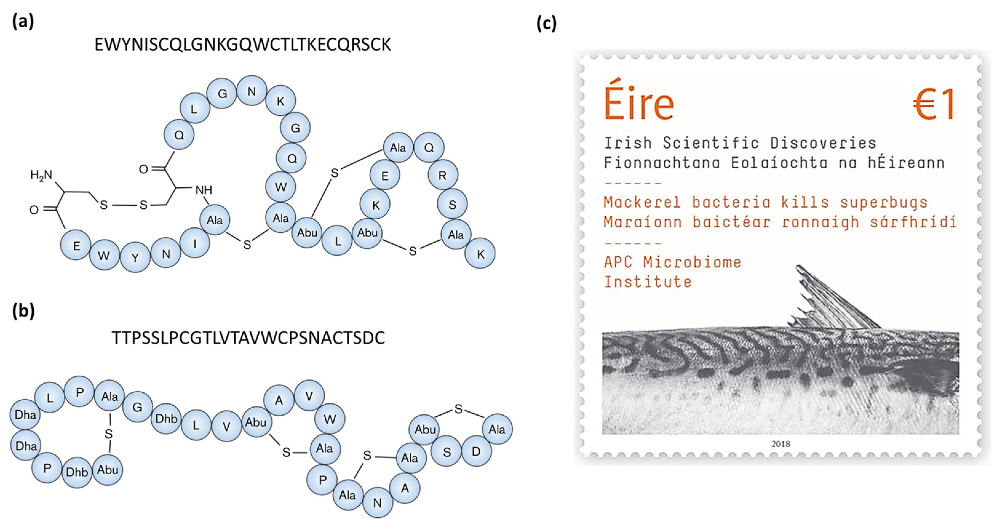
3.1. Bacteriocins from LAB

Lactic acid bacteria (LAB) are important members of the fish gut microbiome with cultivable species from marine fish including Carnobacterium spp., Lactobacillus spp., Lactococcus spp., Enterococcus spp. and Pediococcus spp. [54]. Studies have shown that beneficial LAB in the fish gut may have a significant role in promoting host fish health by improving resistance to infection through immune system modulation and the production of antimicrobial molecules, such as bacteriocins [54].
Pilet et al. [46] screened for antimicrobial-producing strains from the intestines of salmon and trout using Listeria species as target organisms. They identified two antimicrobial-producing species, Carnobacterium piscicola V1 and Carnobacterium divergens V41. C. piscicola V1 was found to produce two class IIa bacteriocins, piscicocins V1a and V1b. The purified peptides were active against the same spectrum of gram-positive indicator strains, which included C. divergens, E. faecalis and L. monocytogenes. Furthermore, piscicocin V1a was significantly more potent than V1b and was deemed to be a novel bacteriocin based on amino acid sequence analysis [47]. In a subsequent study, C. divergens V41 was found to produce divercin V41, also a novel class IIa bacteriocin. The amino acid sequence of divercin V41 was shown to be most similar to those of pediocin PA-1 and enterocin A [48]. Furthermore, piscicocins V1a and V1b and divercin V41 were optimally produced at temperatures below 20 °C and in the presence of up to 4% NaCl [55], likely an adaptation to the marine environment.
Enterococcus mundtii Tw56 was isolated from the intestine of an Odontesthes platensis specimen from the Patagonian region of Argentina, an area subject to relatively low temperatures. Cell-free supernatant (CFS) from this strain was able to inhibit gram-positive and gram-negative strains including Enterococcus spp., Listeria spp., Pseudomonas aeruginosa and Shewanella putrefaciens. It was deduced through thermal and chemical stability assays and PCR that the CFS contained mundticin KS, a class IIa bacteriocin [49].
Two separate studies found nisin Z -producing Lactococcus lactis strains from marine fish intestines with activity against the fish pathogens, Streptococcus iniae and Lactococcus garvieae, respectively [44,45]. Nisin Z is a natural variant of the class I bacteriocin (lanthipeptide) nisin A, differing only by a single amino acid (His27Asn) [56] and contains the characteristic lanthionine and methyllanthionine rings of this bacteriocin class (Figure 2). As with other nisin peptides, nisin Z acts by binding to and forming pores in the membrane of target cells leading to cell death [57]. Sequeiros et al. [45] demonstrated this in their study when they exposed L. garvieae to supernatant from the nisin Z-producing fish isolate (L. lactis TW34) and reported a drop in viable cell counts by 6 logs (CFU/mL) within one hour but without cell lysis, thus indicating bactericidal activity. Heo et al. [44] prepared partially purified nisin Z in a solution containing varying concentrations of NaCl from 0–4% w/v. They showed that nisin Z prepared in 3.5% NaCl, a concentration similar to that of seawater, was the most effective against S. iniae. This demonstrates the increased effectiveness of the marine-derived nisin Z under native conditions.
More recently, Li et al. [51] isolated a novel class IId bacteriocin, CAMT6 from the marine fish isolate Enterococcus durans YQ-6. CFS from this strain possessed a broad spectrum of activity, targeting both gram-positive and gram-negative bacteria, and also inhibited fungi. Measurement of CFS conductivity and cell morphology imaging of L. monocytogenes after exposure to CAMT6 demonstrated that the mode of action of CAMT6 involved disruption of the cell membrane, causing leakage of cell contents. Furthermore, the purified CAMT6 peptide exhibited anti-Listeria activity in a chicken breast model and anti-biofilm activity, demonstrating a potential application in food preservation. The amino acid sequence of CAMT6 is unusual in that, at 12 amino acids in length, it is much shorter than previously reported bacteriocins from enterococci and did not share homology with bacterial-derived proteins, but, rather, was most similar to antimicrobial peptides from humans and sheep.

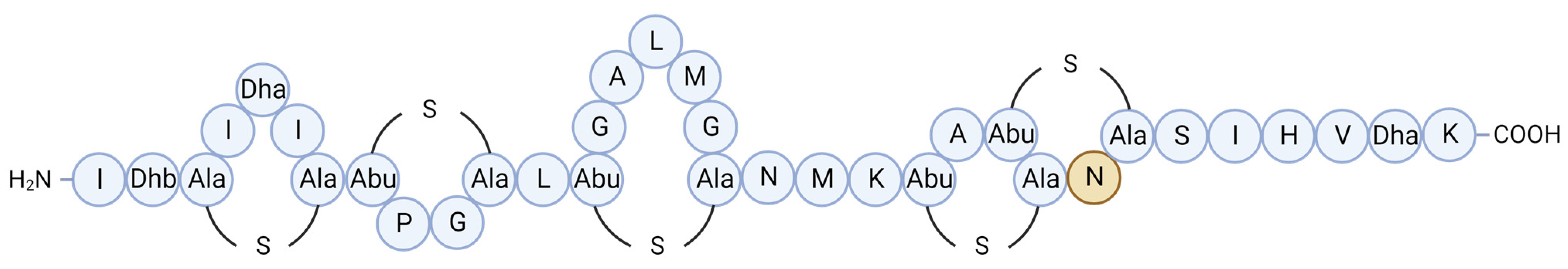
3.2. Bacteriocins from Bacilli

Bacilli isolated from marine fish have also been found to produce highly stable, and often novel, bacteriocins that can target a broad range of target strains:
Bindiya et al. (2015) screened isolates from the gut contents of a deep-sea shark (Centroscyllium fabricii) against various indicator strains. Amongst the antimicrobial producers, they identified Bacillus amyloliquefaciens BTSS-3, a strain with activity against pathogenic bacteria including Salmonella enterica Typhimurium, Clostridium perfringens, S. aureus, Proteus vulgaris, and several Bacillus species [52]. The purified antimicrobial molecule designated BaCf3, was subsequently shown to possess characteristics of being a bacteriocin: it had a mass of approximately 3000 Da, was pH tolerant (from pH 2.0–13.0) and highly thermostable (from 4–100 °C), retaining activity even after autoclaving [53]. Sequence compositional analysis found BaCf3 to be rich in hydrophobic amino acids, such as cysteine and glycine, and was predicted to contain at least one disulfide bridge. The predicted tertiary structure consisted of three anti-parallel β-sheets and resembled that of laterosporulin, a defensin-like bacteriocin from Brevibacillus sp. strain GI-9 [58,59]. Based on its hydrophobic nature and structural similarity to defensin-like bacteriocins, it was predicted that BaCf3 targeted the cell membrane. This was confirmed by microscopic analysis of Bacillus circulans target cells after exposure to BaCf3, whereby leakage of intracellular material and disruption to the cell membrane was observed [59]. Furthermore, BaCf3 was shown to also have anti-biofilm and anti-cancer activities [53,59]. This is one of the first studies that has purified a novel bacteriocin from a deep-sea fish gut isolate, and, significantly, it retained antimicrobial activity under low temperatures (4 °C) and after being subjected to high pressure and heat (autoclaving).
Formicin, a novel two-component lanthipeptide (class I bacteriocin), was discovered by screening bacterial isolates from the intestine of Atlantic mackerel (Scomber scombrus) for antimicrobial activity. The peptide, produced by a Bacillus paralicheniformis isolate, was found to inhibit a range of gram-positive indicator organisms including clinically relevant pathogens such as Listeria monocytogenes, Staphylococcus aureus, Streptococcus mutans, several species of Enterococcus and Clostridia, including Clostridioides difficile [43]. Formicin differs from other two-component lanthipeptides in that the α-peptide contains fewer hydrophobic amino acids and has an overall positive charge of +2, and the β-peptide uniquely contains a negatively charged C-terminus due to the presence of an aspartate residue in the penultimate position (Figure 3a,b). The discovery of formicin and its contribution to scientific discovery in Ireland was commemorated by the Irish national postal service (An Post) together with Science Foundation Ireland by the release of a special issue stamp in 2018 (Figure 3c).
Novel bacteriocin-like molecules derived from bacilli of the marine fish gut microbiome have also been reported, such as CAMT2 [60] and BpSl14 [61], produced by Bacillus amyloliquefaciens ZJHD-06 and Bacillus safensis, respectively. Both molecules exhibited a broad spectrum of activity, targeting both gram-negative and gram-positive strains and even fungi. Elucidation of their chemical structures found that they were more similar to eukaryotic AMPs than to bacterial peptides.
Recent studies combining in vitro and genomic screening have also highlighted the antimicrobial potential of novel Bacilli isolated from fish [62,63]. For example, Bacillus sp. GFP-2, a novel strain of Bacillus velezensis, was isolated from intestinal contents of whitespotted bamboo sharks (Chiloscyllium plagiosum) and found to inhibit the growth of Bacillus subtilis and Escherichia coli. Analysis of the genome sequence of strain GFP-2 found that it encoded genes for various AMPs, such as LCI and glyceraldehyde-3-phosphate dehydrogenase (GAPDH)-related peptides, various antimicrobial secondary metabolites including difficidin, bacillysin, macrolactin and fengycin, as well as lanthipeptides [64].

3.3. Bacteriocins from Actinobacteria

Actinobacteria are abundant in the marine environment, and those associated with higher organisms such as sponges, molluscs and fish are known to produce a wide variety of bioactive compounds with diverse biological activities [65]. One of the most significant antimicrobials from a marine-derived actinomycete is anthracimycin—an antibiotic with potent activity against Bacillus anthracis, the pathogen responsible for causing anthrax. Anthracimycin was discovered by screening Streptomyces isolates from marine sediment [66]. Indeed, Streptomyces are among the most frequently identified producers of bioactive compounds from marine environments, particularly those found in association with higher organisms [65]. Several recent studies have screened Actinobacteria from marine fish microbiomes for antimicrobial activity: Vignesh et al. [67] isolated Actinobacteria from marine fish gut samples that had antimicrobial activity against S. enterica, S. aureus and E. coli. Furthermore, one of the isolates, a Streptomyces strain, also demonstrated anti-fungal and quorum-sensing inhibitory activity. More recently, Vadivel et al. [68] isolated antimicrobial and anti-quorum sensing Streptomyces from marine fish gut samples. Several studies have highlighted that the key to identifying novel bioactive and antimicrobial compounds may now be in the “rare” Actinobacteria—those that are not as easily cultivated. These include genera such as Salinispora, Actinomadura, Microbacterium and Micrococcus [65,69]. Sanchez et al. [70] identified various Actinobacteria, including Rhodococcus, Microbacterium and Micromonospora species (which would be considered part of the “rare” Actinobacteria group) from marine fish digestive tract samples and detected antimicrobial activity against pathogenic Vibrio spp., B. subtilis, S. aureus and E. faecium. Though these studies didn’t identify specific antimicrobial molecules, they highlight the biodiscovery and antimicrobial potential of marine fish gut-derived Actinobacteria.

4. Applications of Marine Fish-Derived Bacteriocins

One of the applications for marine-derived bacteriocins is as an alternative to antibiotics in the treatment of fish diseases within the aquaculture industry [71,72]. Fish and fish products are significant, globally traded commodities, with millions of tonnes consumed each year, either directly as food, as fishmeal or for fish oil. Outbreaks of disease in commercial fish significantly impact global supply, economics and human health [73].
Many studies have also explored the application of bacteriocins from marine fish microbiomes, or the producing strains, in biopreservation of food products, such as seafood products [49,55] and chicken [51]. Schelegueda et al. [49], for example, assessed the potential use of mundticin KS from E. mundtii Tw56 in food biopreservation. The bacteriocins exhibited antimicrobial activity against Listeria innocua over a range of pH (2.0–10.0), and after heat treatment (up to 121 °C × 15 min). Furthermore, CFS from the producing strain retained full activity after storage at −30 °C for 1 year, indicating its potential for use in frozen foods. Duffes et al. [55] studied the anti-Listeria activity of the bacteriocinogenic strains, C. pisccola V1 and C. divergens V41, in vacuum-packed cold smoked salmon over a period of up to 4 weeks. In co-culture assays at 8 °C, both strains reduced the viability of L. monocytogenes by 5–7-fold (log CFU/g) compared to L. monocytogenes alone. The inhibitory effects were even greater at 4 °C, with C. piscola V1, for example, reducing the counts of L. monocytogenes to less than 10 CFU/g after 4 weeks. Crude bacteriocin extracts from the Carnobacterium strains also demonstrated effective anti-Listeria activity, particularly at 4 °C whereby L. monocytogenes was below detectable levels after 1 week. Significantly, the crude extracts were more effective at inhibiting Listeria than nisin under these conditions.
Bacteriocinogenic isolates from marine fish are generating great interest in the aquaculture industry for use as probiotics. For example, Shastry et al. [50] demonstrated that Enterococcus lactis RS5 exhibited not only bacteriocin production but also resistance to bile salts, low pH (53% viability at pH 1.5) and protease digestion—key attributes of probiotic strains. Nguyen et al. [74] explored the probiotic potential of the nisin Z-producing L. lactis WFLU12, originally isolated from the gut of olive flounder [44]. They reported that fish on a diet supplemented with 109 CFU/g of strain WFLU12, exhibited increased growth and were more resistant to infection by Streptococcus parauberis, compared to the control group.
There is also the potential for use as treatments for infection in humans; indeed, many of the bacteriocins listed in Table 3 are capable of targeting human pathogens. Formicin, for example, exhibited effective activity against several clinically relevant species including C. difficile [43]. This is particularly important in cases of antimicrobial resistance whereby treatment with conventional antibiotics has become less effective and novel alternatives are sought.

5. Challenges, Metagenomics and Future Prospects

Some of the challenges in the biodiscovery of bacteriocins from marine microbial communities include the cultivation of the producing bacteria and expression of the bacteriocin biosynthetic genes under in vitro conditions. Without the correct conditions, potentially novel marine microorganisms, and thus their metabolites, may be lost when attempting to culture in vitro [75]. Environmental pressure, temperature and dissolved oxygen concentration are just a few factors to be taken into account as they can all affect the diversity of isolated microorganisms and the metabolites they produce [76,77,78,79]. Often, modified or selective media is required and long incubation times of days, or even weeks, for antimicrobial production to occur. Sanchez et al. [70], for example, implemented the use of selective media for the isolation of bioactive marine Actinobacteria from fish. Vadivel et al. [68] also explored optimization of antimicrobial production in Streptomyces maritimus SQA4, an isolate from squid, using the “one factor at a time” method, whereby one factor or variable in the cultivation step is modified at a time. They observed that substituting carbon, nitrogen, and salt sources, as well as altering pH, had varying effects on antimicrobial activity. Furthermore, production of the bioactive metabolite was detected when the strain was cultivated on solid medium after two days but was not detected when in broth culture until after 10 days of incubation. The growth of some marine bacteria may also be dependent on the presence of specific signaling molecules found in their natural environment [80].
Culture-independent and metagenomics-based technologies, such as next-generation sequencing, metagenomic library construction, metabolomics and genome mining are more frequently being used to study microbial communities [81]. Such methods allow for a more comprehensive understanding of these microbial communities and remove the bias of sampling only culturable microbiota. Frequently used genome mining tools for identifying bacteriocins and other secondary metabolite BGCs include BAGEL and antiSMASH, both standalone and user-friendly programs [82,83]. In using BAGEL (version 3) Collins et al. [43] could identify the formicin biosynthetic operon, from which the masses of the core bacteriocin peptides could be predicted. This was a critical step in the purification and characterization of formicin, as the peptide masses found using colony mass spectrometry did not match any previously characterized bacteriocins. AntiSMASH has been used to great effect to identify microbial secondary metabolite BGCs, such as polyketides, bacteriocins and non-ribosomal peptide synthetases (NRPs), from diverse marine habits including sponges [84,85], sediment [86] and Antarctic fish [87].
Recent advancements in biodiscovery technologies have also seen the development of machine-learning -based tools for high-throughput identification and classification of ribosomally synthesized and post-translationally modified peptides (RiPPs), such as RODEO [88] and DeepRiPP [89]. Other available tools for genome-mining of RiPP BGCs and advancements in this area of research have already been extensively reviewed elsewhere [90,91].
As mentioned above, culture-independent and metagenomic methods are being used to great effect in characterizing the taxonomic diversity of the marine fish gut microbiome. Several studies have also employed the use of metagenomic sequencing to characterize functionality, the presence of antimicrobial resistance genes and bioactive metabolite genes in marine fish microbiota [92,93,94], including those from rarer deep-sea fish species (Figure 4). The culture-independent nature of metagenomics allows for a more comprehensive understanding of these gut communities and removes the bias of sampling only culturable microbiota. Few studies, however, have used metagenomics approaches for screening marine fish microbiomes for bacteriocins specifically. Yi et al. [95] used metagenomic sequencing to study functional dynamics in the gut microbiota of several aquatic animals, including marine and freshwater fish. By comparative gene sequence analysis against multiple protein databases, they identified numerous bacteriocin-associated genes, including secretion systems, and immunity-related membrane transporter systems. However, they identified few core peptide biosynthetic genes, suggesting a role in antimicrobial resistance rather than production by the microbiota.
Whilst in silico genomics screening can identify bacteriocin biosynthetic gene clusters, there is still a need for peptide purification but expression in the native host can be challenging. In fact, it has been suggested that that majority of bacterial BGCs may be “silent”, in that the genes are identifiable yet there is no corresponding bioactivity detectable during in vitro experiments [96]. An emerging technology to combat this is bacteriocin “reincarnation”. This method involves cloning of the biosynthetic operon of an “inactive” antimicrobial into a heterologous host, thereby enabling controlled expression of the antimicrobial gene. Collins et al. [97] utilized this method to reincarnate pediocin-like bacteriocin structural genes that had been identified during in silico analysis of the Lactobacillus pangenome, yet these strains did not exhibit in vitro antimicrobial activity. Ten pediocin-like bacteriocin genes were cloned with a (pediocin) signal sequence and heterologously expressed in E. coli and Lacticaseibacillus paracasei and displayed antimicrobial activity against L. innocua. This study demonstrates the potential for such methods in the expression of such “inactive” bacteriocins, that may be identified in genomic sequences of marine-fish-derived bacteria, and allow for their heterologous expression under more familiar, terrestrial, conditions.
It may be that an approach using a combination of in vitro and in silico methods is key to the discovery of more novel bacteriocins from marine fish gut microbiota.

6. Conclusions

With the increasing demand for novel antimicrobial therapeutics to combat antimicrobial resistance, there is a pressing need to source novel bioactive compounds. The marine environment is vast, with a multitude of factors affecting the diversity of marine life and their biomolecules. The marine fish gut microbiome is no exception, and it is emerging as a productive source of antimicrobial molecules, such as bacteriocins. Key aspects of marine life, such as high salinity, hydrostatic pressure and a range of environmental temperatures all have an impact on the structural and functional diversity of marine antimicrobial molecules, including bacteriocins, often distinguishing them from terrestrial homologs. Characterizing the true diversity of microorganisms in marine fish gut microbiomes and the bioactive molecules they produce still presents challenges but may be key in determining the true potential of this niche. Recent applications of culture-independent technologies, such as metagenomics and next-generation sequencing, have begun to bridge this knowledge gap and are already being used to great effect to resolve the taxonomic diversity of the fish gut microbiome. Bacteriocins from the marine fish gut can be used to combat disease, either directly or indirectly as products of probiotic strains, and for the biopreservation of food products. The marine fish gut microbiome is a potential treasure trove of novel bacteriocins and other antimicrobials but limitations in accessibility and technology have meant its true bioprospective potential has yet to be truly explored.

Author Contributions

S.U.-L., conceptualization, writing—original draft, and visualization; C.S., C.H., supervision, writing—review, and editing; R.P.R., funding acquisition, resources, supervision, writing—review, and editing. All authors have read and agreed to the published version of the manuscript.

Funding

This work was co-funded by a Teagasc Walsh Fellowship (grant number 2017218), Science Foundation Ireland (SFI) under Grant Number SFI/12/RC/2273_P2 and the European Union (ERC, BACtheWINNER, 101054719). Views and opinions expressed are, however, those of the author(s) only and do not necessarily reflect those of the European Union or the European Research Council. Neither the European Union nor the granting authority can be held responsible for them.

Institutional Review Board Statement

Not Applicable.

Data Availability Statement

Not Applicable.

Conflicts of Interest

The authors declare no conflicts of interest.

References

  1. Greenaway, S.F.; Sullivan, K.D.; Umfress, S.H.; Beittel, A.B.; Wagner, K.D. Revised depth of the Challenger Deep from submersible transects; including a general method for precise, pressure-derived depths in the ocean. Deep Sea Res. Part I Oceanogr. Res. Pap. 2021, 178, 103644. [Google Scholar] [CrossRef]
  2. National Oceanic and Atmospheric Administration USDoC. Layers of the Ocean. Available online: https://www.noaa.gov/jetstream/ocean/layers-of-ocean (accessed on 28 March 2023).
  3. Somero, G.N. Biochemical ecology of deep-sea animals. Experientia 1992, 48, 537–543. [Google Scholar] [CrossRef] [PubMed]
  4. Approved Marine Drugs. 2023. Available online: https://www.marinepharmacology.org/approved (accessed on 14 August 2023).
  5. Collaborators, A.R. Global burden of bacterial antimicrobial resistance in 2019: A systematic analysis. Lancet 2022, 399, 629–655. [Google Scholar]
  6. Frieri, M.; Kumar, K.; Boutin, A. Antibiotic resistance. J. Infect. Public Health 2017, 10, 369–378. [Google Scholar] [CrossRef] [PubMed]
  7. Mayer, A.M.S.; Guerrero, A.J.; Rodríguez, A.D.; Taglialatela-Scafati, O.; Nakamura, F.; Fusetani, N. Marine Pharmacology in 2014–2015: Marine Compounds with Antibacterial, Antidiabetic, Antifungal, Anti-Inflammatory, Antiprotozoal, Antituberculosis, Antiviral, and Anthelmintic Activities; Affecting the Immune and Nervous Systems, and Other Miscellaneous Mechanisms of Action. Mar. Drugs 2020, 18, 5. [Google Scholar]
  8. Mayer, A.M.S.; Guerrero, A.J.; Rodríguez, A.D.; Taglialatela-Scafati, O.; Nakamura, F.; Fusetani, N. Marine Pharmacology in 2016–2017: Marine Compounds with Antibacterial, Antidiabetic, Antifungal, Anti-Inflammatory, Antiprotozoal, Antituberculosis and Antiviral Activities; Affecting the Immune and Nervous Systems, and Other Miscellaneous Mechanisms of Action. Mar. Drugs 2021, 19, 49. [Google Scholar] [CrossRef]
  9. Mayer, A.M.S.; Pierce, M.L.; Howe, K.; Rodríguez, A.D.; Taglialatela-Scafati, O.; Nakamura, F.; Fusetani, N. Marine pharmacology in 2018: Marine compounds with antibacterial, antidiabetic, antifungal, anti-inflammatory, antiprotozoal, antituberculosis and antiviral activities; affecting the immune and nervous systems, and other miscellaneous mechanisms of action. Pharmacol. Res. 2022, 183, 106391. [Google Scholar] [CrossRef]
  10. Halloran, K.; Underwood, M.A. Probiotic mechanisms of action. Early Hum. Dev. 2019, 135, 58–65. [Google Scholar] [CrossRef]
  11. Wang, L.; Ravichandran, V.; Yin, Y.; Yin, J.; Zhang, Y. Natural Products from Mammalian Gut Microbiota. Trends Biotechnol. 2019, 37, 492–504. [Google Scholar] [CrossRef]
  12. Wanka, K.M.; Damerau, T.; Costas, B.; Krueger, A.; Schulz, C.; Wuertz, S. Isolation and characterization of native probiotics for fish farming. BMC Microbiol. 2018, 18, 119. [Google Scholar] [CrossRef]
  13. Butt, R.L.; Volkoff, H. Gut Microbiota and Energy Homeostasis in Fish. Front. Endocrinol. 2019, 10, 9. [Google Scholar] [CrossRef] [PubMed]
  14. Ou, W.; Yu, G.; Zhang, Y.; Mai, K. Recent progress in the understanding of the gut microbiota of marine fishes. Mar. Life Sci. Technol. 2021, 3, 434–448. [Google Scholar] [CrossRef] [PubMed]
  15. Cotter, P.D.; Ross, R.P.; Hill, C. Bacteriocins—A viable alternative to antibiotics? Nat. Rev. Microbiol. 2013, 11, 95–105. [Google Scholar] [CrossRef] [PubMed]
  16. Montalbán-López, M.; Scott, T.A.; Ramesh, S.; Rahman, I.R.; van Heel, A.J.; Viel, J.H.; Bandarian, V.; Dittmann, E.; Genilloud, O.; Goto, Y.; et al. New developments in RiPP discovery, enzymology and engineering. Nat. Prod. Rep. 2021, 38, 130–239. [Google Scholar] [CrossRef] [PubMed]
  17. Nissen-Meyer, J.; Rogne, P.; Oppegard, C.; Haugen, S.H.; Kristiansen, E.P. Structure-Function Relationships of the Non-Lanthionine-Containing Peptide (class II) Bacteriocins Produced by Gram-Positive Bacteria. Curr. Pharm. Biotechnol. 2009, 10, 19–37. [Google Scholar] [CrossRef] [PubMed]
  18. Sugrue, I.; Ross, R.P.; Hill, C. Bacteriocin diversity, function, discovery and application as antimicrobials. Nat. Rev. Microbiol. 2024. [Google Scholar] [CrossRef] [PubMed]
  19. Rea, M.C.; Sit, C.S.; Clayton, E.; O’Connor, P.M.; Whittal, R.M.; Zheng, J.; Vederas, J.C.; Ross, R.P.; Hill, C. Thuricin CD, a posttranslationally modified bacteriocin with a narrow spectrum of activity against Clostridium difficile. Proc. Natl. Acad. Sci. USA 2010, 107, 9352–9357. [Google Scholar] [CrossRef] [PubMed] [PubMed Central]
  20. Egerton, S.; Culloty, S.; Whooley, J.; Stanton, C.; Ross, R.P. The Gut Microbiota of Marine Fish. Front. Microbiol. 2018, 9, 873. [Google Scholar] [CrossRef] [PubMed]
  21. Roeselers, G.; Mittge, E.K.; Stephens, W.Z.; Parichy, D.M.; Cavanaugh, C.M.; Guillemin, K.; Rawls, J.F. Evidence for a core gut microbiota in the zebrafish. ISME J. 2011, 5, 1595–1608. [Google Scholar] [CrossRef]
  22. Sullam, K.E.; Essinger, S.D.; Lozupone, C.A.; O’Connor, M.P.; Rosen, G.L.; Knight, R.; Kilham, S.S.; Russell, J.A. Environmental and ecological factors that shape the gut bacterial communities of fish: A meta-analysis. Mol. Ecol. 2012, 21, 3363–3378. [Google Scholar] [CrossRef]
  23. Ward, N.L.; Steven, B.; Penn, K.; Methé, B.A.; Detrich, W.H. Characterization of the intestinal microbiota of two Antarctic notothenioid fish species. Extremophiles 2009, 13, 679–685. [Google Scholar] [CrossRef] [PubMed]
  24. Troussellier, M.; Escalas, A.; Bouvier, T.; Mouillot, D. Sustaining Rare Marine Microorganisms: Macroorganisms As Repositories and Dispersal Agents of Microbial Diversity. Front. Microbiol. 2017, 8, 947. [Google Scholar] [CrossRef] [PubMed]
  25. Romero, J.; Ringø, E.; Merrifield, D. The Gut Microbiota of Fish. Aquac. Nutr. Gut Health Probiotics Prebiotics 2014. [Google Scholar] [CrossRef]
  26. Yano, Y.; Nakayama, A.; Yoshida, K. Population Sizes and Growth Pressure Responses of Intestinal Microfloras of Deep-Sea Fish Retrieved from the Abyssal Zone. Appl. Environ. Microbiol. 1995, 61, 4480–4483. [Google Scholar] [CrossRef] [PubMed]
  27. Ohwada, K.; Tabor, P.S.; Colwell, R.R. Species composition and barotolerance of gut microflora of deep-sea benthic macrofauna collected at various depths in the atlantic ocean. Appl. Environ. Microbiol. 1980, 40, 746–755. [Google Scholar] [CrossRef] [PubMed]
  28. Ghanbari, M.; Kneifel, W.; Domig, K.J. A new view of the fish gut microbiome: Advances from next-generation sequencing. Aquaculture 2015, 448, 464–475. [Google Scholar] [CrossRef]
  29. Huang, Q.; Sham, R.C.; Deng, Y.; Mao, Y.; Wang, C.; Zhang, T.; Leung, K.M.Y. Diversity of gut microbiomes in marine fishes is shaped by host-related factors. Mol. Ecol. 2020, 29, 5019–5034. [Google Scholar] [CrossRef] [PubMed]
  30. Collins, F.W.J. An Investigation into Antimicrobial Production in the Lactobacillus Genus and the Fish Microbiome. Ph.D. Thesis, University College Cork, Cork, Ireland, 2019. [Google Scholar]
  31. Andlid, T.; Juárez, R.-V.; Gustafsson, L. Yeast colonizing the intestine of rainbow trout (Salmo gairdneri) and turbot (Scophtalmus maximus). Microb. Ecol. 1995, 30, 321–334. [Google Scholar] [CrossRef]
  32. Song, W.; Li, L.; Huang, H.; Jiang, K.; Zhang, F.; Chen, X.; Zhao, M.; Ma, L. The Gut Microbial Community of Antarctic Fish Detected by 16S rRNA Gene Sequence Analysis. BioMed Res. Int. 2016, 2016, 3241529. [Google Scholar] [CrossRef]
  33. van der Maarel, M.J.E.C.; Sprenger, W.; Haanstra, R.; Forney, L.J. Detection of methanogenic archaea in seawater particles and the digestive tract of a marine fish species. FEMS Microbiol. Lett. 1999, 173, 189–194. [Google Scholar] [CrossRef]
  34. Vuillemin, A.; Wankel, S.D.; Coskun, Ö.K.; Magritsch, T.; Vargas, S.; Estes, E.R.; Spivack, A.J.; Smith, D.C.; Pockalny, R.; Murray, R.W. Archaea dominate oxic subseafloor communities over multimillion-year time scales. Sci. Adv. 2019, 5, eaaw4108. [Google Scholar] [CrossRef]
  35. Suttle, C.A. Viruses in the sea. Nature 2005, 437, 356–361. [Google Scholar] [CrossRef]
  36. Geoghegan, J.L.; Di Giallonardo, F.; Wille, M.; Ortiz-Baez, A.S.; Costa, V.A.; Ghaly, T.; Mifsud, J.C.O.; Turnbull, O.M.H.; Bellwood, D.R.; Williamson, J.E.; et al. Virome composition in marine fish revealed by meta-transcriptomics. Virus Evol. 2021, 7, veab005. [Google Scholar] [CrossRef] [PubMed]
  37. Johny, T.K.; Saidumohamed, B.E.; Sasidharan, R.S.; Bhat, S.G. Inferences of gut bacterial diversity from next-generation sequencing of 16S rDNA in deep sea blind ray—Benthobatis moresbyi. Ecol. Genet. Genom. 2018, 9, 1–6. [Google Scholar] [CrossRef]
  38. Star, B.; Haverkamp, T.H.; Jentoft, S.; Jakobsen, K.S. Next generation sequencing shows high variation of the intestinal microbial species composition in Atlantic cod caught at a single location. BMC Microbiol. 2013, 13, 248. [Google Scholar] [CrossRef]
  39. Jones, J.; DiBattista, J.D.; Stat, M.; Bunce, M.; Boyce, M.C.; Fairclough, D.V.; Travers, M.J.; Huggett, M.J. The Microbiome of the Gastrointestinal Tract of a Range-Shifting Marine Herbivorous Fish. Front. Microbiol. Orig. Res. 2018, 9, 2000. [Google Scholar] [CrossRef]
  40. Burtseva, O.; Kublanovskaya, A.; Fedorenko, T.; Lobakova, E.; Chekanov, K. Gut microbiome of the White Sea fish revealed by 16S rRNA metabarcoding. Aquaculture 2021, 533, 736175. [Google Scholar] [CrossRef]
  41. Kormas, K.; Nikouli, E.; Kousteni, V.; Damalas, D. Midgut bacterial microbiota of 12 fish species from a marine protected area in the Aegean Sea (Greece). Microb. Ecol. 2023, 86, 1405–1415. [Google Scholar] [CrossRef] [PubMed]
  42. Johny, T.K.; Saidumohamed, B.E.; Sasidharan, R.S.; Bhat, S.G. Metabarcoding data of bacterial diversity of the deep sea shark, Centroscyllium fabricii. Data Brief 2018, 21, 1029–1032. [Google Scholar] [CrossRef] [PubMed]
  43. Collins, F.W.J.; O’Connor, P.M.; O’Sullivan, O.; Rea, M.C.; Hill, C.; Ross, P.R. Formicin—A novel broad-spectrum two-component lantibiotic produced by Bacillus paralicheniformis APC 1576. Microbiology 2016, 162, 1662–1671. [Google Scholar] [CrossRef]
  44. Heo, W.-S.; Kim, E.-Y.; Kim, Y.-R.; Hossain, M.T.; Kong, I.-S. Salt effect of nisin Z isolated from a marine fish on the growth inhibition of Streptococcus iniae, a pathogen of streptococcosis. Biotechnol. Lett. 2012, 34, 315–320. [Google Scholar] [CrossRef] [PubMed]
  45. Sequeiros, C.; Garcés, M.E.; Vallejo, M.; Marguet, E.R.; Olivera, N.L. Potential aquaculture probiont Lactococcus lactis TW34 produces nisin Z and inhibits the fish pathogen Lactococcus garvieae. Arch. Microbiol. 2015, 197, 449–458. [Google Scholar] [CrossRef] [PubMed]
  46. Pilet, M.-F.; Dousset, X.; Barré, R.; Novel, G.; Desmazeaud, M.; Piard, J.-C. Evidence for Two Bacteriocins Produced by Carnobacterium piscicola and Carnobacterium divergens Isolated from Fish and Active Against Listeria monocytogenes. J. Food Prot. 1995, 58, 256–262. [Google Scholar] [CrossRef] [PubMed]
  47. Bhugaloo-Vial, P.; Dousset, X.; Metivier, A.; Sorokine, O.; Anglade, P.; Boyaval, P.; Marion, D. Purification and amino acid sequences of piscicocins V1a and V1b, two class IIa bacteriocins secreted by Carnobacterium piscicola V1 that display significantly different levels of specific inhibitory activity. Appl. Environ. Microbiol. 1996, 62, 4410–4416. [Google Scholar] [CrossRef] [PubMed]
  48. Metivier, A.; Pilet, M.-F.; Dousset, X.; Sorokine, O.; Anglade, P.; Zagorec, M.; Piard, J.-C.; Marlon, D.; Cenatiempo, Y.; Fremaux, C. Divercin V41, a new bacteriocin with two disulphide bonds produced by Carnobacterium divergens V41: Primary structure and genomic organization. Microbiology 1998, 144, 2837–2844. [Google Scholar] [CrossRef] [PubMed]
  49. Schelegueda, L.I.; Vallejo, M.; Gliemmo, M.F.; Marguet, E.R.; Campos, C.A. Synergistic antimicrobial action and potential application for fish preservation of a bacteriocin produced by Enterococcus mundtii isolated from Odontesthes platensis. LWT—Food Sci. Technol. 2015, 64, 794–801. [Google Scholar] [CrossRef]
  50. Shastry, R.P.; Arunrenganathan, R.R.; Rai, V.R. Characterization of probiotic Enterococcus lactis RS5 and purification of antibiofilm enterocin. Biocatal. Agric. Biotechnol. 2021, 31, 101897. [Google Scholar] [CrossRef]
  51. Li, Q.; Chen, Q.; Wu, Y.; Chen, Z.; Liu, Y.; Fang, Z.; Deng, Q. Purification, characterization and structural identification of a novel bacteriocin produced by marine original Enterococcus durans YQ-6, and its inhibition of Listeria monocytogenes. LWT 2023, 173, 114329. [Google Scholar] [CrossRef]
  52. Bindiya, E.S.; Tina, K.J.; Raghul, S.S.; Bhat, S.G. Characterization of Deep Sea Fish Gut Bacteria with Antagonistic Potential, from Centroscyllium fabricii (Deep Sea Shark). Probiotics Antimicrob Proteins 2015, 7, 157–163. [Google Scholar] [CrossRef]
  53. Bindiya, E.S.; Tina, K.J.; Sasidharan, R.S.; Bhat, S.G. BaCf3: Highly thermostable bacteriocin from Bacillus amyloliquefaciens BTSS3 antagonistic on food-borne pathogens. 3 Biotech 2019, 9, 136. [Google Scholar] [CrossRef]
  54. Ringø, E.; Hoseinifar, S.H.; Ghosh, K.; Doan, H.V.; Beck, B.R.; Song, S.K. Lactic acid bacteria in finfish—An update. Front. Microbiol. 2018, 9, 1818. [Google Scholar] [CrossRef] [PubMed]
  55. Duffes, F.; Leroi, F.; Boyaval, P.; Dousset, X. Inhibition of Listeria monocytogenes by Carnobacterium spp. strains in a simulated cold smoked fish system stored at 4 °C. Int. J. Food Microbiol. 1999, 47, 33–42. [Google Scholar] [CrossRef] [PubMed]
  56. Mulders, J.W.; Boerrigter, I.J.; Rollema, H.S.; Siezen, R.J.; de Vos, W.M. Identification and characterization of the lantibiotic nisin, Z.; a natural nisin variant. Eur. J. Biochem. 1991, 201, 581–584. [Google Scholar] [CrossRef] [PubMed]
  57. Breukink, E.; van Kraaij, C.; Demel, R.A.; Siezen, R.J.; Kuipers, O.P.; de Kruijff, B. The C-Terminal Region of Nisin Is Responsible for the Initial Interaction of Nisin with the Target Membrane. Biochemistry 1997, 36, 6968–6976. [Google Scholar] [CrossRef] [PubMed]
  58. Saidumohamed, B.E.; Johny, T.K.; Raveendran, A.T.; Sheela, U.B.; Sreeranganathan, M.; Sasidharan, R.S.; Bhat, S.G. 3D Structure Elucidation and Appraisal of Mode of Action of a Bacteriocin BaCf3 with Anticancer Potential Produced by Marine Bacillus amyloliquefaciens BTSS3. Re:GEN Open 2022, 2, 45–56. [Google Scholar] [CrossRef]
  59. Singh, P.K.; Chittpurna; Ashish; Sharma, V.; Patil, P.B.; Korpole, S. Identification, purification and characterization of laterosporulin, a novel bacteriocin produced by Brevibacillus sp. strain GI-9. PLoS ONE 2012, 7, e31498. [Google Scholar]
  60. An, J.; Zhu, W.; Liu, Y.; Zhang, X.; Sun, L.; Hong, Y.; Xu, C.; Xu, D.; Liu, H. Purification and characterization of a novel bacteriocin CAMT2 produced by Bacillus amyloliquefaciens isolated from marine fish Epinephelus areolatus. Food Control 2015, 51, 278–282. [Google Scholar] [CrossRef]
  61. Saidumohamed, B.E.; Baburaj, A.P.; Johny, T.K.; Sheela, U.B.; Sreeranganathan, M.; Bhat, S.G. A magainin-2 like bacteriocin BpSl14 with anticancer action from fish gut Bacillus safensis SDG14. Anal. Biochem. 2021, 627, 114261. [Google Scholar] [CrossRef] [PubMed]
  62. Emam, A.M.; Dunlap, C.A. Genomic and phenotypic characterization of Bacillus velezensis AMB-y1; a potential probiotic to control pathogens in aquaculture. Antonie Leeuwenhoek 2020, 113, 2041–2052. [Google Scholar] [CrossRef] [PubMed]
  63. Yi, Y.; Zhang, Z.; Zhao, F.; Liu, H.; Yu, L.; Zha, J.; Wang, G. Probiotic potential of Bacillus velezensis JW: Antimicrobial activity against fish pathogenic bacteria and immune enhancement effects on Carassius auratus. Fish Shellfish Immunol. 2018, 78, 322–330. [Google Scholar] [CrossRef]
  64. Wu, J.; Xu, G.; Jin, Y.; Sun, C.; Zhou, L.; Lin, G.; Xu, R.; Wei, L.; Fei, H.; Wang, D.; et al. Isolation and characterization of Bacillus sp. GFP-2, a novel Bacillus strain with antimicrobial activities, from Whitespotted bamboo shark intestine. AMB Express 2018, 8, 84. [Google Scholar] [CrossRef] [PubMed]
  65. Valliappan, K.; Sun, W.; Li, Z. Marine actinobacteria associated with marine organisms and their potentials in producing pharmaceutical natural products. Appl. Microbiol. Biotechnol. 2014, 98, 7365–7377. [Google Scholar] [CrossRef] [PubMed]
  66. Jang, K.H.; Nam, S.-J.; Locke, J.B.; Kauffman, C.A.; Beatty, D.S.; Paul, L.A.; Fenical, W. Anthracimycin, a Potent Anthrax Antibiotic from a Marine-Derived Actinomycete. Angew. Chem. Int. Ed. 2013, 52, 7822–7824. [Google Scholar] [CrossRef] [PubMed]
  67. Vignesh, A.; Ayswarya, S.; Gopikrishnan, V.; Radhakrishnan, M. Bioactive Potential of Actinobacteria Isolated from the Gut of Marine Fishes; NISCAIR-CSIR: New Delhi, India, 2019. [Google Scholar]
  68. Vadivel, M.; Venugopal, G.; Angamuthu, V.; Manikkam, R.; Joseph, J.; Aruni, W. (Eds.) Exploration of Fish Gut Associated Actinobacteria for its Anti-Microbial and Anti-Quorum Sensing Properties. In International Seminar on Promoting Local Resources for Sustainable Agriculture and Development (ISPLRSAD 2020); Atlantis Press: Dordrecht, The Netherlands, 2021. [Google Scholar]
  69. Subramani, R.; Sipkema, D. Marine Rare Actinomycetes: A Promising Source of Structurally Diverse and Unique Novel Natural Products. Mar. Drugs 2019, 17, 249. [Google Scholar] [CrossRef] [PubMed]
  70. Sanchez, L.M.; Wong, W.R.; Riener, R.M.; Schulze, C.J.; Linington, R.G. Examining the Fish Microbiome: Vertebrate-Derived Bacteria as an Environmental Niche for the Discovery of Unique Marine Natural Products. PLoS ONE 2012, 7, e35398. [Google Scholar] [CrossRef] [PubMed]
  71. Desriac, F.; Defer, D.; Bourgougnon, N.; Brillet, B.; Le Chevalier, P.; Fleury, Y. Bacteriocin as Weapons in the Marine Animal-Associated Bacteria Warfare: Inventory and Potential Applications as an Aquaculture Probiotic. Mar. Drugs 2010, 8, 1153–1177. [Google Scholar] [CrossRef] [PubMed]
  72. Sahoo, T.K.; Jena, P.K.; Patel, A.K.; Seshadri, S. Bacteriocins and their applications for the treatment of bacterial diseases in aquaculture: A review. Aquac. Res. 2016, 47, 1013–1027. [Google Scholar] [CrossRef]
  73. FAO. The State of World Fisheries and Aquaculture 2020; Sustainability in Action; Report No.: 978-92-5-132692-3 Contract No.: 63; FAO: Rome, Italy, 2020. [Google Scholar]
  74. Nguyen, T.L.; Park, C.-I.; Kim, D.-H. Improved growth rate and disease resistance in olive flounder, Paralichthys olivaceus, by probiotic Lactococcus lactis WFLU12 isolated from wild marine fish. Aquaculture 2017, 471, 113–120. [Google Scholar] [CrossRef]
  75. Deming, J.W.; Baross, J.A. Survival, Dormancy, and Nonculturable Cells in Extreme Deep-Sea Environments. In Nonculturable Microorganisms in the Environment; Colwell, R.R., Grimes, D.J., Eds.; Springer: Boston, MA, USA, 2000; pp. 147–197. [Google Scholar]
  76. Choi Eun, J.; Nam, S.-J.; Paul, L.; Beatty, D.; Kauffman Christopher, A.; Jensen, P.R.; Fenical, W. Previously Uncultured Marine Bacteria Linked to Novel Alkaloid Production. Chem. Biol. 2015, 22, 1270–1279. [Google Scholar] [CrossRef]
  77. López, R.; Monteón, V.; Chan, E.; Montejo, R.; Chan, M. Oxygen limitation favors the production of protein with antimicrobial activity in Pseudoalteromonas sp. Braz. J. Microbiol. [Publ. Braz. Soc. Microbiol.] 2012, 43, 1206–1212. [Google Scholar] [CrossRef]
  78. Nakayama, A.; Yano, Y.; Yoshida, K. New Method for Isolating Barophiles from Intestinal Contents of Deep-Sea Fishes Retrieved from the Abyssal Zone. Appl. Environ. Microbiol. 1994, 60, 4210–4212. [Google Scholar] [CrossRef] [PubMed]
  79. Zeng, X.; Xiao, X.; Wang, P.; Wang, F.P. Screening and characterization of psychrotrophic, lipolytic bacteria from deep-sea sediments. J. Microbiol. Biotechnol. 2004, 14, 952–958. [Google Scholar]
  80. Nichols, D.; Lewis, K.; Orjala, J.; Mo, S.; Ortenberg, R.; O’Connor, P.; Zhao, C.; Vouros, P.; Kaeberlein, T.; Epstein, S.S. Short Peptide Induces an “Uncultivable” Microorganism To Grow In Vitro. Appl. Environ. Microbiol. 2008, 74, 4889. [Google Scholar] [CrossRef] [PubMed]
  81. Zhang, L.; Chen, F.; Zeng, Z.; Xu, M.; Sun, F.; Yang, L.; Bi, X.; Lin, Y.; Gao, Y.; Hao, H.; et al. Advances in Metagenomics and Its Application in Environmental Microorganisms. Front. Microbiol. Rev. 2021, 12, 766364. [Google Scholar] [CrossRef] [PubMed]
  82. de Jong, A.; van Hijum, S.A.F.T.; Bijlsma, J.J.E.; Kok, J.; Kuipers, O.P. BAGEL: A web-based bacteriocin genome mining tool. Nucleic Acids Res. 2006, 34 (Suppl. S2), W273–W279. [Google Scholar] [CrossRef] [PubMed]
  83. Medema, M.H.; Blin, K.; Cimermancic, P.; de Jager, V.; Zakrzewski, P.; Fischbach, M.A.; Weber, T.; Takano, E.; Breitling, R. antiSMASH: Rapid identification, annotation and analysis of secondary metabolite biosynthesis gene clusters in bacterial and fungal genome sequences. Nucleic Acids Res. 2011, 39, W339–W346. [Google Scholar] [CrossRef] [PubMed]
  84. Jackson, S.A.; Crossman, L.; Almeida, E.L.; Margassery, L.M.; Kennedy, J.; Dobson, A.D.W. Diverse and Abundant Secondary Metabolism Biosynthetic Gene Clusters in the Genomes of Marine Sponge Derived Streptomyces spp. Isolates. Mar. Drugs 2018, 16, 67. [Google Scholar] [CrossRef] [PubMed]
  85. Wang, P.; Xu, S.; Tang, Y.; Wang, H.; Bai, X.; Zhang, H. Genomic and AntiSMASH Analyses of Marine-Sponge-Derived Strain Aspergillus niger L14 Unveiling Its Vast Potential of Secondary Metabolites Biosynthesis. J. Fungi 2022, 8, 591. [Google Scholar] [CrossRef] [PubMed]
  86. Bech, P.K.; Lysdal, K.L.; Gram, L.; Bentzon-Tilla, M.; Strube, M.L. Marine Sediments Hold an Untapped Potential for Novel Taxonomic and Bioactive Bacterial Diversity. mSystems 2020, 5, e00782-20. [Google Scholar] [CrossRef]
  87. Xiao, Y.; Yan, F.; Cui, Y.; Du, J.; Hu, G.; Zhai, W.; Liu, R.; Zhang, Z.; Fang, J.; Chen, L.; et al. A symbiotic bacterium of Antarctic fish reveals environmental adaptability mechanisms and biosynthetic potential towards antibacterial and cytotoxic activities. Front. Microbiol. Orig. Res. 2023, 13, 1085063. [Google Scholar] [CrossRef]
  88. Tietz, J.I.; Schwalen, C.J.; Patel, P.S.; Maxson, T.; Blair, P.M.; Tai, H.-C.; Zakai, U.I.; Mitchell, D.A. A new genome-mining tool redefines the lasso peptide biosynthetic landscape. Nat. Chem. Biol. 2017, 13, 470–478. [Google Scholar] [CrossRef] [PubMed]
  89. Merwin, N.J.; Mousa, W.K.; Dejong, C.A.; Skinnider, M.A.; Cannon, M.J.; Li, H.; Dial, K.; Gunabalasingam, M.; Johnston, C.; Magarvey, N.A. DeepRiPP integrates multiomics data to automate discovery of novel ribosomally synthesized natural products. Proc. Natl. Acad. Sci. USA 2020, 117, 371–380. [Google Scholar] [CrossRef] [PubMed]
  90. Zhong, Z.; He, B.; Li, J.; Li, Y.-X. Challenges and advances in genome mining of ribosomally synthesized and post-translationally modified peptides (RiPPs). Synth. Syst. Biotechnol. 2020, 5, 155–172. [Google Scholar] [CrossRef] [PubMed]
  91. Russell, A.H.; Truman, A.W. Genome mining strategies for ribosomally synthesised and post-translationally modified peptides. Comput. Struct. Biotechnol. J. 2020, 18, 1838–1851. [Google Scholar] [CrossRef] [PubMed]
  92. Johny, T.K.; Puthusseri, R.M.; Bhat, S.G. Metagenomic landscape of taxonomy, metabolic potential and resistome of Sardinella longiceps gut microbiome. Arch. Microbiol. 2021, 204, 87. [Google Scholar] [CrossRef]
  93. Collins, F.W.J.; Walsh, C.J.; Gomez-Sala, B.; Guijarro-García, E.; Stokes, D.; Jakobsdóttir, K.B.; Kristjánsson, K.; Burns, F.; Cotter, P.D.; Rea, M.C.; et al. The microbiome of deep-sea fish reveals new microbial species and a sparsity of antibiotic resistance genes. Gut Microbes 2021, 13, 1–13. [Google Scholar] [CrossRef] [PubMed]
  94. Stevenson, S.J.; Lee, K.C.; Handley, K.M.; Angert, E.R.; White, W.L.; Clements, K.D. Substrate degradation pathways, conserved functions and community composition of the hindgut microbiota in the herbivorous marine fish Kyphosus sydneyanus. Comp. Biochem. Physiol. Part A Mol. Integr. Physiol. 2022, 272, 111283. [Google Scholar] [CrossRef] [PubMed]
  95. Yi, Y.; Liang, L.; Wang, Z.; Ai, P.; You, X.; Bian, C.; Shi, Q.; Dong, B. A Comparative Metagenomics Study on Gastrointestinal Microbiota in Amphibious Mudskippers and Other Vertebrate Animals. Animals 2019, 9, 660. [Google Scholar] [CrossRef] [PubMed]
  96. Seyedsayamdost, M.R. High-throughput platform for the discovery of elicitors of silent bacterial gene clusters. Proc. Natl. Acad. Sci. USA 2014, 111, 7266–7271. [Google Scholar] [CrossRef]
  97. Collins, F.W.J.; Mesa-Pereira, B.; O’Connor, P.M.; Rea, M.C.; Hill, C.; Ross, P.R. Reincarnation of Bacteriocins from the Lactobacillus Pangenomic Graveyard. Front. Microbiol. 2018, 9, 1298. [Google Scholar] [CrossRef]
Figure 1. Representative illustration of the mechanism of action of bacteriocins. In general, bacteriocins form pores in target cell membranes, leading to leakage of intracellular contents and disruption of the membrane potential. They may also interfere with cellular processes by binding to specific receptors. For example, the class I bacteriocin nisin binds to lipid II, inhibiting peptidoglycan synthesis, and forms pores in the membrane.
Microorganisms 12 01346 g001
Figure 2. The structure of the class I bacteriocin (lanthipeptide) nisin Z, a natural nisin A variant, which differs by one amino acid (position 27 where Asn replaces Ala, highlighted in yellow). Nisin Z -producing Lactococcus lactis strains have been found in marine fish intestinal samples by two separate studies, Sequeiros et al. [45] and Heo et al. [44].
Microorganisms 12 01346 g002
Figure 3. Predicted structures and core peptide sequences of the α (a) and β (b) peptides of Formicin, a novel two-component lanthipeptide produced by Bacillus paralicheniformis APC 1576, a mackerel gut isolate. Formicin’s secondary structure differs from that of other similar lanthipeptides, yet it still retains a broad spectrum of activity against gram-positive pathogens. Figure adapted from Collins et al. [43]. The discovery of formicin and its contribution to scientific research was commemorated by the release of a postal stamp released by An Post and Science Foundation Ireland, celebrating scientific discoveries in Ireland (c).
Microorganisms 12 01346 g003
Figure 4. Culture-independent, metagenomics-based studies have enhanced our analysis of the microbiomes of marine fish, including rarer/unusual species such as Anoplogaster cornuta (a) Bathysaurus ferox (b) Centroscyllium fabricii (c) and Cottunculus thomsonii (d). Figure adapted from Collins et al. [93].
Microorganisms 12 01346 g004
Table 3. Bacteriocins, bacteriocin-like inhibitory substances and other AMPs identified from marine fish gut microbiome isolates. Many class IIa bacteriocins have been isolated from this microbial niche, covering a wide spectrum of susceptible organisms.
MoleculeProducerHost, SourceSusceptible Organism(s)Ref.
Class I bacteriocins
FormicinBacillus paralicheniformis APC 1576Scomber scombrus, intestineClostridia spp., Bacillus spp., Listeria spp., Enterococcus spp., Streptococcus mutans, M. luteus[43]
Nisin ZLactococcus lactis subsp. lactisParalichthys olivaceus, intestineStreptococcus iniae[44]
Nisin ZLactococcus lactis TW34Odontesthes platensis, intestineLactococcus garvieae[45]
Class IIa bacteriocins
Piscicocins Vla, VlbCarnobacterium piscola V1salmon/trout, intestineListeria spp.[46,47]
Divercin V41Carnobacterium divergens V41salmon or trout, intestineCarnobacterium piscicola, Listeria spp.[46,48]
Mundticin KSEnterococcus mundtii Tw56 Odontesthes platensis, intestineEnterococcus spp., Listeria spp., M. luteus, Pseudomonas aeruginosa, Shewanella putrefaciens[49]
Enterocin R5Enterococcus
lactis RS5
Sillago indica, gutE. coli, S. enterica Typhimurium, S. aureus, P. aeruginosa. B. subtilis, B. cereus, Proteus vulgaris[50]
Class IId bacteriocins
CAMT6Enterococcus
durans YQ-6
Larimichthys
polyactis, NR
S. aureus, Bacillus spp., S. haemolyticus, P. acnes, Salmonella paratyphi,
V. parahaemolyticus, P. foulis, E. aerogenes, Fusarium sylvaticum, Aspergillus fumigatus
[51]
Other AMPs /bacteriocin-like inhibitory substances
BaCf3Bacillus amyloliquefaciens BTSS3Centroscyllium fabricii, intestineBacillus spp., Clostridium perfringens, Salmonella Typhimurium,
Proteus vulgaris
[52,53]
AMP = antimicrobial peptide; NR = not reported, Ref = reference.
Disclaimer/Publisher’s Note: The statements, opinions and data contained in all publications are solely those of the individual author(s) and contributor(s) and not of MDPI and/or the editor(s). MDPI and/or the editor(s) disclaim responsibility for any injury to people or property resulting from any ideas, methods, instructions or products referred to in the content.

