
Brain Abscesses in Domestic Ruminants: Clinicopathological and Bacteriological Approaches

 , , , , ,
Abstract
:1. Introduction
2. Materials and Methods
2.1. Local Study and Contextualization
2.2. Epidemiological, Clinical, and Postmortem Data
2.3. CSF Samples
2.4. MRI and CT
2.5. Microbiological Culture and Identification of Microorganisms
3. Results
3.1. Animals and Epidemiological Data
3.2. Clinical Signs
3.3. Laboratory Findings
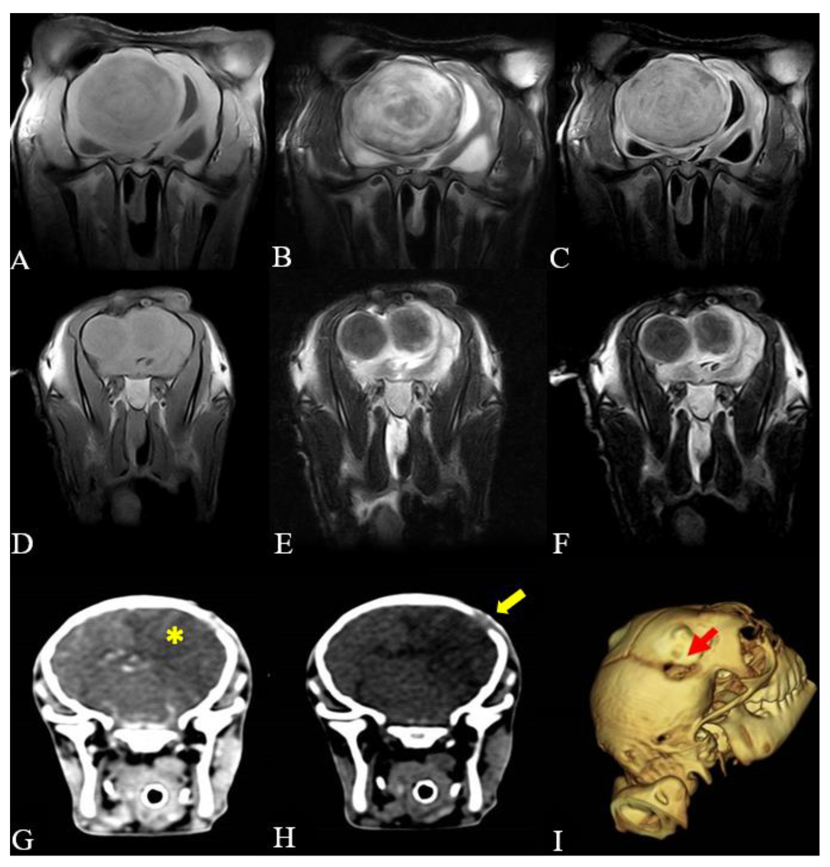
3.4. Image Findings
3.5. Postmortem Evaluation and Microbiological Isolation
4. Discussion
5. Conclusions
Supplementary Materials
Author Contributions
Funding
Data Availability Statement
Acknowledgments
Conflicts of Interest
References
- Braun, U.; Malbon, A.; Kochan, M.; Riond, B.; Janett, F.; Iten, C.; Dennler, M. Computed tomographic findings and treatment of a bull with pituitary gland abscess. Acta Vet. Scand. 2017, 59, 8. [Google Scholar] [CrossRef] [PubMed]
- Yeruham, I.; Orgad, U.; Avidar, Y.; Elad, D. Pituitary abscess and high urea concentration as causes of neurological signs in a cow. Rev. Med. Vet. 2002, 153, 829–831. [Google Scholar]
- Loretti, A.P.; Ilha, M.R.S.; Riet-Correa, G.; Driemeier, D.; Colodel, E.M.; Barros, C.S.L. Síndrome do abscesso pituitário em bezerros associada ao uso de tabuleta nasal para desmame interrompido. Pesq. Vet. Bras. 2003, 23, 9–46. [Google Scholar] [CrossRef]
- Rissi, D.R.; Fighera, R.A.; Irigoyen, L.F.; Kommers, G.D.; Barros, C.S.L. Doenças neurológicas de ovinos na região Central do Rio Grande do Sul. Pesq. Vet. Bras. 2010, 30, 222–228. [Google Scholar] [CrossRef]
- Kessell, A.E.; Finnie, J.W.; Windsor, P.A. Neurological diseases of ruminant livestock in Australia. III: Bacterial and protozoal infections. Aust. Vet. J. 2011, 89, 289–296. [Google Scholar] [CrossRef] [PubMed]
- Dore, V.; Smith, G. Cerebral disorders of calves. Vet. Clin. N. Am. Food Anim. Pract. 2017, 33, 27–41. [Google Scholar] [CrossRef] [PubMed]
- Gerros, T.C.; Mattoon, J.S.; Snyder, S.P. Use of computed tomography in the diagnosis of a cerebral abscess in a goat. Vet. Radiol. Ultrasound 1998, 39, 322–324. [Google Scholar] [CrossRef] [PubMed]
- Chigerwe, M.; Aleman, M. Seizure disorders in goats and sheep. J. Vet. Intern. Med. 2016, 30, 1752–1757. [Google Scholar] [CrossRef] [PubMed]
- Polizopoulou, S.Z.; Giadinis, D.N.; Papahristou, A.; Papaioannou, N. Neurological diseases of small ruminants in Greece: A retrospective study in 114 flocks. Acta Vet. 2016, 66, 160–171. [Google Scholar] [CrossRef]
- Ribeiro, M.G.; Pereira, T.T.; Paz, P.J.L.; Almeida, B.O.; Cerviño, C.S.A.; Rodrigues, C.A.; Santos, G.T.S.; Freire, L.M.S.; Portilho, F.V.R.; Filho, M.F.A.; et al. Bacterial identification in cerebrospinal fluid of domestic species with neurologic signs: A retrospective case-series study in 136 animals (2005–2021). Braz. J. Microbiol. 2023, 54, 449–457. [Google Scholar] [CrossRef]
- Tsuka, T.; Taura, Y. Abscess of bovine brain stem diagnosed by contrast MRI examinations. J. Vet. Med. Sci. 1999, 61, 425–427. [Google Scholar] [CrossRef] [PubMed]
- El-Khodery, S.; Yamada, K.; Aoki, D.; Kamio, K.; Kishimoto, M.; Shimizu, J.; Kobayashi, Y.; Ishii, M.; Inokuma, H.; Yamauchi, S.I.; et al. Brain abscess in a Japanese black calf: Utility of computed tomography (CT). J. Vet. Med. Sci. 2008, 70, 727–730. [Google Scholar] [CrossRef] [PubMed]
- Lee, K.; Yamada, K.; Tsuneda, R.; Kishimoto, M.; Shimizu, J.; Kobayashi, Y.; Furuoka, H.; Matsui, T.; Sasaki, N.; Ishii, M.; et al. Clinical experience of using multidetector-row CT for the diagnosis of disorders in cattle. Vet. Rec. 2009, 165, 559–562. [Google Scholar] [CrossRef] [PubMed]
- Lee, K.J.; Kishimoto, M.; Shimizu, J.; Kobayashi, Y.; Matsumoto, K.; Sasaki, N.; Ishii, M.; Inokuma, H.; Iwasaki, T.; Miyake, Y.I.; et al. Use of contrast-enhanced CT in the diagnosis of abscesses in cattle. J. Vet. Med. Sci. 2011, 73, 113–115. [Google Scholar] [CrossRef] [PubMed]
- Dennler, M.; Carrera, I.; Beckmann, K.; Ritz, J.; Rütten, M.; Kircher, P.R. Imaging diagnosis—Conventional and functional magnetic resonance imaging of a brain abscess in a goat. Vet. Radiol. Ultrasound 2014, 55, 68–73. [Google Scholar] [CrossRef] [PubMed]
- Konradt, G.; Bassuino, D.M.; Prates, K.S.; Bianchi, M.V.; Snel, G.G.M.; Sonne, L.; Driemeier, D.; Pavarini, S.P. Suppurative infectious diseases of the central nervous system in domestic ruminants. Pesq. Vet. Bras. 2017, 37, 820–828. [Google Scholar] [CrossRef]
- Schöb, L.C.; Gerspach, C.; Stirn, M.; Lehmann, R.H.; Riond, B. Findings Related to Cerebrospinal Fluid and Central Nervous System Disorders in Small Ruminants—A Retrospective Study on Sheep and Goats. Animals 2023, 14, 46. [Google Scholar] [CrossRef] [PubMed]
- Quinn, P.J.; Markey, B.K.; Leonard, F.C.; Fitzpatrick, E.S.; Fanning, S.; Hartigan, P.J. Veterinary Microbiology and Microbial Disease, 2nd ed.; Wiley-Blackwell: Chichester, UK, 2011; ISBN 978-1-4051-5823-7. [Google Scholar]
- Clinical and Laboratory Standards Institute—CLSI. Performance Standards for Antimicrobial Disk and Dilution Susceptibility Test for Bacteria Isolated from Animals (CLSI VET 015), 5th ed.; Clinical and Laboratory Standards Institute-CLSI: Wayne, PA, USA, 2020. [Google Scholar]
- Kramer, J.W. Normal hematology of cattle, sheep and goats. In Schalm’s Veterinary Hematology, 5th ed.; Feldman, B.F., Zinkl, J.G., Jain, N.C., Eds.; Lippincott Williams and Wilkins: Philadelphia, PA, USA, 2000; pp. 1075–1084. [Google Scholar]
- Smith, B.P. Large Animal Internal Medicine, 5th ed.; Elsevier: St. Louis, MO, USA, 2015; p. 918. ISBN 978-0-323-08839-8. [Google Scholar]
- Scott, P.R. Cerebrospinal fluid collection and analysis in suspected sheep neurological disease. Small Rumin. Res. 2010, 92, 96–103. [Google Scholar] [CrossRef]
- Giles, L.; Orr, J.; Viora, L.; Quintana, R.G.; Logue, D.; Guevar, J. Ruminant neurological disease: A retrospective cohort study. Vet. Rec. 2017, 181, 372–373. [Google Scholar] [CrossRef]
- Hanche-Olsen, S.; Ottensen, N.; Larsen, H.; Fintl, C. Brain abscess in a 4-month-old filly: A case report. J. Equine Vet. Sci. 2012, 32, 1–4. [Google Scholar] [CrossRef]
- Ertelt, K.; Oevermann, A.; Precht, C.; Lauper, J.; Henke, D.; Gorgas, D. Magnetic resonance imaging findings in small ruminants with brain disease. Vet. Radiol. Ultrasound 2016, 57, 162–169. [Google Scholar] [CrossRef] [PubMed]
- Morris, W.E.; Uzal, F.A.; Cipolla, A.L. Pyogranulomatous meningoencephalitis in a goat due to Corynebacterium ulcerans. Vet. Rec. 2005, 156, 317–318. [Google Scholar] [CrossRef] [PubMed]
- Câmara, A.C.L.; Vale, A.M.; Batista, J.S.; Feijó, F.M.C.; Soto-Blanco, B. Suppurative intracranial processes in 15 domestic ruminants. Pesq. Vet. Bras. 2014, 34, 421–426. [Google Scholar] [CrossRef]
- Guedes, K.M.R.; Riet-Correa, F.; Dantas, A.F.M.; Simões, S.V.D.; Neto, E.G.M.; Nobre, V.M.T.; Medeiros, R.M.T. Doenças do sistema nervoso central em caprinos e ovinos no semi-árido. Pesq. Vet. Bras. 2007, 27, 29–38. [Google Scholar] [CrossRef]
- Fecteau, G.; George, L.W. Bacterial meningitis and encephalitis in ruminants. Vet. Clin. N. Am. Food Anim. Pract. 2004, 20, 363–377. [Google Scholar] [CrossRef]
- Morin, D.E. Brainstem and cranial nerve abnormalities: Listeriosis, otitis media/interna, and pituitary abscess syndrome. Vet. Clin. Am. Food Anim. Pract. 2004, 20, 243–273. [Google Scholar] [CrossRef] [PubMed]
- Müller, K.R.; Blutke, A.; Matiasek, K.; Wieland, M.J. Pituitary abscess syndrome in a Simmental heifer. Vet. Rec. Case Rep. 2014, 2, e000041. [Google Scholar] [CrossRef]
- Stokol, T.; Divers, T.J.; Arrigan, J.W.; McDonough, S.P. Cerebrospinal fluid findings in cattle with central nervous system disorders: A retrospective study of 102 cases (1990–2008). Vet. Clin. Pathol. 2009, 38, 103–112. [Google Scholar] [CrossRef]
- Chowdhury, F.H.; Haque, M.R.; Sarkar, M.H.; Chowdhury, S.M.N.K.; Hossain, Z.; Ranjan, S. Brain abscess: Surgical experiences of 162 cases. Neuroimmunol. Neuroinflamm. 2015, 2, 153–161. [Google Scholar] [CrossRef]
- Sonneville, R.; Ruimy, R.; Benzonana, N.; Riffaud, L.; Carsin, A.; Tadié, J.M.; Piau, C.; Revest, M.; Tattevin, P. An update on bacterial brain abscess in immunocompetent patients. Clin. Microbiol. Infect. 2017, 23, 614–620. [Google Scholar] [CrossRef]
- Brook, I. Microbiology and treatment of brain abscess. J. Clin. Neurosci. 2017, 38, 8–12. [Google Scholar] [CrossRef] [PubMed]
- Chen, M.; Low, D.C.Y.; Low, S.Y.Y.; Muzumbar, D.; Seow, W.T. Management of brain abscesses: Where are we now? Childs Nerv. Syst. 2018, 34, 1871–1880. [Google Scholar] [CrossRef]
- Terra, J.P.; Blume, G.R.; Rabelo, R.E.; Medeiros, J.T.; Rocha, C.G.N.; Chagas, I.N.; Aguiar, M.S.; Sant’Ana, J.F.F. Neurological diseases of cattle in the state of Goiás, Brazil (2010–2017). Pesq. Vet. Bras. 2018, 38, 1752–1760. [Google Scholar] [CrossRef]
- Martin, S.; Drees, R.; Szladovits, B.; Beltran, E. Comparison of medical and/or surgical management of 23 cats with intracranial empyema or abscessation. J. Feline Med. Surg. 2019, 21, 566–574. [Google Scholar] [CrossRef] [PubMed]
- Ochi, F.; Tauchi, H.; Miyata, T.; Moritani, T.; Chisaka, T.; Hamada, J.; Nagai, K.; Ishimae, M.E.; Eguchi, M. Brain abscess associated with polymicrobial infection after intraoral laceration: A pediatric case report. Case Rep. Pediatr. 2020, 2020, 8304302. [Google Scholar] [CrossRef] [PubMed]
- Ribeiro, M.G.; Risseti, R.M.; Bolaños, C.A.D.; Caffaro, K.A.; Morais, A.C.B.; Lara, G.H.B.; Zamprogna, T.O.; Paes, A.C.; Listoni, F.J.P.; Franco, M.M.J. Trueperella pyogenes multispecies infections in domestic animals: A retrospective study of 144 cases (2002 to 2012). Vet. Q. 2015, 35, 82–87. [Google Scholar] [CrossRef]
- El Damaty, H.M.; El-Demerdash, A.S.; El-Aziz, N.K.A.; Yousef, S.G.; Hefny, A.A.; Remela, E.M.A.; Shaker, A.; Elsohaby, I. Molecular characterization and antimicrobial susceptibilities of Corynebacterium pseudotuberculosis isolated from caseous lymphadenitis of smallholder sheep and goats. Animals 2023, 13, 2337. [Google Scholar] [CrossRef]
- Eroksuz, Y.; Otlu, B.; Eroksuz, H.; Gursoy, N.C.; Yerlikaya, Z.; Incili, C.A.; Karabulut, B.; Timurkaan, N.; Timurkan, M.O. Brain abscess and bronchopneumonia caused by Acinetobacter baumannii in a 2-year-old female sheep. Vet. Q. 2018, 38, 67–71. [Google Scholar] [CrossRef]
- Câmara, A.C.L.; Gonzaga, M.C.; Ziober, T.M.; Queiroz, C.R.R.; Fino, T.C.M.; Castro, M.B.; Borges, J.R.J.; Soto-Blanco, B. Cerebrospinal fluid analysis in 58 ruminants showing neurological disorders. Pesq. Vet. Bras. 2020, 40, 346–354. [Google Scholar] [CrossRef]


| Case | Species | Breed | Age | Gender | Weight | Period of Evolution of Signs until Care | Period of Hospitalization Until Death/Euthanasia | Main Findings in Neurological Examination |
|---|---|---|---|---|---|---|---|---|
| 1 | Sheep | Suffolk | 24 months | F | 50 kg | 13 days | 1 day | Lateral recumbency, semicomatous state, unilateral blindness, and seizures |
| 2 | Cow | Dutch | 6 months | F | 180 kg | 2 days | 7 days | Difficulty in grasping, chewing, and swallowing food, and lateral strabismus |
| 3 | Cow | Nelore | 15 months | F | 253 kg | 60 days | 3 days | Apathy, compulsive and circling gait, paresis, head pressing, unilateral blindness, auricular ptosis, decreased facial sensitivity, and ventrolateral strabismus |
| 4 | Goat | Anglo Nubian | 1 month | F | 5.8 kg | 4 days | 4 days | Lateral recumbency, semicomatous state, lateral head tilt, decreased facial sensitivity, and seizures |
| 5 | Goat | Alpine | 36 months | F | 45 kg | 30 days | 21 days | Apathy, compulsive and circling gait, head pressing, excessive vocalization, and amaurosis |
| 6 | Goat | Anglo Nubian | 48 months | F | 45 kg | 3 days | 2 days | Apathy, lateral recumbency, opisthotonus, fasciculations, and horizontal and vertical nystagmus |
| Parameters | Case 1 | Sheep Reference | Case 2 | Case 3 | Bovine Reference | Case 4 | Case 5 | Case 6 | Goat Reference |
|---|---|---|---|---|---|---|---|---|---|
| Haematocrit (%) | 24 | 27–45 | 23 | 25 | 24–46 | 26 | 25 | 44 | 22–38 |
| Red blood cells (106/μL) | 10.28 | 9–15 | 5.02 | 6.24 | 5–10 | NP | NP | 16.95 | 8–18 |
| Hemoglobin (g/dL) | 8.6 | 9–15 | 7.1 | 8.0 | 8–15 | 9.1 | 10.5 | 15.6 | 8–12 |
| Total protein (g/dL) | 8.6 | 6–7.5 | 8.6 | 7.2 | 7.0–8.5 | 6.6 | 9.6 | 10 | 6–7.5 |
| Fibrinogen (mg/dL) | 1000 | 100–500 | 400 | 200 | 300–700 | 1200 | 200 | 1000 | 100–400 |
| Total leukocytes (/μL) | 31,800 | 4000–12,000 | 15,400 | 12,200 | 4000–12,000 | 23,000 | 12,600 | 26,600 | 4000–13,000 |
| Neutrophils (/μL) | 29,900 | 700–6000 | 6300 | 5500 | 600–4000 | 17,500 | 8700 | 23,400 | 1200–7200 |
| Lymphocytes (/μL) | 1900 | 2000–9000 | 8300 | 6000 | 2500–7500 | 4100 | 3500 | 2900 | 2000–9000 |
| Monocytes (/μL) | 0 | 0–750 | 800 | 200 | 25–840 | 1400 | 100 | 300 | 0–550 |
| Eosinophils (/μL) | 0 | 0–1000 | 0 | 400 | 0–2400 | 0 | 300 | 0 | 50–650 |
| Basophils (/μL) | 0 | 0–300 | 0 | 100 | 0–200 | 0 | 0 | 0 | 0–120 |
| Identification | Cerebrospinal Fluid (CSF) | |||
|---|---|---|---|---|
| Color | Aspect | Protein (mg/dL) Ovine (8–70) Bovine (20–40) Caprine (24–40) | Cellularity (<10 cells/µL) | |
| Case 1 | NP | NP | NP | NP |
| Case 2 | Colorless | Clear | 43.6 | 100 cells/µL: Mixed pleocytosis—Predominance of mononuclear cells (50%), followed by neutrophils (27%), typical lymphocytes (19%), macrophages (4%), and rare eosinophils. Presence of cytophagocytosis and free and phagocytosed round structures |
| Case 3 | Colorless | Clear | 108.1 | 95 cells/µL: Mixed pleocytosis—Predominance of mononuclear cells (46%), followed by typical lymphocytes (37%), macrophages (12%), and neutrophils (5%). Presence of clusters of mononuclear cells |
| Case 4 | Xanthochromic | Clear | 783.7 | 149 cells/µL: Mixed pleocytosis—Predominance of neutrophils (57%) followed by mononuclear cells (35%), typical lymphocytes (6%) and macrophages (2%) |
| Case 5 | Colorless | Clear | 44.4 | 1 cell/µL: Predominance of typical lymphocytes (82%), followed by mononuclear cells (14%), neutrophils (2%), and macrophages (2%) |
| Case 6 | Whitish | Turbidity | 100 | 10.450 cells/µL: Neutrophilic pleocytosis—Predominance of neutrophils (79%), followed by mononuclear cells (12%) and typical lymphocytes (9%). Presence of leukophagocytosis |
| Case | Site | Isolation | |
|---|---|---|---|
| Brain Abscesses | CSF | ||
| 1 | Prosencephalon | Trueperella pyogenes, Pasteurella multocida, and Escherichia coli | NP |
| 2 | Rete mirabile | Escherichia coli and Streptococcus spp. | Escherichia coli and Streptococcus spp. |
| 3 | Right cerebral hemisphere | Trueperella pyogenes and Streptococcus spp. | Negative |
| 4 | Prosencephalon | Escherichia coli, Enterobacter sp., and Staphylococcus sp. | Negative |
| 5 | Prosencephalon | Corynebacterium pseudotuberculosis | Corynebacterium pseudotuberculosis |
| 6 | Mesencephalon | Corynebacterium pseudotuberculosis | Negative |
| Case | Pathogen | AMI | AMO | AMP | AZI | CEF | CLI | ENR | ERY | FLO | GEN | PEN | RIF | SUL | TET |
|---|---|---|---|---|---|---|---|---|---|---|---|---|---|---|---|
| Escherichia coli | S | S | R | NP | S | R | NP | NP | NP | S | NP | R | NP | R | |
| 4 | Enterobacter sp. | S | S | R | NP | S | R | NP | NP | NP | S | NP | R | NP | R |
| Staphylococcus sp. | S | R | R | NP | I | R | NP | NP | NP | S | NP | R | NP | I | |
| 6 | Corynebacterium pseudotuberculosis | NP | NP | S | S | S | NP | S | S | S | NP | S | S | S | NP |
Disclaimer/Publisher’s Note: The statements, opinions and data contained in all publications are solely those of the individual author(s) and contributor(s) and not of MDPI and/or the editor(s). MDPI and/or the editor(s) disclaim responsibility for any injury to people or property resulting from any ideas, methods, instructions or products referred to in the content. |
© 2024 by the authors. Licensee MDPI, Basel, Switzerland. This article is an open access article distributed under the terms and conditions of the Creative Commons Attribution (CC BY) license (https://creativecommons.org/licenses/by/4.0/).
Share and Cite
Ferreira, L.V.d.O.; Rocha, T.G.; Takahira, R.K.; Laufer-Amorim, R.; Machado, V.M.d.V.; Ribeiro, M.G.; Pereira, W.A.B.; Oliveira-Filho, J.P.; Borges, A.S.; Amorim, R.M. Brain Abscesses in Domestic Ruminants: Clinicopathological and Bacteriological Approaches. Microorganisms 2024, 12, 1424. https://doi.org/10.3390/microorganisms12071424
Ferreira LVdO, Rocha TG, Takahira RK, Laufer-Amorim R, Machado VMdV, Ribeiro MG, Pereira WAB, Oliveira-Filho JP, Borges AS, Amorim RM. Brain Abscesses in Domestic Ruminants: Clinicopathological and Bacteriological Approaches. Microorganisms. 2024; 12(7):1424. https://doi.org/10.3390/microorganisms12071424
Chicago/Turabian StyleFerreira, Lucas Vinícius de Oliveira, Thaís Gomes Rocha, Regina Kiomi Takahira, Renée Laufer-Amorim, Vânia Maria de Vasconcelos Machado, Márcio Garcia Ribeiro, Wanderson Adriano Biscola Pereira, José Paes Oliveira-Filho, Alexandre Secorun Borges, and Rogério Martins Amorim. 2024. "Brain Abscesses in Domestic Ruminants: Clinicopathological and Bacteriological Approaches" Microorganisms 12, no. 7: 1424. https://doi.org/10.3390/microorganisms12071424
APA StyleFerreira, L. V. d. O., Rocha, T. G., Takahira, R. K., Laufer-Amorim, R., Machado, V. M. d. V., Ribeiro, M. G., Pereira, W. A. B., Oliveira-Filho, J. P., Borges, A. S., & Amorim, R. M. (2024). Brain Abscesses in Domestic Ruminants: Clinicopathological and Bacteriological Approaches. Microorganisms, 12(7), 1424. https://doi.org/10.3390/microorganisms12071424