Research Progress on Immune Evasion of Mycoplasma hyopneumoniae


Abstract
:1. Introduction
2. Mycoplasma hyopneumoniae Achieves Immune Evasion by Altering its Structure or Function
2.1. Genetic Mutation and Post-Translational Protein Processing
2.2. Biofilms
2.3. Adhesion and Invasion
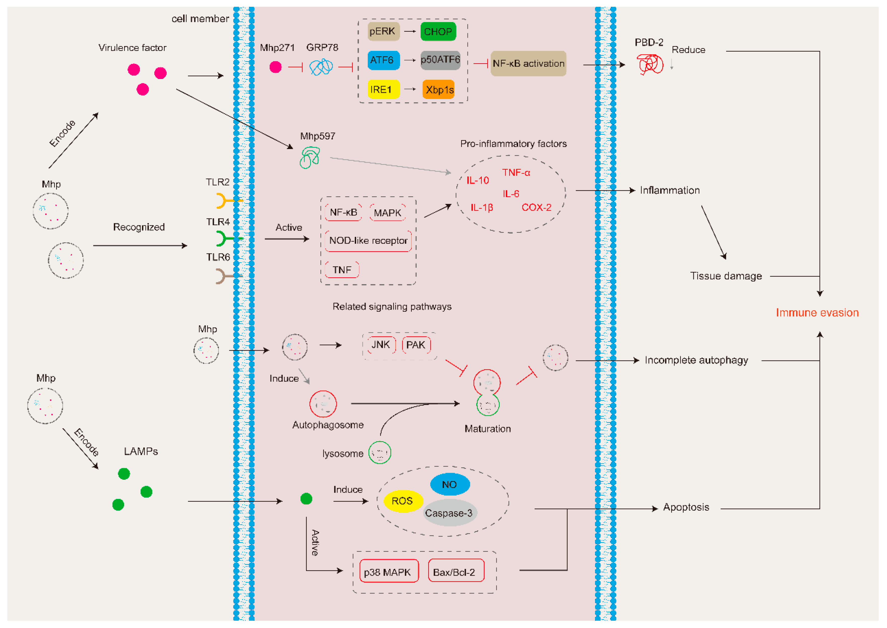
3. Mycoplasma hyopneumoniae Generates Immune Evasion by Modulating the Host Immune System
3.1. Virulence Factors
3.2. Inflammatory Response
3.3. Autophagy and Apoptosis
3.4. Inhibition of the Activity of Immune Effectors or Immune Cells
3.4.1. Anti-Immunoglobulin Strategies
3.4.2. Other Strategies to Inhibit the Activity of Immune Effectors or Immune Cells
4. Conclusions and Future Perspectives
Author Contributions
Funding
Data Availability Statement
Acknowledgments
Conflicts of Interest
References
- Minion, F.C.; Lefkowitz, E.J.; Madsen, M.L.; Cleary, B.J.; Swartzell, S.M.; Mahairas, G.G. The genome sequence of Mycoplasma hyopneumoniae strain 232, the agent of swine mycoplasmosis. J. Bacteriol. 2004, 186, 7123–7133. [Google Scholar] [CrossRef]
- Ross, R.F.; Young, T.F. The nature and detection of mycoplasmal immunogens. Vet. Microbiol. 1993, 37, 369–380. [Google Scholar] [CrossRef] [PubMed]
- Holst, S.; Yeske, P.; Pieters, M. Elimination of from breed-to-wean farms: A review of current protocols with emphasis on herd closure and medication. J. Swine Health Prod. 2015, 23, 321–330. [Google Scholar]
- Maes, D.; Sibila, M.; Kuhnert, P.; Segalés, J.; Haesebrouck, F.; Pieters, M. Update on Mycoplasma hyopneumoniae infections in pigs: Knowledge gaps for improved disease control. Transbound. Emerg. Dis. 2018, 65 (Suppl. 1), 110–124. [Google Scholar] [CrossRef]
- Maes, D.; Segales, J.; Meyns, T.; Sibila, M.; Pieters, M.; Haesebrouck, F. Control of Mycoplasma hyopneumoniae infections in pigs. Vet. Microbiol. 2008, 126, 297–309. [Google Scholar] [CrossRef] [PubMed]
- He, Y.; Xu, M.J.; Zhou, D.H.; Zou, F.C.; Lin, R.Q.; Yin, C.C.; He, X.H.; Liang, R.; Liang, M.; Zhu, X.Q. Seroprevalence of Mycoplasma hyopneumoniae in pigs in subtropical southern China. Trop. Anim. Health Prod. 2011, 43, 695–698. [Google Scholar] [CrossRef] [PubMed]
- Chae, C. Porcine respiratory disease complex: Interaction of vaccination and porcine circovirus type 2, porcine reproductive and respiratory syndrome virus, and Mycoplasma hyopneumoniae. Vet. J. 2016, 212, 1–6. [Google Scholar] [CrossRef] [PubMed]
- Wang, J.Y.; Liang, K.Y.; Chen, L.; Su, X.L.; Liao, D.Y.; Yu, J.W.; He, J. Unveiling the stealthy tactics: Mycoplasma’s immune evasion strategies. Front. Cell. Infect. Microbiol. 2023, 13, 1247182. [Google Scholar] [CrossRef] [PubMed]
- Erwig, L.P.; Gow, N.A.R. Interactions of fungal pathogens with phagocytes. Nat. Rev. Microbiol. 2016, 14, 163–176. [Google Scholar] [CrossRef]
- Rashid, F.; Xie, Z.; Suleman, M.; Shah, A.; Khan, S.; Luo, S. Roles and functions of SARS-CoV-2 proteins in host immune evasion. Front. Immunol. 2022, 13, 940756. [Google Scholar] [CrossRef]
- Lowy, F.D. Staphylococcus aureus infections. N. Engl. J. Med. 1998, 339, 520–532. [Google Scholar] [CrossRef] [PubMed]
- Howden, B.P.; Giulieri, S.G.; Lung, T.W.F.; Baines, S.L.; Sharkey, L.K.; Lee, J.Y.H.; Hachani, A.; Monk, I.R.; Stinear, T.P. Staphylococcus aureus host interactions and adaptation. Nat. Rev. Microbiol. 2023, 21, 380–395. [Google Scholar] [CrossRef] [PubMed]
- Li, G.; Obeng, E.; Shu, J.; Shu, J.; Chen, J.; Wu, Y.; He, Y. Genomic Variability and Post-translational Protein Processing Enhance the Immune Evasion of Mycoplasma hyopneumoniae and Its Interaction with the Porcine Immune System. Front. Immunol. 2020, 11, 510943. [Google Scholar] [CrossRef] [PubMed]
- Deeney, A.S.; Maglennon, G.A.; Chapat, L.; Crussard, S.; Jolivet, E.; Rycroft, A.N. Mycoplasma hyopneumoniae evades phagocytic uptake by porcine alveolar macrophages in vitro. Vet. Res. 2019, 50, 51. [Google Scholar] [CrossRef] [PubMed]
- Tassew, D.D.; Mechesso, A.F.; Park, N.H.; Song, J.B.; Shur, J.W.; Park, S.C. Biofilm formation and determination of minimum biofilm eradication concentration of antibiotics in Mycoplasma hyopneumoniae. J. Vet. Med. Sci. 2017, 79, 1716–1720. [Google Scholar] [CrossRef] [PubMed]
- Meluleni, G.J.; Grout, M.; Evans, D.J.; Pier, G.B. Mucoid Pseudomonas aeruginosa growing in a biofilm in vitro are killed by opsonic antibodies to the mucoid exopolysaccharide capsule but not by antibodies produced during chronic lung infection in cystic fibrosis patients. J. Immunol. 1995, 155, 2029–2038. [Google Scholar] [CrossRef] [PubMed]
- Hoelzle, K.; Ade, J.; Hoelzle, L.E. Persistence in Livestock Mycoplasmas-a Key Role in Infection and Pathogenesis. Curr. Clin. Microbiol. 2020, 7, 81–89. [Google Scholar] [CrossRef]
- Bhugra, B.; Voelker, L.L.; Zou, N.; Yu, H.; Dybvig, K. Mechanism of antigenic variation in Mycoplasma pulmonis: Interwoven, site-specific DNA inversions. Mol. Microbiol. 1995, 18, 703–714. [Google Scholar] [CrossRef] [PubMed]
- Dybvig, K. DNA rearrangements and phenotypic switching in prokaryotes. Mol. Microbiol. 1993, 10, 465–471. [Google Scholar] [CrossRef]
- Niu, Z.T.; Xue, Q.Y.; Wang, H.; Xie, X.Z.; Zhu, S.Y.; Liu, W.; Ding, X.Y. Mutational Biases and GC-Biased Gene Conversion Affect GC Content in the Plastomes of Genus. Int. J. Mol. Sci. 2017, 18, 2307. [Google Scholar] [CrossRef]
- Charlebois, A.; Marois-Créhan, C.; Hélie, P.; Gagnon, C.A.; Gottschalk, M.; Archambault, M. Genetic diversity of Mycoplasma hyopneumoniae isolates of abattoir pigs. Vet. Microbiol. 2014, 168, 348–356. [Google Scholar] [CrossRef] [PubMed]
- Liu, W.; Xiao, S.; Li, M.; Guo, S.; Li, S.; Luo, R.; Feng, Z.; Li, B.; Zhou, Z.; Shao, G.; et al. Comparative genomic analyses of Mycoplasma hyopneumoniae pathogenic 168 strain and its high-passaged attenuated strain. BMC Genom. 2013, 14, 80. [Google Scholar] [CrossRef]
- Citti, C.; Nouvel, L.X.; Baranowski, E. Phase and antigenic variation in mycoplasmas. Future Microbiol. 2010, 5, 1073–1085. [Google Scholar] [CrossRef]
- Yogev, D.; Rosengarten, R.; Watson-McKown, R.; Wise, K.S. Molecular basis of Mycoplasma surface antigenic variation: A novel set of divergent genes undergo spontaneous mutation of periodic coding regions and 5’ regulatory sequences. Embo J. 1991, 10, 4069–4079. [Google Scholar] [CrossRef]
- Glew, M.D.; Marenda, M.; Rosengarten, R.; Citti, C. Surface diversity in Mycoplasma agalactiae is driven by site-specific DNA inversions within the vpma multigene locus. J. Bacteriol. 2002, 184, 5987–5998. [Google Scholar] [CrossRef]
- Citti, C.; Kim, M.F.; Wise, K.S. Elongated versions of Vlp surface lipoproteins protect Mycoplasma hyorhinis escape variants from growth-inhibiting host antibodies. Infect. Immun. 1997, 65, 1773–1785. [Google Scholar] [CrossRef]
- Simmons, W.L.; Zuhua, C.; Glass, J.I.; Simecka, J.W.; Cassell, G.H.; Watson, H.L. Sequence analysis of the chromosomal region around and within the V-1-encoding gene of Mycoplasma pulmonis: Evidence for DNA inversion as a mechanism for V-1 variation. Infect. Immun. 1996, 64, 472–479. [Google Scholar] [CrossRef] [PubMed]
- Noormohammadi, A.H.; Markham, P.F.; Kanci, A.; Whithear, K.G.; Browning, G.F. A novel mechanism for control of antigenic variation in the haemagglutinin gene family of Mycoplasma synoviae. Mol. Microbiol. 2000, 35, 911–923. [Google Scholar] [CrossRef]
- Iverson-Cabral, S.L.; Astete, S.G.; Cohen, C.R.; Totten, P.A. mgpB and mgpC sequence diversity in Mycoplasma genitalium is generated by segmental reciprocal recombination with repetitive chromosomal sequences. Mol. Microbiol. 2007, 66, 55–73. [Google Scholar] [CrossRef] [PubMed]
- Tacchi, J.L.; Raymond, B.B.; Haynes, P.A.; Berry, I.J.; Widjaja, M.; Bogema, D.R.; Woolley, L.K.; Jenkins, C.; Minion, F.C.; Padula, M.P.; et al. Post-translational processing targets functionally diverse proteins in Mycoplasma hyopneumoniae. Open Biol. 2016, 6, 150210. [Google Scholar] [CrossRef]
- Berry, I.J.; Jarocki, V.M.; Tacchi, J.L.; Raymond, B.B.A.; Widjaja, M.; Padula, M.P.; Djordjevic, S.P. N-terminomics identifies widespread endoproteolysis and novel methionine excision in a genome-reduced bacterial pathogen. Sci. Rep. 2017, 7, 11063. [Google Scholar] [CrossRef] [PubMed]
- Raymond, B.B.; Jenkins, C.; Seymour, L.M.; Tacchi, J.L.; Widjaja, M.; Jarocki, V.M.; Deutscher, A.T.; Turnbull, L.; Whitchurch, C.B.; Padula, M.P.; et al. Proteolytic processing of the cilium adhesin MHJ_0194 (P123J) in Mycoplasma hyopneumoniae generates a functionally diverse array of cleavage fragments that bind multiple host molecules. Cell Microbiol. 2015, 17, 425–444. [Google Scholar] [CrossRef] [PubMed]
- Djordjevic, S.P.; Cordwell, S.J.; Djordjevic, M.A.; Wilton, J.; Minion, F.C. Proteolytic processing of the Mycoplasma hyopneumoniae cilium adhesin. Infect. Immun. 2004, 72, 2791–2802. [Google Scholar] [CrossRef] [PubMed]
- Bogema, D.R.; Deutscher, A.T.; Woolley, L.K.; Seymour, L.M.; Raymond, B.B.; Tacchi, J.L.; Padula, M.P.; Dixon, N.E.; Minion, F.C.; Jenkins, C.; et al. Characterization of cleavage events in the multifunctional cilium adhesin Mhp684 (P146) reveals a mechanism by which Mycoplasma hyopneumoniae regulates surface topography. mBio 2012, 3, e00282-11. [Google Scholar] [CrossRef] [PubMed]
- Raymond, B.B.; Tacchi, J.L.; Jarocki, V.M.; Minion, F.C.; Padula, M.P.; Djordjevic, S.P. P159 from Mycoplasma hyopneumoniae binds porcine cilia and heparin and is cleaved in a manner akin to ectodomain shedding. J. Proteome Res. 2013, 12, 5891–5903. [Google Scholar] [CrossRef] [PubMed]
- Seymour, L.M.; Deutscher, A.T.; Jenkins, C.; Kuit, T.A.; Falconer, L.; Minion, F.C.; Crossett, B.; Padula, M.; Dixon, N.E.; Djordjevic, S.P.; et al. A processed multidomain Mycoplasma hyopneumoniae adhesin binds fibronectin, plasminogen, and swine respiratory cilia. J. Biol. Chem. 2010, 285, 33971–33978. [Google Scholar] [CrossRef] [PubMed]
- McAuliffe, L.; Ellis, R.J.; Miles, K.; Ayling, R.D.; Nicholas, R.A.J. Biofilm formation by mycoplasma species and its role in environmental persistence and survival. Microbiology 2006, 152 Pt 4, 913–922. [Google Scholar] [CrossRef] [PubMed]
- Yan, J.; Bassler, B.L. Surviving as a Community: Antibiotic Tolerance and Persistence in Bacterial Biofilms. Cell Host Microbe 2019, 26, 15–21. [Google Scholar] [CrossRef] [PubMed]
- Daubenspeck, J.M.; Totten, A.H.; Needham, J.; Feng, M.; Balish, M.F.; Atkinson, T.P.; Dybvig, K. Mycoplasma genitalium Biofilms Contain Poly-GlcNAc and Contribute to Antibiotic Resistance. Front. Microbiol. 2020, 11, 585524. [Google Scholar] [CrossRef]
- Simmons, W.L.; Dybvig, K. Catalase Enhances Growth and Biofilm Production of Mycoplasma pneumoniae. Curr. Microbiol. 2015, 71, 190–194. [Google Scholar] [CrossRef]
- Bekő, K.; Nagy, E.Z.; Grózner, D.; Kreizinger, Z.; Gyuranecz, M. Biofilm formation and its impact on environmental survival and antibiotic resistance of Mycoplasma anserisalpingitidis strains. Acta Vet. Hung. 2022, 70, 184–191. [Google Scholar] [CrossRef] [PubMed]
- Kang, T.; Zhou, M.; Yan, X.; Song, S.; Yuan, S.; Yang, H.; Ding, H.; Jiang, H.; Zhang, D.; Bai, Y.; et al. Biofilm formation and correlations with drug resistance in Mycoplasma synoviae. Vet. Microbiol. 2023, 283, 109777. [Google Scholar] [CrossRef] [PubMed]
- Chen, H.J.; Yu, S.Q.; Hu, M.R.; Han, X.G.; Chen, D.Q.; Qiu, X.S.; Ding, C. Identification of biofilm formation by Mycoplasma gallisepticum. Vet. Microbiol. 2012, 161, 96–103. [Google Scholar] [CrossRef] [PubMed]
- Mah, T.F.; O’Toole, G.A. Mechanisms of biofilm resistance to antimicrobial agents. Trends Microbiol. 2001, 9, 34–39. [Google Scholar] [CrossRef] [PubMed]
- Simmons, W.L.; Dybvig, K. Biofilms protect Mycoplasma pulmonis cells from lytic effects of complement and gramicidin. Infect. Immun. 2007, 75, 3696–3699. [Google Scholar] [CrossRef] [PubMed]
- Rathore, S.S.; Cheepurupalli, L.; Gangwar, J.; Raman, T.; Ramakrishnan, J. Biofilm of Klebsiella pneumoniae minimize phagocytosis and cytokine expression by macrophage cell line. AMB Express 2022, 12, 122. [Google Scholar] [CrossRef] [PubMed]
- Wu, Y.Z.; Yu, Y.F.; Hua, L.Z.; Wei, Y.N.; Gan, Y.; Chenia, H.Y.; Wang, Y.X.; Xie, X.; Wang, J.; Liu, M.J.; et al. Genotyping and biofilm formation of Mycoplasma hyopneumoniae and their association with virulence. Vet. Res. 2022, 53, 17. [Google Scholar] [CrossRef] [PubMed]
- Raymond, B.B.A.; Turnbull, L.; Jenkins, C.; Madhkoor, R.; Schleicher, I.; Uphoff, C.C.; Whitchurch, C.B.; Rohde, M.; Djordjevic, S.P. Mycoplasma hyopneumoniae resides intracellularly within porcine epithelial cells. Sci. Rep. 2018, 8, 17697. [Google Scholar] [CrossRef] [PubMed]
- DeBey, M.C.; Ross, R.F. Ciliostasis and loss of cilia induced by Mycoplasma hyopneumoniae in porcine tracheal organ cultures. Infect. Immun. 1994, 62, 5312–5318. [Google Scholar] [CrossRef]
- Mucha, S.G.; Ferrarini, M.G.; Moraga, C.; Di Genova, A.; Guyon, L.; Tardy, F.; Rome, S.; Sagot, M.F.; Zaha, A. Mycoplasma hyopneumoniae J elicits an antioxidant response and decreases the expression of ciliary genes in infected swine epithelial cells. Sci. Rep. 2020, 10, 13707. [Google Scholar] [CrossRef]
- Blanchard, B.; Vena, M.M.; Cavalier, A.; Le Lannic, J.; Gouranton, J.; Kobisch, M. Electron microscopic observation of the respiratory tract of SPF piglets inoculated with Mycoplasma hyopneumoniae. Vet. Microbiol. 1992, 30, 329–341. [Google Scholar] [CrossRef] [PubMed]
- Jenkins, C.; Wilton, J.L.; Minion, F.C.; Falconer, L.; Walker, M.J.; Djordjevic, S.P. Two domains within the Mycoplasma hyopneumoniae cilium adhesin bind heparin. Infect. Immun. 2006, 74, 481–487. [Google Scholar] [CrossRef] [PubMed]
- Yu, Y.; Liu, M.; Hua, L.; Qiu, M.; Zhang, W.; Wei, Y.; Gan, Y.; Feng, Z.; Shao, G.; Xiong, Q. Fructose-1,6-bisphosphate aldolase encoded by a core gene of Mycoplasma hyopneumoniae contributes to host cell adhesion. Vet. Res. 2018, 49, 114. [Google Scholar] [CrossRef] [PubMed]
- Seymour, L.M.; Jenkins, C.; Deutscher, A.T.; Raymond, B.B.; Padula, M.P.; Tacchi, J.L.; Bogema, D.R.; Eamens, G.J.; Woolley, L.K.; Dixon, N.E.; et al. Mhp182 (P102) binds fibronectin and contributes to the recruitment of plasmin(ogen) to the Mycoplasma hyopneumoniae cell surface. Cell Microbiol. 2012, 14, 81–94. [Google Scholar] [CrossRef] [PubMed]
- Deutscher, A.T.; Tacchi, J.L.; Minion, F.C.; Padula, M.P.; Crossett, B.; Bogema, D.R.; Jenkins, C.; Kuit, T.A.; Walker, M.J.; Djordjevic, S.P. Mycoplasma hyopneumoniae Surface proteins Mhp385 and Mhp384 bind host cilia and glycosaminoglycans and are endoproteolytically processed by proteases that recognize different cleavage motifs. J. Proteome Res. 2012, 11, 1924–1936. [Google Scholar] [CrossRef] [PubMed]
- Seymour, L.M.; Falconer, L.; Deutscher, A.T.; Minion, F.C.; Padula, M.P.; Dixon, N.E.; Djordjevic, S.P.; Walker, M.J. Mhp107 is a member of the multifunctional adhesin family of Mycoplasma hyopneumoniae. J. Biol. Chem. 2011, 286, 10097–10104. [Google Scholar] [CrossRef] [PubMed]
- Deutscher, A.T.; Jenkins, C.; Minion, F.C.; Seymour, L.M.; Padula, M.P.; Dixon, N.E.; Walker, M.J.; Djordjevic, S.P. Repeat regions R1 and R2 in the P97 paralogue Mhp271 of Mycoplasma hyopneumoniae bind heparin, fibronectin and porcine cilia. Mol. Microbiol. 2010, 78, 444–458. [Google Scholar] [CrossRef]
- Wilton, J.; Jenkins, C.; Cordwell, S.J.; Falconer, L.; Minion, F.C.; Oneal, D.C.; Djordjevic, M.A.; Connolly, A.; Barchia, I.; Walker, M.J.; et al. Mhp493 (P216) is a proteolytically processed, cilium and heparin binding protein of Mycoplasma hyopneumoniae. Mol. Microbiol. 2009, 71, 566–582. [Google Scholar] [CrossRef]
- Burnett, T.A.; Dinkla, K.; Rohde, M.; Chhatwal, G.S.; Uphoff, C.; Srivastava, M.; Cordwell, S.J.; Geary, S.; Liao, X.; Minion, F.C.; et al. P159 is a proteolytically processed, surface adhesin of Mycoplasma hyopneumoniae: Defined domains of P159 bind heparin and promote adherence to eukaryote cells. Mol. Microbiol. 2006, 60, 669–686. [Google Scholar] [CrossRef]
- Tacchi, J.L.; Raymond, B.B.; Jarocki, V.M.; Berry, I.J.; Padula, M.P.; Djordjevic, S.P. Cilium adhesin P216 (MHJ_0493) is a target of ectodomain shedding and aminopeptidase activity on the surface of Mycoplasma hyopneumoniae. J. Proteome Res. 2014, 13, 2920–2930. [Google Scholar] [CrossRef]
- Bogema, D.R.; Scott, N.E.; Padula, M.P.; Tacchi, J.L.; Raymond, B.B.A.; Jenkins, C.; Cordwell, S.J.; Minion, F.C.; Walker, M.J.; Djordjevic, S.P. Sequence TTKF downward arrow QE defines the site of proteolytic cleavage in Mhp683 protein, a novel glycosaminoglycan and cilium adhesin of Mycoplasma hyopneumoniae. J. Biol. Chem. 2011, 286, 41217–41229. [Google Scholar] [CrossRef] [PubMed]
- Minion, F.C.; Adams, C.; Hsu, T. R1 region of P97 mediates adherence of Mycoplasma hyopneumoniae to swine cilia. Infect. Immun. 2000, 68, 3056–3060. [Google Scholar] [CrossRef] [PubMed]
- Jarocki, V.M.; Santos, J.; Tacchi, J.L.; Raymond, B.B.; Deutscher, A.T.; Jenkins, C.; Padula, M.P.; Djordjevic, S.P. MHJ_0461 is a multifunctional leucine aminopeptidase on the surface of Mycoplasma hyopneumoniae. Open Biol. 2015, 5, 140175. [Google Scholar] [CrossRef] [PubMed]
- Machado, L.; Paes, J.A.; Souza Dos Santos, P.; Ferreira, H.B. Evidences of differential endoproteolytic processing on the surfaces of Mycoplasma hyopneumoniae and Mycoplasma flocculare. Microb. Pathog. 2020, 140, 103958. [Google Scholar] [CrossRef]
- Leal Zimmer, F.M.A.; Paes, J.A.; Zaha, A.; Ferreira, H.B. Pathogenicity & virulence of Mycoplasma hyopneumoniae. Virulence 2020, 11, 1600–1622. [Google Scholar] [PubMed]
- Siqueira, F.M.; Gerber, A.L.; Guedes, R.L.; Almeida, L.G.; Schrank, I.S.; Vasconcelos, A.T.; Zaha, A. Unravelling the transcriptome profile of the Swine respiratory tract mycoplasmas. PLoS ONE 2014, 9, e110327. [Google Scholar] [CrossRef] [PubMed]
- Leal Zimmer, F.; Paludo, G.P.; Moura, H.; Barr, J.R.; Ferreira, H.B. Differential secretome profiling of a swine tracheal cell line infected with mycoplasmas of the swine respiratory tract. J. Proteom. 2019, 192, 147–159. [Google Scholar] [CrossRef] [PubMed]
- Siqueira, F.M.; Thompson, C.E.; Virginio, V.G.; Gonchoroski, T.; Reolon, L.; Almeida, L.G.; da Fonsêca, M.M.; de Souza, R.; Prosdocimi, F.; Schrank, I.S.; et al. New insights on the biology of swine respiratory tract mycoplasmas from a comparative genome analysis. BMC Genom. 2013, 14, 175. [Google Scholar] [CrossRef] [PubMed]
- Xie, X.; Hao, F.; Chen, R.; Wang, J.; Wei, Y.; Liu, J.; Wang, H.; Zhang, Z.; Bai, Y.; Shao, G.; et al. Nicotinamide Adenine Dinucleotide-Dependent Flavin Oxidoreductase of Mycoplasma hyopneumoniae Functions as a Potential Novel Virulence Factor and Not Only as a Metabolic Enzyme. Front. Microbiol. 2021, 12, 747421. [Google Scholar] [CrossRef]
- Xu, L.; Hao, F.; Wang, J.; Feng, Z.; Zhang, L.; Yuan, T.; Chen, R.; Zhang, Z.; Shao, G.; Xiong, Q.; et al. Th1 and Th17 mucosal immune responses elicited by nasally inoculation in mice with virulence factors of Mycoplasma hyopneumoniae. Microb. Pathog. 2022, 172, 105779. [Google Scholar] [CrossRef]
- Hao, F.; Xie, X.; Feng, Z.; Chen, R.; Wei, Y.; Liu, J.; Xiong, Q.; Shao, G.; Lin, J. NADH oxidase of Mycoplasma hyopneumoniae functions as a potential mediator of virulence. BMC Vet. Res. 2022, 18, 126. [Google Scholar] [CrossRef] [PubMed]
- Pan, Q.; Xu, Q.; Liu, T.; Zhang, Y.; Xin, J. Mycoplasma hyopneumoniae membrane protein Mhp271 interacts with host UPR protein GRP78 to facilitate infection. Mol. Microbiol. 2022, 118, 208–222. [Google Scholar] [CrossRef] [PubMed]
- Pan, Q.; Wang, X.; Liu, T.; Yu, Y.; Li, L.; Zhou, R.; Li, G.; Xin, J. Mycoplasma hyopneumoniae Inhibits Porcine Beta-Defensin 2 Production by Blocking the Unfolded Protein Response to Facilitate Epithelial Adhesion and Infection. Infect. Immun. 2020, 88, e00164-20. [Google Scholar] [CrossRef] [PubMed]
- Liu, W.; Zhou, D.; Yuan, F.; Liu, Z.; Duan, Z.; Yang, K.; Guo, R.; Li, M.; Li, S.; Fang, L.; et al. Surface proteins mhp390 (P68) contributes to cilium adherence and mediates inflammation and apoptosis in Mycoplasma hyopneumoniae. Microb. Pathog. 2019, 126, 92–100. [Google Scholar] [CrossRef] [PubMed]
- Schmidt, J.A.; Browning, G.F.; Markham, P.F. Mycoplasma hyopneumoniae p65 surface lipoprotein is a lipolytic enzyme with a preference for shorter-chain fatty acids. J. Bacteriol. 2004, 186, 5790–5798. [Google Scholar] [CrossRef] [PubMed]
- Lorenzo, H.; Quesada, O.; Assuncao, P.; Castro, A.; Rodriguez, F. Cytokine expression in porcine lungs experimentally infected with Mycoplasma hyopneumoniae. Vet. Immunol. Immunopathol. 2006, 109, 199–207. [Google Scholar] [CrossRef] [PubMed]
- Rodriguez, F.; Ramirez, G.A.; Sarradell, J.; Andrada, M.; Lorenzo, H. Immunohistochemical labelling of cytokines in lung lesions of pigs naturally infected with Mycoplasma hyopneumoniae. J. Comp. Pathol. 2004, 130, 306–312. [Google Scholar] [CrossRef] [PubMed]
- Rodriguez, F.; Rosales, R.S.; Ramirez, A.S.; Poveda, J.B. Vaccination Upregulates Th1 Cytokines in the Lung of Pigs Experimentally Infected with Mycoplasma hyopneumoniae. Animals 2023, 13, 520. [Google Scholar] [CrossRef] [PubMed]
- Hwang, M.H.; Chang, Z.Q.; Kang, E.H.; Lim, J.H.; Yun, H.I.; Rhee, M.H.; Jeong, K.S.; Park, S.C. Surfactin C inhibits Mycoplasma hyopneumoniae-induced transcription of proinflammatory cytokines and nitric oxide production in murine RAW 264.7 cells. Biotechnol. Lett. 2008, 30, 229–233. [Google Scholar] [CrossRef]
- Li, P.; Zhang, Y.; Li, X.; Zhou, W.; Li, X.; Jiang, F.; Wu, W. Mycoplasma hyopneumoniae Mhp597 is a cytotoxicity, inflammation and immunosuppression associated nuclease. Vet. Microbiol. 2019, 235, 53–62. [Google Scholar] [CrossRef]
- Nueangphuet, P.; Suwanruengsri, M.; Fuke, N.; Uemura, R.; Hirai, T.; Yamaguchi, R. Neutrophil and M2-polarized Macrophage Infiltration, Expression of IL-8 and Apoptosis in Mycoplasma hyopneumoniae Pneumonia in Swine. J. Comp. Pathol. 2021, 189, 31–44. [Google Scholar] [CrossRef] [PubMed]
- Bin, L.; Luping, D.; Bing, S.; Zhengyu, Y.; Maojun, L.; Zhixin, F.; Yanna, W.; Haiyan, W.; Guoqing, S.; Kongwang, H. Transcription analysis of the porcine alveolar macrophage response to Mycoplasma hyopneumoniae. PLoS ONE 2014, 9, e101968. [Google Scholar] [CrossRef] [PubMed]
- Hwang, M.H.; Damte, D.; Lee, J.S.; Gebru, E.; Chang, Z.Q.; Cheng, H.; Jung, B.Y.; Rhee, M.H.; Park, S.C. Mycoplasma hyopneumoniae induces pro-inflammatory cytokine and nitric oxide production through NFkappaB and MAPK pathways in RAW264.7 cells. Vet. Res. Commun. 2011, 35, 21–34. [Google Scholar] [CrossRef] [PubMed]
- Bai, F.; Ni, B.; Liu, M.; Feng, Z.; Xiong, Q.; Shao, G. Mycoplasma hyopneumoniae-derived lipid-associated membrane proteins induce inflammation and apoptosis in porcine peripheral blood mononuclear cells in vitro. Vet. Microbiol. 2015, 175, 58–67. [Google Scholar] [CrossRef] [PubMed]
- Liu, W.; Jiang, P.; Yang, K.; Song, Q.; Yuan, F.; Liu, Z.; Gao, T.; Zhou, D.; Guo, R.; Li, C.; et al. Mycoplasma hyopneumoniae Infection Activates the NOD1 Signaling Pathway to Modulate Inflammation. Front. Cell Infect. Microbiol. 2022, 12, 927840. [Google Scholar] [CrossRef]
- Trueeb, B.S.; Braun, R.O.; Auray, G.; Kuhnert, P.; Summerfield, A. Differential innate immune responses induced by Mycoplasma hyopneumoniae and Mycoplasma hyorhinis in various types of antigen presenting cells. Vet. Microbiol. 2020, 240, 108541. [Google Scholar] [CrossRef] [PubMed]
- Yang, X.; Xing, F.; Wang, L.; Zhao, W.; Fu, Y.; Tu, F.; Li, B.; Fang, X.; Ren, S. Effect of Pregnane X Receptor on CYP3A29 Expression in Porcine Alveolar Macrophages during Mycoplasma hyopneumoniae Infection. Animals 2021, 11, 349. [Google Scholar] [CrossRef]
- Thacker, E.L.; Halbur, P.G.; Ross, R.F.; Thanawongnuwech, R.; Thacker, B.J. Mycoplasma hyopneumoniae potentiation of porcine reproductive and respiratory syndrome virus-induced pneumonia. J. Clin. Microbiol. 1999, 37, 620–627. [Google Scholar] [CrossRef] [PubMed]
- Almeida, H.M.S.; Mechler-Dreibi, M.L.; Sonalio, K.; Ferraz, M.E.S.; Storino, G.Y.; Barbosa, F.O.; Maes, D.; Montassier, H.J.; de Oliveira, L.G. Cytokine expression and Mycoplasma hyopneumoniae burden in the development of lung lesions in experimentally inoculated pigs. Vet. Microbiol. 2020, 244, 108647. [Google Scholar] [CrossRef]
- Muneta, Y.; Minagawa, Y.; Shimoji, Y.; Ogawa, Y.; Hikono, H.; Mori, Y. Immune response of gnotobiotic piglets against Mycoplasma hyopneumoniae. J. Vet. Med. Sci. 2008, 70, 1065–1070. [Google Scholar] [CrossRef]
- Jarocki, V.M.; Raymond, B.B.A.; Tacchi, J.L.; Padula, M.P.; Djordjevic, S.P. Mycoplasma hyopneumoniae surface-associated proteases cleave bradykinin, substance P, neurokinin A and neuropeptide Y. Sci. Rep. 2019, 9, 14585. [Google Scholar] [CrossRef] [PubMed]
- Wang, Z.; Wen, Y.; Zhou, B.; Tian, Y.; Ning, Y.; Ding, H. Incomplete autophagy promotes the replication of Mycoplasma hyopneumoniae. J. Microbiol. 2021, 59, 782–791. [Google Scholar] [CrossRef] [PubMed]
- Wen, Y.; Chen, Z.; Tian, Y.; Yang, M.; Dong, Q.; Yang, Y.; Ding, H. Incomplete autophagy promotes the proliferation of Mycoplasma hyopneumoniae through the JNK and Akt pathways in porcine alveolar macrophages. Vet. Res. 2022, 53, 62. [Google Scholar] [CrossRef] [PubMed]
- Rudin, C.M.; Thompson, C.B. Apoptosis and disease: Regulation and clinical relevance of programmed cell death. Annu. Rev. Med. 1997, 48, 267–281. [Google Scholar] [PubMed]
- Liu, M.; Du, G.; Liu, B.; Hu, Y.; Liu, J.; Jia, Y.; Minion, F.C.; Shao, G.; Zhao, R. Cholesterol exacerbates Mycoplasma hyopneumoniae-induced apoptosis via stimulating proliferation and adhesion to porcine alveolar macrophages. Vet. Microbiol. 2017, 211, 112–118. [Google Scholar] [CrossRef] [PubMed]
- Bai, F.; Ni, B.; Liu, M.; Feng, Z.; Xiong, Q.; Xiao, S.; Shao, G. Mycoplasma hyopneumoniae-derived lipid-associated membrane proteins induce apoptosis in porcine alveolar macrophage via increasing nitric oxide production, oxidative stress, and caspase-3 activation. Vet. Immunol. Immunopathol. 2013, 155, 155–161. [Google Scholar] [CrossRef] [PubMed]
- Ni, B.; Bai, F.F.; Wei, Y.; Liu, M.J.; Feng, Z.X.; Xiong, Q.Y.; Hua, L.Z.; Shao, G.Q. Apoptosis induced by lipid-associated membrane proteins from Mycoplasma hyopneumoniae in a porcine lung epithelial cell line with the involvement of caspase 3 and the MAPK pathway. Genet. Mol. Res. 2015, 14, 11429–11443. [Google Scholar] [CrossRef] [PubMed]
- Paes, J.A.; Virginio, V.G.; Cancela, M.; Leal, F.M.A.; Borges, T.J.; Jaeger, N.; Bonorino, C.; Schrank, I.S.; Ferreira, H.B. Pro-apoptotic effect of a Mycoplasma hyopneumoniae putative type I signal peptidase on PK(15) swine cells. Vet. Microbiol. 2017, 201, 170–176. [Google Scholar] [CrossRef] [PubMed]
- Robb, C.T.; Regan, K.H.; Dorward, D.A.; Rossi, A.G. Key mechanisms governing resolution of lung inflammation. Semin. Immunopathol. 2016, 38, 425–448. [Google Scholar] [CrossRef]
- Arfi, Y.; Minder, L.; Di Primo, C.; Le Roy, A.; Ebel, C.; Coquet, L.; Claverol, S.; Vashee, S.; Jores, J.; Blanchard, A.; et al. MIB-MIP is a mycoplasma system that captures and cleaves immunoglobulin G. Proc. Natl. Acad. Sci. USA 2016, 113, 5406–5411. [Google Scholar] [CrossRef]
- Arfi, Y.; Lartigue, C.; Sirand-Pugnet, P.; Blanchard, A. Beware of Mycoplasma Anti-immunoglobulin Strategies. mBio 2021, 12, e0197421. [Google Scholar] [CrossRef] [PubMed]
- Kilian, M.; Brown, M.B.; Brown, T.A.; Freundt, E.A.; Cassell, G.H. Immunoglobulin A1 protease activity in strains of Ureaplasma urealyticum. Acta Pathol. Microbiol. Immunol. Scand. B 1984, 92, 61–64. [Google Scholar] [CrossRef] [PubMed]
- Kilian, M.; Freundt, E.A. Exclusive occurrence of an extracellular protease capable of cleaving the hinge region of human immunoglobulin A1 in strains of Ureaplasma urealyticum. Isr. J. Med. Sci. 1984, 20, 938–941. [Google Scholar]
- Spooner, R.K.; Russell, W.C.; Thirkell, D. Characterization of the immunoglobulin A protease of Ureaplasma urealyticum. Infect. Immun. 1992, 60, 2544–2546. [Google Scholar] [CrossRef] [PubMed]
- Cizelj, I.; Bercic, R.L.; Dusanic, D.; Narat, M.; Kos, J.; Dovc, P.; Bencina, D. Mycoplasma gallisepticum and Mycoplasma synoviae express a cysteine protease CysP, which can cleave chicken IgG into Fab and Fc. Microbiology 2011, 157, 362–372. [Google Scholar] [CrossRef] [PubMed]
- Grover, R.K.; Zhu, X.Y.; Nieusma, T.; Jones, T.; Boero, I.; MacLeod, A.S.; Mark, A.; Niessen, S.; Kim, H.J.; Kong, L.; et al. A Structurally Distinct Human Mycoplasma Protein that Generically Blocks Antigen-Antibody Union. Science 2014, 343, 656–661. [Google Scholar] [CrossRef] [PubMed]
- Guiraud, J.; Le Roy, C.; Rideau, F.; Sirand-Pugnet, P.; Lartigue, C.; Bébéar, C.; Arfi, Y.; Pereyre, S. Improved transformation efficiency in Mycoplasma hominis enables disruption of the MIB-MIP system targeting human immunoglobulins. Microbiol. Spectr. 2023, 11, e0187323. [Google Scholar] [CrossRef] [PubMed]
- Zhao, H.; Zhang, Y.; Wang, Z.; Liu, M.; Wang, P.; Wu, W.; Peng, C. MBOVPG45_0375 Encodes an IgG-Binding Protein and MBOVPG45_0376 Encodes an IgG-Cleaving Protein in Mycoplasma bovis. Front. Vet. Sci. 2021, 8, 644224. [Google Scholar] [CrossRef] [PubMed]
- Torres-Puig, S.; Crespo-Pomar, S.; Akarsu, H.; Yimthin, T.; Cippà, V.; Démoulins, T.; Posthaus, H.; Ruggli, N.; Kuhnert, P.; Labroussaa, F.; et al. Functional surface expression of immunoglobulin cleavage systems in a candidate Mycoplasma vaccine chassis. Commun. Biol. 2024, 7, 779. [Google Scholar] [CrossRef]
- Brezski, R.J.; Jordan, R.E. Cleavage of IgGs by proteases associated with invasive diseases: An evasion tactic against host immunity? mAbs 2010, 2, 212–220. [Google Scholar] [CrossRef]
- Yu, Y.; Wang, J.; Han, R.; Wang, L.; Zhang, L.; Zhang, A.Y.; Xin, J.; Li, S.; Zeng, Y.; Shao, G.; et al. Mycoplasma hyopneumoniae evades complement activation by binding to factor H via elongation factor thermo unstable (EF-Tu). Virulence 2020, 11, 1059–1074. [Google Scholar] [CrossRef] [PubMed]
- Henthorn, C.R.; Chris Minion, F.; Sahin, O. Utilization of macrophage extracellular trap nucleotides by Mycoplasma hyopneumoniae. Microbiology 2018, 164, 1394–1404. [Google Scholar] [CrossRef] [PubMed]
- Shen, Y.; Hu, W.; Wei, Y.; Feng, Z.; Yang, Q. Effects of Mycoplasma hyopneumoniae on porcine nasal cavity dendritic cells. Vet. Microbiol. 2017, 198, 1–8. [Google Scholar] [CrossRef] [PubMed]
- Caruso, J.P.; Ross, R.F. Effects of Mycoplasma hyopneumoniae and Actinobacillus (Haemophilus) pleuropneumoniae infections on alveolar macrophage functions in swine. Am. J. Vet. Res. 1990, 51, 227–231. [Google Scholar] [CrossRef] [PubMed]
- Wang, H.; Zhang, Z.; Xie, X.; Liu, B.; Wei, Y.; Gan, Y.; Yuan, T.; Ni, B.; Wang, J.; Zhang, L.; et al. Paracellular Pathway-Mediated Mycoplasma hyopneumoniae Migration across Porcine Airway Epithelial Barrier under Air-Liquid Interface Conditions. Infect. Immun. 2020, 88, e00470-20. [Google Scholar] [CrossRef] [PubMed]
- Vicca, J.; Stakenborg, T.; Maes, D.; Butaye, P.; Peeters, J.; de Kruif, A.; Haesebrouck, F. In vitro susceptibilities of Mycoplasma hyopneumoniae field isolates. Antimicrob. Agents Chemother. 2004, 48, 4470–4472. [Google Scholar] [CrossRef] [PubMed]
- Tao, Y.; Shu, J.; Chen, J.; Wu, Y.; He, Y. A concise review of vaccines against Mycoplasma hyopneumoniae. Res. Vet. Sci. 2019, 123, 144–152. [Google Scholar] [CrossRef]
- Okamba, F.R.; Moreau, E.; Cheikh Saad Bouh, K.; Gagnon, C.A.; Massie, B.; Arella, M. Immune responses induced by replication-defective adenovirus expressing the C-terminal portion of the Mycoplasma hyopneumoniae P97 adhesin. Clin. Vaccine Immunol. 2007, 14, 767–774. [Google Scholar] [CrossRef]
- de Oliveira, N.R.; Jorge, S.; Gomes, C.K.; Rizzi, C.; Pacce, V.D.; Collares, T.F.; Monte, L.G.; Dellagostin, O.A. A novel chimeric protein composed of recombinant Mycoplasma hyopneumoniae antigens as a vaccine candidate evaluated in mice. Vet. Microbiol. 2017, 201, 146–153. [Google Scholar] [CrossRef]


| Adhesin | Binding Site | Reference |
|---|---|---|
| P146 | Plasminogen | [34] |
| Fructose-1,6-bisphosphate aldolase (FBA) | Fibronectin | [53] |
| MHJ_0194 (P123) | Proteoglycans, glycoproteins, plasminogen | [32] |
| Mhp182 (P102) | Fibronectin | [54,55] |
| Mhp107 | Heparin | [56] |
| P116 | Fibronectin, plasminogen | [36] |
| Mhp271 | Heparin, fibronectin | [57] |
| Mhp493 (P216) | Heparin | [58] |
| Mhp183 (P97) | Heparin | [52,55] |
| P159 | Glycosaminoglycan | [59] |
| MHJ_0493 (P216) | Glycosaminoglycan | [60] |
| Mhp683 | Glycosaminoglycan | [61] |
Disclaimer/Publisher’s Note: The statements, opinions and data contained in all publications are solely those of the individual author(s) and contributor(s) and not of MDPI and/or the editor(s). MDPI and/or the editor(s) disclaim responsibility for any injury to people or property resulting from any ideas, methods, instructions or products referred to in the content. |
© 2024 by the authors. Licensee MDPI, Basel, Switzerland. This article is an open access article distributed under the terms and conditions of the Creative Commons Attribution (CC BY) license (https://creativecommons.org/licenses/by/4.0/).
Share and Cite
Jiang, B.; Zhang, Y.; Li, G.; Quan, Y.; Shu, J.; Feng, H.; He, Y. Research Progress on Immune Evasion of Mycoplasma hyopneumoniae. Microorganisms 2024, 12, 1439. https://doi.org/10.3390/microorganisms12071439
Jiang B, Zhang Y, Li G, Quan Y, Shu J, Feng H, He Y. Research Progress on Immune Evasion of Mycoplasma hyopneumoniae. Microorganisms. 2024; 12(7):1439. https://doi.org/10.3390/microorganisms12071439
Chicago/Turabian StyleJiang, Bin, Ying Zhang, Gaojian Li, Yanping Quan, Jianhong Shu, Huapeng Feng, and Yulong He. 2024. "Research Progress on Immune Evasion of Mycoplasma hyopneumoniae" Microorganisms 12, no. 7: 1439. https://doi.org/10.3390/microorganisms12071439