Article

Presence and Role of the Type 3 Fimbria in the Adherence Capacity of Enterobacter hormaechei subsp. hoffmannii

1
Departamento de Biología, Facultad de Química y Biología, Universidad de Santiago de Chile, Av. Libertador Bernardo O’Higgins 3363, Santiago 9170022, Chile
2
Programa de Microbiología y Micología, Instituto de Ciencias Biomédicas, Facultad de Medicina, Universidad de Chile, Av. Independencia 1027, Independencia, Santiago 8380453, Chile
3
Instituto Milenio de Inmunología e Inmunoterapia, Facultad de Medicina, Universidad de Chile, Av. Independencia 1027, Independencia, Santiago 8380453, Chile
4
Departamento de Bioquímica y Biología Molecular, Facultad de Ciencias Químicas y Farmacéuticas, Universidad de Chile, Santiago 8380453, Chile
5
Laboratorio de Biofilms Microbianos, Departamento de Microbiología, Instituto de Investigaciones Biológicas Clemente Estable, Montevideo 11600, Uruguay
*
Author to whom correspondence should be addressed.
Microorganisms 2024, 12(7), 1441; https://doi.org/10.3390/microorganisms12071441
Submission received: 17 June 2024 / Revised: 8 July 2024 / Accepted: 10 July 2024 / Published: 16 July 2024
(This article belongs to the Special Issue Pathogenic Mechanisms of Bacterial Infections)

Abstract

:
Enterobacter hormaechei, one of the species within the Enterobacter cloacae complex, is a relevant agent of healthcare-associated infections. In addition, it has gained relevance because isolates have shown the capacity to resist several antibiotics, particularly carbapenems. However, knowledge regarding colonization and virulence mechanisms of E. hormaechei has not progressed to the same extent as other Enterobacteriaceae species as Escherichia coli or Klebsiella pneumoniae. Here, we describe the presence and role of the type 3 fimbria, a chaperone-usher assembled fimbria, which was first described in Klebsiella spp., and which has been detected in other representatives of the Enterobacteriaceae family. Eight Chilean E. cloacae isolates were examined, and among them, four E. hormaechei isolates were found to produce the type 3 fimbria. These isolates were identified as E. hormaechei subsp. hoffmannii, one of the five subspecies known. A mutant E. hormaechei subsp. hoffmannii strain lacking the mrkA gene, encoding the major structural subunit, displayed a significantly reduced adherence capacity to a plastic surface and to Caco-2 cells, compared to the wild-type strain. This phenotype of reduced adherence capacity was not observed in the mutant strains complemented with the mrkA gene under the control of an inducible promoter. Therefore, these data suggest a role of the type 3 fimbria in the adherence capacity of E. hormaechei subsp. hoffmannii. A screening in E. hormaechei genomes contained in the NCBI RefSeq Assembly database indicated that the overall presence of the type 3 fimbria is uncommon (5.94–7.37%), although genes encoding the structure were detected in representatives of the five E. hormaechei subspecies. Exploration of complete genomes indicates that, in most of the cases, the mrkABCDF locus, encoding the type 3 fimbria, is located in plasmids. Furthermore, sequence types currently found in healthcare-associated infections were found to harbor genes encoding the type 3 fimbria, mainly ST145, ST78, ST118, ST168, ST66, ST93, and ST171. Thus, although the type 3 fimbria is not widespread among the species, it might be a determinant of fitness for a subset of E. hormaechei representatives.

1. Introduction

Enterobacter hormaechei is a Gram-negative bacterium belonging to the Enterobacteriaceae family and one of the most common representatives of the Enterobacter cloacae complex (ECC) [1]. E. hormaechei is a frequent agent of diverse infections in humans, mainly opportunistic infections in healthcare-associated settings, including urinary tract infections and bloodstream infections, among others [1,2,3]. Five E. hormaechei subspecies have been established: E. hormaechei subsp. hormaechei, E. hormaechei subsp. hoffmannii, E. hormaechei subsp. oharae, E. hormaechei subsp. steigerwaltii, and E. hormaechei subsp. xiangfangensis [1,4]. Many aspects regarding knowledge about E. hormaechei biology are undergoing evolution. In fact, taxonomy is one of these aspects. A recent scheme for species distribution proposed E. hormaechei to be a single species, without a subspecies, leaving E. hoffmannii and E. xiangfangensis as separate species (not subspecies) [5]. Representatives of E. hormaechei subsp. oharae and E. hormaechei subsp. steigerwaltii would be part of the E. xiangfangensis species [5]. This scheme has not been incorporated yet by the List of Prokaryotic Names with Standing in Nomenclature [4], but the situation reflects that more research is required to gain insights into E. hormaechei life.
It is well established that Enterobacter species have developed resistance to multiple antibiotics, and this is the reason why they have been part of the ESKAPE and ESCAPE groups of bacteria [6,7]. In addition, carbapenem-resistant and third-generation cephalosporin-resistant Enterobacter species were considered within the critical group, along with other Enterobacteriaceae family members, in the Bacterial Priority Pathogens List recently published by the World Health Organization [8]. However, the emerging status of Enterobacter species, and particularly E. hormaechei, has not been accompanied by progress in knowledge of the colonization and infectious mechanisms, at least to a similar extent as other relative species such as Klebsiella pneumoniae and Escherichia coli. Therefore, gaining insights into the colonization and virulence determinants of E. hormaechei is required, in parallel to surveilling the antibiotic resistance status, because evolution might be followed up, and alternative therapies could be designed [9].
The type 3 fimbria is an adherence structure assembled by the chaperone–usher pathway, first discovered in Klebsiella and found widely distributed among the K. pneumoniae species [10,11]. The type 3 fimbria is composed of the major structural subunit MrkA, the minor subunit MrkF, and the tip subunit MrkD, which would act as the adhesin [12]. MrkB is the chaperone that assists structural subunit assembly at the usher MrkC, an outer membrane porin-like protein [12]. The type 3 fimbria system has been relatively well studied in K. pneumoniae; in fact, the MrkA protein has been proposed as a target for anti-adherence therapies [13,14]. Besides K. pneumoniae, type 3 fimbria can be produced by other representatives of the Enterobacteriaceae family, such as Klebsiella oxytoca, E. coli, Citrobacter freundii, and Citrobacter koseri [15]. In addition, the presence of type 3 fimbria has been detected in some Enterobacter species, and the mrkB gene has been found in E. hormaechei subsp. oharae [16,17,18], but its actual contribution to their adherence capacity has not been addressed. In this study, we report the evaluation of the role of type 3 fimbria in the adherence capacity of an E. hormaechei subsp. hoffmannii clinical isolate obtained in Chile and the presence of the locus encoding the structure among E. hormaechei genomes.

2. Materials and Methods

Strains: A group of eight strains belonging to the E. cloacae complex (Eclo1-UCH, Eclo5-UCH, Eclo6-UCH, Eh12-UCH, Eh13-UCH, Eh18-UCH, Eclo29-UCH, and Eh31-UCH), previously isolated in Santiago (Chile) during the SENTRY antimicrobial resistance study [19], were initially selected for this work (Table 1). Four of these strains (Eh12-UCH, Eh13-UCH, Eh18-UCH, and Eh31-UCH) were positive for the detection of the mrkA gene by PCR using primers mrkA-F and mrkA-R (primers listed in Table S1). PCR mixes comprised 1X reaction buffer, 1.5 mM MgCl2, 0.8 mM dNTPs, 10 pmol primers, and 0.6 U Taq DNA polymerase (GoTaq® G2, Promega, Madison, WI, USA). Amplification conditions were (1) one cycle at 94 °C for 5 min; (2) 30 cycles including denaturation at 94 °C for 30 s, annealing at 57 °C for 30 s, and extension at 72 °C for 30 s; (3) a final extension cycle at 72 °C for 10 min. Later, the four isolates were identified as E. hormaechei subsp. hoffmannii after genome sequencing (see “Genome/bioinformatical analyses” below).
Antimicrobial resistance profile: Susceptibility to common antibiotics was tested by the Kirby–Bauer method, according to the guidelines of the Clinical and Laboratory Standard Institute (CLSI) [22]. Antibiotics tested were amikacin (30 μg), cefepime (30 μg), cefotaxime (30 μg), cefotaxime–clavulanic acid (30/10 μg), cephalothin (30 μg), chloramphenicol (30 μg), kanamycin (30 μg), levofloxacin (10 μg), streptomycin (10 μg), tetracycline (30 μg), trimethoprim (5 μg) (all obtained from Oxoid, Thermo Fisher, Waltham, MA, USA); ampicillin (10 μg) and gentamicin (10 μg) (both obtained from BD, Franklin Lakes, NJ, USA), ciprofloxacin (5 μg), imipenem (10 μg), meropenem (10 μg), nalidixic acid (30 μg), nitrofurantoin (300 μg), piperacillin–tazobactam (100/10 μg), and trimethoprim–sulfamethoxazole (1.25/23.75 μg) (all obtained from Mast Group, Bootle, UK). E. coli strain ATCC 25922 was used as a control.
Genome/bioinformatical analyses: Draft genomic sequences were obtained for strains Eh12-UCH, Eh13-UCH, Eh18-UCH, and Eh31-UCH, as they were recognized as positive for mrkA. Briefly, strains were grown in lysogeny broth (LB, Lennox formula, Thermo Fisher Scientific, Waltham, MA, USA), and genomic DNA was purified using the Wizard Genomic DNA Purification kit (Promega, Madison, WI, USA). The integrity of the product was determined by electrophoresis in 1% agarose gel and ethidium bromide staining. Sequencing was performed at MicrobesNG (Birmingham, UK) using the Illumina MiSeq platform (Illumina, San Diego, CA, USA). Draft genomes were provided after assembly with SPAdes 3.14 [23]. Assembly metrics (Genome length and N50) were obtained by using QUAST v5.0.2 [24], while percentages of completeness and contamination were obtained with CheckM v1.2.2 [25]. The species were identified using “Identify Species” (available at https://pubmlst.org/species-id, accessed on 27 April 2024) [26]. Subspecies were identified by performing a phylogenetic analysis with kSNP 3.1 [27], including genomes of strains E. hormaechei subsp. hoffmannii DSM 14563, E. hormaechei subsp. hormaechei ATCC 49162, E. hormaechei subsp. oharae DSM 16687, E. hormaechei subsp. steigerwaltii DSM 16691, and E. hormaechei subsp. xiangfangensis LMG 27195 as controls (Table S2) [5]. The main features of the sequences obtained are shown in Table 2. The tree image was obtained from the Interactive Tree of Life (iToL) server. In addition, subspecies were established by determining the average nucleotide identity (ANI) using the FastANI software v1.1 [28]. The subspecies for which the highest ANI value was obtained, being this ≥96%, was considered as the subspecies of the interrogated genome. Sequence types were identified using the E. cloacae scheme in the in silico multiple locus sequence typing software mlst 2.18 [29,30]. Furthermore, the results of determining the antibiotic resistance profiles were complemented by detecting antibiotic resistance genes and their associated profiles using ResFinder 4.5.0 [31,32].
Screening of the type 3 fimbria encoding locus mrkABCDF, and the individual genes, was performed using the Large-Scale Blast Score Ratio (LS-BSR) software with the blastn or the tblastn options (available at https://github.com/jasonsahl/LS-BSR, accessed on 1 May 2024). Sequences were selected from the K. pneumoniae Kp13 strain’s genome (Table S2) [33]. The genomes screened included the Chilean isolates and E. hormaechei genomes obtained from NCBI Assembly RefSeq database (https://www.ncbi.nlm.nih.gov/genbank/, accessed on 1 May 2024) [34]. Records with BSR ≥ 0.9 were considered positive, according to a previous report of our group and others [35,36,37]. Template sequences selected for screening and their accession codes are shown in Table S2. The presence of the mrkABCDF locus in chromosomes or plasmids was explored in annotations (genomic files in .gbff extension) of fully sequenced genomes contained in the NCBI Assembly RefSeq database. Subspecies were determined by performing the ANI analysis, and sequence types were determined by using the mlst software, as indicated above.
Allelic exchange and complementation: The mrkA, which encodes the type 3 fimbria major structural subunit, was knocked out in Eh13-UCH by the allelic exchange procedure, according to the protocol described by Sharan et al. [21]. Briefly, the strain was transformed with approximately 100 ng of the purified pSIM9 plasmid by electroporation at 1700 V and recombinants were selected in LB agar plates containing 12.5 μg/mL chloramphenicol (Sigma Aldrich, St Louis, MO, USA) at 30 °C. Although the strain was resistant to disks containing 10 μg/mL of ampicillin, it was sensitive to 100 μg/mL in LB (Sigma Aldrich, St Louis, MO, USA). The next day, chloramphenicol-resistant colonies were transformed with approximately 500 ng of a purified linear DNA fragment containing the kanamycin resistance gene aph flanked by 40 nt sequences identical to both mrkA ends. This product was generated by PCR using primers mrkA-mutF and mrkA-mutR, described in Table S1, and the plasmid pCLF4 as template. PCR conditions were (1) initial denaturation at 94 °C for 5 min; (2) 30 cycles including denaturation at 94 °C for 30 s, annealing at 56 °C for 30 s, and elongation at 72 °C for 1 min; (3) final elongation at 72 °C for 10 min. Electroporation was performed at 17,000 V and recombinant strains were selected by seeding the bacterial suspension in LB plates containing kanamycin (50 μg/mL, Sigma Aldrich, St Louis, MO, USA). Verification of the allelic exchange was performed by PCR from colony lysates and then from purified DNA using primers mrkA-Nde and K1 (Table S1). PCR conditions were (1) initial denaturation at 94 °C for 5 min; (2) 30 cycles including denaturation at 94 °C for 30 s, annealing at 56 °C for 30 s, and elongation at 72 °C for 30 s; (3) final elongation at 72 °C for 10 min.
Complementation of the Eh13-UCHmrkA strain was performed by transformation with the pVB1 expression plasmid harboring the mrkA gene. For this purpose, the mrkA gene was amplified by PCR from Eh13-UCH purified genomic DNA using primers mrkA-nde and mrkA-bam. The product was purified, digested with restriction endonucleases NdeI (FastDigest, Thermo Fisher Scientific, Waltham, MA, USA) and BamHI (FastDigest, Thermo Fisher Scientific, Waltham, MA, USA) and then ligated into the pVB1 plasmid, previously digested with the same enzymes and purified. E. coli DH5α was electroporated with the ligation mix and clones harboring the plasmid were selected by seeding onto LB agar plates containing ampicillin (100 μg/mL). Presence of recombinant plasmids was checked by colony PCR using primers pVB1-F and mrkA-bam. Recombinant plasmid was purified and checked again by PCR and by digestion with NdeI and BamHI endonucleases. Finally, the purified recombinant plasmid was introduced in Eh13-UCHΔmrkA by electroporation at 1700 V and selection of clones in LB agar plates containing ampicillin (100 μg/mL). Expression of the mrkA gene in the complemented Eh13-UCHmrkA/mrkA was induced by adding 2 mM of m-tuolic acid (Sigma Aldrich, St Louis, MO, USA) to the culture media.
Western blot: The presence of the type 3 fimbriae was established by detecting MrkA by Western blot in heat-extracted proteins. Briefly, bacteria were cultured in 10 mL of LB broth, containing supplements if necessary (antibiotics kanamycin, ampicillin, or m-tuolic acid if necessary). After overnight incubation at 37 °C, without shaking, tubes were centrifuged at 3000× g for 10 min at room temperature (RT). The sediment was gently suspended in 1 mL of phosphate buffer saline (PBS, Merck Millipore, Burlington, MA, USA), centrifuged again under the same conditions, and suspended in 100 µL of PBS. The suspension was heated at 60 °C for 30 min and then centrifuged at 3000× g for 10 min at RT. The supernatant was recovered, centrifuged again under the same conditions, and the supernatant was recovered again. This fraction represents the heat-extracted proteins that have been shown to contain fimbrial major structural subunits [38]. The concentration of proteins was determined by the Bradford method [39]. A volume containing 1 µg of heat-extracted proteins was subjected to sodium dodecyl sulfate–polyacrylamide gel electrophoresis (SDS-PAGE) under denaturing conditions in a two phases polyacrylamide gel (4% concentrating phase and 15% separating phase) for 90 min at 100 V. The separated proteins were transferred to a nitrocellulose membrane in a wet transference apparatus (Mini Trans-Blot, BioRad, Hercules, CA, USA), and then, the protein binding sites were blocked with 1% bovine serum albumin (BSA, Merck Millipore, Burlington, MA, USA) dissolved in Tris-buffered saline solution containing 0.05% Tween 20 (TBS-T, Merck Millipore, Burlington, MA, USA). The membrane was incubated with a polyclonal rabbit anti-MrkA antibody (Poly Express custom antibody production, Genscript, Piscataway, NY, USA) in a 1:1000 dilution for 1 h at RT. After three washes with TBS-T buffer, 10 min at RT each, the membrane was incubated with a secondary goat anti-rabbit IgG conjugated to alkaline phosphatase (Thermo Fisher Scientific, Waltham, MA, USA) in a 1:5000 dilution for 1 h at RT. The membrane was washed three times with TBS-T and once with distilled water to finally reveal the presence of immunoreactive bands by adding a mix of the chromogenic alkaline phosphatase substrates nitro blue tetrazolium chloride (NBT) and 5-bromo-4-chloro-3-indolyl-phosphate (BCIP) (Novex, Thermo Fisher Scientific, Waltham MA, USA).
Immunogold staining: The presence of the type 3 fimbria was also established in Eh13-UCH, and its derivative strains, by detecting the MrkA protein by immunogold staining over whole non-permeabilized bacteria. An aliquot of 1 mL of an overnight culture in LB, grown at 37 °C without shaking and containing supplements, if necessary (antibiotics and/or m-tuolic acid), was concentrated by centrifugation at 3000× g and gently suspended in 100 μL of LB. Ten microliters of this suspension was deposited over 200-mesh formvar/carbon nickel grids (Electron Microscopy Sciences, Hatfield, PA, USA) and incubated for 20 min at 37 °C inside a humidified chamber. After three washes with PBS, grids were incubated with the blocking solution, 1% BSA 0.01 M glycine in PBS, for 1 h at RT (Glycine obtained from Merck Millipore, Burlington, MA, USA). Three washes were then performed with washing solution, 1% BSA 0.05% Tween 20 in PBS (PBS-BT), and the grids were incubated with the primary antibody anti-MrkA in a 1:10 dilution, dissolved in PBS-BT, for 1 h at RT. Three washes with PBS-BT were carried out, and the grids were incubated with the secondary antibody, anti-rabbit IgG conjugated with 10 nm gold particles (Sigma Aldrich, St Louis, MO, USA) in a 1:10 dilution for 1 h at RT. After three washes with PBS-BT, bacteria attached to the grids were fixed with 2% glutaraldehyde (Merck Millipore, Burlington, MA, USA) for 10 min at RT and then washed three times with ultrapure water. Finally, grids were incubated with 0.5% phosphotungstic acid (Merck Millipore, Burlington, MA, USA) for 30 s, washed three times with ultrapure water, and allowed to dry for 1 h at 37 °C. Bacteria were visualized in a Hitachi HT7700 transmission electron microscope (Minato-ku, Tokyo, Japan) at 80 kV at the Center for the Development of Nanoscience and Nanotechnology, Universidad de Santiago de Chile.
Adherence assays: The role of the type 3 fimbria was established by assessing the adherence capacity of Eh13-UCH and its derivative strains over empty polystyrene cell culture plates and over Caco-2 cells. In the first case, 96 black well plates with optical bottoms were used (Thermo Fisher Scientific 165305, Waltham, MA, USA). Approximately 106 colony forming units (CFU) of each strain were put in each well in a final volume of 100 µL of LB or Dulbecco’s modified Eagle medium (DMEM) containing 4.5 mg/mL glucose, sodium pyruvate, and glutamine (Thermo Fisher Scientific, Waltham, MA, USA). Antibiotics kanamycin, ampicillin, and/or the inducer m-tuolic acid were included for those wells receiving the mutant Eh13-UCHmrkA strain and strains harboring the pBV1 plasmid when necessary. The plate was incubated at 37 °C for 3 h or 48 h. After the incubation time was completed, for the 3 h assay, wells were washed three times with PBS, and attached bacteria were stained with 4′,6-diamidino-2-phenylindole (DAPI, Merck Millipore, Burlington, MA, USA) in a concentration of 1 µg/mL for 1 min. Then, three washes with distilled water were performed, and fluorescence intensity was measured from the bottom in a Synergy HT multi-plate reader (BioTek Instruments, Winooski, VT, USA). For the 48 h assay, wells were washed three times with PBS after the incubation time was completed and attached bacteria were fixed with 99% methanol (Merck Millipore, Burlington, MA, USA) for 15 min at RT. Wells were washed three times with distilled water and bacteria were stained with 0.1% crystal violet (Merck Millipore, Burlington, MA, USA) for 30 min at RT. After three washes with distilled water, 100 µL of a mix of ethanol/acetone (80%/20%) (Merck Millipore, Burlington, MA, USA) was added to each well. This volume was recovered and optical density (OD) at 595 nm was measured.
Confluent layers of Caco-2 cells were used for cell adherence assays. This model has been previously tested with E. hormaechei [40]. Cells were maintained in DMEM high glucose supplemented with 10% fetal bovine serum (Cytiva, Amersham, UK) and 1% antibiotic/antimycotic mix (Thermo Fisher Scientific, Waltham, MA, USA), at 37 °C in an atmosphere containing 95% air/5% CO2. Approximately 1.9 × 10⁴ cells were seeded onto 24-well plates (Nest, Wuxi, Jiangsu, China) and kept until reaching confluence. Before the infection, cells were washed once with PBS and then infected at a multiplicity of infection (MOI) of 10 bacteria per cell. Infection lasted for 30 min or 3 h at 37 °C under the 95% air/5% CO2 atmosphere. After five washes with PBS, cells were lysed with 0.1% Triton X-100 (Winkler Ltd., Santiago, Chile), and cell-associated viable bacteria were quantified by performing serial dilutions and seeding drops onto LB plates.
Statistical analyses: Data were analyzed with the Brown–Forsythe and Welch’s ANOVA tests followed by Dunnet’s T3 multiple comparison test, using the GraphPad Prism v9 software. Significant differences were established when p < 0.05.

3. Results

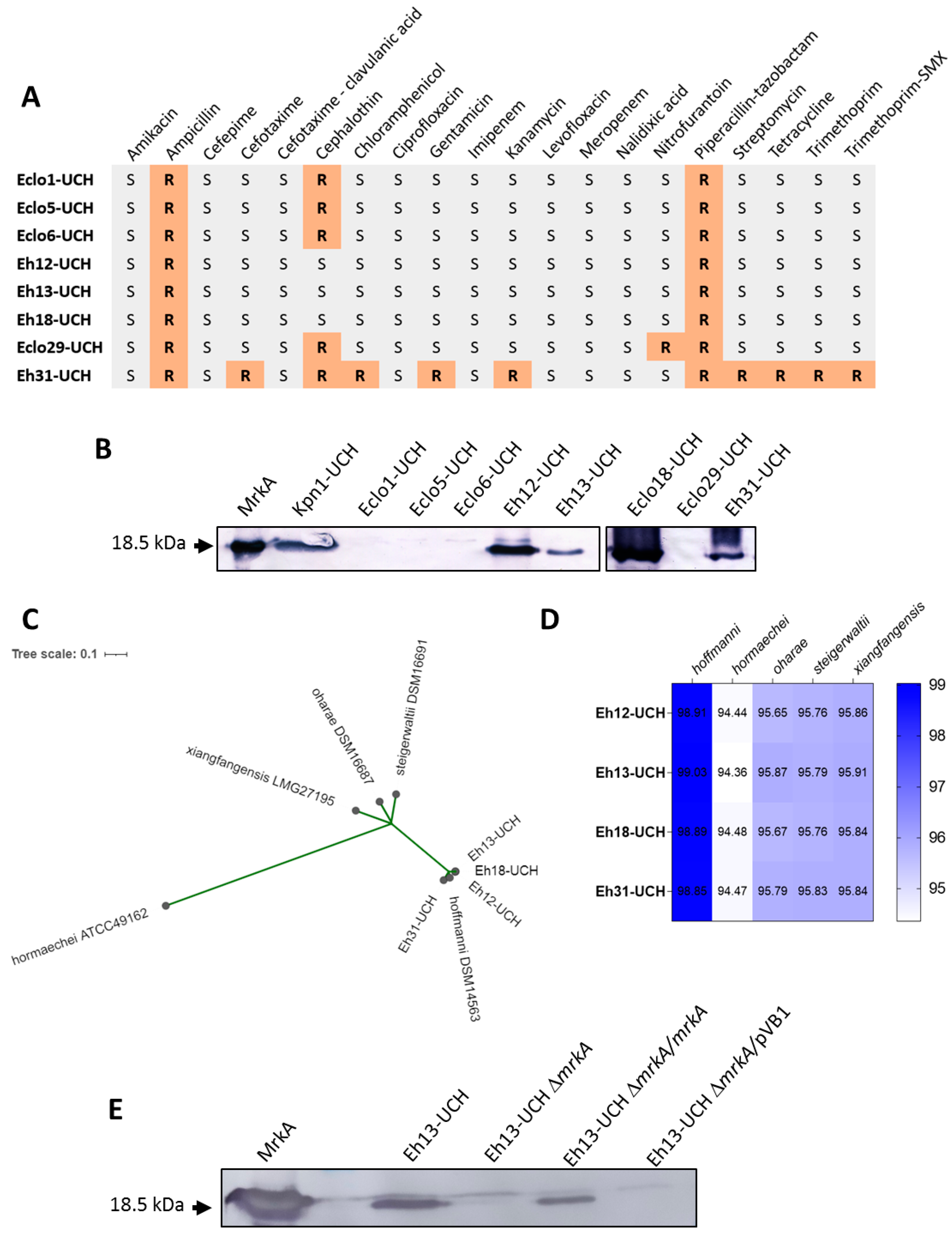
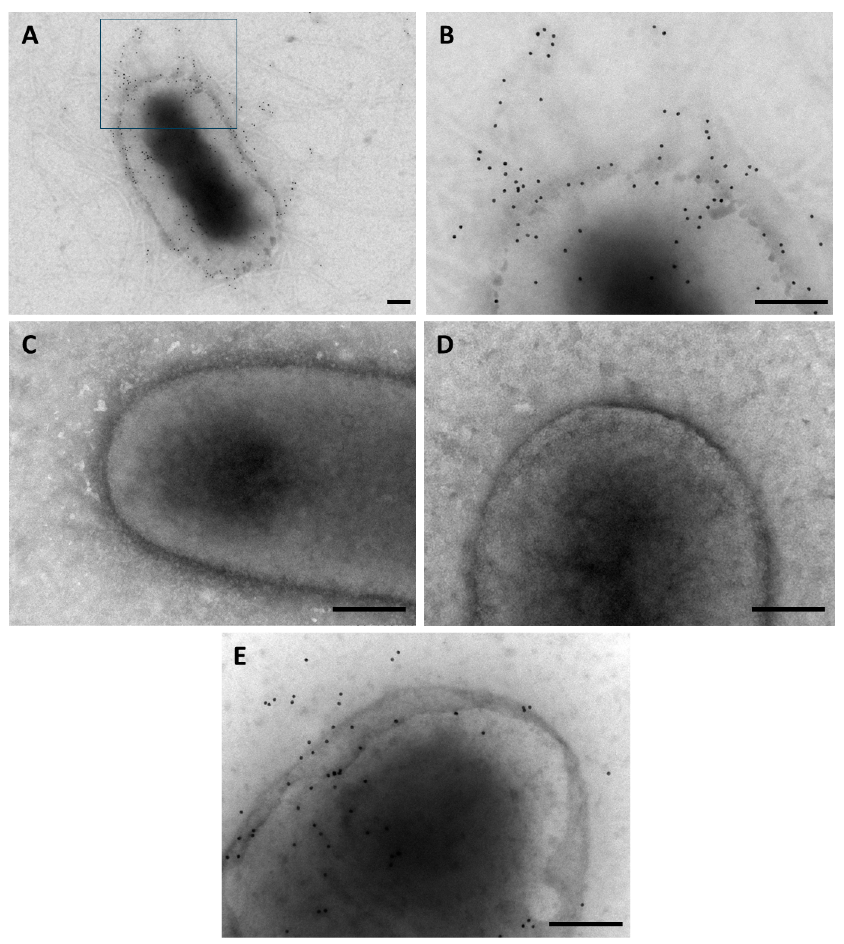
3.1. Production of Type 3 Fimbria by E. hormaechei subsp. hoffmannii

Eight clinical isolates identified as Enterobacter cloacae (Table 1) were included in a screening of loci encoding fimbriae assembled by the chaperone–usher pathway. Disk susceptibility test indicated that seven of them were sensitive to most of the antibiotics tested (Figure 1A). All the strains were resistant to ampicillin and piperacillin–tazobactam, while five were resistant to cephalothin. Only one strain, Eh31-UCH, showed resistance to multiple antibiotics, including resistance to beta-lactam drugs apparently determined by an extended spectrum β-lactamase (ESBL), as clavulanic acid inhibited cefotaxime resistance. No resistance to carbapenems was detected. Four of the strains, Eh12-UCH, Eh13-UCH, Eh18-UCH, and Eh31-UCH, were positive for the detection of the mrkA gene, encoding the major structural subunit of the type 3 fimbriae, by PCR. This result was further confirmed by the detection of MrkA protein in heat-extracted proteins by Western blot (Figure 1B). The four strains that resulted negative in the detection of mrkA by PCR were also negative in the detection of MrkA by Western blot (Figure 1B). Sequencing of the positive strain’s genomes allowed identification as Enterobacter hormaechei. Multiple locus sequence type analysis indicated that Eh12-UCH, Eh13-UCH, and Eh18-UCH belong to the sequence type ST145 and Eh31-UCH to sequence type ST118 (Table 2). A phylogenetic analysis was conducted to identify the isolates using the E. hormaechei subspecies scheme [1,4,5]. Thus, genomes of type strains representing E. hormaechei subsp. hoffmannii, E. hormaechei subsp. hormaechei, E. hormaechei subsp. oharae, E. hormaechei subsp. steigerwaltii, and E. hormaechei subsp. xiangfangensis were included in a core genome SNP-based parsimony tree. The result indicated that the four isolates are E. hormaechei subsp. hoffmannii (Figure 1C). This was also proved by the determination of the ANI among the four Chilean isolates’ genomes and the reference genomes (Figure 1D). Furthermore, the prediction of the presence of antibiotic-resistance genes was consistent with the resistance phenotypes observed for Eh12-UCH, Eh13-UCH, Eh18-UCH, and Eh31-UCH. Thus, the presence of blaACT-14 might explain the resistance of Eh12-UCH, Eh13-UCH, and Eh18-UCH to ampicillin and piperacillin–tazobactam (Table S3). On the other hand, ten different resistance genes were found in the Eh31-UCH genome, which explains the observed multi-resistant phenotype. Noteworthy is the presence of two genes encoding ESBLs, blaTEM-1B and blaSHV-12 (Table S3).
In order to determine the role of the type 3 fimbriae in the adherence capacity of E. hormaechei subsp. hoffmannii, we selected Eh13 as the representative and removed the mrkA gene by allelic exchange. The mrkA gene was then provided back by transformation with the recombinant pVB1-mrkA plasmid. The outcomes of these processes were checked by Western blot (Figure 1E). MrkA was detected in heat-extracted proteins obtained from the wild-type Eh13-UCH strain and the complemented mutant Eh13-UCHΔmrkA/mrkA strain, but it was not detected in extracts from the mutant Eh13-UCHΔmrkA strain nor the mutant Eh13-UCHΔmrkA harboring the empty pVB1 plasmid (Figure 1D).
The presence of the type 3 fimbria was also established by immunogold staining in whole non-permeabilized bacteria. Gold particles were widely distributed along the entire surface of Eh13-UCH suggesting the presence of the type 3 fimbria (Figure 2A,B). In contrast, particles were not observed, or were very scarce, at the surface of the mutant strains Eh13-UCHΔmrkA and Eh13-UCHΔmrkA (Figure 2C,D), suggesting the absence of the structure. As expected, gold particles were evident in the complemented Eh13-UCHΔmrkA/mrkA strain, indicating that the fimbria is present at the bacterial surface (Figure 2E).

3.2. Role of the Type 3 Fimbria in Adherence Capacity of E. hormaechei subsp. hoffmannii Eh13-UCH Strain

In order to evaluate the role of type 3 fimbria in the adherence capacity of E. hormaechei subsp. hoffmanni, adherence assays were performed with the Eh13-UCH strain and its derivatives over a plastic surface and Caco-2 cells. First, assays were carried out by incubating the strains over cell culture plates for 3 h. After removing the medium and repeatedly washing the wells, the attached bacteria were stained with DAPI, and fluorescence intensity was measured. Fluorescence levels were similar among all the strains tested when the assay was carried out in LB. The only significant difference was noticed with the mutant Eh13-UCHΔmrkA strain harboring the empty pVB1 plasmid, which showed a lower adherence level than the wild type (Figure 3A). However, when the assay was performed in DMEM, a significant reduction in the adherence level was evident in both the mutant strain Eh13-UCHΔmrkA and the mutant strain Eh13-UCHΔmrkA/pVB1, compared to the wild-type’s level (Figure 3A). In fact, the fluorescence intensity was significantly higher in the wild-type EH13-UCH after incubation in DMEM, compared to the result obtained from the incubation in LB. In agreement with this observation, the complemented Eh13-UCHΔmrkA/mrkA strain in DMEM showed an adherence level similar to that obtained with the wild type in DMEM (Figure 3A). Then, a similar assay was performed, but the incubation lasted for 48 h, and the presence of attached bacteria was measured by crystal violet staining. Regardless of the medium (LB or DMEM), results were consistent with the behavior observed in the 3 h assay performed in DMEM (Figure 3B). Thus, a significantly lower adherence capacity was observed in mutant strains Eh13-UCHΔmrkA and the mutant strain Eh13-UCHΔmrkA/pVB1, compared to the wild type, but this was not observed in the case of the complemented Eh13-UCHΔmrkA/mrkA strain (Figure 3B).
In addition, infection assays were carried out over Caco-2 cells for 30 min or 3 h. Adherence capacity was expressed as the percentage of cell-associated bacteria, which means the percentage that represents the number of colony-forming units (CFU) recovered after lysis of the cell layer, relative to the initial inoculum. A significantly lower level of adherence was noticed in the mutant strains Eh13-UCHΔmrkA and Eh13-UCHΔmrkA/pVB1, compared to the wild type, at both times, 30 min and 3 h post-infection (Figure 3C,D). The difference was insignificant in the case of the complemented Eh13-UCHΔmrkA/mrkA strain, although after 30 min of infection, the average percentage of cell-associated bacteria was approximately three times lower compared to the wild type (Figure 3C). In contrast, the percentage of cell-associated bacteria for Eh13-UCHΔmrkA/mrkA after 3 h was similar to that observed for the wild type (Figure 3D).

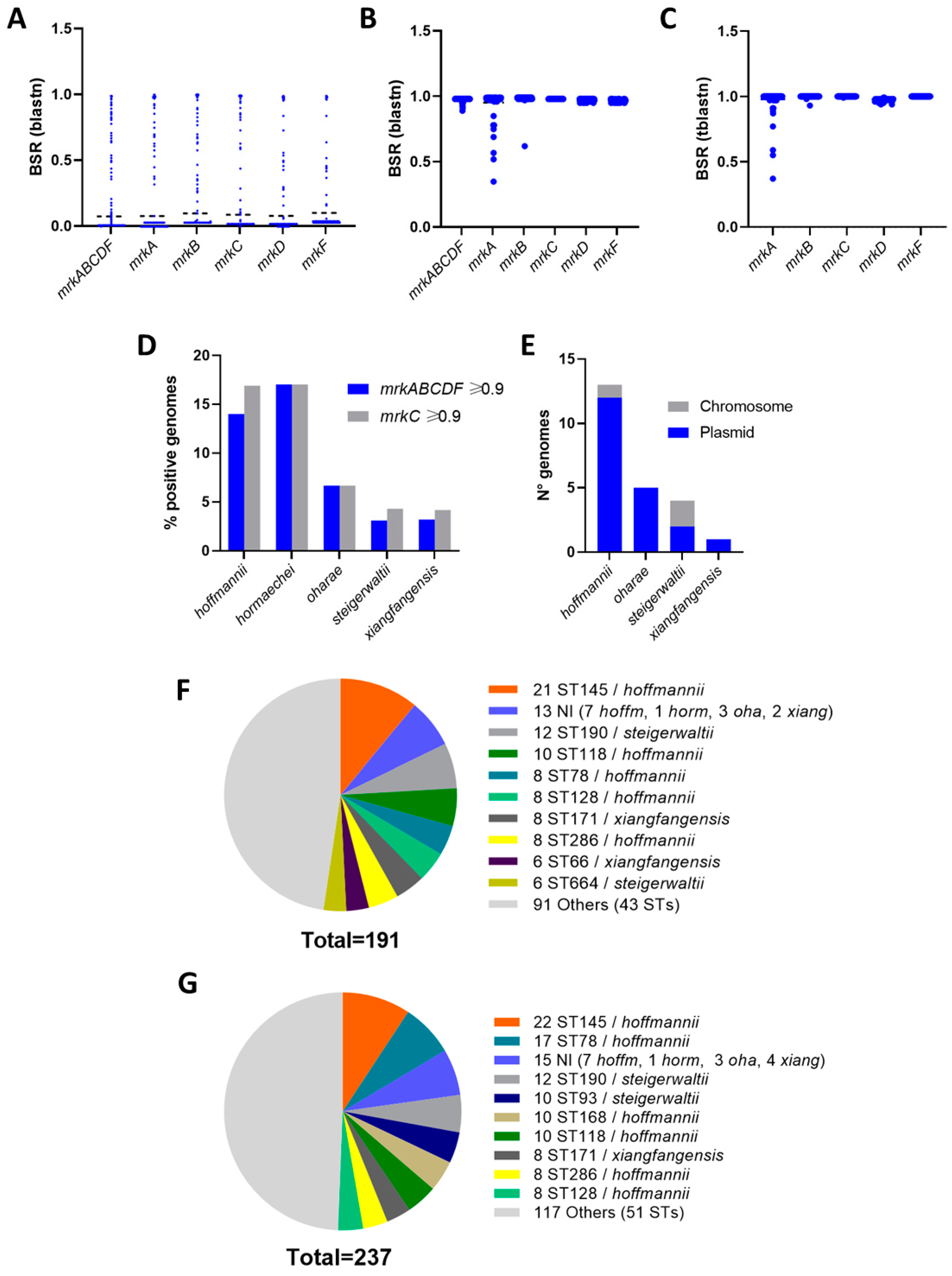
3.3. Distribution of the mrkABCDF Locus among E. hormaechei

After obtaining evidence of the role of the type 3 fimbria in Eh13-UCH, we determined the distribution of the mrkABCDF locus among E. hormaechei genomes contained in the NCBI Assembly RefSeq database. Screening of 3215 genomes with blastn indicated that the presence of the mrkABCDF locus was uncommon, with only 191 genomes (5.94%) obtaining blast-score ratios (BSR) equal to or higher than 0.9 (Figure 4A,B). A similar distribution was observed for the individual genes, with 197 (6.12%), 208 (6.46%), 214 (6.66%), 214 (6.66%), and 223 (6.93%) genomes showing BSR ≥ 0.9 for mrkA, mrkB, mrkC, mrkD, and mrkF, respectively (Figure 4A,B). Screening of the individual genes using tblastn showed slightly higher BSRs, and the numbers of records with BSR ≥ 0.9 were 213 (6.63%), 230 (7.15%), 237 (7.37%), 214 (6.65%), and 223 (6.93%), for mrkA, mrkB, mrkC, mrkD, and mrkF, respectively (Figure 4C). Regardless of the algorithm used for the screening, blastn or tblastn, the higher degree of variation among records with BSR ≥ 0.9 for the mrkABCDF locus was found in the mrkA gene, which encodes the major structural subunit (Figure 4B,C).
We selected sets of genomes as type 3 pili positives for further analyses according to two criteria. The first group included 191 genomes showing BSR ≥ 0.9 for the mrkABCDF locus screening using blastn, and the second group included 237 genomes showing BSR ≥ 0.9 for the screening of mrkC, using tblastn. Thus, we identified the subspecies within both groups of genomes, positives for the type 3 pili genes, according to the ANI analysis. ANI values ranged from 98.10% to 99.95% for this identification. We considered the five subspecies recognized in the List of Prokaryotic names with Standing in Nomenclature [1,4]. Positive genomes were identified in the five cases, with the highest percentages obtained for E. hormaechei subsp. hoffmannii (14–17%, among 714 genomes) and E. hormaechei subsp. hormaechei (17%, among 47 genomes) (Figure 4D). Then, we explored annotations in a subset of 23 complete genomes to establish if the mrkABCDF locus was located on the chromosome or in any other extra-chromosome element. We found that the locus was located in plasmids, in most of the cases, regardless of the subspecies (Figure 4E). Regretfully, no E. hormaechei subsp. hormaechei complete genomes were found in the NCBI Assembly RefSeq database to be included in the analysis.
Finally, we determined the positive genomes’ sequence type according to the E. cloacae MLST scheme [29,30]. We found 52 different sequence types among genomes with BSR ≥ 0.9 for the screening of the mrkABCF locus and 60 sequence types among genomes with BSR ≥ 0.9 for the screening of the mrkC gene. Most of the positive genomes belonged to the ST145 sequence type, all identified as E. hormaechei subsp. hoffmannii (Figure 4F). Four other sequence types of this subspecies were also found among the most frequent, ST118, ST78, ST28, and ST286 (Figure 4F). In the set selected according to the mrkC BSR ≥ 0.9 criterion, ST168-genomes were also found as representatives of E. hormaechei subsp. hoffmannii (Figure 4G). Sequence types of E. hormaechei subsp. steigerwaltii and E. hormaechei subsp. xiangfangensis were also found among the most common, including ST93, ST190, and ST664, and ST66, and ST171, respectively (Figure 4F,G).

4. Discussion

In this work, we have described the presence and the role of type 3 fimbria in the adherence capacity of E. hormaechei subsp. hoffmannii. The presence was evidenced by the detection of the major subunit MrkA in heat-extracted proteins and also at the surface of whole bacteria in a representative strain isolated in Chile, the Eh13-UCH strain. This strain was identified as E. hormaechei subsp. hoffmannii, according to its draft genomic sequence, compared with type strains representing four E. hormaechei subspecies, E. hormaechei subsp. hoffmanni, E. hormaechei subsp. hormaechei, E. hormaechei subsp. oharae, E. hormaechei subsp. steigerwaltii, and E. hormaechei subsp. xiangfangensis [1,4].
Research to find and characterize colonization and virulence factors of E. hormaechei, and even in representatives of the Enterobacter cloacae complex, has not been carried out to the same extent as other species such as Escherichia coli or Klebsiella spp. Indeed, epidemiological relevance is an important factor supporting research priorities. According to the 2019 global mortality report [41], E. coli and K. pneumoniae are among the top four, being the most relevant representatives of the Enterobacteriaceae family. Enterobacter is included as a genus (Enterobacter spp.) within the top ten taxa [41]. Within the Enterobacter genus, E. hormaechei is an important pathogen, particularly relevant in healthcare-associated infections and for the emergence of antibiotic-resistant clones [42,43,44,45]. In this work, one of the four strains, identified as a type 3 fimbria-producing E. hormaechei subsp. hoffmannii, Eh31-UCH, was isolated from a sepsis case. It showed the capacity to resist multiple antibiotics, including a third-generation cephalosporin, likely by the production of ESBLs. We believe that research to gain knowledge into the pathogenic mechanisms and to identify colonization and virulence determinants is valuable for both antibiotic-susceptible and antibiotic-resistant strains. However, particularly for those strains displaying multiple resistance, this could help in exploring and developing novel therapies as alternatives to antibiotics. In this scenario, several anti-virulence, including anti-adherence, therapies have been proposed for Enterobacteriaceae representatives, as compounds to inhibit the production of attachment determinants, receptor analogs, probiotics, or vaccines, among others [9,46,47,48].
The type 3 fimbria was first described in Klebsiella spp., and most of the progress regarding knowledge about the structure has been made in species representative of that genus. Thus, the fimbria has even been proposed as a basis for potential anti-adherence therapies [13,14]. However, the presence of the type 3 fimbriae has been reported in other species as E. coli, C. freundii, and C. koseri [15]. In fact, an early work reported the reactivity of anti-type 3 fimbria with structures produced by several other representatives of the Enterobacteriaceae family, including some Enterobacter species [16]. Later, the presence of the mrkB gene was reported in E. hormaechei subsp. oharae isolates obtained in Brazil [18]. As higher BSR values were obtained in our screenings of the mrkABCDF locus, and also of the individual genes, sequences seem to be conserved between K. pneumoniae Kp13 and the positive E. hormaechei strains analyzed here. This observation is consistent with results reported by Ong et al., which indicated the high degree of conservation of the mrkABCDF locus among all the Enterobacteriaceae species in which the type 3 fimbria was detected (Klebsiella spp., E. coli, C. freundii, and C. koseri) [15].
Regarding the role of the type 3 fimbriae in the adherence capacity of E. hormaechei subsp. hoffmannii, we found that the Eh13-UCH mutant strain lacking mrkA had a significantly lower adherence capacity to a plastic plate compared to the wild type. The difference was particularly evident when the assay was performed in DMEM rather than LB, suggesting that the production of the type 3 fimbria could be favored in this medium. Previous data of higher production of the bundle-forming pilus and higher secretion of effector proteins by enteropathogenic E. coli cultured in DMEM, compared to LB, support this hypothesis [49,50]. Also, in agreement with this finding, the Eh13-UCH mutant strain lacking mrkA had a significantly lower adherence capacity to the human colonic epithelial cell line Caco-2, an assay that is also performed in DMEM. In both cases, adherence to the plastic surface and Caco-2 cells, and the complementation of the mutant strain with the mrkA gene, provided in the pVB1 expression plasmid, restored the adherent phenotype. This was evident at 3 h and 48 h over the plastic surface, in DMEM, and at 3 h over Caco-2 cells. Given that the cell-adherent phenotype of the complemented strain was not clear at 30 min, we hypothesize that type 3 fimbria production and/or assembly could take longer in that case. Although the colonic epithelium represents a reservoir rather than the most common E. hormaechei infection sites [1], the Caco-2 cell line has been previously used as a model to evaluate the adherence capacity of this bacterium [40]. In fact, according to the results reported by Rafferty et al., the number of CFU associated with the Caco-2 cells was higher than the number of CFU attached to primary human aortic endothelial cells [40]. This suggests that Caco-2 cells produce receptors for binding of the type 3 fimbria. If toxigenic or cytopathic effects occur over Caco-2 cells on E. hormaechei subsp. hoffmannii infection, and whether those are influenced by type 3 fimbria-directed binding, is yet to be determined. However, type 3 fimbria is conserved among Enterobacteriaceae species in which it has been detected, and it is involved in the adherence of K. pneumoniae to bladder and endothelial cells [51]. In addition, a role in the in vivo K. pneumoniae colonization of lungs and bladder in the mouse model has been reported [14,52]. Therefore, we hypothesize that it might also determine E. hormaechei attachment to other organs in which it causes infection.
According to our results, the overall presence of the type 3 fimbria among E. hormaechei is low. The highest percentages of genomes positive for mrkABCDF or mrkC in our study were observed among E. hormaechei subsp. hoffmannii (14/17%) and E. hormaechei subsp. hormaechei (17%) genomes. Even lower percentages were found among E. hormaechei subsp. oharae, E. hormaechei subsp. steigerwaltii, and E. hormaechei subsp. xiangfangensis. We performed the screening first with the mrkABCDF locus in order to find genomes harboring the minimum required number of genes to direct production of the type 3 fimbria, in a single piece of DNA. However, we also performed the screening of mrkC because the mrkABCDF might be incomplete in draft genomes, which represent most of the NCBI Assembly RefSeq database, due to incomplete sequencing. This is consistent with previous reports, in which type 3 fimbria-encoding genes have been detected among E. cloacae complex strains or E. hormaechei strains [17,18]. To our knowledge, there is only one report in which the presence of a type 3 fimbria-encoding gene was found in an E. hormaechei subspecies [18]. It corresponds to the mrkB gene detected in subpopulations of an Enterobacter hormaechei subsp. oharae displaying higher adherence capacities [18]. The presence of the mrkABCDF locus in plasmids, in most of the cases that we analyzed, suggests that it was acquired by representatives of several different lineages by horizontal transfer, as we found it in more than 50 different STs. The higher percentage of positivity for E. hormaechei subsp. hoffmannii and E. hormaechei subsp. hormaechei suggests that representatives of these subspecies could have experienced more encounters with type 3 fimbria-producing bacteria or that they have been more exposed to the free type 3 fimbria-encoding plasmids during their evolution. Alternatively, they might incorporate and/or deliver the type 3 fimbria-encoding plasmids more efficiently compared to representatives of the rest of the subspecies.
Even though there is a low distribution of type 3 fimbria among E. hormaechei, our results suggest that it might be a determinant of the colonization and pathogenic capacities in strains that have acquired the locus. This is consistent with the well-known role of the fimbria in K. pneumoniae and also with the fact that the most common mrkABCDF- and mrkC-positive sequence types found here have been found in carbapenem-resistant strains associated with illness in humans. For example, E. cloacae complex strains belonging to sequence types ST145, ST118, ST78, ST168, and ST66 were found in France, while ST145, ST93, and ST171 were found in the same type of strains isolated in China, and ST93 in Poland [53,54,55]. Furthermore, as bacterial adherence is a complex process, additional adhesive structures are expected to be produced by E. hormaechei, and future research will help to find and characterize them.
On the other hand, the characterization of E. hormaechei isolates at the subspecies level reinforces the relevance of the group, especially in nosocomial infections. For example, E. hormaechei subsp. hoffmannii was found as the most common agent of nosocomial infections in a University Hospital in Taiwan [56], and one of the agents was involved in an outbreak in a neonatal intensive care unit in Germany [57]. In addition, E. hormaechei strains, including E. hormaechei subsp. xiangfangensis, E. hormaechei subsp. steigerwaltti, and E. hormaechei subsp. hoffmannii, were found as the most common agents among E. cloacae complex isolates from hospitals in South Korea [58]. In that study, E. hormaechei subsp. hoffmannii and E. hormaechei subsp. xiangfangensis strains belonging to sequence types ST78 and ST66, respectively, both recognized in our study for including mrkABCDF-positive representatives, were identified [58].
Given that the taxonomy of Enterobacter spp. has been evolving, the identity of the isolates at the species and/or subspecies level has not always been established according to a single scheme. In 2020, Wu et al. proposed an update on the taxonomy of the Enterobacter genus based on comparative genomics and phylogenomic analyses [5]. In that work, E. hormaechei subsp. hoffmannii was proposed to be renamed as E. hoffmannii; so, the Eh12-UCH, Eh13-UCH, Eh18-UCH, and Eh31-UCH strains could also be considered as E. hoffmannii. In addition, E. hormaechei subsp. oharae and E. hormaechei subsp. steigerwaltii were considered as synonyms of E. xiangfangensis. Therefore, several database records identified here as positive for mrkABCDF or mrkC, indexed as E. hormaechei subsp. oharae or E. hormaechei subsp. steigerwaltii, might represent E. xiangfangensis. This proposal remains to be considered in the List of Prokaryotic Names with Standing in Nomenclature [4]. However, some studies have already incorporated the new guidelines and have identified these species. For example, a study carried out in France found E. hoffmannii, E. hormaechei, and E. xiangfangensis among strains obtained from septic shock lethal cases in newborns [59]. Another study reanalyzed the presence of AmpC variants with E. hoffmannii and E. xiangfangensis strains isolated from different clinical samples in China [60]. A third study carried out in France, found E. hoffmanni and E. xiangfangensis among strains causing neonatal sepsis cases, attributed to contamination of the incubators [61]. Finally, a study carried out in Spain identified E. hoffmannii strains belonging to the ST78 sequence type and E. xiangfangensis strains belonging to the ST66 and ST171 sequence types as causative agents of bloodstream infections [62]. On the other hand, a very recent study in which genomes of 256 clinical strains belonging to the E. cloacae complex were analyzed suggests that the subspecies classification scheme for E. hormaechei has a better correlation with the molecular features, including phylogeny, presence of virulence genes, and capsule type [63]. Indeed, regardless of this dynamic scenario, the current epidemiological relevance of the Enterobacter species is evident in the results of all these studies.
Overall, it is expected that the knowledge about Enterobacter species will move forward regarding taxonomy and phylogenomics. These advances should certainly move along with increasing knowledge regarding pathogenicity mechanisms and the role and distribution of the key virulence factors.

5. Conclusions

  • The type 3 fimbria is an adherence determinant of Enterobacter hormaechei subsp. hoffmannii.
  • The type 3 fimbria is uncommon among E. hormaechei, although it can be harbored by representatives of the five subspecies. E. hormaechei subsp. hoffmannii and E. hormaechei subsp. hormaechei displayed the highest positivity rates. Production of type 3 fimbria may confer fitness advantages regarding adherence and colonization capacities.

Supplementary Materials

The following supporting information can be downloaded at: https://www.mdpi.com/article/10.3390/microorganisms12071441/s1, Table S1: Primers used in this work; Table S2: Sequences recovered from databases, used in this study. Table S3: Antibiotic resistance genes identified in E. hormaechei subsp. hoffmannii sequenced in this study and their known resistance-associated profiles according to ResFinder. Reference [64] is cited in the Supplementary Materials.

Author Contributions

Conceptualization, V.F.-Y., R.M.V., C.T., P.S. and F.D.C.; methodology, V.F.-Y., R.M.V., C.T., P.S. and F.D.C.; software, V.F.-Y. and F.D.C.; validation, V.F.-Y., C.T., P.S. and F.D.C.; formal analysis, V.F.-Y., R.M.V., C.T., P.S. and F.D.C.; investigation, V.F.-Y., V.I., A.T., I.S., V.S., C.A., I.M. and F.D.C.; resources, V.F.-Y., R.M.V., C.T. and F.D.C.; data curation, V.F.-Y., C.T. and F.D.C.; writing—original draft preparation, V.F.-Y. and F.D.C.; writing—review and editing, F.D.C.; visualization, V.F.-Y. and F.D.C.; supervision, V.F.-Y., C.T. and F.D.C.; project administration, V.F.-Y. and F.D.C.; funding acquisition, F.D.C., R.M.V., C.T. and F.D.C. All authors have read and agreed to the published version of the manuscript.

Funding

This research was funded by the Fondo Nacional de Desarrollo Científico y Tecnológico (Fondecyt) grants 1200979, 1211647 and 11150966, from the Agencia Nacional de Investigación y Desarrollo (ANID), Ministerio de Ciencia, Tecnología, Conocimiento e Innovación, Gobierno de Chile and with the support of the Líneas de Apoyo a la Investigación del Instituto de Ciencias Biomédicas, Facultad de Medicina, Universidad de Chile. VF doctoral thesis is supported by Beca de Doctorado Nacional 21201275, Agencia Nacional de Investigación y Desarrollo (ANID), Ministerio de Ciencia, Tecnología, Conocimiento e Innovación, Gobierno de Chile.

Institutional Review Board Statement

The use of the clinical strains (Kpn1-UCH, Eclo1-UCH, Eclo5-UCH, Eclo6-UCH, Eh12-UCH, Eh13-UCH, Eh18-UCH, Eclo29-UCH, and Eh31-UCH), which were already stored at the Programa de Microbiología y Micología, Instituto de Ciencias Biomédicas, Facultad de Medicina, Universidad de Chile, at the beginning of this project, was approved by the Ethics Committee for Research on Human Beings, Facultad de Medicina, Universidad de Chile (Ethics Approval Document N° 003, issued on 4 May 2023).

Data Availability Statement

The original contributions presented in the study are included in the article and Supplementary Materials, further inquiries can be directed to the corresponding author.

Acknowledgments

The authors are grateful to JMI Laboratories for providing the clinical isolates used in this work.

Conflicts of Interest

The authors declare no conflicts of interest.

References

  1. Davin-Regli, A.; Lavigne, J.P.; Pagès, J.M. Enterobacter spp.: Update on taxonomy, clinical aspects, and emerging antimicrobial resistance. Clin. Microbiol. Rev. 2019, 32, e00002-19. [Google Scholar] [CrossRef] [PubMed]
  2. Martins, E.R.; Bueno, M.F.C.; Francisco, G.R.; Casella, T.; de Oliveira Garcia, D.; Cerdeira, L.T.; Gerber, A.L.; de Almeida, L.G.P.; Lincopan, N.; de Vasconcelos, A.T.R.; et al. Genome and plasmid context of two rmtG-carrying Enterobacter hormaechei isolated from urinary tract infections in Brazil. J. Glob. Antimicrob. Resist. 2020, 20, 36–40. [Google Scholar] [CrossRef]
  3. da Silva, C.L.; Miranda, L.E.; Moreira, B.M.; Rebello, D.; Carson, L.A.; Kellum, M.E.; de Almeida, M.C.; Sampaio, J.L.; O’Hara, C.M. Enterobacter hormaechei bloodstream infection at three neonatal intensive care units in Brazil. Pediatr. Infect. Dis. J. 2002, 21, 175–177. [Google Scholar] [CrossRef] [PubMed]
  4. Parte, A.C.; Sardà Carbasse, J.; Meier-Kolthoff, J.P.; Reimer, L.C.; Göker, M. List of prokaryotic names with standing in nomenclature (LPSN) moves to the DSMZ. Int. J. Syst. Evol. Microbiol. 2020, 70, 5607–5612. [Google Scholar] [CrossRef] [PubMed]
  5. Wu, W.; Feng, Y.; Zong, Z. Precise species identification for Enterobacter: A genome sequence-based study with reporting of two novel species, Enterobacter quasiroggenkampii sp. nov. and Enterobacter quasimori sp. nov. mSystems 2020, 5, e00527-20. [Google Scholar] [CrossRef] [PubMed]
  6. Rice, L.B. Federal funding for the study of antimicrobial resistance in nosocomial pathogens: No ESKAPE. J. Infect. Dis. 2008, 197, 1079–1081. [Google Scholar] [CrossRef] [PubMed]
  7. De Rosa, F.G.; Corcione, S.; Pagani, N.; Di Perri, G. From ESKAPE to ESCAPE, from KPC to CCC. Clin. Infect. Dis. 2015, 60, 1289–1290. [Google Scholar] [CrossRef] [PubMed]
  8. WHO Bacterial Priority Pathogens List, 2024: Bacterial Pathogens of Public Health Importance to Guide Research, Development and Strategies to Prevent and Control Antimicrobial Resistance. Available online: https://www.who.int/publications/i/item/9789240093461 (accessed on 17 May 2024).
  9. Campos, J.C.M.; Antunes, L.C.; Ferreira, R.B. Global priority pathogens: Virulence, antimicrobial resistance and prospective treatment options. Future Microbiol. 2020, 15, 649–677. [Google Scholar] [CrossRef]
  10. Gerlach, G.F.; Allen, B.L.; Clegg, S. Molecular characterization of the type 3 (MR/K) fimbriae of Klebsiella pneumoniae. J. Bacteriol. 1988, 170, 3547–3553. [Google Scholar] [CrossRef]
  11. Nuccio, S.P.; Bäumler, A.J. Evolution of the chaperone/usher assembly pathway: Fimbrial classification goes Greek. Microbiol. Mol. Biol. Rev. 2007, 71, 551–575. [Google Scholar] [CrossRef]
  12. Murphy, C.N.; Clegg, S. Klebsiella pneumoniae and type 3 fimbriae: Nosocomial infection, regulation and biofilm formation. Future Microbiol. 2012, 7, 991–1002. [Google Scholar] [CrossRef] [PubMed]
  13. Choi, M.; Tennant, S.M.; Simon, R.; Cross, A.S. Progress towards the development of Klebsiella vaccines. Expert. Rev. Vaccines 2019, 18, 681–691. [Google Scholar] [CrossRef] [PubMed]
  14. Wang, Q.; Chang, C.S.; Pennini, M.; Pelletier, M.; Rajan, S.; Zha, J.; Chen, Y.; Cvitkovic, R.; Sadowska, A.; Heidbrink Thompson, J.; et al. Target-agnostic identification of functional monoclonal antibodies against Klebsiella pneumoniae multimeric MrkA fimbrial subunit. J. Infect. Dis. 2016, 213, 1800–1808. [Google Scholar] [CrossRef] [PubMed]
  15. Ong, C.L.; Beatson, S.A.; Totsika, M.; Forestier, C.; McEwan, A.G.; Schembri, M.A. Molecular analysis of type 3 fimbrial genes from Escherichia coli, Klebsiella and Citrobacter species. BMC Microbiol. 2010, 10, 183. [Google Scholar] [CrossRef] [PubMed]
  16. Old, D.C.; Adegbola, R.A. Antigenic relationships among type-3 fimbriae of Enterobacteriaceae revealed by immunoelectronmicroscopy. J. Med. Microbiol. 1985, 20, 113–121. [Google Scholar] [CrossRef] [PubMed]
  17. Livrelli, V.; De Champs, C.; Di Martino, P.; Darfeuille-Michaud, A.; Forestier, C.; Joly, B. Adhesive properties and antibiotic resistance of Klebsiella, Enterobacter, and Serratia clinical isolates involved in nosocomial infections. J. Clin. Microbiol. 1996, 34, 1963–1969. [Google Scholar] [CrossRef] [PubMed]
  18. Brust, F.R.; Boff, L.; da Silva Trentin, D.; Pedrotti Rozales, F.; Barth, A.L.; Macedo, A.J. Macrocolony of NDM-1 producing Enterobacter hormaechei subsp. oharae generates subpopulations with different features regarding the response of antimicrobial agents and biofilm formation. Pathogens 2019, 8, 49. [Google Scholar] [CrossRef]
  19. Gales, A.C.; Castanheira, M.; Jones, R.N.; Sader, H.S. Antimicrobial resistance among Gram-negative bacilli isolated from Latin America: Results from SENTRY Antimicrobial Surveillance Program (Latin America, 2008–2010). Diagn. Microbiol. Infect. Dis. 2012, 73, 354–360. [Google Scholar] [CrossRef]
  20. Santiviago, C.A.; Reynolds, M.M.; Porwollik, S.; Choi, S.H.; Long, F.; Andrews-Polymenis, H.L.; McClelland, M. Analysis of pools of targeted Salmonella deletion mutants identifies novel genes affecting fitness during competitive infection in mice. PLoS Pathog. 2009, 5, e1000477. [Google Scholar] [CrossRef]
  21. Sharan, S.K.; Thomason, L.C.; Kuznetsov, S.G.; Court, D.L. Recombineering: A homologous recombination-based method of genetic engineering. Nat. Protoc. 2009, 4, 206–223. [Google Scholar] [CrossRef]
  22. Performance Standards for Antimicrobial Susceptibility Testing, M100, 32nd ed.; Clinical and Laboratory Standards Institute: Wayne, PA, USA, 2022. Available online: https://clsi.org/standards/products/microbiology/documents/m100/ (accessed on 1 November 2022).
  23. Prjibelski, A.; Antipov, D.; Meleshko, D.; Lapidus, A.; Korobeynikov, A. Using SPAdes De Novo Assembler. Curr. Protoc. Bioinform. 2020, 70, e102. [Google Scholar] [CrossRef]
  24. Gurevich, A.; Saveliev, V.; Vyahhi, N.; Tesler, G. QUAST: Quality assessment tool for genome assemblies. Bioinformatics 2013, 29, 1072–1075. [Google Scholar] [CrossRef] [PubMed]
  25. Parks, D.H.; Imelfort, M.; Skennerton, C.T.; Hugenholtz, P.; Tyson, G.W. CheckM: Assessing the quality of microbial genomes recovered from isolates, single cells, and metagenomes. Genome Res. 2015, 25, 1043–1055. [Google Scholar] [CrossRef] [PubMed]
  26. Jolley, K.A.; Bliss, C.M.; Bennett, J.S.; Bratcher, H.B.; Brehony, C.; Colles, F.M.; Wimalarathna, H.; Harrison, O.B.; Sheppard, S.K.; Cody, A.J.; et al. Ribosomal multilocus sequence typing: Universal characterization of bacteria from domain to strain. Microbiology 2012, 158, 1005–1015. [Google Scholar] [CrossRef]
  27. Gardner, S.N.; Slezak, T.; Hall, B.G. kSNP3.0: SNP detection and phylogenetic analysis of genomes without genome alignment or reference genome. Bioinformatics 2015, 31, 2877–2878. [Google Scholar] [CrossRef] [PubMed]
  28. Jain, C.; Rodriguez-R, L.M.; Phillippy, A.M.; Konstantinidis, K.T.; Aluru, S. High throughput ANI analysis of 90K prokaryotic genomes reveals clear species boundaries. Nat. Commun. 2018, 9, 5114. [Google Scholar] [CrossRef]
  29. Seemann, T. mlst. Github. Available online: https://github.com/tseemann/mlst (accessed on 1 May 2024).
  30. Jolley, K.A.; Bray, J.E.; Maiden, M.C.J. Open-access bacterial population genomics: BIGSdb software, the PubMLST.org website and their applications. Wellcome Open Res. 2018, 3, 124. [Google Scholar] [CrossRef] [PubMed]
  31. Bortolaia, V.; Kaas, R.S.; Ruppe, E.; Roberts, M.C.; Schwarz, S.; Cattoir, V.; Philippon, A.; Allesoe, R.L.; Rebelo, A.R.; Florensa, A.F.; et al. ResFinder 4.0 for predictions of phenotypes from genotypes. J. Antimicrob. Chemother. 2020, 75, 3491–3500. [Google Scholar] [CrossRef]
  32. Camacho, C.; Coulouris, G.; Avagyan, V.; Ma, N.; Papadopoulos, J.; Bealer, K.; Madden, T.L. BLAST+: Architecture and applications. BMC Bioinform. 2009, 10, 421. [Google Scholar] [CrossRef]
  33. Ramos, P.I.; Picão, R.C.; Almeida, L.G.; Lima, N.C.; Girardello, R.; Vivan, A.C.; Xavier, D.E.; Barcellos, F.G.; Pelisson, M.; Vespero, E.C.; et al. Comparative analysis of the complete genome of KPC-2-producing Klebsiella pneumoniae Kp13 reveals remarkable genome plasticity and a wide repertoire of virulence and resistance mechanisms. BMC Genom. 2014, 15, 54. [Google Scholar] [CrossRef]
  34. Sayers, E.W.; Bolton, E.E.; Brister, J.R.; Canese, K.; Chan, J.; Comeau, D.C.; Connor, R.; Funk, K.; Kelly, C.; Kim, S.; et al. Database resources of the national center for biotechnology information. Nucleic Acids Res. 2022, 50, D20–D26. [Google Scholar] [CrossRef] [PubMed]
  35. Fernández-Yáñez, V.; Suazo, P.; Hormazábal, C.; Ibaceta, V.; Arenas-Salinas, M.; Vidal, R.M.; Silva-Ojeda, F.; Arellano, C.; Muñoz, I.; Del Canto, F. Distribution of papA and papG variants among Escherichia coli genotypes: Association with major extraintestinal pathogenic lineages. Int. J. Mol. Sci. 2024, 25, 6657. [Google Scholar] [CrossRef]
  36. Wang, L.; Wu, P.; Su, Y.; Wei, Y.; Guo, X.; Yang, L.; Wang, M.; Liu, B. Detection of genus and three important species of Cronobacter using novel genus- and species-specific genes identified by large-scale comparative genomic analysis. Front. Microbiol. 2022, 13, 885543. [Google Scholar] [CrossRef] [PubMed]
  37. Webb, J.R.; Buller, N.; Rachlin, A.; Golledge, C.; Sarovich, D.S.; Price, E.P.; Mayo, M.; Currie, B.J. A persisting nontropical focus of Burkholderia pseudomallei with limited genome evolution over five decades. mSystems 2020, 5, e00726-20. [Google Scholar] [CrossRef] [PubMed]
  38. Del Canto, F.; Botkin, D.J.; Valenzuela, P.; Popov, V.; Ruiz-Perez, F.; Nataro, J.P.; Levine, M.M.; Stine, O.C.; Pop, M.; Torres, A.G.; et al. Identification of Coli Surface Antigen 23, a novel adhesin of enterotoxigenic Escherichia coli. Infect. Immun. 2012, 80, 2791–2801. [Google Scholar] [CrossRef] [PubMed]
  39. Bradford, M.M. A rapid and sensitive method for the quantitation of microgram quantities of protein utilizing the principle of protein-dye binding. Anal. Biochem. 1976, 72, 248–254. [Google Scholar] [CrossRef] [PubMed]
  40. Rafferty, B.; Dolgilevich, S.; Kalachikov, S.; Morozova, I.; Ju, J.; Whittier, S.; Nowygrod, R.; Kozarov, E. Cultivation of Enterobacter hormaechei from human atherosclerotic tissue. J. Atheroscler. Thromb. 2011, 18, 72–81. [Google Scholar] [CrossRef]
  41. GBD 2019 Antimicrobial Resistance Collaborators. Global mortality associated with 33 bacterial pathogens in 2019: A systematic analysis for the Global Burden of Disease Study 2019. Lancet 2022, 400, 2221–2248. [Google Scholar] [CrossRef]
  42. Yeh, T.K.; Lin, H.J.; Liu, P.Y.; Wang, J.H.; Hsueh, P.R. Antibiotic resistance in Enterobacter hormaechei. Int. J. Antimicrob. Agents 2022, 60, 106650. [Google Scholar] [CrossRef]
  43. Xu, T.; Xue, C.X.; Huang, J.; Wu, J.; Chen, R.; Zhou, K. Emergence of an epidemic hypervirulent clone of Enterobacter hormaechei coproducing mcr-9 and carbapenemases. Lancet Microbe 2022, 3, e474–e475. [Google Scholar] [CrossRef]
  44. Knecht, C.A.; García Allende, N.; Álvarez, V.E.; Prack Mc Cormick, B.; Massó, M.G.; Campos, J.; Fox, B.; Alonso, F.M.; Donis, N.; Canigia, L.F.; et al. New sequence type of an Enterobacter cloacae complex strain with the potential to become a high-risk clone. J. Glob. Antimicrob. Resist. 2022, 31, 162–164. [Google Scholar] [CrossRef] [PubMed]
  45. Donà, V.; Nordmann, P.; Kittl, S.; Schuller, S.; Bouvier, M.; Poirel, L.; Endimiani, A.; Perreten, V. Emergence of OXA-48-producing Enterobacter hormaechei in a Swiss companion animal clinic and their genetic relationship to clinical human isolates. J. Antimicrob. Chemother. 2023, 78, 2950–2960. [Google Scholar] [CrossRef] [PubMed]
  46. Cegelski, L.; Marshall, G.R.; Eldridge, G.R.; Hultgren, S.J. The biology and future prospects of antivirulence therapies. Nat. Rev. Microbiol. 2008, 6, 17–27. [Google Scholar] [CrossRef] [PubMed]
  47. Krachler, A.M.; Orth, K. Targeting the bacteria-host interface: Strategies in anti-adhesion therapy. Virulence 2013, 4, 284–294. [Google Scholar] [CrossRef] [PubMed]
  48. Lau, W.Y.V.; Taylor, P.K.; Brinkman, F.S.L.; Lee, A.H.Y. Pathogen-associated gene discovery workflows for novel antivirulence therapeutic development. eBioMedicine 2023, 88, 104429. [Google Scholar] [CrossRef] [PubMed]
  49. Puente, J.L.; Bieber, D.; Ramer, S.W.; Murray, W.; Schoolnik, G.K. The bundle-forming pili of enteropathogenic Escherichia coli: Transcriptional regulation by environmental signals. Mol. Microbiol. 1996, 20, 87–100. [Google Scholar] [CrossRef] [PubMed]
  50. Kenny, B.; Abe, A.; Stein, M.; Finlay, B.B. Enteropathogenic Escherichia coli protein secretion is induced in response to conditions similar to those in the gastrointestinal tract. Infect. Immun. 1997, 65, 2606–2612. [Google Scholar] [CrossRef] [PubMed]
  51. Tarkkanen, A.M.; Virkola, R.; Clegg, S.; Korhonen, T.K. Binding of the type 3 fimbriae of Klebsiella pneumoniae to human endothelial and urinary bladder cells. Infect. Immun. 1997, 65, 1546–1549. [Google Scholar] [CrossRef]
  52. Murphy, C.N.; Mortensen, M.S.; Krogfelt, K.A.; Clegg, S. Role of Klebsiella pneumoniae type 1 and type 3 fimbriae in colonizing silicone tubes implanted into the bladders of mice as a model of catheter-associated urinary tract infections. Infect. Immun. 2013, 81, 3009–3017. [Google Scholar] [CrossRef]
  53. Emeraud, C.; Petit, C.; Gauthier, L.; Bonnin, R.A.; Naas, T.; Dortet, L. Emergence of VIM-producing Enterobacter cloacae complex in France between 2015 and 2018. J. Antimicrob. Chemother. 2022, 77, 944–951. [Google Scholar] [CrossRef]
  54. Chen, J.; Tian, S.; Nian, H.; Wang, R.; Li, F.; Jiang, N.; Chu, Y. Carbapenem-resistant Enterobacter cloacae complex in a tertiary Hospital in Northeast China, 2010–2019. BMC Infect. Dis. 2021, 21, 611. [Google Scholar] [CrossRef] [PubMed]
  55. Izdebski, R.; Baraniak, A.; Zabicka, D.; Sekowska, A.; Gospodarek-Komkowska, E.; Hryniewicz, W.; Gniadkowski, M. VIM/IMP carbapenemase-producing Enterobacteriaceae in Poland: Epidemic Enterobacter hormaechei and Klebsiella oxytoca lineages. J. Antimicrob. Chemother. 2018, 73, 2675–2681. [Google Scholar] [CrossRef] [PubMed]
  56. Chen, C.J.; Lu, P.L.; Jian, S.H.; Fu, H.L.; Huang, P.H.; Chang, C.Y. Molecular epidemiology, risk factors and clinical outcomes of carbapenem-nonsusceptible Enterobacter cloacae complex infections in a Taiwan University Hospital. Pathogens 2022, 11, 151. [Google Scholar] [CrossRef] [PubMed]
  57. Morhart, P.; Gerlach, R.G.; Kunz, C.; Held, J.; Valenza, G.; Wölfle, J.; Reutter, H.; Hanslik, G.J.; Fahlbusch, F.B. Application of next-generation sequencing to Enterobacter hormaechei subspecies analysis during a neonatal intensive care unit outbreak. Children 2023, 10, 1696. [Google Scholar] [CrossRef] [PubMed]
  58. Ganbold, M.; Seo, J.; Wi, Y.M.; Kwon, K.T.; Ko, K.S. Species identification, antibiotic resistance, and virulence in Enterobacter cloacae complex clinical isolates from South Korea. Front. Microbiol. 2023, 14, 1122691. [Google Scholar] [CrossRef] [PubMed]
  59. Girlich, D.; Ouzani, S.; Emeraud, C.; Gauthier, L.; Bonnin, R.A.; Le Sache, N.; Mokhtari, M.; Langlois, I.; Begasse, C.; Arangia, N.; et al. Uncovering the novel Enterobacter cloacae complex species responsible for septic shock deaths in newborns: A cohort study. Lancet Microbe 2021, 2, e536–e544. [Google Scholar] [CrossRef]
  60. Feng, Y.; Hu, Y.; Zong, Z. Reexamining the association of AmpC variants with Enterobacter species in the context of updated taxonomy. Antimicrob. Agents Chemother. 2021, 65, e0159621. [Google Scholar] [CrossRef] [PubMed]
  61. Hernandez-Alonso, E.; Bourgeois-Nicolaos, N.; Lepainteur, M.; Derouin, V.; Barreault, S.; Waalkes, A.; Augusto, L.A.; Gera, S.; Gleizes, O.; Tissieres, P.; et al. Contaminated incubators: Source of a multispecies Enterobacter outbreak of neonatal sepsis. Microbiol. Spectr. 2022, 10, e0096422. [Google Scholar] [CrossRef]
  62. Lumbreras-Iglesias, P.; de Toro, M.; Vázquez, X.; García-Carús, E.; Rodicio, M.R.; Fernández, J. High-risk international clones ST66, ST171 and ST78 of Enterobacter cloacae complex causing blood stream infections in Spain and carrying blaOXA-48 with or without mcr-9. J. Infect. Public. Health 2023, 16, 272–279. [Google Scholar] [CrossRef]
  63. Qiu, X.; Ye, K.; Ma, Y.; Zhao, Q.; Wang, L.; Yang, J. Genome sequence-based species classification of Enterobacter cloacae complex: A study among clinical isolates. Microbiol. Spectr. 2024, 12, e0431223. [Google Scholar] [CrossRef]
  64. Datsenko, K.A.; Wanner, B.L. One-step inactivation of chromosomal genes in Escherichia coli K-12 using PCR products. Proc. Natl. Acad. Sci. USA 2000, 97, 6640–6645. [Google Scholar] [CrossRef] [PubMed]
Figure 1. Presence of the type 3 fimbriae among Enterobacter cloacae complex strains and E. hormaechei subsp. hoffmannii strains: (A) Antibiotic susceptibility profile of eight E. cloacae complex strains. S: susceptible, R: resistant. Trimethropim-SMX: trimethoprim–sulfamethoxazole. (B) The type 3 fimbria major structural subunit MrkA was detected by Western blot in heat-extracted proteins. The purified mature MrkA protein and extracts obtained from the Klebsiella pneumoniae strain Kpn1-UCH were used as positive controls. (C) Maximum parsimony phylogenetic tree to identify E. hormaechei subspecies. Genomes of strains Eh12-UCH, Eh13-UCH, Eh18-UCH, and Eh31-UCH were included along with genomes of the type strains E. hormaechei subsp. hoffmannii DSM 14563, E. hormaechei subsp. hormaechei ATCC 49162, E. hormaechei subsp. oharae DSM 16687, E. hormaechei subsp. steigerwaltii DSM 16691, and E. hormaechei subsp. xiangfangensis LMG 27195 [5]. The tree was built based on 48,321 core SNPs. (D) Identification of E. hormaechei subspecies by average nucleotide identity (ANI) analysis. The heat map represents the results for the same set of genomes included in (C). (E) Detection of MrkA by Western blot in heat-extracted proteins obtained from the mutant strain Eh13-UCHΔmrkA and its derivative complemented strains.
Microorganisms 12 01441 g001
Figure 2. Detection of type 3 fimbria by immunogold staining. The presence of the structure was established using anti-MrkA and a secondary antibody conjugated with 10 nm gold particles over whole non-permeabilized bacteria: (A) Wild-type Eh13-UCH. (B) Higher magnification for the square depicted in (A) for wild-type Eh13-UCH. (C) Mutant Eh13-UCHΔmrkA. (D) The mutant strain harboring the empty pVB1 plasmid (Eh13-UCHΔmrkA/pVB1). (E) Complemented mutant strain (Eh13-UCHΔmrkA/mrkA). Bars: 200 nm.
Microorganisms 12 01441 g002
Figure 3. Evaluation of the adherence capacity of Eh13-UCH and its derivative strains: (A) Adherence over cell culture plates after 3 h of incubation, expressed as arbitrary fluorescence units (AFU). (B) Adherence over cell culture plates after 48 h of incubation, expressed as optical density measured at λ = 545 nm to detect crystal violet absorbance. LB: lysogeny broth. DMEM: Dulbecco’s modified Eagle medium. For (A,B), asterisks over the bars indicate significant differences compared to the level detected in the wild-type Eh13-UCH in LB. (C,D) Adherence capacity over Caco-2 cells after 30 min (C) or 3 h (D) of infection at a multiplicity of infection (MOI) of 10 bacteria/cell. Results are expressed as the percentage of cell-associated bacteria relative to the initial inoculum. For (C) and (D), asterisks over the bars indicate significant differences compared to the level detected in the wild-type Eh13-UCH. For all the cases, bars represent the mean ± the standard error. * p < 0.05, ** p < 0.01, *** p < 0.0001 according to Welch’s ANOVA test followed by Dunnett’s T3 multiple comparison test.
Microorganisms 12 01441 g003
Figure 4. Distribution of the type 3 pili genes among E. hormaechei genomes contained in the NCBI Assembly RefSeq database: (A) Screening for the mrkABCDF locus and its individual genes using blastn. The graph shows the blast-score ratio (BSR) values. (B,C) Distribution of BSR values in the screening of individual genes among records that showed BSR equal to or higher than 0.9 for the screening of the mrkABCDF locus, using blastn (B) or tblastn (C). Horizontal dotted lines in (AC) represent the means. (D) Identification of subspecies among positive records selected according to two criteria, BSR ≥ 0.9 for the mrkABCDF locus with blastn or BSR ≥ 0.9 for mrkC with tblastn. Numbers above the bars indicate the percentage of positive records among genomes representing each subspecies. Total numbers (100%) were 714 E. hormaechei subsp. hoffmannii, 47 E. hormaechei subsp. hormaechei, 165 E. hormaechei subsp. oharae, 1257 E. hormaechei subsp. steigerwaltii, and 1032 E. hormaechei subsp. xiangfangensis. (E) Localization of the mrkABCDF locus, analyzed in 23 complete genomes. No complete genomes representing E. hormaechei subsp. hormaechei were found. (F,G) Distribution of sequence type among E. hormaechei genome records positive for type 3 fimbriae, selected according to both criteria, BSR ≥ 0.9 in the screening for the mrkABCDF locus using blastn (F) or BSR ≥ 0.9 in the screening for mrkC with tblastn (G).
Microorganisms 12 01441 g004
Table 1. Strains and plasmids used in this work.
StrainSpecie/ComplexOrigin/IllnessUtilityReference
Eclo1-UCHEnterobacter cloacaePneumoniaDetection of type 3 fimbriaThis study
Eclo5-UCHEnterobacter cloacaeSepsisDetection of type 3 fimbriaThis study
Eclo6-UCHEnterobacter cloacaeSepsisDetection of type 3 fimbriaThis study
Eh12-UCHEnterobacter hormaechei subsp. hoffmanniiSepsisDetection of type 3 fimbriaThis study
Eh13-UCHEnterobacter hormaechei subsp. hoffmanniiSepsisDetection of type 3 fimbria and functional analysesThis study
Eh18-UCHEnterobacter hormaechei subsp. hoffmanniiPneumoniaDetection of type 3 fimbriaThis study
Eclo29-UCHEnterobacter cloacaeSepsisDetection of type 3 fimbriaThis study
Eh31-UCHEnterobacter hormaechei subsp. hoffmanniiSepsisDetection of type 3 fimbriaThis study
Kpn1-UCHKlebsiella pneumoniaePneumoniaDetection of type 3 fimbriaThis study
Eh13-UCHΔmrkAEnterobacter hormaechei subsp. hoffmanniiObtained after removal of the mrkA gene in Eh13-UCHDetection of type 3 fimbriaThis study
Eh13-UCHΔmrkA/pVB1Enterobacter hormaechei subsp. hoffmanniiEh13-UCHΔmrkA transformed with the empty pVB1 plasmidDetection of type 3 fimbriaThis study
Eh13-UCHΔmrkA/mrkAEnterobacter hormaechei subsp. hoffmanniiEh13-UCHΔmrkA transformed with the pVB1 plasmid containing mrkADetection of type 3 fimbriaThis study
Plasmids
pCLF4--Template to amplify the kanamycin resistance cassette used for allelic exchange[20]
pSIM9--Provision of the λ Red recombinase system[21]
pVB1--Expression plasmid, promoter inducible with m-tuolic acidDualSystems Biotech, (Schlieren, Switzerland)
Table 2. Main features of the draft genomes obtained in this study.
StrainSequence TypeGenome Length (bp)N50Completeness
(%)
Contamination
(%)
NCBI Nucleotide Accession Code
Eh12-UCHST1455,115,276270,30499.890.2JBCNUO000000000
Eh13-UCHST1454,744,088309,64099.890.2JBCNUP000000000
Eh18-UCHST1455,112,462269,96999.890.2JBCNUQ000000000
Eh31-UCHST1185,900,299165,59499.7812.04JBCNUR000000000
Disclaimer/Publisher’s Note: The statements, opinions and data contained in all publications are solely those of the individual author(s) and contributor(s) and not of MDPI and/or the editor(s). MDPI and/or the editor(s) disclaim responsibility for any injury to people or property resulting from any ideas, methods, instructions or products referred to in the content.

