Review

Insights into Pathogenesis of Trachoma

by
Panagiotis Toumasis
1,
Georgia Vrioni
2,
Ioannis T. Tsinopoulos
1,3,
Maria Exindari
1,4 and
George Samonis
5,6,*
1
MSc in Ocular Surgery, School of Medicine, Aristotle University of Thessaloniki, 541 24 Thessaloniki, Greece
2
Department of Microbiology, School of Medicine, National and Kapodistrian University of Athens, 115 25 Athens, Greece
3
Second Department of Ophthalmology, School of Medicine, Aristotle University of Thessaloniki, Papageorgiou General Hospital, 564 29 Thessaloniki, Greece
4
Department of Microbiology, School of Medicine, Aristotle University of Thessaloniki, 541 24 Thessaloniki, Greece
5
School of Medicine, University of Crete, 715 00 Heraklion, Greece
6
Metropolitan Hospital, 185 47 Piraeus, Greece
*
Author to whom correspondence should be addressed.
Microorganisms 2024, 12(8), 1544; https://doi.org/10.3390/microorganisms12081544 (registering DOI)
Submission received: 19 April 2024 / Revised: 31 May 2024 / Accepted: 25 July 2024 / Published: 28 July 2024
(This article belongs to the Section Public Health Microbiology)

Abstract

:
Trachoma is the most common infectious cause of blindness worldwide. This review investigates the pathogenesis of trachoma, focusing on its causative agent, transmission pathways, disease progression, and immune responses. Trachoma is caused by serovars A–C of the bacterium Chlamydia trachomatis (Ct). Transmission occurs through direct or indirect exchanges of ocular and nasal secretions, especially in regions with poor hygiene and overcrowded living conditions. The disease is initiated in early childhood by repeated infection of the ocular surface by Ct. This triggers recurrent chronic inflammatory episodes, leading to the development of conjunctival scarring and potentially to trichiasis, corneal opacity, and visual impairment. Exploring the pathogenesis of trachoma not only unveils the intricate pathways and mechanisms underlying this devastating eye disease but also underscores the multifaceted dimensions that must be considered in its management.

1. Introduction

Trachoma, an enduring relentless scourge on human populations, persistently exacts a profound toll on public health, standing as the leading infectious cause of blindness globally [1,2]. Trachoma is a type of chronic keratoconjunctivitis caused by the bacterium Chlamydia trachomatis (Ct). Nowadays, trachoma is endemic in much of sub-Saharan Africa, areas of the Middle East crescent, and pockets of Asia and South America, being a public health problem in a total of 40 countries [3]. The World Health Organization (WHO) has included trachoma in a comprehensive portfolio of neglected tropical diseases earmarked for targeted elimination [4]. The persistent nature of trachoma, coupled with its potential to cause irreversible harm, accentuates the urgency of concerted efforts to combat and eradicate this infectious affliction.
Trachoma has been extensively documented, holding a significant place in historical texts. The earliest evidence comes from China around 2600 B.C., while the initial detailed account of the disease dates back to 1500 B.C. in the Ebers Papyrus—a compilation of medical prescriptions discovered in Egypt [5]. Despite being such an ancient ailment, our understanding of trachoma continues to evolve, and its impact persists in contemporary times, especially in impoverished and overcrowded communities worldwide.
This narrative review aims to delve into the pathogenesis of trachoma, analyzing the causative agent, transmission pathways, the natural history of the disease, and the immune responses elicited within the human organism, serving as a valuable knowledge repository. Also, this review endeavors to contribute to the ongoing discourse on trachoma, fostering a deeper understanding of its complexities and paving the way for informed discussions on preventive and therapeutic interventions, encouraging continued efforts towards the ultimate goal of totally eliminating this particularly disabling disease.

2. The Causative Agent

Ct is a Gram-negative, anaerobic, intracellular obligate eubacterium that falls within the genus Chlamydia and, more broadly, the Chlamydiaceae family [6]. Ct infects humans at the epithelial layer of mucosal surfaces. Ct is classified into multiple serovars, which are serologically variant strains distinguished on the basis of specific epitopes of the major outer membrane protein (MOMP) [7,8]. A single-copy chromosomal gene, ompA, encodes this protein, which constitutes more than half of the cell wall mass and plays a role in adhering to the surface of host cells [8]. There are 15 major Ct serovars [9]. These serovars display well-documented and unique tissue tropism. Specifically, serovars A–C are recognized as the causative agents of trachoma [9]. Except for serovar B variants, which are linked to a minimal occurrence of urogenital disease, the serovars associated with trachoma are seldom found in the genital tract [10]. Serovars D–K are significant contributors to urogenital tract infections worldwide, such as urethritis, cervicitis, proctitis, epididymitis, and pelvic inflammatory disease, but they are not associated with trachoma [9]. Yet, serovars D–K have the potential to induce ocular infections: neonatal conjunctivitis (Ct is the most common cause of neonatal conjunctivitis, “ophthalmia neonatorum”) typically presents during the first 5 to 14 days of life, acquired during delivery, and adult inclusion conjunctivitis, which is considered a sexually transmitted infection (STI), is transmitted through the hand-to-eye spread of infected genital secretions, primarily affecting sexually active individuals [11,12]. Both serovars A–C and serovars D–K can affect the eyes, but the impact varies significantly. While the serovars A–C induce trachoma, a chronic ocular infection with a high risk of blindness, serovars D–K result in inclusion conjunctivitis, a transient and self-limiting infection of the conjunctival mucous membrane without severe complications. Serovars L1–L3 cause an invasive infection of the lymph nodes near the genitals, called lymphogranuloma venereum (LGV) [9]. The serovars of Ct are grouped into three main human biovars: serovars A–C have been grouped into the trachoma biovar, serovars D–K have been grouped into the genital tract biovar, and serovars L1–L3 constitute the LGV biovar [9] (Table 1).
The diverse manifestations of Ct infections are attributed to inactivating mutations found in the genomic sequences of the tryptophan synthase operon (trpRBA), located within a region known as the plasticity zone (PZ) [13,14,15]. Ocular serovars A, Ba, and C have mutations in the TrpA gene, which codes the α subunits of tryptophan synthase, resulting in the synthesis of a truncated, enzymatically inactive TrpA. In contrast, all urogenital serovars (D–K) encode a full-length, active TrpA [15]. Also, the infrequent genital isolates of serovar B, a serovariant typically linked to trachoma, exhibit intact trp genes responsible for encoding a functional TrpBA complex—a feature exclusive to genital serovars [15]. A potential explanation could be that ocular serovars might have the ability to obtain tryptophan from a source within the conjunctival sac or infected cells, whereas this source is not accessible to pathogens in the urogenital tract.
Ct has a life cycle consisting of two morphologically distinct forms: the elementary body (EB), which is the small, metabolically inactive extracellular form of the microorganism in which it moves between host cells, namely human epithelial cells, and the reticulate body (RB), the larger, metabolically active, intracellular stage [7,9,16]. The ΕΒ possesses a sturdy cell wall, enabling it to endure adverse conditions. The chlamydial developmental cycle begins when the infectious EB attaches to the host cell and enters it through receptor-mediated endocytosis. The EB, surrounded by a vacuole formed by the host cell membrane and chlamydial proteins, develops into a peri-nuclear inclusion. Once engulfed within the inclusion, the EB undergoes a transformation into the RB form which replicates by binary fission over the following 30 to 72 h. The massive number of RBs then proceed to transition back to EBs with a condensation of nuclear material and an overall reduction in size, before being released into the environment through the lysis of the host cell. These new ΕΒs then attach to new host cells.

3. Transmission

Trachoma spreads through the exchange of ocular and nasal secretions, both directly and indirectly between individuals (Figure 1). Direct transmission occurs through face-to-face activities, like face touching. In indirect transmission, fomites play a crucial role, wherein contaminated objects such as bedding or towels act as intermediaries in passing on the infection [17,18]. An experimental study has shown that Ct can remain viable on plastic, cotton cloth, and skin for over 24 h [17]. An intriguing aspect of the transmission dynamics is the involvement of eye-seeking flies, particularly Musca sorbens, which selectively gather protein from human exudates to support their egg production [19]. There is still uncertainty about whether Ct infects and replicates within vector fly species. However, a recent study, where flies from the same genus as Musca sorbens were fed with Ct, confirmed the presence of viable EBs in fly crops up to 24 h post feeding and the presence of Ct DNA up to 48 h post feeding [20]. Furthermore, it demonstrated that these flies can regurgitate and transmit Ct at their next feeding [20]. Also, a recent study has demonstrated that managing fly populations with insecticides decreases the incidence of active trachoma disease [21]. A combination of these modes of transmission probably functions in most environments, with their relative importance varying between different communities and between members of a community.
In endemic areas, specific factors contribute to the swift and sustained transmission of the disease (Figure 2). The transmission of trachoma is amplified by the lack of proper personal and community hygiene [22]. This deficiency is often rooted in several interconnected factors. Limited access to clean water may pose a critical challenge, as it hinders individuals’ ability to maintain basic personal hygiene practices, such as regular handwashing and face cleaning. However, water storage capacity and the way that water is utilized are more essential for facial cleanliness [23]. In communities where sanitation facilities such as toilets and sewage systems are inadequate or unavailable, the disposal of waste becomes a persistent issue, further contributing to the compromised hygiene of the community [22]. Moreover, attitudes and a lack of awareness play a pivotal role in shaping hygiene behaviors [24]. In some cases, communities may not fully realize the importance of hygiene practices in preventing trachoma transmission. Additionally, overcrowded living conditions further facilitate the contagious nature of trachoma [25]. In such environments, where individuals not only live in close proximity but also share sleeping quarters, the risk of direct contact and the exchange of infectious particles increases substantially.

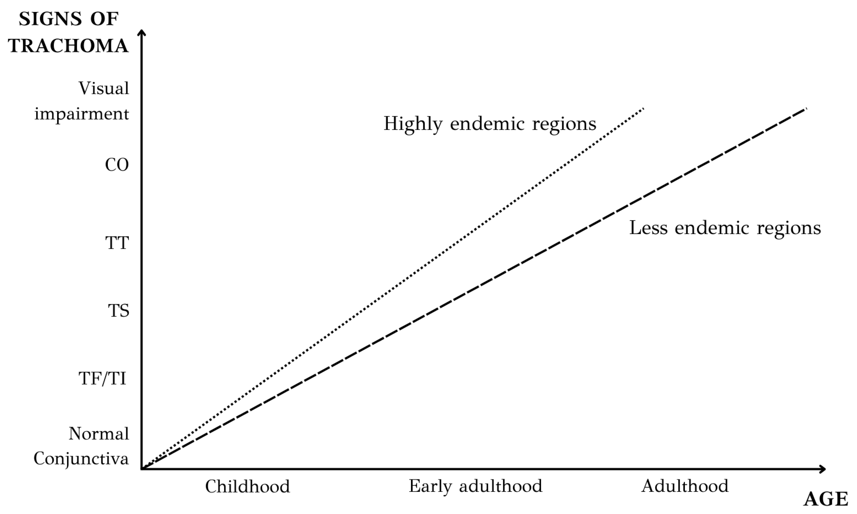
4. Natural History and Clinical Features of Trachoma

Trachoma can be conceptualized as progressing in two distinct but overlapping stages. Ιn the first stage, repeated infections with ocular strains of Ct induce recurrent episodes of chronic conjunctival inflammation, which results in the manifestation of the clinical signs of what is referred to as “active trachoma” [26]. A key clinical sign of active trachoma is follicular conjunctivitis, most prominently of the upper tarsal conjunctiva. Follicular conjunctivitis is identified by the presence of follicles, clusters of lymphoid tissue located beneath the epithelial layer of the tarsal conjunctiva. In some cases, the inflammation can be more intense, with severe papillary hypertrophy, which refers to the diffuse infiltration, edema, and enlargement of vascular tufts that can obscure deep tarsal vessels [26].
The correlation between active disease and infection in trachoma is intricate, revealing a disparity between clinical manifestations of active trachoma and the identification of Ct [27] (Figure 3). After infection with Ct, there is a short ‘preclinical’ phase lasting a few days before the onset of inflammation symptoms, known as the incubation period. A mathematical model describing the natural history of trachoma infection and disease estimated the median incubation period at 17 days (95% CI: 11–28) [28]. This period is followed by a phase of evident disease where the infectious agent and clinical signs coexist. Finally, there is a recovery period during which the human immune response acts in clearing the infectious agent or reducing it to undetectable levels, while the clinical signs of active trachoma persist and gradually diminish. Clinical manifestations endure for an uncertain yet presumably prolonged duration following the clearance of the infectious agent, unless there is a subsequent encounter with the chlamydial antigen. The mathematical model mentioned before estimated that the duration of the disease is longer than that of the infection, with a median of 21 weeks (range: 15–32 weeks) compared to 17 weeks (range: 12–24 weeks) [28]. Additionally, the estimated median duration of inflammation following the infection was 5 weeks (range: 3–8 weeks) [28].
Persistent re-infections contribute to a chronic cicatricial process [2]. With time, inflammation-induced tissue damage results in scarring of the eyelid, marking the onset of the second stage of the disease. The recovery of the inflamed conjunctiva involves some residual conjunctival scarring. A mathematical model describing the transmission of Ct and its progression to cicatricial disease estimated a threshold, forecasting that more than 100 infections during an individual’s lifetime are needed to generate trachomatous scarring [29]. Conjunctival scarring encompasses anything from a few linear or stellate scars to fibrous bands that shorten the fornix and create symblepharons (adhesions of the palpebral conjunctiva to the bulbar conjunctiva). Chronic conjunctival inflammation is commonly observed in adults who have developed conjunctival scarring [30,31,32]. This subsequent stage of conjunctival inflammation typically lacks follicular characteristics, with a rare detection of Ct infection. Instead, it is marked by a significant innate epithelial immune response. Factors such as non-chlamydial infections, along with the dryness and irritation of the scarred conjunctiva, may also play a role in promoting inflammation and advancing the progression in the presence of established scarring [33].
As the scar tissue contracts, it causes the eyelids to turn inward, resulting in entropion. The condition known as trichiasis occurs when eyelashes make contact with the eye. In the case of trachoma, trichiasis often originates from entropion, although this is not a strict rule. Many patients with trachomatous trichiasis have minimal or no entropion and trichiasis arises from misdirected lashes in a normal position (aberrant lashes) or lashes growing from abnormal locations (metaplastic lashes) [34]. The same mathematical model mentioned before predicts that more than 150 infections are necessary for the development of trachomatous trichiasis [29]. The potential harm resulting from the cornea being scratched by inward-turned eyelashes extends beyond immediate discomfort. Continuous friction from misdirected eyelashes injures the corneal surface, leading to corneal opacification. This, in turn, can result in impaired vision and, in severe cases, blindness. Untreated trichiasis can lead to corneal opacity and vision loss in as little as a year in about one-third of individuals [35]. It is essential to emphasize that corneal opacification and blindness likely result from traumatic damage caused by both trichiasis and secondary infections from other pathogens, such as bacteria or fungi [36,37].
The clinical features mentioned above are classified using the simplified WHO trachoma grading system [2,38]. This simplified grading system, suggested by the WHO in 1987 and amended in 2018, is specifically designed to enable a quick evaluation of the prevalence and severity of the disease within a population. It is as follows:
  • Trachomatous Follicular (TF) inflammation: The stage of active trachoma with predominantly follicular inflammation. Diagnosing this stage requires the presence of at least five or more follicles on the upper tarsal conjunctiva, each measuring 0.5 mm or more in diameter.
  • Trachomatous Intense (TI) inflammation: This stage of active trachoma is identified when significant inflammatory thickening of the upper tarsal conjunctiva obscures more than half of the normal deep tarsal vessels. The tarsal conjunctiva displays a reddish hue, a coarse texture, and increased thickness—the results of papillary hypertrophy. It should be mentioned that the key sign for the diagnosis of TI is thickened, velvety edematous conjunctiva, not redness.
  • Trachomatous Scarring (TS): This stage is identified through the detection of scarring in the tarsal conjunctiva, which is apparent as white lines, bands, or sheets (fibrosis) in the tarsal conjunctiva. Typically, the scars exhibit a shiny and fibrous appearance, featuring straight, angular, or feathered edges. Scarring, particularly diffuse fibrosis, might obscure the blood vessels in the tarsal area, but it is crucial not to mistake this for diffuse inflammatory thickening.
  • Trachomatous Trichiasis (TT): This stage is identified when at least one eyelash from the upper eyelid rubs on the eyeball, or there is evidence of recent removal of in-turned eyelashes from the upper eyelid.
  • Corneal Opacity (CO): This stage is recognized when there is a clearly visible corneal opacity covering the pupil. The corneal opacity is so dense that at least part of the pupil margin is blurred when viewed through the opacity. This definition aims to identify corneal opacities that result in substantial visual impairment (Figure 4).
The signs of trachoma exhibit a significant correlation with age (Figure 5). In a typical endemic setting, repeated chlamydial infection of the conjunctiva begins early in life. The prevalence of active trachoma (TF/TI) peaks in preschool children [39,40,41]. A recent meta-analysis focused on the pediatric population showed that the key factors associated with active trachoma among children are the absence of latrines, children’s unclean faces, and the lack of reported soap use for washing [42]. The occurrence of active trachoma diminishes as individuals age, with only a small number of adults displaying signs of active disease and even fewer presenting proof of infection [28,39,43,44]. On the contrary, there is an escalation in the prevalence of trachomatous conjunctival scarring as individuals age, underscoring the cumulative nature of the damage. The long-term sequelae of trachoma infection in adults relates to their exposure to active trachoma during the first months of life. The scarring sequelae of trachoma typically emerge in adulthood, generally starting around the third decade of life. However, in areas with a higher prevalence of severe disease, these effects may manifest earlier. Furthermore, there is a correlation between age and the duration of active disease but not with the duration of infection [28]. As individuals age, there is a statistically significant decline in the duration of active disease. Although a reduction in the duration of infection is observed, this decrease is non-significant.
Trachoma exhibits a notable correlation with gender. Male and female children tend to be equally affected by active trachoma. On the other hand, studies consistently highlight that females engaged in childcare tasks present a higher prevalence of active disease compared to those not involved in caregiving duties [39,45]. As a logical consequence, a higher prevalence of blinding complications of trachoma is observed among women compared to men, supporting the hypothesis of a multifaceted interplay between biological, social, and cultural factors [39,46,47].

5. Histopathology and Immunopathology

The conjunctiva, a thin and transparent mucous membrane covering the front part of the eye and lining the inner surface of the eyelids, exhibits a layered histological arrangement [48]. Externally, there is a stratified non-keratinizing squamous epithelium of three to five cell layers, with interspersed goblet cells that produce important mucins for the tear film that covers the epithelium. Beneath the epithelium lies the stroma, which consists of superficial lymphoid and fibrous tissue.
Tissue changes in active follicular trachoma begin with the dilation of capillaries and small venules, accompanied by the infiltration of inflammatory cells (lymphocytes, macrophages, neutrophils, dendritic cells and plasma cells) [49]. B cells are surrounded by a mantle of T cells, forming lymphoid follicles [49]. Once they reach a significant size, lymphoid follicles alter the shape of the covering epithelium and become observable in the inverted tarsal conjunctiva. Also, there is a generalized increase in the amounts of types I, III, and IV collagen, which typically present in the stroma, and a deposition of new type V collagen [50]. Trachomatous scarring involves epithelial atrophy (epithelial cells become thinned), a decrease in tear film volume with altered composition, and a depletion of goblet cells [51]. The stroma loses its regular architecture and transforms into a dense scar composed of type V collagen, while these fibers firmly adhere to the tarsal plate, resulting in distortion [51,52]. Immunohistochemical staining in conjunctival biopsy samples from individuals with trachomatous scarring showed the infiltration of immune cells with a phenotype suggestive of natural killer (NK) cells [53]. Also, contrary to our expectations based on previous studies, a significant portion of inflammatory cells in this study were found to be CD45-negative [53]. It was anticipated that the majority, if not all, of these inflammatory cells would exhibit CD45 expression, as CD45 is typically present on nearly all nucleated cells of hematopoietic origin. Regarding trachomatous trichiasis conjunctival tissue, it exhibits notably elevated expression levels of proteins S100A7 and CTGF in the epithelium, along with a significantly increased expression of IL-1β in the subepithelial tissue [54].
Diseases caused by chlamydia are based on intense and chronic inflammation elicited and maintained by re-infection or persistent infection. Ct infection triggers an innate immune response. Ct-infected epithelial cells directly initiate this process by detecting the presence of the pathogen through their pattern recognition receptors [55,56]. Findings from both in vitro and in vivo studies revealed a significant proinflammatory reaction, marked by the production of several chemokines and cytokines [56,57,58,59]. The former act as chemoattractants, organizing the migration of immune cells towards the site of infection, and the latter are involved in the up-regulation of inflammatory reactions. Another suggestion, though with little evidence from studies on humans, is that the disease is mediated by antigen-dependent delayed-type hypersensitivity or autoimmunity [30,55,60].
The infection can resolve or progress to eyelid scarring. The resolution of Ct is believed to rely on the presence of interferon gamma (IFNγ) derived from an immune response mediated by CD4+ T helper 1 (TH1) cells. Examinations of gene expression in children residing in trachoma-endemic regions reveal a significant rise in the conjunctival expression of IFNγ and factors associated with TH1 cells, such as IL-12B and indoleamine-2,3-dioxygenase (IDO1), along with increased activity in NK cell pathways marked by NCR1, CD56, and CD247 [57,58,59]. In cases of active trachoma without detectable Ct, levels of IFNγ are not notably elevated, suggesting a swift regulatory response once the infection has cleared. Infection and active trachoma are associated with a significant TH17 response, which is characterized by heightened levels of IL-17, IL-21, and IL-22 [59]. Also, some evidence suggests that TH17–IL-17 responses could potentially aggravate the inflammatory and scarring reaction [61].
The primary clinical indicator associated with the formation of scarring is intense conjunctival inflammation, which correlates with a heightened expression of various pro-inflammatory factors [59]. Additionally, studies have found that both active and scarring trachoma show increased amounts of matrix metalloproteinases (MMPs) 7, 9, and 12 [31,57,59,62]. MMPs are a family of extracellular matrix (ECM) remodeling endopeptidases that have the capacity to degrade almost every component of the ECM, promoting the contraction of scars. A single-nucleotide polymorphism (SNP) in the catalytic domain of MMP-9 possibly leads to reduced function and is linked to a decreased likelihood of scarring [63]. Moreover, active trachoma and scarring have been correlated with the heightened expression of numerous fibrogenic growth factors.

6. Conclusions

Exploring the pathophysiology of trachoma not only unveils the intricate pathways and mechanisms underlying this devastating eye disease but also underscores the multifaceted dimensions that must be considered in its management.
The first crucial dimension revolves around prevention strategies based on the transmission pathways and the complex interplay of risk factors contributing to the disease. The initial pivotal aspect involves prevention strategies rooted in understanding the transmission pathways and the intricate interplay of risk factors contributing to trachoma. To interrupt the cycle of trachoma transmission, it is imperative to promote hygiene by educating communities on proper face washing. Additionally, environmental improvement is essential, necessitating the implementation of water and sanitation infrastructure projects. Socio-economic development plays a crucial role, requiring a focus on poverty alleviation strategies, the economic empowerment of communities, and enhancements in overall living conditions. Furthermore, it is crucial to emphasize surveillance and monitoring through the implementation of robust systems. This approach aims to detect populations at risk and areas that may be experiencing great numbers of trachoma cases, ensuring timely and targeted preventive and therapeutic measures.
Another crucial dimension is the mismatch noted between infection and disease. This fact holds significant implications for trachoma programs. Making antibiotic treatment decisions based solely on observable conjunctival inflammation of at least moderate intensity, aimed at individual targets, would overlook many individuals with mild clinical responses who are still infected and require treatment.
The WHO-led Global Alliance for the Elimination of Trachoma recommends the implementation of the SAFE strategy which tackles the disease at different stages: Surgery to correct trichiasis, Antibiotics to treat chlamydial infection, and Facial cleanliness as well as Environmental improvements to suppress the transmission of infection. The strides taken by the WHO in curbing the prevalence of trachoma represent a commendable achievement, reflecting a commitment to global public health. However, it is crucial to recognize that the battle against this eye disease is far from over.
Trachoma’s impact extends far beyond the field of medicine, reaching into the social, economic, and educational domains of affected communities. Addressing each dimension with precision and synergy can provide hope that trachoma can be effectively limited, ultimately alleviating its impact on the social burden and improving global health.

Author Contributions

Conceptualization, P.T. and M.E.; methodology, P.T., M.E. and G.S.; validation, P.T. and M.E.; formal analysis, P.T. and G.V.; investigation, P.T. and G.V.; data curation, P.T., G.V., M.E. and G.S.; writing—original draft preparation, P.T.; writing—review and editing, P.T., G.V., I.T.T., M.E. and G.S.; supervision, M.E. and G.S. All authors have read and agreed to the published version of the manuscript.

Funding

This research received no external funding.

Data Availability Statement

Data are contained within the article.

Conflicts of Interest

The authors declare no conflicts of interest.

References

  1. Flaxman, S.R.; Bourne, R.R.A.; Resnikoff, S.; Ackland, P.; Braithwaite, T.; Cicinelli, M.V.; Das, A.; Jonas, J.B.; Keeffe, J.; Kempen, J.H.; et al. Global causes of blindness and distance vision impairment 1990–2020: A systematic review and meta-analysis. Lancet Glob. Health 2017, 5, e1221–e1234. [Google Scholar] [CrossRef] [PubMed]
  2. Solomon, A.W.; Burton, M.J.; Gower, E.W.; Harding-Esch, E.M.; Oldenburg, C.E.; Taylor, H.R.; Traore, L. Trachoma. Nat. Rev. Dis. Primers 2022, 8, 32. [Google Scholar] [CrossRef]
  3. World Health Organization. Trachoma. Available online: https://www.who.int/news-room/fact-sheets/detail/trachoma (accessed on 1 April 2024).
  4. World Health Organization. Ending the Neglect to Attain the Sustainable Development Goals: A Road Map for Neglected Tropical Diseases 2021–2030. Available online: https://www.who.int/publications/i/item/9789240052932 (accessed on 2 April 2024).
  5. Yaghoobi, G.; Anani Sarab, G. The History of Trachoma and Current Prevalence (Spotlight on Iran): A Review Article. Iran. J. Public Health 2018, 47, 1458–1465. [Google Scholar] [PubMed]
  6. Fan, H.; Scidmore, M.A. Chlamydia. In Reference Module in Biomedical Sciences; Elsevier: Amsterdam, The Netherlands, 2015. [Google Scholar] [CrossRef]
  7. Jury, B.; Fleming, C.; Huston, W.M.; Luu, L.D.W. Molecular pathogenesis of Chlamydia trachomatis. Front. Cell. Infect. Microbiol. 2023, 13, 1281823. [Google Scholar] [CrossRef] [PubMed]
  8. Hepler, R.W.; Nahas, D.D.; Lucas, B.; Kaufhold, R.; Flynn, J.A.; Galli, J.D.; Swoyer, R.; Wagner, J.M.; Espeseth, A.S.; Joyce, J.G.; et al. Spectroscopic analysis of chlamydial major outer membrane protein in support of structure elucidation. Protein Sci. 2018, 27, 1923–1941. [Google Scholar] [CrossRef]
  9. Grygiel-Gorniak, B.; Folga, B.A. Chlamydia trachomatis—An Emerging Old Entity? Microorganisms 2023, 11, 1283. [Google Scholar] [CrossRef] [PubMed]
  10. Lesiak-Markowicz, I.; Schotta, A.M.; Stockinger, H.; Stanek, G.; Markowicz, M. Chlamydia trachomatis serovars in urogenital and ocular samples collected 2014–2017 from Austrian patients. Sci. Rep. 2019, 9, 18327. [Google Scholar] [CrossRef] [PubMed]
  11. Makker, K.; Nassar, G.N.; Kaufman, E.J. Neonatal Conjunctivitis. In StatPearls; Ineligible Companies: Treasure Island, FL, USA, 2024. [Google Scholar]
  12. Lee, W.A.; Chen, C.C. Adult inclusion conjunctivitis diagnosed by polymerase chain reaction and Giemsa stain. IDCases 2022, 27, e01367. [Google Scholar] [CrossRef] [PubMed]
  13. Abdelsamed, H.; Peters, J.; Byrne, G.I. Genetic variation in Chlamydia trachomatis and their hosts: Impact on disease severity and tissue tropism. Future Microbiol. 2013, 8, 1129–1146. [Google Scholar] [CrossRef]
  14. Fehlner-Gardiner, C.; Roshick, C.; Carlson, J.H.; Hughes, S.; Belland, R.J.; Caldwell, H.D.; McClarty, G. Molecular basis defining human Chlamydia trachomatis tissue tropism. A possible role for tryptophan synthase. J. Biol. Chem. 2002, 277, 26893–26903. [Google Scholar] [CrossRef]
  15. Caldwell, H.D.; Wood, H.; Crane, D.; Bailey, R.; Jones, R.B.; Mabey, D.; Maclean, I.; Mohammed, Z.; Peeling, R.; Roshick, C.; et al. Polymorphisms in Chlamydia trachomatis tryptophan synthase genes differentiate between genital and ocular isolates. J. Clin. Investig. 2003, 111, 1757–1769. [Google Scholar] [CrossRef] [PubMed]
  16. Elwell, C.; Mirrashidi, K.; Engel, J. Chlamydia cell biology and pathogenesis. Nature reviews. Microbiology 2016, 14, 385–400. [Google Scholar]
  17. Versteeg, B.; Vasileva, H.; Houghton, J.; Last, A.; Shafi Abdurahman, O.; Sarah, V.; Macleod, D.; Solomon, A.W.; Holland, M.J.; Thomson, N.; et al. Viability PCR shows that non-ocular surfaces could contribute to transmission of Chlamydia trachomatis infection in trachoma. PLoS Negl. Trop. Dis. 2020, 14, e0008449. [Google Scholar] [CrossRef] [PubMed]
  18. Last, A.; Versteeg, B.; Shafi Abdurahman, O.; Robinson, A.; Dumessa, G.; Abraham Aga, M.; Shumi Bejiga, G.; Negussu, N.; Greenland, K.; Czerniewska, A.; et al. Detecting extra-ocular Chlamydia trachomatis in a trachoma-endemic community in Ethiopia: Identifying potential routes of transmission. PLoS Negl. Trop. Dis. 2020, 14, e0008120. [Google Scholar] [CrossRef]
  19. Emerson, P.M.; Lindsay, S.W.; Alexander, N.; Bah, M.; Dibba, S.M.; Faal, H.B.; Lowe, K.O.; McAdam, K.P.; Ratcliffe, A.A.; Walraven, G.E.; et al. Role of flies and provision of latrines in trachoma control: Cluster-randomised controlled trial. Lancet 2004, 363, 1093–1098. [Google Scholar] [CrossRef]
  20. Brewer, N.; McKenzie, M.S.; Melkonjan, N.; Zaky, M.; Vik, R.; Stoffolano, J.G.; Webley, W.C. Persistence and Significance of Chlamydia trachomatis in the Housefly, Musca domestica L. Vector Borne Zoonotic Dis. 2021, 21, 854–863. [Google Scholar] [CrossRef]
  21. Robinson, A.; Bristow, J.; Holl, M.V.; Makalo, P.; Alemayehu, W.; Bailey, R.L.; Macleod, D.; Birkett, M.A.; Caulfield, J.C.; Sarah, V.; et al. Responses of the putative trachoma vector, Musca sorbens, to volatile semiochemicals from human faeces. PLoS Negl. Trop. Dis. 2020, 14, e0007719. [Google Scholar] [CrossRef]
  22. Ageed, A.; Khan, M. Eliminating Trachoma in Africa: The Importance of Environmental Interventions. Cureus 2024, 16, e52358. [Google Scholar] [CrossRef]
  23. Chen, X.; Munoz, B.; Wolle, M.A.; Woods, G.; Odonkor, M.; Naufal, F.; Mkocha, H.; West, S.K. Environmental factors and hygiene behaviors associated with facial cleanliness and trachoma in Kongwa, Tanzania. PLoS Negl. Trop. Dis. 2021, 15, e0009902. [Google Scholar] [CrossRef] [PubMed]
  24. Churko, C.; Asfaw, M.A.; Zerdo, Z. Knowledge, Attitude, Practices and Associated Factors towards Trachoma among People Living in Arba Minch Zuria District, Gamo Zone, Southern Ethiopia. Clin. Ophthalmol. 2021, 15, 3075–3085. [Google Scholar] [CrossRef]
  25. Bailey, R.; Osmond, C.; Mabey, D.C.; Whittle, H.C.; Ward, M.E. Analysis of the household distribution of trachoma in a Gambian village using a Monte Carlo simulation procedure. Int. J. Epidemiol. 1989, 18, 944–951. [Google Scholar] [CrossRef] [PubMed]
  26. Ahmad, B.; Patel, B.C. Trachoma. In StatPearls; StatPearls Publishing Copyright © 2024; StatPearls Publishing LLC/Ineligible Companies: Treasure Island, FL, USA, 2024. [Google Scholar]
  27. Wright, H.R.; Taylor, H.R. Clinical examination and laboratory tests for estimation of trachoma prevalence in a remote setting: What are they really telling us? Lancet Infect. Dis. 2005, 5, 313–320. [Google Scholar] [CrossRef] [PubMed]
  28. Grassly, N.C.; Ward, M.E.; Ferris, S.; Mabey, D.C.; Bailey, R.L. The natural history of trachoma infection and disease in a Gambian cohort with frequent follow-up. PLoS Negl. Trop. Dis. 2008, 2, e341. [Google Scholar] [CrossRef] [PubMed]
  29. Gambhir, M.; Basáñez, M.G.; Burton, M.J.; Solomon, A.W.; Bailey, R.L.; Holland, M.J.; Blake, I.M.; Donnelly, C.A.; Jabr, I.; Mabey, D.C.; et al. The development of an age-structured model for trachoma transmission dynamics, pathogenesis and control. PLoS Negl. Trop. Dis. 2009, 3, e462. [Google Scholar] [CrossRef] [PubMed]
  30. Hu, V.H.; Holland, M.J.; Burton, M.J. Trachoma: Protective and pathogenic ocular immune responses to Chlamydia trachomatis. PLoS Negl. Trop. Dis. 2013, 7, e2020. [Google Scholar] [CrossRef] [PubMed]
  31. Hu, V.H.; Weiss, H.A.; Ramadhani, A.M.; Tolbert, S.B.; Massae, P.; Mabey, D.C.; Holland, M.J.; Bailey, R.L.; Burton, M.J. Innate immune responses and modified extracellular matrix regulation characterize bacterial infection and cellular/connective tissue changes in scarring trachoma. Infect. Immun. 2012, 80, 121–130. [Google Scholar] [CrossRef] [PubMed]
  32. Burton, M.J.; Rajak, S.N.; Bauer, J.; Weiss, H.A.; Tolbert, S.B.; Shoo, A.; Habtamu, E.; Manjurano, A.; Emerson, P.M.; Mabey, D.C.; et al. Conjunctival transcriptome in scarring trachoma. Infect. Immun. 2011, 79, 499–511. [Google Scholar] [CrossRef] [PubMed]
  33. Hu, V.H.; Macleod, D.; Massae, P.; Afwamba, I.; Weiss, H.A.; Mabey, D.C.W.; Bailey, R.L.; Burton, M.J. Non-Chlamydial Bacterial Infection and Progression of Conjunctival Scarring in Trachoma. Investig. Ophthalmol. Vis. Sci. 2018, 59, 2339–2344. [Google Scholar] [CrossRef] [PubMed]
  34. Rajak, S.N.; Habtamu, E.; Weiss, H.A.; Bedri, A.; Gebre, T.; Bailey, R.L.; Mabey, D.C.; Khaw, P.T.; Gilbert, C.E.; Emerson, P.M.; et al. The clinical phenotype of trachomatous trichiasis in Ethiopia: Not all trichiasis is due to entropion. Investig. Ophthalmol. Vis. Sci. 2011, 52, 7974–7980. [Google Scholar] [CrossRef]
  35. Bowman, R.J.; Faal, H.; Myatt, M.; Adegbola, R.; Foster, A.; Johnson, G.J.; Bailey, R.L. Longitudinal study of trachomatous trichiasis in the Gambia. Br. J. Ophthalmol. 2002, 86, 339–343. [Google Scholar] [CrossRef]
  36. Burton, M.J.; Bowman, R.J.; Faal, H.; Aryee, E.A.; Ikumapayi, U.N.; Alexander, N.D.; Adegbola, R.A.; Mabey, D.C.; Foster, A.; Johnson, G.J.; et al. The long-term natural history of trachomatous trichiasis in the Gambia. Investig. Ophthalmol. Vis. Sci. 2006, 47, 847–852. [Google Scholar] [CrossRef] [PubMed]
  37. Burton, M.J.; Kinteh, F.; Jallow, O.; Sillah, A.; Bah, M.; Faye, M.; Aryee, E.A.; Ikumapayi, U.N.; Alexander, N.D.; Adegbola, R.A.; et al. A randomised controlled trial of azithromycin following surgery for trachomatous trichiasis in the Gambia. Br. J. Ophthalmol. 2005, 89, 1282–1288. [Google Scholar] [CrossRef] [PubMed]
  38. Solomon, A.W.; Kello, A.B.; Bangert, M.; West, S.K.; Taylor, H.R.; Tekeraoi, R.; Foster, A. The simplified trachoma grading system, amended. Bull. World Health Organ. 2020, 98, 698–705. [Google Scholar] [CrossRef] [PubMed]
  39. West, S.K.; Munoz, B.; Turner, V.M.; Mmbaga, B.B.; Taylor, H.R. The epidemiology of trachoma in central Tanzania. Int. J. Epidemiol. 1991, 20, 1088–1092. [Google Scholar] [CrossRef] [PubMed]
  40. Ngondi, J.; Onsarigo, A.; Adamu, L.; Matende, I.; Baba, S.; Reacher, M.; Emerson, P.; Zingeser, J. The epidemiology of trachoma in Eastern Equatoria and Upper Nile States, southern Sudan. Bull. World Health Organ. 2005, 83, 904–912. [Google Scholar] [PubMed]
  41. Ngondi, J.; Gebre, T.; Shargie, E.B.; Adamu, L.; Ejigsemahu, Y.; Teferi, T.; Zerihun, M.; Ayele, B.; Cevallos, V.; King, J.; et al. Evaluation of three years of the SAFE strategy (Surgery, Antibiotics, Facial cleanliness and Environmental improvement) for trachoma control in five districts of Ethiopia hyperendemic for trachoma. Trans. R. Soc. Trop. Med. Hyg. 2009, 103, 1001–1010. [Google Scholar] [CrossRef] [PubMed]
  42. Gebrie, A.; Alebel, A.; Zegeye, A.; Tesfaye, B.; Wagnew, F. Prevalence and associated factors of active trachoma among children in Ethiopia: A systematic review and meta-analysis. BMC Infect. Dis. 2019, 19, 1073. [Google Scholar] [CrossRef] [PubMed]
  43. Solomon, A.W.; Holland, M.J.; Burton, M.J.; West, S.K.; Alexander, N.D.; Aguirre, A.; Massae, P.A.; Mkocha, H.; Muñoz, B.; Johnson, G.J.; et al. Strategies for control of trachoma: Observational study with quantitative PCR. Lancet 2003, 362, 198–204. [Google Scholar] [CrossRef] [PubMed]
  44. Taylor, H.R.; Siler, J.A.; Mkocha, H.A.; Muñoz, B.; Velez, V.; Dejong, L.; West, S. Longitudinal study of the microbiology of endemic trachoma. J. Clin. Microbiol. 1991, 29, 1593–1595. [Google Scholar] [CrossRef]
  45. Congdon, N.; West, S.; Vitale, S.; Katala, S.; Mmbaga, B.B. Exposure to children and risk of active trachoma in Tanzanian women. Am. J. Epidemiol. 1993, 137, 366–372. [Google Scholar] [CrossRef]
  46. Dolin, P.J.; Faal, H.; Johnson, G.J.; Ajewole, J.; Mohamed, A.A.; Lee, P.S. Trachoma in the Gambia. Br. J. Ophthalmol. 1998, 82, 930–933. [Google Scholar] [CrossRef]
  47. Courtright, P.; Sheppard, J.; Schachter, J.; Said, M.E.; Dawson, C.R. Trachoma and blindness in the Nile Delta: Current patterns and projections for the future in the rural Egyptian population. Br. J. Ophthalmol. 1989, 73, 536–540. [Google Scholar] [CrossRef] [PubMed]
  48. Shumway, C.L.; Motlagh, M.; Wade, M. Anatomy, Head and Neck, Eye Conjunctiva. In StatPearls; StatPearls Publishing: Treasure Island, FL, USA, 2024. [Google Scholar]
  49. el-Asrar, A.M.; Van den Oord, J.J.; Geboes, K.; Missotten, L.; Emarah, M.H.; Desmet, V. Immunopathology of trachomatous conjunctivitis. Br. J. Ophthalmol. 1989, 73, 276–282. [Google Scholar] [CrossRef]
  50. Abu el-Asrar, A.M.; Geboes, K.; al-Kharashi, S.A.; Tabbara, K.F.; Missotten, L. Collagen content and types in trachomatous conjunctivitis. Eye 1998, 12 Pt 4, 735–739. [Google Scholar] [CrossRef]
  51. al-Rajhi, A.A.; Hidayat, A.; Nasr, A.; al-Faran, M. The histopathology and the mechanism of entropion in patients with trachoma. Ophthalmology 1993, 100, 1293–1296. [Google Scholar] [CrossRef] [PubMed]
  52. Hu, V.H.; Holland, M.J.; Cree, I.A.; Pullin, J.; Weiss, H.A.; Massae, P.; Makupa, W.; Mabey, D.C.; Bailey, R.L.; Burton, M.J.; et al. In vivo confocal microscopy and histopathology of the conjunctiva in trachomatous scarring and normal tissue: A systematic comparison. Br. J. Ophthalmol. 2013, 97, 1333–1337. [Google Scholar] [CrossRef]
  53. Hu, V.H.; Luthert, P.J.; Derrick, T.; Pullin, J.; Weiss, H.A.; Massae, P.; Mtuy, T.; Makupa, W.; Essex, D.; Mabey, D.C.; et al. Immunohistochemical Analysis of Scarring Trachoma Indicates Infiltration by Natural Killer and Undefined CD45 Negative Cells. PLoS Negl. Trop. Dis. 2016, 10, e0004734. [Google Scholar] [CrossRef] [PubMed]
  54. Derrick, T.; Luthert, P.J.; Jama, H.; Hu, V.H.; Massae, P.; Essex, D.; Holland, M.J.; Burton, M.J. Increased Epithelial Expression of CTGF and S100A7 with Elevated Subepithelial Expression of IL-1β in Trachomatous Trichiasis. PLoS Negl. Trop. Dis. 2016, 10, e0004752. [Google Scholar] [CrossRef]
  55. Stephens, R.S. The cellular paradigm of chlamydial pathogenesis. Trends Microbiol. 2003, 11, 44–51. [Google Scholar] [CrossRef]
  56. Rasmussen, S.J.; Eckmann, L.; Quayle, A.J.; Shen, L.; Zhang, Y.X.; Anderson, D.J.; Fierer, J.; Stephens, R.S.; Kagnoff, M.F. Secretion of proinflammatory cytokines by epithelial cells in response to Chlamydia infection suggests a central role for epithelial cells in chlamydial pathogenesis. J. Clin. Investig. 1997, 99, 77–87. [Google Scholar] [CrossRef]
  57. Burton, M.J.; Bailey, R.L.; Jeffries, D.; Mabey, D.C.; Holland, M.J. Cytokine and fibrogenic gene expression in the conjunctivas of subjects from a Gambian community where trachoma is endemic. Infect. Immun. 2004, 72, 7352–7356. [Google Scholar] [CrossRef] [PubMed]
  58. Natividad, A.; Freeman, T.C.; Jeffries, D.; Burton, M.J.; Mabey, D.C.; Bailey, R.L.; Holland, M.J. Human conjunctival transcriptome analysis reveals the prominence of innate defense in Chlamydia trachomatis infection. Infect. Immun. 2010, 78, 4895–4911. [Google Scholar] [CrossRef] [PubMed]
  59. Ramadhani, A.M.; Derrick, T.; Macleod, D.; Massae, P.; Mtuy, T.; Jeffries, D.; Roberts, C.H.; Bailey, R.L.; Mabey, D.C.W.; Holland, M.J.; et al. Immunofibrogenic Gene Expression Patterns in Tanzanian Children with Ocular Chlamydia trachomatis Infection, Active Trachoma and Scarring: Baseline Results of a 4-Year Longitudinal Study. Front. Cell. Infect. Microbiol. 2017, 7, 406. [Google Scholar] [CrossRef] [PubMed]
  60. Taylor, H.R.; Johnson, S.L.; Schachter, J.; Caldwell, H.D.; Prendergast, R.A. Pathogenesis of trachoma: The stimulus for inflammation. J. Immunol. 1987, 138, 3023–3027. [Google Scholar] [CrossRef] [PubMed]
  61. Vicetti Miguel, R.D.; Quispe Calla, N.E.; Pavelko, S.D.; Cherpes, T.L. Intravaginal Chlamydia trachomatis Challenge Infection Elicits TH1 and TH17 Immune Responses in Mice That Promote Pathogen Clearance and Genital Tract Damage. PLoS ONE 2016, 11, e0162445. [Google Scholar] [CrossRef] [PubMed]
  62. Burton, M.J.; Rajak, S.N.; Hu, V.H.; Ramadhani, A.; Habtamu, E.; Massae, P.; Tadesse, Z.; Callahan, K.; Emerson, P.M.; Khaw, P.T.; et al. Pathogenesis of progressive scarring trachoma in Ethiopia and Tanzania and its implications for disease control: Two cohort studies. PLoS Negl. Trop. Dis. 2015, 9, e0003763. [Google Scholar] [CrossRef]
  63. Natividad, A.; Cooke, G.; Holland, M.J.; Burton, M.J.; Joof, H.M.; Rockett, K.; Kwiatkowski, D.P.; Mabey, D.C.; Bailey, R.L. A coding polymorphism in matrix metalloproteinase 9 reduces risk of scarring sequelae of ocular Chlamydia trachomatis infection. BMC Med. Genet. 2006, 7, 40. [Google Scholar] [CrossRef]
Figure 1. Routes of trachoma transmission−direct (fingers between humans) and indirect (fomites and eye-seeking flies) means of Ct spread.
Microorganisms 12 01544 g001
Figure 2. Interplay of factors contributing to trachoma.
Microorganisms 12 01544 g002
Figure 3. Relationship between the appearance of clinical signs of active trachoma and the load of the causative agent (Ct). (A) Quadrilateral table of the four states of infection and disease with states indexed from 1 to 4. (B) Diagram illustrating the kinetics of trachoma, highlighting that active inflammatory disease persists for a period even after the load of Ct becomes undetectable. (1) Time of infection—the load of Ct increases. (2) Incubation period—infection, but no clinical signs are observed. (3) Evident disease—clinical signs have become apparent. (4) Recovery period—infection cleared, and clinical signs persist but gradually resolve. (Disclaimer: the duration of each period is not representative).
Microorganisms 12 01544 g003
Figure 4. The natural history of trachoma. Persistent re-infections contribute to the chronic cicatricial process (1). The recovery of the inflamed conjunctiva (2) involves some residual conjunctival scarring (3). [The clinical images are adapted from Ref. [30], CC BY 4.0 (https://creativecommons.org/licenses/by/4.0/ accessed on 26 May 2024)].
Microorganisms 12 01544 g004
Figure 5. Diagram illustrating the correlation of clinical signs of trachoma with age. In highly endemic regions, blinding complications of trachoma emerge at an earlier age. (TF/TI: Trachomatous Follicular/Intense inflammation, TS: Trachomatous Scarring, TT: Trachomatous Trichiasis, CO: Corneal Opacity).
Microorganisms 12 01544 g005
Table 1. Chlamydia trachomatis–biovars, serovars, and associated diseases.
BiovarsSerovarsMain Diseases
TrachomaA–C *Trachoma
GenitalD–KNeonatal conjunctivitis
Adult inclusion conjunctivitis
Pneumonia (neonates)
LGVL1–L3LGV
LGV = Lymphogranuloma venereum; * B/Ba: linked to both, ocular and genital tract infection.
Disclaimer/Publisher’s Note: The statements, opinions and data contained in all publications are solely those of the individual author(s) and contributor(s) and not of MDPI and/or the editor(s). MDPI and/or the editor(s) disclaim responsibility for any injury to people or property resulting from any ideas, methods, instructions or products referred to in the content.

