Article

Inhibition of the Growth of Escherichia coli and Staphylococcus aureus Microorganisms in Aesthetic Orthodontic Brackets through the In Situ Synthesis of Ag, TiO2 and Ag/TiO2 Nanoparticles

by
Paola Ariselda Sánchez Reyna
1,
Oscar Fernando Olea Mejía
2,
María G. González-Pedroza
3,
Norma M. Montiel-Bastida
1,
Bernabe Rebollo-Plata
4 and
Raúl A. Morales-Luckie
2,*
1
Center for Advanced Studies and Research on Dentistry, Autonomous University of the State of Mexico (UAEMex), Toluca 50200, Mexico
2
Department of Materials Science, Center for Research in Sustainable Chemistry (CCIQS), Autonomous University of the State of Mexico (UAEMex), Km 14.5, Carr. Toluca-Atlacomulco, Toluca 50200, Mexico
3
Department of Biotechnology, Faculty of Sciences, Autonomous University of the State of Mexico (UAEMex), Km 14.5, Carr. Toluca-Atlacomulco, Toluca 50200, Mexico
4
Tecnológico Nacional de México, Instituto Tecnológico Superior de Irapuato, Carr. Irapuato-Silao Km 12.5, Irapuato 36821, Mexico
*
Author to whom correspondence should be addressed.
Microorganisms 2024, 12(8), 1583; https://doi.org/10.3390/microorganisms12081583
Submission received: 5 July 2024 / Revised: 29 July 2024 / Accepted: 30 July 2024 / Published: 3 August 2024
(This article belongs to the Special Issue Antimicrobial Properties of Nanoparticle)

Abstract

:
Plaque control is especially important during orthodontic treatment because areas of the teeth near brackets and wires are difficult to clean with a toothbrush, resulting in debris buildup of food or dental plaque, thus causing caries and periodontal disease. The objective of this study was to evaluate the antimicrobial properties of silver nanoparticles (AgNPs), titanium dioxide nanoparticles (TiO2NPs), and silver/titanium dioxide nanoparticles (Ag/TiO2NPs), synthesized on the surface of α-alumina ceramic brackets. The AgNPs and TiO2NPs were synthesized by a simple chemical method, and these were characterized by XRD, SEM, and XPS TEM; the antimicrobial activity was tested against Staphylococcus aureus and Escherichia coli by diffusion test. The results of this study demonstrated that by this simple chemical method, silver and titanium dioxide nanoparticles can be synthesized on the surface of α-alumina esthetic brackets, and these NPs possess good antimicrobial activity and the possibility of reducing dental caries, periodontal disease, and white spot generated during orthodontic treatment.

1. Introduction

Nanoscience is an emerging field that includes nanostructured materials, including the use of metallic nanoparticles such as Ag, Au, Ir, Pd, and Pt, which are used in different disciplines due to their multiple properties. In recent years, silver nanoparticles (AgNPs) have been one of the most broad-spectrum antibacterial materials used due to their high cytotoxicity in a wide range of microbial and fungal species, including antibiotic-resistant strains, and low toxicity in humans [1,2,3,4,5]. It has been possible to demonstrate that AgNPs can bind directly with the cell membrane of bacteria and then damaging or altering their functionality [4,6]. This is due to its high reactivity to substances such as proteins, enzymes, DNA, RNA, etc., because of interactions that occur against thiol functional groups, carboxylate, phosphate, hydroxyl, imidazole, indole, or amine [7,8,9]. These interactions can occur easily or combined which may cause a series of events that interfere microbial processes [10,11]. The factors that affect the toxicity of AgNPs are the particle size, since with smaller size, there will be greater toxicity, this being due to the surface area they present [12,13]; the shape of the nanoparticles also has to do with the properties that triangular nanoparticles can provide, for example, they have greater antibacterial activity than spherical ones [14]. There are many reports on the synthesis of AgNPs, and in all of them, one of the most important aspects that must be controlled is the precipitation of AgNPs from its precursor Ag+; and the second is the size or shape of the AgNPs after their formation. This is possible thanks to the synthesis method. Synthesis of AgNPs can be carried out by different methods: chemical reduction [15,16,17], photochemical reduction [18], gamma [19], electron beam [20,21], microwave [22,23], laser [24], ultra violet [25], electrochemistry [26], sonochemistry [27], and synthesis ultrasound [28,29,30].
TiO2 photoactive materials are used to sterilize different materials when irradiated with sunlight or ultraviolet light. TiO2 can inhibit microbial adhesion for its excellent photocatalytic activity [31]. By irradiating TiO2 with a UV light power greater than its bandwidth, an electron is promoted from the valence band to the conduction band; then, high-energy electron–hole pairs reacting with water or oxygen molecules form various reactive oxygen species (ROS), which are strong oxidizing agents that cause damage to the cell membrane and therefore cell death [32,33,34]. However, the effect is decreased in the dark, which limits their biomedical applications [35,36,37]. TiO2 nanoparticles (TiO2NPs) are anatase phase antibacterial photocatalysts that have attracted great interest for inactivation of bacteria, fungi, viruses, and even cancer cells [38,39,40,41,42]. Compared with regular particles, solids with nanometric TiO2 and high specificity morphological dimensions may offer a single reaction means for the construction of photoactive nanoscale devices due to its high specific surface. In recent years, great efforts have been devoted to the design and manufacture of nanostructured systems with tunable physicochemical properties for advanced photocatalytic applications [43,44,45]. In recent decades, many methods have been developed to synthesize TiO2NPs such as, for example, sol-gel, hydrothermal, solvothermal, microemulsion, etc. [46,47,48].
The combination of TiO2 and Ag provides an active photocatalytic semiconductor and a noble metal that can extend and promote applications through photochemistry and heterogeneous catalysis [49,50,51]. In fact, anchoring AgNPs as electron traps is favorable for the separation of photoelectrons and excited holes to improve the quantum efficiency of TiO2, which further helps in eliminating bacteria, so that a layer of TiO2 facilitates the dispersion of AgNPs, and therefore incorporating TiO2/Ag makes an excellent route to enhance the antibacterial behavior. The conventional method used to prepare Ag/TiO2 nanoparticles (Ag/TiO2NPs) is the sol-gel chemical reduction, thermal reduction, or photochemical reduction of silver ions. Several methods have been proposed for the Ag/TiO2 preparation: a single step sol-gel route [52], photoreduction of Ag+ in a suspension of TiO2 [53], and electrochemical deposition of AgNPs on the surface of TiO2 [54].
Currently, the use of antibacterial agents for the removal of dental plaque is limited, and anchoring is complicated, given the nature of the surfaces of some dental materials, for example elastomeric modules, resins, etc. [55,56,57,58]. However, in this work, we present the use of novel elements for the generation of nanoparticles, the effectiveness of using a bimetallic system, as well as a more specific and safe anchoring technique, which guarantees the antimicrobial effect, against two very representative microorganisms in bacterial models.
To verify the antibacterial effect of the materials generated against these microorganisms, a technique known as Kirby Bauer has been used, which is a microbiological technique that shows the halos of inhibition generated by the materials and the antibacterial activity of brackets decorated with nanoparticles. As has been seen, both AgNPs and TiO2NPs have certain antibacterial activity and together have a characteristic behavior. These particles were synthesized and supported on ceramic brackets manufactured from Al2O3, used in orthodontic treatment with fixed appliances. Their antimicrobial properties were characterized and evaluated, recognizing it as a potential system to reduce plaque accumulation and bacterial growth in orthodontic procedures.

2. Materials and Methods

Materials: the reagents were silver nitrate (AgNO3), sodium dodecyl sulfate (SDS), titanium dioxide (TiO2) 10 wt% dispersed in water, and hydrazine (N2H4 · H2O); all reagents were purchased from SIGMA-ALDRICH company, St Louis, MO, USA.

2.1. Synthesis and Incorporation of Nanoparticles in Brackets

2.1.1. Ag Nanoparticles Supported on Brackets

AgNPs were synthesized in situ on a support of brackets of α-Al2O3, starting with a 20 mL solution of 10 mM of AgNO3 solution mixed with a 20 mL solution of SDS 10% for 30 min with magnetic stirring, and the mixture was subsequently added a solution N2H4 ·H2O (108 mM) as a reducing agent, followed by continued magnetic stirring for 30 min. Finally, the support with AgNPs was removed from the reaction mixture and washed with deionized water before being subsequently dried in an oven at 70 °C for 24 h.

2.1.2. TiO2 Nanoparticles Supported on Brackets

Nanoparticles of TiO2 were anchored to the support of brackets of α-Al2O3: a 20 mL solution of TiO2 at 10 mM solution 10 wt% dispersed in water was mixed with a SDS 10% for 30 min with magnetic stirring; finally, the TiO2 nanoparticles support was removed from the reaction mixture and washed with deionized water to remove the excess, and then subsequently dried in an oven at 70 °C for 24 h.

2.1.3. Ag/TiO2 Nanoparticles Supported on Brackets

The Ag/TiO2NPs on the support of brackets of α-Al2O3 were obtained as follows: 10 mL of TiO2 solution 10 mM 10% wt, dispersed in water, with a 10 mL of AgNO3 solution 10 mM was mixed, and to this mixture was added a 10% solution of 20 mL of SDS, before magnetically stirring for 30 min, then a N2H4 ·H2O (108 mM) solution was added as a reducing agent; subsequently, magnetic stirring continued for 30 min. Finally, the support with Ag/TiO2 nanoparticles was removed from the reaction mixture and washed with deionized water, and then subsequently dried in an oven at 70 °C for 24 h.

2.2. Characterization Techniques

The dry samples of 2 mL of silver, titanium dioxide, and silver/titanium dioxide nanoparticles were analyzed separately in a JEOL brand scanning electron microscope, microscope (Tokyo, Japan), with a JSM-6510LV Tungsten Filament coupled to an X-ray detector, for chemical analysis by OXFORD brand Energy Dispersion (EDS) (Atlanta, GA, USA), with a resolution of 137 eV.
For Transmission Electron Microscopy (TEM), the sample was analyzed using a drop of the nanoparticle solutions separately on Cu grids, and they were analyzed in a transmission electron microscope JEOL-2100 of 200 kV brand LaB6 filament (Tokyo, Japan) with a resolution of 0.23 nm point to point and 0.14 nm line by line. Micrographs were acquired digitally using a Gatan CCD camera, model SC200 (Pleasanton, USA).
Atomic Force Microscopy (AFM) VEECO Brand CP-II atomic force microscope (NY, USA: SPM Digital. Touch and touch modes have a 5 µm XY scan range and 1 µm Z scan range.
X-ray Diffraction (XRD) dry samples of the generated nanoparticles were analyzed in a D8 Advance Bruker LynxEye X-ray diffractometer (Karlsruhe, Germany) that has a copper anode X-ray source (Kα1 = 0.1540 Å).
For X-ray Photoelectron Spectrometry (XPS), the generated and dried nanoparticles were analyzed with a JEOL JPS-9200 X-ray photoelectron spectrometer (Tokyo, Japan); the computer uses a Mg Ka (hv = 1253.6 eV) X-ray source operated at 10 kV and 20 mA (200 W) and is equipped with a hemispherical electrostatic analyzer with a mean radius of 100 mm and a multichannel detector.

2.3. Antibacterial Activity

The antibacterial activity of the support with AgNPs, TiO2NPs, and Ag/TiO2 NPs was evaluated against both Gram-negative and Gram-positive bacteria, including species of Escherichia coli and Staphylococcus aureus, using the agar diffusion method [59]. The bacterial strains used in this study were acquired from the stock culture collection of the Biochemistry laboratory of the Faculty of Dentistry of the National Autonomous University of Mexico (UNAM). The strains were originally collected from clinical samples of the oral cavity of patients from the previously mentioned institution. These strains are native to central Mexico, having all been characterized through a set of culture media assays and biochemical tests. Luri Bertani (LB) and agar broth were used for growing E. coli and S. aureus. The endemic E. coli and S. aureus bacteria were inoculated in Petri dishes with Mueller Hinton agar and at a concentration of 1.5 × 10⁸ CFU/mL. The experiments were carried out as recommended by the Clinical and Laboratory Standards Institute [60]. The tests were performed in triplicate by placing a bracket previously treated with silver nanoparticles (AgNPs), titanium dioxide nanoparticles (TiO2NPs), and silver nanoparticles/titanium dioxide (Ag/TiO2NPs) separately and a control bracket (without treatment) at 37 °C for 24 h.

3. Results and Discussion

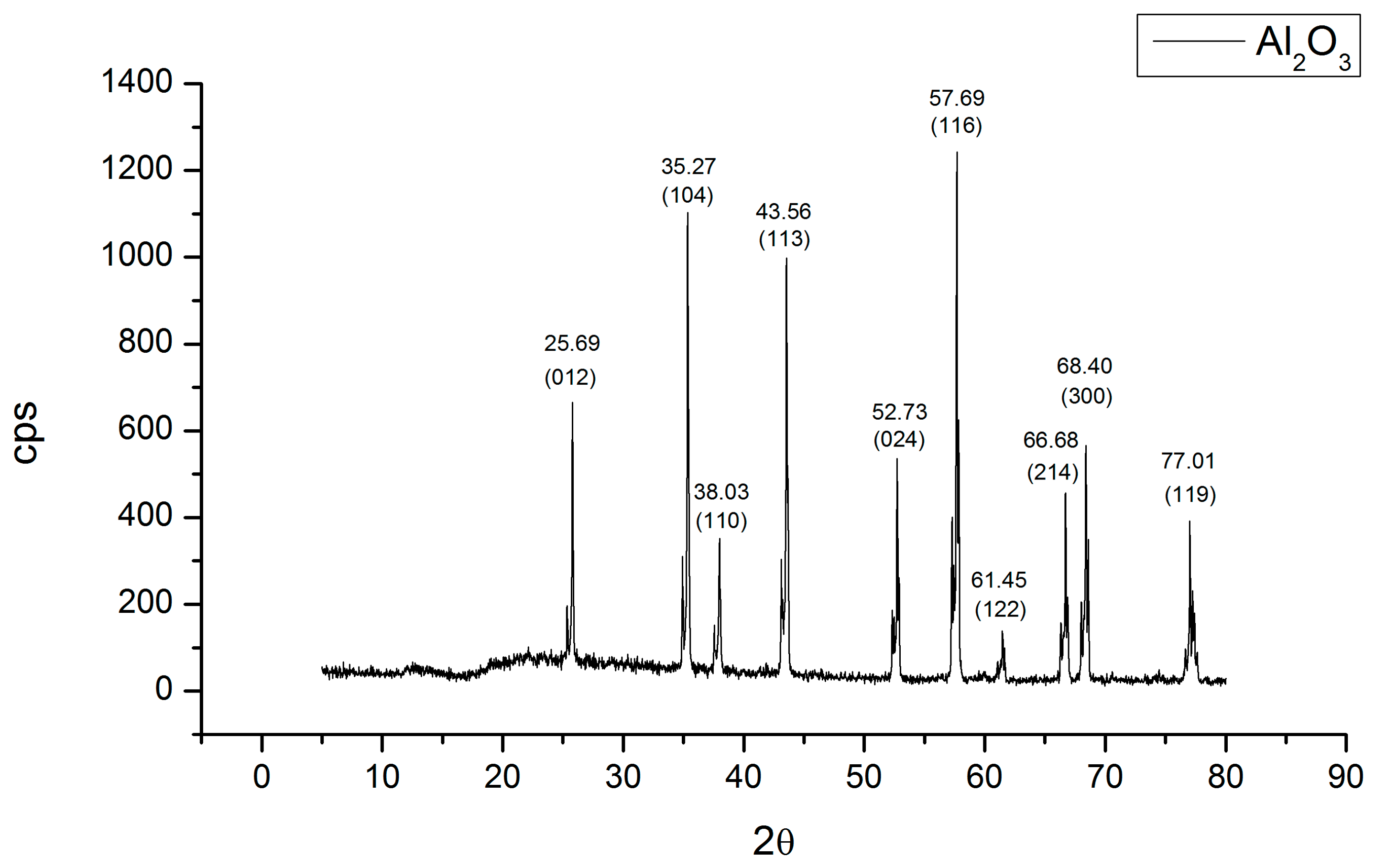
3.1. Elemental and Superficial Characterization of the Al2O3 Bracket

The elemental analysis by EDS, shown in Figure 1a, carried out on the surface of the bracket shows only the presence of oxygen and aluminum, and the atomic percentage reported is 60% for oxygen and 40% for aluminum; this stoichiometric relationship corresponds to aluminum oxide as can be seen, inferring the chemical composition of Al2O3. Figure 1b clearly shows the surface of the bracket, where a typical morphology of a ceramic bracket is observed. The bracket Al2O3 support was characterized for AFM where the topography of a polycrystalline material can be observed, with an average grain size of 400 nm; it has high roughness, and in Figure 1c, it is observed that the grain size is not homogeneous and a difference between the peaks and valleys is perceived.
Figure 2 shows the XRD diffractogram that was compared with the JCPDS-ICCD-46-1212 card, indicating that the Al2O3 is in the alpha phase. In the diffractogram, you can see the presence of a small peak to the left of the main diffraction, 2θ~25.6°; to the left, this small peak appears with an intensity of a quarter of the main peak, and thus it is repeated. In the remaining diffractions, since the surface of the bracket is not flat, it presents different heights in its topography, and this causes the appearance of this double peak at 52.7° and 57.6° due to the difference in the distance of the topography of the material.

3.2. Morphological Characterization of the Synthesized Nanoparticles

The synthesized AgNPs were characterized using the TEM technique to obtain information on the particle size and distribution of Ag deposited on the surface of the Al2O3 supports. As shown in the micrographs in Figure 3a,b,e,f, the presence of AgNPs, TiO2NPs, and Ag/TiO2NPs is corroborated, all being quasi-spherical. Regarding the HRTEM analysis where the information on the interplanar distance of the AgNPs is shown, this being 0.235 nm and 0.144 nm, respectively in the (111) and (220) planes, consistent with the JCPDS 04-0783 card. We observe, in Figure 3g(I), a distribution of AgNPs with a mean size of 4.2 nm on the surface of the Al2O3 bracket.
Likewise, the HRTEM technique was used to obtain information on the interplanar distance of the TiO2NPs, these being 0.243 nm, 0.189 nm, and 0.165 nm corresponding to (103), (200), and (211), respectively, according to the JCPDS card 021-1272. TiO2NPs with a mean size of 3.6 nm are observed on the surface of the bracket. In Figure 3e,f, a small number of titanium dioxide nanoparticles with an average size of 31 nm were deposited on the surface of the Al2O3 support. It is also observed that the silver nanoparticles are deposited on the surface of the individual TiO2 crystallites; this can be observed in Figure 3f depending on the difference in shades.

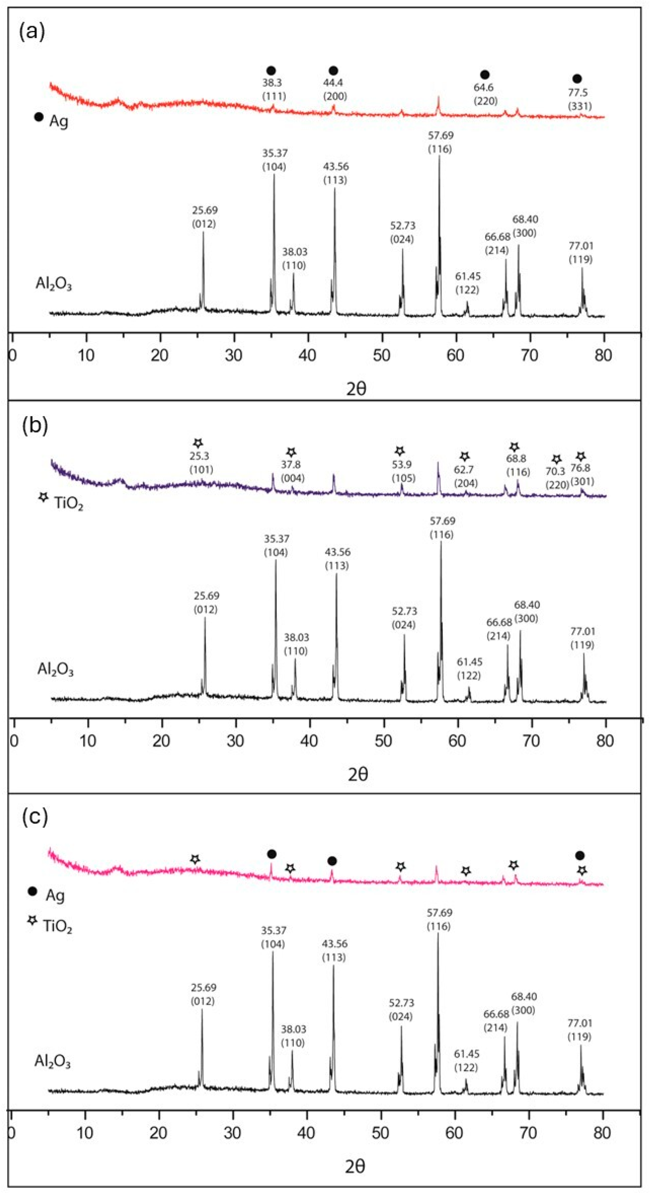
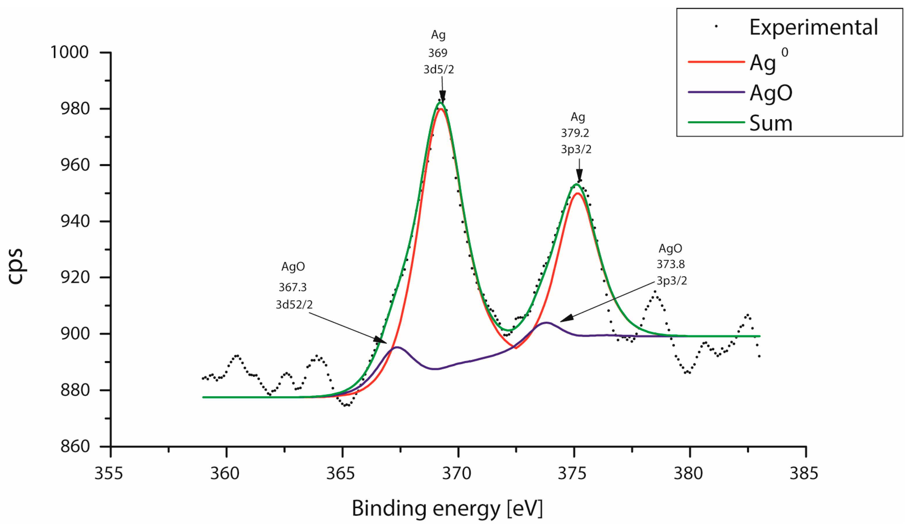
3.3. Characterization of the Nanoparticles Supported on the Surface of the Al2O3 Brackets

Figure 4a shows the elemental composition of the surface of the Ag/TiO2NPs deposited on the bracket as determined by EDS, where the presence of aluminum, titanium, silver, and oxygen is mainly observed; these samples were coated with gold. As seen in Figure 4b, the mapping of the distribution of the nanoparticles is homogeneous, with the Ag concentration being higher. Therefore, it can be assumed that the Ag/TiO2NPs were deposited on the Al2O3 surface support.
In Figure 5a, the pattern of X-ray diffraction of AgNPs deposited on the support surface Al2O3, shows that the peaks at 38.3, 44.4, 64.6, and 75.5 of AgNPs coincide with the peaks of Al2O3; we observed that they are less intense, so we assume that the support surface was covered by AgNPs. Also, in Figure 5b, the pattern of X-ray diffraction of Ag/TiO2 NPs deposited on the support surface of Al2O3 shows that the peaks at 23.3°, 37.8°, 53.9°, 62.7°, 68.8°, 70.3°, and 76.8° of TiO2NPs match those shown by Al2O3 but are less intense, so we assume that the support surface was covered by NP’s TiO2.
In Figure 5c, one can see the pattern of X-ray diffraction of the combination of Ag/TiO2NPs deposited on the support surface Al2O3 shown that peaks at 38.3°, 44.4°, and 75.5° of Ag NPs and peaks at 23.3°, 37.8°, 53.9°, 62.7°, 68.8°, and 76.8° of the TiO2NPs match those of Al2O3; both materials are deposited individually, making properties make the most of materials. The characterization technique is consistent with the analysis on elemental chemical mapping, where this distribution was observed. The diffractograms show that the peaks associated with the alumina decrease with the increment in the concentration of Ag and TiO2 [61,62,63].
XPS spectra were obtained after etching with Ar+ for 10 s. The C 1s (binding energy, BE = 285.00 eV) was used as a reference for calibrating binding energy in all spectra of the samples. For the sample, AgNPs were analyzed by XPS to determine the oxidation state of the Ag. Figure 6 shows the XPS spectrum and corresponding oxidation states of the silver deconvolutions, indicating mostly the presence of metallic silver, as it can be seen that a large part of the silver is metallic while a small contribution is also observed assigned to Ag-O; the above indicates the almost total reduction of silver ions which confirms the effectiveness of the reduction method proposed for the formation of silver nanoparticles from the silver (Ag+1) ions.
In Figure 7, the Ti deconvolution was performed, and there is only one signal corresponding to Ti-O, which confirms the effectiveness of the proposed reduction method to anchor the titanium dioxide nanoparticles and the generation of titanium dioxide nanoparticles. Because the XPS is highly sensitive to the surface, the AgNPs are exposed largely on the surface and cover a portion of the surface of the TiO2 particles; as observed in Ag3d5/2 signals and Ti2p3/2, they can be comparable in magnitude despite having different concentrations. This argument is consistent with the morphological characteristics of the samples [64,65,66,67].
Dodecyl-sulfate (DS) surfactant molecules were used in 10 wt% to provide a hydrophobic character to the photocatalyst particles and increase the dispersibility of the originally hydrophilic photocatalyst particles in the polymer film [68]. The mechanism of anchoring the NPs to the surface of the alumin is described with respect to the high electronic density of the oxygen of the alumina that electrostatically attract the NPs, and there is also a mechanical retention due to the roughness of the surface of the bracket.

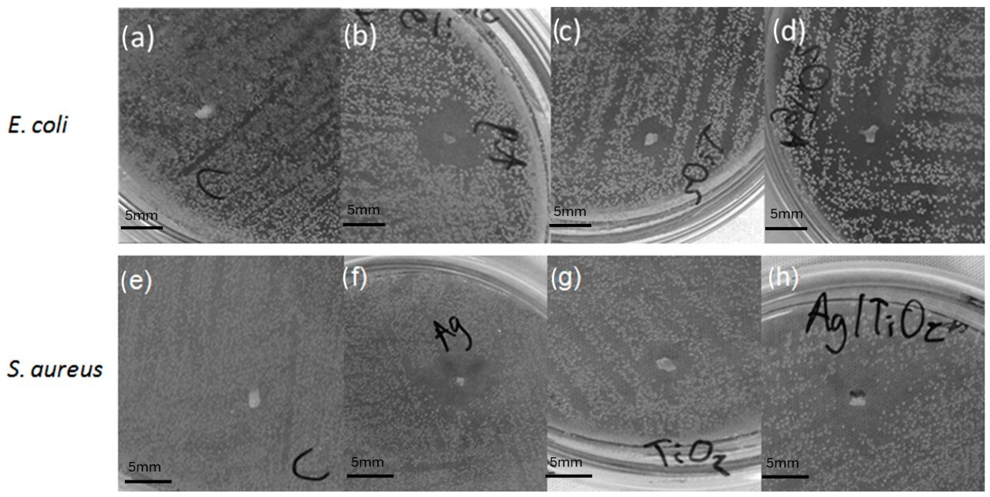
3.4. Antimicrobial Activity

In the microbiological testing, the samples prepared with AgNPs, TiO2NPs, and Ag/TiO2NPs on brackets exhibited a bactericidal effect, which can be observed by formation of an inhibitory halo. This occurs in both microorganisms E. coli and S. aureus, Gram (−) and Gram (+), respectively; this can contrast the inhibitory halo in Figure 8, with respect to the control bracket. This is consistent with several investigations where E. coli inactivation is mainly due to the destruction of the cell wall by Ag and TiO2 [69,70].
In Figure 8, the antimicrobial activity in E. coli of AgNPs, TiO2 NPs, and Ag/TiO2NPs and a control bracket is shown, and it is shown in Figure 8b,d that both AgNPs and Ag/TiO2NPs have a similar inhibitory halo, while in Figure 8c, it can be seen that TiO2NPs presents a significant inhibition halo, all compared to the control non-inhibitory halo presented as shown in Figure 8a. As for the antimicrobial activity in S. aureus of AgNPs, TiO2NPs, and Ag/TiO2NPs and a bracket control, it is observed that in Figure 8f, the AgNPs have a greater inhibitory halo than the TiO2NPs inhibitory halo in Figure 8g, and in Figure 8h, the Ag/TiO2NPs have an inhibitory halo slightly higher than those of AgNPs and TiO2NPs alone, all compared to the control bracket which did not present an inhibitory halo, as shown in Figure 8e (Table 1). In general, all treatments had a greater effect against E. coli bacteria than S. aureus.
This difference in bacterial resistance of Gram-negative and Gram-positive bacteria agrees with previously published research [71,72,73]. The tolerance of Gram-negative bacteria to different nanoparticles could be attributed to the layer of lipopolysaccharide (LPS), providing an external bacteria cell membrane. This LPS layer not only contributes to cell integrity but also hinders the absorption of Ag+ and/or AgNPs [74]. It is unclear whether this trend is universal for all species of Gram-negative and Gram-positive bacteria [75]. It is important to highlight that the Ag/TiO2NPs increase considerably in size; according to the literature, we expected to obtain greater antibacterial activity; however, they are almost similar. This may be due to the arrangement of the atoms in the system according to the atomic radii of each of the elements or because when the system dries, it agglomerates, and the size of the supported nanoparticle becomes larger. Other authors have shown that the antimicrobial activity on other types of materials, such as wires used in orthodontics, show a better antibacterial effect with smaller silver nanoparticles [76], and even with other methods of synthesis of titanium dioxide nanoparticles, such as N-doped TiO2 thin films coated on stainless steel brackets under visible light irradiation, an important antibacterial effect was achieved, as well as the results obtained in this work using a simpler method [77].
According to the literature, we can infer that the mechanism of action of the metallic nanoparticles is the following: nanoparticles can bind directly to the cell membrane of bacteria and then damage or alter its functionality. This is due to its high reactivity to substances such as proteins, enzymes, DNA, RNA, etc. [7,8,9].

4. Conclusions

In this study, synthesis of AgNPs, TiO2NPs, and Ag/TiO2NPs on the surface of Al2O3 was performed by a simple, inexpensive chemical reduction method. These nanoparticles are quasi-spherical, with defined sizes that offer different behavior alone and in combination. In the microbiological testing, an inhibitory effect was achieved in all samples, having a greater inhibitory halo in samples of Ag and Ag/TiO2 than TiO2 in the samples of both bacteria. Finally, these results show that supporting Ag and Ag/TiO2NPs in brackets can be an alternative to avoid generating stains on teeth during orthodontic treatment; in future work, it will be necessary to carry out more specific tests that simulate dentobacterial plaque until treatment is assured in patients.

Author Contributions

Writing and experimentation, P.A.S.R.; writing—review and editing, R.A.M.-L.; methodology-microbiology test, M.G.G.-P.; formal analysis, N.M.M.-B.; microscopic analysis, O.F.O.M.; statistical analysis, B.R.-P.; writing preparation of the original draft and discussion, P.A.S.R.; sample preparation for analysis and interpretation, M.G.G.-P.; supervision, R.A.M.-L. All authors have read and agreed to the published version of the manuscript.

Funding

This research received no external funding.

Data Availability Statement

The original contributions presented in the study are included in the article, further inquiries can be directed to the corresponding author.

Acknowledgments

To Rosa Maria Gomez Espinoza for the acquisition of the XRD spectra and to Gustavo Lopez Tellez for the technical support in the XPS analysis.

Conflicts of Interest

The authors declare no conflicts of interest.

References

  1. Oves, M.; Rauf, M.A.; Aslam, M.; Qari, H.A.; Sonbol, H.; Ahmad, I.; Zaman, G.S.; Saeed, M. Green synthesis of silver nanoparticles by Conocarpus Lancifolius plant extract and their antimicrobial and anticancer activities. Saudi J. Biol. Sci. 2022, 29, 460–471. [Google Scholar] [CrossRef] [PubMed]
  2. Kamaruzaman, N.H.; Noor, N.N.M.; Mohamed, R.M.S.R.; Al-Gheethi, A.; Ponnusamy, S.K.; Sharma, A.; Vo, D.-V.N. Applicability of bio-synthesized nanoparticles in fungal secondary metabolites products and plant extracts for eliminating antibiotic-resistant bacteria risks in non-clinical environments. Environ. Res. 2022, 209, 112831. [Google Scholar] [CrossRef] [PubMed]
  3. Huq, M.A.; Ashrafudoulla, M.; Rahman, M.M.; Balusamy, S.R.; Akter, S. Green synthesis and potential antibacterial applications of bioactive silver nanoparticles: A review. Polymers 2022, 14, 742. [Google Scholar] [CrossRef] [PubMed]
  4. Linklater, D.P.; Le Guével, X.; Bryant, G.; Baulin, V.A.; Pereiro, E.; Perera, P.G.T.; Wandiyanto, J.V.; Juodkazis, S.; Ivanova, E.P. Lethal Interactions of Atomically Precise Gold Nanoclusters and Pseudomonas aeruginosa and Staphylococcus aureus Bacterial Cells. ACS Appl. Mater. Interfaces 2022, 14, 32634–32645. [Google Scholar] [CrossRef] [PubMed]
  5. Bilal, M.; Zhao, Y.; Rasheed, T.; Ahmed, I.; Hassan, S.T.; Nawaz, M.Z.; Iqbal, H. Biogenic Nanoparticle–Chitosan Conjugates with Antimicrobial, Antibiofilm, and Anticancer Potentialities: Development and Characterization. Int. J. Environ. Res. Public Health 2019, 16, 598. [Google Scholar] [CrossRef] [PubMed]
  6. Siddiqi, K.S.; Husen, A. Properties of zinc oxide nanoparticles and their activity against microbes. Nanoscale Res. Lett. 2018, 13, 141. [Google Scholar] [CrossRef] [PubMed]
  7. Krishnamoorthy, R.; Athinarayanan, J.; Periyasamy, V.S.; Alshuniaber, M.A.; Alshammari, G.; Hakeem, M.J.; Ahmed, M.A.; Alshatwi, A.A. Antibacterial Mechanisms of Zinc Oxide Nanoparticle against Bacterial Food Pathogens Resistant to Beta-Lactam Antibiotics. Molecules 2022, 27, 2489. [Google Scholar] [CrossRef]
  8. Duan, S.; Wu, R.; Xiong, Y.-H.; Ren, H.-M.; Lei, C.; Zhao, Y.-Q.; Zhang, X.-Y.; Xu, F.-J. Multifunctional antimicrobial materials: From rational design to biomedical applications. Prog. Mater. Sci. 2022, 125, 100887. [Google Scholar] [CrossRef]
  9. Chatterjee, S.; Lou, X.-Y.; Liang, F.; Yang, Y.-W. Surface-functionalized gold and silver nanoparticles for colorimetric and fluorescent sensing of metal ions and biomolecules. Coord. Chem. Rev. 2022, 459, 214461. [Google Scholar] [CrossRef]
  10. Jia, H.; Hou, W.; Wei, L.; Xu, B.; Liu, X. The structures and antibacterial properties of nano-SiO2 supported silver/zinc–silver materials. Dent. Mater. 2008, 24, 244–249. [Google Scholar] [CrossRef]
  11. Marambio-Jones, C.; Hoek, E. A review of the antibacterial effects of silver nanomaterials and potential implications for human health and the environment. J. Nanoparticle Res. 2010, 12, 1531–1551. [Google Scholar] [CrossRef]
  12. Liu, W.; Wu, Y.; Wang, C.; Li, H.C.; Wang, T.; Liao, C.Y.; Cui, L.; Zhou, Q.F.; Yan, B.; Jiang, G.B. Impact of silver nanoparticles on human cells: Effect of particle size. Nanotoxicology 2010, 4, 319–330. [Google Scholar] [CrossRef]
  13. Mao, B.-H.; Luo, Y.-K.; Wang Jr, B.; Chen, C.-W.; Cheng, F.-Y.; Lee, Y.-H.; Yan, S.-J.; Wang, Y.-J. Use of an in silico knowledge discovery approach to determine mechanistic studies of silver nanoparticles-induced toxicity from in vitro to in vivo. Part. Fibre Toxicol. 2022, 19, 6. [Google Scholar] [CrossRef]
  14. Ji, Q.-T.; Hu, D.-K.; Mu, X.-F.; Tian, X.-X.; Zhou, L.; Yao, S.; Wang, X.-H.; Xiang, S.-Z.; Ye, H.-J.; Fan, L.-J. Cucurbit [7] uril-Mediated Supramolecular Bactericidal Nanoparticles: Their Assembly Process, Controlled Release, and Safe Treatment of Intractable Plant Bacterial Diseases. Nano Lett. 2022, 22, 4839–4847. [Google Scholar] [CrossRef]
  15. Wang, X.; Liu, Z.; Bani-Fwaz, M.Z.; Marzouki, R.; Ali, I.H.; El-kott, A.F.; Alhomaid, F.A. Ag nanoparticles immobilized on guanidine modified-KIT-5 mesoporous nanostructure: Evaluation of its catalytic activity for synthesis of propargylamines and investigation of its antioxidant and anti-lung cancer effects. Arab. J. Chem. 2022, 15, 103548. [Google Scholar] [CrossRef]
  16. Khanna, P.K.; Singh, N.; Charan, S.; Subbarao, V.; Gokhale, R.; Mulik, U.P. Synthesis and characterization of Ag/PVA nanocomposite by chemical reduction method. Mater. Chem. Phys. 2005, 93, 117–121. [Google Scholar] [CrossRef]
  17. Wang, H.; Qiao, X.; Chen, J.; Ding, S. Preparation of silver nanoparticles by chemical reduction method. Colloids Surf. A Physicochem. Eng. Asp. 2005, 256, 111–115. [Google Scholar] [CrossRef]
  18. Dizman, H.M.; Kazancioglu, E.O.; Shigemune, T.; Takahara, S.; Arsu, N. High sensitivity colorimetric determination of L-cysteine using gold nanoparticles functionalized graphene oxide prepared by photochemical reduction method. Spectrochim. Acta Part A Mol. Biomol. Spectrosc. 2022, 264, 120294. [Google Scholar] [CrossRef]
  19. Chen, P.; Song, L.; Liu, Y.; Fang, Y.-e. Synthesis of silver nanoparticles by γ-ray irradiation in acetic water solution containing chitosan. Radiat. Phys. Chem. 2007, 76, 1165–1168. [Google Scholar] [CrossRef]
  20. Attia, R.M.; Yousif, N.M.; Balboul, M.R. Facile synthesis of r GO/Ag/PVA nanocomposites polymeric films by electron beam irradiation for optoelectronic applications. Opt. Mater. 2022, 126, 112233. [Google Scholar] [CrossRef]
  21. Li, K.; Zhang, F.-S. A novel approach for preparing silver nanoparticles under electron beam irradiation. J. Nanoparticle Res. 2010, 12, 1423–1428. [Google Scholar] [CrossRef]
  22. Nthunya, L.N.; Gutierrez, L.; Mhlanga, S.D.; Richards, H.L. Bio-mediated synthesis of silver nanoparticles via conventional and irradiation-assisted methods and their application for environmental remediation in agriculture. In Green Synthesis of Silver Nanomaterials; Elsevier: Amsterdam, The Netherlands, 2022; pp. 219–239. [Google Scholar]
  23. Zhang, J.; Ahmadi, M.; Fargas, G.; Perinka, N.; Reguera, J.; Lanceros-Méndez, S.; Llanes, L.; Jiménez-Piqué, E. Silver Nanoparticles for Conductive Inks: From Synthesis and Ink Formulation to Their Use in Printing Technologies. Metals 2022, 12, 234. [Google Scholar] [CrossRef]
  24. Naganthran, A.; Verasoundarapandian, G.; Khalid, F.E.; Masarudin, M.J.; Zulkharnain, A.; Nawawi, N.M.; Karim, M.; Che Abdullah, C.A.; Ahmad, S.A. Synthesis, Characterization and Biomedical Application of Silver Nanoparticles. Materials 2022, 15, 427. [Google Scholar] [CrossRef]
  25. Harada, M.; Saijo, K.; Sakamoto, N. Characterization of metal nanoparticles prepared by photoreduction in aqueous solutions of various surfactants using UV–vis, EXAFS and SAXS. Colloids Surf. A Physicochem. Eng. Asp. 2009, 349, 176–188. [Google Scholar] [CrossRef]
  26. Abbas, A.; Amin, H.M.A. Silver Nanoparticles Modified Electrodes for Electroanalysis: An Updated Review and a Perspective. Microchem. J. 2022, 175, 107166. [Google Scholar] [CrossRef]
  27. Calderón-Jiménez, B.; Montoro Bustos, A.R.; Pereira Reyes, R.; Paniagua, S.A.; Vega-Baudrit, J.R. Novel pathway for the sonochemical synthesis of silver nanoparticles with near-spherical shape and high stability in aqueous media. Sci. Rep. 2022, 12, 882. [Google Scholar] [CrossRef]
  28. Sultan, M.; Siddique, M.; Khan, R.; Fallatah, A.M.; Fatima, N.; Shahzadi, I.; Waheed, U.; Bilal, M.; Ali, A.; Abbasi, A.M. Ligustrum lucidum Leaf Extract-Assisted Green Synthesis of Silver Nanoparticles and Nano-Adsorbents Having Potential in Ultrasound-Assisted Adsorptive Removal of Methylene Blue Dye from Wastewater and Antimicrobial Activity. Materials 2022, 15, 1637. [Google Scholar] [CrossRef]
  29. Barbhuiya, R.I.; Singha, P.; Asaithambi, N.; Singh, S.K. Ultrasound-assisted rapid biological synthesis and characterization of silver nanoparticles using pomelo peel waste. Food Chem. 2022, 385, 132602. [Google Scholar] [CrossRef]
  30. Tian, S.; Hu, Y.; Chen, X.; Liu, C.; Xue, Y.; Han, B. Green synthesis of silver nanoparticles using sodium alginate and tannic acid: Characterization and anti-S. aureus activity. Int. J. Biol. Macromol. 2022, 195, 515–522. [Google Scholar] [CrossRef]
  31. Zheng, M.; Wang, Z.-S.; Zhu, Y.-W. Preparation of silver nanoparticle via active template under ultrasonic. Trans. Nonferrous Met. Soc. China 2006, 16, 1348–1352. [Google Scholar] [CrossRef]
  32. Briones, M.A.; Altamirano, G.V.; Moreano, R.M.; Nieto, N.T. Biomateriales inteligentes usados en odontología. Arch. Venez. De Farmacol. Ter. 2022, 41, 1–7. [Google Scholar] [CrossRef]
  33. Estrada-Monje, A.; Zitzumbo-Guzman, R.; Banuelos-Diaz, J.A.; Zaragoza-Contreras, E.A. Ultrasonic dispersion and activation of TiO2 nanoparticles and its effect on bacterial inhibition in EVA films. Mater. Chem. Phys. 2019, 235, 121760. [Google Scholar] [CrossRef]
  34. Ripolles-Avila, C.; Martinez-Garcia, M.; Hascoët, A.-S.; Rodríguez-Jerez, J.J. Bactericidal efficacy of UV activated TiO2 nanoparticles against Gram-positive and Gram-negative bacteria on suspension. CyTA-J. Food 2019, 17, 408–418. [Google Scholar] [CrossRef]
  35. Ge, X.; Ren, C.; Ding, Y.; Chen, G.; Lu, X.; Wang, K.; Ren, F.; Yang, M.; Wang, Z.; Li, J. Micro/nano-structured TiO2 surface with dual-functional antibacterial effects for biomedical applications. Bioact. Mater. 2019, 4, 346–357. [Google Scholar] [CrossRef]
  36. Shang, C.; Cheung, L.M.; Ho, C.-M.; Zeng, M. Repression of photoreactivation and dark repair of coliform bacteria by TiO2-modified UV-C disinfection. Appl. Catal. B Environ. 2009, 89, 536–542. [Google Scholar] [CrossRef]
  37. Yan, T.; Cai, S.; Hu, Z.; Li, Z.; Fang, X. Ultrafast speed, dark current suppression, and self-powered enhancement in TiO2-based ultraviolet photodetectors by organic layers and Ag nanowires regulation. J. Phys. Chem. Lett. 2021, 12, 9912–9918. [Google Scholar] [CrossRef]
  38. Çeşmeli, S.; Avci, C.B. Application of titanium dioxide (TiO2) nanoparticles in cancer therapies. J. Drug Target. 2019, 27, 762–766. [Google Scholar] [CrossRef]
  39. Lee, H.U.; Lee, S.C.; Lee, Y.-C.; Son, B.; Park, S.Y.; Lee, J.W.; Oh, Y.-K.; Kim, Y.; Choi, S.; Lee, Y.-S. Innovative three-dimensional (3D) eco-TiO2 photocatalysts for practical environmental and bio-medical applications. Sci. Rep. 2014, 4, 6740. [Google Scholar] [CrossRef]
  40. Mano, S.S.; Kanehira, K.; Sonezaki, S.; Taniguchi, A. Effect of polyethylene glycol modification of TiO2 nanoparticles on cytotoxicity and gene expressions in human cell lines. Int. J. Mol. Sci. 2012, 13, 3703–3717. [Google Scholar] [CrossRef]
  41. Ziental, D.; Czarczynska-Goslinska, B.; Mlynarczyk, D.T.; Glowacka-Sobotta, A.; Stanisz, B.; Goslinski, T.; Sobotta, L. Titanium dioxide nanoparticles: Prospects and applications in medicine. Nanomaterials 2020, 10, 387. [Google Scholar] [CrossRef]
  42. Subhapriya, S.; Gomathipriya, P. Green synthesis of titanium dioxide (TiO2) nanoparticles by Trigonella foenum-graecum extract and its antimicrobial properties. Microb. Pathog. 2018, 116, 215–220. [Google Scholar] [CrossRef]
  43. Massima Mouele, E.S.; Dinu, M.; Cummings, F.; Fatoba, O.O.; Zar Myint, M.T.; Kyaw, H.H.; Parau, A.C.; Vladescu, A.; Francesconi, M.G.; Pescetelli, S. Effect of calcination time on the physicochemical properties and photocatalytic performance of carbon and nitrogen co-doped TiO2 nanoparticles. Catalysts 2020, 10, 847. [Google Scholar] [CrossRef]
  44. Bogdan, J.; Jackowska-Tracz, A.; Zarzyńska, J.; Pławińska-Czarnak, J. Chances and limitations of nanosized titanium dioxide practical application in view of its physicochemical properties. Nanoscale Res. Lett. 2015, 10, 57. [Google Scholar] [CrossRef]
  45. Baygi, N.J.; Saghir, A.V.; Beidokhti, S.M.; Khaki, J.V. Modified auto-combustion synthesis of mixed-oxides TiO2/NiO nanoparticles: Physical properties and photocatalytic performance. Ceram. Int. 2020, 46, 15417–15437. [Google Scholar] [CrossRef]
  46. Gupta, S.; Tripathi, M. A review on the synthesis of TiO2 nanoparticles by solution route. Open Chem. 2012, 10, 279–294. [Google Scholar] [CrossRef]
  47. Nabi, G.; Khalid, N.; Tahir, M.B.; Rafique, M.; Rizwan, M.; Hussain, S.; Iqbal, T.; Majid, A. A review on novel eco-friendly green approach to synthesis TiO2 nanoparticles using different extracts. J. Inorg. Organomet. Polym. Mater. 2018, 28, 1552–1564. [Google Scholar] [CrossRef]
  48. Vijayalakshmi, R.; Rajendran, V. Synthesis and characterization of nano-TiO2 via different methods. Arch. Appl. Sci. Res 2012, 4, 1183–1190. [Google Scholar]
  49. Bui, V.K.H.; Park, D.; Lee, Y.-C. Chitosan combined with ZnO, TiO2 and Ag nanoparticles for antimicrobial wound healing applications: A mini review of the research trends. Polymers 2017, 9, 21. [Google Scholar] [CrossRef]
  50. Petronella, F.; Diomede, S.; Fanizza, E.; Mascolo, G.; Sibillano, T.; Agostiano, A.; Curri, M.; Comparelli, R. Photodegradation of nalidixic acid assisted by TiO2 nanorods/Ag nanoparticles based catalyst. Chemosphere 2013, 91, 941–947. [Google Scholar] [CrossRef]
  51. Meydanju, N.; Pirsa, S.; Farzi, J. Biodegradable film based on lemon peel powder containing xanthan gum and TiO2–Ag nanoparticles: Investigation of physicochemical and antibacterial properties. Polym. Test. 2022, 106, 107445. [Google Scholar] [CrossRef]
  52. Mariappan, A.; Pandi, P.; Rajeswarapalanichamy, R.; Neyvasagam, K.; Sureshkumar, S.; Gatasheh, M.K.; Hatamleh, A.A. Bandgap and visible-light-induced photocatalytic performance and dye degradation of silver doped HAp/TiO2 nanocomposite by sol-gel method and its antimicrobial activity. Environ. Res. 2022, 211, 113079. [Google Scholar] [CrossRef]
  53. Ramakrishnan, V.M.; Rajesh, G.; Selvakumar, P.; Flores, M.; Muthukumarasamy, N.; Velauthapillai, D.; Chi, N.T.L.; Pugazhendhi, A. TiO2/AgO composites by one step photo reduction technique as electron transport layers (ETL) for dye-sensitized solar cells. Chemosphere 2022, 305, 134953. [Google Scholar] [CrossRef]
  54. Butmee, P.; Samphao, A.; Tumcharern, G. Reduced graphene oxide on silver nanoparticle layers-decorated titanium dioxide nanotube arrays as SERS-based sensor for glyphosate direct detection in environmental water and soil. J. Hazard. Mater. 2022, 437, 129344. [Google Scholar] [CrossRef]
  55. Argueta-Figueroa, L.; Morales-Luckie, R.A.; Scougall-Vilchis, R.J.; Olea-Mejía, O.F. Synthesis, characterization and antibacterial activity of copper, nickel and bimetallic Cu–Ni nanoparticles for potential use in dental materials. Prog. Nat. Sci. Mater. Int. 2014, 24, 321–328. [Google Scholar] [CrossRef]
  56. Hernández-Gómora, A.E.; Lara-Carrillo, E.; Robles-Navarro, J.B.; Scougall-Vilchis, R.J.; Hernández-López, S.; Medina-Solís, C.E.; Morales-Luckie, R.A. Biosynthesis of silver nanoparticles on orthodontic elastomeric modules: Evaluation of mechanical and antibacterial properties. Molecules 2017, 22, 1407. [Google Scholar] [CrossRef]
  57. Argueta-Figueroa, L.; Scougall-Vilchis, R.J.; Morales-Luckie, R.A.; Olea-Mejía, O.F. An evaluation of the antibacterial properties and shear bond strength of copper nanoparticles as a nanofiller in orthodontic adhesive. Australas. Orthod. J. 2015, 31, 42–48. [Google Scholar] [CrossRef]
  58. Garcia-Contreras, R.; Scougall-Vilchis, R.J.; Contreras-Bulnes, R.; Sakagami, H.; Morales-Luckie, R.A.; Nakajima, H. Mechanical, antibacterial and bond strength properties of nano-titanium-enriched glass ionomer cement. J. Appl. Oral Sci. 2015, 23, 321–328. [Google Scholar] [CrossRef]
  59. Bauer, A. Antibiotic susceptibility testing by a standardized single disc method. Am. J. Clin. Pathol. 1966, 45, 149–158. [Google Scholar] [CrossRef]
  60. Tholen, D.W.; Linnet, K.; Kondratovich, M.; Armbruster, D.A.; Garrett, P.E.; Jones, R.L.; Kroll, M.H.; Lequin, R.M.; Pankratz, T.J.; Scassellati, G. Protocols for determination of limits of detection and limits of quantitation; approved guideline. CLSI EP17-A 2004, 24, 34. [Google Scholar]
  61. Khojasteh, F.; Mersagh, M.R.; Hashemipour, H. The influences of Ni, Ag-doped TiO2 and SnO2, Ag-doped SnO2/TiO2 nanocomposites on recombination reduction in dye synthesized solar cells. J. Alloys Compd. 2022, 890, 161709. [Google Scholar] [CrossRef]
  62. Zhang, H.; Chen, G. Potent antibacterial activities of Ag/TiO2 nanocomposite powders synthesized by a one-pot sol− gel method. Environ. Sci. Technol. 2009, 43, 2905–2910. [Google Scholar] [CrossRef] [PubMed]
  63. Atarod, M.; Nasrollahzadeh, M.; Sajadi, S.M. Euphorbia heterophylla leaf extract mediated green synthesis of Ag/TiO2 nanocomposite and investigation of its excellent catalytic activity for reduction of variety of dyes in water. J. Colloid Interface Sci. 2016, 462, 272–279. [Google Scholar] [CrossRef] [PubMed]
  64. Akhavan, O.; Ghaderi, E. Self-accumulated Ag nanoparticles on mesoporous TiO2 thin film with high bactericidal activities. Surf. Coat. Technol. 2010, 204, 3676–3683. [Google Scholar] [CrossRef]
  65. Ashkarran, A. Antibacterial properties of silver-doped TiO2 nanoparticles under solar simulated light. J. Theor. Appl. Phys. 2011, 4, 1–8. [Google Scholar]
  66. Tunc, I.; Bruns, M.; Gliemann, H.; Grunze, M.; Koelsch, P. Bandgap determination and charge separation in Ag@ TiO2 core shell nanoparticle films. Surf. Interface Anal. 2010, 42, 835–841. [Google Scholar] [CrossRef]
  67. Massaro, C.; Rotolo, P.; De Riccardis, F.; Milella, E.; Napoli, A.; Wieland, M.; Textor, M.; Spencer, N.; Brunette, D. Comparative investigation of the surface properties of commercial titanium dental implants. Part I: Chemical composition. J. Mater. Sci. Mater. Med. 2002, 13, 535–548. [Google Scholar] [CrossRef] [PubMed]
  68. Györgyey, Á.; Janovak, L.; Adam, A.; Kopniczky, J.; Tóth, K.L.; Deák, Á.; Panayotov, I.; Cuisinier, F.; Dékány, I.; Turzo, K. Investigation of the in vitro photocatalytic antibacterial activity of nanocrystalline TiO2 and coupled TiO2/Ag containing copolymer on the surface of medical grade titanium. J. Biomater. Appl. 2016, 31, 55–67. [Google Scholar] [PubMed]
  69. Liu, N.; Zhu, Q.; Zhang, N.; Zhang, C.; Kawazoe, N.; Chen, G.; Negishi, N.; Yang, Y. Superior disinfection effect of Escherichia coli by hydrothermal synthesized TiO2-based composite photocatalyst under LED irradiation: Influence of environmental factors and disinfection mechanism. Environ. Pollut. 2019, 247, 847–856. [Google Scholar] [CrossRef] [PubMed]
  70. Xu, X.; Wu, C.; Guo, A.; Qin, B.; Sun, Y.; Zhao, C.; Zhang, F.; Cai, A. Visible-light photocatalysis of organic contaminants and disinfection using biomimetic-synthesized TiO2-Ag-AgCl composite. Appl. Surf. Sci. 2022, 588, 152886. [Google Scholar] [CrossRef]
  71. Deng, K.; Li, Y.; Liang, X.; Shen, C.; Zeng, Z.; Xu, X. Virus-inspired nanoparticles as versatile antibacterial carriers for antibiotic delivery against Gram-negative and Gram-positive bacteria. Chin. Chem. Lett. 2022, 33, 1619–1622. [Google Scholar] [CrossRef]
  72. Kong, J.; Zhang, S.; Shen, M.; Zhang, J.; Yoganathan, S. Evaluation of copper (I)-doped zinc oxide composite nanoparticles on both gram-negative and gram-positive bacteria. Colloids Surf. A Physicochem. Eng. Asp. 2022, 643, 128742. [Google Scholar] [CrossRef]
  73. Su, J.; Li, H.; Hu, J.; Wang, D.; Zhang, F.; Fu, Z.; Han, F. LcCCL28-25, Derived from Piscine Chemokine, Exhibits Antimicrobial Activity against Gram-Negative and Gram-Positive Bacteria In Vitro and In Vivo. Microbiol. Spectr. 2022, 10, e02515-21. [Google Scholar] [CrossRef]
  74. Karami, A.; Xie, Z.; Zhang, J.; Kabir, M.S.; Munroe, P.; Kidd, S.; Zhang, H. Insights into the antimicrobial mechanism of Ag and I incorporated ZnO nanoparticle derivatives under visible light. Mater. Sci. Eng. C 2020, 107, 110220. [Google Scholar] [CrossRef]
  75. Li, M.; Noriega-Trevino, M.E.; Nino-Martinez, N.; Marambio-Jones, C.; Wang, J.; Damoiseaux, R.; Ruiz, F.; Hoek, E.M. Synergistic bactericidal activity of Ag-TiO2 nanoparticles in both light and dark conditions. Environ. Sci. Technol. 2011, 45, 8989–8995. [Google Scholar] [CrossRef]
  76. Espinosa-Cristóbal, L.F.; López-Ruiz, N.; Cabada-Tarín, D.; Reyes-López, S.Y.; Zaragoza-Contreras, A.; Constandse-Cortéz, D.; Donohué-Cornejo, A.; Tovar-Carrillo, K.; Cuevas-González, J.C.; Kobayashi, T. Antiadherence and antimicrobial properties of silver nanoparticles against Streptococcus mutans on brackets and wires used for orthodontic treatments. J. Nanomater. 2018, 2018, 9248527. [Google Scholar] [CrossRef]
  77. Cao, S.; Liu, B.; Fan, L.; Yue, Z.; Liu, B.; Cao, B. Highly antibacterial activity of N-doped TiO2 thin films coated on stainless steel brackets under visible light irradiation. Appl. Surf. Sci. 2014, 309, 119–127. [Google Scholar] [CrossRef]
Figure 1. Microscopic and elemental characterization: (a) SEM-EDS elemental characterization; (b) micrograph of the bracket; and (c) AFM micrograph of the bracket surface.
Microorganisms 12 01583 g001
Figure 2. XRD pattern of the support (bracket). The peaks were compared with JCPDS-ICCD-46-1212 card, corresponding to α-Al2O3.
Microorganisms 12 01583 g002
Figure 3. TEM micrographs: (a) Ag nanoparticles; (b) high resolution analysis of Ag nanoparticles; (c) TiO2 nanoparticles; (d) high resolution analysis of TiO2 nanoparticles; (e,f) Ag/nanoparticles TiO2. (g) Nanoparticle size histograms; (I) AgNPs, (II) TiO2NPs, and (III) Ag/TiO2NPs.
Microorganisms 12 01583 g003
Figure 4. Analysis by SEM-EDS. (a) Spectrum of the elemental analysis of Ag/TiO2 NPs, (b) elemental mapping; Ti (Red), Ag (Green) on surface characterization of the nanoparticles supported on the surface of the brackets.
Microorganisms 12 01583 g004
Figure 5. X-ray diffraction (XRD). (a) XRD pattern of AgNPs anchored Al2O3 support. (b) XRD pattern of TiO2NPs anchored Al2O3 support. (c) Pattern X-ray diffraction of the Ag/TiO2NPs anchored Al2O3 support.
Microorganisms 12 01583 g005
Figure 6. XPS spectra of AgNPs on Al2O3 support.
Microorganisms 12 01583 g006
Figure 7. XPS Spectra TiO2NPs on Al2O3 support.
Microorganisms 12 01583 g007
Figure 8. It shows the inhibitory halos of brackets (mm) on E. coli: (a) bracket without nanoparticles; (b) bracket with AgNPs; (c) bracket with TiO2NPs; (d) bracket with AgNPs, TiO2NPs, and Ag/TiO2NPs. It shows the inhibitory halos of brackets on S. aureus: (e) bracket without nanoparticles; (f) bracket with AgNPs; (g) bracket with TiO2NPs; (h) bracket with Ag/TiO2NPs.
Microorganisms 12 01583 g008
Table 1. Results of diffusion tests on disk.
Diameter of Inhibition Halos (mm)
StrainControlAgTiO2Ag/TiO2
S. aureusNP8.8 ± 0.36.0 ± 0.68.7 ± 0.8
E. coliNP8.3 ± 0.25.0 ± 0.38.9 ± 0.1
Results are represented by the mean ± standard error.
Disclaimer/Publisher’s Note: The statements, opinions and data contained in all publications are solely those of the individual author(s) and contributor(s) and not of MDPI and/or the editor(s). MDPI and/or the editor(s) disclaim responsibility for any injury to people or property resulting from any ideas, methods, instructions or products referred to in the content.

