Article

Exploring the Role of the Environment as a Reservoir of Antimicrobial-Resistant Campylobacter: Insights from Wild Birds and Surface Waters

1
Environmental Research and Innovation (ERIN) Department, Luxembourg Institute of Science and Technology (LIST), 41 Rue du Brill, L-4422 Belvaux, Luxembourg
2
Epidemiology and Microbial Genomics, Laboratoire National de Santé (LNS), 1 Rue Louis Rech, L-3555 Dudelange, Luxembourg
*
Author to whom correspondence should be addressed.
Microorganisms 2024, 12(8), 1621; https://doi.org/10.3390/microorganisms12081621
Submission received: 22 July 2024 / Revised: 2 August 2024 / Accepted: 5 August 2024 / Published: 8 August 2024
(This article belongs to the Special Issue Pathogen Infection in Wildlife 2.0)

Abstract

:
Antimicrobial resistance (AMR) is a growing global health challenge, compromising bacterial infection treatments and necessitating robust surveillance and mitigation strategies. The overuse of antimicrobials in humans and farm animals has made them hotspots for AMR. However, the spread of AMR genes in wildlife and the environment represents an additional challenge, turning these areas into new AMR hotspots. Among the AMR bacteria considered to be of high concern for public health, Campylobacter has been the leading cause of foodborne infections in the European Union since 2005. This study examines the prevalence of AMR genes and virulence factors in Campylobacter isolates from wild birds and surface waters in Luxembourg. The findings reveal a significant prevalence of resistant Campylobacter strains, with 12% of C. jejuni from wild birds and 37% of C. coli from surface waters carrying resistance genes, mainly against key antibiotics like quinolones and tetracycline. This study underscores the crucial role of the environment in the spread of AMR bacteria and genes, highlighting the urgent need for enhanced surveillance and control measures to curb AMR in wildlife and environmental reservoirs and reduce transmission risks to humans. This research supports One Health approaches to tackling antimicrobial resistance and protecting human, animal, and environmental health.

1. Introduction

With 4.95 million deaths associated with drug-resistant bacterial infections in 2019, antimicrobial resistance (AMR) is considered as the new silent pandemic [1]. The emergence of AMR represents a complex interplay of factors, including the overuse and misuse of antimicrobials in human and veterinary medicine, as well as in agricultural practices [2]. This selective pressure drives the evolution of antimicrobial-resistant bacteria (ARB) by promoting their survival and proliferation [3]. With the emergence of resistant strains, different risks of dissemination can occur, ranging from the spread of the acquired genes through horizontal gene transfer or mobile genetic elements to the geographic dispersion of the resistant clone itself thanks to international travel and trade [4]. AMR poses a significant threat to global public health, challenging the efficacy of antimicrobials and complicating the treatment of infectious diseases [5], contributes to increased morbidity, mortality, and healthcare costs [6], and jeopardises the success of medical procedures such as organ transplantation, chemotherapy, and surgery [7]. In addition to its direct impact on human health, AMR also compromises animal health and welfare, affecting food production systems and posing challenges to sustainable agriculture [8].
Environmental reservoirs, including surface waters, soil, and wildlife habitats, serve as vast repositories for diverse microbial communities, providing ample opportunities for the exchange and acquisition of resistance genes [9]. Factors such as agricultural runoff, the improper disposal of antimicrobial residues, and the discharge of untreated sewage contribute to the contamination of environmental matrices with antimicrobial agents and ARB [10]. Once introduced into these ecosystems, ARB can persist, survive, and sometimes proliferate, with a potential risk of the transmission of resistant microorganisms to human and animal populations [11]. As such, AMR is a global One Health challenge, involving the transfer of bacteria and genes between humans, animals, and the environment [12].
Zoonotic ARB present in animals and food can, consequently, also compromise the effective treatment of infectious diseases in humans [13]. Among zoonotic bacteria, Campylobacter spp. represents a significant bacterial pathogen with substantial implications for both animal and human health. In Europe, Campylobacter has been the leading cause of bacterial gastroenteritis since 2005, representing more than 60% of all reported cases in 2022 [14]. The highest notification rate in 2022 was observed in Luxembourg (141.3 cases per 100,000) [14]. However, the true incidence of campylobacteriosis may be higher due to underreporting, diagnostic challenges, and asymptomatic infections [15]. Therefore, Campylobacter infections pose a considerable burden on public health systems, causing symptoms ranging from mild diarrhoea to severe abdominal pain, and occasionally leading to complications such as Guillain–Barré syndrome [16]. The virulence of Campylobacter strains depends on various factors, including bacteria motility, adhesion to the intestinal mucosa, the invasion of epithelial cells, toxin production, and protein secretion [17]. Campylobacteriosis is primarily associated with the consumption and cross-contamination of contaminated food, particularly poultry products. However, these bacteria are also prevalent in the environment and can colonise the gastrointestinal tracts of various animals, including poultry, cattle, and wild birds [18].
Despite the growing recognition of the importance of environmental reservoirs in the transmission of AMR, there remains a paucity of data regarding the prevalence and distribution of resistant Campylobacter spp. in wild birds and surface waters. This study aims to address this knowledge gap by investigating the virulence and AMR profiles of Campylobacter isolates obtained from wild birds and surface waters in Luxembourg. By elucidating the dynamics of AMR dissemination in these ecological niches, this research will provide valuable insights into the potential role of the environment as a reservoir of ARB and antimicrobial resistance genes (ARGs).

2. Materials and Methods

2.1. Campylobacter Isolates Collection

A total of 263 Campylobacter isolates recovered by the passive filtration method from wild bird faeces and surface waters collected between 2019 and 2021 in Luxembourg [19] were included in the study: 119 C. jejuni (110 isolated from wild birds and 9 isolated from surface waters) and 144 C. coli (2 isolated from wild birds and 142 isolated from surface waters) (Supplementary Data S1). All strains were inoculated on chocolate agar plates with Vitox (PO5090A, Oxoid, Basingstoke, UK) or mCCDA plates (PO5091A, Oxoid, Basingstoke, UK) and incubated over 48 h at 42 °C under microaerobic conditions using the CampyGen gas-generating system (DN0025, Oxoid, Basingstoke, UK).

2.2. Disk Diffusion Method

AMR was assessed using the disk diffusion method on Mueller–Hinton agar with 5% sheep blood (PB0431, Oxoid, Basingstoke, UK) for various classes of antibiotics. For aminoglycosides, gentamicin (CN, 10 µg) (CT0024B, Oxoid, Basingstoke, UK) was used. Beta-lactam resistances were tested with ampicillin (AMP, 10 µg) (CT0003B, Oxoid, Basingstoke, UK) and amoxicillin/clavulanic acid (AMC, 20/10 µg) (CT0223B, Oxoid, Basingstoke, UK). Quinolone resistances were evaluated using nalidixic acid (NAL, 30 µg) (CT0031B, Oxoid, Basingstoke, UK) and ciprofloxacin (CIP, 5 µg) (CT0425B, Oxoid, Basingstoke, UK). Macrolide resistances were assessed with erythromycin (ERY, 15 µg) (CT0020B, Oxoid, Basingstoke, UK). Resistance to phenicols was tested with florfenicol (FFC, 30 µg) (CT1754B, Oxoid, Basingstoke, UK). Finally, tetracycline resistance (TET, 30 µg) (CT0054B, Oxoid, Basingstoke, UK) was also considered. The testing followed the French Microbiology Society (SFM) and EUCAST recommendations (Recommendations 2020 v1.1 April) [20]. The breakpoints were set as follows: AMP, 14 mm; AMC, 14 mm; CIP, 26 mm; ERY, 20 mm; and TET, 30 mm [20]. Additionally, according to Tang et al. [21], the breakpoints for NAL and FFC were 32 mm and 16 mm, respectively.

2.3. Genomic DNA Extraction and Whole-Genome Sequencing

DNA was extracted by using the QIAamp DNA Mini Kit (Qiagen, Venlo, The Netherlands) according to the manufacturer’s instructions. DNA was quantified with the Qubit® 2.0 Fluorometer (Invitrogen, Merelbeke, Belgium) and the Qubit® dsDNA HS Assay kit (Life Technologies, Gistel, Belgium). The DNA concentration was adjusted to be within the range between 3 and 17 ng/µL for subsequent sequencing. Libraries were prepared using the Nextera™ DNA Flex Library Prep Kit (Illumina, San Diego, CA, USA) and sequenced on the MiSeq™ platform (Illumina, USA), achieving 250-bp paired-end reads. The datasets of the sequence raw reads used for this study can be found in the ENA projects PRJEB57730 and PRJEB75211.

2.4. Genomic Assembly and Characterisation

The paired-end raw read data were de novo assembled using Spades v.3.11.1 (default parameters) implemented on Ridom SeqSphere+ v8.3.1 (Ridom GmbH, Münster, Germany) [22]. The Multi-locus sequence typing (MLST, 7 loci) [23] and core genome MLST (cgMLST, 637 loci) was assigned by using a scheme available in Ridom SeqSphere+, and isolates were classified in Sequence Type (ST). NCBI AMRFinderPlus v3.10.5 [24] (database v2021-06-01.1), deployed with the Ridom SeqSphere+ installation, was used to find genes and point mutations related to AMR, biocide, stress resistance, and virulence (BLAST identity > 90% and aligned = 100%).

2.5. Result Interpretation and Statistical Analyses

Multidrug-resistant (MDR) bacteria are defined as those displaying resistance to at least three classes of antibiotics. Measurements of proportion and their 95% confidence intervals (CI95s) were determined by following a binomial law approximated by a normal law (Wilson method). The concordance rate between the AMR genotype and the phenotype refers to the correspondence between the observed phenotypic antibiotic resistance and the presence of resistance genes or mutations associated with that phenotype. The concordance rate was calculated as the number of isolates with a matching phenotype and genotype (the difference between the total number of isolates and the difference between the number of resistant genotypes and resistant phenotypes) divided by the total number of isolates tested. Cohen’s kappa coefficient was used to determine the agreement between the pairs of phenotypes and genotypes of AMR and resistance determinants for all isolates. The interpretation of the kappa coefficient, expressed as the strength of agreement, was: <0.00: poor; 0.00–0.20: slight; 0.21–0.40: fair; 0.41–0.60: moderate; 0.61–0.80: substantial; and 0.81–1.00: almost perfect [25]. The association between AMR and other characteristics (bird species, upstream/downstream from a wastewater treatment plan, arsenic resistance) was determined using the chi-squared test (Rstudio v2022.02.0+443), with a probability value of p < 0.05 considered to be statistically significant. UPGMA trees were constructed by pairwise analyses of alleles of MLST and cgMLST, with missing targets ignored using the default settings (Ridom SeqSphere+ v8.3.1). UPGMA trees for virulome analyses were constructed by a pairwise analysis of 126 virulence genes, with missing values considered as their own category (Ridom SeqSphere+ v8.3.1).

3. Results

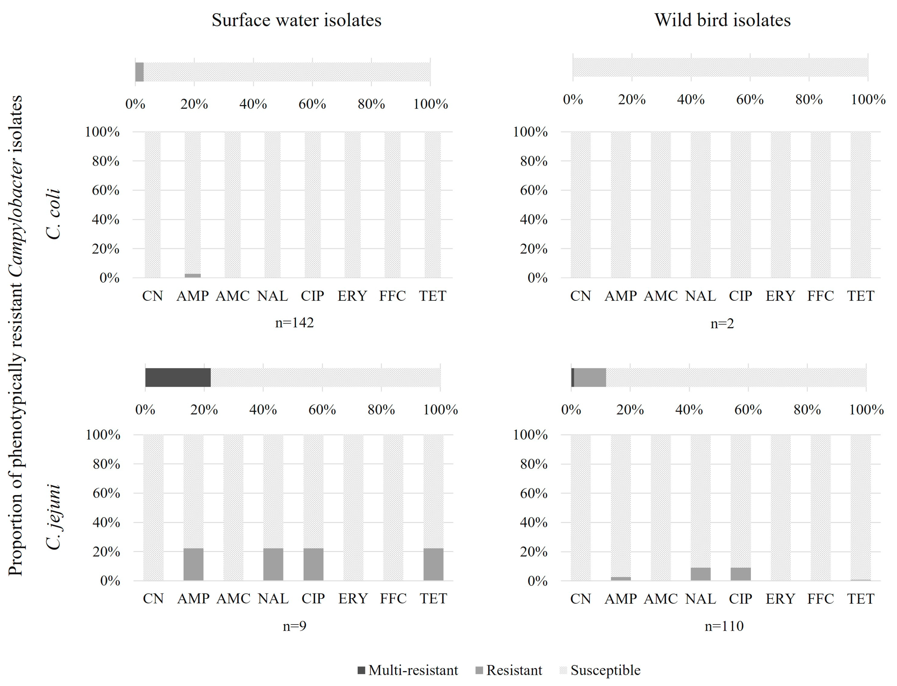
3.1. AMR Phenotypes and Genotypes of Isolates

The Campylobacter coli and C. jejuni isolates were analysed for their resistance to eight antibiotics using the disk diffusion method. C. coli were mainly isolated from surface waters and 3% were resistant, whereas C. jejuni were mainly isolated from wild birds and 12% were resistant to at least one antibiotic (CN, AMP, AMC, NAL, CIP, ERY, FFC, and TET). One C. jejuni isolate, from western jackdaw, was resistant to nalidixic acid, ciprofloxacin, and tetracycline. In surface waters and wild birds, Campylobacter showed resistance to beta-lactams, quinolone, and tetracycline (Figure 1, Table 1).
Genotypically, genes or mutations conferring AMR were detected for aminoglycosides, beta-lactams, quinolones, macrolides, and tetracycline (Figure 2, Table 2). In surface waters, 48% of C. coli isolates possessed resistance genes corresponding to at least one antibiotic, and one isolate was MDR (aminoglycoside, beta-lactam, and quinolone). Most resistant genotypes possessed a mutation responsible for aminoglycoside resistance (36.6% of isolates) or genes conferring beta-lactam resistance (13.4% of isolates). Among the nine C. jejuni isolates from surface waters, six showed resistance genes to beta-lactam and two possessed resistance genes to beta-lactam and tetracycline and mutations conferring resistance to quinolone. In wild birds, 89% of isolates possessed resistance genes to beta-lactam, where, among them, 11% also possessed mutations conferring resistance to quinolone, aminoglycoside, or tetracycline and one isolate possessed an MDR genotype (beta-lactam, macrolide, and tetracycline). Another MDR profile was isolated from birds with genes conferring tetracycline resistance and mutations conferring macrolide and quinolone resistance.
The concordance of resistant genotypes (predicted by AMRFinder from whole-genome sequencing data) and phenotypes (confirmed by disk diffusion) was between 98 and 100%, consistent with predictions observed for the database validation [24], except for the genes aadE-Cc detected by AMRFinder, but without a resistant phenotype observed for aminoglycoside and some blaOXA genes which could confer beta-lactam resistance (Table 3). This concordance was verified by the Cohen’s kappa coefficient measuring the inter-rater reliability, which confirmed a slight concordance between phenotypes and genotypes for beta-lactam resistance in C. coli and C. jejuni (Table 4).
Macrolide resistance was associated with an A103V mutation in the ribosomal protein L22 gene. ARG tet(O) was associated with tetracycline resistance. aadE genes coding for aminoglycoside-modifying enzymes were detected and conferred aminoglycoside resistance. Point mutations in the ribosomal protein P12 (RpsL) acting as a streptomycin-interacting residue were also detected. A gyrA mutation resulting in the amino acid substitution T86I was associated with resistance to (fluoro)quinolones (ciprofloxacin and nalidixic acid). Nineteen variants of the blaOXA genes involved in beta-lactam resistance were detected in 126 isolates (Table S1). Nine isolates were phenotypically ampicillin-resistant, and six of them (67%) also carried at least one gene coding for a beta-lactamase of the OXA-like family.

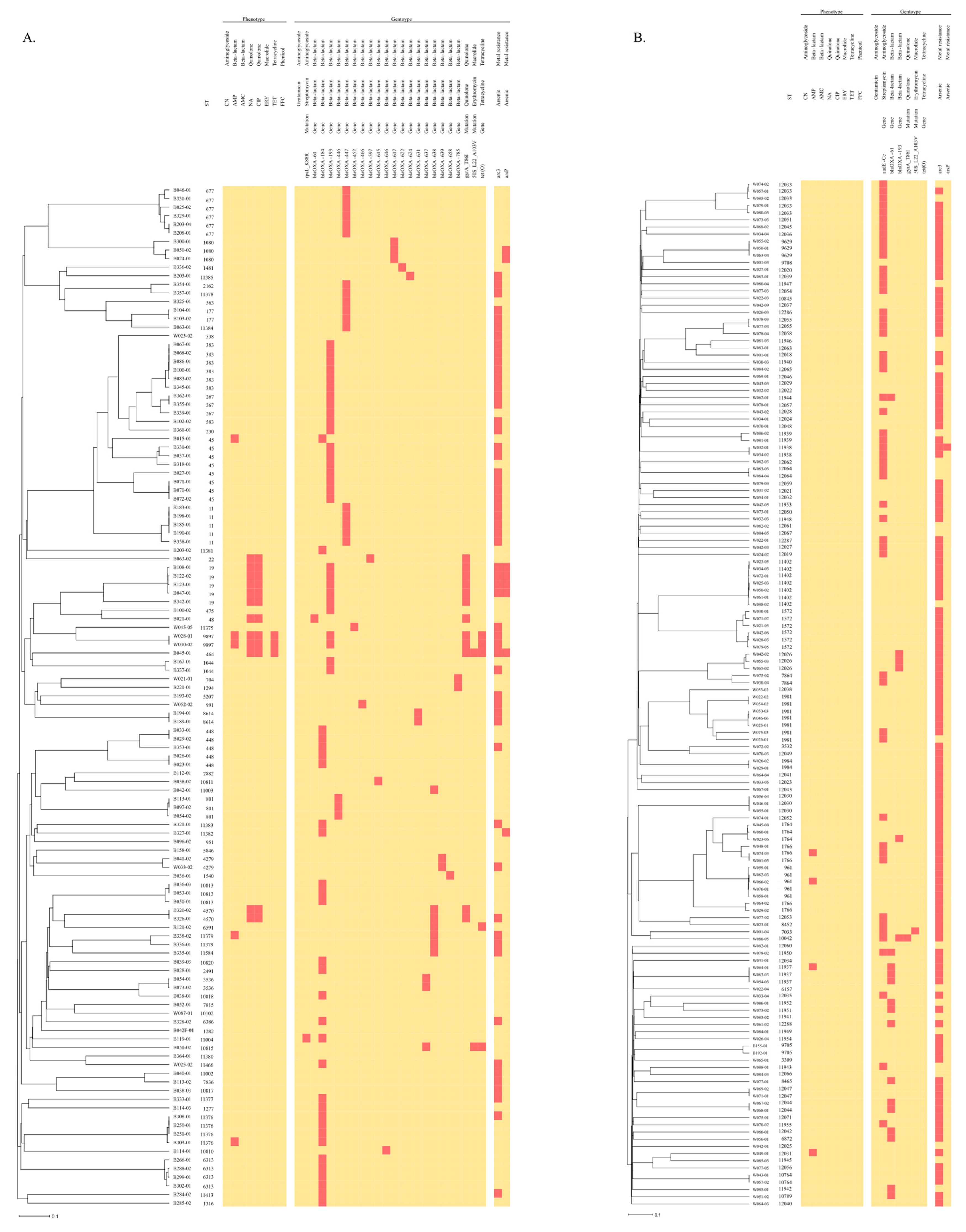
3.2. Association between AMR, ST, and Heavy Metals

An association was established between STs and the AMR profile resulting from both analyses, WGS in silico prediction, and antibiograms (Figure 3). All isolates assigned to the same ST showed a similar resistance profile to the same antibiotic classes, with two exceptions: C. coli ST 1766 and ST 1981. Only one of three isolates from ST 1766 expressed resistance to AMP and only two of seven isolates from ST1981 were in silico resistant to aminoglycosides.
Two STs exhibited MDR profiles in silico only: ST 10042 (C. coli), associated with streptomycin, quinolone, and beta-lactam (blaOXA-193) resistances, and ST 10815 (C. jejuni), harbouring erythromycin, tetracycline, and beta-lactam (blaOXA-637) resistances determinants. In addition, two C. jejuni isolates from ST 9897 were classified as MDR in silico and phenotypically for the following antibiotic classes: quinolone (NA and CIP, gyrA_T86), beta-lactam (blaOXA-193), and tetracycline (tet(O)). These two strains were isolated on the same day, but at two different places (distance of around 10 km) in the Alzette river. C. jejuni ST 11004 was the only isolate possessing a mutation for streptomycin resistance, but without phenotypical resistance against gentamycin.
The bird species of the isolates showed no relation with the AMR pattern. Likewise, no more resistant isolates were detected in surface water after a wastewater treatment plan (WWTP). However, MDR strains were only detected after a WWTP (Figure S1). Additional genes conferring arsenic resistance, arcP and arc3, were also detected in our isolates. These arsenic resistance genes were significantly more prevalent in C. jejuni than C. coli (Figure 4). However, the presence of heavy metal tolerance genes was not correlated with the presence of ARGs.

3.3. Virulence Genes of Isolates

The genomic analysis of 263 C. coli and C. jejuni isolates revealed, in total, 119 virulence-associated genes (70 and 114, respectively) related to motility, chemotaxis, adhesion, and invasion (Table 5). All the isolates harboured the genes flgB, flgC, flgF, flgG, flgI, flhA, fliE, fliF, fliG, fliI, fliL, fliM, fliS, and fliW, all involved in the flagellar structure, pseB and flaC involved in flagellin synthesis and consequently implicated in the adhesion and invasion of host cells, and hldD involved in lipooligosaccharide (LOS) synthesis.
Several virulence factors (n = 49) were only present in C. jejuni isolates. The cadF gene encodes an outer membrane protein mediating the specific binding of C. jejuni to fibronectin, promoting C. jejuni adhesion. Campylobacter invasion antigens (Cia), required to invade host cells, were also present in all the C. jejuni isolates. The wlaN gene, associated with Guillain–Barré syndrome [26], was identified in six (5.5%) C. jejuni isolates from wild birds. cdtABC genes, coding for cytolethal distending toxin (CDT) production, were only present in 47% (56 out of 119) of C. jejuni isolates. Only one C. coli harboured the pVir plasmid-encoding genes (Cjp54, virB11, virB4, virB8, and virD4) homologous to the type IV secretion system found in Helicobacter pylori [27]. The number of virulence-associated genes was higher in C. jejuni than in the C. coli isolates (Figure S2). The strains with the most detected virulence genes belonged to ST 19 and ST 464. The dendrograms in Figures S3 and S4 group isolates based on the similarity of their virulomes. Generally, isolates from the same ST corresponded to a cluster according to their virulome, except C. jejuni ST 448 and C. coli ST 1981, 11402, 12026, and 12033.

4. Discussion

Wild birds, and more generally wildlife, are recognised as vectors and spreaders for a wide range of microorganisms, including Campylobacter, which is transmissible to other animals and humans [17]. An additional risk factor linked to Campylobacter environmental reservoirs is the spread of their AMR. Campylobacter spp. stands as a notable bacterial pathogen contributing significantly to the AMR crisis. Previous studies have shown that wild birds, including migratory species, can harbour Campylobacter strains with various AMR profiles [28,29,30]. Surface waters, acting as repository for faecal contaminants from diverse sources, have also been implicated in the dissemination of AMR, with Campylobacter isolates, exhibiting resistance to multiple antimicrobials, frequently detected in these environments [31]. Hence, elucidating the prevalence and distribution of ARGs in Campylobacter isolated from the environment, including wild birds and surface waters, is crucial in understanding the dynamics of AMR dissemination and elaborating effective mitigation strategies.
In our study, the main isolated species in wild birds was C. jejuni, on the contrary to surface waters, where C. coli was predominant, in accordance with previously reported studies [29,32]. Mutations in the ribosomal protein L22 and gyrA, as well as the presence of tet(O) and blaOXA genes, were identified as the prevalent mechanisms conferring macrolide, quinolone, tetracycline, and beta-lactam resistance, respectively, and in accordance with other global observations [26,33,34,35]. In surface waters, almost half of the C. coli isolates presented resistance genes to at least one antibiotic. Resistances to aminoglycosides (36.6%) and beta-lactams (13.4%) were the most observed. In wild birds, almost 90% of the isolates possessed ARGs to beta-lactams, which is similar to the proportion of beta-lactam-resistant C. jejuni in turkeys, according to a recent study [26]. This antibiotic class is not typically prescribed for treating campylobacteriosis and is rarely used for routine monitoring [34]. Most cases of Campylobacter gastroenteritis do not require treatment, as they are generally short-lived, self-limited events. However, when symptoms are prolonged or very severe, common antimicrobial agents prescribed are macrolides, such as erythromycin, and fluoroquinolones, such as ciprofloxacin [36]. Macrolides are classified as critically important antibiotics by the World Health Organisation (WHO), and fluoroquinolones as the highest priority critically important antimicrobials because of their consideration as a last resort of sole therapy for treating serious MDR infection in humans [37]. The WHO even considers fluoroquinolone-resistant Campylobacter spp. as a high-priority pathogen [38]. However, high to extremely high levels of resistance to ciprofloxacin and tetracycline were reported in human C. jejuni and C. coli [39]. Erythromycin resistance for C. jejuni was not detected or was very low, but was higher in C. coli, and combined resistance with ciprofloxacin was rare [39]. Resistance genes against antibiotics commonly used to treat campylobacteriosis, macrolides, and fluoroquinolones were observed in 5% of all our isolates.
While our study identified a diverse array of known AMR genetic determinants in Campylobacter isolates from wild birds and surface waters, the observed phenotype resistance was not necessarily equated. In our study, only 3% of C. coli isolates from surface water and 12% of C. jejuni isolates from wild birds expressed resistance. The proportion of susceptible isolates was higher than that in other European and non-European studies, e.g., 30% of C. coli isolates from surface waters and 67–78% of Campylobacter isolates from wild birds were resistant [29,32,40]. All resistant C. coli isolates in this study exhibited resistance to ampicillin, constituting 3% of the total C. coli isolates. Similarly, 3% of C. jejuni isolates from birds were resistant to ampicillin. Among the C. jejuni isolates from birds, 8% were resistant to quinolones (NAL and CIP), and 1% demonstrated resistance to both quinolones and tetracycline (NAL, CIP, and TET). Our findings indicated slightly lower resistance rates compared to previous studies, which reported 11.1–12.5% resistance to quinolones and 6.0–22.2% resistance to both quinolones and tetracycline [40,41]. This difference may be attributed to the fact that most Campylobacter strains in Luxembourg water are associated with wild birds rather than poultry, probably due to the low poultry and slaughtering intensity in the country [42]. It is expected that wild bird strains are not under antimicrobial selective pressure and so exhibit lower resistance compared to poultry isolates, explaining the lower resistance rate observed in the Luxembourg environment.
The concordance between genome-based predicted and phenotypic AMR profiles for quinolone, macrolide, and tetracycline underscores the reliability of whole-genome-sequencing-based approaches for defining resistances in Campylobacter isolates [24,43]. Nevertheless, this concordance was low for beta-lactams and aminoglycosides. Using an automated annotation pipeline to detect ARGs could mislead some mechanisms. The phenotypic resistance profile could be impacted by transcriptional regulation, the presence or absence of efflux pumps and their expression, membrane permeability, some frameshift mutations, and/or the existence of mosaic or new resistance genes [26,44]. Some technical issues, such as poor-quality sequences, assembly errors, and/or incorrect analyses or incomplete databases, may also contribute to discrepancies in genomic and phenotypic AMR profiles [45,46]. In addition, our phenotypic resistance profiles were observed by the disk diffusion method and analysed according to cut-off values relevant in clinical applications. Other methods like broth microdilution (MIC) or the E-test (BioMerieux, Craponne, France) could highlight the lower resistance seen in genotypes.
In the present study, most of the blaOXA genes did not correspond to phenotypic resistance to ampicillin. This observation that the majority of Campylobacter isolates harbouring the blaOXA-61 gene were still susceptible to ampicillin was previously reported [47,48]. A single-nucleotide G-T transversion in the blaOXA-61-like promoter area is responsible for the expression level of beta-lactamase and, thus, ampicillin resistance [34]. In our study, only one strain harbouring the blaOXA-61 gene was confirmed as being resistant to ampicillin, whereas the 15 others remained susceptible. Therefore, further investigations are required to understand the relationship between genetic determinants of resistance and the environmental factors shaping antimicrobial susceptibility to refine our strategies for predicting and mitigating the emergence and spread of AMR, including in environmental reservoirs.
Excluding blaOXA genes, 25.5% of the isolated Campylobacter possessed ARGs: 12% of C. jejuni from wild birds and 37% of C. coli from surface waters had genes known to confer AMR. These proportions align with our phenotype observations, with a 97% concordance rate. In Luxembourg, 62% and 82% of C. jejuni and C. coli human isolates, respectively, are resistant to at least one antibiotic agent (CN, AMC, CIP, ERY, and TET) [49]. Through wastewater treatment plants, resistant Campylobacter can be introduced into the environment. Although the proportion of resistant Campylobacter isolated from wild birds and surface water was lower than that in human isolates, it remains a concern, as there is a risk of persistence, spread, and sources of contamination over time.
In our study, 2% of isolated C. coli and C. jejuni strains (n = 5) harboured at least three resistance genomic determinants and can be considered as MDR Campylobacter. Notably, in surface waters, MDR strains (n = 3) were only detected downstream from a WWTP. Two C. jejuni isolates, identified as MDR both in silico and phenotypically (quinolone, beta-lactam, and tetracycline), sharing the same ST (ST 9897), were collected on the same day, from the same river, at locations 10 km apart. Additionally, ST 9897 has largely been found in human stools in the United Kingdom since 2021, according to the PubMLST database [50]. This finding reinforces the hypothesis that Campylobacter strains, including resistant ones, have a significant spreading capacity, with water acting as a vehicle for the geographic dispersion of AMR. Surface waters play a crucial role in transmitting Campylobacter among humans, farm animals, and wild animals, including wild birds, through close contact [32]. Additionally, bird migration facilitates the establishment of disease far from the original infection site. Furthermore, some bird species that have adapted to anthropogenic environments can easily transmit zoonotic diseases through close contact with livestock, domestic animals, and humans in urban and agricultural settings [51]. A previous study reported that generalist and recurring lineages of C. jejuni found in infected patients are also present in several species of wild birds [19], highlighting their potential as reservoirs for human infection. These findings also emphasise the role of the water cycle in collecting clinical strains from patients through wastewater, which then disperse into surface waters, contaminating both wild and food animals.
The presence of virulence-associated genes in environmental Campylobacter genomes has also been studied. Genes encoding adhesion, invasion, and cytotoxin production underscore the adaptability of environmental Campylobacter to colonise and cause disease in hosts, including humans. The number of virulence-associated genes was higher in C. jejuni than in C. coli isolates, concurring with other recent observations [33,52,53]. Nevertheless, this conclusion may be biased, because the virulome of Campylobacter spp. has mainly been studied in C. jejuni, and most of the commonly used virulence factor databases were constructed based on the reference genome of this species. Consequently, the lower number of virulence factors detected in C. coli may be attributed to either the absence of C. coli-specific genes in the queried database or a sequence identity between C. coli virulence genes and those in the C. jejuni reference genomes below the VFDB tool threshold [33]. All the isolates harboured genes implicated in the flagellar structure and LOS (lipooligosaccharide) synthesis. The flagella contribute to the natural motility of Campylobacter required to move into the mucus layer covering the intestinal epithelial cells and colonise the intestine [54]. LOS is located on the Campylobacter surface and is involved in the adhesion and invasion of epithelial cells. Moreover, the mimicry between the LOS structure of some C. jejuni strains and the neuronal gangliosides can cause a cross-reactive antibody response leading to Guillain–Barré syndrome [54]. A previous study described that specific virulence genes were associated with particular STs [33]. In our study, the genomic diversity was high and did not allow for associating an ST with a specific virulome.
The AMR of Campylobacter may not be directly linked to the degree of virulence the strains exhibit, consistent with a previous study [33]. Despite possessing fewer virulence genes, C. coli exhibited a higher proportion of antimicrobial resistance genes. Additionally, no correlation was observed between bird species and AMR profiles, nor between heavy metal tolerance genes and ARGs, contrary to the hypothesis of synergistic co-resistance between heavy metals and antimicrobials [55].
In conclusion, the use of WGS is invaluable for the monitoring and epidemiology of Campylobacter AMR. WGS offers accurate predictions of AMR phenotypes, while also providing detailed insights into their genetic determinants and broader genomic context [56]. Simultaneously, WGS enables the traceability of infections by genomic comparisons of different strains, thereby identifying potential transmission pathways. Our study further highlights that a rapid analysis to determine the ST of Campylobacter could reliably infer the AMR characteristics of strains if the database includes previously analysed STs. This finding underscores the potential of WGS and rapid ST determination as powerful tools for tracking and managing AMR in microbial populations.
The proportion of resistant Campylobacter isolates observed in our study is noticeable, since wild birds, unlike domestic animals and humans, are not directly exposed to antimicrobials. In particular, the main resistances observed in these environmental niches were against antibiotics commonly used in human and veterinary medicine: macrolides and quinolones. Our findings point out the key roles of wild birds and surface waters as environmental reservoirs and vehicles for spreading resistant Campylobacter. Moreover, migratory bird carriers can disperse ARGs or MDR bacteria over large distances. Consequently, the environment plays a critical role in public health by conveying AMR to wildlife throughout the water cycle. These findings underscore the need for enhanced surveillance and control measures to mitigate the spread of AMR in the environment and reduce the risk of transmission to humans and animals. Embracing a holistic One Health approach that includes the environmental dimension of bacterial infection and AMR is essential to preserve the health of humans, animals, wildlife, and the environment.

Supplementary Materials

The following supporting information can be downloaded at: https://www.mdpi.com/article/10.3390/microorganisms12081621/s1, Figure S1: Proportion of susceptible Campylobacter isolates without ARGs (and mutations) and resistant isolates with one, two, or three antibiotic resistance genes coming from surface water, with a distinction between water upstream wastewater treatment plan (WWTP) and downstream WWTP; Figure S2: Number of C. coli and C. jejuni isolates per number of virulence-associated genes; Figure S3: UPGMA dendrogram of C. jejuni isolates according to their virulence genes; ST, Sequence Type; Figure S4: UPGMA dendrogram of C. coli isolates according to their virulence genes; ST, Sequence Type. Table S1: Distribution of 126 isolates with blaOXA genes. Supplementary Data S1: Accession numbers and associated metadata of the built collection of C. jejuni and C. coli isolates originating from wild bird faeces and surface waters sampled between 2019 and 2021.

Author Contributions

Conceptualisation, L.H.; methodology, L.H.; validation, L.H, C.R. and H.-M.C.; formal analysis, L.H.; investigation, L.H., C.W. and J.M.; data curation, L.H. and C.W.; writing—original draft preparation, L.H.; writing—review and editing, L.H., C.W., J.M., C.R. and H.-M.C.; visualization, L.H.; supervision, H.-M.C.; project administration, L.H.; funding acquisition, C.R. All authors have read and agreed to the published version of the manuscript.

Funding

This research was funded by the National Research Fund of Luxembourg (FNR), grant number C17/BM/11684203, CampylOmic.

Data Availability Statement

The datasets of sequence raw reads used for this study can be found in the ENA projects PRJEB57730 and PRJEB75211.

Acknowledgments

The authors thank the collaborators of the Luxembourgish bird protection association Natur & Emwelt for their help in the establishment of the wild bird faecal collection.

Conflicts of Interest

The authors declare no conflicts of interest.

References

  1. Murray, C.J.; Ikuta, K.S.; Sharara, F.; Swetschinski, L.; Robles Aguilar, G.; Gray, A.; Han, C.; Bisignano, C.; Rao, P.; Wool, E.; et al. Global burden of bacterial antimicrobial resistance in 2019: A systematic analysis. Lancet 2022, 399, 629–655. [Google Scholar] [CrossRef] [PubMed]
  2. Laxminarayan, R.; Duse, A.; Wattal, C.; Zaidi, A.K.M.; Wertheim, H.F.L.; Sumpradit, N.; Vlieghe, E.; Hara, G.L.; Gould, I.M.; Goossens, H.; et al. Antibiotic resistance-the need for global solutions. Lancet Infect. Dis. 2013, 13, 1057–1098. [Google Scholar] [CrossRef] [PubMed]
  3. Van Boeckel, T.P.; Brower, C.; Gilbert, M.; Grenfell, B.T.; Levin, S.A.; Robinson, T.P.; Teillant, A.; Laxminarayan, R. Global trends in antimicrobial use in food animals. Proc. Natl. Acad. Sci. USA 2015, 112, 5649–5654. [Google Scholar] [CrossRef] [PubMed]
  4. Tacconelli, E.; Carrara, E.; Savoldi, A.; Harbarth, S.; Mendelson, M.; Monnet, D.L.; Pulcini, C.; Kahlmeter, G.; Kluytmans, J.; Carmeli, Y.; et al. Discovery, research, and development of new antibiotics: The WHO priority list of antibiotic-resistant bacteria and tuberculosis. Lancet Infect. Dis. 2018, 18, 318–327. [Google Scholar] [CrossRef] [PubMed]
  5. O’Neill, J. Tackling Drug-Resistance Infections Globally: Final Report and Recommendations. Available online: https://amr-review.org/sites/default/files/160518_Final%20paper_with%20cover.pdf (accessed on 27 January 2023).
  6. Wild, A.C.; Moinova, H.R.; Mulcahy, R.T. Regulation of γ-glutamylcysteine synthetase subunit gene expression by the transcription factor Nrf2. J. Biol. Chem. 1999, 274, 33627–33636. [Google Scholar] [CrossRef] [PubMed]
  7. World Health Organization. Antimicrobial resistance. Bull. World Health Organ. 2015, 93, 363–364. [Google Scholar]
  8. Góchez, D.; Raicek, M.; Ferreira, J.P.; Jeannin, M.; Moulin, G.; Erlacher-Vindel, E. OIE annual report on antimicrobial agents intended for use in animals: Methods used. Front. Vet. Sci. 2019, 6, 317. [Google Scholar] [CrossRef] [PubMed]
  9. Pruden, A.; Pei, R.; Storteboom, H.; Carlson, K.H. Antibiotic resistance genes as emerging contaminants: Studies in northern Colorado. Environ. Sci. Technol. 2006, 40, 7445–7450. [Google Scholar] [CrossRef] [PubMed]
  10. Bengtsson-Palme, J.; Kristiansson, E.; Larsson, D.G.J. Environmental factors influencing the development and spread of antibiotic resistance. FEMS Microbiol. Rev. 2018, 42, 68–80. [Google Scholar] [CrossRef]
  11. Berendonk, T.U.; Manaia, C.M.; Merlin, C.; Fatta-Kassinos, D.; Cytryn, E.; Walsh, F.; Bürgmann, H.; Sørum, H.; Norström, M.; Pons, M.N.; et al. Tackling antibiotic resistance: The environmental framework. Nat. Rev. Microbiol. 2015, 13, 310–317. [Google Scholar] [CrossRef]
  12. Larsson, D.G.J.; Flach, C.-F. Antibiotic resistance in the environment. Nat. Rev. Microbiol. 2021, 20, 257–269. [Google Scholar] [CrossRef] [PubMed]
  13. Conesa, A.; Garofolo, G.; Di Pasquale, A.; Cammà, C. Monitoring AMR in Campylobacter jejuni from Italy in the last 10 years (2011–2021): Microbiological and WGS data risk assessment. EFSA J. 2022, 20, 200406. [Google Scholar] [CrossRef] [PubMed]
  14. EFSA; ECDC. The European Union One Health 2022 Zoonoses Report. EFSA J. 2023, 21, 8442. [Google Scholar] [CrossRef]
  15. Hansson, I.; Sandberg, M.; Habib, I.; Lowman, R.; Engvall, E.O. Knowledge gaps in control of Campylobacter for prevention of campylobacteriosis. Transbound. Emerg. Dis. 2018, 65, 30–48. [Google Scholar] [CrossRef] [PubMed]
  16. Kaakoush, N.O.; Castaño-Rodríguez, N.; Mitchell, H.M.; Man, S.M. Global epidemiology of campylobacter infection. Clin. Microbiol. Rev. 2015, 28, 687–720. [Google Scholar] [CrossRef] [PubMed]
  17. Wysok, B.; Sołtysiuk, M.; Stenzel, T. Wildlife Waterfowl as a Source of Pathogenic Campylobacter Strains. Pathogens 2022, 11, 113. [Google Scholar] [CrossRef] [PubMed]
  18. Sheppard, S.K.; Dallas, J.F.; Strachan, N.J.C.; MacRae, M.; McCarthy, N.D.; Wilson, D.J.; Gormley, F.J.; Falush, D.; Ogden, L.D.; Maiden, M.C.J.; et al. Campylobacter genotyping to determine the source of human infection. Clin. Infect. Dis. 2009, 48, 1072–1078. [Google Scholar] [CrossRef] [PubMed]
  19. Hock, L.; Herold, M.; Walczak, C.; Schoos, A.; Penny, C.; Cauchie, H.M.; Ragimbeau, C. Environmental dynamics of Campylobacter jejuni genotypes circulating in Luxembourg: What is the role of wild birds? Microb. Genom. 2023, 9, 001031. [Google Scholar] [CrossRef]
  20. Société Française de Microbiologie. CASFM/EUCAST; Société Française de Microbiologie: Paris, France, 2020. [Google Scholar]
  21. Tang, Y.; Sahin, O.; Pavlovic, N.; Lejeune, J.; Carlson, J.; Wu, Z.; Dai, L.; Zhang, Q. Rising fluoroquinolone resistance in Campylobacter isolated from feedlot cattle in the United States. Sci. Rep. 2017, 7, 494. [Google Scholar] [CrossRef]
  22. Jünemann, S.; Sedlazeck, F.J.; Prior, K.; Albersmeier, A.; John, U.; Kalinowski, J.; Mellmann, A.; Goesmann, A.; Von Haeseler, A.; Stoye, J.; et al. Updating benchtop sequencing performance comparison. Nat. Biotechnol. 2013, 31, 294–296. [Google Scholar] [CrossRef]
  23. Dingle, K.E.; Colles, F.M.; Wareing, D.R.A.; Ure, R.; Fox, A.J.; Bolton, F.E.; Bootsma, H.J.; Willems, R.J.L.; Urwin, R.; Maiden, M.C.J. Multilocus sequence typing system for Campylobacter jejuni. J. Clin. Microbiol. 2001, 39, 14–23. [Google Scholar] [CrossRef] [PubMed]
  24. Feldgarden, M.; Brover, V.; Haft, D.H.; Prasad, A.B.; Slotta, D.J.; Tolstoy, I.; Tyson, G.H.; Zhao, S.; Hsu, C.H.; McDermott, P.F.; et al. Validating the AMRFINder tool and resistance gene database by using antimicrobial resistance genotype-phenotype correlations in a collection of isolates. Antimicrob. Agents Chemother. 2019, 63, e00483-19. [Google Scholar] [CrossRef]
  25. Landis, J.R.; Koch, G.G. The Measurement of Observer Agreement for Categorical Data. Biometrics 1977, 33, 159. [Google Scholar] [CrossRef]
  26. El-Adawy, H.; Hotzel, H.; García-Soto, S.; Tomaso, H.; Hafez, H.M.; Schwarz, S.; Neubauer, H.; Linde, J. Genomic insight into Campylobacter jejuni isolated from commercial turkey flocks in Germany using whole-genome sequencing analysis. Front. Vet. Sci. 2023, 10, 1092179. [Google Scholar] [CrossRef] [PubMed]
  27. Ghielmetti, G.; Seth-Smith, H.M.B.; Roloff, T.; Cernela, N.; Biggel, M.; Stephan, R.; Egli, A. Whole-genome-based characterization of Campylobacter jejuni from human patients with gastroenteritis collected over an 18 year period reveals increasing prevalence of antimicrobial resistance. Microb. Genom. 2023, 9, 000941. [Google Scholar] [CrossRef]
  28. Waldenström, J.; Axelsson-Olsson, D.; Olsen, B.; Hasselquist, D.; Griekspoor, P.; Jansson, L.; Teneberg, S.; Svensson, L.; Ellström, P. Campylobacter jejuni colonization in wild birds: Results from an infection experiment. PLoS ONE 2010, 5, e9082. [Google Scholar] [CrossRef]
  29. Mencía-Gutiérrez, A.; Martín-Maldonado, B.; Pastor-Tiburón, N.; Moraleda, V.; González, F.; García-Peña, F.J.; Pérez-Cobo, I.; Revuelta, L.; Marín, M. Prevalence and antimicrobial resistance of Campylobacter from wild birds of prey in Spain. Comp. Immunol. Microbiol. Infect. Dis. 2021, 79, 147–9571. [Google Scholar] [CrossRef]
  30. Batista, R.; Saraiva, M.; Lopes, T.; Silveira, L.; Coelho, A.; Furtado, R.; Castro, R.; Correia, C.B.; Rodrigues, D.; Henriques, P.; et al. Genotypic and Phenotypic Characterization of Pathogenic Escherichia coli, Salmonella spp., and Campylobacter spp., in Free-Living Birds in Mainland Portugal. Int. J. Environ. Res. Public Health 2023, 20, 223. [Google Scholar] [CrossRef]
  31. Wieczorek, K.; Osek, J. Antimicrobial resistance mechanisms among Campylobacter. BioMed Res. Int. 2013, 2013, 340605. [Google Scholar] [CrossRef]
  32. Szczepanska, B.; Andrzejewska, M.; Spica, D.; Klawe, J.J. Prevalence and antimicrobial resistance of Campylobacter jejuni and Campylobacter coli isolated from children and environmental sources in urban and suburban areas. BMC Microbiol. 2017, 17, 80. [Google Scholar] [CrossRef]
  33. Garcia-Fernandez, A.; Janowicz, A.; Marotta, F.; Napoleoni, M.; Arena, S.; Primavilla, S.; Pitti, M.; Romantini, R.; Tomei, F.; Garofolo, G.; et al. Antibiotic resistance, plasmids, and virulence-associated markers in human strains of Campylobacter jejuni and Campylobacter coli isolated in Italy. Front. Microbiol. 2023, 14, 1293666. [Google Scholar] [CrossRef]
  34. Ocejo, M.; Oporto, B.; Lavín, J.L.; Hurtado, A. Whole genome-based characterisation of antimicrobial resistance and genetic diversity in Campylobacter jejuni and Campylobacter coli from ruminants. Sci. Rep. 2021, 11, 8998. [Google Scholar] [CrossRef] [PubMed]
  35. Hodges, L.M.; Taboada, E.N.; Koziol, A.; Mutschall, S.; Blais, B.W.; Inglis, G.D.; Leclair, D.; Carrillo, C.D. Systematic Evaluation of Whole-Genome Sequencing Based Prediction of Antimicrobial Resistance in Campylobacter jejuni and C. coli. Front. Microbiol. 2021, 12, 776967. [Google Scholar] [CrossRef]
  36. Papadopoulos, D.; Petridou, E.; Papageorgiou, K.; Giantsis, I.A.; Delis, G.; Economou, V.; Frydas, I.; Papadopoulos, G.; Hatzistylianou, M.; Kritas, S.K. Phenotypic and molecular patterns of resistance among Campylobacter coli and Campylobacter jejuni isolates, from pig farms. Animals 2021, 11, 2394. [Google Scholar] [CrossRef] [PubMed]
  37. World Health Organization. WHO List of Medically Important Antimicrobials: A Risk Management Tool for Mitigating; World Health Organization: Geneva, Switzerland, 2024. [Google Scholar]
  38. Koutsoumanis, K.; Allende, A.; Álvarez-Ordóñez, A.; Bolton, D.; Bover-Cid, S.; Chemaly, M.; Davies, R.; De Cesare, A.; Herman, L.; Hilbert, F.; et al. Role played by the environment in the emergence and spread of antimicrobial resistance (AMR) through the food chain. EFSA J. 2021, 19, e06651. [Google Scholar] [CrossRef]
  39. EFSA; ECDC. The European Union Summary Report on antimicrobial resistance in zoonotic and indicator bacteria from humans, animals and food in 2021–2022. EFSA J. 2024, 10, 2598. [Google Scholar] [CrossRef]
  40. Andrzejewska, M.; Grudlewska-Buda, K.; Śica, D.; Skowron, K.; Ćwiklińska-Jurkowska, M.; Szady-Grad, M.; Indykiewicz, P.; Wiktorczyk-Kapischke, N.; Klawe, J.J.; King, L.; et al. Genetic relatedness, virulence, and drug susceptibility of Campylobacter isolated from water and wild birds. Front. Cell. Infect. Microbiol. 2022, 12, 1005085. [Google Scholar] [CrossRef]
  41. Casalino, G.; D’Amico, F.; Dinardo, F.R.; Bozzo, G.; Napoletano, V.; Camarda, A.; Bove, A.; Lombardi, R.; D’Onghia, F.P.; Circella, E. Prevalence and Antimicrobial Resistance of Campylobacter jejuni and Campylobacter coli in Wild Birds from a Wildlife Rescue Centre. Animals 2022, 12, 2889. [Google Scholar] [CrossRef]
  42. Mughini-Gras, L.; Penny, C.; Ragimbeau, C.; Schets, F.M.; Blaak, H.; Duim, B.; Wagenaar, J.A.; de Boer, A.; Cauchie, H.M.; Mossong, J.; et al. Quantifying potential sources of surface water contamination with Campylobacter jejuni and Campylobacter coli. Water Res. 2016, 101, 36–45. [Google Scholar] [CrossRef]
  43. Painset, A.; Day, M.; Doumith, M.; Rigby, J.; Jenkins, C.; Grant, K.; Dallman, T.J.; Godbole, G.; Swift, C. Comparison of phenotypic and WGS-derived antimicrobial resistance profiles of Campylobacter jejuni and Campylobacter coli isolated from cases of diarrhoeal disease in England and Wales, 2015–16. J. Antimicrob. Chemother. 2020, 75, 883–889. [Google Scholar] [CrossRef]
  44. Hormeño, L.; Campos, M.J.; Vadillo, S.; Quesada, A. Occurrence of tet(O/M/O) mosaic gene in tetracycline-resistant Campylobacter. Microorganisms 2020, 8, 710. [Google Scholar] [CrossRef] [PubMed]
  45. Bortolaia, V.; Kaas, R.S.; Ruppe, E.; Roberts, M.C.; Schwarz, S.; Cattoir, V.; Philippon, A.; Allesoe, R.L.; Rebelo, A.R.; Florensa, A.F.; et al. ResFinder 4.0 for predictions of phenotypes from genotypes. J. Antimicrob. Chemother. 2020, 75, 3491–3500. [Google Scholar] [CrossRef] [PubMed]
  46. Elhadidy, M.; Ali, M.M.; El-Shibiny, A.; Miller, W.G.; Elkhatib, W.F.; Botteldoorn, N.; Dierick, K. Antimicrobial resistance patterns and molecular resistance markers of Campylobacter jejuni isolates from human diarrheal cases. PLoS ONE 2020, 15, e227833. [Google Scholar] [CrossRef] [PubMed]
  47. Griggs, D.J.; Peake, L.; Johnson, M.M.; Ghori, S.; Mott, A.; Piddock, L.J.V. β-lactamase-mediated β-lactam resistance in Campylobacter species: Prevalence of Cj0299 (blaOXA-61) and evidence for a novel β-lactamase in C. jejuni. Antimicrob. Agents Chemother. 2009, 53, 3357–3364. [Google Scholar] [CrossRef]
  48. Shen, Z.; Wang, Y.; Zhang, Q.; Shen, J. Antimicrobial Resistance in Campylobacter spp. Microbiol. Spectr. 2018, 6, 10. [Google Scholar] [CrossRef] [PubMed]
  49. EFSA; ECDC. The European Union Summary Report on Antimicrobial Resistance in zoonotic and indicator bacteria from humans, animals and food in 2018/2019. EFSA J. 2021, 19, 6490. [Google Scholar] [CrossRef]
  50. Jolley, K.A.; Bray, J.E.; Maiden, M.C.J. Open-access bacterial population genomics: BIGSdb software, the PubMLST.org website and their applications. Wellcome Open Res. 2018, 3, 1–20. [Google Scholar] [CrossRef]
  51. Smith, O.M.; Snyder, W.E.; Owen, J.P. Are we overestimating risk of enteric pathogen spillover from wild birds to humans? Biol. Rev. 2020, 95, 652–679. [Google Scholar] [CrossRef] [PubMed]
  52. Bravo, V.; Katz, A.; Porte, L.; Weitzel, T.; Varela, C.; Gonzalez-Escalona, N.; Blondel, C.J. Genomic analysis of the diversity, antimicrobial resistance and virulence potential of clinical Campylobacter jejuni and Campylobacter coli strains from chile. PLoS Negl. Trop. Dis. 2021, 15, e0009207. [Google Scholar] [CrossRef]
  53. Hull, D.M.; Harrel, E.; Harden, L.; Thakur, S. Detection of resistance and virulence plasmids in Campylobacter coli and Campylobacter jejuni isolated from North Carolina food animal production, 2018–2019. Food Microbiol. 2023, 116, 104348. [Google Scholar] [CrossRef]
  54. Lopes, G.V.; Ramires, T.; Kleinubing, N.R.; Scheik, L.K.; Fiorentini, Â.M.; Padilha da Silva, W. Virulence factors of foodborne pathogen Campylobacter jejuni. Microb. Pathog. 2021, 161, 105265. [Google Scholar] [CrossRef] [PubMed]
  55. Edet, U.O.; Bassey, I.U.; Joseph, A.P. Heavy metal co-resistance with antibiotics amongst bacteria isolates from an open dumpsite soil. Heliyon 2023, 9, e13457. [Google Scholar] [CrossRef] [PubMed]
  56. Mencía-Ares, O.; Borowiak, M.; Argüello, H.; Cobo-Díaz, J.F.; Malorny, B.; Álvarez-Ordóñez, A.; Carvajal, A.; Deneke, C. Genomic Insights into the Mobilome and Resistome of Sentinel Microorganisms Originating from Farms of Two Different Swine Production Systems. Microbiol. Spectr. 2022, 10, e02896-22. [Google Scholar] [CrossRef] [PubMed]
Figure 1. Proportion of phenotypically antibiotic-resistant Campylobacter isolates from surface waters and wild birds. CN, gentamycin; AMP, ampicillin; AMC, amoxicillin/clavulanic acid; NAL, nalidixic acid; CIP, ciprofloxacin; ERY, erythromycin; FFC, florfenicol; and TET, tetracycline.
Microorganisms 12 01621 g001
Figure 2. Proportion of genotypically antibiotic-resistant Campylobacter isolates from surface waters and wild birds.
Microorganisms 12 01621 g002
Figure 3. UPGMA dendrogram of cgMLST typing of C. jejuni (A) and C. coli (B) strains compiled with heatmap of AMR phenotype and genotype patterns. The red squares indicate the phenotypic resistance or the presence of AMR determinants. ST, Sequence Type.
Microorganisms 12 01621 g003
Figure 4. (A) Proportion of isolates from wild birds and surface waters possessing resistance gene(s) to arsenic and (B) proportion of susceptible Campylobacter isolates without ARGs and resistant isolates with one, two, or three ARGs in function of the resistance to arsenic.
Microorganisms 12 01621 g004
Table 1. Phenotypic antibiotic resistance profiles of Campylobacter isolates. AMP, ampicillin; NAL, nalidixic acid; CIP, ciprofloxacin; TET, tetracycline; and CI95, 95% confidence interval.
C. coliC. jejuni
AMR ProfilesBird (n = 2)Water (n = 142)Bird (n = 110)Water (n = 9)
Susceptible2 (100%)138 (97.2%, CI95: 93.0–98.9%)97 (88.2%, CI95: 80.8–93.0%)7 (78%)
AMP 4 (2.8%, CI95: 1.1–7.0%)3 (2.7%, CI95: 0.9–7.7%)
NAL-CIP 9 (8.2%, CI95: 4.4–14.8%)
NAL-CIP-TET 1 (0.9%, CI95: 0.2–5.0%)
AMP-NAL-CIP-TET 2 (22%)
Table 2. Genotypic antibiotic resistance profiles of Campylobacter isolates.
C. coliC. jejuni
AMR ProfilesBird (n = 2)Water (n = 142)Bird (n = 110)Water (n = 9)
Susceptible2 (100%)74 (52.1%, CI95: 43.9–60.2%)10 (9.1%, CI95: 5.0–15.9%)1 (11.1%)
Aminoglycoside 48 (33.8%, CI95: 26.5–41.9%)
Beta-lactam 16 (11.3%, CI95: 7.1–17.5%)87 (79.1%, CI95: 70.6–85.6%)6 (66.7%)
Aminoglycoside–Macrolide 1 (0.9%, CI95:0.2–3.9%)
Aminoglycoside–Beta-lactam 2 (1.2%, CI95: 0.4–5%)1 (0.9%, CI95: 0.2–5%)
Beta-lactam–Quinolone 9 (8.2%, CI95: 4.4–14.8%)
Beta-lactam–Tetracycline 1 (0.9%, CI95: 0.2–5%)
Aminoglycoside–Beta-lactam–Quinolone 1 (0.9%, CI95: 0.2–3.9%)
Beta-lactam–Macrolide–Tetracycline 1 (0.9%, CI95: 0.2–5%)
Beta-lactam–Quinolone–Tetracycline 2 (22%)
Macrolide–Quinolone–Tetracycline 1 (0.9%, CI95: 0.2–5%)
CI95, 95% confidence interval.
Table 3. Concordance between resistant genotypes (AMRFinder) and phenotypes (disk diffusion) of isolates.
C. coliC. jejuni
Bird (n = 2)Water (n = 142)Bird (n = 110)Water (n = 9)
AminoglycosideNo. of isolates with R phenotype0000
No. of isolates with R genotype05210
Concordance (%)1006399100
Beta-lactamNo. of isolates with R phenotype0432
No. of isolates with R genotype019998
Concordance (%)100891333
QuinoloneNo. of isolates with R phenotype00102
No. of isolates with R genotype01102
Concordance (%)10099100100
MacrolideNo. of isolates with R phenotype0000
No. of isolates with R genotype0120
Concordance (%)1009998100
TetracyclineNo. of isolates with R phenotype0012
No. of isolates with R genotype0032
Concordance (%)10010098100
Table 4. Concordance between phenotype and genotype AMR predictions.
Phenotype: SusceptiblePhenotype: Resistant
AntimicrobialGenotype: SusceptibleGenotype: ResistantGenotype: ResistantGenotype: SusceptibleCohen’s Kappa Coefficient 95% CIInterpretation
C. coliAminoglycoside925200--
Beta-lactam12218130.04−0.11–0.2Slight
Quinolone143100--
Macrolide143100--
Tetracylcine144000--
C. jejuniAminoglycoside118100--
Beta-lactam12102500.010–0.02Slight
Quinolone107012011Almost perfect
Macrolide117200--
Tetracylcine1142300.740.40–1Substantial
Table 5. No. of isolates with virulence genes.
C. coliC. jejuni
Virulence GeneBird (n = 2)Water (n = 142)Bird (n = 110)Water (n = 9)
FlagellinflaC21421109
Flagellar proteinsflgB, flgC, flgF, flgG, flgI21421109
Flagellar biosynthesis proteinflhA21421109
Flagellar proteinfliE, fliF, fliG, fliI, fliL, fliM, fliS, fliW21421109
Lipooligosaccharide (LOS) synthesishldD21421109
Pseudaminic acid synthesis (Flagellin)pseB21421109
Chemotaxis proteincheW21411109
Flagellar proteinflgH, flgJ, flgQ21411109
Flagellar motor proteinmotA21411109
Chemotaxis proteincheV21421099
Flagellar biosynthesis proteinfliR21421099
Chemotaxis proteincheY21401109
Flagellar proteinflhG01421109
Flagellar proteinflgK21411099
Lipooligosaccharide (LOS) synthesisgmhA01391098
Pseudaminic acid synthesis (flagellin)pseC01091109
LOS synthesishldE2961099
RNA polymerase factor sigma-54 (Pse)rpoN2871109
Pseudaminic acid synthesis (flagellin)pseI2651109
Flagellar proteinflgE2431109
Flagellar proteinflaD294506
Pseudaminic acid synthesis (flagellin)pseA240927
Capsular polysaccharidekpsT2151109
Chemotaxis proteincheA091098
Flagellar motor proteinfliN061109
Flagellar proteinflgM051109
Pseudaminic acid synthesis (flagellin)pseF031109
Pseudaminic acid synthesis (flagellin)pseG021109
Flagellar proteinflaG011109
Flagellar proteinflgQ011109
Outer membrane fibronectin-binding proteincadF001109
Invasion antigenciaB, ciaC001109
AdherenceeptC001109
Flagellar proteinflgA, flgP, flgR, flgS001109
Flagellar biosynthesis proteinflhB, flhF001109
Flagellar biosynthesis proteinfliA, fliH, fliP, fliY001109
Lipooligosaccharide (LOS) synthesisgmhB001109
AdhesinjlpA001109
Flagella motor protein motB001109
AdhesinpebA001109
Flagellar proteinpflA001109
Lipooligosaccharide (LOS) synthesiswaaC001109
Capsular polysaccharidekpsS001099
Capsular polysaccharidekpsD, kpsE011097
Capsular polysaccharidekpsM021087
Capsular synthesisCj1419c220847
Cytolethal distending toxin (CDT)cdtC00999
Capsular synthesisCj1420c221776
Capsule proteinkpsC00876
Capsule biosynthesis and transportCj1417c00857
Lipooligosaccharide (LOS) synthesisgmhA204804
Capsule synthesishddA05784
Flagella proteinpseH00727
Major outer membrane proteinporA01571
Cytolethal distending toxin (CDT)cdtA00543
Cytolethal distending toxin (CDT)cdtB00533
Lipooligosaccharide (LOS) synthesiswaaF01504
Lipooligosaccharide (LOS) synthesishtrB00457
Capsule proteincysC00474
Flagella proteinptmA, ptmB00428
Flagella proteinflgD01426
Lipooligosaccharide (LOS) synthesisneuC1035100
Capsule synthesisCj1416c00404
Flagella proteinfliD01394
Capsule synthesishddC01371
Motility accessory factor PseEpseE maf500325
Lipooligosaccharide (LOS) synthesisCj113500237
Capsule proteinCj1427c01241
Lipooligosaccharide (LOS) synthesiswaaV00241
FlagellinflaA, flaB00154
Motility accessory factor PseDpseD maf200141
Motility accessory factormaf40092
Capsule proteinrfbC0190
Lipooligosaccharide (LOS) synthesisneuA1, neuB10090
Flagellar proteinfliK0081
Lipooligosaccharide (LOS) synthesisCj1137c, Cj11380070
Lipooligosaccharide (LOS) synthesiswlaN0060
Lipooligosaccharide (LOS) synthesisCj11360050
Lipooligosaccharide (LOS) synthesiscstIII0050
Capsule synthesisfcl0140
Capsule proteinCj1432c, Cj1435c, Cj1436c, Cj1440c0110
Capsule proteinglf0110
Capsule proteinkfiD0110
Capsule proteinCj1426c0010
Type IV secretion system proteinCjp54, virB11, virB4, virB8, virD40100
Disclaimer/Publisher’s Note: The statements, opinions and data contained in all publications are solely those of the individual author(s) and contributor(s) and not of MDPI and/or the editor(s). MDPI and/or the editor(s) disclaim responsibility for any injury to people or property resulting from any ideas, methods, instructions or products referred to in the content.

