Article

Analysis of the Probiotic Potential of Lactiplantibacillus plantarum LB1_P46 Isolated from the Mexican Fermented Pulque Beverage: A Functional and Genomic Analysis

by
Martha Giles-Gómez
1,
Ximena Morales Huerta
1,
Rodolfo Pastelin-Palacios
1,
Constantino López-Macías
2,
Mayrene Sarai Flores Montesinos
3,
Fernando Astudillo-Melgar
3 and
Adelfo Escalante
3,*
1
Departamento de Biología, Facultad de Química, Universidad Nacional Autónoma de México, Ciudad de México 04510, Mexico
2
Unidad de Investigación Médica en Inmunoquímica, Hospital de Especialidades del Centro Médico Nacional “Siglo XXI”, Instituto Mexicano del Seguro Social (IMSS), Cuauhtémoc, Ciudad de México 06720, Mexico
3
Departamento de Ingeniería Celular y Biocatálisis, Instituto de Biotecnología, Universidad Nacional Autónoma de México, Cuernavaca 62210, Morelos, Mexico
*
Author to whom correspondence should be addressed.
Microorganisms 2024, 12(8), 1652; https://doi.org/10.3390/microorganisms12081652
Submission received: 20 July 2024 / Revised: 6 August 2024 / Accepted: 8 August 2024 / Published: 12 August 2024
(This article belongs to the Special Issue Probiotic Bacteria in Fermented Foods)

Abstract

:
The traditional Mexican fermented beverage pulque has been considered a healthy product for treating gastrointestinal disorders. Lactic acid bacteria (LAB) have been identified as one of the most abundant microbial groups during pulque fermentation. As traditional pulque is consumed directly from the fermentation vessel, the naturally associated LABs are ingested, reaching the consumer’s small intestine alive, suggesting their potential probiotic capability. In this contribution, we assayed the probiotic potential of the strain of Lactiplantibacillus plantarum LB1_P46 isolated from pulque produced in Huitzilac, Morelos State, Mexico. The characterization included resistance to acid pH (3.5) and exposure to bile salts at 37 °C; the assay of the hemolytic activity and antibiotic resistance profiling; the functional traits of cholesterol reduction and β-galactosidase activity; and several cell surface properties, indicating that this LAB possesses probiotic properties comparable to other LAB. Additionally, this L. plantarum showed significance in in vitro antimicrobial activity against several Gram-negative and Gram-positive bacteria and in vivo preventive anti-infective capability against Salmonella in a BALB/c mouse model. Several functional traits and probiotic activities assayed were correlated with the corresponding enzymes encoded in the complete genome of the strain. The genome mining for bacteriocins led to the identification of several bacteriocins and a ribosomally synthesized and post-translationally modified peptide encoding for the plantaricin EF. Results indicated that L. plantarum LB1_P46 is a promising probiotic LAB for preparing functional non-dairy and dairy beverages.

1. Introduction

Pulque is a traditional Mexican alcoholic non-distilled fermented beverage produced from the fermentation of the fresh sap (aguamiel) extracted from several Agave (maguey) species, mainly Agave americana, A. salmiana, A. mapisaga and produced mainly in the Central Mexican Plateau. Pulque is a milky white, viscous, acidic, and slightly alcoholic (less than 6 °GL) beverage produced in Mexico since the pre-Hispanic period. It is the most studied traditional fermented beverage because of its historical, religious, economic, social, and medical relevance. Additionally, maguey cultivation for pulque production maintains a relevant traditional agroforestry significance [1,2,3].
Studies on the microbiology of pulque have focused on identifying those microorganisms responsible for the fermentation of the sap to yield pulque and the development of the final sensorial properties of the beverage. Pioneer studies on the microbiology of pulque reported Lactobacillus, Leuconostoc, Zymomonas, and Saccharomyces as the main microorganisms responsible for fermentation of the main sugars (sucrose, glucose, and fructose) present in aguamiel, resulting in the development of lactic acid, alcoholic fermentation, and the production of extracellular polysaccharides like dextran, responsible of the characteristic acidic, alcoholic, and viscous properties of pulque [4]. Further studies have led to the detection of a great diversity of lactic acid bacteria (LAB) in aguamiel and pulque [5,6]. Recent applications of metagenomics and amplicon sequencing (variable regions of the 16S rDNA gene and ITS regions) led to determining the complete microbial composition of aguamiel and pulque samples [7,8,9,10], proposing for the first time the dynamics of the microbial diversity during aguamiel fermentation to produce pulque, and proposing a microbial core responsible for the fermentation, highlighting the abundance of LAB during the fermentation process [7,8].
Traditional fermented foods and beverages have been considered a source of a great diversity of LAB, many identified as potential probiotic bacteria [11,12,13,14]. Several LAB strains of lactobacilli, Lactococcus lactis, Leuconostoc spp., Pediococcus spp., and Streptococcus thermophilus, have been included in the latest relation of Qualified Presumption of Safety (QPS) by the European Food Safety Authority (EFSA), providing a safety assessment approach for microorganisms intended for use in food or feed chains [15]. Additionally, the Food and Drug Administration (FDA) included in the Recently Published GRAS Notices and FDA Letters for food and beverages as regulated products to Lactiplantibacillus plantarum, Lacticaseibacillus rhamnosus, S. thermophilus, and Lactobacillus acidophilus, among other LAB [16].
Pulque has traditionally been considered a functional-like beverage due to the different benefits associated with its consumption [3,17,18]. Recent studies have focused on the study of potential probiotic benefits of several LAB isolated from aguamiel and pulque from diverse geographical origins, including the in vitro anti-infective capabilities of an isolate of L. mesenteroides against enterobacterial bacteria like Escherichia coli, Salmonella enterica subsp. enterica serovar Typhi (Salmonella Typhi), Salmonella enterica subsp. enterica serovar Typhimurium (Salmonella Typhimurium), and the Gram-positive pathogen Listeria monocytogenes; and in vivo anti-infective assays in a mouse model against Salmonella Typhimurium [19]. The administration of L. sanfranciscensis LBH1068 in a mouse model resulted in a significant reduction in weight loss, a decrease in gut permeability, and cytokine modulation resulted in improved mouse health [20]. Other studies include the in vitro activity against Staphylococcus aureus and Helicobacter pylori by Lactobacillus spp. and Pediococcus spp. [21] and the in vitro antimicrobial activity of L. plantarum, Levilactobacillus brevis, and Lacticaseibacillus paracasei against Listeria monocytogenes [22]. The consumption of aguamiel and pulque has been demonstrated as a source of vitamins and minerals [23,24,25]. Additionally, the metagenomic analysis of aguamiel and pulque also demonstrated the enrichment of the biosynthetic metabolic pathways for the synthesis of folates [8].
Lactiplantibacillus plantarum, previously designated as Lactobacillus plantarum, is a LAB isolated from diverse fermented foods, including fermented vegetables, meats, dairy products, and cereals. L. plantarum has relevant commercial importance as a starter culture for food fermentations and as a probiotic bacteria in humans and animals for its ability to survive gastric transit and colonize the intestinal tract of consumers [26,27]. L. plantarum produces several bacteriocins known as plantaricins, used as food preservatives and starters for dairy, meat, and fish fermentations, showing a diverse spectrum of antimicrobial activity against several Gram-positive and Gram-negative bacteria, including pathogenic bacteria [11,28]. Plantaricins include plantaricin A, produced by L. plantarum C11, showing narrow activity against other LAB such as L. casei, L. sakei, L. viridescens, and other L. plantarum. Plantaricin C has a broad spectrum against several Gram-positive bacteria. Other plantaricins, like the plantaricin C19 produced by L. plantarum isolated from traditional fermented foods, have broad inhibitory activity against pathogenic Listeria monocytogenes. The plantaricin 35d showed antimicrobial activity against Staphylococcus aureus, L. monocytogenes, Aeromonas hydrophila, and the plantaricin MG showed activity against L. monocytogenes, S. aureus, Salmonella Typhimurium, and E. coli [28]. Because of the relevance of this LAB, several L. plantarum strains have been isolated from traditional fermented sources from India, Indonesia, Turkey, and Mexico and assayed for probiotic capabilities [22,29,30,31].
In the present contribution, we report functional probiotic traits and the in vitro and in vivo antimicrobial and anti-infective activity of the Lactiplantibacillus plantarum strain LB1_P46. As we previously reported the sequence of the complete genome of two L. plantarum isolates from traditional Mexican fermented pulque [32], we correlated several assayed functional probiotic characteristics with the encoded proteins in the genome possibly involved with the antimicrobial and potential probiotic properties. A genome-mining analysis identified several genes for bacteriocins and ribosomally synthesized and post-translationally modified peptides (RiPPs).

2. Materials and Methods

2.1. Bacterial Strains and Cultivation Conditions

Lactiplantibacillus plantarum LB1_P46 was isolated from fermented pulque beverage produced in the municipality of Huitzilac (19°01′42′′ N 99°16′02′′ W), Morelos State, Mexico, as described previously. Its identity was determined by whole genome sequence comparison in the BLAST application at NCBI, assigning the identity of L. plantarum (query cover 100%, identity 99.80%). The assembled sequence of the isolate LB1_P46 was deposited in the GenBank database under BioProject PRJNA1021245, Biosample SAMN37547333 (LB1_P46) [32]. This LAB was cultivated routinary in MRS broth (DIFCO) and agar MRS (DIFCO) overnight at 30 °C or 37 °C, as required.
Bacterial strains used to assay the in vitro antimicrobial activity were provided by the culture collection of Facultad de Química-Universidad Nacional Autónoma de México. Salmonella enterica subsp. enterica serovar Typhimurium ATCC14028, S. enterica subsp. enterica serovar Typhi ATCC9992, Pseudomonas aeruginosa ATCC27853, and Escherichia coli 112 were grown in LB broth or agar (DIFCO). Staphylococcus aureus ATCC6538, Listeria monocytogenes CFQ-B-103, Streptococcus pyogenes CFQ-B-128, Enterococcus faecalis CFQ-B60, and B. cereus CFQ-B-230 were grown in BHI broth or agar (DIFCO). The streptomycin-resistant Salmonella enterica subsp. enterica serovar Typhimurium L1334 (strr) was used for in vivo anti-infective activity assays and grown in LB broth or plates (DIFCO) supplemented with 100 μg/mL streptomycin [19].

2.2. Safety Traits of L. plantarum

2.2.1. Hemolytic Activity

As reported by [22], the hemolytic activity was determined by streaking a colony of L. plantarum and the probiotic control bacteria in Columbia agar medium supplemented with 5% defibrinated blood in quadruplicate and incubated at 37 °C for 24 h. Expected hemolytic activity was the appearance and color of a halo: transparent, β-hemolytic activity; green, α-hemolytic; no halo, no activity.

2.2.2. Antibiotic Resistance

The assay for antibiotic resistance of the L. plantarum LB1_P46 was performed using the Antibiotic Susceptibility Testing Strips (ATB™ STREP 5 test strips, bioMérieux, Marcy-l’Étoile, France). The strain was grown in MRS broth as described until it reached a turbidity equivalent to 1 unit of the standard of McFarland. A quantity of 200 μL of the previous suspension was inoculated in the ATB S medium (bioMérieux) as described by the provider. A quantity of 135 μL of this solution was inoculated in each well of the ATB STREP 5 and was incubated at 37 °C for 24 h. The results were analyzed by registering the development (resistance to antibiotics) or absence (sensibility) of turbidity in the wells, determining if the analyzed strains were antibiotic sensitive, intermediate sensitive, or resistant. The Multiple Antibiotic Resistance (MAR) index (Equation (1)) was calculated as follows, according to [29]:
M A R   i n d e x = N u m b e r   o f   a n t i b i o t i c   t h a t   i s o l a t e   i s   r e s i s t a n t   T o t a l   n u m e r   o f   a n t i b i o t i c s   t e s t e d

2.3. Functional Traits

2.3.1. Cholesterol Reduction

The cholesterol reduction assay was performed as reported by [22]. The L. plantarum strain was grown in MRS broth at 37 °C for 20 h. Meanwhile, the MRS–cholesterol broth was prepared by adding 0.5% bile salts (Sigma-Aldrich, Naucalpan de Juárez, Mexico) and 100 μg/mL of cholesterol (Sigma-Aldrich dissolved in ethanol (J. T. Baker, Xalostoc, Mexico). Analyzed strains were inoculated in the MRS–cholesterol broth with an inoculum of 1% v/v and incubated for 24 h at 37 °C. The control condition was MRS–cholesterol broth inoculated with 1% v/v of MRS broth. Grown cultures were centrifuged at 3000× g for 10 min, the supernatants were recovered, and the absorbance was determined at 570 nm. The percent of cholesterol reduction (CR) (Equation (2)) was calculated as follows:
C R   ( % ) = A b l a n k A L A B A b l a n k × 100
where Ablank and ALAB are, respectively, the absorbance of the blank and the LAB supernatant cell suspension.

2.3.2. β-Galactosidase Activity

The β-galactosidase activity was assayed as reported previously [22]. L. plantarum LB1_P46 was streaked in quadruplicate in lactose medium plates (0.5% lactose, 0.5% peptone, 0.3% meat extract, and 1.5% bacteriological agar) supplemented with 50 μg/mL of 5-bromo-4-chloro-3-idolyl-β-D-galactopyranoside (X-gal) (Sigma-Aldrich). Plates were incubated at 37 °C for 24–48 and analyzed by the presence of blue-stained colonies indicative of β-galactosidase activity.

2.4. Cell Surface Properties

2.4.1. Hydrophobicity

The hydrophobicity (H) assay was determined by analyzing the Microbial Adhesion To Solvents (MATS) as determined previously [22] with slight modifications: Mild-exponential growing cultures in MRS broth at 37 °C of the strain LB1_P46 were centrifuged at 5000× g for 30 min, followed by two washes PBS (pH 7.2) and resuspended in this buffer at an OD600 nm = 1.0. The resultant cellular suspension was separately mixed with chloroform and hexane 1:1 v/v) and vortexed for 30 s, then incubated for one hour at room temperature, and the DO600 nm was measured. The H-value (Equation (3)) was calculated by the following:
H ( % ) = A 0 A A 0 × 100
where A0 and A are, respectively, the absorbance determined by DO600 nm before and after treatment with organic solvents.

2.4.2. Auto-Aggregation

This assay was performed as previously reported [22]: Cells grown in MRS broth were harvested at the mild exponential phase and centrifuged as above. The resultant pellet was twice washed in PBS pH 7.2 and resuspended in PBS to an OD600 nm = 0.5. This cellular suspension was incubated at 25 °C, and auto-aggregation was monitored by measuring the absorbance at 600nm at 0, 2, 4, 6, 20, and 24 h. Auto-aggregation (A) (Equation (4)) was calculated as follows:
A ( % ) = A 0 A A 0 × 100
where A0 and A indicate the absorbance before and after extraction with the organic solvents used.

2.4.3. Coaggregation

The coaggregation assay was assayed as reported previously [22]: Bacterial cell suspensions were prepared as previously for the auto-aggregation assay and mixed separately with an equal volume of the above-described L. monocytogenes, Salmonella Typhi and E. coli and incubated at 25 °C. Coaggregation was monitored by measuring the OD600 nm at 0, 2, 4, 6, 20, and 24 h. Coaggregation (C) (Equation (5)) was calculated as follows:
C ( % ) = A X + A L A B ( A m i x ) ( A X + A L A B ) × 100
where AX is the initial absorbance (DO600 nm) of L. monocytogenes, Salmonella Typhi, or E. coli; ALAB is the initial absorbance (DO600 nm) of the assayed LAB strain, and Amix is the absorbance (DO600 nm) of the mixture at different times.
All the assays described in Section 2.3 and Section 2.4 were performed in biological triplicates, and the commercial probiotic Lactobacillus casei Shirota, isolated from a commercial probiotic beverage, was used as a positive probiotic control.

2.5. In Vitro Assays

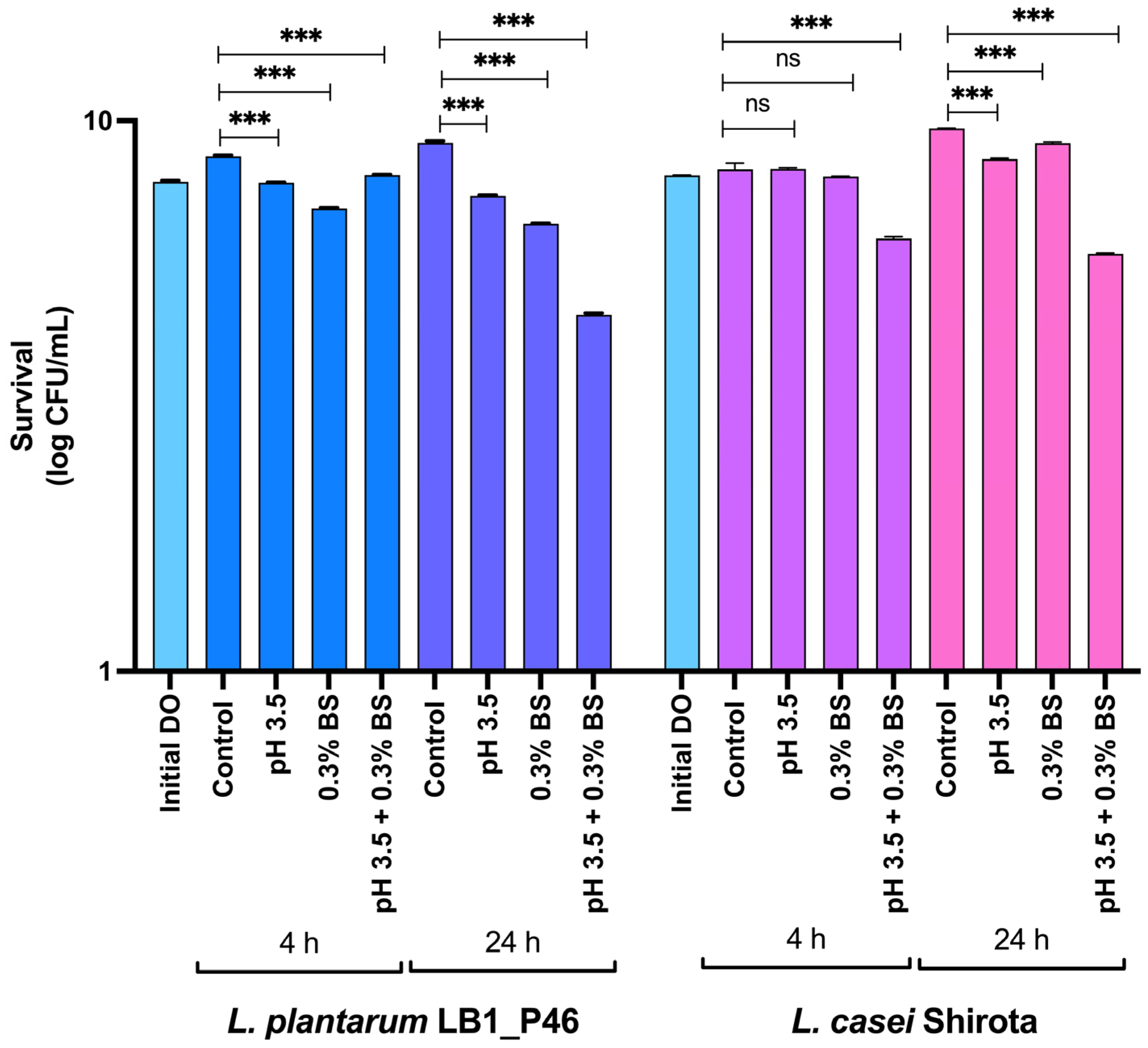
2.5.1. Bile Salt and Acid Resistance

Bile salt and acid resistance assays were assayed as reported previously by [19] with slight modifications. An overnight preculture of the strain LB1_P46 in 5 mL MRS broth was incubated for acid resistance at 37 °C. Resultant growth was adjusted to a final DO600 nm = 0.2 in 0.5 mL to inoculate a new culture of 4.5 mL MRS acidified to pH 3.5 with 1N HCl (Baker) and incubated at 37 °C. Growth was monitored at 4 and 24 h by plating an aliquot of the culture in agar MRS plates incubated as above. For the bile salt resistance assay, an inoculum of 0.5 mL of the above culture of the acid resistance assay was used to inoculate 4.5 mL of MRS broth supplemented with 0.3% bile salts (oxgall, Oxoid, Basingstoke, UK), incubated at 37 °C for 4 and 24 h, and growth was monitored as above. Additionally, an assay that included acidified MRS (pH 3.5) supplemented with 0.3% bile salts was performed and monitored as above. All experiments were performed in biological duplicates, and the commercial probiotic Lactobacillus casei Shirota isolated from a commercial probiotic beverage was used as a positive control.

2.5.2. Antibacterial Assays against Pathogenic Bacteria

In vitro antibacterial activity of L. plantarum LB1_P46 isolate was assayed against pathogenic bacteria as reported by [19]: An aliquot of 0.1 mL of a cell suspension containing 9 log CFU/mL of an overnight culture of strains LB1_P46 and LB1_P47 grown in APT was dropped onto two fresh APT plates in quadruplicate for each plate for each assayed pathogenic bacterium and incubated overnight at 37 °C. The resultant spotted growth was overlaid with 5 mL of BHI or LB soft agar containing 0.5 mL of the overnight culture of assayed pathogenic bacteria adjusted to an optical density of OD600 nm = 0.2. The plates were incubated overnight at 37 °C and evaluated for antibacterial activity by measuring each zone of inhibition with a millimeter ruler around the spotted growth of L. plantarum.

2.5.3. In Vivo Preventive Anti-Infective Activity of L. plantarum LB1_P46 Isolate against Salmonella Typhimurium

The in vivo preventive anti-infective activity of L. plantarum LB1_P46 against the streptomycin-resistant (strr) Salmonella Typhimurium strain L1334 was assayed in male BALB/c mice with 8–9 weeks old in the vivarium of the Departamento de Medicina Experimental de la Facultad de Medicina, UNAM—Hospital General de México. The experiment was performed as described previously with several modifications [19]. Three groups were assayed: the PBS group without administration of LAB, the LB1_P46 group, and the L. casei Shirota group (probiotic bacteria group). All the groups comprised ten mice each. Strain LB1_P46 and L. casei Shirota were grown as described above, centrifuged (5000× g, for 5 min at 4 °C), and resuspended in 1 mL of sterile PBS buffer (0.8% NaCl, 0.121% K2HPO4, 0.034% KH2PO4, pH 7.4), and the cell density was adjusted to 9.3 log CFU/mL in 300 μL per dose. The experiment was carried out as follows in five stages: (1) adaptation of mice to the laboratory environment for four days, (2) daily administration of L. plantarum LB1_P46 or L. casei Shirota with an esophagogastric probe for seven days, (3) one resting day without bacterial administration, (4) infection at the next day with Salmonella Typhimurium adjusted to 7 log CFU/mL with an esophagogastric probe, and (5) after two days, mice were sacrificed, and the target blank organs (liver and spleen) were extracted and analyzed. Throughout the experiment, mice were provided with food and water ad libitum. The mice infection group was treated as described for experimental and probiotic groups, and PBS buffer was administrated before the infection with Salmonella. Organ extraction and processing were performed as previously [19], and cell suspensions were serially diluted in sterile saline and plated on agar LB supplemented with 100 mg/mL streptomycin (Sigma-Aldrich). The total log CFU/mL was determined by duplicate after incubation at 37 °C for 24 h.

2.6. Statistical Analysis

Statistical analyses were performed in the XLSTAT program 2024.2.2 (1422). A paired t-test was used to determine significant differences between the observed results of the functional traits and antibacterial activity assays between the L. plantarum LB1_P46 and L. casei Shirota as probiotic control. An analysis of variance (ANOVA) with Dunnett’s post hoc test was performed to determine if the observed differences in the log CFU/mL in L. plantarum LB1_P46 and L. casei Shirota after 4 and 24 h of exposure to pH 3.5, 0.3% bile salts, and the combined condition pH 3.5 + 0.3% bile salt were significant by comparing the means of the assayed conditions for each strain with their respective controls. For the in vivo preventive anti-infective activity of L. plantarum LB1_P46 against Salmonella Typhimurium, to determine if the observed differences between the growth of Salmonella in the PBS condition and the administration of the LABs assayed were significant, an ANOVA with Tukey’s post hoc test was performed for the detected log CFU/g in the liver and spleen.

3. Results

3.1. Safety Traits of L. plantarum

The safety traits assayed for L. plantarum LB1_P46 were hemolytic activity and antibiotic resistance/susceptibility profiling. L. plantarum LB1_P46 resulted in negative hemolytic activity, whereas the profiling for antibiotic resistance/susceptibility is shown in Table 1. Results indicating sensitivity and intermediate sensitivity to PEP (<0.24 mg/L), PES, AMOP, ERY, QDA, CLI, LVX, CMP, and TSU antibiotics, but resistance to >0.125 mg/L of PEP, CTXP, TET, and VAN. The resulting MAR index for the strain LB1_P46 = 0.333 was calculated from the observed resistance to 4 of the 12 assayed antibiotics.
We searched the genome of strain LB1_P46 for possible corresponding genes encoding resistance to the assayed antibiotics, which resulted in the genes shown in Table 2.

3.2. Functional Traits and Cell Surface Properties

The L. plantarum strain LB1_P46 was assayed for cholesterol reduction and β-galactosidase activity as functional traits, and for the cell surface properties of hydrophobicity, auto-aggregation, and aggregation. The results of these assays are shown in Table 3. The strain LB1_P46 showed a significant (p < 0.05) increment in the reduction of cholesterol of 56.03%, an increment of 21.17% compared to the control probiotic bacteria. Microbial cholesterol degradation was proposed mainly through the enzymatic deconjugation of bile acids by a bile-salt hydrolase or cholesterol conversion into coprostanol by a cholesterol reductase activity [34]. The search for annotated proteins encoded in the chromosome related to cholesterol degradation led to the identification in the genome annotation by Prokka of two enzymes possibly involved in bile acid degradation: the bile acid 7-alpha dehydratase (complement 2,710,282..2,710,726) forms secondary bile acids via the 7-alpha-dehydroxylation of primary bile acids, and the 3-alpha-hydroxycholanate dehydrogenase (NADP(+)) (complement 1,739,730..1,740,449) involved in the modification of secondary bile acids to form 3-beta-bile acids (also known as iso-bile acids) via a 3-oxo intermediate [35].
The strain LB1_P46 showed significantly (p < 0.05) lower values for all cell surface characteristics assayed compared to the probiotic control strain (Table 3). For hydrophobicity in chloroform and hexane, the strain LB1_P46 showed 58.38% and 83.75%, respectively, compared to the observed values in the control strain in both solvents. Regarding auto-aggregation, the strain LB1_P46 showed a significant (p < 0.05) reduction (~33%) of the observed for the control strain. Finally, the percentage of coaggregation reduced significantly (p < 0.05) with pathogenic bacteria at 24 h compared to the values observed for the control strain: ~28% for EPEC, ~22% for L. monocitogenes, and ~23% for Salmonella Typhi.
Regarding the β-galactosidase activity, the strain LB1_P46 was positive for this trait (Table 1), resulting in blue colonies growing on lactose medium plates supplemented with X-gal, indicating the capability to transport and ferment lactose despite the non-dairy origin of this LAB. Analysis of the genome region encoding this hydrolytic enzyme revealed the presence of a β-galactosidase enzyme (locus_tag RUO99_14335 protein_id WNW15683.1) and a β-galactosidase small subunit (locus_tag RUO99_14405, protein_id WNW15697.1) located in a chromosomal region of 20,099 bp (3,021,825–3,041,923 bp), including several genes encoding transporters, carbohydrate metabolism enzymes, two LacI family DNA-binding transcriptional regulators (locus_tag RU099_14340 and RU099_14380), and three genes encoding the respective PTS lactose/cellobiose transporter subunits II (locus_tag RUO99_05865 protein_id WNW16884.1, locus_tag RUO99_11285 protein_i WNW15120.1, and locus_tag RUO99_03815l protein_id WNW16520.1, respectively).
Analysis of the genome annotation by Prokka led to the identification of two encoded lactose permease (LacS). Nevertheless, GenBank annotation indicated these two proteins as glycoside-pentoside-hexuronide (GPH): cation symporters (locus_tag RUO99_14330, protein_id WNW15682.1 and locus_tag RUO99_14415, protein_id WNW15699.1, complement 3,044,336..3,046,294, respectively). Protein-BLAST analysis identified a lactose permease with an identity of 99.69% and 99.54% of L. plantarum, respectively. Although it is reported that L. plantarum transports lactose by LacS, further evidence is required to determine the main role of LacS or the PTS lactose/cellobiose transporter in transporting this sugar.

3.3. In Vitro Assays

Bile Salt and Acid Resistance

Results of the exposure at 4 and 24 h of strain LB1_P46 to acid pH (3.5), 0.3% bile salts, and a combined stress condition of acidified MRS (pH 3.5) supplemented with 0.3% of bile salts at 37 °C are shown in Figure 1. For the strain LB1_P46, all the assayed conditions significantly reduced the log CFU/mL of the respective control condition at 4 and 24 h of exposure. The combined pH 3.5 + 0.3% bile salts at 24 h proved to be the most stressful condition assayed, resulting in a final log CFU/mL = 4.46 compared to the log CFU/mL in the control at 24 h = 9.07. The probiotic control showed no significant differences in survival at individual pH 3.3 and 0.3% bile salt exposure. Nevertheless, at 24 h of exposure, the probiotic strain looks more resistant to the combined pH 3.5 + 0.3% bile salts at 4 and 24 h.
Analysis in the sequenced genome of the strain LB1_P46led for the bsh and clpL genes encoding a bile salt hydrolase and a Clp ATPase involved in acid and bile tolerance, respectively, reported previously in L. plantarum isolated from Indian fermented foods [11], led to the identification of these genes: bsh (locus_tag RUO99_14595, protein_id WNW15732.1) encoding a choloylglycine hydrolase and clpL (locus_tag RUO99_14745, protein_id WNW15759.1) encoding an ATP-dependent protease ClpL.
Regarding acid resistance of LAB, DltA (L. rhamnosus) and DltB (f L. reuteri), with the putative function of d-anylation of LTA, were proposed and detected in two L. plantarum from Indian fermented foods as involved proteins with acid and defensin resistance [11]. For its part, the bacterial community analysis of the Chinese baijiu fermentation showed that Lactobacillus was the most dominant bacterium during fermentation, and acid resistance genes identified were argR, aspA, ilvE, gshA, DnaK, and cfa, associated with the survival of Lactobacillus in the late stages of fermentation with high contents of acid and ethanol [36]. All these genes and an acid shock protein were detected in the chromosomal sequence of the strain LB1_046 by the analysis of the Prokka and GenBank annotations (Table 4).

3.4. Antibacterial Assays

3.4.1. In Vitro Antibacterial Assays

The antimicrobial activity assays indicated that L. plantarum LB1_P46 can significantly inhibit in vitro (p < 0.05) the growth of Salmonella Typhimurium, E. coli, P. aeruginosa, L. monocytogenes, S. pyogenes, S. aureus, E. faecalis, and B. cereus (Table 5, Figure S1); the strain of L. casei Shirota was used as the control probiotic strain for this assay (Figure S2).
The results showed bacteriostatic activity in the assays associated with cell-to-cell contact against all pathogenic bacteria assayed. This suggests that the observed antimicrobial activity is likely related to a cell-associated diffusing enzyme or peptide with antimicrobial activity. Cell-to-cell contact assays showed a higher inhibition of L. plantarum LB1_P46 against Salmonella Typhimurium and L. monocitogenes (15.5 mm and 27.2 mm, respectively). In contrast, a lower inhibition was observed against P. aeruginosa (6.3 mm) (Table 5, Figure S1). L. casei Shirota used as control probiotic bacteria showed antimicrobial activity against all pathogenic bacteria assayed, still, to a lesser extent than the observed inhibition zones of the strain LB1_P46, ranging to 16.6 mm against B. cereus to 4.9 mm to P. aeruginosa (Table 5, Figure S2).
To identify the possible enzyme or peptide responsible for this antimicrobial activity of the strain LB1_P46, we performed an in silico mining for biosynthetic clusters encoding any bacteriocin or a possible RiPP in the antiSMASH 7.0 bacterial version database [37] and the BAGEL4 server [38]. Results indicated the presence of two chromosomal regions located from 392,458 to 414,826 bp in the genome of L. plantarum LB1_P46 encoding for a predicted bacteriocin biosynthetic cluster (392,458–403,458), including an enterocin_X_chain_beta (RUO99_01860), a putative bacteriocin immunity protein (RUO99_01875), two plantaricin J genes (RUO99_01880 and RUO99_01885), a plantaricin A (RUO99_01895), and a bacteriocin-production-related histidine kinase (RUO99_01900). The second region comprises a RiPP-like encoding region (420,459–414,826 bp), including two response regulators, PlnC and PlnD activators (RUO99_01905, and RUO99_01910, respectively), an immunity protein PlnI membrane-bound protease CAAX (RUO99_01920), the subunits E and F of the plantaricin EF (RUO99_01925 and RUO99_01930, respectively), a bacteriocin ABC-transporter, ATP binding and permease protein PlnG (RUO99_01935), and the accessory factor for ABC-transporter PlnH (RUO99_01940) (Figure 2). The mined genomic structure corresponding to the RiPP-like region showed the typical structure of an operon encoding class II bacteriocins [39,40].
Other enzymes with possible antimicrobial activity encoded in the chromosome of L. plantarum LB1_P46 are five enzymes involved in the peptidoglycan catabolic process, four with lysozyme activity (GH25 family lysozyme, locus_tag, protein ID): RUO99_04720 WNW16683.1, RUO99_05000 WNW16738.1, RUO99_07360 WNW17169.1, RUO99_04720 WNW16683.1, RUO99_11435 WNW17283.1; and one enzyme with N-acetylmuramoyl-L-alanine amidase (RUO99_08270 WNW14576.1).

3.4.2. In Vivo Anti-Infective Activity of L. plantarum LB1_P46 Isolate against Salmonella Typhimurium

The anti-infective effect of the oral administration of L. plantarum LB1_P46 in BALB/c male mice of age 8–9 weeks against Salmonella Typhimurium strr was determined by quantification of the log CFU/mL of Salmonella in dissected liver and spleen of infected mice compared. All the experimental groups were monitored for weight to discard a possible gain associated with the LAB administration, resulting in no weight gain. The mice of the LB1_P46 group were bright-eyed and alert, had a smooth coat with a sheen, responded to stimuli, and showed interest in their environment. In contrast, the PBS mice group showed visible signs of infection after administering Salmonella, being hunched over and exhibiting lethargy, little interest in the environment, and fur clumping.
The administration of the strain LB1_P46 to the BALB/c mice before the infection with the strain Salmonella Typhimurium L1334 resulted in a significant reduction in the log CFU/g tissue (liver and spleen) of Salmonella than in the non-LAB supplemented group (Figure 3). The observed log CFU in the PBS group was 6.08 for the liver. The observed values for the LB1_P46 group were 3.92 and 5.02 for the L casei Shirota group. The PBS group showed a log CFU of 4.86 for the spleen, reducing to 3.02 and 3.81 for the LB1_46 and the L. casei Shirota groups, respectively. These results showed that administering L. plantarum LB1_P46 to BALB/c male mice confers an effective preventive anti-infective effect against the strain of Salmonella assayed compared to that observed with the commercial probiotic strain.
Several genes have been associated with the in vivo antipathogenic effect of L. plantarum, including luxS associated with the putative production of AI-2 and AI-3 conferring an autoinduction ability, dltB and dltD with the putative function of d-anylation of LTA, conferring an anti-inflammatory potential in a murine model of colitis (dltB) and resistance to human β defensin-2 [11], and seven unspecified genes related to infection for Salmonella, tuberculosis, and legionellosis [30]. The analysis of the genome sequence of the strain LB1_P46 led to identifying luxS (annotated as S-ribosylhomocysteine lyase in the GenBank annotation but LuxS by Prokka) locus_tag RUO99_03160, protein_id WNW16399.1, dltD (D-alanyl-lipoteichoic acid biosynthesis protein DltD), locus_tag RUO99_08340, protein_id WNW14589.1, and dltB (product D-alanyl-lipoteichoic acid biosynthesis protein DltB), locus_tag RUO99_08350, protein_id WNW14591.1.

4. Discussion

L. plantarum LB1_P46 showed a MAR index = 0.33, resulting from resistance to the PEP, CTXP, and VAN antibiotics. Several strains of L. plantarum isolated from traditional fermented sources have been demonstrated as resistant LAB for several antibiotics, mainly synthetic quinolones such as nalidixic acid and ciprofloxacin [22,29], targeted mainly against pathogenic Gram-negative bacteria [41], and resistance to the glycopeptide vancomycin [42]. Resistance to vancomycin was proposed to be caused by the presence of peptidoglycan precursors terminating in the depsipeptide D-Ala-D-Lac observed in vanA and vanB resistance classes to vancomycin observed in Enterococci, resulting in the weak affinity of the antibiotic to the LAB’s cell walls [43]. Other L. plantarum isolated from fermented sources exhibited MAR indexes higher than 0.2, indicating multiple antibiotic-resistant bacteria [29]. Nevertheless, four L. plantarum isolated from pulque beverages from the States of Puebla and Oaxaca in Mexico showed MAR < 0.2 indexes (calculated from published values in Ruiz-Ramírez et al.) [22].
The analysis of the sequence of the complete chromosome of the strain LB1_P46 led to identifying the encoded resistance proteins for PEP and possibly for CTXP (a serine hydrolase member of the GO:0030655—beta-lactam antibiotic catabolic process) [44,45]; TET (TetM/TetW/TetO/TetS family tetracycline resistance ribosomal protection protein, tetracycline resistance MFS efflux pump, and ABC multidrug transporters) [46,47,48]; and VAN (VanZ family protein), a member of the VanA glycopeptide resistance gene cluster, reported to confer resistance to lipoglycopeptide antibiotics independent of activity of the encoded proteins activity of the vanHAX genes [49]. The ABC transporter permease/daunorubicin/doxorubicin resistance ABC transporter permease proteins DrrA and DrrB are reported as involved in the resistance to these antibiotics.
Other potential MFS, MDR, and ABC multidrug efflux transporters were also detected, and, remarkably, 21 genes encoded the TetR/AcrR family transcriptional regulator and two genes encoded the TetR-like C-terminal domain-containing protein (Table 2). TetR was reported as a sensor of the cellular environment and its dynamics. Genes under the control of TetR are involved in functions like antibiotic production, osmotic stress, and multidrug resistance, including multidrug efflux pumps, pathogenesis, and metabolic modulation processes. Remarkably, the first TetR sensor was reported to regulate the tetracycline efflux pump, resulting in resistance to this antibiotic [50]. Our antibiotic resistance assay did not include the quinolone antibiotics ciprofloxacin and ofloxacin; nevertheless, Lactobacillus resistance to these antibiotics is a common trait in this LAB. Antibiotic profiling in other L. plantarum isolated from Mexican pulque [22] and boza from Turkey [29] report resistance to quinolone antibiotics. Proposed resistance mechanisms to quinolones include chromosomal mutations in the target enzymes, DNA gyrase (gyrAB) and DNA topoisomerase IV subunit V (parCE), down-regulation of outer membrane porin coupled with increases in active drug efflux, and the possible acquisition of transmissible quinolone-resistance genes [22,51].
Potential probiotic strains isolated from traditional fermented sources resistant to several antibiotics help control intestinal infections due to probiotics’ intrinsic preventive and therapeutic properties. Still, their associated antibiotic-resistant traits could be considered helpful in promoting faster recovery of patients by promoting the rapid establishment of desirable microbiota if resistant probiotic LAB are administered simultaneously with antibiotics [29]. Nevertheless, as Lactobacillus (and Lactiplantibacillus) species are generally considered nonpathogenic bacteria, many strains of these LABs are used to produce diverse foods and products for humans and animals. Still, multidrug resistance observed for several LABs exceeds the resistance levels recommended by the EFSA [29,42].
A relevant characteristic of the strain LB1_P46 is its capability to grow at 37 °C under stress-challenged conditions, compared to the optimal growth environmental temperature of this bacteria in its natural environment in the sap in maguey species and during the pulque fermentation process. Acid tolerance (pH 3.5), bile tolerance, and combined acid and bile tolerance were assayed for strain LB1_P46 at 37 °C. Acid resistance in lactobacilli involved in traditional Chinese fermented foods was associated with the activity of proteins ArgR, Cfa, AspA, IlvE, DNaK, and GshAB [36]. All these genes were encoded in the genome of the strain LB1_P46 (Table 4). The proposed role of acid-resistance genes involves the transcriptional regulator ArgR of the arginine deaminase pathway (ADI), which was proposed to be under antirepression control by the small RNA S042 of argR in Lactococcus lactis. The expression of the ADI pathway results in the intracellular production of ammonia, which neutralizes intracellular protons and increases intracellular pH [36,52]. Cyclopropane fatty acyl phospholipid synthase (CFA), encoded by cfa, is an enzyme that catalyzes a modification of the acyl chains of phospholipid bilayers through methylenation of unsaturated fatty acyl chains to their cyclopropane derivatives. Mutants of E. coli in cfa and cfa-rpoS showed a reduced resistance mutant, showing enhanced acid sensitivity. In E. coli, the cytoplasmic membrane CFA protects the cells against several environmental stressors, including acid pH [36,53,54]. The amino acid-dependent system involving the deamination of aspartate by aspartate ammonia-lyase (AspA) results in the reversible formation of fumaric acid and ammonium ion, which was proposed to increase intracellular pH. No pH changes in a ΔaspA mutant was observed [36,55,56].
The branched-chain amino acid aminotransferase (IlvE) is involved in the biosynthesis and degradation of branched-chain amino acids, such as in the production of branched-chain fatty acids in Streptococcus mutants. ΔilvE mutants resulted in a decreased acid tolerance, and the transcriptional analysis of the ilvE regulator was upregulated during growth under acid pH [36,57]. The molecular chaperone DnaK (dnaK) was reported as involved in the extended response to acid pH in Streptococcus infantarius isolated from traditional Mexican fermented pozol. Upregulation of dnaK was observed during exposure to pH 3.6 and 4.0 [58]. Its upregulation was demonstrated to be associated with the thermo-acidophilic stress of Alicyclobacillus acidoterrestris, a Gram-positive bacteria isolated from orchard soil, spoiled fruit juices, and soils closer to volcanoes and hot springs, growing in acid pHs from 2.0 to 6.0 [59].
Regarding GshAB, LAB developed a glutathione (GSH)-mediated acid-tolerance response. However, several reports indicate the absence of GshA or GshAB in the synthesis of GSH in several LAB like L. salivarius or L. plantarum, resolving availability by transport of GSH from the extracellular medium [60]. The GSH-mediated acid response involves using glutathione to avoid rapid acidification of the intracellular pH [61]. The presence of two GshAB in the strain LB1_P46 requires deep analysis to demonstrate its possible role in the GSH biosynthesis and the possible participation of GSH in a mechanism of acid resistance as proposed for other LAB.
Regarding bile tolerance, the product of the bsh gene (locus_tag RUO99_14595) is a choloylglycine hydrolase (a bile salt hydrolase) according to GenBank annotation of the genome of the strain LB1_P46. This enzyme cleaves the peptide bond of the bile acids, removing the amino acid residing in the steroid core, and the unconjugated resulting bile acids are then precipitated at acid pH [62]. The ClpL protein was reported as involved in acid and bile tolerance in L. plantarum isolates from Indian fermented foods [11] and was detected in the genome of the LB1_P46 strain. Nevertheless, the ClpL protein was reported in several LAB, including the probiotic strain of L. plantarum IS-10506 as expressed in response to heat shock, but is also considered a desirable trait for surviving probiotic bacteria during the manufacturing and administration process [30]. Another enzyme possibly involved in bile tolerance in the strain LB1_P46 is the bile acid 7-alpha dehydratase (locus_tag RUO99_12795), an enzyme reported to catalyze the fourth step in the bile acid 7-alpha-dehydroxylation pathway, resulting in secondary bile acids via the 7-alpha-dehydroxylation of primary bile acids, a pathway carried out by intestinal anaerobic bacteria [35].
Cell surface characteristics such as hydrophobicity, auto-aggregation, and coaggregation are crucial for probiotic bacteria. Obtained values for cell surface characteristics assayed for the strain LB1_P46 are in the range of previous LAB isolated from pulque beverage analyzed for similar traits [22]. Hydrophobicity correlates with the adhesion ability of probiotic bacteria to the host’s epithelial cells and is considered an essential characteristic of probiotics’ cell surface properties. Coaggregation is considered an effective host defensive mechanism against pathogenic microorganisms in the host’s gastrointestinal system, whereas auto-aggregation is a desirable precondition for biofilm development [63,64]. The operon dltABCD was detected in the chromosome of the strain LB1_P46 in the region of 1,764,114 to 1,762,640. It was reported in several LAB encoding the pathway of esterification of lipoteichoic acid (TLA) by D-alanine; resulting esters are reported to play an essential role in controlling the net anionic charge for the poly (GroP) moiety of LTA [65]. This operon has been proposed to play a pleiotropic role in the properties of the cell surface. Inactivation of the genes in the operon has resulted in the loss of integrative coaggregation (dltA mutant), loss of acid tolerance (dltC mutant), and increased cellular length and increased cellular susceptibility of CTAB and chlorhexidine (dltD mutant) associated with a decreased in D-alanylation of LTA [65,66].
A remarkably functional trait of the strain LB1_P46 is its capability to transport lactose, probably by the identified lactose permease (LacS), and to hydrolyze the disaccharide as a carbon source by the action of the β-galactosidase enzyme. We previously observed this bacteria’s ability to grow in skim milk to a final 9.14 log CFU/mL at 37 °C for 24 h [67], suggesting the potential use of this strain in the elaboration of dairy probiotic preparations.
The capability to produce bacteriocins confers a competitive advantage to probiotic bacteria while colonizing the host’s intestinal tract. L. plantarum produces diverse bacteriocins (plantaricins) that have acquired great importance as bio preservatives in the preparation of diverse dairy, meat, and fish products, and for their potential use in treating infective diseases, including a protective role in urinary tract infections [28]. We identified three predicted bacteriocins in the chromosomal sequence of the strain LB1_P46: two plantaricin J, one plantaricin A, and the two subunits of the plantaricin EF, including immunity proteins and transporters. Four lysozyme enzymes were also identified, including one member of the GH25 lysozyme family and one enzyme with N-acetylmuramoyl-L-alanine amidase. Interestingly, the lysozyme family GH25 exhibits both β-1,4-N-acetyl- and β-1,4-N,6-O-diacetylmuramidase activities and is structurally unrelated to the GH22-24, GH73, and GH108 lysozyme families [38]. All identified plantaricins and cell wall hydrolytic enzymes could confer a competitive advantage during the development of this bacteria in the fermentation process for pulque production.
Lactobacillus isolates have antagonistic activity against pathogenic bacteria, including microorganisms like Staphylococcus aureus, Enterococcus faecalis, Klebsiella pneumonia, Pseudomonas aeruginosa, E. coli, Salmonella Typhi, and Shigella sp. [68], whereas the in vivo preventive and anti-infective of probiotic Lactobacillus have been assayed against Salmonella. Typhimurium [69]. Our results showed the in vitro antagonistic effect of L. plantarum LB1_P46 against Salmonella Typhimurium, E. coli, P. aeruginosa, L. monocytogenes, S. pyogenes, S. aureus, E. faecalis, and B. cereus; and the anti-infective treatment of the oral delivery of the strain of L. plantarum LB1_P46 against the infection of Salmonella Typhimurium in a BALB/c male mouse model, resulting in a relevant probiotic trait of this LAB isolated from pulque beverage. The oral administration of probiotic bacteria has been used to reduce the infection of pathogenic bacteria involving immune-enhancing properties by releasing anti-inflammatory cytokines within the gut by impacting various types of immune cells, including dendritic cells, monocytes, natural killer cells, macrophages, lymphocytes, and epithelial cells simultaneously (reviewed in [70]). Several genes have been identified and proposed as activating the immune response during the in vivo anti-infective treatment of probiotic Lactobacilllus against pathogenic bacteria, including luxS and dltBD [11,65,66]. DltB and DltD proteins involved in the D-anylation of LTA and encoded in the dltABCD operon (encoded in the chromosome of the strain LB1_P46) have been associated with anti-inflammatory activity. Mutants in dltD exhibited reduced anti-inflammatory potential. Mutants of L. plantarum in dltB showed significantly reduced immunomodulatory activity compared to its parental strain both in in vitro and in vivo models. In contrast, mutants in dltD in L. rhamnosus GG, cytokine stimulation of human intestinal epithelial cells, and peripheral blood mononuclear cells were not significantly altered [66]. Additionally, the LTA glycolipid anchor and the length of the Gro-P backbone have been reported to have immunostimulatory potential in other Gram-positive bacteria [66], resulting in interest in the role of the operon dltABCD and the structure of LTA in the L. plantarum strain LB1_P46.

5. Conclusions

The present study investigates the probiotic characteristics of L. plantarum LB1_P46 isolated from traditional Mexican fermented pulque beverage, including several functional traits, such as the determination of antagonistic in vitro activity against several pathogenic bacteria, and in vivo anti-infective effect against the infection of Salmonella Typhimurium in a murine model. Along with the functional and antimicrobial properties assayed, we identified several probiotic genes, operons, and biosynthetic clusters in the genome of this bacteria that are probably responsible for the observed probiotic characteristics, indicating that L. plantarum LB1_P46 is a potential probiotic candidate that can be used in non-dairy and dairy functional products. Additionally, our results provide scientific evidence supporting several beneficial effects of consuming traditional pulque beverage.

Supplementary Materials

The following supporting information can be downloaded at: https://www.mdpi.com/article/10.3390/microorganisms12081652/s1, Figure S1: Antimicrobial activity of Lactiplantibacillus plantarum LB1_P46 against pathogenic bacteria; Figure S2: Antimicrobial activity of Lactobacillus casei Shirota as probiotic control against pathogenic bacteria.

Author Contributions

Conceptualization, M.G.-G., R.P.-P., C.L.-M. and A.E.; methodology, X.M.H., F.A.-M. and M.S.F.M.; formal analysis, X.M.H., F.A.-M., M.G.-G. and A.E.; writing—original draft preparation, M.G.-G. and A.E.; writing—review and editing, A.E.; funding acquisition, A.E. All authors have read and agreed to the published version of the manuscript.

Funding

This research was funded by project PAPIIT IN227023 (DGAPA, UNAM).

Data Availability Statement

The raw data supporting the conclusions of this article will be made available by the authors upon request.

Acknowledgments

The authors acknowledge María Antonieta Silva Chávez, Alejandro Camacho Cruz, Georgina Teresa Hernández Chávez, Luz María Martínez Mejía, and Noemí Flores Mejía for their technical support.

Conflicts of Interest

The authors declare no conflicts of interest.

References

  1. Moreno-Terrazas, R.; Escalante, A.; Verdugo-Valdez, A.G.; De la Rosa, M.; Ortiz Basurto, R.I.; Orantes-García, C.; Palafox-González, L.; Castro Díaz, A.S.; Lappe-Oliveras, P. Bebidas refrescantes y alcohólicas de agave. In Panorama del Aprovechamiento de Agaves en México; CONACYT, CIATEJ, AGARED: Guadalajara, Mexico, 2017; pp. 123–163. ISBN 978-607-97548-2-2. [Google Scholar]
  2. Ramírez Rancaño, M. El Rey Del Pulque: Ignacio Torres Adalid y La Industria Pulquera; Plaza y Valdes, UNAM, Instituto de Investigaciones Sociales: Ciudad de México, Mexico, 2000; ISBN 968-856-812-0. [Google Scholar]
  3. Valdivieso Solís, D.G.; Vargas Escamilla, C.A.; Mondragón Contreras, N.; Galván Valle, G.A.; Gilés-Gómez, M.; Bolívar, F.; Escalante, A. Sustainable production of pulque and maguey in Mexico: Current situation and perspectives. Front. Sustain. Food Syst. 2021, 5, 678168. [Google Scholar] [CrossRef]
  4. Sánchez-Marroquín, A.; Hope, P.H. Agave juice, fermentation and chemical composition studies of some species. J. Agric. Food Chem. 1953, 1, 246–249. [Google Scholar] [CrossRef]
  5. Escalante, A.; Giles Gomez, M.; Hernandez, G.; Cordova Aguilar, M.; Lopez Munguia, A.; Gosset, G.; Bolivar, F. Analysis of bacterial community during the fermentation of pulque, a traditional Mexican alcoholic beverage, using a polyphasic approach. Int. J. Food Microbiol. 2008, 124, 126–134. [Google Scholar] [CrossRef]
  6. Escalante, A.; Elena Rodríguez, M.; Martínez, A.; López-Munguía, A.; Bolívar, F.; Gosset, G. Characterization of bacterial diversity in Pulque, a traditional Mexican alcoholic fermented beverage, as determined by 16S rDNA analysis. FEMS Microbiol. Lett. 2004, 235, 273–279. [Google Scholar] [CrossRef] [PubMed]
  7. Astudillo-Melgar, F.; Hernández-Chávez, G.; Rodríguez-Alegría, M.E.; Bolívar, F.; Escalante, A. Analysis of the microbial diversity and population dynamics during the pulque fermentation process. Fermentation 2023, 9, 342. [Google Scholar] [CrossRef]
  8. Chacón-Vargas, K.; Torres, J.; Giles-Gómez, M.; Escalante, A.; Gibbons, J.G. Genomic profiling of bacterial and fungal communities and their predictive functionality during pulque fermentation by whole-genome shotgun sequencing. Sci. Rep. 2020, 10, 15115. [Google Scholar] [CrossRef] [PubMed]
  9. Huezo-Sánchez, A.R.; Ortega-Rodríguez, E.M.; Pérez-Armendáriz, B.; El-Kassis, E.G. Characterization of Bacterial Diversity in Aguamiel and Two Types of Pulque from the Zacatlán Region, México. Fermentation 2023, 9, 564. [Google Scholar] [CrossRef]
  10. Rocha-Arriaga, C.; Espinal-Centeno, A.; Martinez-Sánchez, S.; Caballero-Pérez, J.; Alcaraz, L.D.; Cruz-Ramírez, A. Deep microbial community profiling along the fermentation process of pulque, a biocultural resource of Mexico. Microbiol. Res. 2020, 241, 126593. [Google Scholar] [CrossRef]
  11. Goel, A.; Halami, P.M.; Tamang, J.P. Genome Analysis of Lactobacillus plantarum isolated from some Indian fermented foods for bacteriocin production and probiotic marker genes. Front. Microbiol. 2020, 11, 40. [Google Scholar] [CrossRef]
  12. Tamang, J.P.; Cotter, P.D.; Endo, A.; Han, N.S.; Kort, R.; Liu, S.Q.; Mayo, B.; Westerik, N.; Hutkins, R. Fermented foods in a global age: East meets West. Compr. Rev. Food Sci. Food Saf. 2020, 19, 184–217. [Google Scholar] [CrossRef]
  13. Zielińska, D.; Kolożyn-Krajewska, D. Food-origin lactic acid bacteria may exhibit probiotic properties: Review. BioMed Res. Int. 2018, 2018, 5063185. [Google Scholar] [CrossRef]
  14. Sionek, B.; Szydłowska, A.; Zielińska, D.; Neffe-Skocińska, K.; Kołożyn-Krajewska, D. Beneficial bacteria isolated from food in relation to the next generation of probiotics. Microorganisms 2023, 11, 1714. [Google Scholar] [CrossRef] [PubMed]
  15. Koutsoumanis, K.; Allende, A.; Alvarez-Ordóñez, A.; Bolton, D.; Bover-Cid, S.; Chemaly, M.; DeCesare, A.; Hilbert, F.; Lindqvist, R.; Nauta, M.; et al. Update of the list of qualified presumption of safety (QPS) recommended microbiological agents intentionally added to food or feed as notified to EFSA 19: Suitability of taxonomic units notified to EFSA until September 2023. EFSA J. 2024, 22, e8517. [Google Scholar] [CrossRef]
  16. FDA. Recently Published GRAS Notices and FDA Letters; FDA: Silver Spring, MD, USA, 2024.
  17. Escalante, A.; López Soto, D.R.; Velázquez Gutiérrez, J.E.; Giles-Gómez, M.; Bolívar, F.; López-Munguía, A. Pulque, a traditional Mexican alcoholic fermented beverage: Historical, microbiological, and technical aspects. Front. Microbiol. 2016, 7, 1026. [Google Scholar] [CrossRef] [PubMed]
  18. Escalante, A.; Giles-Gómez, M.; Esquivel Flores, G.; Matus Acuña, V.; Moreno-Terrazas, R.; López-Munguía, A.; Lappe-Oliveras, P. Pulque Fermentation. In Handbook of Plant-Based Fermented Food and Beverage Technology; Hui, Y.H., Ed.; CRC Press: Boca Raton, FL, USA, 2012; pp. 691–706. ISBN 978-1-4398-7069-3. [Google Scholar]
  19. Giles-Gómez, M.; Sandoval García, J.G.; Matus, V.; Campos Quintana, I.; Bolívar, F.; Escalante, A. In vitro and in vivo probiotic assessment of Leuconostoc mesenteroides P45 isolated from pulque, a Mexican traditional alcoholic beverage. SpringerPlus 2016, 5, 708. [Google Scholar] [CrossRef] [PubMed]
  20. Torres-Maravilla, E.; Lenoir, M.; Mayorga-Reyes, L.; Allain, T.; Sokol, H.; Langella, P.; Sánchez-Pardo, M.E.; Bermúdez-Humarán, L.G. Identification of novel anti-inflammatory probiotic strains isolated from pulque. Appl. Microbiol. Biotechnol. 2016, 100, 385–396. [Google Scholar] [CrossRef] [PubMed]
  21. Cervantes-Elizarrarás, A.; Cruz-Cansino, N.; Ramírez-Moreno, E.; Vega-Sánchez, V.; Velázquez-Guadarrama, N.; Zafra-Rojas, Q.; Piloni-Martini, J. In vitro probiotic potential of lactic acid bacteria isolated from aguamiel and pulque and antibacterial activity against pathogens. Appl. Sci. 2019, 9, 601. [Google Scholar] [CrossRef]
  22. Ruiz-Ramírez, Y.; Valadez-Blanco, R.; Calderón-García, C.; Chikindas, M.L.; Ponce-Alquicira, E. Probiotic and functional potential of lactic acid bacteria isolated from pulque and evaluation of their safety for food applications. Front. Microbiol. 2023, 14, 1241581. [Google Scholar] [CrossRef] [PubMed]
  23. Backstrand, J.R.; Goodman, A.H.; Allen, L.H.; Pelto, G.H. Pulque intake during pregnancy and lactation in rural Mexico: Alcohol and child growth from 1 to 57 months. Eu. J. Clin. Nutr. 2004, 58, 1626–1634. [Google Scholar] [CrossRef]
  24. Backstrand, J.R.; Allen, L.H.; Black, A.K.; de Mata, M.; Pelto, G.H. Diet and iron status of nonpregnant women in rural Central Mexico. Am. J. Clin. Nutr. 2002, 76, 156–164. [Google Scholar] [CrossRef]
  25. Backstrand, J.R.; Allen, L.H.; Martinez, E.; Pelto, G.H. Maternal Consumption of pulque, a traditional Central Mexican alcoholic beverage: Relationships to infant growth and development. Public Health Nutr. 2001, 4, 883–891. [Google Scholar] [CrossRef]
  26. Zheng, J.; Wittouck, S.; Salvetti, E.; Franz, C.M.A.P.; Harris, H.M.B.; Mattarelli, P.; O’Toole, P.W.; Pot, B.; Vandamme, P.; Walter, J.; et al. A Taxonomic note on the genus Lactobacillus: Description of 23 novel genera, emended description of the genus Lactobacillus Beijerinck 1901, and union of Lactobacillaceae and Leuconostocaceae. Int. J. Syst. Evol. Microbiol. 2020, 70, 2782–2858. [Google Scholar] [CrossRef]
  27. De Vries, M.C.; Vaughan, E.E.; Kleerebezem, M.; De Vos, W.M. Lactobacillus plantarum—Survival, functional and potential probiotic properties in the human intestinal tract. Int. Dairy. J. 2006, 16, 1018–1028. [Google Scholar] [CrossRef]
  28. Abdulhussain Kareem, R.; Razavi, S.H. Plantaricin bacteriocins: As safe alternative antimicrobial peptides in food preservation—A review. J. Food Saf. 2020, 40, e12735. [Google Scholar] [CrossRef]
  29. Gökmen, G.G.; Sarıyıldız, S.; Cholakov, R.; Nalbantsoy, A.; Baler, B.; Aslan, E.; Düzel, A.; Sargın, S.; Göksungur, Y.; Kışla, D. A Novel Lactiplantibacillus plantarum strain: Probiotic properties and optimization of the growth conditions by response surface methodology. World J. Microbiol. Biotechnol. 2024, 40, 66. [Google Scholar] [CrossRef]
  30. Umanets, A.; Surono, I.S.; Venema, K. I Am Better than I Look: Genome based safety assessment of the probiotic Lactiplantibacillus plantarum IS-10506. BMC Genom. 2023, 24, 518. [Google Scholar] [CrossRef] [PubMed]
  31. Garcia-Gonzalez, N.; Battista, N.; Prete, R.; Corsetti, A. Health-promoting role of Lactiplantibacillus plantarum isolated from fermented foods. Microorganisms 2021, 9, 349. [Google Scholar] [CrossRef] [PubMed]
  32. Gutiérrez Rodríguez, E.K.; Astudillo-Melgar, F.; Larios, V.; Bolívar, F.; Escalante, A.; Giles-Gómez, M. The complete genome of two Lactiplantibacillus plantarum isolates from the traditional Mexican fermented pulque beverage assembled with a combination of PacBio and Illumina platforms. Microbiol. Resour. Announc. 2024, 13, e00985-23. [Google Scholar] [CrossRef] [PubMed]
  33. Seemann, T. Prokka: Rapid Prokaryotic Genome Annotation. Bioinformatics 2014, 30, 2068–2069. [Google Scholar] [CrossRef]
  34. Bourgin, M.; Labarthe, S.; Kriaa, A.; Lhomme, M.; Gérard, P.; Lesnik, P.; Laroche, B.; Maguin, E.; Rhimi, M. Exploring the bacterial impact on cholesterol cycle: A numerical study. Front. Microbiol. 2020, 11, 1121. [Google Scholar] [CrossRef]
  35. Dawson, J.A.; Mallonee, D.H.; Björkhem, I.; Hylemon, P.B. Expression and characterization of a C24 bile acid 7 alpha-dehydratase from Eubacterium sp. strain VPI 12708 in Escherichia coli. J. Lipid Res. 1996, 37, 1258–1267. [Google Scholar] [CrossRef] [PubMed]
  36. Li, X.; Tan, G.; Chen, P.; Cai, K.; Dong, W.; Peng, N.; Zhao, S. Uncovering acid resistance genes in lactic acid bacteria and impact of non-viable bacteria on bacterial community during Chinese strong-flavor baijiu fermentation. Food Res. Int. 2023, 167, 112741. [Google Scholar] [CrossRef] [PubMed]
  37. Blin, K.; Shaw, S.; Steinke, K.; Villebro, R.; Ziemert, N.; Lee, S.Y.; Medema, M.H.; Weber, T. antiSMASH 5.0: Updates to the secondary metabolite genome mining pipeline. Nucleic Acids Res. 2019, 47, W81–W87. [Google Scholar] [CrossRef] [PubMed]
  38. van Heel, A.J.; de Jong, A.; Song, C.; Viel, J.H.; Kok, J.; Kuipers, O.P. BAGEL4: A User-friendly web server to thoroughly mine RiPPs and bacteriocins. Nucleic Acids Res. 2018, 46, W278–W281. [Google Scholar] [CrossRef] [PubMed]
  39. Ekblad, B.; Kyriakou, P.K.; Oppegård, C.; Nissen-Meyer, J.; Kaznessis, Y.N.; Kristiansen, P.E. Structure–function analysis of the two-peptide bacteriocin plantaricin EF. Biochemistry 2016, 55, 5106–5116. [Google Scholar] [CrossRef] [PubMed]
  40. Zhang, T.; Zhang, Y.; Li, L.; Jiang, X.; Chen, Z.; Zhao, F.; Yi, Y. Biosynthesis and production of class II bacteriocins of food-associated lactic acid bacteria. Fermentation 2022, 8, 217. [Google Scholar] [CrossRef]
  41. Pham, T.D.M.; Ziora, Z.M.; Blaskovich, M.A.T. Quinolone antibiotics. MedChemComm 2019, 10, 1719–1739. [Google Scholar] [CrossRef] [PubMed]
  42. Campedelli, I.; Mathur, H.; Salvetti, E.; Clarke, S.; Rea, M.C.; Torriani, S.; Ross, R.P.; Hill, C.; O’Toole, P.W. Genus-wide assessment of antibiotic resistance in Lactobacillus spp. Appl. Environ. Microbiol. 2018, 85, e01738-18. [Google Scholar] [CrossRef]
  43. Selim, S. Mechanisms of Gram-positive vancomycin resistance (Review). Biomed. Rep. 2022, 16, 7. [Google Scholar] [CrossRef]
  44. Clark, D.P.; Pazdernik, N.J.; McGehee, M.R. Plasmids. In Molecular Biology; Elsevier: Amsterdam, The Netherlands, 2019; pp. 712–748. ISBN 978-0-12-813288-3. [Google Scholar]
  45. D’Andrea, M.M.; Arena, F.; Pallecchi, L.; Rossolini, G.M. CTX-M-Type β-Lactamases: A successful story of antibiotic resistance. Int. J. Med. Microbiol. 2013, 303, 305–317. [Google Scholar] [CrossRef]
  46. Li, W.; Atkinson, G.C.; Thakor, N.S.; Allas, Ü.; Lu, C.; Chan, K.-Y.; Tenson, T.; Schulten, K.; Wilson, K.S.; Hauryliuk, V.; et al. Mechanism of tetracycline resistance by ribosomal protection protein Tet(O). Nat. Commun. 2013, 4, 1477. [Google Scholar] [CrossRef]
  47. Orelle, C.; Mathieu, K.; Jault, J.-M. Multidrug ABC transporters in Bacteria. Res. Microbiol. 2019, 170, 381–391. [Google Scholar] [CrossRef] [PubMed]
  48. Wan, Y.; Wang, M.; Chan, E.W.C.; Chen, S. Membrane transporters of the major facilitator superfamily are essential for long-term maintenance of phenotypic tolerance to multiple antibiotics in E. coli. Microbiol. Spectr. 2021, 9, e01846-21. [Google Scholar] [CrossRef] [PubMed]
  49. Vimberg, V.; Zieglerová, L.; Buriánková, K.; Branny, P.; Balíková Novotná, G. VanZ reduces the binding of lipoglycopeptide antibiotics to Staphylococcus aureus and Streptococcus pneumoniae cells. Front. Microbiol. 2020, 11, 566. [Google Scholar] [CrossRef] [PubMed]
  50. Deng, W.; Li, C.; Xie, J. The underling mechanism of bacterial TetR/AcrR family transcriptional repressors. Cell Signal 2013, 25, 1608–1613. [Google Scholar] [CrossRef] [PubMed]
  51. Chang, M.-X.; Zhang, J.-F.; Sun, Y.-H.; Li, R.-S.; Lin, X.-L.; Yang, L.; Webber, M.A.; Jiang, H.-X. contribution of different mechanisms to ciprofloxacin resistance in Salmonella spp. Front. Microbiol. 2021, 12, 663731. [Google Scholar] [CrossRef] [PubMed]
  52. Wu, H.; Song, S.; Tian, K.; Zhou, D.; Wang, B.; Liu, J.; Zhu, H.; Qiao, J. A Novel small RNA S042 increases acid tolerance in Lactococcus lactis F44. Biochem. Biophys. Res. Commun. 2018, 500, 544–549. [Google Scholar] [CrossRef] [PubMed]
  53. Chen, Y.Y.; Gänzle, M.G. Influence of cyclopropane fatty acids on heat, high pressure, acid and oxidative resistance in Escherichia coli. Int. J. Food Microbiol. 2016, 222, 16–22. [Google Scholar] [CrossRef] [PubMed]
  54. Keseler, I.M.; Gama-Castro, S.; Mackie, A.; Billington, R.; Bonavides-Martínez, C.; Caspi, R.; Kothari, A.; Krummenacker, M.; Midford, P.E.; Muñiz-Rascado, L.; et al. The EcoCyc Database in 2021. Front Microbiol 2021, 12, 711077. [Google Scholar] [CrossRef]
  55. Viola, R.E. L-Aspartase: New tricks from an old enzyme. Adv. Enzymol. Relat. Areas Mol. Biol. 2000, 74, 295–341. [Google Scholar] [CrossRef]
  56. Hu, Y.; Lu, P.; Zhang, Y.; Li, L.; Chen, S. Characterization of an aspartate-dependent acid survival system in Yersinia pseudotuberculosis. FEBS Lett. 2010, 584, 2311–2314. [Google Scholar] [CrossRef] [PubMed]
  57. Santiago, B.; MacGilvray, M.; Faustoferri, R.C.; Quivey, R.G. The branched-chain amino acid aminotransferase encoded by ilvE is involved in acid tolerance in Streptococcus mutans. J. Bacteriol. 2012, 194, 2010–2019. [Google Scholar] [CrossRef]
  58. Domínguez-Ramírez, L.L.; Rodríguez-Sanoja, R.; Tecante, A.; García-Garibay, M.; Sainz, T.; Wacher, C. Tolerance to acid and alkali by Streptococcus infantarius subsp. infantarius strain 25124 isolated from fermented nixtamal dough: Pozol. studies in APT broth. Food Microbiol. 2020, 90, 103458. [Google Scholar] [CrossRef] [PubMed]
  59. Jiao, L.; Ran, J.; Xu, X.; Wang, J. Heat, Acid and Cold Stresses Enhance the Expression of DnaK Gene in Alicyclobacillus Acidoterrestris. Food Res. Int. 2015, 67, 183–192. [Google Scholar] [CrossRef]
  60. Lee, K.; Pi, K.; Kim, E.B.; Rho, B.-S.; Kang, S.-K.; Lee, H.G.; Choi, Y.-J. Glutathione-mediated response to acid stress in the probiotic bacterium Lactobacillus salivarius. Biotechnol. Lett. 2010, 32, 969–972. [Google Scholar] [CrossRef]
  61. Pophaly, S.D.; Singh, R.; Pophaly, S.D.; Kaushik, J.K.; Tomar, S.K. current status and emerging role of glutathione in food grade lactic acid bacteria. Microb. Cell Fact. 2012, 11, 114. [Google Scholar] [CrossRef]
  62. Begley, M.; Hill, C.; Gahan, C.G.M. Bile salt hydrolase activity in probiotics. Appl. Environ. Microbiol. 2006, 72, 1729–1738. [Google Scholar] [CrossRef]
  63. Haghshenas, B.; Kiani, A.; Mansoori, S.; Mohammadi-noori, E.; Nami, Y. Probiotic properties and antimicrobial evaluation of silymarin-enriched Lactobacillus bacteria isolated from traditional curd. Sci. Rep. 2023, 13, 10916. [Google Scholar] [CrossRef]
  64. Sadeghi, M.; Panahi, B.; Mazlumi, A.; Hejazi, M.A.; Komi, D.E.A.; Nami, Y. Screening of potential probiotic lactic acid bacteria with antimicrobial properties and selection of superior bacteria for application as biocontrol using machine learning models. LWT 2022, 162, 113471. [Google Scholar] [CrossRef]
  65. Debabov, D.V.; Kiriukhin, M.Y.; Neuhaus, F.C. Biosynthesis of lipoteichoic acid in Lactobacillus rhamnosus: Role of DltD in d-alanylation. J. Bacteriol. 2000, 182, 2855–2864. [Google Scholar] [CrossRef]
  66. Vélez, M.P.; Verhoeven, T.L.A.; Draing, C.; Von Aulock, S.; Pfitzenmaier, M.; Geyer, A.; Lambrichts, I.; Grangette, C.; Pot, B.; Vanderleyden, J.; et al. Functional analysis of d-alanylation of lipoteichoic acid in the probiotic strain Lactobacillus rhamnosus GG. Appl. Environ. Microbiol. 2007, 73, 3595–3604. [Google Scholar] [CrossRef] [PubMed]
  67. Galicia Robles, E.M.; Giles Gómez, M. Elaboración de Bebidas Lácteas Fermentadas con las Cepas Probióticas Leuconostoc mesenteroides P45 y Lactobacillus plantarum P46; Universidad Nacional Autónoma de México: Ciudad de México, Mexico, 2020. [Google Scholar]
  68. Prabhurajeshwar, C.; Chandrakanth, K. Evaluation of antimicrobial properties and their substances against pathogenic bacteria in-vitro by probiotic Lactobacilli strains isolated from commercial yoghurt. Clin. Nutr. Exp. 2019, 23, 97–115. [Google Scholar] [CrossRef]
  69. Shi, Y.; Li, J.; Shen, Y.; Sun, Z. Using probiotics to mute Salmonella enteric serovar Typhimurium: An opinion. Front. Bioeng. Biotechnol. 2020, 8, 558. [Google Scholar] [CrossRef] [PubMed]
  70. Petrariu, O.-A.; Barbu, I.C.; Niculescu, A.-G.; Constantin, M.; Grigore, G.A.; Cristian, R.-E.; Mihaescu, G.; Vrancianu, C.O. Role of probiotics in managing various human diseases, from oral pathology to cancer and gastrointestinal diseases. Front. Microbiol. 2024, 14, 1296447. [Google Scholar] [CrossRef] [PubMed]
Figure 1. The survival of L. plantarum LB1_P46 and L. casei Shirota to pH 3.5 and 0.3% bile salt exposition at 4 and 24 h. The initial DO600 nm = 0.2 in the inoculum. All data are shown as the average and SD values for each determination. BS, bile salts. Significant differences in one-way ANOVA with Dunnett’s post hoc test between controls and assayed conditions are indicated: *** p < 0.0001; ns, non-significant differences.
Microorganisms 12 01652 g001
Figure 2. The chromosomal region of L. plantarum LB1_P46 encodes a bacteriocin-encoding gene cluster and a RiPP-like region as predicted by the antiSMASH 7.0 bacterial version database [37] and the BAGEL4 server [38]. The upper section of the figure showing the genomic location of the mined region bacteriocin-RiPP-like regions (29,199 bp) corresponds to the output analysis in the BAGEL4 server of the entire chromosome of L. plantarum LB1_P46. Detailed locus_tag assignment of the genes in the bacteriocin—RiPP-like encoding region (392,458–414,826 bp) was generated in the SnapGene 7.2.1 program (San Diego, USA, GSL Biotech LLC). Detailed information on the corresponding locus_tag, protein ID, encoded product, genomic location, and predicted function was retrieved from the antiSMASH and BAGEL4 analysis output. NA, not assigned function in the BAGEL4 output results.
Microorganisms 12 01652 g002
Figure 3. Anti-infective effect of L. plantarum LB1_P46 on Salmonella Typhimurium strr in mouse liver and spleen (blue bars). The yellow bars correspond to the infection (PBS) group without LAB administration, and the lavender bars correspond to the probiotic group with L. casei Shirota. Values are the average and SD of log CFU/g liver and spleen samples obtained from ten mice per group. Significant differences in one-way ANOVA with Tukey’s post hoc test between assayed groups are indicated: *** p < 0.0001.
Microorganisms 12 01652 g003
Table 1. Antibiotic susceptibility profile of L. plantarum LB1_P46.
AntibioticConcentration (mg/L)Antibiotic Susceptibility
Penicillin (PEP)0.031–0.5R
0.063–1.0R
0.125–2.0R
0.25–4.0I
Penicillin–Streptomycin (PES)0.12–2.0I
Amoxicillin (AMOP)2.0–4.0S
Cefotaxime (CTXP)0.5–2.0R
0.5–1.0R
Erythromycin (ERY)0.25S
Quinupristin/dalfopristin (QDA)1.0S
Clindamycin (CLI)0.25S
Tetracycline (TET)2.0R
Levofloxacin (LVX)2.0–4.0I
Chloramphenicol (CMP)4.0S
Vancomycin (VAN)1.0R
Cotrimoxazole (TSU)
(trimetoprima y sulfametoxazol)
0.5/9.5–2/38S
R, resistant; S, sensitive; I, intermediate sensitivity.
Table 2. Antibiotic resistance encoding genes and products in the complete genome of L. plantarum LB1_P46.
Gene/locus_tag 1Encoded ProteinProtein_id 1
RUO99_00065MFS transporter 1, Multidrug resistance protein MdtG 2WNW15827.1
RUO99_00280TetR/AcrR family transcriptional regulator 1WNW15867.1
RUO99_00405TetM/TetW/TetO/TetS family tetracycline resistance ribosomal protection protein 1, Tetracycline resistance protein TetO 2WNW15891.1
RUO99_00755TetR/AcrR family transcriptional regulator 1WNW15960.1
RUO99_01090Small multidrug resistance protein 1WNW16015.1
RUO99_01335TetR/AcrR family transcriptional regulator 1WNW16064.1
RUO99_01430TetR-like C-terminal domain-containing protein 1WNW16081.1
RUO99_02075Multidrug efflux MFS transporter 1WNW16202.1
RUO99_03140Tetracycline resistance M.F.S. efflux pump 1WNW16395.1
RUO99_03385TetR/AcrR family transcriptional regulator 1WNW16441.1
RUO99_03440VanZ family protein 1WNW16450.1
RUO99_03670TetR/AcrR family transcriptional regulator 1WNW16492.1
RUO99_03685 (pseudo)Multidrug efflux SMR transporter 1
RUO99_04150TetR/AcrR family transcriptional regulator 1WNW16574.1
RUO99_04695TetR/AcrR family transcriptional regulator 1WNW16678.1
RUO99_05535MDR family MFS transporter 1, Multidrug resistance protein 3 2WNW16825.1
RUO99_06080TetR family transcriptional regulator 1WNW16925.1
RUO99_06590TetR/AcrR family transcriptional regulator 1WNW17024.1
RUO99_06840TetR/AcrR family transcriptional regulator 1WNW17073.1
RUO99_07040 (pseudo)TetR/AcrR family transcriptional regulator 1
RUO99_07045MFS transporter/Putative multidrug resistance protein MdtD 1WNW17111.1
RUO99_07060TetR family transcriptional regulator 1WNW17114.1
RUO99_07745MFS transporter 1, Multidrug resistance protein 3 2WNW14477.1
RUO99_07940ABC transporter permease 1, Daunorubicin/doxorubicin resistance ABC transporter permease protein DrrB 2WNW14514.1
RUO99_07945Daunorubicin resistance protein DrrA family ABC transporter ATP-binding protein 1WNW14515.1
RUO99_09860Serine hydrolase (GO:0030655—beta-lactam antibiotic catabolic process) 1WNW14850.1
RUO99_10165ABC transporter ATP-binding protein 1, Multidrug resistance ABC transporter ATP-binding/permease protein BmrA 2WNW14908.1
RUO99_10430VanZ family protein 1WNW14958.1
RUO99_11265ABC transporter ATP-binding protein 1, Linearmycin resistance ATP-binding protein LnrL 2WNW15116.1
RUO99_11750A.B.C. transporter ATP-binding protein 1, Putative multidrug resistance A.B.C. transporter ATP-binding/permease protein YheH 2WNW15206.1
RUO99_11750A.B.C. transporter ATP-binding protein 1, Putative multidrug resistance A.B.C. transporter ATP-binding/permease protein YheI 2WNW15206.1
RUO99_11780TetR/AcrR family transcriptional regulator 1WNW15212.1
RUO99_12010TetR-like C-terminal domain-containing protein 1WNW15254.1
RUO99_12025ABC transporter ATP-binding protein 1, Multidrug resistance A.B.C. transporter ATP-binding and permease protein 2WNW15256.1
RUO99_12525TetR/AcrR family transcriptional regulator 1WNW15353.1
RUO99_12560TetR/AcrR family transcriptional regulator 1WNW15360.1
RUO99_12610TetR/AcrR family transcriptional regulator 1WNW17292.1
RUO99_12660TetR/AcrR family transcriptional regulator 1WNW15377.1
RUO99_12810TetR/AcrR family transcriptional regulator 1WNW15405.1
RUO99_13645Multidrug efflux SMR transporter 1WNW17299.1
RUO99_13760VanZ family protein 1WNW15579.1
RUO99_14075TetR family transcriptional regulator 1WNW15636.1
RUO99_14165TetR/AcrR family transcriptional regulator 1WNW15651.1
RUO99_14295TetR/AcrR family transcriptional regulator 1WNW15675.1
1 The NCBI Prokaryotic Genome Annotation Pipeline (P.G.A.P.), 2 Prokka annotation pipeline [33].
Table 3. Functional traits and cell properties of L. plantarum LB1_P46.
AssayL. plantarum LB1_P46L. casei Shirota
β-galactosidase activity++
Assayed trait 1
Cholesterol assimilation (%)56.03 ± 0.96 b46.21 ± 0.27
Hydrophobicity (%)
Chloroform 37.65 ± 1.08 a64.49 ± 0.43
Hexane29.13 ± 3.14 NS34.78 ± 0.27
Auto-aggregation (%)20.67 ± 2.21 b30.81 ± 1.56
Coaggregation (%)
EPEC39.12 ± 0.98 b54.34 ± 1.04
L. monocitogenes30.30 ± 1.24 c38.86 ± 1.78
Salmonella Typhi56.13 ± 1.71 b73.17 ± 1.96
1 All data for functional assays and cell properties are shown in the average obtained from three independent experiments and SD values. For the β-galactosidase assay, each LAB was streaked in quadruplicate in lactose medium plates, and the resultant blue-colony phenotype was registered as positive. The letters correspond to the paired t-test results, indicating significant differences at p < 0.05 between the strain LB1_P46 and L. casei Shirota as probiotic control. *** p < 0.0001, ** p < 0.01, * p < 0.05. a = ***, b = **, c = *, NS, non-significant differences.
Table 4. Genes proposed as being involved in pH and bile salt resistance identified in the genome of L. plantarum LB1_P46.
Gene/locus_tag 1Gene 1,2Encoded ProteinProtein_id 1
RUO99_00625NA 1,2Hsp20/alpha-crystallin family protein 1, acid shock protein 2WNW15934.1
RUO99_05920argR1, argR_1 2Arginine repressor 1,2 WNW16894.1
RUO99_07080NA 1, cfa_2 2Cyclopropane-fatty-acyl-phospholipid synthase family protein 1,2WNW17118.1
RUO99_11515aspA2Aspartate ammonia-lyase 1,2 WNW15160.1
RUO99_10045NA 1, ilvE 2Branched-chain amino acid aminotransferase 1,2WNW14887.1
RUO99_12925NA 1, cfa_2 2Cyclopropane-fatty-acyl-phospholipid synthase family protein 1,2WNW15428.1
RUO99_08375dnaK1,2Molecular chaperone DnaK 1,2WNW14596.1
RUO99_09755gshAB_2 2Glutamate-cysteine ligase 1, glutathione biosynthesis bifunctional protein GshAB 2WNW14833.1
RUO99_09835NA 1, gshAB_1 2Bifunctional glutamate--cysteine ligase GshA/glutathione synthetase GshB 1, glutathione biosynthesis bifunctional protein GshAB 2WNW14847.1
1 The NCBI Prokaryotic Genome Annotation Pipeline (PGAP), 2 Prokka annotation pipeline; NA, Not assigned.
Table 5. Antimicrobial activity of L. plantarum LB1_P46 against several Gram-positive and Gram-negative pathogens.
PathogenL. plantarum LB1_p46L. casei Shirota
Growth Inhibition Zone (mm) 1
Salmonella Typhimurium ATCC1402817.5 ± 1.8 a7.4 ± 0.4
E. coli 112911.2 ± 0 a7.1 ± 0.0
P. aeruginosa ATCC278536.3 ± 0.6 a4.9 ± 0.4
L monocytogenes CFQ-B-10327.2 ± 0.6 a9.6 ± 1.2
S. pyogenes CFQ-B-21813.8 ± 0.9 a9.9 ± 0.4
S. aureus ATCC653813.7 ± 0.5 a9.9 ± 0.4
E. faecalis CFQ-B-14.0 ± 0.5 b13.3 ± 0.5
B. cereus CFQ-B-23013.5 ± 1.4 a16.6 ± 0.4
1 The value corresponds to the average and SD values of eight inhibition zones per assayed pathogen. Figures S1 and S2 show the plate assays for L. plantarum LB1_P46 and L. casei Shirota against pathogenic bacteria. The letters correspond to the paired t-test results indicating significant differences at p < 0.05 between the strain LB1_P46 and L. casei Shirota as probiotic control. a = ***, b = *. *** p < 0.0001, * p < 0.05
Disclaimer/Publisher’s Note: The statements, opinions and data contained in all publications are solely those of the individual author(s) and contributor(s) and not of MDPI and/or the editor(s). MDPI and/or the editor(s) disclaim responsibility for any injury to people or property resulting from any ideas, methods, instructions or products referred to in the content.

