Article

Salmonirosea aquatica gen. nov., sp. nov., a Novel Genus within the Family Spirosomaceae, Was Isolated from Brackish Water in the Republic of Korea

Biological Resources Research Department, Nakdonggang National Institute of Biological Resources (NNIBR), Sangju 37242, Republic of Korea
*
Author to whom correspondence should be addressed.
Microorganisms 2024, 12(8), 1671; https://doi.org/10.3390/microorganisms12081671 (registering DOI)
Submission received: 24 May 2024 / Revised: 12 August 2024 / Accepted: 13 August 2024 / Published: 14 August 2024
(This article belongs to the Special Issue Aquatic Microorganisms and Their Application in Aquaculture)

Abstract

:
A Gram-stain-negative, obligately aerobic, non-motile, rod-shaped bacterial strain designated SJW1-29T was isolated from brackish water samples collected from the Seomjin River, Republic of Korea. The purpose of this study was to characterize strain SJW1-29T and determine its taxonomic position as a potential new genus within the family Spirosomaceae. The strain grew within the range of 10–30 °C (optimum, 25 °C), pH 5.0–10.0 (optimum, 7.0), and 1–4% NaCl (optimum, 3%). Phylogenetic analysis based on the 16S rRNA gene revealed that strain SJW1-29T belongs to the family Spirosomaceae and is closely related to Persicitalea jodogahamensis Shu-9-SY12-35CT (91.3% similarity), Rhabdobacter roseus R491T (90.6%), and Arundinibacter roseus DMA-K-7aT (90.0%), while the similarities to strains within the order Cytophagales were lower than 90.0%. The genome is 7.1 Mbp with a G+C content of 50.7 mol%. The use of genome-relatedness indices confirmed that this strain belongs to a new genus. The major polar lipid profile consisted of phosphatidylethanolamine, and MK-7 was the predominant menaquinone. The predominant fatty acids were summed feature 3 (C16:1 ω7c and/or C16:1 ω6c), iso-C15:0, iso-C17:0 3-OH, and C16:0, representing more than 80% of the total fatty acids. The phenotypic, chemotaxonomic, genetic, and phylogenetic properties suggest that strain SJW1-29T represents a novel species within a new genus in the family Spirosomaceae, for which the name Salmonirosea aquatica gen. nov., sp. nov., is proposed. The type strain of Salmonirosea aquatica is SJW1-29T (=KCTC 72493T = NBRC 114061T = FBCC-B16924T).

1. Introduction

The family Spirosomaceae, established by Larkin and Borrall [1], belongs to the order Cytophagales within the phylum Bacteroidota [2]. Based on genome-scale data, several genera previously classified under the family Cytophagaceae were reclassified into the family Spirosomaceae [3]. At present, the family Spirosomaceae comprises 26 genera with validly published names (LPSN, accessed on 25 April 2024) [3], with Spirosoma as its type genus. Members of the family Spirosomaceae have been isolated from diverse habitats, including freshwater sources, marine environments, soil, rhizosphere, and plants. These bacteria are Gram-negative, rod-shaped, non-spore-forming, and frequently pigmented [4]. The predominant menaquinone is MK-7, and the major polar lipid is phosphatidylethanolamine.
Members of the family Spirosomaceae exhibit significant potential in various environmental and biotechnological applications. They play a critical role in the biodegradation of pollutants, making them valuable for bioremediation of soil and water environments [5,6]. They contribute to soil health improvement through symbiotic relationships with plants and are involved in the production of antibiotics and enzymes, which have industrial and pharmaceutical applications [7,8]. Furthermore, Spirosomaceae are capable of synthesizing biopolymers, which are useful in developing biodegradable materials [9,10,11]. Additionally, Spirosomaceae serve as model organisms for studying microbial adaptation to environmental stressors [12].
The river microbiome, particularly that of the family Spirosomaceae, is influenced by various environmental factors. These factors include nutrient availability, water temperature, pH levels, salinity, and anthropogenic activities such as pollution and urbanization. In the Seomjin River, the microbial community structure is shaped by both natural and human-induced changes, affecting the ecological balance and health of the river system. Studies have shown that variations in nutrient concentrations, temperature, and pH significantly impact microbial diversity and activity in river ecosystems [13,14]. Additionally, urbanization and pollution have been observed to alter microbial communities, potentially leading to shifts in ecological functions and overall river health [15,16].
Key indicator species in river ecosystems include various bacteria, algae, and invertebrates, which help assess the ecological status and water quality [17]. In particular, the representative organism of the Seomjin River is Liobagrus somjinensis, which shows very high numbers compared with other regions across the country. Conservation strategies for the Seomjin River focus on protecting key species and maintaining the river’s ecological integrity through habitat restoration, pollution control, and sustainable land-use practices [18,19]. Urbanization impacts the hydrology and ecology of rivers by altering water flow, increasing sediment load, and introducing pollutants, which affect microbial communities and the overall health of the river [20,21]. Long-term trends in river ecosystem changes indicate potential consequences such as loss of biodiversity, altered nutrient cycling, and compromised ecosystem services, emphasizing the need for effective management practices [22,23].
This study presents strain SJW1-29T, a novel member of the family Spirosomaceae isolated from brackish water in the Seomjin River, Republic of Korea. Through phenotypic, genotypic, chemotaxonomic, and phylogenetic analyses, SJW1-29T is proposed as a novel species of a new genus within the family Spirosomaceae. The aim of this research is to enhance our understanding of the ecological roles and potential applications of Spirosomaceae in river ecosystems, providing insights into their contributions to environmental health and biotechnological innovations.

2. Material and Methods

2.1. Isolation and Ecology

A sample was collected from brackish water in the Seomjin River, Republic of Korea (35°00′26.3″ N, 127°47′11.7″ E). The Seomjin River, a prominent river in Korea, is distinguished by its absence of estuarine banks. This unique characteristic plays a crucial role in preserving the integrity of the brackish water region, fostering recognized biodiversity, and demonstrating remarkable environmental resilience.
Isolation was carried out using a standard dilution plating method on Marine agar 2216 (MA; BD Difco, Detroit, MI, USA), followed by incubation at 20 °C for 2 weeks. Subsequently, the optimal growth temperature was determined, and working cultures of strain SJW1-29T were maintained on MA at 25 °C. To preserve the strain, glycerol suspensions (20% in distilled marine broth, w/v) were prepared, and cultures were stored at −80 °C. Strain SJW1-29T, representing the novel strain, was deposited at the Korean Collection for Type Cultures (KCTC), the NITE Biological Resource Center (NBRC), and the Freshwater Bioresources Culture Collection (FBCC) under accession numbers KCTC 72493T, NBRC 114061T, and FBCC-B16924T, respectively, for further systematic research.

2.2. 16S rRNA Gene Phylogeny

For the 16S rRNA sequence analysis, the genomic DNA of strain SJW1-29T was extracted using the DNeasy Blood and Tissue Kit (Qiagen, Albany, NY, USA) following the manufacturer’s instructions. The 16S rRNA gene sequencing was conducted using two pairs of forward and reverse primers (27F: 5′-AGAGTTTGATCCTGGCTCAG-3′ and 1492R: 5′-GGTTACCTTGTTACGACTT-3′) [24]. PCR parameters comprised an initial denaturation at 94 °C for 5 min, followed by 25 cycles of denaturation at 94 °C for 30 s, annealing at 56 °C for 1 min, and extension at 72 °C for 1 min 30 s, with a final extension step at 72 °C for 5 min. Sequencing was carried out at Macrogen (Seoul, Republic of Korea) using an ABI 3730 DNA analyzer (Applied Biosystems, Thermo Fisher Scientific, Waltham, MA, USA). The amplified gene fragments underwent Sanger sequencing using four primers: 27F, 518F, 907R, and 1492R.
The obtained 16S rRNA gene sequences were compared with sequences of bacterial species with validly published names, accessible in the EzBioCloud database (www.ezbiocloud.net/eztaxon, accessed on 25 April 2024) [25]. Multiple sequence alignments were conducted using the EzEditor2 software (www.ezbiocloud.net, accessed on 25 April 2024) [26]. Phylogenetic trees were constructed utilizing the neighbor joining (NJ) [27], maximum likelihood (ML) [28], and maximum parsimony (MP) [29] methods integrated into MEGA 7.0 software [30]. The robustness of the neighbor joining, maximum likelihood, and maximum parsimony trees was verified through bootstrap analyses based on 1000 random replicates [31].

2.3. Genome Sequencing, Assembly and Annotation

The complete genome of strain SJW1-29T was sequenced at DNALink (Seoul, Republic of Korea) using a PacBio RS II (Pacific Biosciences, Menlo Park, CA, USA) sequencing instrument with a 20 kb library. De novo assembly of the genome sequences was conducted using PacBio SMRT Analysis 2.3.0 [32]. Following assembly, contigs were annotated using the Rapid Annotation using Subsystem Technology (RAST) server v.2.0 (https://rast.nmpdr.org, accessed on 22 April 2024) [33] and the NCBI prokaryotic genome annotation pipeline [34]. To evaluate potential contamination in the genome assemblies, the Contamination Estimator by 16S (ContEst16S) tool (https://www.ezbiocloud.net, accessed on 13 March 2024) [35] was employed. Functional categorization of genes based on Clusters of Orthologous Group (COG) was performed by querying the KEGG (Kyoto Encyclopedia of Genes and Genomes) database (https://www.genome.jp/kegg, accessed on 24 April 2024) [36]. A genome map for strain SJW1-29T was also generated using the EzBioCloud Server (https://www.ezbiocloud.net, accessed on 13 March 2024). The DNA G+C content of strain SJW1-29T was determined based on the genome sequence.
The genomic sequence of strain SJW1-29T was uploaded to the Type Strain Genome Server (TYGS), a free bioinformatics platform for a whole-genome-based taxonomic analysis (https://tygs.dsmz.de, accessed on 24 April 2024) [37]. The phylogenomic tree was generated with the FastME 2.1.6.1 software to highlight the position of each new bacterial strain among its closest relatives from the genome BLAST distance phylogeny (GBDP) [38]. In addition, we performed phylogenomic comparisons using key parameters, including average nucleotide identity (ANI) [39], average amino acid identity (AAI) [40], digital DNA-DNA hybridization (dDDH) [41], and percentage of conserved proteins (POCP) [42] values between strain SJW1-29T and other closely related type strains in the family Spirosomaceae. These additional analyses support the novelty of the genus.

2.4. Morphological and Phenotypic Characterization

For phenotypic characterization, strain SJW1-29T was routinely cultured on MA at 25 °C for 5 days. Cellular morphology and cell size were examined using phase-contrast microscopy (Nikon 80i, Tokyo, Japan) and transmission electron microscopy (TEM) (CM200; Philips, Amsterdam, The Netherlands). For TEM, cells were negatively stained with 2.0% uranyl acetate on a carbon-coated copper grid. Gram staining was performed using a Gram staining kit (MBcell, Seoul, Republic of Korea). Cellular motility was observed in wet mounts using the hanging drop method. Growth on MA medium under anaerobic conditions was assessed in an anaerobic chamber filled with a gas mixture.
The temperature range and optimum for bacterial growth were determined in MA at temperatures ranging from 4 °C to 50 °C (4, 10, 15, 20, 25, 30, 37, 42, and 50 °C). To determine the pH range and optimum, strains were cultivated in artificial seawater medium (ASW), as described by Choo et al. [43], supplemented with 0.5% peptone and 0.1% yeast extract, at different pH levels ranging from 5.0 to 10.0 (at intervals of 1.0 pH units). Buffers such as MES (pH 5.0–6.0), MOPS (pH 6.5–7.0), HEPES (pH 7.5–8.0), Tris (pH 8.5–9.0), and CHES (pH 9.5–10.0) were added at a final concentration of 0.05 M to maintain the pH. The requirement for and tolerance to NaCl was determined by culturing strains in NaCl-free ASW containing 0.5% peptone and 0.1% yeast extract, with different concentrations of NaCl (0–5% NaCl at intervals of 0.5%; 5.0–15.0% NaCl at intervals of 2.5%). The turbidity of each culture was monitored daily using a spectrophotometer (NovaspecPro; Biochrom Ltd., Cambridge, UK) for up to 7 days. Degradation of Tween-20 (1.0%, w/v), Tween-40 (1.0%, w/v), and Tween-80 (1.0%, w/v) was tested on MA supplemented with each component, according to the method described by Smibert and Krieg [44].
Hydrolysis of casein (10% skimmed milk, w/v) and starch (1.0%, w/v) was determined based on the formation of clear zones around colonies after applying suitable staining solutions [45]. Following the manufacturer’s instructions, other enzymatic activities were evaluated using an API ZYM kit (BioMérieux, Lyon, France). Substrate usage was determined using the API 20NE system (BioMérieux) and API 50CH (BioMérieux).

2.5. Chemotaxonomic Characterization

To determine the fatty acid composition, cells of strain SJW1-29T were cultivated to the late exponential phase (5 days) on MA at 25 °C. Analysis was conducted using the method described by the Sherlock Microbial Identification System version 6.1 (MIDI), utilizing the TSBA6 library [46]. Respiratory isoprenoid quinones were purified by thin-layer chromatography (TLC) following the protocol outlined in Minnikin et al. [47] and analyzed via reverse-phase HPLC.
Polar lipids of strain SJW1-29T were extracted from lyophilized bacterial cells and examined using two-dimensional TLC [47]. Differential spots were detected by spraying with appropriate detection reagents. All polar lipids on the TLC plates were visualized by spraying with a phosphomolybdic acid ethanol solution (Sigma-Aldrich, St. Louis, MO, USA). Specific lipids containing functional groups were identified by spraying with zinzadze reagent (molybdenum blue spray reagent, 1.3%; Sigma-Aldrich) for phospholipids, 0.2% ninhydrin solution (Sigma-Aldrich) for aminolipids, and α-naphthol solution for glycolipids.

3. Results and Discussion

3.1. Isolation of Strain

Strain SJW1-29T was isolated from brackish water from the Seomjin River, Republic of Korea, using a standard dilution plating method on MA. After an incubation period of 2 weeks, salmon-pink-colored colonies were observed on the MA medium. The optimum temperature for growth was determined, and cultures were subsequently maintained on MA at 25 °C.

3.2. 16S rRNA Gene Phylogeny

The nearly full length of the 16S rRNA gene sequence of strain SJW1-29T comprises 1465 nucleotides (NCBI GenBank accession number MN596019). Comparison of the 16S rRNA gene sequence with the EZBioCloud server revealed that strain SJW1-29T belongs to the family Spirosomaceae and was most closely related to Persicitalea jodogahamensis Shu-9-SY12-35CT (91.3% similarity), followed by Rhabdobacter roseus R49T (90.6%) and Arundinibacter roseus DMA-K-7aT (90.0%). However, the similarity to other strains of Spirosomaceae was lower than 90.0%. These values were significantly lower than the cut-off value of 94% for allocating a strain to a novel genus [48].
The phylogenetic trees (Figure 1, Figures S1 and S2) indicate that strain SJW1-29T forms a distinct lineage within the members of the family Spirosomaceae. This distinct phylogenetic position, coupled with the low similarity values, supports the proposal of a novel genus. Phylogenetic analyses using the neighbor joining, maximum likelihood, and maximum parsimony methods consistently placed SJW1-29T in a separate clade, reinforcing its unique taxonomic status.
The use of 16S rRNA gene sequencing is a foundational approach in bacterial taxonomy and phylogeny. The significant divergence observed in SJW1-29T’s 16S rRNA gene sequence from other members of the family Spirosomaceae highlights its potential novelty. Studies by García-López et al. [4] have demonstrated the importance of integrating multiple phylogenetic methods to establish robust phylogenetic relationships among novel bacterial taxa. Additionally, the application of genome-scale data, as emphasized by Oren and Garrity [2], enhances the resolution of bacterial classification beyond what 16S rRNA analysis alone can achieve. This polyphasic approach ensures a more accurate and comprehensive understanding of the phylogenetic and taxonomic positioning of novel strains like SJW1-29T.

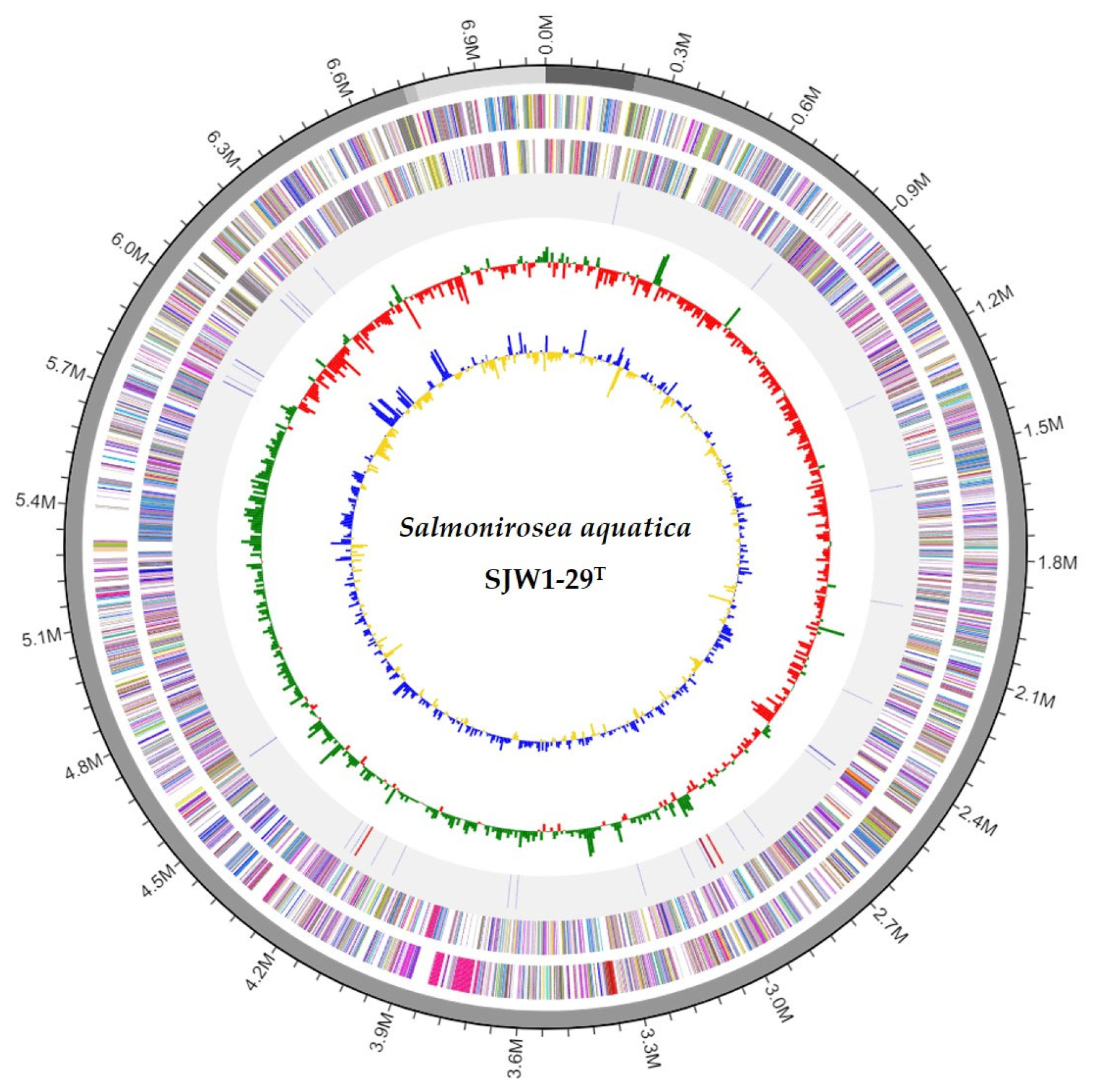
3.3. Genome Features

The genome features of strain SJW1-29T were analyzed in this study. The DNA G+C content of strain SJW1-29T was 50.7%. A summary of the genome properties and statistics of strain SJW1-29T is provided in Table S1. The draft genome sequence of SJW1-29T, representing the novel strain, had a size of 7,065,248 bp, consisting of four contigs with a contig N50 value of 6,512,965 bp. The genome contained a total of 6297 coding sequences, 9 rRNA genes, and 40 tRNA genes (Figure 2).
Contamination was excluded by comparing a fragment of the 16S rRNA gene with the ContEst16S results. The genome sequence met the proposed minimum standards for bacterial taxonomy [49]. A RAST analysis was performed to predict the functional gene content of the strain SJW1-29T genome (Figure 3). The analysis revealed the presence of genes involved in various biological processes, including carbohydrates (351 genes), amino acids and derivatives (334 genes), protein metabolism (247 genes), and cofactors, vitamins, prosthetic groups, and pigments (188 genes). A total of 5315 protein-coding genes were identified in the genome of strain SJW1-29T, with 5315 genes (84.4%) assigned to COG categories. The COG category assignment of genes included cell wall/membrane/envelope biogenesis (M; 6.2%, 328 genes) and inorganic ion transport and metabolism (P; 5.6%, 299 genes), whereas 45.3% of genes were classified as having unknown functions within the COGs (Figure S3).
The genome annotations and functional characterization analysis revealed that the genome sequence of strain SJW1-29T contains multiple genes encoding putative polysaccharide-degrading enzymes. These include eleven glucosidases, two chitinases, four cellulases, eight xylanases, six amylases, one alginate lyase, and six pectinesterases [50].
The phylogenomic tree constructed based on TYGS analysis revealed the relationship between strain SJW1-29T and closely related type strains (Figure S4). It is most closely related to species of the genus Persicitalea and other genera within the family Spirosomaceae. The analysis clearly shows that strain SJW1-29T forms a distinct clade, separate from its closest related species, with a low bootstrap value of 79%, supporting its classification as a new genus.
Considering the results obtained from complete phylogenetic analyses, to confirm that strain SJW1-29T is indeed a new taxon, ANI, AAI, dDDH, and POCP values of strain SJW1-29T and other related members of the family Spirosomaceae were calculated (Figure S5 and Table 1). The genome-relatedness indices, including ANI, AAI, and dDDH, showed that this strain represents a distinct species with values below the species demarcation thresholds (ANI < 95%, AAI < 95%, and dDDH < 70%) when compared with the closest type strains within the family Spirosomaceae [39,51,52,53,54,55,56]. Additionally, the POCP results indicated less than 66% similarity with other genera within the family Spirosomaceae. POCP can serve as a powerful genomic index to define the genus boundaries of prokaryotic groups [43]. However, it should also be recognized that POCP is only a genomic index. For the genus-level separation of the family Bacillaceae, the POCP boundary was more than 50% [57,58]. Even in this family Spirosomaceae, the POCP value is more than 50% between genera, but it can be judged as a different genus based on several taxonomic grounds. Since this new taxon’s most closely related species belong to the family Spirosomaceae, we suggest that this new genus should also be classified within this family as a member of the order Cytophagales within the class Cytophagia.

3.4. Morphological and Phenotypic Characteristics

The cells of SJW1-29T were observed to be Gram-stain-negative and had a rod shape, ranging in size from 0.6 to 0.8 µm × 1.3 to 1.7 µm (Figure S6). Colonies formed by the strain on the MA medium appeared regular, round, and salmon pink after 5 days of incubation at 25 °C. Strain SJW1-29T exhibited a 15–30 °C temperature range with an optimum growth temperature of 25 °C. It grew within a pH range of 5.0–10.0, with optimal growth occurring at pH 7.0. The strain showed tolerance to NaCl concentrations ranging from 1.0% to 5.0% (w/v), with an optimum NaCl concentration for growth of 3.0%. Analyses of differential characteristics showed that SJW1-29T and related species differed in multiple characteristics, including isolation source, salinity range, the optimum temperature for growth, enzyme activities, and carbon source utilization patterns (Table 1).
The phenotypic characteristics of strain SJW1-29T align with those typically observed in the family Spirosomaceae, such as Gram-stain negativity and rod-shaped morphology. The ability of SJW1-29T to grow in a broad range of temperatures and pH levels, as well as its tolerance to varying NaCl concentrations, suggests ecological versatility, which is a common trait in environmental isolates from brackish waters. This adaptability is crucial for survival in fluctuating estuarine environments where salinity and other abiotic factors vary significantly [11].
Comparative phenotypic analysis, as shown in Table 1, reveals distinct biochemical capabilities that further substantiate the novelty of SJW1-29T. For instance, the strain’s unique enzymatic profile, including its ability to hydrolyze specific substrates and its distinct fatty acid composition, differentiates it from closely related species. These phenotypic traits support the classification of SJW1-29T as a new genus and highlight its potential functional roles in its native habitat. The presence of multiple genes encoding polysaccharide-degrading enzymes, as indicated by genome annotation, suggests a role in the degradation of complex carbohydrates in the ecosystem, contributing to nutrient cycling and organic matter decomposition in the Seomjin River estuary [10].
Table 1. Differential characteristics between strain SJW1-19T and the most closely related species of the family Spirosomaceae. Genus: 1, Salmonirosea gen. nov. (data from this study); 2, Persicitalea jodogahamensis (only species in the genus) [59]; 3, Rhabdobacter roseus (only species in the genus) [60]; 4, Arundinibacter roseus (only species in the genus) [61]; 5, Telluribacter humicola (only species in the genus) [62]; 6, Dyadobacter fermentans (type species in the genus) [63]; 7, Ravibacter arvi (only species in the genus) [64]. +, positive; −, negative; ND, data not available.
Characteristic1234567
Isolation sourceBrackish waterSea waterSoilFreshwaterSoilPlantSoil
16S rRNA gene similarity (%)ref.91.390.690.089.188.484.8
Genome size (bp)7,065,2486,413,5896,917,9645,828,2786,558,7316,967,7905,775,379
ANI valueref.75.071.971.871.169.267.3
AAI valueref.78.470.874.670.667.063.4
dDDH valueref.21.018.517.518.218.819.9
POCP valueref.62.663.966.063.052.646.0
Colony colorSalmon pinkPale pinkLight pinkLight pinkPinkYellowPale yellow
Cell morphologyRodsStraight rod to curved rodsRodsRodsRodsRodsRods
Flexirubin reactionNDNDND+ND
Temperature for growth (°C)15–3025–3010–375–3710–4215–3715–40
Highest NaCl tolerated (%, w/v)<5.0<7.0<5.0<4.0<4.0<1.5<1.0
Genome size (Mb)7.07NDND5.83ND6.97ND
Assimilation of (API 20NE):
Glucose fermentation+W++
Urease++NDND+
Enzyme activity (API ZYM)
Alkaline phosphatase+++ND+
Esterase (C4), cystine arylamidase, trypsin, β-galactosidase++++ND+
Lipase (C14)ND+
α-chymotrypsin++++ND
Acid phosphatase++++ND+
β-glucosidase++++ND+
α-mannosidase++++ND+
Major quinone MK-7MK-7MK-7MK-7MK-7MK-7MK-7
Major polar lipids PE, APL, AL, LPE, AL, LPE, PL, AL, LPE, AL, GL, LAL, LPE, PL, AL, LPE, APL, AL
DNA G+C content (mol%) §50.750.854.045.748.951.550.6
MK, menaquinone; PE, phosphatidylethanolamine; APL, unidentified aminophospholipid; AL, unidentified aminolipid; GL, unidentified glycolipid; PL, unidentified phospholipid; L, unidentified lipids; § the G+C content was calculated based on the nucleotide content of the sequenced genomes.

3.5. Chemotaxonomic Characterization

The major cellular fatty acids of strain SJW1-29T were identified as summed feature 3 (C16:1 ω7c and/or C16:1 ω6c; 55.3%), followed by iso-C15:0 (11.9%), iso-C17:0 3-OH (8.2%), and C16:0 (8.0%). A comparison of the full fatty acid profiles of strain SJW1-29T can be found in Table 2. The predominant respiratory quinone in strain SJW1-29T was menaquinone-7 (MK-7), a common quinone found in the family Spirosomaceae. The major polar lipid of strain SJW1-29T was phosphatidylethanolamine (PE). Additionally, one unidentified aminophospholipid (APL), one unidentified aminolipid (AL), and seven unidentified lipids (L1–7) were also detected. No glycolipid was detected in the chromatogram when sprayed with α-naphthol solution and incubated at 110 °C for 15 min (Figure S7).

4. Conclusions

The identification and characterization of Salmonirosea aquatica gen. nov., sp. nov. have significant implications for microbial taxonomy and environmental microbiology. This study expands the known diversity within the family Spirosomaceae and underscores the importance of exploring brackish water environments to discover novel bacterial taxa. The discovery of S. aquatica contributes to our understanding of phylogenetic relationships within Spirosomaceae, revealing unique evolutionary lineages due to its low 16S rRNA gene sequence similarity (<94%) with closest relatives. Additionally, genome-based analyses, including ANI, AAI, dDDH, and POCP, further support its classification as a new genus. These findings emphasize the need for continued phylogenetic studies to uncover and classify new genera and species.
Practically, S. aquatica’s unique phenotypic and chemotaxonomic characteristics, including its ability to degrade various polysaccharides and tolerate diverse environmental conditions, highlight its potential applications in biotechnology and environmental management. For instance, its polysaccharide-degrading enzymes could be used in bioremediation to break down complex organic pollutants in aquatic environments. Future research should explore the functional roles of S. aquatica in its native habitat, its interactions within microbial communities, and its contribution to nutrient cycling and ecosystem health. Additionally, biotechnological studies can investigate the potential applications of its enzymes in industrial processes and environmental cleanup efforts.
In conclusion, the isolation and characterization of Salmonirosea aquatica enrich our understanding of microbial diversity and open new avenues for applied research in environmental biotechnology and ecosystem management. This study lays the groundwork for future explorations into the functional capabilities and ecological significance of novel bacterial taxa.

4.1. Description of Salmonirosea gen. nov.

Salmonirosea (Sal.mo.ni.ro’se.a. L. masc. n. salmo, a salmon; L. masc. adj. roseus, pink; N.L. fem. n. Salmonirosea, an organism with a salmon-pink color).
Cells are Gram-stain-negative, rod, and non-motile, and colonies on MA are salmon pink. Oxidase- and catalase-positive. Cells are obligately aerobic. Its major respiratory quinone is menaquinone-7 (MK-7). The major fatty acid components (>10.0%) included include summed feature 3 (C16:1 ω7c and/or C16:1 ω6c; 55.6%) and iso-C15:0, a major profile similar to those of its closest related strains. Major polar lipids are phosphatidylethanolamine. The type species of the new genus is Salmonirosea.

4.2. Description of Salmonirosea aquatica sp. nov.

Salmonirosea aquatica (a.qua′ti.ca. L. fem. adj. aquatica, living, growing, or found in or by water, aquatic).
Gram-stain-negative, oxidase- and catalase-positive, and aerobic. Cells are rod-shaped (0.6–0.8 µm in diameter and 1.3–1.7 in length, Figure S4). Growth occurs at 15–30 °C (optimum 25 °C), pH 5.0–10.0 (optimum pH 7.0), and in the presence of 1.0–5.0% (w/v) NaCl (optimum, 3.0% NaCl). Catalase and oxidase reactions are positive. Tween-20 and Tween-40 are hydrolyzed, but Tween-80, casein, and starch are not. Positive for esculin hydrolysis and PNPG (β-galactosidase) but negative for nitrate reduction, indole production, glucose fermentation, arginine dihydrolase, urease, and gelatinase (in API 20NE). With API ZYM, positive for alkaline phosphatase, esterase lipase (C8), leucine arylamidase, valine arylamidase, α-chymotrypsin, acid phosphatase, naphthol-AS-BI-phosphohydrolase, α-galactosidase, α-glucosidase, β-glucosidase, N-acetyl-β-glucosaminidase, and α-mannosidase, but negative for esterase (C4), lipase (C14), cystine arylamidase, trypsin, β-galactosidase, β-glucuronidase, and α-fucosidase. Substrates used include erythritol, D-arabinose, D-xylose, L-xylose, D-galactose, D-glucose, D-fructose, D-mannose, L-sorbose, arbutin, aesculin ferric citrate, salicin, D-melibiose, D-melezitose, gentiobiose, D-lyxose, D-tagatose, potassium 2-ketogluconate, and potassium 5-ketogluconate (API 50CH). The in silico G+C content of the whole genome of the type strain is 50.7 mol%.
The type strain SJW1-29T (=KCTC 72493T = NBRC 114061T = FBCC-B16924T) was isolated from brackish water collected in the Republic of Korea.

Supplementary Materials

The following supporting information can be downloaded at: https://www.mdpi.com/article/10.3390/microorganisms12081671/s1, Table S1: summary of complete genome sequence of strain SJW1-29T, Figure S1: maximum likelihood tree based on nearly complete 16S rRNA gene sequences showing the phylogenetic position of strain SJW1-29T with closely related taxa, Figure S2: maximum parsimony tree based on nearly complete 16S rRNA gene sequences showing the phylogenetic position of strain SJW1-29T with closely related taxa, Figure S3: COG functional classification of proteins in strain SJW1-29T genome, Figure S4: whole-genome sequence-based phylogenetic tree of strains SJW1-29T using TYGS design tree with FastME 2.1.4 software based on Genome BLAST Distance Phylogeny (GBDP) parameters, Figure S5: heatmap of orthologous average nucleotide identity (OrthoANI) between strain SJW1-29T and closely related relatives, Figure S6: transmission electron micrograph of SJW1-29T cells Figure S7: two-dimensional thin-layer chromatogram showing the polar lipids of SJW1-29T.

Author Contributions

Conceptualization: K.B. and A.C.; methodology: K.B.; formal analysis: K.B.; data curation: K.B., S.J. and A.C.; writing—original draft preparation: K.B. and A.C.; writing—review and editing: all authors; funding acquisition: A.C. and J.G.; supervision: A.C. All authors have read and agreed to the published version of the manuscript.

Funding

This study was supported by a grant from the Nakdonggang National Institute of Biological Resources (NNIBR), funded by the Ministry of Environment (MOE) of the Republic of Korea (NNIBR20241103) and by the Korea Environment Industry & Technology Institute (KEITI) through a project to make multi-ministerial national biological research resources more advanced, funded by the Korea Ministry of Environment (MOE) (2021003420003).

Data Availability Statement

The GenBank/EMBL/DDBJ accession number for the 16S rRNA gene sequence of strain SJW1-29T is MN596019. The GenBank/EMBL/DDBJ accession number for the whole-genome sequence of strain SJW1-29T is WHLY00000000.

Conflicts of Interest

The authors declare no conflicts of interest.

Abbreviations

MA, marine agar 2216; ASW, artificial seawater medium; NJ, neighbor joining; ML, maximum likelihood; MP, maximum parsimony; RAST, Rapid Annotation using the Subsystem Technology; COGs, clusters of orthologous groups; ANI, average nucleotide identity; AAI, average amino acid identity; dDDH, digital DNA-DNA hybridization; POCP, percentage of conserved proteins; TEM, transmission electron microscopy.

References

  1. Larkin, J.M.; Borrall, R. Spirosomaceae, a new family to contain the genera Spirosoma Migula 1894, Flectobacillus Larkin et al. 1977, and Runella Larkin and Williams 1978. Int. J. Syst. Evol. Microbiol. 1978, 28, 595–596. [Google Scholar] [CrossRef]
  2. Oren, A.; Garrity, G. Notification of changes in taxonomic opinion previously published outside the IJSEM. Int. J. Syst. Evol. Microbiol. 2020, 70, 4061–4090. [Google Scholar] [CrossRef] [PubMed]
  3. Parte, A.C.; Sardà Carbasse, J.; Meier-Kolthoff, J.P.; Reimer, L.C.; Göker, M. List of Prokaryotic names with Standing in Nomenclature (LPSN) moves to the DSMZ. Int. J. Syst. Evol. Microbiol. 2020, 70, 5607–5612. [Google Scholar] [CrossRef]
  4. García-López, M.; Meier-Kolthoff, J.P.; Tindall, B.J.; Gronow, S.; Woyke, T.; Kyrpides, N.C.; Hahnke, R.L.; Göker, M. Analysis of 1000 type-strain genomes improves taxonomic classification of Bacteroidetes. Front. Microbiol. 2019, 10, 2083. [Google Scholar] [CrossRef] [PubMed]
  5. Dias, R.L.; Ruberto, L.; Calabro, A.; Balbo, A.L.; Del Panno, M.T.; Mac Cormack, W.P. Hydrocarbon removal and bacterial community structure in on-site biostimulated biopile systems designed for bioremediation of diesel-contaminated Antarctic soil. Polar Biol. 2015, 38, 677–687. [Google Scholar] [CrossRef]
  6. El Fantroussi, S.; Agathos, S.N. Is bioaugmentation a feasible strategy for pollutant removal and site remediation? Curr. Opin. Microbiol. 2005, 8, 268–275. [Google Scholar] [CrossRef]
  7. Dixit, R.; Wasiullah; Malaviya, D.; Pandiyan, K.; Singh, U.B.; Sahu, A.; Shukla, R.; Singh, B.P.; Rai, J.P.; Sharma, P.K.; et al. Bioremediation of heavy metals from soil and aquatic environment: An overview of principles and criteria of fundamental processes. Sustainability 2015, 7, 2189–2212. [Google Scholar] [CrossRef]
  8. Donati, E.R.; Sani, R.K.; Goh, K.M.; Chan, K.-G. Editorial: Recent advances in bioremediation/biodegradation by extreme microorganisms. Front. Microbiol. 2019, 10, 1851. [Google Scholar] [CrossRef]
  9. Khan, F.I.; Husain, T.; Hejazi, R. An overview and analysis of site remediation technologies. J. Environ. Manag. 2004, 71, 95–122. [Google Scholar] [CrossRef]
  10. Hatti-Kaul, R.; Törnvall, U.; Gustafsson, L.; Börjesson, P. Industrial biotechnology for the production of bio-based chemicals—A cradle-to-grave perspective. Trends Biotechnol. 2007, 25, 119–124. [Google Scholar] [CrossRef]
  11. Duarte, M.; Nielsen, A.; Camarinha-Silva, A.; Vilchez-Vargas, R.; Bruls, T.; Wos-Oxley, M.L.; Jauregui, R.; Pieper, D.H. Functional soil metagenomics: Elucidation of polycyclic aromatic hydrocarbon degradation potential following 12 years of in situ bioremediation. Environ. Microbiol. 2017, 19, 2992–3011. [Google Scholar] [CrossRef] [PubMed]
  12. Olaniran, A.O.; Pillay, D.; Pillay, B. Biostimulation and bioaugmentation enhances aerobic biodegradation of dichloroethenes. Chemosphere 2006, 63, 600–608. [Google Scholar] [CrossRef] [PubMed]
  13. Halim, M.; Conte, P.; Piccolo, A. Potential availability of heavy metals to phytoextraction from contaminated soils induced by exogenous humic substances. Chemosphere 2003, 52, 265–275. [Google Scholar] [CrossRef] [PubMed]
  14. Gupta, G.; Kumar, V.; Pal, A.K. Microbial degradation of high molecular weight polycyclic aromatic hydrocarbons with emphasis on pyrene. Polycycl. Aromat. Compd. 2019, 39, 124–138. [Google Scholar] [CrossRef]
  15. Lee, J.H.; Lee, M.J.; LEE, G.G. National level assessment of Biodiversity importance—Focusing on South Korea. KSCE J. Civ. Eng. 2014, 19, 46–62. [Google Scholar] [CrossRef]
  16. Lee, M.; Par, B.S.; Baek, S.H. Tidal Influences on Biotic and Abiotic Factors in the Seomjin River Estuary and Gwangyang Bay, Korea. Estuaries Coast 2018, 41, 1977–1993. [Google Scholar] [CrossRef]
  17. Hwang, J.H.; Jang, D.; Kim, Y.H. Stratification and salt-wedge in the Seomjin river estuary under the idealized tidal influence. Ocean Sci. J. 2017, 52, 469–487. [Google Scholar] [CrossRef]
  18. Prieto-Barajas, C.M.; Valencia-Cantero, E.; Santoyo, G. Microbial mat ecosystems: Structure types, functional diversity, and biotechnological application. Electron. J. Biotechnol. 2018, 31, 48–56. [Google Scholar] [CrossRef]
  19. Lee, D.I.; Park, C.K.; Cho, H.S. Ecological modeling for water quality management of Kwangyang Bay, Korea. J. Environ. Manag. 2005, 74, 327–337. [Google Scholar] [CrossRef]
  20. Findlay, S.J.; Taylor, M.P. Why rehabilitate urban river systems? Area 2006, 38, 312–325. [Google Scholar] [CrossRef]
  21. Gurnell, A.; Lee, M.; Souch, C. Urban rivers: Hydrology, geomorphology, ecology and opportunities for change. Geogr. Compass 2007, 1, 1118–1137. [Google Scholar] [CrossRef]
  22. Paul, M.J.; Meyer, J.L. Streams in the urban landscape. Annu. Rev. Ecol. Syst. 2001, 32, 333–365. [Google Scholar] [CrossRef]
  23. Bunn, S.E.; Arthington, A.H. Basic principles and ecological consequences of altered flow regimes for aquatic biodiversity. Environ. Manag. 2002, 30, 493–507. [Google Scholar] [CrossRef] [PubMed]
  24. Frank, J.A.; Reich, C.I.; Sharma, S.; Weisbaum, J.S.; Wilson, B.A.; Olsen, G.J. Critical evaluation of two primers commonly used for amplification of bacterial 16S rRNA genes. Appl. Environ. Microbiol. 2008, 74, 2461–2470. [Google Scholar] [CrossRef]
  25. Yoon, S.H.; Ha, S.M.; Kwon, S.; Lim, J.; Kim, Y.; Seo, H.; Chun, J. Introducing EzBioCloud: A taxonomically united database of 16S rRNA gene sequences and whole-genome assemblies. Int. J. Syst. Evol. Microbiol. 2017, 67, 1613–1617. [Google Scholar] [CrossRef] [PubMed]
  26. Jeon, Y.S.; Lee, K.; Park, S.C.; Kim, B.S.; Cho, Y.J.; Ha, S.M.; Chun, J. EzEditor: A versatile sequence alignment editor for both rRNA- and protein-coding genes. Int. J. Syst. Evol. Microbiol. 2014, 64, 689–691. [Google Scholar] [CrossRef] [PubMed]
  27. Saitou, N.; Nei, M. The neighbor-joining method: A new method for reconstructing phylogenetic trees. Mol. Biol. Evol. 1987, 4, 406–425. [Google Scholar] [CrossRef] [PubMed]
  28. Felsenstein, J. Evolutionary trees from DNA sequences: A maximum likelihood approach. J. Mol. Evol. 1981, 17, 368–376. [Google Scholar] [CrossRef] [PubMed]
  29. Fitch, W.M. Toward defining the course of evolution: Minimum change for a specific tree topology. Syst. Zool. 1971, 20, 406–416. [Google Scholar] [CrossRef]
  30. Kumar, S.; Stecher, G.; Tamura, K. MEGA7: Molecular evolutionary genetics analysis version 7.0 for bigger datasets. Mol. Biol. Evol. 2016, 33, 1870–1874. [Google Scholar] [CrossRef]
  31. Felsenstein, J. Confidence limits on phylogenies: An approach using the bootstrap. Evolution 1985, 39, 783–791. [Google Scholar] [CrossRef]
  32. Chin, C.S.; Alexander, D.H.; Marks, P.; Klammer, A.A.; Drake, J.; Heiner, C.; Clum, A.; Copeland, A.; Huddleston, J.; Eichler, E.E.; et al. Nonhybrid, finished microbial genome assemblies from long-read SMRT sequencing data. Nat. Methods 2013, 10, 563–569. [Google Scholar] [CrossRef]
  33. Aziz, R.K.; Bartels, D.; Best, A.A.; DeJongh, M.; Disz, T.; Edwards, R.A.; Formsma, K.; Gerdes, S.; Glass, E.M.; Kubal, M.; et al. The RAST server: Rapid annotations using subsystems technology. BMC Genom. 2008, 9, 75. [Google Scholar] [CrossRef] [PubMed]
  34. Tatusova, T.; DiCuccio, M.; Badretdin, A.; Chetvernin, V.; Nawrocki, E.P.; Zaslavsky, L.; Lomsadze, A.; Pruitt, K.D.; Borodovsky, M.; Ostell, J. NCBI prokaryotic genome annotation pipeline. Nucleic Acids Res. 2016, 44, 6614–6624. [Google Scholar] [CrossRef] [PubMed]
  35. Lee, I.; Chalita, M.; Ha, S.M.; Na, S.I.; Yoon, S.H.; Chun, J. ContEst16S: An algorithm that identifies contaminated prokaryotic genomes using 16S RNA gene sequences. Int. J. Syst. Evol. Microbiol. 2017, 67, 2053–2057. [Google Scholar] [CrossRef]
  36. Kanehisa, M.; Goto, S. KEGG: Kyoto encyclopedia of genes and genomes. Nucleic Acids Res. 2000, 28, 27–30. [Google Scholar] [CrossRef] [PubMed]
  37. Meier-Kolthoff, J.P.; Göker, M. TYGS is an automated high-throughput platform for state-of-the-art genome-based taxonomy. Nat. Commun. 2019, 10, 2182. [Google Scholar] [CrossRef] [PubMed]
  38. Lefort, V.; Desper, R.; Gascuel, O. FastME 2.0: A comprehensive, accurate, and fast distance-based phylogeny inference program. Mol. Biol. Evol. 2015, 32, 2798–2800. [Google Scholar] [CrossRef]
  39. Price, M.N.; Dehal, P.S.; Arkin, A.P. FastTree: Computing large minimum evolution trees with profiles instead of a distance matrix. Mol. Biol. Evol. 2009, 26, 1641–1650. [Google Scholar] [CrossRef]
  40. Lee, I.; Ouk Kim, Y.; Park, S.-C.; Chun, J. OrthoANI: An improved algorithm and software for calculating average nucleotide identity. Int. J. Syst. Evol. Microbiol. 2016, 66, 1100–1103. [Google Scholar] [CrossRef]
  41. Kim, D.; Park, S.; Chun, J. Introducing EzAAI: A pipeline for high throughput calculations of prokaryotic average amino acid identity. J. Microbiol. 2021, 59, 476–480. [Google Scholar] [CrossRef] [PubMed]
  42. Meier-Kolthoff, J.P.; Auch, A.F.; Klenk, H.P.; Göker, M. Genome sequence-based species delimitation with confidence intervals and improved distance functions. BMC Bioinform. 2013, 14, 60. [Google Scholar] [CrossRef] [PubMed]
  43. Qin, Q.L.; Xie, B.B.; Zhang, X.Y.; Chen, X.L.; Zhou, B.C.; Zhou, J.; Oren, A.; Zhang, Y.Z. A proposed genus boundary for the prokaryotes based on genomic insights. J. Bacteriol. 2014, 196, 2210–2215. [Google Scholar] [CrossRef]
  44. Choo, Y.-J.; Lee, K.; Song, J.; Cho, J.-C. Puniceicoccus vermicola gen. nov., sp. nov., a novel marine bacterium, and description of Puniceicoccaceae fam. nov., Puniceicoccales ord. nov., Opitutaceae fam. nov., Opitutales ord. nov. and Opitutae classis nov. in the phylum ‘Verrucomicrobia’. Int. J. Syst. Evol. Microbiol. 2007, 57, 532–537. [Google Scholar] [CrossRef]
  45. Smibert, R.M.; Krieg, N.R. Phenotypic characterization. In Methods for General and Molecular Bacteriology; Gerhardt, P., Murray, R.G.E., Wood, W.A., Krieg, N.R., Eds.; American Society for Microbiology: Washington, DC, USA, 1994; pp. 607–654. [Google Scholar]
  46. Tindall, B.J.; Sikorski, J.; Smibert, R.A.; Krieg, N.R. Phenotypic Characterization and the Principles of Comparative Systematics. In Methods for General and Molecular Microbiology; Reddy, C.A., Ed.; ASM Press: Washington, DC, USA, 2007; pp. 330–393. [Google Scholar]
  47. Sasser, M. Identification of Bacteria by Gas Chromatography of Cellular Fatty Acids; Technical Note 101; MIDI: Newark, NJ, USA, 2001. [Google Scholar]
  48. Minnikin, D.E.; O’Donnell, A.G.; Goodfellow, M.; Alderson, G.; Athalye, M.; Schaal, A.; Parlett, J.H. An integrated procedure for the extraction of bacterial isoprenoid quinones and polar lipids. J. Microbiol. Methods 1984, 2, 233–241. [Google Scholar] [CrossRef]
  49. Kim, M.; Oh, H.S.; Park, S.C.; Chun, J. Towards a taxonomic coherence between average nucleotide identity and 16S rRNA gene sequence similarity for species demarcation of prokaryotes. Int. J. Syst. Evol. Microbiol. 2014, 64, 346–351. [Google Scholar] [CrossRef] [PubMed]
  50. Chun, J.; Oren, A.; Ventosa, A.; Christensen, H.; Arahal, D.R.; da Costa, M.S.; Rooney, A.P.; Yi, H.; Xu, X.W.; De Meyer, S.; et al. Proposed minimal standards for the use of genome data for the taxonomy of prokaryotes. Int. J. Syst. Evol. Microbiol. 2018, 68, 461–466. [Google Scholar] [CrossRef]
  51. Baek, K.; Choi, A. Draft genome sequence of the polysaccharide degrading bacterium Cytophagaceae strain SJW1-29. Korean J. Microbiol. 2020, 56, 422–425. [Google Scholar] [CrossRef]
  52. Konstantinidis, K.T.; Tiedje, J.M. Trends between gene content and genome size in prokaryotic species with larger genomes. Proc. Natl. Acad. Sci. USA 2004, 101, 3160–3165. [Google Scholar] [CrossRef]
  53. Jain, C.; Rodriguez-R, L.M.; Phillippy, A.M.; Konstantinidis, K.T.; Aluru, S. High throughput ANI analysis of 90K prokaryotic genomes reveals clear species boundaries. Nat. Commun. 2018, 9, 5114. [Google Scholar] [CrossRef]
  54. Goris, J.; Konstantinidis, K.T.; Klappenbach, J.A.; Coenye, T.; Vandamme, P.; Tiedje, J.M. DNA–DNA hybridization values and their relationship to whole-genome sequence similarities. Int. J. Syst. Evol. Microbiol. 2007, 57, 81–91. [Google Scholar] [CrossRef] [PubMed]
  55. Konstantinidis, K.T.; Rosselló-Móra, R.; Amann, R. Uncultivated microbes in need of their own taxonomy. ISME J. 2017, 11, 2399–2406. [Google Scholar] [CrossRef] [PubMed]
  56. Stackebrandt, E.; Goebel, B.M. Taxonomic note: A place for DNA-DNA reassociation and 16S rRNA sequence analysis in the present species definition in Bacteriology. Int. J. Syst. Evol. Microbiol. 1994, 44, 846–849. [Google Scholar] [CrossRef]
  57. Hölzer, M. POCP-nf: An automatic Nextflow pipeline for calculating the percentage of conserved proteins in bacterial taxonomy. Bioinformatics 2024, 40, btae175. [Google Scholar] [CrossRef] [PubMed]
  58. Aliyu, H.; Lebre, P.; Blom, J.; Cowan, D.; De Maayer, P. Phylogenomic re-assessment of the thermophilic genus Geobacillus. Syst. Appl. Microbiol. 2016, 39, 527–533. [Google Scholar] [CrossRef] [PubMed]
  59. Yoon, J.; Ishikawa, S.; Kasai, H.; Yokota, A. Persicitalea jodogahamensis gen. nov., sp. nov., a marine bacterium of the family ‘Flexibacteraceae’, isolated from seawater in Japan. Int. J. Syst. Evol. Microbiol. 2007, 57, 1014–1017. [Google Scholar] [CrossRef]
  60. Dahal, R.H.; Kim, J. Rhabdobacter roseus gen. nov., sp. nov., isolated from soil. Int. J. Syst. Evol. Microbiol. 2016, 66, 308–314. [Google Scholar] [CrossRef] [PubMed]
  61. Szuróczki, S.; Khayer, B.; Spröer, C.; Toumi, M.; Szabó, A.; Felföldi, T.; Schumann, P.; Tóth, E. Arundinibacter roseus gen. nov., sp. nov., a new member of the family Cytophagaceae. Int. J. Syst. Evol. Microbiol. 2019, 69, 2076–2081. [Google Scholar] [CrossRef]
  62. Lee, D.; Jang, J.H.; Cha, S.; Seo, T. Telluribacter humicola gen. nov., sp. nov., a new member of the family Cytophagaceae isolated from soil in South Korea. Antonie Van Leeuwenhoek 2016, 109, 1525–1533. [Google Scholar] [CrossRef]
  63. Chelius, M.K.; Triplett, E.W. Dyadobacter fermentans gen. nov., sp. nov., a novel gram-negative bacterium isolated from surface-sterilized Zea mays stems. Int. J. Syst. Evol. Microbiol. 2000, 50, 751–758. [Google Scholar] [CrossRef]
  64. Chaudhary, D.K.; Dahal, R.H.; Altankhuu, K.; Kim, J. Ravibacter arvi gen. nov., sp. nov., isolated from farmland soil during development of new culture techniques. Int. J. Syst. Evol. Microbiol. 2017, 67, 5252–5260. [Google Scholar] [CrossRef] [PubMed]
Figure 1. Neighbor joining tree based on nearly complete 16S rRNA gene sequences showing the phylogenetic position of strain SJW1-29T with closely related taxa. Bootstrap values > 70 are indicated and based on 1000 replications (NJ/ML/MP; -, no-value-filled circles indicate that the corresponding nodes were recovered by all treeing methods. An open circle indicates that the corresponding node was recovered by the neighbor joining and maximum parsimony methods. The bold font represents the novel species identified in this study. Haliscomenobacter hydrossis DSM 1100T (CP002691) was used as an out-group. The scale bar depicts 0.02 substitutions per nucleotide position.
Microorganisms 12 01671 g001
Figure 2. Circular map of the strain SJW1-29T genome. From outside to the center: the colored bands in ring 1 represent contigs; ring 2 represents the annotated genes on the forward strand (color determined by COG category); ring 3 shows the annotated genes on the reverse strand (color determined by COG category); ring 4 displays the RNA genes (rRNAs are displayed in red and tRNAs are displayed in purple); ring 5 shows the GC skew (higher-than-average values are displayed in green, while lower-than-average values are displayed in red) and ring 6 shows the GC ratio (higher-than-average values in blue and lower-than-average values in yellow).
Microorganisms 12 01671 g002
Figure 3. Subsystem category distribution of strain SJW1-29T based on the RAST annotation server (https://rast.nmpdr.org/, accessed on 21 May 2024).
Microorganisms 12 01671 g003
Table 2. Fatty acid profiles of strain SJW1-29T and the type strains of the related genera of the family Spirosomaceae. Genus: 1, Salmonirosea gen. nov. (data from this study); 2, Persicitalea jodogahamensis (only species in the genus) [11]; 3, Rhabdobacter roseus (only species in the genus) [10]; 4, Arundinibacter roseus (only species in the genus) [59]; 5, Telluribacter humicola (only species in the genus) [60]; 6, Dyadobacter fermentans (type species in the genus) [61]; 7, Ravibacter arvi (only species in the genus) [62]. Values are percentages of the total fatty acids. tr, trace amount (<1%).
Fatty Acid1234567
Saturated
C14:01.9-1.0-tr-tr
C16:08.012.06.67.47.75.610.1
Unsaturated
C13:1-2.01.6--1.3-
C14:1 ω5c----3.7 -
C16:1 ω5c1.86.617.514.213.515.96.9
Hydroxy
C14:0 2-OH1.1-----tr
C16:0 3-OH1.22.21.32.3-2.34.6
C16:0 N alcohol----1.1--
Iso-C15:0 3-OH2.62.11.01.53.21.72.5
Iso-C16:0 2-OH------1.1
Iso-C16:0 3-OH2.61.51.2---tr
Iso-C17:0 3-OH8.26.23.76.5-4.48.5
Iso-C17:1 ω9c---1.33.9--
Branched chain
Iso-C10:0----4.3--
Iso-C14:01.1-----tr
Iso-C15:011.931.521.416.613.819.49.9
Iso-C17:0----1.8-tr
Iso-C15:0 G---1.4- -
Anteiso-C15:01.51.7-1.73.5-tr
Summed features 3 *55.329.940.741.839.944.452.1
* Summed features represent groups of two or three fatty acids that could not be separated using the MIDI system. Summed feature 3 comprised C16:1 ω7c and/or C16:1 ω6c.
Disclaimer/Publisher’s Note: The statements, opinions and data contained in all publications are solely those of the individual author(s) and contributor(s) and not of MDPI and/or the editor(s). MDPI and/or the editor(s) disclaim responsibility for any injury to people or property resulting from any ideas, methods, instructions or products referred to in the content.

