Article

Agro Active Potential of Bacillus subtilis PE7 against Didymella bryoniae (Auersw.), the Causal Agent of Gummy Stem Blight of Cucumis melo

by
Seo Kyoung Jeong
1,
Seong Eun Han
2,
Prabhakaran Vasantha-Srinivasan
3,
Woo Jin Jung
1,2,
Chaw Ei Htwe Maung
2,* and
Kil Yong Kim
1,2,*
1
Department of Plant Protection and Quarantine, Chonnam National University, Gwangju 61186, Republic of Korea
2
Department of Agricultural Chemistry, Environmentally-Friendly Agricultural Research Center, College of Agriculture and Life Sciences, Chonnam National University, Gwangju 61186, Republic of Korea
3
Department of Applied Biology, College of Agriculture and Life Sciences, Chonnam National University, Gwangju 61186, Republic of Korea
*
Authors to whom correspondence should be addressed.
Microorganisms 2024, 12(8), 1691; https://doi.org/10.3390/microorganisms12081691
Submission received: 11 July 2024 / Revised: 27 July 2024 / Accepted: 13 August 2024 / Published: 16 August 2024
(This article belongs to the Special Issue Antifungal Activity of Bacillus Species against Plant Pathogens)

Abstract

:
Microbial agents such as the Bacillus species are recognized for their role as biocontrol agents against various phytopathogens through the production of diverse bioactive compounds. This study evaluates the effectiveness of Bacillus subtilis PE7 in inhibiting the growth of Didymella bryoniae, the pathogen responsible for gummy stem blight (GSB) in cucurbits. Dual culture assays demonstrate significant antifungal activity of strain PE7 against D. bryoniae. Volatile organic compounds (VOCs) produced by strain PE7 effectively impede mycelial formation in D. bryoniae, resulting in a high inhibition rate. Light microscopy revealed that D. bryoniae hyphae exposed to VOCs exhibited abnormal morphology, including swelling and excessive branching. Supplementing a potato dextrose agar (PDA) medium with a 30% B. subtilis PE7 culture filtrate significantly decreased mycelial growth. Moreover, combining a 30% culture filtrate with half the recommended concentration of a chemical fungicide yielded a more potent antifungal effect than using the full fungicide concentration alone, inducing dense mycelial formation and irregular hyphal morphology in D. bryoniae. Strain PE7 was highly resilient and was able to survive in fungicide solutions. Additionally, B. subtilis PE7 enhanced the nutrient content, growth, and development of melon plants while mitigating the severity of GSB compared to fungicide and fertilizer treatments. These findings highlight B. subtilis PE7 as a promising biocontrol candidate for integrated disease management in crop production.

1. Introduction

Netted melon (Cucumis melo L. var. reticulatus Naud) is a member of the Cucurbitaceae family, distinguished by its netted rind and sweet, aromatic flesh [1]. This annual, sprawling vine features large, lobed leaves and tendrils that enable it to climb. Netted melons are globally significant due to their high nutritional value, sweet flavor, and versatility in culinary uses. They are a major agricultural product with substantial economic impact, contributing to the livelihoods of farmers and the agricultural economy. In addition, their popularity and high market demand across various regions make them a key commodity in international trade. They are rich in vitamins, antioxidants, and dietary fiber, making them a nutritious dietary addition. China is the largest producer of netted melons, supplying a substantial portion of the global market [2]. Melon production can be influenced by various factors, with insect pests and diseases playing a significant role in hampering productivity. Insect pests such as aphids, whiteflies, and thrips feed on plant sap, causing stunted growth and transmitting viral diseases that result in the distortion and discoloration of leaves [3].
Pathogen attacks also pose a significant obstacle to attaining high yields in this crucial fruit crop. Gummy stem blight (GSB), caused by Didymella bryoniae (Auersw.), significantly reduces melon yields and compromises the quality of the fruits [4,5]. The infection begins with the germination of spores produced by the pathogen D. bryoniae. Infection occurs through natural openings or wounds on plant parts, with subsequent colonization, lesion development, and spore production contributing to the spread of the disease, particularly under favorable environmental conditions [6]. Moisture and temperature significantly influence the germination, sporulation, and colonization of D. bryoniae conidia in plant tissue, as well as symptom development. The ideal conditions for infection occur within a relative humidity (RH) of 85% and a temperature range of 61–75 °F [7]. Rahman et al. [8] reported that the incidence of D. bryoniae can increase rapidly under warm and humid conditions in both greenhouses and natural fields, leading to substantial yield reductions ranging from 15% to 50%. Infected fruits may exhibit symptoms such as lesions, discoloration, and decay, making them less appealing to consumers [9,10]. These phytopathogenic fungi are widespread and have been reported in various countries across different continents, leading to significant yield losses by affecting fruit quality and yield, in addition to directly contributing to economic losses for watermelon growers [11]. GSB poses a serious threat to agriculture worldwide, because it can occur on or within seeds and transplants, facilitating its transmission across continents. Therefore, concerns have been raised regarding the global spread of this disease [12]. GSB management relies on fungicides [8], with chlorothalonil, mancozeb, azoxystrobin, and propiconazole being the most commonly used compounds in watermelon crops [13]. Seblani et al. [14] recently highlighted the importance of GSB management due to the lack of resistant cultivars and the significant ability of fungi to develop resistance to commercial fungicides. Given the growing consumer demand for pesticide-free products, alternative disease control methods, particularly biological control strategies, have recently garnered increasing attention [13,15,16].
Biologically active compounds from microbial antagonists are increasingly recognized as preferable and harmless alternatives to chemical pesticides for environmentally friendly plant disease management [17]. Members of the Bacillus genus play a significant role in suppressing plant diseases, leveraging their considerable potential to combat various phytopathogens. These bacteria employ a range of mechanisms, including volatile organic compounds, antibiotics, lytic enzymes, and the indirect activation of the plant’s native immune system, making them effective agents in disease control [18]. The Gram-positive bacterium Bacillus subtilis effectively suppresses plant diseases while simultaneously promoting plant growth [19,20]. B. subtilis employs diverse mechanisms to compete with plant pathogens, exerting antagonistic activity on fungal strains by restricting their resources and impeding their spatial development [21]. Additionally, B. subtilis synthesizes a variety of antimicrobial compounds, including antibiotics, peptides, and secondary metabolites, which play a crucial role in inhibiting the growth and development of pathogens [22,23]. Furthermore, strains of B. subtilis activate the plant’s innate immune system, enhancing its resistance to pathogenic attacks. This systemic resistance helps prevent and manage severe diseases in melons, such as Fusarium wilt and GSB [24,25]. Despite the widespread recognition of B. subtilis as a bioactive strain with plant growth–promoting properties, no previous studies have explored the interactions of the specific strain B. subtilis PE7 and its associated volatile organic compounds (VOCs) with commercial fungicides, and how they affect D. bryoniae growth. Given the potential advantages of B. subtilis as a promising agro-active agent for crop growth and disease management, this study sought to (1) investigate the antagonistic activity of B. subtilis PE7 against D. bryoniae, (2) examine the antifungal effects of VOCs released by B. subtilis PE7 on the growth of D. bryoniae, (3) evaluate the antifungal effect of active metabolites of B. subtilis PE7 and its potential synergistic interaction with fungicides on the growth of D. bryoniae, (4) determine the effect of the fungicide on the survival of B. subtilis PE7, and (5) assess whether B. subtilis PE7 promotes the growth of melon plants.

2. Materials and Methods

2.1. Culture Conditions and Microorganisms

The microbial strain B. subtilis PE7 (Acc. No. 92549P) from the Korean Agricultural Culture Collection (KACC) employed in this study was initially isolated from cabbage kimchi. B. subtilis was subcultured on a tryptone soy agar (TSA) medium at 30 °C, and strain PE7 was inoculated in a tryptone soy broth (TSB) medium for 2 days in a shaking incubator. The broth culture was then preserved at −80 °C after being mixed with a sterile 50% glycerol solution for subsequent experiments. The fungal pathogen D. bryoniae (Acc. No. KACC 40669) was obtained from the KACC and subcultured on a potato dextrose agar (PDA) medium at 25 °C for 7 days.

2.2. Dual Culture and Mycelial Growth Inhibition Assay

A mycelial plug (5 mm diameter) obtained from a 6-day-old colony of D. bryoniae was placed on one side of a sterile PDA plate. Simultaneously, a colony of B. subtilis PE7 was streaked on the opposite side of the same medium. The PDA medium inoculated solely with D. bryoniae was employed as a control. The experiment was conducted in triplicate. The plates were then incubated at 25 °C for 7 days, and mycelial growth inhibition was calculated using Formula (1)
Mycelial growth inhibition (%) = R1 − R2/R1 × 100
where R1 represents the radius of the D. bryoniae colony in the control plate, whereas R2 is the radius of the D. bryoniae colony in the dual-culture plate. This experiment was adapted following the protocol described by Maung et al. [20].

2.3. Inhibitory Activity of B. subtilis PE7 VOCs on D. bryoniae Growth

The antifungal activity of VOCs was assessed as described by Maung et al. [26]. A 100 µL aliquot of a 1-day-old broth culture containing B. subtilis PE7 (108 CFU mL−1) was spread onto a TSA medium, after which a 5 mm diameter mycelial plug from a 6-day-old D. bryoniae colony was placed at the center of the PDA medium. The bacterial and fungal plates were sealed together with parafilm, while a control TSA plate spread with 100 µL sterile distilled water was similarly sealed with the fungal plate. Three replicate plates were used for each experimental condition, and all plates were incubated at 25 °C for 7 days. The effect of B. subtilis PE7 VOCs on D. bryoniae mycelial growth was calculated using Formula (2)
Mycelial growth inhibition (%) = (D − d)/D × 100
where D represents the diameter of the D. bryoniae colony in the control plate, whereas d denotes the diameter of the D. bryoniae colony in the VOCs-affected plate. Small pieces of mycelia from both the control and VOCs-affected plates were examined under a light microscope to observe the hyphal morphology of D. bryoniae.

2.4. Effect of Chemical Fungicide on B. subtilis PE7 Growth

A single colony of B. subtilis PE7 was preinoculated in 100 mL of a pink–brown broth (PBB) medium and incubated at 30 °C and 130 rpm for 3 days. The preinoculated broth culture (500 µL, 107 CFU mL−1) was then transferred into 500 mL of fresh PBB medium and incubated at 30 °C and 130 rpm for an additional 2 days. Subsequently, 100 mL of the broth culture of strain PE7 (108 CFU mL−1) was transferred to a sterile flask containing 100 mL of sterile distilled water, and a chemical fungicide (50% trifloxystrobin) was added to achieve a final concentration of 250 ppm. A control was established using the diluted broth culture without the addition of the chemical fungicide. The flasks were incubated at 30 °C in a shaking incubator at 130 rpm, and samples were collected from each flask at 0, 1, 3, 5, and 7 days after incubation (DAI). Cell numbers in each sample were determined via the serial dilution and plate count method by spreading the cultures on the TSA medium.

2.5. In Vitro Examination of D. bryoniae Growth Inhibition by B. subtilis PE7

An individual colony of B. subtilis PE7 was preinoculated in a pink–brown broth (PBB) medium at 30 °C and 130 rpm for 2 days. Subsequently, 500 µL of the preinoculated broth culture of B. subtilis PE7 (107 CFU mL−1) was reinoculated into a flask containing 500 mL of fresh PBB medium and incubated at 30 °C and 130 rpm in a shaking incubator for 7 days. After incubation, the broth culture was centrifuged at 12,000 rpm and 4 °C for 15 min. The resulting supernatant was collected and filtered using a 0.2 µm syringe filter. This culture filtrate (CF) was used for antifungal assays against D. bryoniae. The CF of strain PE7 was blended with a sterile-cool PDA medium in a flask to achieve a final concentration of 30% (30% CF). Separately, 125 ppm of the chemical fungicide (50% trifloxystrobin) was added to another flask containing a sterile-cool PDA medium with 30% CF, resulting in a concentration of 30% CF + 50% F. The PDA medium mixed with each fungicide concentration (125 ppm and 250 ppm) was denoted as 50% F and 100% F, respectively. Here, 100% F represented the recommended concentration of the chemical fungicide (250 ppm of 50% trifloxystrobin), and 50% F indicated half of the recommended concentration (125 ppm of 50% trifloxystrobin). The PDA medium without the addition of culture filtrate or fungicide served as the control, with triplicate plates employed for each treatment. Subsequently, a 5 mm diameter mycelial plug from a 6-day-old colony of D. bryoniae was placed at the center of each PDA plate for each treatment, and the plates were incubated at 25 °C for 5 days. Mycelial growth inhibition was calculated as described in Section 2.3.

2.6. Preparation of D. bryoniae Conidial Suspension for Pot Experiments

Oriental melons were surface-sterilized with 1% NaOCl for 3 min, followed by 70% ethanol for 3 min, and then rinsed three times with double-distilled water. A sterile cork borer was used to create two wounds (approximately 2.5 mm in depth) spaced 5 cm apart on the surface of each melon. Subsequently, a mycelial plug of D. bryoniae was inoculated into each wound. The melons were then placed in plastic boxes and incubated at 25 °C with a 12-h photoperiod and 88% RH for 12 days. The melon tissues containing black fruiting bodies of D. bryoniae were homogenized with sterile distilled water, and the resulting conidia were filtered through two layers of sterile cheesecloth. The concentration of conidia in the suspension was determined by counting using a hemocytometer under a light microscope and then adjusted with sterile distilled water to achieve a 105 conidia mL−1 density.

2.7. In Vivo Pot Experiment

Cucumis melo L. var. reticulatus Naud seeds were initially sown in organic bed soil for 3 weeks. Upon reaching the two-leaf stage, each melon seedling was transplanted into pots containing a soil mixture (soil; sand: vermiculite, 1:1:1 v/v/v). The experiment included three treatments as follows: fertilizer as the control (Con), fertilizer with the recommended concentration of fungicide (FF), and B. subtilis PE7 culture (PE7). Each treatment was replicated three times, with five melon plants per replicate. Equivalent amounts of fertilizer used in the PBB medium were prepared for soil drenching in the Con and FF treatments. For the FF treatment, the recommended concentration (250 ppm) of fungicide (50% trifloxystrobin) was applied via foliar spraying according to label instructions. For the PE7 treatment, we employed a 7-day-old broth culture of B. subtilis PE7 (107 CFU mL−1) inoculated in the PBB medium at 30 °C and 130 rpm. At 7 days post-transplantation, 25 mL of each treatment was applied to the melon plants via soil drenching as the first and second treatments at weekly intervals. For the PE7 and FF treatments, foliar spraying was conducted with 15 mL of bacterial culture and 15 mL of fungicide solution, respectively. For the third treatment, 50 mL was applied via soil drenching and 25 mL via foliar spraying of each treatment. Subsequently, for the fourth, fifth, sixth, and seventh treatments, 65 mL of each treatment was soil drenched, and 35 mL of bacterial culture or fungicide solution was sprayed on the leaves. During the sixth and seventh treatments, a total of 10 mL of conidial suspension (1 × 10⁵ spore mL−1) was thoroughly sprayed on the leaves of each melon plant. At 16 days after pathogen inoculation, the disease severity of melon leaves was assessed using a rating scale modified from Hassan et al. [27]: 1 = 0% of leaf area infected, 2 = 1–5% of leaf area infected, 3 = 6–10% of leaf area infected, 4 = 11–30% of leaf area infected, 5 = 31–50% of leaf area infected, and 6 = 51–100% of leaf area infected. The percent disease index (PDI) was calculated using Equation (3), as described by Rahman et al. [8]
Percent disease index (PDI) = ∑ (class rating × class frequency)/Total number of leaves counted × maximum rating value × 100
The plant growth analyses included measurements of leaf number, leaf area, and lengths, as well as the fresh and dry weights of shoots and roots. Dry weights were determined after oven-drying at 70 °C for 3 days. Additionally, the chlorophyll content of melon leaves was measured at weekly intervals from the fourth to the seventh week after transplantation using a chlorophyll meter (SPAD 502-plus).

2.8. Quantification of B. subtilis PE7 in Pot Soil

The potting soil from melon plants subjected to the PE7 treatment was collected and air-dried under ambient room conditions. Subsequently, 1 g of soil was blended with 9 mL of sterile distilled water and placed in a shaker for 15 min. The cell numbers of strain PE7 were determined through a serial dilution and plate count method on the TSA medium. The colonies of strain PE7 were counted following incubation at 30 °C for 2 days.

2.9. Nutrient Analysis of Melon Plants

The oven-dried melon plants from each treatment group (Con, FF, PE7) were ground into a fine powder using a grinder. The dry powders from three plants in each treatment were then utilized for nutrient analysis. Essential nutrients, including phosphorus (P), potassium (K), calcium (Ca), magnesium (Mg), sulfur (S), boron (B), copper (Cu), iron (Fe), manganese (Mn), and molybdenum (Mo), were quantified by adding 0.5 g of each sample, 1 mL of hydrogen peroxide, and 9 mL of nitric acid into a Kjeldahl digestion tube. The mixtures were then allowed to react overnight. The samples were then digested by heating at 100 °C for 30 min, 150 °C for 30 min, and 200 °C for 1 h using a digestion block. After digestion, the samples were diluted with double-distilled water (DDH2O) to a final volume of 100 mL, filtered through two layers of Whatman Grade 6 filter papers, and analyzed using an inductively coupled plasma optical emission spectrometer (ICP-OES). To determine the nitrogen (N) content of the melon plants, 50 mg of each dried sample was placed into a titanium capsule and analyzed using a vario MAX cube elemental analyzer (ELEMENTAR, Langenselbold, Germany).

2.10. Data Analysis

The data on mycelial growth inhibition caused by the culture filtrate of B. subtilis PE7, as well as the growth parameters and nutrient contents of melon plants, were subjected to analysis of variance (ANOVA) using SAS version 9.4. Mean comparisons were conducted using the least significant difference (LSD) test at a significance level of p < 0.05.

3. Results

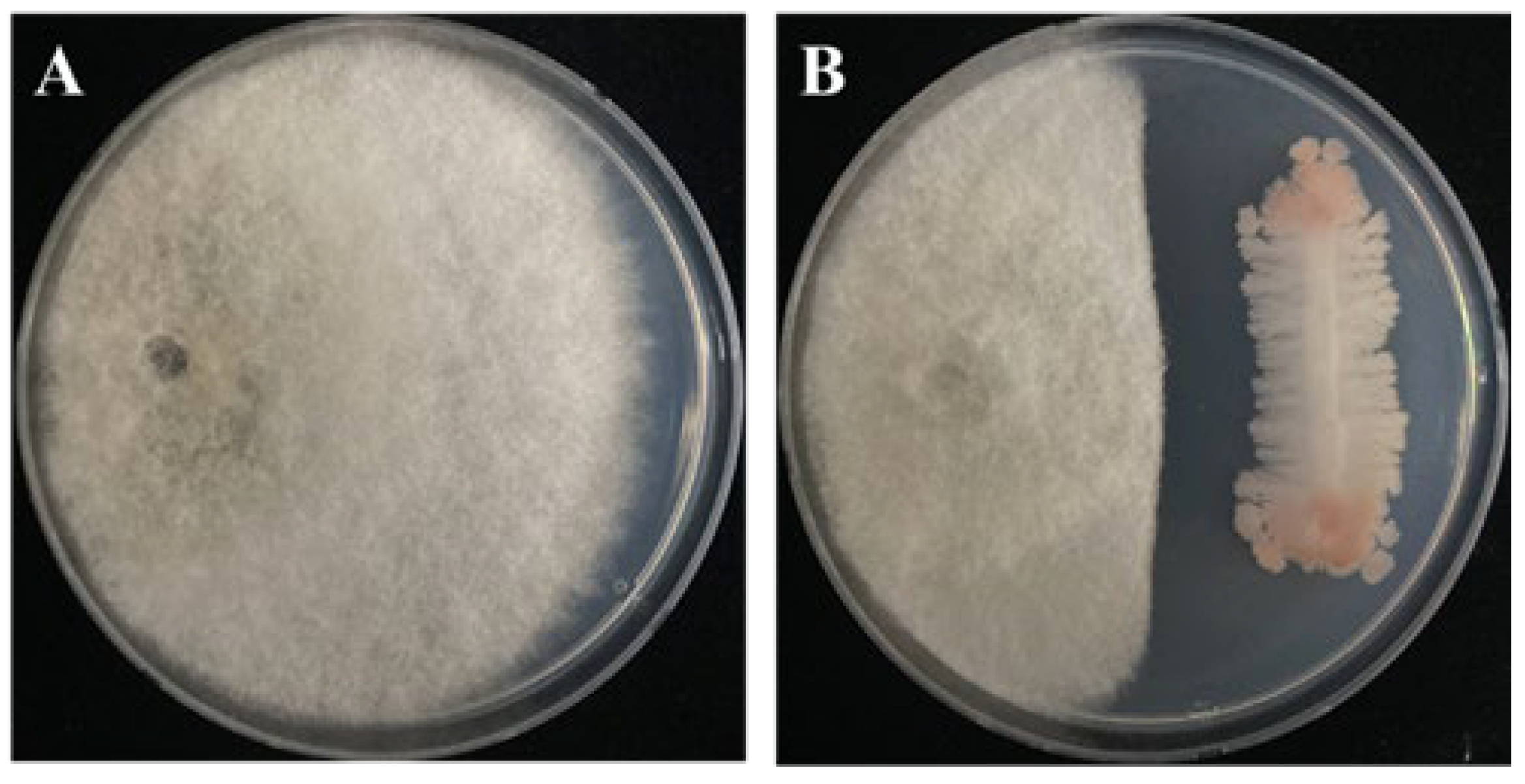
3.1. Antagonistic Activity of B. subtilis PE7 against D. bryoniae

The dual culture assay results revealed that B. subtilis PE7 significantly inhibited the growth of the pathogen D. bryoniae compared to the control (Figure 1A). At 7 days of incubation, the inhibition rate was 57.14 ± 1.2%, as shown in Figure 1B.

3.2. Inhibitory Effect of B. subtilis PE7 VOCs on D. bryoniae

The mycelial growth of the fungal pathogen D. bryoniae was markedly influenced by the VOCs of B. subtilis PE7, resulting in an inhibition rate of 85.4 ± 3.10% (Figure 2B) compared to the control (Figure 2A). Examination under light microscopy revealed that the hyphae affected by VOCs were intricately twisted and swollen (Figure 2D), whereas the control hyphae displayed normal and straight structures (Figure 2C).

3.3. Influence of Chemical Fungicide on the Survival of B. subtilis PE7

The growth of B. subtilis PE7 was examined following treatment with the commercial fungicide over a 7-day incubation period (Figure 3). The presence of fungicide had no remarkable impact on the growth of B. subtilis PE7, because the growth rate remained steady compared to the control even at a 250 ppm concentration (Figure 3A). No significant difference was observed between the control and fungicide treatment after 7 days of incubation. Strain PE7 displayed consistent growth and maintained similar colony numbers from 0 to 7 days after incubation, as illustrated in Figure 3B.

3.4. B. subtilis PE7 Culture Filtrate and Fungicide Co-Exposure for Targeted Suppression of D. bryoniae

The mycelial growth of D. bryoniae was notably inhibited after treatment with different concentrations of B. subtilis PE7 culture filtrate (CF) and a commercial fungicide (F). The highest inhibition percentage occurred with the combined treatment of 30% CF and 50% F, which was statistically significant compared to other treatments. Furthermore, there was no significant difference in mycelial inhibition rate between the 50% F and 100% F treatments (p < 0.05). Notably, the least mycelial growth inhibition was observed in the treatment with 30% CF alone (Figure 4).

3.5. Effects of B. subtilis PE7 CF and Fungicide on D. bryoniae Hyphal Morphology

The mycelial growth was significantly inhibited in both the CF and F treatments compared to the control, which exhibited normal mycelial development. The highest inhibition occurred with the combined treatment (30% CF + 50% F) compared to other treatments (30% F, 50% F, and 100% F). Microscopic examination of the hyphal morphology of the fungal pathogen D. bryoniae revealed abnormal expansion and alterations in the morphology of D. bryoniae treated with fungicide and CF. Notably, more severe damage characterized by twisting, excessive branching, bulging, and irregular twisting was observed in the treatment with 30% CF and the combined treatment (30% CF + 50% F) compared to the other fungicide treatments (Figure 5).

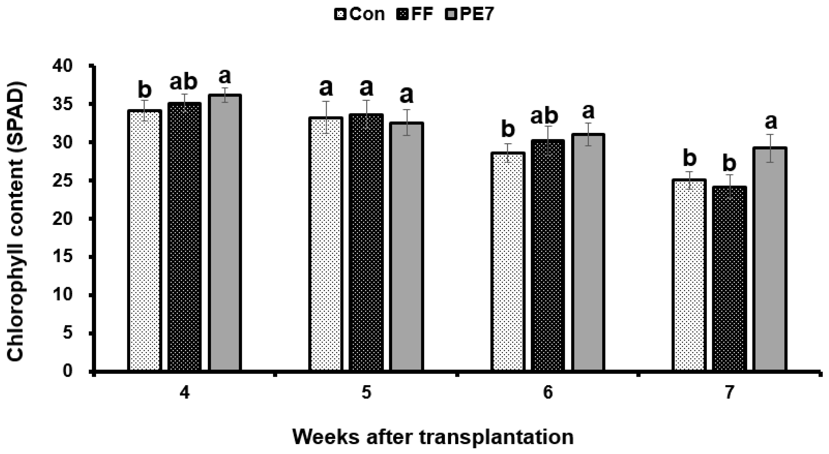
3.6. Effect of B. subtilis PE7 Culture on Melon Plant Growth

The effects of different treatments on the growth of melon plants under potted conditions were evaluated over a 16-day period after D. bryoniae inoculation. Analysis of the results indicated enhanced plant growth, particularly in terms of shoot length (108.87 ± 5 cm) in the PE7 broth culture treatment compared to both the FF treatment (82.7 ± 5 cm) and the control (86.02 ± 1 cm). Notably, no significant difference in melon plant growth was observed between the FF treatment and the control, as depicted in Figure 6A. Moreover, the fresh weight of shoots also increased in the PE7 treatment (94.2 ± 4 g) compared to FF and the control (Table 1). Similarly, root length in melon plants showed an increase in the PE7 treatment (24.98 ± 1 cm) compared to the FF treatment (23.72 ± 0.7 cm) and the control (21.4 ± 1 cm). Fresh root weight was significantly increased in the PE7 treatment group (19.62 ± 4 g) compared to FF and the control (Figure 6B). Moreover, we observed an increase in the leaf number (24.8 ± 0.9) and leaf area (1388.6 cm2) in the PE7 treatment (Table 1). Furthermore, the chlorophyll content of melon plants increased considerably following treatment with the PE7 broth culture over a 4-week period post-transplantation compared to the control. However, during the initial 3 weeks post-transplantation, there was no significant disparity in chlorophyll content between the PE7 and FF treatments, with significance emerging only in the fourth week post-transplantation (Figure 7).

3.7. Effects of B. subtilis PE7 Culture and Fungicide on the Disease Severity of D. bryoniae-Infected Melon Plants

The PDI of GSB was assessed across the following three distinct treatments: Con, FF, and PE7. The highest disease index was observed in the control group, whereas both the FF and PE7 treatments exhibited reduced disease severity. However, there was no statistically significant difference in disease severity between the PE7 and FF treatments 16 days following inoculation with D. bryoniae (Figure 8).

3.8. Nutrient Analysis of Melon Plants

Compared to the control group (Con), melon plants treated with the broth culture of B. subtilis PE7 (PE7) exhibited significantly higher levels of major macronutrients, as detailed in Table 2. Specifically, the PE7-treated plants exhibited notable increases in nitrogen (N) at 199.23 mg plant−1, potassium (K) at 488.27 mg plant−1, magnesium (Mg) at 98.76 mg plant−1, and calcium (Ca) at 328.86 mg plant−1. However, there were no significant differences observed in macronutrient levels (P, K, Mg, and Ca) between the control and PE7-treated groups. Furthermore, the PE7 treatment resulted in elevated levels of micronutrients in melon plants compared to both the Con and FF treatments. Notably, the concentrations of manganese (Mn), boron (B), copper (Cu), iron (Fe), and zinc (Zn) increased to 7.56, 344.34, 1450.49, 30.82, and 0.50 mg plant−1, respectively, in the PE7-treated plants. However, there were no significant differences in micronutrient levels (Cu and Fe) between the FF and PE7 treatments. Additionally, the concentration of molybdenum (Mo) was found to be lowest in the PE7-treated plants at 4.07 mg plant−1 compared to the plants in the Con group at 7.25 mg plant−1 and the FF treatment at 6.43 mg plant−1.

4. Discussion

Bacillus subtilis strains are known for their ability to suppress plant pathogens through various antagonistic actions, including the production of antifungal compounds [28,29], outcompeting the pathogens for nutrients and space [21,30], inducing systemic resistance in plants by priming them to defend against pathogen attacks [31,32,33], forming biofilms on plant surfaces that create physical barriers preventing pathogen attachment and colonization [34,35], and producing enzymes such as chitinases and proteases that directly inhibit fungal growth and disrupt the pathogen’s lifecycle [36,37]. The dual culture methodology facilitates precise quantification of the antagonistic impact exerted by Bacillus strains against fungal pathogens. This is achieved by assessing key growth parameters, including radial growth inhibition, alterations in hyphal morphology, and reduction in biomass [38,39]. Our findings revealed that B. subtilis PE7 inhibits the growth of D. bryoniae, the causal agent of GSB, following a 7-day exposure period.
Grahovac et al. [40] recently reported that various strains of B. subtilis can generate a wide spectrum of VOCs characterized by diverse chemical compositions and biological properties, exhibiting the capacity to hinder the growth of fungal pathogens either through direct effects on fungal cells or by interfering with their physiological processes. Our findings were highly consistent with these observations. Specifically, VOCs emitted by B. subtilis significantly inhibited the mycelial growth of D. bryoniae. Al-Mutar et al. [41] also reported that extracellular lipopeptides and VOCs emitted from B. subtilis strain DHA41 exhibited strong antifungal activity against D. bryoniae, F. oxysporum, F. graminearum, and Sclerotinia sclerotiorum. Similarly, VOCs produced by B. amyloliquefaciens L3, including 2-ethyl-1-hexanol, 2-heptanone, and 2-nonanone, exhibited complete inhibition of F. oxysporum mycelial growth [6]. Leveraging the biocontrol potential of B. subtilis VOCs offers a sustainable and eco-friendly basis for managing fungal diseases in agriculture. The use of VOCs as natural fungicides can reduce the reliance on chemical pesticides and promote environmentally friendly crop protection practices. Waghunde et al. [42] reported that VOCs released by B. subtilis can lead to branching or fragmentation of hyphae in fungal pathogens, resulting in the development of irregular structures and impaired hyphal growth. Similarly, upon exposure to VOCs from B. subtilis PE7, D. bryoniae displayed distorted hyphal structures characterized by swelling and twisting. Ahmed et al. [43] reported that B. amyloliquefaciens WS-10 produced bioactive compounds with strong antifungal properties against various pathogens. The study emphasized the role of VOCs in disrupting the cell walls of fungal pathogens, thereby inhibiting their growth.
The potential synergism between commercial fungicides and B. subtilis is a crucial consideration in integrated pest management strategies. Therefore, assessing the potential interactions between fungicides and beneficial bacteria is critical to avoid negative impacts on biocontrol efficacy [44]. Certain chemical fungicides such as tebuconazole can have antagonistic effects on B. subtilis when applied at high rates, inhibiting its growth or activity and creating unfavorable conditions for its survival and proliferation, thereby potentially compromising its biocontrol efficacy [45]. The influence of chemical fungicides on the survival or synergistic interactions of B. subtilis is highly complex, and understanding the dynamics between fungicides and B. subtilis is essential for optimizing biocontrol strategies and promoting sustainable agriculture practices. Our findings indicate a consistent growth rate in B. subtilis PE7 when exposed to trifloxystrobin, maintaining colony numbers similar to those observed in the control group. This result aligns with those of Kondoh et al. [46], who reported that the chemical pesticide flutolanil had no remarkable effect on the growth of B. subtilis RB14-C. These findings support the potential for the co-administration of biocontrol agents with chemical fungicides, because bacterial survival did not appear to be negatively affected by the fungicide.
Synergistic interactions between Bacillus species and chemical fungicides in controlling fungal pathogens can enhance disease management strategies, including complementary modes of action [47]. In our study, treatment with 30% CF of strain PE7 significantly inhibited the growth of D. bryoniae. This inhibition is likely due to active metabolites accumulated in the CF of strain PE7. Numerous studies have shown that Bacillus species produce a variety of antifungal substances, including cyclic lipopeptides, polypeptides, proteins, and non-peptides, which serve as their primary defense against phytopathogenic microorganisms. Among these antimicrobial compounds, cyclic lipopeptides released by B. subtilis such as iturins, bacillomycins, fengycins, and surfactins are well-known for their inhibitory effects on the growth of phytopathogenic fungi by disturbing cell membranes, inducing osmotic imbalance, and leading to cell death [48,49]. Noh et al. [50] demonstrated that exposure to a CF of B. subtilis NM4 reduced the mycelial growth of Alternaria solani and led to hyphal deformations and abnormal bulbous morphology, which was attributed to the presence of surfactins and fengycins in the CF. Similarly, the bulging and severe swelling in the hyphal structures of D. bryoniae observed in the 30% CF treatment could be attributed to the action of these lipopeptides or related compounds. Moreover, our findings demonstrated that combining 30% CF of B. subtilis PE7 with 50% commercial fungicide resulted in the highest inhibition rate against D. bryoniae, surpassing the outcomes of individual treatments and the control group. This suggests synergism between the fungicide and strain PE7 metabolites, enhancing the growth inhibition of the fungal pathogen. By leveraging the biocontrol capabilities of Bacillus species alongside chemical fungicides, our current reliance on fungicides can be markedly decreased while maintaining effective disease management. This integrated approach can help minimize the use of synthetic chemicals while achieving sustainable disease control outcomes [51]. For instance, combining the chemical fungicide carbendazim 12% + mancozeb 63% with the bio-agent Trichoderma harzianum significantly reduced Fusarium wilt disease (21.72%) compared to the control (64.2%) [52]. Synergistic interactions can result in a broader spectrum of activity, increased pathogen inhibition, and enhanced plant protection against fungal pathogens [53]. Bacillus subtilis produces antifungal compounds that can inhibit fungal growth, including the elongation of hyphae [39]. When combined with chemical fungicides, which also inhibit hyphal growth, the overall impact on fungal morphology can be enhanced, leading to reduced hyphal length and branching [53,54]. Therefore, the synergistic interactions between antimicrobial metabolites produced by B. subtilis PE7 and chemical fungicides (30% CF + 50% F) result in more pronounced effects on fungal hyphal morphology than either treatment alone. This combined approach can provide enhanced control of fungal pathogens by disrupting hyphal growth and structure, thereby impeding the progression of fungal infections and reducing disease severity in plants [55].
In our pot experiment, the soil drench and foliar application of B. subtilis PE7 to melon plants significantly reduced the disease severity of GSB compared to the control. The efficacy of strain PE7 was comparable to that of the fungicide treatment. This outcome may be attributed to the cells and metabolites of strain PE7 disrupting the conidial germination and developmental processes of D. bryoniae, thereby reducing GSB in melon plants. Severe infections of GSB can weaken plants, reduce photosynthetic activity, and impair nutrient uptake, leading to decreased growth and yield. Additionally, gummy exudate from stem cankers can attract secondary pathogens, further compromising plant health [14]. The production of antimicrobial compounds and the alteration of microbial populations by B. subtilis can create an unfavorable environment for pathogen survival and proliferation, leading to decreased disease severity in plants [56]. A similar study by Sayago et al. [57] revealed that the administration of B. subtilis ALBA01 resulted in a lower PDI with a higher biocontrol level (>50%) against the soil-borne pathogen Setophoma terrestris in onions. Furthermore, B. subtilis B579 exhibited a remarkable 73% reduction in F. oxysporum disease incidence in Cucumis sativus (cucumber) plants [58].
Bacillus subtilis is known for its plant growth–promoting properties, including the production of substances such as indole-3-acetic acid (IAA) and siderophores [18,59]. Our previous studies have shown that inoculation with B. subtilis PE7 culture significantly enhances the overall growth of melon and tomato plants. Additionally, the presence of IAA in the PE7 culture positively affects root density and promotes nutrient acquisition, increasing both macro- and micronutrients in melon plants [60,61]. In vivo plant growth assays in the present study also revealed a notable enhancement in both root and shoot development, along with an increase in fresh leaf count in melon plants treated with PE7 strains, surpassing the effects observed with fungicide treatment and the control treated with fertilizer. Our nutrient analysis results further suggest that B. subtilis PE7 significantly improves the macro- and micronutrient content in C. melo compared to the control and FF treatment. Similar to our findings, microbial extracts from Bacillus LKE15 significantly improve the contents of major nutrients such as magnesium, potassium, and phosphorus in oriental melons [62]. Biologically active strains of B. subtilis employ a range of mechanisms that influence plant root and shoot growth. These mechanisms include the modulation of plant hormone production pathways such as cytokinins and gibberellins [63], nutrient solubilization in the soil [64], induction of stress tolerance mechanisms [65], triggering of the plant’s systemic resistance mechanisms [66], root colonization [67], regulation of gene expression in both the plant and the microbe through plant-microbe signaling [68,69,70], and promotion of enhanced root system architecture [71,72,73]. Furthermore, B. subtilis can enhance photosynthetic efficiency by optimizing photosystem functionality and augmenting light harvesting and utilization efficiency, thereby indirectly bolstering chlorophyll biosynthesis [74]. This is strongly supported by our current findings, which show a significant improvement in melon plant chlorophyll content following treatment with B. subtilis compared to fungicide application. In addition to direct antagonistic interactions, B. subtilis can exert indirect effects on pathogens by modulating the microbial community structure in the rhizosphere. In our study, the high numbers of viable cells of strain PE7 in the pot soil could be another reason for the growth enhancement of melon plants. Zhang et al. [75] demonstrated that microbial consortia involving Bacillus strains can significantly alter the rhizosphere bacterial community, enhancing plant resistance to pathogens. The study showed a substantial reduction in disease incidence when microbial consortia were used, suggesting a synergistic effect in biocontrol.

5. Conclusions

This study highlights the significant potential of B. subtilis PE7 as an effective biocontrol agent against D. bryoniae, the causative agent of GSB in cucurbits. By examining the impact of VOCs on the morphology of D. bryoniae, our findings demonstrated the potent antifungal properties of strain PE7. Additionally, our results revealed that PE7 culture filtrate, either alone or in combination with chemical fungicides, significantly inhibits D. bryoniae growth, offering a promising alternative to conventional fungicide treatments. The observed enhancement of the nutrient index and growth of melon plants, along with the reduction in disease severity, further underscores the potential of B. subtilis PE7 in integrated disease management strategies for sustainable crop production. This research provides valuable insights into harnessing microbial agents for effective disease control and crop health enhancement, paving the way for the development of innovative agricultural practices. In future studies, we will investigate the antifungal action of B. subtilis PE7 on different fungal pathogens and explore the active compounds responsible for the growth inhibition of these fungal pathogens. These compounds will be isolated using various chromatographic methods and identified through liquid chromatography–mass spectrometry (LC/MS) and nuclear magnetic resonance (NMR) analyses.

Author Contributions

Conceptualization, K.Y.K. and C.E.H.M.; methodology, S.K.J.; software, S.K.J.; validation, S.E.H. and C.E.H.M.; formal analysis, S.E.H. and C.E.H.M.; investigation, S.K.J. and S.E.H.; resources, K.Y.K. and W.J.J.; data curation, S.E.H. and C.E.H.M.; writing—original draft preparation, P.V.-S.; writing—review and editing, P.V.-S., C.E.H.M. and K.Y.K.; visualization, S.K.J., S.E.H. and K.Y.K.; supervision, K.Y.K.; project administration, W.J.J.; funding acquisition, W.J.J. and P.V.-S. All authors have read and agreed to the published version of the manuscript.

Funding

This research was supported by the Korea Institute of Planning and Evaluation for Technology in Food, Agriculture and Forestry (IPET) through the Agricultural Machinery/Equipment Localization Technology Development Program, funded by the Ministry of Agriculture, Food and Rural Affairs (MAFRA) (Grant No. 122020-03).

Data Availability Statement

All the data are available within the manuscript.

Acknowledgments

We thank the Korea Institute of Planning and Evaluation for Technology in Food, Agriculture, and Forestry (IPET) through the Agricultural Machinery/Equipment Localization Technology Development Program, Ministry of Agriculture, Food, and Rural Affairs (MAFRA) (Grant No. 122020-03) for funding this research.

Conflicts of Interest

The authors declare no conflicts of interest.

References

  1. Hu, Y.; Li, Y.; Zhu, B.; Huang, W.; Chen, J.; Wang, F.; Chen, Y.; Wang, M.; Lai, H.; Zhou, Y. Genome-wide identification of the expansion gene family in netted melon and their transcriptional responses to fruit peel cracking. Front. Plant Sci. 2024, 15, 1332240. [Google Scholar] [CrossRef] [PubMed]
  2. Xiao, J.; Sun, Y.; He, Y.; Tang, X.; Yang, S.; Huang, J. Comparison of Rhizospheric and Endophytic Bacterial Compositions between Netted and Oriental Melons. Microbiol. Spect. 2023, 11, e04027-22. [Google Scholar] [CrossRef] [PubMed]
  3. Kim, K.; Kim, D.; Kwon, T.H.; Lee, B.H.; Lee, S.E. Effective Phyto-sanitary Treatment for Export of Oriental Melons (Cucumis melo var L.) Using Ethyl Formate and Modified Atmosphere Packaging to Control Trialeurodes vaporariorum (Hemiptera: Aleyrodidae). Insects 2023, 14, 442. [Google Scholar] [CrossRef] [PubMed]
  4. Luo, Q.; Tan, G.F.; Ma, Y.Q.; Meng, P.H.; Zhang, J. Cucurbitaceous Vegetables’ Gummy Stem Blight Research. Agronomy 2022, 12, 1283. [Google Scholar] [CrossRef]
  5. Keinath, A.P. Reproduction of Didymella bryoniae on nine species of cucurbits under field conditions. Plant Dis. 2014, 98, 1379–1386. [Google Scholar] [CrossRef]
  6. Wu, Z.W.; Cai, X.W.; Mao, X.W.; Zhou, M.G.; Hou, Y.P. Sensitivity and resistance risk analysis of Didymella bryoniae populations to fluopyram. J. Integr. Agric. 2023, 23, 2306–2317. [Google Scholar] [CrossRef]
  7. Paret, M.L.; Dufault, N.S.; Newark, M.; Freeman, J.H. Management of gummy stem blight (black rot) on cucurbits in Florida. Plant Pathol. Dep. UF/IFAS Ext. 2021, 2018, PP280. [Google Scholar] [CrossRef]
  8. Rahman, M.Z.; Kibria, M.G.; Talukder, M.M.R.; Akhter, M.S.; Amia, M.F. Evaluation of fungicides for control of gummy stem blight of watermelon caused by Didymella bryoniae. Bangladesh J. Plant Pathol. 2019, 35, 47–52. [Google Scholar]
  9. Noh, J.; Kim, J.H.; Lim, J.H.; Kim, T.B.; Seong, M.H.; Jung, G.T.; Kim, J.M.; Cheong, S.S.; Oh, N.K.; Lee, W.H. Occurrence of diseases and case of clinical diagnosis on watermelon in South Korea, 2008–2012. Res. Plant Dis. 2014, 20, 8–14. [Google Scholar] [CrossRef]
  10. Pereira, A.S.; dos Santos, G.R.; Sarmento, R.A.; da Silva Galdino, T.V.; de Oliveira Lima, C.H.; Picanço, M.C. Key factors affecting watermelon yield loss in different growing seasons. Sci. Hortic. 2017, 218, 205–212. [Google Scholar] [CrossRef]
  11. Café-Filho, A.C.; Santos, G.R.; Laranjeira, F.F. Temporal and spatial dynamics of watermelon gummy stem blight epidemics. Eur. J. Plant Pathol. 2010, 128, 473–482. [Google Scholar] [CrossRef]
  12. Rennberger, G.; Keinath, A.P. Susceptibility of fourteen new cucurbit species to gummy stem blight caused by Stagonosporopsis citrulli under field conditions. Plant Dis. 2018, 102, 1365–1375. [Google Scholar] [CrossRef] [PubMed]
  13. Jones, J.G.; Korir, R.C.; Walter, T.L.; Everts, K.L. Reducing chlorothalonil use in fungicide spray programs for powdery mildew, anthracnose, and gummy stem blight in melons. Plant Dis. 2020, 104, 3213–3220. [Google Scholar] [CrossRef] [PubMed]
  14. Seblani, R.; Keinath, A.P.; Munkvold, G. Gummy stem blight: One disease, three pathogens. Mol. Plant Pathol. 2023, 24, 825–837. [Google Scholar] [CrossRef] [PubMed]
  15. Haggag, M.; Wafaa, M.H. Sustainable agriculture management of plant diseases. Online J. Biol. Sci. 2002, 2, 280–284. [Google Scholar]
  16. Gimode, W.; Bao, K.; Fei, Z.; McGregor, C. QTL associated with gummy stem blight resistance in watermelon. Theor. Appl. Genet. 2020, 134, 573–584. [Google Scholar] [CrossRef] [PubMed]
  17. Sharma, M.; Tarafdar, A.; Ghosh, R.; Gopalakrishanan, S. Biological Control as a Tool for Eco-Friendly Management of Plant Pathogens. In Advances in Soil Microbiology: Recent Trends and Future Prospects; Springer: Berlin/Heidelberg, Germany, 2017; pp. 153–188. [Google Scholar]
  18. Maung, C.E.H.; Choub, V.; Cho, J.Y.; Kim, K.Y. Control of the bacterial soft rot pathogen, Pectobacterium carotovorum by Bacillus velezensis CE 100 in cucumber. Microb. Pathog. 2022, 173, 105807. [Google Scholar] [CrossRef] [PubMed]
  19. Kumbar, B.; Mahmood, R.; Nagesha, S.N.; Nagaraja, M.S.; Prashant, D.G.; Kerima, O.Z.; Karosiya, A.; Chavan, M. Field application of Bacillus subtilis isolates for controlling late blight disease of potato caused by Phytophthora infestans. Biocatal. Agric. Biotechnol. 2019, 22, 101366. [Google Scholar] [CrossRef]
  20. Maung, C.E.H.; Baek, W.S.; Choi, T.G.; Kim, K.Y. Control of grey mould disease on strawberry using the effective agent, Bacillus amyloliquefaciens Y1. Biocontrol Sci. Technol. 2021, 31, 468–482. [Google Scholar] [CrossRef]
  21. Vehapi, M.; Inan, B.; Kayacan-Cakmakoglu, S.; Sagdic, O.; Özçimen, D. Optimization of growth conditions for the production of Bacillus subtilis using central composite design and its antagonism against pathogenic fungi. Probiotics Antimicrob. Proteins 2023, 15, 682–693. [Google Scholar] [CrossRef] [PubMed]
  22. Harish, B.N.; Nagesha, S.N.; Ramesh, B.N.; Shyamalamma, S.; Nagaraj, M.S.; Girish, H.C.; Pradeep, C.; Shiva Kumar, K.S.; Tharun Kumar, K.S.; Pavan, S.N.; et al. Molecular characterization and antifungal activity of lipopeptides produced from Bacillus subtilis against plant fungal pathogen Alternaria alternata. BMC Microbiol. 2023, 23, 179. [Google Scholar] [CrossRef] [PubMed]
  23. Tang, R.; Tan, H.; Dai, Y.; Huang, Y.; Yao, H.; Cai, Y.; Yu, G. Application of antimicrobial peptides in plant protection: Making use of the overlooked merits. Front. Plant Sci. 2023, 14, 1139539. [Google Scholar] [CrossRef] [PubMed]
  24. Ngo, T.Q.H.; Thai, T.H.; Tran, T.X.P.; Tran, D.H. Evaluation of Bacillus sp. strains for biological control of gummy stem blight, Didymella bryoniae (Auersw.) in watermelon (Citrullus lanatus). Crop Res. 2023, 58, 238–242. [Google Scholar] [CrossRef]
  25. Chen, Z.; Wang, Z.; Xu, W. Bacillus velezensis WB induces systemic resistance in watermelon against Fusarium wilt. Pest Manag. Sci. 2024, 80, 1423–1434. [Google Scholar] [CrossRef] [PubMed]
  26. Maung, C.E.H.; Lee, H.G.; Cho, J.Y.; Kim, K.Y. Antifungal compound, methyl hippurate from Bacillus velezensis CE 100 and its inhibitory effect on growth of Botrytis cinerea. World J. Microbiol. Biotechnol. 2021, 37, 159. [Google Scholar] [CrossRef] [PubMed]
  27. Hassan, H.A.; Elmenisy, A.; Abdel-Ghafar, N.Y. Prediction of Tomato Early Blight Disease Under Climate Change Conditions in Egypt. Arab Univ. J. Agric. Sci. 2019, 27, 1985–1995. [Google Scholar] [CrossRef]
  28. Awan, Z.A.; Shoaib, A.; Schenk, P.M.; Ahmad, A.; Alansi, S.; Paray, B.A. Antifungal potential of volatiles produced by Bacillus subtilis BS-01 against Alternaria solani in Solanum lycopersicum. Front. Plant Sci. 2023, 13, 1089562. [Google Scholar] [CrossRef] [PubMed]
  29. Trung, N.T.; Thao, N.T.; Le Thanh, N.S.; Dai Nguyen, N.P.; Tuyet, N.T.A.; Cuong, N.T.; Chan, S.S.; Khoo, K.S.; Show, P.L. Antifungal activity of secondary metabolites purified from Bacillus subtilis isolated in Vietnam and evaluated on in vitro and in vivo models. Int. Biodeterior. Biodegrad. 2023, 179, 105558. [Google Scholar]
  30. Qiao, J.; Zhang, R.; Liu, Y.; Liu, Y. Evaluation of the Biocontrol Efficiency of Bacillus subtilis Wettable Powder on Pepper Root Rot Caused by Fusarium solani. Pathogens 2023, 12, 225. [Google Scholar] [CrossRef] [PubMed]
  31. Choudhary, D.K.; Johri, B.N. Interactions of Bacillus spp. and plants—With special reference to induced systemic resistance (ISR). Microbiol. Res. 2009, 164, 493–513. [Google Scholar] [CrossRef] [PubMed]
  32. Yang, P.; Zhao, Z.; Fan, J.; Liang, Y.; Bernier, M.C.; Gao, Y.; Zhao, L.; Opiyo, S.O.; Xia, Y. Bacillus proteolyticus OSUB18 triggers induced systemic resistance against bacterial and fungal pathogens in Arabidopsis. Front. Plant Sci. 2023, 14, 1078100. [Google Scholar] [CrossRef] [PubMed]
  33. Samaniego-Gámez, B.Y.; Valle-Gough, R.E.; Garruña-Hernández, R.; Reyes-Ramírez, A.; Latournerie-Moreno, L.; Tun-Suárez, J.M.; Villanueva-Alonzo, H.D.J.; Nuñez-Ramírez, F.; Diaz, L.C.; Samaniego-Gámez, S.U.; et al. Induced Systemic Resistance in the Bacillus spp.—Capsicum chinese Jacq.—PepGMV Interaction, Elicited by Defense-Related Gene Expression. Plants 2023, 12, 2069. [Google Scholar] [CrossRef] [PubMed]
  34. Arnaouteli, S.; Bamford, N.C.; Stanley-Wall, N.R.; Kovács, Á.T. Bacillus subtilis biofilm formation and social interactions. Nat. Rev. Microbiol. 2021, 19, 600–614. [Google Scholar] [CrossRef] [PubMed]
  35. Verma, P.; Chauhan, P.; Kumar, N.; Mishra, N.; Pandey, S.; Bajpai, R.; Yadav, J.K.; Sahay, R.; Bahadur, L.; Mishra, A. Microbial biofilm approaches in phytopathogen management. In Microbial Biomolecules; Academic Press: Cambridge, MA, USA, 2023; pp. 77–96. [Google Scholar]
  36. Malik, M.S.; Rehman, A.; Khan, I.U.; Khan, T.A.; Jamil, M.; Rha, E.S.; Anees, M. Thermo-neutrophilic cellulases and chitinases characterized from a novel putative antifungal biocontrol agent: Bacillus subtilis TD11. PLoS ONE 2023, 18, e0281102. [Google Scholar] [CrossRef] [PubMed]
  37. Rosazza, T.; Eigentler, L.; Earl, C.; Davidson, F.A.; Stanley-Wall, N.R. Bacillus subtilis extracellular protease production incurs a context-dependent cost. Mol. Microbiol. 2023, 120, 105–121. [Google Scholar] [CrossRef] [PubMed]
  38. Kim, H.S.; Park, J.; Cho, S.W.; Park, K.H.; Lee, G.P.; Ban, S.J.; Lee, C.R.; Kim, C.S. Isolation and characterization of Bacillus strains for biological control. J. Microbiol. 2003, 41, 196–201. [Google Scholar]
  39. Liang, N.; Charron, J.B.; Jabaji, S. Comparative transcriptome analysis reveals the biocontrol mechanism of Bacillus velezensis E68 against Fusarium graminearum DAOMC 180378, the causal agent of Fusarium head blight. PLoS ONE 2023, 18, e0277983. [Google Scholar] [CrossRef] [PubMed]
  40. Grahovac, J.; Pajčin, I.; Vlajkov, V. Bacillus VOCs in the Context of Biological Control. Antibiotics 2023, 12, 581. [Google Scholar] [CrossRef] [PubMed]
  41. Al-Mutar, D.M.K.; Noman, M.; Alzawar, N.S.A.; Qasim, H.H.; Li, D.; Song, F. The extracellular lipopeptides and volatile organic compounds of Bacillus subtilis DHA41 display broad-spectrum antifungal activity against soil-borne pytopathogenic Fungi. J. Fungi 2023, 9, 797. [Google Scholar] [CrossRef] [PubMed]
  42. Waghunde, R.R.; Khunt, M.D.; Shelake, R.M.; Hiremani, N.; Patil, V.A.; Kim, J.Y. Endophytes: A potential bioagent for plant disease management. In Microbial Endophytes and Plant Growth; Academic Press: Cambridge, MA, USA, 2023; pp. 19–34. [Google Scholar]
  43. Ahmed, W.; Zhou, G.; Yang, J.; Munir, S.; Ahmed, A.; Liu, Q.; Zhao, Z.; Ji, G. Bacillus amyloliquefaciens WS-10 as a potential plant growth-promoter and biocontrol agent for bacterial wilt disease of flue-cured tobacco. Egypt. J. Biol. Pest Control 2022, 32, 25. [Google Scholar] [CrossRef]
  44. Salazar, B.; Ortiz, A.; Keswani, C.; Minkina, T.; Mandzhieva, S.; Pratap Singh, S.; Rekadwad, B.; Borriss, R.; Jain, A.; Singh, H.B.; et al. Bacillus spp. as bio-factories for antifungal secondary metabolites: Innovation beyond whole organism formulations. Microb. Ecol. 2023, 86, 1–24. [Google Scholar] [CrossRef] [PubMed]
  45. Liu, L.; Zhao, K.; Cai, L.; Zhang, Y.; Fu, Q.; Huang, S. Combination effects of tebuconazole with Bacillus subtilis to control rice false smut and the related synergistic mechanism. Pest Manag. Sci. 2023, 79, 234–243. [Google Scholar] [CrossRef] [PubMed]
  46. Kondoh, M.; Hirai, M.; Shoda, M. Integrated biological and chemical control of damping-off caused by Rhizoctonia solani using Bacillus subtilis RB14-C and flutolanil. J. Biosci. Bioeng. 2001, 91, 173–177. [Google Scholar] [CrossRef]
  47. Ons, L.; Bylemans, D.; Thevissen, K.; Cammue, B.P. Combining biocontrol agents with chemical fungicides for integrated plant fungal disease control. Microorganisms 2020, 8, 1930. [Google Scholar] [CrossRef] [PubMed]
  48. Liu, J.; Hagberg, I.; Novitsky, L.; Hadj-Moussa, H.; Avis, T.J. Interaction of antimicrobial cyclic lipopeptides from Bacillus subtilis influences their effect on spore germination and membrane permeability in fungal plant pathogens. Fungal Biol. 2014, 118, 855–861. [Google Scholar] [CrossRef] [PubMed]
  49. Wang, T.; Liang, Y.; Wu, M.; Chen, Z.; Lin, J.; Yang, L. Natural products from Bacillus subtilis with antimicrobial properties. Chin. J. Chem. Eng. 2015, 23, 744–754. [Google Scholar] [CrossRef]
  50. Noh, J.S.; Hwang, S.H.; Maung, C.E.H.; Cho, J.Y.; Kim, K.Y. Enhanced control efficacy of Bacillus subtilis NM4 via integration of chlorothalonil on potato early blight caused by Alternaria solani. Microb. Pathog. 2024, 190, 106604. [Google Scholar] [CrossRef] [PubMed]
  51. Ayaz, M.; Li, C.H.; Ali, Q.; Zhao, W.; Chi, Y.K.; Shafiq, M.; Ali, F.; Yu, X.Y.; Yu, Q.; Zhao, J.T.; et al. Bacterial and Fungal Biocontrol Agents for Plant Disease Protection: Journey from Lab to Field, Current Status, Challenges, and Global Perspectives. Molecules 2023, 28, 6735. [Google Scholar] [CrossRef] [PubMed]
  52. Musheer, N.; Jamil, A.; Choudhary, A. Solo and combined applications of fungicides and bio-agents to reduce severity of Fusarium oxysporum and induce antioxidant metabolites in Ocimum tenuiflorum L. J. Plant Pathol. 2023, 105, 237–251. [Google Scholar] [CrossRef]
  53. Gu, Z.W.; Yin, J.H.; Wu, H.; Liang, Y.Q.; Wu, W.H.; Lu, Y.; Li, R.; Tan, S.B.; He, C.P.; Yi, K.X. Synergistic mechanism of Bacillus subtilis Czk1 combined with propiconazole and tebuconazole mixtures against Pyrrhoderma noxium. Chem. Biol. Technol. Agric. 2023, 10, 113. [Google Scholar] [CrossRef]
  54. de Novais, C.B.; Giovannetti, M.; de Faria, S.M.; Sbrana, C. Two herbicides, two fungicides and spore-associated bacteria affect Funneliformis mosseae extraradical mycelium structural traits and viability. Mycorrhiza 2019, 29, 341–349. [Google Scholar] [CrossRef] [PubMed]
  55. Hirozawa, M.T.; Ono, M.A.; Suguiura, I.M.D.S.; Bordini, J.G.; Ono, E.Y.S. Lactic acid bacteria and Bacillus spp. as fungal biological control agents. J. Appl. Microbiol. 2023, 134, 083. [Google Scholar] [CrossRef]
  56. Ahmad, I.; Ahmad, M.; Hussain, A.; Jamil, M. Integrated use of phosphate-solubilizing Bacillus subtilis strain IA6 and zinc-solubilizing Bacillus sp. strain IA16: A promising approach for improving cotton growth. Folia Microbiol. 2021, 66, 115–125. [Google Scholar] [CrossRef] [PubMed]
  57. Sayago, P.; Juncosa, F.; Albarracín Orio, A.G.; Luna, D.F.; Molina, G.; Lafi, J.; Ducasse, D.A. Bacillus subtilis ALBA01 alleviates onion pink root by antagonizing the pathogen Setophoma terrestris and allowing physiological status maintenance. Eur. J. Plant Pathol. 2020, 157, 509–519. [Google Scholar] [CrossRef]
  58. Jabborova, D.; Enakiev, Y.; Sulaymanov, K.; Kadirova, D.; Ali, A.; Annapurna, K. Plant growth promoting bacteria Bacillus subtilis promote growth and physiological parameters of Zingiber officinale Roscoe. Plant Sci. Today 2021, 8, 66–71. [Google Scholar] [CrossRef]
  59. Shafi, J.; Tian, H.; Ji, M. Bacillus species as versatile weapons for plant pathogens: A review. Biotechnol. Biotechnol. Equip. 2017, 31, 446–459. [Google Scholar] [CrossRef]
  60. Chen, F.; Wang, M.; Zheng, Y.; Luo, J.; Yang, X.; Wang, X. Quantitative changes of plant defense enzymes and phytohormone in biocontrol of cucumber Fusarium wilt by Bacillus subtilis B579. World J. Microbiol. Biotechnol. 2010, 26, 675–684. [Google Scholar] [CrossRef]
  61. Han, S.E.; Cho, J.Y.; Kim, K.Y.; Maung, C.E.H. Role of an antagonistic bacterium, Bacillus subtilis PE7, in growth promotion of netted melon (Cucumis melo L. var. reticulatus Naud.). Can. J. Microbiol. 2023, 70, 40–51. [Google Scholar] [CrossRef] [PubMed]
  62. Han, S.E.; Kim, K.S.; Maung, C.E.H.; Kim, K.Y. Growth enhancement of tomato by a plant growth promoting bacterium, Bacillus subtilis PE7. Korean J. Soil Sci. Fertil. 2023, 56, 398–406. [Google Scholar] [CrossRef]
  63. Jensen, C.N.G.; Pang, J.K.Y.; Hahn, C.M.; Gottardi, M.; Husted, S.; Moelbak, L.; Kovács, Á.T.; Fimognari, L.; Schulz, A. Differential influence of Bacillus subtilis strains on Arabidopsis root architecture through common and distinct plant hormonal pathways. Plant Sci. 2024, 339, 111936. [Google Scholar] [CrossRef] [PubMed]
  64. Lastochkina, O.; Garshina, D.; Allagulova, C.; Fedorova, K.; Koryakov, I.; Vladimirova, A. Application of endophytic Bacillus subtilis and salicylic acid to improve wheat growth and tolerance under combined drought and Fusarium root rot stresses. Agronomy 2020, 10, 1343. [Google Scholar] [CrossRef]
  65. Kumar, S.; Anjali, A.R.; Masurkar, P.; Singh, U.B.; Tripathi, R.; Bhupenchandra, I.; Minkina, T.; Keswani, C. Bacillus subtilis-Mediated induction of disease resistance and promotion of plant growth of vegetable crops. In Applications of Bacillus and Bacillus Derived Genera in Agriculture, Biotechnology and Beyond; Springer Nature: Singapore, 2024; pp. 165–211. [Google Scholar]
  66. Pomerleau, M.; Charron-Lamoureux, V.; Leonard, L.; Grenier, F.; Rodrigue, S.; Beauregard, P.B. Adaptive laboratory evolution reveals biofilm regulating genes as key players in B. subtilis root colonization. mSystems 2024, 9, e0084323. [Google Scholar] [CrossRef] [PubMed]
  67. Blake, C.; Christensen, M.N.; Kovács, Á.T. Molecular aspects of plant growth promotion and protection by Bacillus subtilis. Mol. Plant-Microbe Interact. 2021, 34, 15–25. [Google Scholar] [CrossRef]
  68. Chang, P.E.; Wu, Y.H.; Tai, C.Y.; Lin, I.H.; Wang, W.D.; Tseng, T.S.; Chuang, H.W. Examining the transcriptomic and biochemical signatures of Bacillus subtilis strains: Impacts on plant growth and abiotic stress tolerance. Int. J. Mol. Sci. 2023, 24, 13720. [Google Scholar] [CrossRef] [PubMed]
  69. Xu, Z.; Liu, Y.; Zhang, N.; Xun, W.; Feng, H.; Miao, Y.; Shao, J.; Shen, Q.; Zhang, R. Chemical communication in plant–microbe beneficial interactions: A toolbox for precise management of beneficial microbes. Curr. Opin. Microbiol. 2023, 72, 102269. [Google Scholar] [CrossRef] [PubMed]
  70. Posada, L.F.; Álvarez, J.C.; Romero-Tabarez, M.; de-Bashan, L.; Villegas-Escobar, V. Enhanced molecular visualization of root colonization and growth promotion by Bacillus subtilis EA-CB0575 in different growth systems. Microbiol. Res. 2018, 217, 69–80. [Google Scholar] [CrossRef] [PubMed]
  71. Wang, J.; Zhang, Y.; Jin, J.; Li, Q.; Zhao, C.; Nan, W.; Wang, X.; Ma, R.; Bi, Y. An intact cytokinin-signaling pathway is required for Bacillus sp. LZR216-promoted plant growth and root system architecture alteration in Arabidopsis thaliana seedlings. Plant Growth Regul. 2018, 84, 507–518. [Google Scholar] [CrossRef]
  72. Roy, B.; Maitra, D.; Biswas, A.; Chowdhury, N.; Ganguly, S.; Bera, M.; Dutta, S.; Golder, S.; Roy, S.; Ghosh, J.; et al. Efficacy of high-altitude biofilm-forming novel Bacillus subtilis species as plant growth-promoting Rhizobacteria on Zea mays L. Appl. Biochem. Biotechnol. 2023, 196, 643–666. [Google Scholar] [CrossRef] [PubMed]
  73. Mohamed, H.I.; Gomaa, E.Z. Effect of plant growth promoting Bacillus subtilis and Pseudomonas fluorescence on growth and pigment composition of radish plants (Raphanus sativus) under NaCl stress. Photosynthetica 2012, 50, 263–272. [Google Scholar] [CrossRef]
  74. Kang, S.M.; Radhakrishnan, R.; Lee, K.E.; You, Y.H.; Ko, J.H.; Kim, J.H.; Lee, I.J. Mechanism of plant growth promotion elicited by Bacillus sp. LKE15 in oriental melon. Acta Agric. Scand. Sect. B Soil Plant Sci. 2015, 65, 637–647. [Google Scholar]
  75. Zhang, J.; Ahmed, W.; Dai, Z.; Zhou, X.; He, Z.; Wei, L.; Ji, G. Microbial consortia: An engineering tool to suppress clubroot of Chinese cabbage by changing the rhizosphere bacterial community composition. Biology 2022, 11, 918. [Google Scholar] [CrossRef]
Figure 1. Antifungal activity of B. subtilis PE7 against D. bryoniae on the PDA medium. (A) Control plate with D. bryoniae only. (B) Plate with B. subtilis PE7 and D. bryoniae co-culture showing inhibition at 7 days of incubation.
Microorganisms 12 01691 g001
Figure 2. Inhibitory effect of B. subtilis PE7 VOCs on D. bryoniae. (A) Control plate with D. bryoniae only. (B) Plate showing inhibition of D. bryoniae by VOCs from B. subtilis PE7. (C) Normal hyphal structure of D. bryoniae in the control. (D) Twisted and swollen hyphal structure of D. bryoniae affected by VOCs from B. subtilis PE7 (scale bar = 25 µm).
Microorganisms 12 01691 g002
Figure 3. Effect of chemical fungicide (50% trifloxystrobin, 250 ppm) on the survival of B. subtilis PE7. (A) Growth rate comparison between the control and fungicide-treated B. subtilis PE7 at a concentration of 250 ppm. Different letters indicate significant differences between control and fungicide treatment according to Student’s t-test (p < 0.05). (B) Consistent colony numbers of strain PE7 from 0 to 7 days after incubation in both control and fungicide treatments.
Microorganisms 12 01691 g003
Figure 4. Inhibition of D. bryoniae mycelial growth treated with varying concentrations of B. subtilis PE7 culture filtrate (CF) and fungicide (F), as well as their combination (100% F and 50% F correspond to 250 ppm and 125 ppm of 50% trifloxystrobin, respectively). The data were presented as the mean ± standard deviation of three replicates. Different letters in the bar graph indicate significant differences between treatments, as determined by the least significant difference (LSD) test at a p < 0.05 significance level.
Microorganisms 12 01691 g004
Figure 5. Comparison of abnormal mycelial growth and deformed hyphal morphology of D. bryoniae, characterized by bulbous or condensed structures, in treatments with B. subtilis PE7 culture filtrate (CF), fungicide (F), and their combination, relative to the control displaying normal morphology (scale bar = 25 µm).
Microorganisms 12 01691 g005
Figure 6. Influence of B. subtilis PE7 culture on the growth of melon plants. (A) Comparison of shoot length after 16 days postinoculation of D. bryoniae. (B) Comparison of fresh root weight. Con: fertilizer only; FF: fertilizer combined with fungicide; PE7: Broth culture of B. subtilis PE7.
Microorganisms 12 01691 g006
Figure 7. Chlorophyll content of melon leaves of different treatments at different weeks (4–7) post-transplantation. Con: fertilizer only; FF: fertilizer combined with fungicide; PE7: Broth culture of B. subtilis PE7. Different letters above each bar (grouped by post-transplantation week) indicate significant differences between treatments, as determined by the least significant difference (LSD) test at a p < 0.05 significance level.
Microorganisms 12 01691 g007
Figure 8. Reduction in disease severity of GSB in melon by B. subtilis PE7 culture and fungicide compared to the control at 16 days after D. bryoniae inoculation. Con: fertilizer only; FF: fertilizer combined with fungicide; PE7: Broth culture of B. subtilis PE7. Different letters in the bar graph indicate significant differences between treatments, as determined by the least significant difference (LSD) test at a p < 0.05 significance level.
Microorganisms 12 01691 g008
Table 1. Plant growth parameters under different treatments at 16 days postinoculation with D. bryoniae. Con: fertilizer only; FF: fertilizer combined with fungicide; PE7: Broth culture of B. subtilis PE7. The data were presented as the mean ± standard deviation of six replicates. Different letters in the same column indicate significant differences between treatments, as determined by the least significant difference (LSD) test at a p < 0.05 significance level.
Length (cm)Fresh Weight (g)Dry Weight (g)Leaf NumberLeaf Area (cm2)Strain PE7 in Pot Soil
ShootRootShootRootShootRoot
Con86.02 ± 1.22 b21.40 ± 1.76 a73.52 ± 3.78 b13.77 ± 2.56 b8.14 ± 0.52 b1.17 ± 0.21 b22.67 ± 0.52 b1111.83 ± 35.76 c-
FF82.72 ± 5.65 b23.72 ± 0.79 a77.06 ± 9.06 b15.31 ± 3.83 ab7.90 ± 0.69 b1.16 ± 0.26 b24.33 ± 1.97 a1269.07 ± 12.36 b-
PE7108.87 ± 5.17 a24.98 ± 1.71 a94.22 ± 4.23 a19.62 ± 4.21 a9.41 ± 0.25 a1.54 ± 0.40 a24.83 ± 0.98 a1388.66 ± 96.14 a1.73 ± 0.34 × 10⁷
CFU mL−1
Table 2. Effect of different treatments on the nutrient content of melon plants. (Con: fertilizer only; FF: fertilizer combined with fungicide; PE7: Broth culture of B. subtilis PE7). Different letters in the same column indicate significant differences between treatments, as determined by the least significant difference (LSD) test at a p < 0.05 significance level.
TreatmentMacronutrient (mg plant−1)
NPKMgCa
Con132.84 ± 5.37 b11.57 ± 0.57 a416.66 ± 8.98 b83.98 ± 8.52 a284.83 ± 12.86 b
FF131.94 ± 5.47 b11.46 ± 1.17 a416.66 ± 8.98 b86.75 ± 7.66 a340.66 ± 14.57 a
PE7199.23 ± 7.92 a11.13 ± 0.92 a488.27 ± 20.50 a98.76 ± 10.63 a328.86 ± 16.50 a
TreatmentMicronutrient (mg plant−1)
MnBCuFeMoZn
Con3.56 ± 0.20 b171.60 ± 1.73 b962.57 ± 275.3 a31.38 ± 11.27 a7.25 ± 0.51 a0.24 ± 0.03 c
FF3.78 ± 0.11 b162.85 ± 2.41 b1275.7 ± 227.5 a29.01 ± 5.63 a6.43 ± 0.51 a0.34 ± 0.01 b
PE77.56 ± 0.59 a344.34 ± 24.53 a1450.4 ± 322.1 a30.82 ± 13.03 a4.07 ± 0.98 b0.50 ± 0.06 a
N, nitrogen; P, phosphorous; K, potassium; Mg, Magnesium; Ca, Calcium; Mn, Manganese; B, Boron; Cu, Copper; Fe, Iron; Mo, Molybdenum; Zn, Zinc.
Disclaimer/Publisher’s Note: The statements, opinions and data contained in all publications are solely those of the individual author(s) and contributor(s) and not of MDPI and/or the editor(s). MDPI and/or the editor(s) disclaim responsibility for any injury to people or property resulting from any ideas, methods, instructions or products referred to in the content.

