Article

Epibiotic Bacteria Isolated from the Non-Indigenous Species Codium fragile ssp. fragile: Identification, Characterization, and Biotechnological Potential

National Institute of Marine Sciences and Technologies (INSTM), University of Carthage, Tunis 2025, Tunisia
*
Authors to whom correspondence should be addressed.
Microorganisms 2024, 12(9), 1803; https://doi.org/10.3390/microorganisms12091803 (registering DOI)
Submission received: 29 February 2024 / Revised: 26 April 2024 / Accepted: 29 April 2024 / Published: 30 August 2024
(This article belongs to the Special Issue Holobionts in Aquaculture)

Abstract

:
Due to their richness in organic substances and nutrients, seaweed (macroalgae) harbor a large number of epiphytic bacteria on their surfaces. These bacteria interact with their host in multiple complex ways, in particular, by producing chemical compounds. The released metabolites may have biological properties beneficial for applications in both industry and medicine. In this study, we assess the diversity of culturable bacterial community of the invasive alga Codium fragile ssp. fragile with the aim to identify key groups within this epiphytic community. Seaweed samples were collected from the Northern Tunisian coast. A total of fifty bacteria were isolated in pure culture. These bacterial strains were identified by amplification of the ribosomal intergenic transcribed spacer between the 16S and the 23S rRNA genes (ITS-PCR) and by 16S rRNA sequencing. Antimicrobial activity, biochemical, and antibiotic resistance profile characterization were determined for the isolates. Isolated strains were tested for their antimicrobial potential against human and fish bacterial pathogens and the yeast Candida albicans, using the in vitro drop method. About 37% of isolated strains possess antibacterial activity with a variable antimicrobial spectrum. Ba1 (closely related to Pseudoalteromonas spiralis), Ba12 (closely related to Enterococcus faecium), and Bw4 (closely related to Pseudoalteromonas sp.) exhibited strong antimicrobial activity against E. coli. The isolated strain Ba4, closely related to Serratia marcescens, demonstrated the most potent activity against pathogens. The susceptibility of these strains to 12 commonly used antibiotics was investigated. Majority of the isolates were resistant to oxacillin, cefoxitin, tobramycin, and nitrofurantoin. Ba7 and Ba10, closely related to the Vibrio anguillarum strains, had the highest multidrug resistance profiles. The enzymes most commonly produced by the isolated strains were amylase, lecithinase, and agarase. Moreover, nine isolates produced disintegration zones around their colonies on agar plates with agarolitic index, ranging from 0.60 to 2.38. This investigation highlighted that Codium fragile ssp. fragile possesses an important diversity of epiphytic bacteria on its surface that could be cultivated in high biomass and may be considered for biotechnological application and as sources of antimicrobial drugs.

1. Introduction

Algal surfaces provide suitable substrates for microbial colonization and secrete various organic substances that serve as nutrients for bacterial growth and microbial biofilm formation. Microbial communities living on the seaweed surface are highly complex and consist of a dynamic group of microorganisms, including bacteria, fungi, diatoms, protozoa, spores, and the larvae of marine invertebrates. These organisms play crucial roles in every marine ecological process, hence the growing interest in studying their populations and functions [1,2]. Novel and diverse molecules with a large antimicrobial spectrum derived from algae and their associated bacteria could have a huge potential for use in biotechnological research. Different studies have reported that the micro- and macro-organisms of an opportunistic nature colonize surfaces of seaweed, producing protective compounds [3]. Surface-associated marine organisms such as microbes, diatoms and larval forms of marine invertebrates have been detailed to be related with algal thallus [4,5]. These epibiotic communities, especially epiphytic bacteria, provide hormones, vitamins, minerals, carbon dioxide, and a large number of bioactive metabolites to the seaweed and therefore play an important role in the morphogenesis, growth, and immune defense. In return, the seaweed provides a habitat, oxygen, and carbohydrates such as algal polysaccharides to the associated microorganisms [6,7]. The importance of microbial diversity on the macroalgae surface, especially among the bacterial genus, is highly host-specific as new species emerge from this algal environment [8]. The secondary metabolites produced by these bacteria are widely recognized for their importance in biotechnological uses [9,10,11]. Antimicrobial properties [12] and antifungal activities [13] of the epiphytic bacterial communities from the macroalgae collected from the Tunisian coasts have been reported.
The green alga Codium fragile ssp. fragile (CFF) has spread rapidly in temperate areas throughout the globe from its native range in Japan [14]. This alien algae were observed for the first time on the Tunisian coasts in 1985 [15]. The distribution of the algae on the Tunisian coasts has since expanded over the last decade [16,17]. Studies on the bacteria associated with algae from the Tunisian coasts have gradually emerged over the last ten years; but none has targeted the invasive algae Codium fragile. As an invasive algae, having made various adaptations to settle into their new environment, Codium fragile have absolutely hosted a specific epiphytic bacterial community to protect themselves from exogenous pathogens [18]. This specific bacterial community could also be a source of new bioactive compounds of biotechnological interest. The current study has been undertaken based on the occurrence of the invasive seaweed Codium fragile on the northern coast of Tunisia to evaluate its associated bacteria and potential biotechnological application. After the isolation of culturable bacterial strains from the algal surface, a molecular identification of Codium fragile-associated bacteria was undertaken, and antimicrobial activity against various pathogens was evaluated. In addition, antibiotic resistance and enzymatic production profiles were performed. The main objectives of this study were to isolate culturable Codium fragile-associated bacteria and evaluate their biological activities, including enzyme production capacity, antibacterial activity, and antibiotic resistance, in order to provide additional data on invasive seaweeds collected from the Southern Mediterranean coasts.

2. Materials and Methods

2.1. Sampling Site

CFF samples were collected in January 2022 from a rocky shore at the La Marsa site (36°53′06″ N and 10°20′14″ E) in the northern coast of Tunisia (Southern Mediterranean Sea). The whole thalli were sampled carefully with their holdfasts by snorkeling at a 2 m depth. The freshly collected samples were immediately transported in an ice box to the laboratory and kept at 4 °C until analysis.

2.2. Taxonomic Identification of Seaweed Sample

In the laboratory, the algal material was cleaned with seawater to remove the maximum number of epiphytes, and then kept in 2% formaldehyde seawater. Fixed materials were identified using a binocular microscope (Alphaphot-2.YS2-H; Nikon, Tokyo, Japan). The identification was based on different taxonomical keys. To determinate the subspecies of Codium fragile, the morpho-anatomical details were analyzed following the keys reported in the literature [19,20,21]. The green invasive alga C. fragile ssp. fragile (CFF) has a spongy, irregularly dichotomous branched thallus. The internal structure is composed of intertwined colorless medullary filaments that are amorphously cylindrical, and a green palisade-like layer of vesicles called utricles prolonging into a long pointed mucron. The presence of the pointed mucron and surface covered with a multitude of hairs, which gives it this aspect fluffy characteristic of C. fragile, confirmed the identity of the species (Figure 1).

2.3. Isolation of Seaweed Associated Bacteria

Fresh seaweed transported to the laboratory in sterile plastic bags were first rinsed with sterile seawater for a few seconds to remove the non-attached bacteria. Ten grams of seaweed were weighed and homogenized with 9 mL of sterile seawater, vortexed, serially diluted and 0.1 mL aliquots were plated on agar in triplicate, according to Ismail et al. [12]. The plates were incubated at 20 °C for a minimum of 7 days on Zobell Marine Agar (HiMedia) [22]. The individual bacterial strains were then isolated by repeated streaking. The pure cultures were stored at −80 °C in Marine Broth supplemented with 20% glycerol until further study.

2.4. Morphological Characterization

Morphological characterization tests were conducted based on the colony morphology, Gram staining, and catalase and oxidase tests, according to Dalton et al. [23].

2.5. Genotypic Characterization and Identification of Isolated Strains

The bacterial DNA was extracted using the Easy-Pure Bacteria Genomic DNA Kit (Trans-Gen Biotech, Beijing, China). Bacterial strain identification at the species and inter-species levels were performed via PCR 16S-23S intergenic spacer region amplification and using primers ITS-F (3′-GTCGTAACAAGGTAGCCGTA-5′) and ITS-R (3′-CTACGGCTACCTTGTTACGA-5′) as previously described by [24].
All the amplification products were visualized on a 1% agarose gel stained with 5 µL of SYBR Green (Atlas Clear-Sight DNA Stain, Bio-Atlas, Istanbul, Türkiye). ITS profiles were visually analyzed to group together the bacterial isolates exhibiting the same band pattern. At least one representative for each ITS group was identified by partial 16S rRNA sequencing. The amplification of the bacterial 16S rRNA gene was performed using the universal primers F27 (5′-AGAGTTTGATCCTGGCTGGCTCAG-3′) and R1492 (5′-TACGGCTACCTTGTTACGACTT-3′). Subsequent alignment of the sequence was performed on the NCBI database (http://www.ncbi.nlm.nih.gov/BLAST/Blast.cgi (accessed on 22 June 2023)).
A phylogenetic dendrogram was constructed using the neighbor-joining method, and tree topology was evaluated by bootstrap analysis of 1000 data sets using MEGA 6 [25].
The partial 16S rRNA gene sequences of each isolate were submitted to the NCBI GenBank database under the following accession numbers: ON908586, ON908587, ON908588, ON908589, ON908590, OR139885, ON908592, ON908593, ON908594, ON908595, ON908596, ON908597, ON908598, ON908599, ON908604, ON908609, ON908608, ON908605, ON908600, ON908601, ON908603, ON908602, ON908607, ON908606, OR139886, OR139887, OR139888, OR139889, OR139890, OR139891.

2.6. Antibiotic Resistance Profile Analysis

Antimicrobial susceptibility testing was performed on 24 isolates by the disk diffusion method on Mueller Hinton (MH, BIO RAD, Hercules, CA, USA) plates [12]. Twelve antimicrobial agents (BIO RAD, Marnes-la-Coquette, France) were tested (Table 1). A strain was determined to be sensitive to an antimicrobial compound if any growth inhibition zone was observed around the disk. The interpretation was performed in accordance with the French Society of Microbiology’s norms [26].

Determination of MAR Index

Determination of the multiple antibiotic resistances (MAR) index was calculated according to [27], using the following formula:
MAR = a/b
in which (a) is the number of antibiotics to which an isolate is resistant and (b) is the total number of the antibiotics used in the study.

2.7. Screening of Qualitative Enzymatic Production of CFF-Associated Bacteria

Several biochemical and enzymatic tests like the lipase, DNase, lecithinase, amylase, hemolysis, gelatinase, chitinase, cellulase, and agarose tests were carried out. All tests were performed in triplicate.

2.7.1. Lipase

Bacteria were streaked on agar plates beforehand prepared with Tween-80. The results of this test were readable after 24 h at 37 °C, through the formation of an opaque zone denoting a lipase positive response [28].

2.7.2. DNase

After incubation for 48 h at 28 °C, the surface of the agar medium (with a single colony) was covered with the toluidine blue reagent. A pink halo around the culture was formed in a DNase positive response [29].

2.7.3. Lecithinase

Bacterial streaks were inoculated on nutrient agar containing a sterile egg yolk emulsion, followed by an incubation of 24 to 72 h at 30 °C. Opaque areas in the transparent halo would indicate degradation of the egg yolk lecithin by bacterial production of the lecithinase enzyme [30].

2.7.4. Amylase

Bacteria were inoculated on specific starch agarin a single streak, and then incubated for 24 to 72 h at 30 °C. The hydrolysis of amylases was indicated by the presence of a clear zone around the colonies after the addition of Lugol. The absence of staining around the culture would show the degradation of starch (amylase-positive bacteria), while areas containing starch would stain brown (amylase-negative bacteria) [31].

2.7.5. Hemolysis

Agar plates were prepared with horse blood. A streak of bacteria was added and incubated at 30 °C. Results were observed after 48 to 72 h and distinguished with a colorless area [32].

2.7.6. Gelatinase

Nutrient agar medium containing 1% gelatin was inoculated with bacterial isolates and the plates were incubated at 30 or 37 °C for 2 to 5 days. A solution of mercury chloride was used to highlight the degradation of gelatin with a clear halo around the colonies [33].

2.7.7. Chitinase

This test was carried out on nutrient agar supplemented with 1% chitin. After inoculation of bacteria in streaks, plates were incubated for 72 h at 30 °C. The appearance of light areas around the colonies would indicate the production of chitinase [34].

2.7.8. Cellulase

The bacterial isolates were streaked on a plate and incubated for 72 h at 30 °C. Afterwards, 1% Congo red aqueous solution was added, and this allowed for the demonstration of cellulose decomposition after 15 min [35].

2.7.9. Agarase

A qualitative test for the agarase enzyme was performed with iodine using Lugol’s staining process. Selected isolates were grown in ZMB (HiMedia, Kennett Square, PA, USA), pH 7.6 ± 0.2. An 8 mm well was cut in the agar, and 100 µL broth culture of each strain was added into the well and incubated at 30°C for 3 days. After incubation, the agarolytic activity was measured by adding 2 mL Lugol’s iodine on the entire surface of the plate and leaving it for 15 min [36].
Additionally, the agarolytic index (AI) was calculated as the ratio between the diameter of the clear zone and the colony. This index denotes the ability of the bacteria to produce agarase enzymes [36].
The calculating formula is shown below:
AI = Clear zone diameter (mm) − Colony diameter (mm)/Colony diameter (mm)

2.8. Antimicrobial Tests of CFF Associated Bacteria

To study the antibiotic activity, the spot method described by Fleming et al. [37] was used. A fresh culture of pathogenic bacteria (Escherchia coli (ATCC 14948), Vibrio anguillarum (ATCC 12964), Vibrio alginolyticus (ATCC 17749), Pseudomonas aeruginosa (ATCC 27853), Salmonella typhymurium (C52), and Staphylococcus aureus (ATCC 25923)), and the unicellular yeast (Candida albicans (ATCC 10231)) were prepared and cultured in 9 mL of marine broth and incubated at 30 °C for 24 h. After incubation, 9 mL of the target strain was diluted in Mueller Hinton Agar (MH) to 106 CFU/mL. Then, the mixture was poured onto the MH layer. The Petri dishes were left at room temperature to allow for the drying of the strains. Then, fresh cultures of all isolated bacteria in nutrient broth were prepared. The spot test consisted of depositing a volume of 5 μL of the fresh culture from each strain on the dried plate. The same volume of sterile marine broth was also deposited as a negative control. The plates were then incubated at 30 °C for 24 h. The antibacterial activity was known from the appearance of light areas around the spots. The diameter of the inhibition zones was measured in millimeters, and the diameter of the spot was never considered in the expression of the results [38].

3. Results

3.1. Molecular Identification of Isolates

Culturable epiphytic bacteria from the CFF collected from the La Marsa site, were isolated. Fifty bacterial isolates were obtained from the surface of Codium. In addition, six bacterial isolates were isolated from the sediment and water. The obtained results show a higher number of culturable bacteria from the alga thallus compared to the sediment or the surrounding water.
ITS-PCR fingerprinting was applied to assess the bacterial diversity of the selected isolates. The ITS profiles showed reproducible patterns consisting of 1 to 45 bands, with sizes ranging from 50 to about 1000 bp (Figure 2). Thirteen different ITS haplotypes were obtained. Six haplotypes, H5 (Ba3), H6 (Bw4), H7 (Ba4), H10 (Ba20), H11 (Ba17), and H13 (Ba18), were represented by one isolate. The following ITS profiles were found with at least two isolates: H3 (Ba6, Ba9), H4 (Bw3, A43), H8 (A26,Ba2, Ba8, Ba19), H9 (Ba21, Ba22, Ba23, Ba24), H12 (Ba1,Ba5, A22, Ba11) H1 (A32,A34,A35,Ba7,Ba10), H2 (Bs1, Bs2,A38, A39,Bw1, Bw2, Ba12, Ba13, Ba14,Ba15, Ba16, A47, A49, A44, A45).
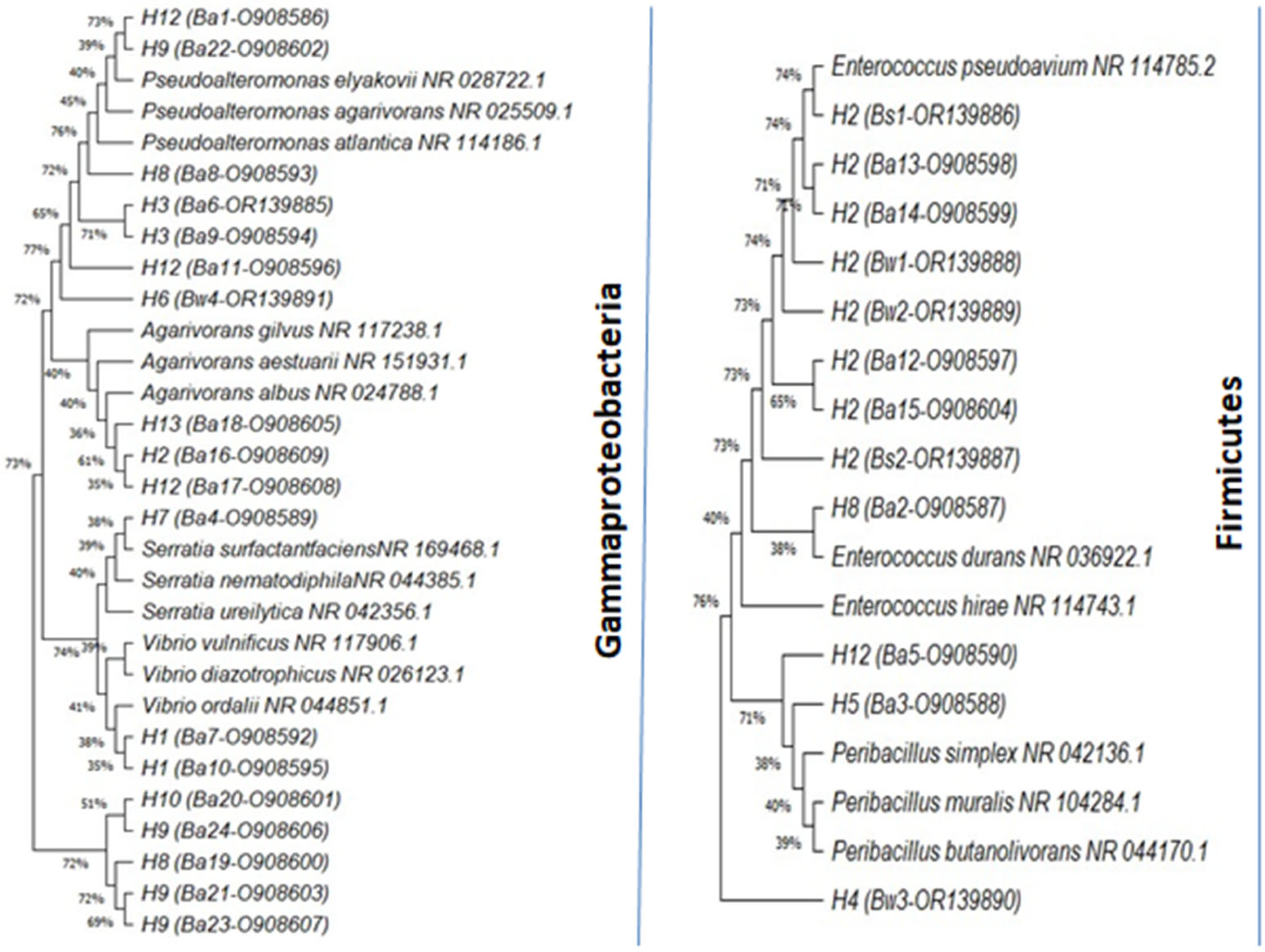
A total of 30 bacterial isolates were selected and subjected to identification, characterization, and phylogenetic analysis, among which 24 were from the alga, 2 were from the sediment, and 4 were from the surrounding water (Table 2). Phylogenetic analysis revealed that the selected isolates belonged to two phylla, namely the Gammaproteobacteria (63%) and the Firmicutes (37%), showing 98–100% identity to the published species sequences. Based on 16S rRNA gene sequences compared to those of their close relatives, the differential alignment of bacterial isolates with different species was highlighted (Figure 3). Among the isolated strains, five strains assigned to genus Pseudoalteromonas included the following: P. spiralis (Ba1), P. agarivorans (Ba8), P. piscicida (Ba6 and Ba9), and P. shioyasakiensis (Ba11). The genus Pseudomonas included P. khazarica (Ba19, Ba20, Ba21, Ba23 and Ba24), while the genus Vibrio was represented by V. anguillarum (Ba7et Ba10) and V. atlanticus (Bw3). The genus Agarivorans included A. litoreus (Ba16), A. abus (Ba17), and Agarivorans sp. (Ba18), while the genus Peribacillus was represented by P. frigoritolerans (Ba3 and Ba5), and the genus Serratia was represented by S. marcescens. Finally, the genus Enterococcus was represented by E. faecium (Ba2, Ba12, Ba13, Ba14, Ba15, Bs1, Bs2, Bw1, and Bw2).

3.2. Antibiotic Resistant Profile of CFF Associated Bacteria

The resistance profiles of 24 bacterial isolates from the CFF samples to 12 antibiotics are shown in Figure 4. A high percentage of the strains show antibiotic resistance to cefoxitin (90%) and oxacillin (87%), while a lower percentage of the strains show resistance to streptomycin (40%), tobramycin (37%), cefotaxime (30%), and to nitrofurantoin (27%). Most of the strains were resistant to imipenem (93% sensible), pipemidic acid (90%), and trimethoprim/sulfamethoxazole (87%), and all the isolated strains showed sensitivity to chloramphenicol, norfloxacin, and tetracycline. A phenotype involving various antibiotic resistances was observed. Ten dominant multi-resistance profiles were found for all bacterial isolates against six families of antibiotics (quinolones, penicillin, aminoglycoside, nitrofuran, pyridopyrimidine, and carbapenem). Over 40% of the strains showed a MAR index higher than 0.4. Isolates Ba7 and Ba10, closely related to Vibrio anguillarum, displayed the highest Mar index of 0.6 (Figure 4).
The antibiotic resistance profiles of the associated bacteria isolated from sediment and from water are shown in Figure 5. All isolates revealed sensitivity to cefoxitin, norfloxacin, tetracycline, chloramphenicol, and trimethoprim/sulfamethoxazole, with Pseudoalteromonas sp. presenting the highest MAR index with 0.4.

3.3. Screening of Qualitative Enzyme Production of CFF Associated Bacteria

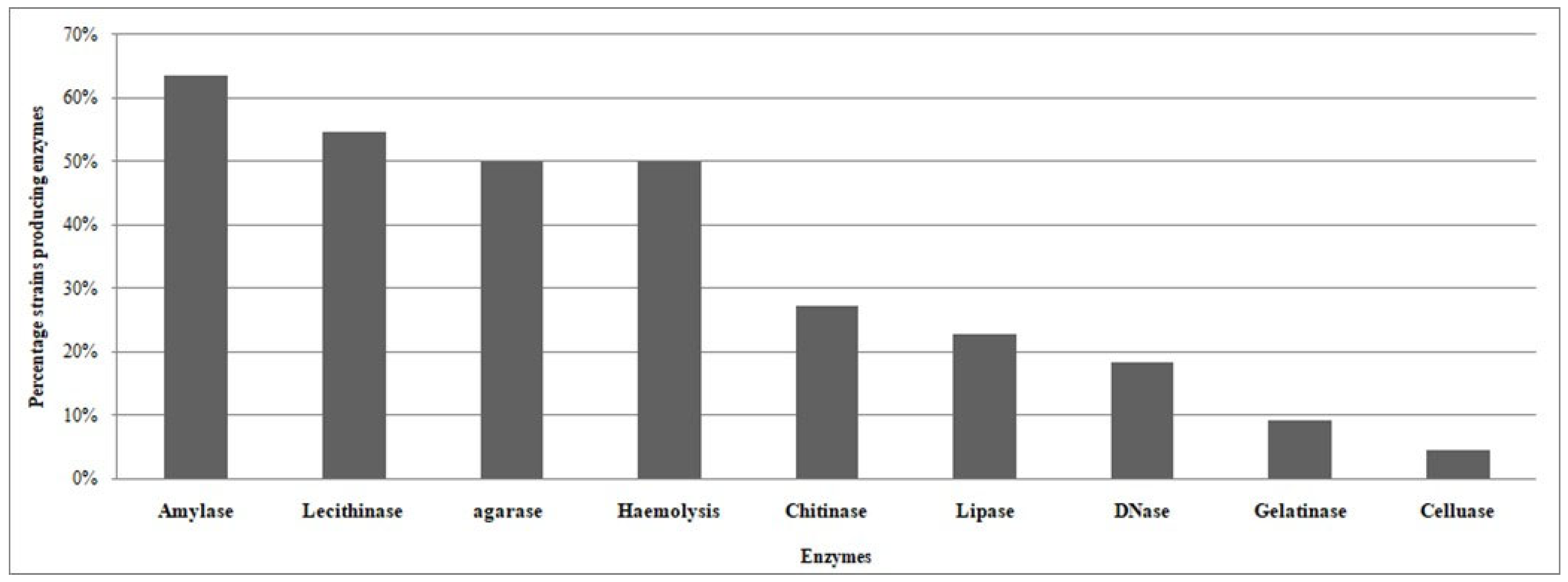
Table 3 presents an enzymatic production profile of the CFF-associated isolates. Most of the isolates show the ability to produce enzymes, with 71% producing more than one enzyme. Three strains, Ba8 and Ba22 (closely related to Pseudoalteromonas agarivorans), and Ba23 (closely related to Pseudomonas khazarica), showed the ability to produce at least five enzymes, while three CFF-associated strains, Ba2 (Enterococcus faecium), Ba3 (Peribacillusfrigoritolerans), and Ba10 (Vibrio anguillarum), had the ability to produce 50% of the tested enzymes. The most produced enzymes by the CFF-associated strains were amylase, lecithinase, and agarase with percentages of 64%, 55%, 50%, respectively (Figure 6). The most productive genera among isolates were the Pseudoalteromonas, Pseudomonas, Enterococcus and Vibrio strains. These strains had the ability to produce chitinase, amylase, and cause hemolysis, while Agarivorans isolates had the ability to produce agarase and lecithinase.

3.4. Qualitative Assays of Agarase Enzyme Production

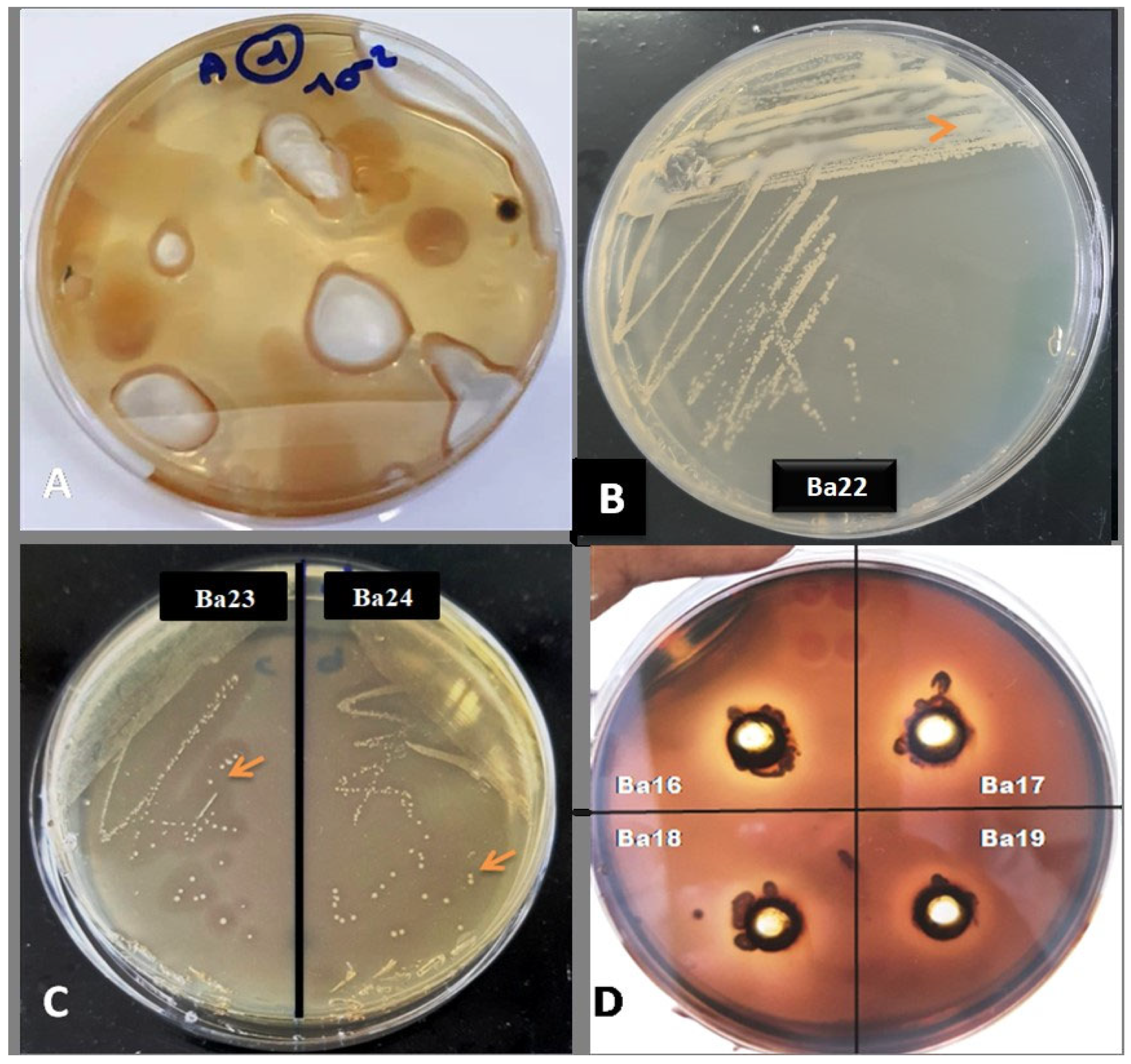
Nine strains demonstrate important agarose activity as shown in Figure 7. All the isolates (Ba16, Ba17, Ba18, Ba19, Ba20, Ba21, Ba22, Ba23, and Ba24) showed a clear light-colored zone around the culture when the dark brown iodine was poured on the culture plates. The potential of the agarolitic bacteria in producing agarase enzymes can be seen qualitatively by calculating their agarolitic index. Table 4 shows the agarolitic index enzyme activity obtained for the CFF bacterial isolates. A clear zone forming around the colony with a different halo upon the addition of drops of iodine solution would demonstrate the enzymatic activity of extracellular agarose produced by agarolitic isolates from the CFF (Table 3). Ba16, Ba17, and Ba18, closely related to Agarivorans litoreus, Agarivoransabus, Agarivorans sp., respectively, gave the higher agarolitic index.

3.5. Antimicrobial Activity of CFF Associated Bacteria

Antimicrobial activity observed for the isolated strains is shown in Table 5. Out of the 24 strains, 9 displayed antimicrobial activity against E. coli, consisting of 8 Gammaproteobacteria (Ba1, Ba3, Ba4, Ba7, Ba8, Ba10, Ba11 and Ba12) and 9 Firmicutes (Ba12).Conforming to the results, the strongest antimicrobial activity was obtained for the strains Ba1 (closely related to Pseudoalteromonas spiralis), Ba12 (closely related to Enterococcus faecium), and Bw4 (closely related to Pseudoalteromonas sp.) with an inhibition against E. coli showing a diameter of more than 20 mm. The isolate Ba4 (closely related to Serratia marcescens) had the largest antimicrobial activity, inhibiting the six pathogenic bacteria tested as well asthe yeast. Only one strain, Ba4 (closely related to Serratia marcescens), displayed a wide screen of antimicrobial activity, showing positive inhibition of all the tested pathogenic species.

4. Discussion

Our findings suggest that Codium fragile ssp. fragile hosts a varied community of cultivable bacteria, affirming that the CFF thallus is indeed a conducive matrix for bacterial accumulation and growth. Phylogenetic analysis revealed that the selected isolates represented 98–100% identity to the published species sequences. However, six isolates’ sequences had a percentage identity below 98.7% (Ba16, Ba17, Ba18, Ba20, Ba24 and Bw4), which suggests that these may be new species [39]. The majority of the isolated strains belong to the phyla Gammaproteobacteria. These findings are in accordance with several studies showing that Proteobacteria phyla are common members of macroalgal bacterial communities [40], with dominance in the surface of green algae [41]. A study on Codium tomentosum showed that the majority of the bacterioflora is largely represented by Proteobacteria (94.6%) and secondarily by Bacteroidetes, Spirochaetes, and Firmicutes [42].
At the genus level, it is observed that Pseudoalteromonas, Pseudomonas, and Enterococcus were the most abundant genera in the current study, where bacteria were collected from the surface of the entire thallus. Epiphytic bacteria like Azotobacter have been evidenced at the surface of the utricles of Codium fragile [43], while cyanobacterial colonies may develop between the bases of the utricles in Codium decorticatum [44]. Illustrations with scanning electron microscopy revealed the presence of bacteria, mainly of the Bacillus type, at the level of the apical region of the utricle [45].
In the present study, a phenotype involving various antibiotic resistances was observed. Ten dominant multi-resistance profiles were found for all the bacterial isolates against six families of antibiotics (quinolones, penicillin, aminoglycoside, nitrofuran, pyridopyrimidine, and carbapenem). The MAR index of the majority of the bacteria ≥ 0.2, as mentioned in previous studies, increases the consumption of antibiotics and thus contributes to an increase in environmental waste [46]. The ecological basis of antibiotic resistance is the adaptation of bacteria to a polluted environment. Bacteria present in these contaminated aquatic matrices share or exchange transferable elements of DNA with other bacteria, and this can occur between different bacterial species [47]. Seawater is often contaminated with drug residues [48]. This contamination encourages the spread of resistance and even of multi-resistant bacteria [49]. Few data are available on multidrug resistance patterns of marine vegetation-associated bacteria. Resistance to Aztreonam, Ceftazidime, Amoxicillin, and Rifampicin has been reported for epi-endophytic bacterial strains isolated from Posidonia oceanica seagrass from the eastern coast of Tunisia, with a high MAR index of 0.67 [50].
The isolates investigated in this study showed diversified enzymatic activity. The majority of the isolates associated with CFF have the capacity to produce amylase. Amylases have the potential to be used in a wide range of industrial processes; for example, a large variety of microbial α-amylases have applications in food, textile, paper, and detergent industries [51]. CFF-associated bacterial strains have the ability to produce Lecithinases (59%). Lecithinases are also capable of hemolysis, and they work synergistically with phospholipases to hydrolyze lipids and lecithin. In this study, the average of chitinase-positive strains appears to be 36.4%, particularly considering those isolates obtained from CFF, the surrounding water, and sediment. Degradation of chitin in the aquatic biosphere is a very active and efficient process mainly carried out by bacteria. It is well known that chitinolytic bacteria are ubiquitous in marine environments. However, in our study, we found chitinolytic strains in sediment and in water. Chitinolytic enzymes have a wide range of potential applications in biotechnology, and the search for innovative chitinolytic bacteria continues to be a fascinating subject. Some applications of chitinases in the food and wine industries have been successfully tested at the laboratory level [52]. Only a few strains were found positive for cellulase. However, due to the crucial applications of these enzymes, positive strains could have highly valuable biotechnological uses [53].
Currently, a number of microorganisms have been reported to secrete agarase, and are mainly in a marine environment, either in the sea water, marine sediments, or associated to red algae [54,55]. An agar-degrading bacteria, Agarivorans sp., has been isolated from the red alga Grateloupia filicina. The enzyme has been characterized as part of the glycoside hydrolase family, β-agarase. By degrading agar, the enzyme produced neoagaro-oligosaccharides with biological properties [56]. Agarase is primarily used to produce oligosaccharides from agar. Agar-derived oligosaccharides have many functions, including hepatoprotective potential [57], anti-oxidation [58] and have potential applications in the food, cosmetic [59], and medical industries. In addition, agarase can also be used to degrade the cell walls of seaweed to generate protoplasts [60,61]. Agarase has also been reported to be used to recover DNA from agarose gel [62,63]. Recently, efforts have been made to find even more active agarases in the environment.
All the isolates grown on solid media were aerobic, showed clear bright zones around the colonies which degraded agar as shown by the iodine test, and showed pits on the surface of the agar medium. It is concluded that the higher agarolitic activity detected from isolates Ba16, Ba17, and Ba18 had an agarolitic index equivalent to 2.38, 2.13, and 2.38, respectively. Our results are comparable to those found by [27], who obtained an agarolytic index of 3.75 mm and 2.53 mm for isolates Sg8 and A13 respectively, isolated from marine algae in India. Agarolitic bacteria could be divided into two groups, namely those which only softened the agar and those which caused extensive liquefaction of agar. The isolated strains of agarolytic bacteria produced at least two enzyme complexes, one cell-free and the other cell-bound, which hydrolyzed agar with the formation of oligosaccharides [64]. A new agarase was thus purified from an agarolytic bacterium, Bacillus megaterium, a novel agar-degrading bacterium isolated from marine sediment [65]. Agar-degrading organisms have been isolated from a wide range of environments, including seawater, marine sediments, marine algae, marine mollusks, fresh water, and soils. Agarase activity has been investigated in various bacteria, including the genus Alteromonas, Pseudomonas, Vibrio, Cytophaga, Agarivorans, Thalassomonas, Pseudoalteromonas, Bacillus, and Acinetobacter [66].
In the present work, the agarolitic isolates were susceptible to Chloramphenicol, imipenem, Trimethoprim/sulfamethoxazole, Cefotaxime, Tobramycin, Streptomycin, Nitrofurantoin, Tetracycline, Pipemidic acid, and Norfloxacin. All the strains isolated from CFF were sensitive to Oxacillin and Cefoxitin. In particular, theisolated agarolytic strains were susceptible to penicillin, kanamycin, nitrofurantoin, and tobramycin [67]. Representatives of the aerobic genus Agarivorans, which is related to Proteobacteria, may generate agarase and catalyze the hydrolysis of agar. A unique strain WH0801T was recently isolated from the surface of seaweed in the shallow coastal region of Weihai, China, for its agarolytic activity and has been suggested as a new species, Agarivorans gilvus [68]. Agarolytic bacteria are likely involved in the degradation of algal polysaccharides. The presence of these bacteria may play an important role in the nutrient cycling of marine ecosystems and can have biotechnological applications in the production of agar-based products. Agarases are hydrolytic enzymes used in biotechnological and commercial applications including the following: decomposing algal polysaccharides, the creation of simple sugars, biofilm removal in bioreactors, making bread and low-calorie foods in the food sector [69], as well as applications in cosmetics, and medical industries [60]. Agarase can be used for molecular biology applications such as the extraction of DNA or RNA fragments from agarose gel [70]. Some agar oligosaccharides obtained with agarase enzyme have anti-oxidative, antibacterial, antimutagenic, and immune-modulating properties [71].
In this study, 37% of the isolates displayed antibacterial activity, which is in accordance with findings from research conducted along the coast of Scotland, where 22% of the isolated strains from Codium fragile presented antimicrobial activity [72].The strain Ba4 (closely related to Serratia marcescens) was the most active strain, inhibiting the growth of all the pathogenic species used. Interestingly, a previously isolated strain from the genus Serratia, associated with the coralline red alga Amphiroa anceps, demonstrated antimicrobial activity [4]. The isolate showed antibacterial activity against E. coli, Staphylococcus aureus, and Klebsiella sp., and exhibited antifungal properties. Besides Serratia nematodiphila and Serratia marcescens were isolated from the Malaysian marine environment and have been reported to produce strong antimicrobial compounds acting against Staphylococcus aureus and Candida albicans. Nevertheless, the isolate did not show any activity against E. coli [73,74]. Arivuselvam et al. [74] reported that the Serratia marcescens JSSCPM1 strain exhibited significant antibacterial activity against the Gram-negative bacterial strains E. coli NCIM 2065, K. pneumoniae NCIM 2706, and P. aeruginosa.
Species of the genus Pseudoalteromonas are often associated with eukaryotic hosts [75,76]. The presence of the Pseudoalteromonas species in diverse habitats worldwide suggests that their adaptive and survival strategies are varied, efficient, and hold significant potential for both fundamental and practical research. Many Pseudoalteromonas species have been demonstrated to produce antibacterial products which appear to help them in the colonization of surfaces, including those of their hosts [76]. Pseudoalteromonas species display a broad range of antibiotic, agarolytic, algicidal, and antifouling effects. The production of agarases, toxins, bacteriolytic substances, and other enzymes by many Pseudoalteromonas species may assist the bacterial cells in their competition for nutrients and space, as well as in their protection against predators grazing at surfaces [77]. Furthermore, the marine bacterium, Pseudoalteromonas phenolica sp. nov. O-BC30T was active against themethicillin-resistant Staphylococcus aureus and the hypersensitive Escherichia coli mutant (KO 1489) [78].
In this study, the isolate Ba12, identified as closely related to Enterococcus faecium, demonstrated antibacterial activity against E. coli. Enterococcus is also known to be one of the most commonly used lactic acid-producing bacteria and has since become a focus for its use in commercially farmed aquatic species [79]. Moreover, probiotics are used to improve water quality and control of bacterial infections, can improve the digestibility of nutrients, increase tolerance to stress, and encourage reproduction [80]. Thus, our findings can be of great interest for aquaculture industry to develop sustainable commercial probiotic products prepared from the Enterococcus species isolated from algae.
Concerning the antimicrobial activity of the associated bacteria in same area, the results of the present study is in agreement with that found by [12] and [81], which showed that several bacterial strains isolated from the Ulva surface have an important antimicrobial activity.

5. Conclusions

Codium fragile is a marine alga widely distributed in the Mediterranean Sea, particularly along the Tunisian coast. This alga has been found to harbor diverse bacterial communities, including agarolytic bacteria, a polysaccharide commonly found in the cell walls of red macroalgae which are known to degrade agar. The associated bacterial communities of Codium fragile ssp. fragile collected from the northern Tunisian coast are diverse and displayed different enzymatic profiles. The use of culture-dependent and culture-independent methods provides a more comprehensive understanding of the bacterial communities associated with marine algae and their potential biotechnological applications. Culturable associated bacteria presented a multidrug resistance profile, suggesting their adaptation and ability to encode antibiotic resistance genes. Further studies are needed to explore the ecological and biotechnological significance of these bacterial communities.

Author Contributions

Conceptualization, W.C., L.K. and M.E.B.; methodology, W.C., A.I. and B.H.; software, W.C. and B.H.; investigation, W.C.; writing—original draft preparation, W.C.; writing—review and editing, L.K., A.I. and M.E.B.; supervision, L.K. and M.E.B. All authors have read and agreed to the published version of the manuscript.

Funding

This research is part of the project PRI—National Project IRESA/INSTM “Développement de Procédés Biologiques Innovants en Alternative à l’Antibiothérapie” (PROBIA2).

Data Availability Statement

The raw data supporting the conclusions of this article will be made available by the authors upon request.

Acknowledgments

The authors thank M. Chahin Benmesaoud for his invaluable assistance during the collection of algae samples used in this study. We would like to express our sincere gratitude to Rhadia Mrouana for her invaluable assistance throughout this research. Her technical expertise, dedication, and attention to detail were instrumental in the successful execution of our experiments.

Conflicts of Interest

The authors declare no conflicts of interest.

References

  1. Goecke, F.; Labes, A.; Wiese, J.; Imhoff, J.F. Chemical interactions between marine macroalgae and bacteria. Mar. Ecol. Prog. Ser. 2010, 409, 267–300. [Google Scholar] [CrossRef]
  2. Zozaya-Valdés, E.; Roth-Schulze, A.J.; Thomas, T. Effects of temperature stress and aquarium conditions on the red macroalga Delisea pulchra and its associated microbial community. Front. Microbiol. 2016, 7, 161. [Google Scholar] [CrossRef]
  3. Srinivasan, R.; Kannappan, A.; Shi, C.; Lin, X. Marine Bacterial Secondary Metabolites: A Treasure House for Structurally Unique and Effective Antimicrobial Compounds. Mar. Drugs 2021, 19, 530. [Google Scholar] [CrossRef]
  4. Karthick, P.; Mohanraju, R. Antimicrobial Potential of Epiphytic Bacteria Associated with Seaweeds of Little Andaman, India. Front. Microbiol. 2018, 9, 611. [Google Scholar] [CrossRef]
  5. Menaa, F.; Wijesinghe, P.A.U.I.; Thiripuranatha, G.; Uzair, B.; Iqbal, H.; Ali Khan, B.; Menaa, B. Ecological and Industrial Implications of Dynamic Seaweed-Associated Microbiota Interactions Mar. Drugs 2020, 18, 641. [Google Scholar] [CrossRef]
  6. Martin, M.; Portetelle, D.; Michel, G.; Vandenbol, M. Microorganisms living on macroalgae: Diversity, interactions, and biotechnological applications. Appl. Microbiol. Biotechnol. 2014, 98, 2917–2935. [Google Scholar] [CrossRef] [PubMed]
  7. Mei, X.; Wu, C.; Zhao, J.; Yan, T.; Jiang, P. Community structure of bacteria associated with drifting Sargassumhorneri, the causative species of golden tide in the Yellow Sea. Front. Microbiol. 2019, 10, 1192. [Google Scholar] [CrossRef]
  8. Goecke, F.; Thiel, V.; Wiese, J.; Labes, A.; Imhoff, J.F. Algae as an important environment for bacteria—Phylogenetic relationships among new bacterial species isolated from algae. Phycologia 2013, 52, 14–24. [Google Scholar] [CrossRef]
  9. Armstrong, E.; Yan, L.; Boyd, K.G.; Wright, P.C.; Burgess, J.G. The symbiotic role of marine microbes on living surfaces. Hydrobiologia 2001, 461, 37–40. [Google Scholar] [CrossRef]
  10. Kelecom, A. Secondary metabolites from marine microorganisms. An. Acad. Bras. Cienc. 2002, 74, 151–170. [Google Scholar] [CrossRef]
  11. Burgess, J.G.; Boyd, K.G.; Armstrong, E.; Jiang, Z.; Yan, L.; Berggren, M. The development of a marine natural product-based antifouling paint. Biofouling 2003, 19, 197–205. [Google Scholar] [CrossRef]
  12. Ismail, A.; Ktari, L.; Ahmed, M.; Bolhuis, H.; Bouhaouala-Zahar, B.; Stal, L.J.; Boudabbous, A.; El Bour, M. Heterotrophic bacteria associated with the green alga Ulva rigida: Identification and antimicrobial potential. J. Appl. Phycol. 2018, 30, 2883–2899. [Google Scholar] [CrossRef]
  13. Chen, J.; Zang, Y.; Yang, Z.; Qu, T.; Sun, T.; Liang, S.; Zhu, M.; Wang, Y.; Tang, X. Composition and Functional Diversity of Epiphytic Bacterial and Fungal Communities on Marine Macrophytes in an Intertidal Zone. Front. Microbiol. 2022, 13, 839465. [Google Scholar] [CrossRef] [PubMed]
  14. Trowbridge, C.D.; Todd, C.D. Host-plant changes of marine specialist herbivores: Ascoglossan sea slugs on the introduced macroalgae. Ecol. Monogr. 2001, 71, 219–243. [Google Scholar] [CrossRef]
  15. Djellouli, A. Sur la présence de Codium fragile (Suringar) Hariot (Codiaceae, Ulvophyceae) en Tunisie. Bull. Société Linnéenne Provence 1987, 39, 103–105. [Google Scholar]
  16. Sghaier, Y.R.; Zakhama-Sraieb, R.; Mouelhi, S.; Vazquez, M.; Valle, C.; Ramos-Espla, A.A.; Astier, J.M.; Verlaque, M.; Charfi-Cheikhrouha, F. Review of alien marine macrophytes in Tunisia. Mediterr. Mar. Sci. 2016, 17, 109–123. [Google Scholar] [CrossRef]
  17. Cherif, W.; Ktari, L.; El Bour, M.; Boudabous, A.; Grignon-Dubois, M. Codium fragile subsp. fragile (Suringar) Hariot in Tunisia: Morphological data and status of knowledge. ALGAE 2016, 31, 129–136. [Google Scholar] [CrossRef]
  18. Morrissey, K.L.; Iveša, L.; Delva, S.; D’Hondt, S.; Willems, A.; De Clerck, O. Impacts of environmental stress on resistance and resilience of algal-associated bacterial communities. Ecol. Evol. 2021, 11, 15004–15019. [Google Scholar] [CrossRef]
  19. Silva, P.C. The dichotomous species of Codium in Britain. J. Mar. Biol. Assoc. 1955, 34, 565–577. [Google Scholar] [CrossRef]
  20. Trowbridge, C.D. Ecology of the green macroalga Codium fragile (Suringar) Hariot 1889: Invasive and noninvasive subspecies. Oceanogr. Mar. Biol. 1998, 36, 1–64. [Google Scholar]
  21. Hubbard, C.B.; Garbary, D.J. Morphological variation of Codium fragile (Chlorophyta) in eastern Canada. Bot. Mar. 2002, 45, 476–485. [Google Scholar] [CrossRef]
  22. Lemos, M.L.; Toranzo, A.E.; Barja, J.L. Antibiotic activity of epiphytic bacteria isolated from intertidal seaweeds. Microb. Ecol. 1985, 11, 149–163. [Google Scholar] [CrossRef]
  23. Dalton, H.M.; Poulsen, L.K.; Halasz, P.; Angles, M.L.; Goodman, A.E.; Marshall, K.C. Substratum induced morphological changes in a marine bacterium and their relevance to bioflm structure. J. Bacteriol. 1994, 176, 6900–6906. [Google Scholar] [CrossRef]
  24. Hassen, W.; Neifar, M.; Cherif, H.; Mahjoubi, M.; Souissi, Y.; Raddadi, N.; Fava, F.; Cherif, A. Assessment of genetic diversity and bioremediation potential of Pseudomonas isolated from pesticide-contaminated artichoke farm soils. Biotech 2018, 8, 263. [Google Scholar]
  25. Tamura, K.; Stecher, G.; Kumar, S. MEGA11: Molecular evolutionary genetics analysis version 11. Mol. Biol. Evol. 2021, 38, 3022–3027. [Google Scholar] [CrossRef]
  26. Soussy, C.J. Antibiogram Committee of the French Society of Microbiology; Centre Hospitalier Universitaire Henri Mondor: Créteil, France, 2008. [Google Scholar]
  27. Krumperman, P.H. Multiple antibiotic resistance indexing of Escherichia coli to identify high-risk sources of fecal contamination of foods. Appl. Environ. Microbiol. 1983, 46, 165–170. [Google Scholar] [CrossRef]
  28. Sierra, G. A simple method for the detection of lipolytic activity of microorganisms and some observations on the influence of the contact between cells and fatty substrates. Antonie Van Leeuwenhoek 1957, 23, 15–22. [Google Scholar] [CrossRef]
  29. Schreiber, L.; Kjeldsen, K.U.; Funch, P.; Jensen, J.; Obst, M.; LópezLegentil, S.; Schramm, A. Endozoicomonas are specifc, facultative symbionts of sea squirts. Front. Microbiol. 2016, 7, 1042. [Google Scholar] [CrossRef]
  30. Garrity, P.; Jones, G.; Krieg, D.; Ludwig, N.R.; Rainey, W.F.A.; Schleifer, K.H.; Whitman, W. (Eds.) Bergey’s Manual of Systematic Bacteriology, 2nd ed.; Springer: New York, NY, USA, 2009; Volume 3. [Google Scholar]
  31. Krishnan, A.; Kumar, G.; Loganathan, K.; Rao, B. Optimization, production and partial purifcation of extracellular αamylase from Bacillus sp. marini. Arch. Appl. Sci. Res. 2011, 3, 33–42. [Google Scholar]
  32. Satpute, S.; Bhawsar, B.D.; Dhakephalkar, P.; Chopade, B. Assessment of different screening methods for selecting biosurfactant producing marine bacteria. Indian J. Mar. Sci. 2008, 37, 243–250. [Google Scholar]
  33. Smith, H.L.; Goodner, K. Detection of bacterial gelatinases by gelatin-agar plate methods. J. Bacteriol. 1958, 76, 662–665. [Google Scholar] [CrossRef]
  34. Kaur, K.; Dattajirao, V.; Shrivastava, V.; Bhardwaj, U. Isolation and characterization of chitosan-producing bacteria from beaches of Chennai, India. Enzym. Res. 2012, 2012, 421683. [Google Scholar] [CrossRef]
  35. Sinha, P.; Bedi, S. Production of sugars from cellulosic wastes by enzymatic hydrolysis. Int. J. Curr. Res. 2018, 10, 72141–72144. [Google Scholar]
  36. Kandasamy, K.P.; Subramanian, R.K.; Srinivasan, R.; Ragunath, S.G.; Balaji, G.; Gracy, M.; Latha, K. Shewanellaalgae and Microbulbiferelongatus from marine macro-algae—Isolation and characterization of agarhydrolysing bacteria. Access Microbiol. 2020, 2, e000170. [Google Scholar] [CrossRef]
  37. Fleming, H.P.; Etchells, J.L.; Costilow, R.N. Microbiol inhibition by an isolate of Pediococcus from cucumber brines. Appl. Microbiol. 1975, 30, 1040–1042. [Google Scholar] [CrossRef]
  38. Ismail, A.; Ktari, L.; Ahmed, M.; Bolhuis, H.; Boudabbous, A.; Stal, L.J. Antimicrobial activities of bacteria associated with the brown alga Padina pavonica. Front. Microbiol. 2016, 7, 1072. [Google Scholar] [CrossRef]
  39. Stackebrandt, E.; Ebers, J. Taxonomic parameters revisited: Tarnished gold standards. Microbiol. Today 2006, 33, 152–155. [Google Scholar]
  40. Florez, J.Z.; Camus, C.; Hengst, M.B.; Buschmann, A.H. A functional perspective analysis of macroalgae and epiphytic bacterial community interaction. Front. Microbiol. 2017, 8, 2561. [Google Scholar] [CrossRef]
  41. Fisher, M.M.; Wilcox, L.W.; Graham, L.E. Molecular characterization of epiphytic bacterial communities on Charophycean green algae applied and environmental. Microbiology 1998, 64, 4384–4389. [Google Scholar]
  42. Le Pennec, G.; Gall, E.A. The microbiome of Codium tomentosum: Original state and in the presence of copper. World J. Microbiol. Biotechnol. 2019, 35, 167. [Google Scholar] [CrossRef]
  43. Head, W.D.; Carpenter, E.J. Nitrogen fixation associated with the marine macroalga Codium fragile. Limnol. Oceanogr. 1975, 20, 815–823. [Google Scholar] [CrossRef]
  44. Rosenberg, G.; Paerl, H.W. Nitrogenfxation by blue-green algae associated with siphonous green seaweed Codium decorticatum: Effects on ammonium uptake. Mar. Biol. 1981, 61, 151–158. [Google Scholar] [CrossRef]
  45. Singh, R.P.; Reddy, C.R.K. Seaweed-microbial interactions: Key functions of seaweed-associated bacteria. FEMS Microbiol. Ecol. 2014, 88, 213–230. [Google Scholar] [CrossRef]
  46. Polianciuc, S.I.; Gurzău, A.E.; Kiss, B.; Ştefan, M.G.; Loghin, F. Antibiotics in the environment: Causes and consequences. Med. Pharm. Rep. 2020, 93, 231–240. [Google Scholar] [CrossRef]
  47. Tahrani, L.; Van Loco, J.; Anthonissen, R.; Verschaeve, L.; Ben Mansour, H.; Reyns, T. Identification and risk assessment of human and veterinary antibiotics in the wastewater treatment plants and the adjacent sea in Tunisia. Water Sci. Technol. 2017, 76, 3000–3021. [Google Scholar] [CrossRef]
  48. Afsa, S.; Hamden, K.; Martin, P.A.L.; Mansour, H.B. Occurrence of 40 pharmaceutically active compounds in hospital and urban wastewaters and their contribution to Mahdia coastal seawater contamination. Environ. Sci. Pollut. Res. 2020, 27, 1941–1955. [Google Scholar] [CrossRef]
  49. Alibi, S.; Beltifa, A.; Hassen, W.; Jaziri, A.; Soussia, L.; Zbidi, F.; Ben Mansour, H. Coastal Surveillance and Water Quality monitoring in the Rejiche Sea-Tunisia. Water Environ. Res. 2021, 93, 2025–2033. [Google Scholar] [CrossRef] [PubMed]
  50. Jebara, A.; Hassen, W.; Ousleti, A.; Mabrouk, L.; Jaziri, A.; Di Bella, G.; Ben Mansour, H. Multidrug-resistant epi-endophytic bacterial community in Posidonia oceanica of Mahdia coast as biomonitoring factor for antibiotic contamination. Arch Microbiol. 2022, 204, 229. [Google Scholar] [CrossRef]
  51. De Souza, P.M.; de Oliveirae, P.M. Application of microbial α-amylase in industry—A review. Braz. J. Microbiol. 2010, 41, 850–861. [Google Scholar] [CrossRef]
  52. Fenice, M.; Giambattista, R.D.; Leuba, J.L.; Federici, F. Inactivation of Mucorplumbeus by the combined actions of chitinase and high hydrostatic pressure. Int. J. Food Microbiol. 1999, 52, 109–113. [Google Scholar] [CrossRef]
  53. Raveendran, C.; Naveenan, T.; Varatharajan, G.R. Optimization of alkaline cellulase production by the marine-derived fungus Chaetomium sp. using agricultural and industrial wastes as substrates. Bot. Mar. 2010, 53, 275–282. [Google Scholar] [CrossRef]
  54. Lavilla-Pitogo, C.R. Agar-digesting bacteria associated with ‘rotten thallus syndrome’ of Gracilaria sp. Aquaculture 1992, 102, 1–7. [Google Scholar] [CrossRef]
  55. Schroeder, D.C.; Jaffer, M.A.; Coyne, V.E. Investigation of the role of a beta (1–4) agarase produced by Pseudoalteromonas gracilis B9 in eliciting disease symptoms in the red alga Gracilaria gracilis. Microbiology 2003, 149, 2919–2929. [Google Scholar] [CrossRef]
  56. Nikapitiya, C.; Oh, C.H.; Lee, Y.D.; Lee, S.K.; Whang, I.S.; Lee, J.H. Characterization of a glycoside hydrolase family 50 thermostable β-agaraseAgrA from marine bacteria Agarivorans sp. AG17. Fish. Aquat. Sci. 2010, 13, 36–48. [Google Scholar] [CrossRef]
  57. Chen, H.; Yan, X.; Zhu, P.; Lin, J. Antioxidant activity and hepatoprotective potential of agaro-oligosaccharides in vitro and in vivo. Nutr. J. 2006, 5, 31. [Google Scholar] [CrossRef]
  58. Chen, X.; Fu, X.; Huang, L.; Xu, J.; Gao, X. Agar oligosaccharides: A review of preparation, structures, bioactivities and application. Carbohydr. Polym. 2021, 265, 118076. [Google Scholar] [CrossRef]
  59. Wang, J.; Jiang, X.; Mou, H.; Guan, H. Anti-oxidation of agar oligosaccharides produced by agarase from a marine bacterium. J. Appl. Phycol. 2004, 16, 333–340. [Google Scholar] [CrossRef]
  60. Kobayashi, R.; Takisada, M.; Suzuki, T.; Kirimura, K.; Usami, S. Neoagarobiose as a novel moisturizer with whitening effect. Biosci. Biotechnol. Biochem. 1997, 61, 162–163. [Google Scholar] [CrossRef] [PubMed]
  61. Chen, L.C.M.; Craigie, J.S.; Xie, Z.K. Protoplast production from Porphyralinearis using a simplified agarase procedure capable of commercial application. J. Appl. Phycol. 1994, 6, 35–39. [Google Scholar] [CrossRef]
  62. Dipakkore, S.; Reddy, C.; Jha, B. Production and seeding of protoplasts of Porphyraokhaensis (Bangiales, Rhodophyta) in laboratory culture. J. Appl. Phycol. 2005, 17, 331–337. [Google Scholar] [CrossRef]
  63. Burmeistera, M.; Lehrachb, H. Isolation of large DNA fragments from agarose gels using agarase. Trend Genet. 1989, 5, 41. [Google Scholar] [CrossRef]
  64. Aanshi, K.; Bhupendra. Isolation, Characterization and Purification of an Extracellular Enzyme Agarase from Agarolytic bacteria. Int. J. Curr. Res. 2015, 7, 19345–19349. [Google Scholar]
  65. Rajeswari, S.; Jaiganesh, R.; Muthukumar, R.; Jaganathan, M.K. Isolation and Characterization of an Agarase Producing Bacteria from Marine Sediment. Int. J. ChemTech Res. 2016, 9, 437–446. [Google Scholar]
  66. Fu, X.T.; Kim, S.M. Agarase: Review of major sources, categories, purification method, enzyme characteristics and applications. Mar. Drugs 2010, 8, 200–218. [Google Scholar] [CrossRef]
  67. Yu, F.S.; Hui, C.Y.; Yen, L. The discoveery of agarolytic bacterium with agrarse gene containing plasmid, and some enzymology characteristics. Int. J. Appl. Sci. Eng. 2009, 7, 25–41. [Google Scholar]
  68. Du, Z.J.; Lv, G.Q.; Rooney, A.P.; Miao, T.T.; Xu, Q.Q.; Chen, G.J. Agarivorans gilvus sp. nov. isolated from seaweed. Int. J. Syst. Evol. Microbiol. 2011, 61, 493–496. [Google Scholar] [CrossRef]
  69. Comba-González, N.B.; Ruiz-Toquica, J.S.; López-Kleine, L.; Montoya-Castaño, D. Epiphytic bacteria of macroalgae of the Genus Ulva and their potential in producing enzymes having biotechnological interest. J. Mar. Biol. Oceanogr. 2016, 5, 1–9. [Google Scholar] [CrossRef]
  70. Osoegawa, K.; Woon, P.Y.; Zhao, B.; Frengen, E.; Tateno, M.; Catanese, J.J.; de Jong, P.J. An improved approach for construction of bacterial artificial chromosome libraries. Genomics 1998, 52, 1–8. [Google Scholar] [CrossRef] [PubMed]
  71. Kong, J.Y. Production and functional properties of agaro-oligosaccharides. In Proceedings of Symposium on Scientific Study and Industrialization of Health Food; Health Food Society of Taiwan: Taipai, China, 2001; pp. 60–81. [Google Scholar] [CrossRef]
  72. Boyd, K.G.; Adams, D.R.; Burgess, J.G. Antibacterial and repellent activities of marine bacteria associated with algal surfaces. Biofouling 1999, 14, 227–236. [Google Scholar] [CrossRef]
  73. Jafarzade, M.; Shayesteh, F.; Usup, G.; Ahmad, A. Influence of culture conditions and medium composition on the production of antibacterial compounds by marine Serratia sp. WPRA3. J. Microbiol. 2013, 51, 373–379. [Google Scholar] [CrossRef]
  74. Arivuselvam, R.; Dera, A.A.; Parween Ali, S.; Alraey, Y.; Saif, A.; Hani, U.; Arumugam Ramakrishnan, S.; Azeeze, M.S.T.A.; Rajeshkumar, R.; Susil, A. Isolation, Identification, and Antibacterial Properties of Prodigiosin, a Bioactive Product Produced by a New Serratiamarcescens JSSCPM1 Strain: Exploring the Biosynthetic Gene Clusters of Serratia Species for Biological Applications. Antibiotics 2023, 12, 1466. [Google Scholar] [CrossRef]
  75. Atencio, L.A.; Dal Grande, F.; Young, G.O.; Gavilán, R.; Guzmán, H.M.; Schmitt, I.; Mejía, L.C.; Gutiérrez, M. Antimicrobial-producing Pseudoalteromonas from the marine environment of Panama shows a high phylogenetic diversity and clonal structure. J. Basic Microbiol. 2018, 58, 747–769. [Google Scholar] [CrossRef]
  76. Skovhus, T.L.; Ramsing, N.B.; Holmström, C.; Kjelleberg, S. Real-Time Quantitative PCR for assessment of abundance of Pseudoalteromonas Species in marine samples. Appl. Environ. Microbiol. 2004, 70, 2373–2382. [Google Scholar] [CrossRef]
  77. Holmström, C.; Kjelleberg, S. Marine Pseudoalteromonas species are associated with higher organisms and producebiologically active extracellular agents. FEMS Microbiol. Ecol. 1999, 30, 285–293. [Google Scholar] [CrossRef]
  78. Isnansetyo, A.; Kamei, Y. MC21-A, a bactericidal antibiotic produced by a new marine bacterium, Pseudoalteromonas phenolicasp. nov. O-BC30(T), against methicillin-resistant Staphylococcus aureus. Antimicrob. Agents Chemother. 2003, 47, 480–488. [Google Scholar] [CrossRef]
  79. Hanchi, H.; Mottawea, W.; Sebei, K.; Hammami, R. The Genus Enterococcus: Between Probiotic Potential and Safety Concerns-An Update. Front. Microbiol. 2018, 9, 1791. [Google Scholar] [CrossRef]
  80. Martínez Cruz, P.; Ibáñez, A.L.; Monroy Hermosillo, O.A.; Ramírez Saad, H.C. Use of Probiotics in Aquaculture. Int. Sch. Res. Not. 2012, 2012, 916845. [Google Scholar] [CrossRef]
  81. Hmani, I.; Ktari, L.; Ismail, A.; EL Bour, M. Biotechnological potential of Ulva ohnoi epiphytic bacteria: Enzyme production and antimicrobial activities. Front. Mar. Sci. 2023, 10, 1042527. [Google Scholar] [CrossRef]
Figure 1. Photographs of Codium fragile subsp. fragile. (A) Dichotomous thallus with a spongy base, (B) surface covered with a multitude of hairs (C), branched cords composed of multiple utricles ending in a mucron, (D,E) utricles in the cross-section of the thallus, (F) Close-up view of long and pointed mucron on top of the utricle.
Microorganisms 12 01803 g001
Figure 2. ITS-PCR fingerprinting patterns of epiphytic bacterial isolates resolved by agarose gel electrophoresis. Haplotypes:H1→H13, M: molecular size marker 100. H1 (A32,A34,A35,Ba7, Ba10), H2 (Bs1, Bs2,A38, A39,Bw1, Bw2, Ba13, Ba14,Ba15, Ba16, A47, A49, A44, A45), H3 (Ba6, Ba9), H4 (Bw3, A43), H5 (Ba3),H6 (Bw4), H7 (Ba4), H8 (A26,Ba2, Ba8, Ba19), H9 (Ba21, Ba22, Ba23, Ba24), H10 (Ba20), H11 (Ba17), H12 (Ba1,Ba5, A22, Ba11), and H13 (A28).
Microorganisms 12 01803 g002
Figure 3. Neighbor-joining phylogenetic tree based on 16S rRNA gene sequence of bacteria isolated from the Codium fragile surface, surrounding water, and sediment.
Microorganisms 12 01803 g003
Figure 4. Antibiotic resistance profile of the CFF-associated bacteria: Ba1→Ba23; isolated from the alga surface, CHL: chloramphenicol, IPM: imipenem, SXT: trimethoprim/sulfamethoxazole, FOX: cefoxitin, COX: cefotaxime, TOB: tobramycin, SMN: streptomycin, F: nitrofurantoin, OX: oxacillin, TE30: tetracycline, PI: pipemidic acid, NOR: norfloxacin, PAR: Phenotype of antibiotic resistance.
Microorganisms 12 01803 g004
Figure 5. Antibiotic resistance profile of associated bacteria Bs1→Bs2: isolated from sediment, Bw1→w4; isolated from water. CHL: chloramphenicol, IPM: imipenem, SXT: trimethoprim/sulfamethoxazole, FOX: cefoxitin, COX: cefotaxime, TOB: tobramycin, SMN: streptomycin, F: nitrofurantoin, OX: oxacillin, TE30: tetracycline, PI: pipemidic acid, NOR: norfloxacin, PAR: Phenotype of antibiotic resistance.
Microorganisms 12 01803 g005
Figure 6. Percentage of the CFF isolated strains producing enzymes.
Microorganisms 12 01803 g006
Figure 7. Agar liquefying by agarolytic bacterium strain after 5 days of incubation in solid medium (A), Clearance zone around the colony of the strain (Ba23) and (Ba24) (B), agarolytic bacterial colonies (Ba 22) showing liquefaction (C), and halo zone for the qualitative test for agarase enzymes performed with iodine using Lugol’s staining process (D); arrows pointing to clearance zones.
Microorganisms 12 01803 g007
Table 1. The antibiotics and concentrations used in this study.
ClassAbbreviationAntibioticAmount (µg)
PhenicolCHLChloramphenicol30
CarbapenemIPMImipenem10
DiaminopyrimidinesSXTTrimethoprim/Sulfamethoxazole1.25/23.75
QuinolonesFOXCefoxitin30
QuinolonesCOXCefotaxime5
AminoglycosideTOBTobramycin10
AminoglycosideSMNStreptomycin10
NitrofuranFNitrofurantoin300
PenicillinOXOxacillin1
TE30Tetracycline30
PyridopyrimidinePIPipemidicacid20
QuinoloneNORNorfloxacin5
Table 2. Epibiotic bacteria isolated from the invasive macroalga Codium fragile ssp. fragile surface, sediment, and surrounding water.
ITS HaplotypeIsolatesOxCatClosestspecies in NCBISize (bp)Identity (%)Accession NumberPhylum
H1Ba10++Vibrio anguillarum105099.62ON908595γp
H1Ba7++Vibrio anguillarum111199.82ON908592γp
H2Ba12++Enterococcus faecium114099.56ON908597F
H2Ba13++Enterococcus faecium113699.74ON908598F
H2Ba14++Enterococcus faecium112299.91ON908599F
H2Ba15++Enterococcus faecium100599.80ON908604F
H2Ba16++Agarivoranslitoreus105694.33ON908609γp
H2Bs1++Enterococcus faecium115798.88OR139886F
H2Bs2++Enterococcus faecium112899.65OR139887F
H2Bw1++Enterococcus faecium111099.91OR139888F
H2Bw2++Enterococcus faecium114599.74OR139889F
H3Ba6++Pseudoalteromonas piscicida112499.82OR139885γp
H3Ba9++Pseudoalteromonaspiscicida108699.72ON908594γp
H4Bw3++Vibrio atlanticus110499.37OR139890γp
H5Ba3++Peribacillusfrigoritolerans114099.22ON908588F
H6Bw4++Pseudoalteromonas sp.98890.26OR139891γp
H7Ba4++Serratia marcescens112999.56ON908589γp
H8Ba8++Pseudoalteromonasagarivorans1079100ON908593γp
H8Ba19++Pseudomonas khazarica113998.43ON908600γp
H8Ba2++Enterococcus faecium110198.73ON908587F
H9Ba21++Pseudomonas khazarica110498.46ON908603γp
H9Ba22+Pseudoalteromonasagarivorans112999.47ON908602γp
H9Ba23++Pseudomonas khazarica107298.42ON908607γp
H9Ba24++Pseudomonas khazarica110997.93ON908606γp
H10Ba20+Pseudomonas khazarica78497.96ON908601γp
H11Ba17++Agarivoransabus105694.33ON908608γp
H12Ba1++Pseudoalteromonas spiralis133399.38ON908586γp
H12Ba5++Peribacillusfrigoritolerans111799.91ON908590F
H12Ba11+Pseudoalteromonasshioyasakiensis101198.81ON908596γp
H13Ba18++Agarivorans sp.100992.54ON908605γp
Isolates: (Ba1→Ba23: isolated from alga surface, Bs1→Bs2: isolated from sediment, Bw1→Bw4: isolated from water), OX: oxidase, Cat: catalase, bp: base pair, γp: Gammaproteobacteria, F: Firmicutes. (+) presence of activity, (−) absence of activity.
Table 3. Screening of qualitative enzymatic production of CFF-associated bacteria, sediment, and surrounding water.
REFIsolatesAmylaseLecithinaseHemolysisChitinaseDNaseLipaseGelatinaseCellulaseAgarase
CFF-associated bacteria
Ba1Pseudoalteromonasspiralis++
Ba2Enterococcus aecium++++
Ba3Peribacillusfrigoritolerans++++
Ba4Serratia marcescens++
Ba5Peribacillusfrigoritolerans++
Ba8Pseudoalteromonasagarivorans+++++
Ba9Pseudoalteromona spiscicida+++
Ba10Vibrio anguillarum++++
Ba11Pseudoalteromonasshioyasakiensis+
Ba12Enterococcus faeciumxxxxxxxx
Ba13Enterococcus faecium++++
Ba14Enterococcus faecium++++
Ba15Enterococcus faecium+
Ba16Agarivoranslitoreus+
Ba17Agarivoransabus+
Ba18Agarivorans sp.++
Ba19Pseudomonas khazarica+
Ba20Pseudomonas khazarica+++
Ba21Pseudomonas khazarica+++
Ba22Pseudoalteromonasagarivorans++++++
Ba23Pseudomonas khazarica++++++
Ba24Pseudomonas khazarica+++
Sediment and surrounding water
Bs1Enterococcus faecium
Bs2Enterococcus faecium++
Bw1Enterococcusfaecium+
Bw2Enterococcus faecium++++
Bw3Vibrio atlanticus+
Bw4Pseudoalteromonas sp.+++
Ba1→Ba24; isolated from alga surface, Bs1→Bs2: isolated from sediment, Bw1→Bw4; isolated from water, +: positive response, −: negative response, x: not tested.
Table 4. Agarolitic index halo zone of a qualitative test for the agarase enzyme.
IsolatesAgarolitic Index
Ba162.38 ± 0
Ba172.13 ± 0
Ba182.38 ± 0
Ba191.13 ± 0
Ba200.63 ± 0
Ba211.13 ± 0
Ba221.13 ± 0
Ba230.71 ± 0.57
Ba240.88 ± 0
Table 5. Antimicrobial activity of bacterial isolates from CFF surface, surrounding water, and sediment.
IsolatesRef Pathogenic bacteriaYeast
E.cV.anV.alP.aS.tS.aC.a
Pseudoalteromonas spiralisBa123 mm ±1.2000000
Peribacillus frigoritoleransBa310 mm ±1.5000000
Serratia marcescensBa48 mm ±0.610 mm ±18 mm ±0.68 mm ±0.610 mm ±112 mm ±0.613 mm ±1
Vibrio anguillarumBa713 mm ±0.6000000
Pseudoalteromonas agarivoransBa815 mm ±2.1000000
Vibrio anguillarumBa1013 mm ±1.5000000
Pseudoalteromonas shioyasakiensisBa1113 mm ±0.6000000
Enterococcus faeciumBa1222 mm ±2.6000000
Pseudoalteromonas sp.Bw422 mm ±2.1000000
Ba: bacteriaisolatedfromalga surface, Bw: isolatedfrom water, E.c: Escherichia coli, V.an: Vibrio anguillarum, V.al: Vibrio alginolyticus, P.a: Pseudomonas aeruginosa, S.t: Salmonella typhimurium, S.a: Staphylococcus aureus, C.a: Candida albicans.
Disclaimer/Publisher’s Note: The statements, opinions and data contained in all publications are solely those of the individual author(s) and contributor(s) and not of MDPI and/or the editor(s). MDPI and/or the editor(s) disclaim responsibility for any injury to people or property resulting from any ideas, methods, instructions or products referred to in the content.