Article

Calcium Rescues Streptococcus pneumoniae D39 ΔmntE Manganese-Sensitive Growth Phenotype

1
Department of Biological Sciences, Idaho State University, Pocatello, ID 83209, USA
2
Department of Microbiology and Molecular Genetics, McGovern Medical School, University of Texas Health Science Center, Houston, TX 77030, USA
3
MD Anderson Cancer Center UT Health Graduate School of Biomedical Sciences, University of Texas Health Science Center, Houston, TX 77030, USA
*
Author to whom correspondence should be addressed.
Microorganisms 2024, 12(9), 1810; https://doi.org/10.3390/microorganisms12091810
Submission received: 26 July 2024 / Revised: 25 August 2024 / Accepted: 28 August 2024 / Published: 1 September 2024
(This article belongs to the Special Issue Advance Research on Bacterial Biofilm)

Abstract

:
Calcium (Ca2+) functions as a universal signal messenger in eukaryotes but in bacteria, the physiological roles for Ca2+ are limited. Here, we examine the role of Ca2+ in Streptococcus pneumoniae during manganese (Mn2+) intoxication. S. pneumoniae mntE mutants, lacking the Mn2+ efflux transporter, exhibit impaired growth due to accumulation of Mn2+ when exposed to elevated exogenous Mn2+. This Mn2+-sensitive growth defect is restored to wild-type growth level by exogenous Ca2+, in a Ca2+-dependent manner. Despite growth restoration of the mntE mutant to wild-type levels, cellular Mn2+ remains elevated in this strain. Bacterial capsule production is also increased for the mntE mutant, resulting in reduced adherence capacity to surfaces and poor biofilm formation, which is consistent with it experiencing Mn2+ intoxication. Ca2+ presence did not significantly impact bacterial capsule production or biofilm formation. Further analysis of the cell morphology demonstrates that Ca2+ contributes to cell division and reduces cell chain lengths. Together, these data describe the first role of Ca in S. pneumoniae that has potential implications in bacterial virulence since Ca affects cell division and likely Mn2+-associated cellular processes.

1. Introduction

Calcium (Ca2+) is an essential metal micronutrient present in the eukaryotic host environment that plays both structural and regulatory roles in a myriad of cellular processes ranging from cell division, transport, to host immune defenses against pathogens [1,2]. However, in bacteria, the physiological roles for Ca2+ remain elusive. A growing body of evidence indicates that Ca2+ may serve as a signaling molecule in regulating bacterial cellular processes from cell division, transport, stress response, competence, to lifestyle switches that include sporulation and biofilm formation [3,4,5,6,7].
More than three-quarters of all human microbial infections are due to biofilms [8,9]. These biofilms are not only associated with medical devices but also with the mucosa of the gastrointestinal and respiratory tracts. The concentration of Ca2+ in respiratory-associated human body fluids is 1–5 mM [10] and is up to 10,000 times higher than host cell cytosolic Ca2+ concentrations [11,12]. Bacterial pathogens can induce changes in host cell cytosolic Ca2+ concentrations via bacterial surface-associated protein or toxin interactions with host cells [13,14,15]. Such alterations in host Ca2+ levels have been shown to facilitate bacterial adherence, enhance cell aggregation, and increase the release of extracellular DNA, all of which contribute to strengthening biofilms formed by Streptococcus strains [16,17]. We note here that adherence is necessary for initial Streptococcus pneumoniae colonization in the nasopharynx of the human upper respiratory tract, and that biofilm formation is important for S. pneumoniae persistence and survival within the host [18].
In S. pneumoniae, Ca2+ is an obligatory metal micronutrient required at 150 µM for viability in laboratory medium [19]. Increasing the Ca2+ concentration to ≥1 mM induces two different cell states: genetic competence during exponential growth to take up DNA and cell lysis when entering the stationary phase of growth to release DNA [19,20]. It is important to note that S. pneumoniae is an extracellular pathogen and its natural environment is human body fluids in which the Ca2+ concentration as described above is ≥1 mM, the concentration at which S. pneumoniae competence and lysis are obtained under laboratory conditions [19].
As with host cells, the intracellular Ca2+ concentration range in bacteria is 80–100 nM [21,22], indicating that bacteria like their hosts also regulate Ca2+ storage and flux across their membranes. It is also imperative that bacterial pathogens sense Ca2+ levels, since Ca2+ concentrations differ among host infection sites and fluctuate during bacterial disease progression. To date, several Ca2+-sensing and Ca2+-dependent regulatory systems, in addition to homologs of eukaryotic Ca2+-leak channel and pumps, have been identified among bacteria that function in virulence and pathogenesis [10]. Much remains to be learned for Ca2+ homeostasis in many bacterial pathogens, including S. pneumoniae.
In comparison to Ca2+, Mn2+ is well recognized to serve a versatile role in bacteria at the host–pathogen interface, aiding in bacterial virulence, and is both essential for bacterial viability and toxic in excess [23]. As such, many bacteria, including Streptococcus pneumoniae, employ complex regulatory networks to control Mn2+ homeostatic levels that optimize colonization and growth within the host. Like Ca2+, the Mn2+ concentration varies across body sites and fluid ranging from 650 nM in the nasopharynx where S. pneumoniae commonly colonizes to 1100 nM Mn2+ in the lung; the blood contains 400–700 nM Mn2+ [24]. Note that these concentrations reflect total Mn2+ and do not represent the bioavailable free Mn2+ that bacteria like S. pneumoniae physically experience and use during pathogenesis in the host. As such, the topic of when and where do pathogenic bacteria experience Mn2+ intoxication remains obscure. It is hypothesized that Mn2+ levels are not stagnant in the host, and that S. pneumoniae must be equipped to quickly adapt to constant metal ion fluctuations.
S. pneumoniae acquires Mn2+ via the Mn2+-specific ABC-type importer PsaBCA, which is negatively regulated by the Mn2+-dependent DtxR family transcriptional corepressor PsaR [25,26]. Excess cellular Mn2+ is exported out of the cell by the Mn2+-specific cation diffusion facilitator (CDF) transporter MntE to prevent Mn2+ intoxication [27]. An additional metal-dependent P1B-type ATPase efflux transporter MgtA (formerly CaxP) is shown to function as a failsafe when cellular Mn2+ reaches ≥100 µM [28]. Previous characterization of the regulation of mgtA expression by a 5′ Mn/Ca2+-sensing yybP-ykoY family riboswitch revealed a potential intersectional role between Ca2+ and Mn2+ [28], prompting deeper examination of the Ca2+ impact on S. pneumoniae growth during Mn2+ stress.
This study is the first step in uncovering such cross-sectional roles for Ca2+ in Mn2+ physiological processes in S. pneumoniae by delving into the molecular complexities surrounding the cellular metal ion composition and its effect on bacterial virulence components.

2. Materials and Methods

2.1. Bacterial Growth Conditions

Brain heart infusion (BHI) and Todd Hewitt (TH) broth were of standard composition (BD Biosciences, San Jose, CA, USA) and prepared using ultrapure water (≈17.1 Ω). Both BHI and TH are general-purpose rich media for cultivation of fastidious microorganisms, including Streptococcus strains. Bacterial strains used in this study are listed in Table S1. All S. pneumoniae strains were grown in BHI or TH broth at 37 °C in a 5% CO2 atmosphere. Briefly, S. pneumoniae bacterial frozen stocks were inoculated into broth, serially diluted, and propagated overnight. The next morning, exponentially growing cultures were diluted to 0.005 OD620 in prewarmed broth supplemented with MnCl2 and CaCl2 as indicated, and growth was monitored over time at OD620.
All Escherichia coli and Bacillus subtilis strains were grown in BHI broth aerobically at 37 °C with vigorous shaking. Briefly, single colonies were inoculated into broth and propagated overnight. The next morning, overnight cultures were diluted to 0.01 OD620 in prewarmed broth. After approximately four generations of growth, exponentially growing cells were diluted to 0.005 OD620 into fresh prewarmed broth supplemented with MnCl2 and CaCl2 as indicated, and growth was monitored over time at OD620.

2.2. Dilution Drop Test

S. pneumoniae cultures were grown in BHI broth to approximately 0.40 OD620, then serially diluted 10-fold to 10−5 in BHI broth. An aliquot of 5 µL of each dilution was spotted onto BHI agar plates containing 250 U/mL of filtered bovine liver catalase (Worthington Biochemical, Lakewood, NJ, USA) and varying concentrations of MnCl2 and CaCl2 as indicated [29]. All plates were incubated at 37 °C in a 5% CO2 atmosphere for 24 h and documented photographically.

2.3. Metal Quantification

The total cell-associated metal ions for Mn2+, Fe2+, Zn2+, Cu2+, Mg2+, and Ca2+ were quantified from S. pneumoniae cultured for 3.5 h in BHI broth with MnCl2 and CaCl2 as indicated using slightly modified standard protocol previously described [30,31]. Briefly, cells (200 mL) were harvested by centrifugation at 4 °C, suspended in 1/200 volume of original culture with cold phosphate-buffered saline (PBS) pH 7.4, 2 mM EDTA, and washed twice with cold metal-free PBS (10 g/L chelex-100 resin was used to remove metals) pH 7.4 before storing at -80 °C. Metal quantifications of inactivated cell pellets were determined by the Center for Applied Isotope Studies (University of Georgia, Athens) using a Perkin Elmer 8300 ICP-OES (Perkin Elmer, Shelton, CT, USA). Metal quantifications were normalized to wet cell weight (wcw).

2.4. Uronic Acid Assay

S. pneumoniae was grown for 3.5 h in BHI broth with MnCl2 and CaCl2 as indicated prior to determining uronic acid concentrations using a standard protocol previously described [29,32]. Briefly, 10 mL culture was harvested by centrifugation after approximately 3.5 h of growth in BHI with MnCl2 or CaCl2 as indicated. Cell pellets were suspended in 1/20 the original culture volume with 150 mM Tris-HCl (pH 7.0)/1 mM MgSO4, incubated with 0.1% deoxycholate, and subjected to enzymatic digestion of the cell wall (100 U mutanolysin), nucleic acids (0.1 mg/mL DNase and 0.1 mg/mL RNase), and proteins (0.1 mg/mL proteinase K). The supernatant was collected after centrifugation, mixed with a 98% (v/v) sulfuric acid–12.5 mM tetraborate solution, boiled, cooled on ice, and mixed with 0.15% (w/v) 3-phenylphenol in 0.5% NaOH. The absorbance was read at 520 nm at room temperature within 5 min. Samples incubated with 0.5% NaOH served as the background, and measurements were subtracted out prior to normalization to total protein determined using the DC protein assay (Bio-Rad, Hercules, CA, USA).

2.5. Bacteria Adherence Assay

S. pneumoniae capsule properties were assessed using a mucoviscosity assay adapted from Walker, K.A. et al., 2019 [29,33]. Briefly, S. pneumoniae strains were grown to approximately 0.20–0.40 OD620 in BHI with MnCl2 and CaCl2 as indicated and harvested by centrifugation at 4 °C. Cell pellets were resuspended in 1/10 (WT strain) or 1/30 (ΔmntE strain) the original culture volume with cold PBS pH 7.4. Cell suspensions were then normalized to 1.0 OD620/1 mL with cold PBS pH 7.4, centrifuged at 1000× g for 3 min at 4 °C, and the OD620 of supernatant was measured. The fraction of cells remaining in solution was calculated by dividing the final OD620 by the initial OD620. Measurements were performed in duplicate for each independent growth condition. We note that S. pneumoniae strains expressing thicker capsules do not form tight pellets, and the supernatants therefore have higher absorbance readings [29].

2.6. Biofilm Quantification

Biofilm formation on abiotic surfaces was assessed by crystal violet staining similar to published protocols [34,35]. Briefly, S. pneumoniae was grown in TH broth to approximately 0.4 OD620 and harvested by centrifugation at 4 °C. Cell pellets were resuspended and diluted to 0.04 OD620 in 1 mL TH broth, TH/0.2% D-glucose, or TH/0.3% yeast extract. Each media composition was examined with or without 300 µM MnCl2 and/or 1 mM CaCl2. Equal aliquots of diluted cell suspension and their corresponding media composition were transferred to a sterile 96-well round-bottom plate (Fisher brand, Rockingham County, NH, USA) in triplicate. After 24 h incubation at 37 °C in a 5% CO2 atmosphere, plates were washed with ultrapure water, stained with 0.1% crystal violet for 15 min, and air-dried before being solubilized with 95% ethanol. Absorbance was measured at 570 nm. Each media composition was used as a standard negative control for background through the process.

2.7. RNA Extraction and Differential Sequence Analysis

Total RNA was extracted using standard protocol from S. pneumoniae grown for 3.5 h in BHI with MnCl2 and CaCl2 as indicated. Total RNA samples devoid of DNA were sent to the Molecular Research Core Facility (Idaho State University, Pocatello) to determine RNA integrity and further preparation. Briefly, rRNA was depleted and cDNA libraries were generated using the NEBNext ultra II directional RNA library kit (New England Biolabs, Ipswich, MA, USA). Libraries were quantified by Qubit and pooled in equimolar concentrations. Sequencing was performed by the Nevada Genomics Center (University of Nevada, Reno) on one NextSeq2000 P2 SE100 (Illumina, San Diego, CA, USA) 200-cycle sequencing run.
Raw sequencing reads from mRNAseq were quality- and adapter-trimmed using Cutadapt V4.9 [36] with a minimum read length cutoff of 20 nucleotides, prior to mapping on the S. pneumoniae D39 (RefSeq NC_0085833) genome using Bowtie2 with the very sensitive option. Read counts were tabulated with HTSeq [37], and differential gene expression analysis was performed as described previously using DEseq2 [38]. Genes were defined as differentially expressed if their p-value adjusted for multiple testing (Padj) was <0.05 and transcript level change was 1.5-fold.

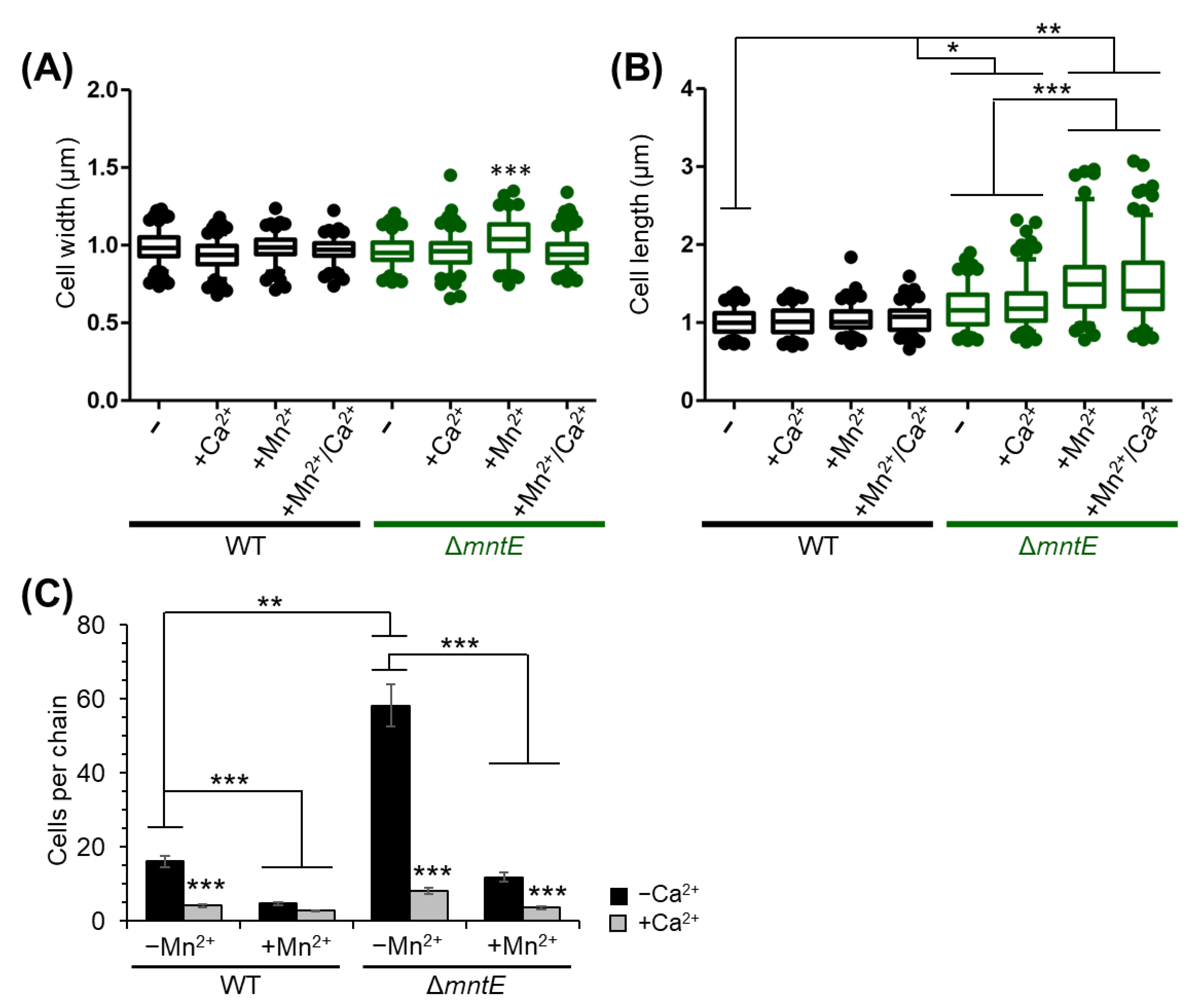
2.8. Cell Morphology Measurements

Images of S. pneumoniae cells grown for 3.5 h in BHI broth with or without MnCl2 or CaCl2 as indicated were collected using a Leica DM6B upright brightfield microscope (Leica, Deerfield, IL, USA). The lengths and widths of approximately 100–150 cell bodies for each condition were measured using ImageJ software version 1.51m9, Java 1.8.0_101 (64-bit). The number of cell bodies were counted from 50–100 chains to determine average number of cells/chain.

2.9. Statistical Analysis

All growth and assays were independently conducted in at least triplicate. Statistical analyses were performed using GraphPad Prism (Graphpad Prism 9 Software, La Jolla, CA, USA). One-way ANOVA and unpaired t-tests were selected with 95% confidence interval. Accepted p-values are indicated in figures.

3. Results

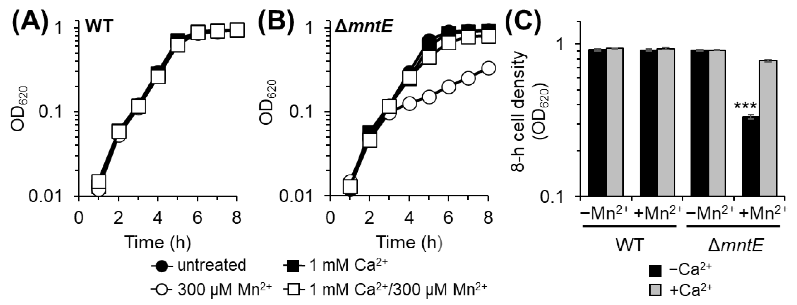
3.1. Ca Rescues Mn2+-Sensitive S. pneumoniae ΔmntE Growth Phenotype

Previous reports demonstrate that S. pneumoniae lacking the Mn2+-specific CDF efflux transporter MntE (ΔmntE) is sensitive to elevated exogenous Mn2+ due to its inability to export Mn out of the cell and is thus considered Mn2+-stressed [27,30,39]. Likewise, we show here that the S. pneumoniae ΔmntE strain fails to reach wild-type (WT) cell densities after 8 h when cultured in rich medium with excess exogenous Mn (Figure 1). The addition of 1 mM Ca2+ to Mn2+-stressed ΔmntE permits growth patterns similar to unstressed cells, resulting in the complete rescue of its Mn2+-sensitive growth defect (Figure 1B,C). Ca2+ alone in the absence of Mn2+ stress had no significant effect on the growth of both the WT and ΔmntE strains (Figure 1).
To further evaluate the ability of Ca2+ to rescue the ΔmntE strain Mn stress growth defect, a serial dilution spot test was performed on rich agar medium supplemented with catalase to prevent hydrogen peroxide intoxication and allow S. pneumoniae colonies to form. In comparison to the WT strain, the ΔmntE mutant showed significantly diminished growth at 500 µM Mn2+ and no growth was observed ≥700 µM Mn (Figure 2A, top). In the presence of 1 mM Ca2+, the ΔmntE mutant resembled a WT strain growth pattern up to 500 µM Mn2+ (Figure 2A, bottom). Residual growth up to 10−2 dilution of the ΔmntE mutant was observed at 700 µM Mn2+ in the presence of Ca2+ (Figure 2A).
The Mn concentration was then fixed at 500 µM to examine the minimum Ca2+ concentration needed for the recovery of the ΔmntE mutant to the WT strain or unstressed ΔmntE mutant growth levels. During Mn2+ stress, residual growth was observed for the ΔmntE strain when Ca2+ was supplied between 100 and 500 µM. The growth density of the ΔmntE strain rose as the Ca2+ levels increased (Figure 2). Complete rescue of Mn2+-stressed ΔmntE was only visible for Ca2+ concentrations ≥1 mM (Figure 2B). These findings are consistent with those observed in broth cultures (Figure 1) and together demonstrate that Ca2+ presence is capable of impacting S. pneumoniae growth and likely has an intersecting role with Mn metabolism.

3.2. Ca2+ Rescue of Mn2+ Sensitivity Varies among Bacteria

Given the significant impact of Ca2+ on S. pneumoniae growth during Mn2+ stress and the importance of maintaining optimum intracellular Mn2+ concentrations among pathogenic bacteria in general, we examined the extent to which Ca2+ could rescue other bacteria from Mn2+ stress. Two individual bacteria were chosen, B. subtilis and E. coli, whose Mn2+ homeostatic mechanisms including import, export, and their regulation are well characterized [40,41]. We considered B. subtilis to be characteristically similar to S. pneumoniae in that both are Gram-positive organisms possessing a Mn2+-centric physiology, requiring relatively high Mn2+ levels for growth compared to other bacteria [30,40,42]. In contrast, E. coli is a Gram-negative organism with an iron-centric physiology that conditionally imports Mn2+ in response to oxidative stress or Fe2+ scarcity [43].
Akin to the S. pneumoniae WT strain (Figure 1A), the addition of Ca2+ or Mn2+ to rich growth medium did not significantly impact the growth of either the B. subtilis or E. coli WT strains (Figure 3A,D). Consistent with the published literature [41,44], B. subtilis, impaired in regulating intracellular Mn2+ levels (ΔmntR), and E. coli, deficient in Mn2+ efflux (ΔmntP), exhibited reduced growth in the presence of 50 and 500 µM Mn2+, respectively (Figure 3B,E). This reduced growth results from Mn2+ intracellular accumulation and intoxication [41,44]. The addition of 1 mM Ca2+ in the presence of Mn2+ stress did not significantly impact the growth of the B. subtilis ΔmntR strain (Figure 3B,C). We note that Mn2+-stressed B. subtilis ΔmntR cultures reached WT stationary cell density after 6 h incubation, independent of Ca2+ presence (Figure 3B). Incubation of B. subtilis ΔmntR with increased Mn2+ (100 µM) enhanced the growth defect, which was not further affected by Ca2+ presence (Figure S1).
In contrast to the B. subtilis ΔmntR strain growth, the addition of 1 mM Ca2+ during Mn2+ stress partially restored the growth of the E. coli ΔmntP mutant to half that of the WT strain cell density (Figure 3D,F). Together, these data along with those of S. pneumoniae suggest that the roles of Ca2+ in relation to cellular Mn2+ demand likely vary among bacteria and may instead depend on host environment nutrient conditions and other natural environmental selective pressures.

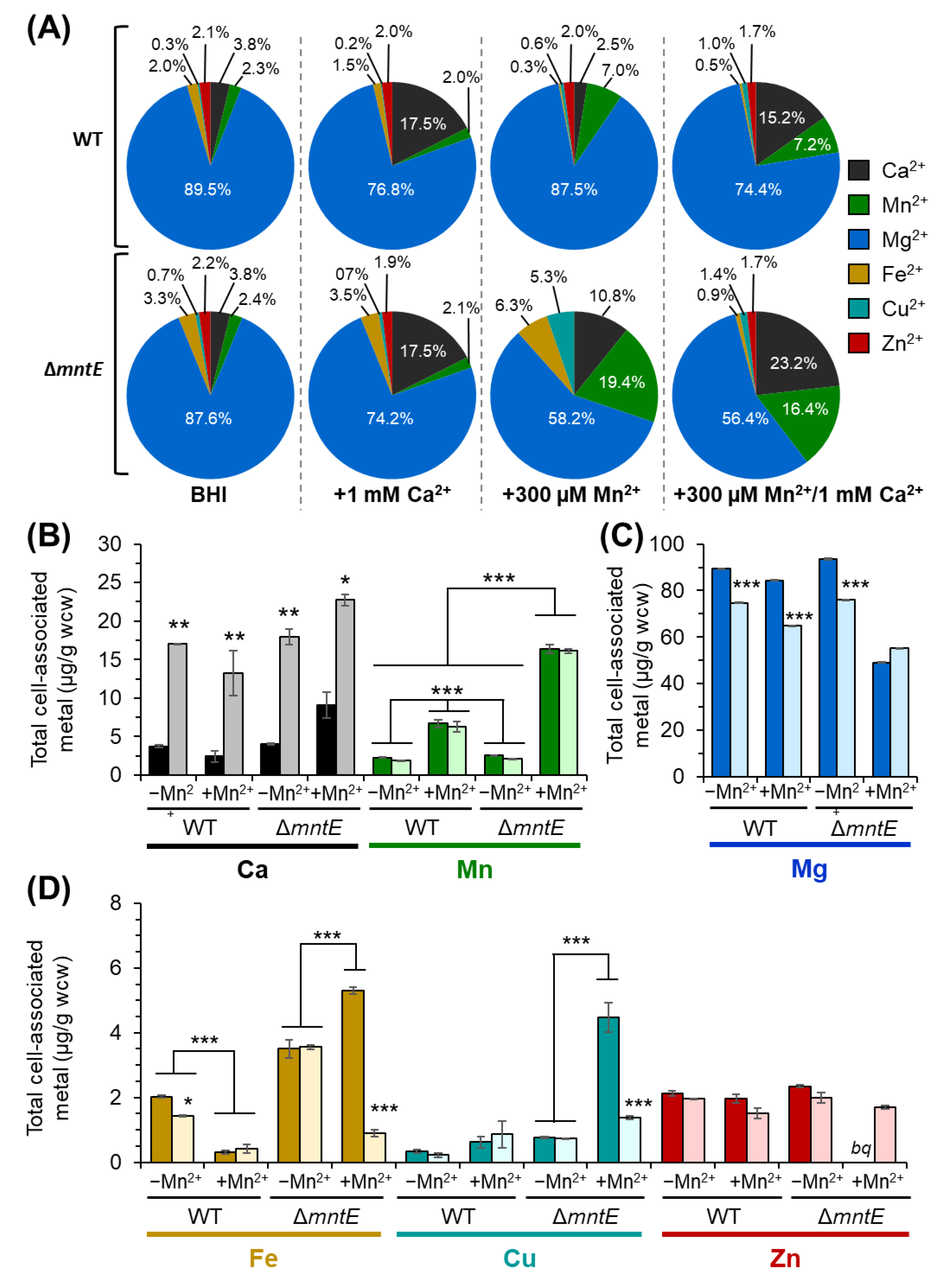
3.3. S. pneumoniae ΔmntE Accumulates High Cellular Mn2+ Despite Growth Restoration by Ca2+

To investigate the mechanism by which Ca2+ rescues the S. pneumoniae ΔmntE strain Mn2+-sensitive growth phenotype, we next assessed the metal ion composition of bacterial cells. During routine growth in rich medium, the S. pneumoniae WT strain cell-associated Ca2+ and Mn2+ concentration yielded 3.8 ± 0.15 and 2.3 ± 0.07 µg/g wcw (wet cell weight), respectively (Figure 4B). Likewise, similar Ca2+ and Mn2+ levels were observed for unstressed ΔmntE cells, 4.1 ± 0.09 and 2.6 ± 0.06 µg/g wcw, respectively. Cellular Ca2+ increased 4.5-fold to 17.0 ± 0.05 and 18.0 ± 1.05 µg/g wcw for S. pneumoniae WT and ΔmntE strains, respectively, when cultured in the presence of 1 mM Ca2+ (Figure 4B), indicating that S. pneumoniae is capable of importing Ca2+ but the mechanism of how Ca2+ enters S. pneumoniae is not understood at this time. Incubation with Ca2+ alone did not significantly alter other cellular metals examined, including Mn2+, zinc (Zn2+), iron (Fe2+), and copper (Cu2+), for both the WT and ΔmntE strains (Figure 4A,D) but did slightly reduce magnesium (Mg2+) by ≈18% from approximately 89.4 ± 0.45 to 74.8 ± 0.25 µg/g for WT and 93.5 ± 1.00 to 76.0 ± 0.20 µg/g wcw for ΔmntE (Figure 4C).
Culturing S. pneumoniae WT and ΔmntE strains with 300 µM Mn yielded a 3-fold and 8-fold increase in cell-associated Mn for the WT (2.25 ± 0.70 vs. 6.71 ± 0.51 µg/g wcw) and ΔmntE (2.56 ± 0.06 vs. 16.4 ± 0.55 µg/g wcw) strains, respectively (Figure 4B). The significant accumulation of cellular Mn2+ is consistent with the ΔmntE mutant being incapable of efficiently exporting Mn2+ out of the cell [27,39]. Interestingly, the Ca2+ levels increased 2-fold to 9.1 ± 1.7 µg/g wcw for the Mn2+-stressed ΔmntE mutant but were relatively unchanged for the WT strain when compared to unstressed cells (Figure 4B). The ΔmntE mutant cell-associated Mg2+ levels were reduced by almost 2-fold during Mn2+ stress, independent of Ca2+ presence (Figure 4C). Cell-associated Fe2+ and Cu2+ rose 1.5-fold (from 3.5 ± 0.28 to 5.3 ± 0.12 µg/g wcw) and 5-fold (from 0.8 ± 0.04 to 4.5 ± 0.46 µg/g wcw), respectively, for the ΔmntE mutant during Mn2+ stress (Figure 4D), which is consistent with previous reports showing elevated Mn2+ interferes with the expression of Fe2+ and Cu2+ homeostatic proteins [30]. No significant change in Zn2+ was observed for the WT strain cultured with Mn (Figure 4D); Zn2+ was below the quantifiable detection for the ΔmntE mutant. We note here that a high cellular Mn2+ concentration can disrupt Zn2+ homeostasis in S. pneumoniae by possibly binding to the Zn2+-sensing regulatory protein SczA and antagonizing its activity to properly regulate the expression of the Zn2+-specific CDF exporter CzcD [45] (see Section 3.6 for further support).
When strains were cultured with 1 mM Ca2+ during Mn2+ stress, the cell-associated Mn2+ levels remained elevated for the ΔmntE mutant at 16.1 ± 0.30 µg/g wcw (Figure 4B), indicating that the ΔmntE strain continues to experience Mn2+ intoxication and that exogenous Ca2+ is not inhibiting Mn2+ import and likely not altering Mn2+ export through the secondary Mn2+ efflux transporter MgtA [28]. Fe2+ and Cu2+ were significantly reduced 5-fold and 3-fold, respectively, when Ca2+ was added to the Mn2+-stressed ΔmntE strain (Figure 4D); no significant change in Fe2+ or Cu2+ was observed for the WT strain.

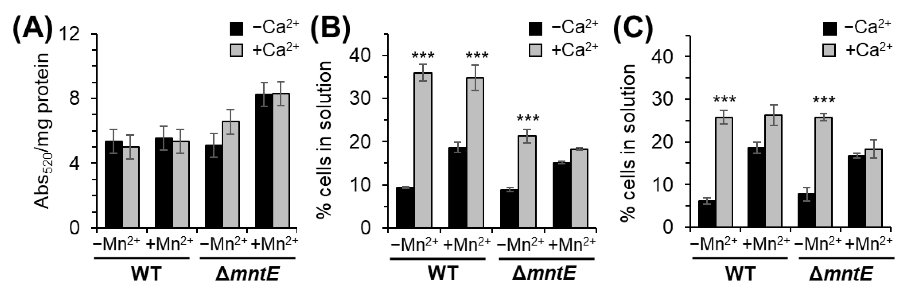
3.4. Ca2+ Does Not Alter S. pneumoniae Capsular Polysaccharide Production

To confirm that the S. pneumonie ΔmntE strain was still physically experiencing Mn2+ intoxication despite growth rescue, we next evaluated the production of CPS using two different methods: uronic acid assay (a component of the S. pneumoniae D39 bacterial capsule) [32] and mucoviscosity [29,33]. During Mn2+ stress, elevated intracellular Mn2+ binds to and activates phosphoglucomutase Pgm, a Mn2+-requiring enzyme that is critical for the S. pnuemoniae biosynthesis of CPS [29]. As such, hyperactivation of phosphoglucomutase Pgm by Mn2+ leads to increased CPS production, which prevents S. pneumoniae cell adherence to surfaces [29]. For these experiments, the concentration of Mn2+ was reduced to 100 µM to ensure growth of the ΔmntE mutant over time during Mn2+ stress [29,30].
Direct measurement of uronic acid revealed the WT strain CPS production was not significantly altered by Ca2+ or Mn2+ (Figure 5A). Compared to the WT strain, the unstressed ΔmntE mutant produced 24% more uronic acid (Figure 5A). The ΔmntE strain CPS levels increased an additional 26% during Mn2+ stress, producing ≈45% more uronic acid than WT (Figure 5A). The addition of Ca2+ to Mn2+-stressed ΔmntE did not significantly affect CPS production. These data are consistent with elevated cell-associated Mn2+ (Figure 5B) and suggest that cellular Mn2+ actively binds to and activates Pgm, leading to thicker CPS.
Qualitative assessment of CPS thickness using mucoviscosity assay during routine growth in rich medium revealed similar mucoviscosity levels for both the WT and ΔmntE strains (Figure 5B), which are consistent with those observed by McFarland et al., 2021 [29]. Upon Mn2+ exposure, both the WT and ΔmntE strains showed a significant increase in likelihood of remaining in solution, up to 2-fold compared to unstressed conditions (Figure 5B). Culturing with Ca2+ also reduced cell adherence ability; WT and ΔmntE cells were 4- and 2-fold less likely to adhere to the surface compared to unstressed cells, respectively (Figure 5B). Similar adherence levels were observed in the presence of Ca2+ during Mn2+ stress. We note that Ca2+-treated cells appeared “fluffy” and that the cell pellets were easily disrupted if care was not taken. Decreasing exogenous Ca2+ did not change these observations and had no significant effect on S. pneumoniae cells’ adherence ability (Figure 5C). Together, these data demonstrate that exogenous Ca2+ does not influence CPS production in S. pneumoniae D39 but may play a role in overall extracellular bacterial surface charge possibly via the expression of adhesion molecules, molecular binding determinants of adhesion molecules, or by other unknown mechanisms.

3.5. Mn2+ Stress Inhibits S. pneumoniae Biofilm Formation

Like many other upper respiratory tract bacterial pathogens, S. pneumoniae forms a biofilm during asymptomatic carriage [46]. These biofilms can increase persistence in the host, as well as S. pneumoniae spread among the host population. Furthermore, initial colonization, biofilm formation, and dispersion of S. pneumoniae cells from the infection site are each influenced by multiple competing factors and environmental resources, including host nutritional immunity and viral co-infection [47,48]. Since changes in host Ca2+ levels have been shown to facilitate bacterial adherence [16,17] and cellular Mn2+ influences CPS production [29], we investigated the effect of Ca2+ and Mn2+ on biofilm formation. For these studies, S. pneumoniae was cultivated in TH broth as an alternative to BHI because of its ability to induce consistent biofilm formation [35]. The WT and ΔmntE strains showed no significant difference in biofilm formation when cultured in TH broth for 24 h (Figure 6). The addition of Ca2+ also did not significantly affect the biofilm formation of either strain. The WT strain biofilm formation was also not affected by Mn2+ or the combination of Mn2+/Ca2+ (Figure 6A). No detectable biofilm formation was observed for the ΔmntE mutant during Mn2+ stress independent of Ca2+ presence (Figure 6B).
We note that other host environmental nutrient factors, including sugar carbohydrates, can also effectively facilitate bacterial biofilm formation [34]. Sugar carbohydrate effectiveness is dependent on its concentration and the bacteria type, as well as other associated factors [49]. Glucose can inhibit initial colonization and early-stage bacterial biofilm formation but can also increase biofilm thickness at later stages of growth [50,51]. We found that supplementation of TH broth with 0.2% glucose or 0.3% yeast extract slightly increased biofilm formation by ≈20% and ≈34% for all conditions examined using the WT strain (Figure 6A); the ΔmntE mutant showed similar pattern increases (Figure 6B). Together, these data suggest that carbohydrates and Mn2+, but not Ca2+ have a role in S. pneumoniae biofilm formation. Additional studies are underway to understand the mechanism of how Mn2+ modulates biofilm formation.

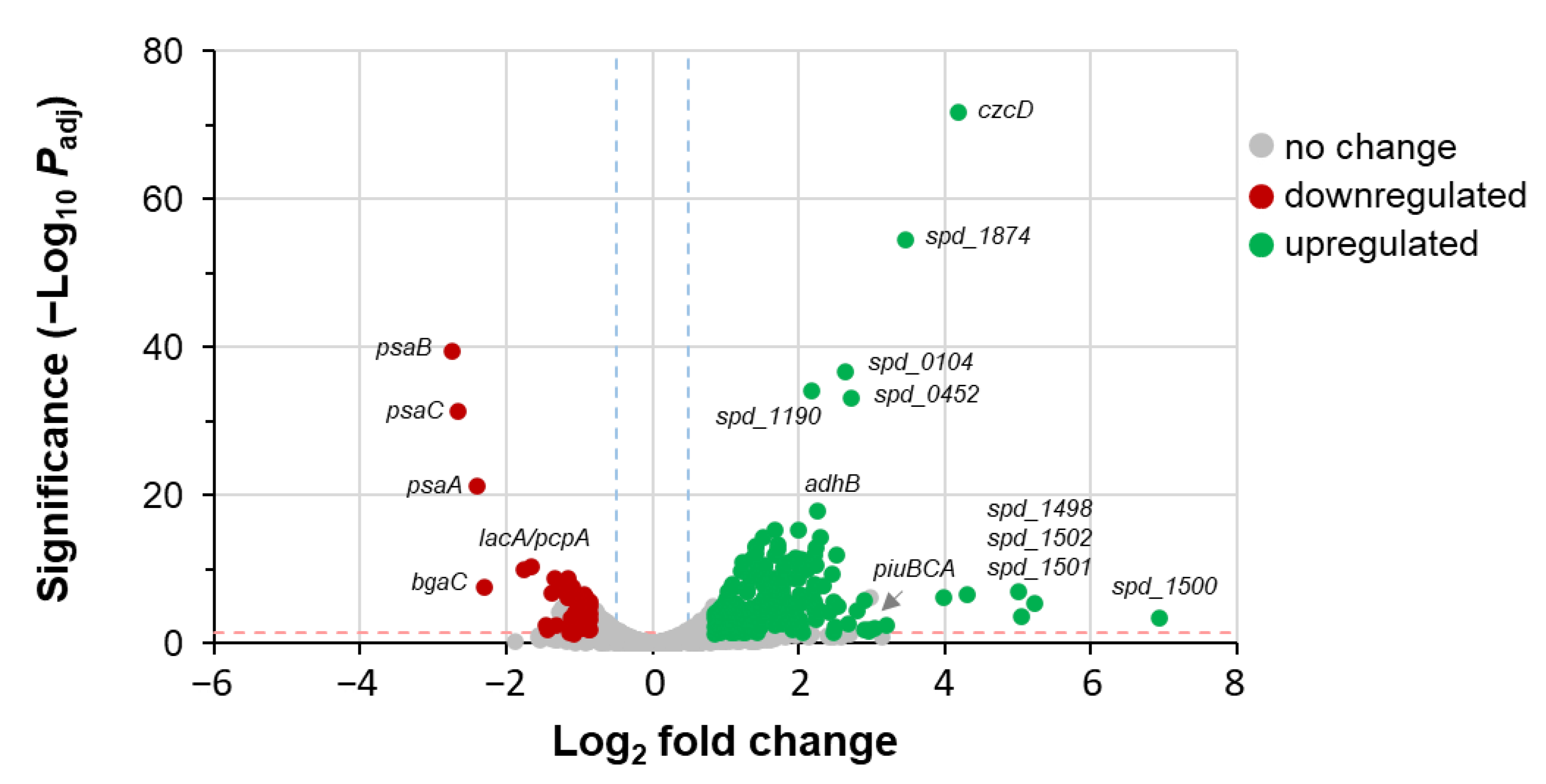
3.6. Ca2+ Does Not Alter S. pneumoniae Gene Expression

Having eliminated a direct involvement of Ca2+ in modulating S. pneumoniae Mn2+ transport mechanisms, we sought to gain insight into the mechanism by which Ca2+ rescues the Mn2+-sensitive ΔmntE strain growth phenotype (Figure 1). Since Ca2+ is implicated in several sensor pathways in other pathogenic bacteria that regulate genes encoding virulence and resistance proteins [10], we hypothesized that Ca2+ might function as a signaling molecule by directly aiding the modulation of S. pneumoniae gene expression or by altering cellular protein activities by coordinating with EF-hand motifs found in calmodulin-like proteins that require Mn2+.
To investigate this further, differential RNAseq analysis was performed to provide a global snapshot of the relative transcript levels of genes during S. pneumoniae growth. Comparison of the transcriptome expression profiles of the WT and Mn2+-stressed ΔmntE mutant yielded data (Figure 7) consistent with previous reports [30,42]. A total of 273 genes were significantly differentially expressed; there were 65 downregulated (Table S2) and 208 upregulated genes (Table S3). Our data show that during Mn2+ stress, the psaBCA genes encoding the Mn2+-specific ABC-type importer PsaBCA are downregulated 8- to 11-fold. The czcD gene encoding the Zn2+-specific CDF efflux transporter CzcD is upregulated 5-fold. The latter observations likely result from the mismetallation of the Zn2+ regulatory proteins AdcR and SczA by the accumulation of excess cellular Mn2+ [45]. piuBCAD encoding the Fe2+ ABC-type transporter PiuBCAD was induced 8-fold, consistent with previous work [30]. rffD (spd_0940)-encoding UDP-N-acetyl-D-mannosaminuronic acid dehydrogenase, which functions in producing sugar precursors for CPS biosynthesis, was upregulated 12-fold in addition to other genes involved in sugar transport and metabolism, which is reflective of the observed increased CPS produced by S. pneumoniae during Mn2+ stress (Figure 5). Despite observations of a 4.5-fold elevated cell-associated Ca2+ (Figure 4A), no significant difference in gene expression was measured in Mn2+-stressed ΔmntE cultured with and without 1 mM Ca2+. Several possibilities are discussed later.

3.7. Mn2+ Impacts S. pneumoniae Cell Division, Which Is Rescued by Ca2+

Accumulation of cellular Mn2+ can lead to dysregulation of S. pneumoniae cell division via hyperactivation of the Mn2+-dependent protein phosphatase PhpP [30]. Such S. pneumoniae cells show on average elongated cell bodies, some cell lysis, and increased chaining [30]. We report similar findings here in Figure 8. The average WT cell width and length was 0.99 ± 0.09 and 1.01 ± 0.16, respectively. In general, the WT cell widths and lengths were not significantly changed by Ca2+ and Mn2+ presence. The ΔmntE cells measured slightly wider than the WT cells during Mn2+ intoxication at 1.05 ± 0.12 µm vs. 0.98 ± 0.08 µm, respectively (Figure 8A). The addition of Ca2+ to the Mn2+-stressed ΔmntE mutant returned cell widths to the WT or unstressed ΔmntE cell sizes.
During routine growth in rich medium, ΔmntE cells were on average slightly more elongated than WT cells (1.01 ± 0.16 µm vs. 1.20 ± 0.26 µm for ΔmntE). The ΔmntE cell lengths significantly increased to 1.54 ± 0.46 µm when cultured with Mn2+. The increase appeared to be independent of Ca2+ presence, although the median cell length for ΔmntE grown with Mn2+/Ca2+ was lower than with Mn2+ alone and was approaching closer to the unstressed ΔmntE average cell lengths (Figure 8B). Chaining was also most prevalent for the ΔmntE mutant, with an average of 58 cells per chain observed compared to 16 for the WT strain during routine growth (Figure 8C). The addition of Mn2+ to the ΔmntE mutant resulted in significantly less chaining likely due to increased cell lysis and its reduced ability to divide when toxified by Mn2+ [30]. In all cases, the addition of Ca2+ led to a significant decrease in chain length; cells were mostly found in pairs or short chains of ≤8 cells per chain depending on the strain (Figure 8C). Cell lysis was not observed microscopically during Ca2+ treatment. Taken together with the above findings, the data suggest that Ca2+ likely has a role in cell division impacting Mn2+ physiology. Further investigation is underway to determine the actual mechanism.

4. Discussion

Unlike in eukaryotes, the role of Ca2+ in bacteria physiology and virulence remains elusive. Past studies are mostly correlative and have collectively suggested that bacteria may utilize Ca2+ as an intracellular signaling messenger whose presence or absence impacts multiple cellular processes, including biofilm formation [10,52]. The present study brings evidence for the involvement of Ca2+ in cellular processes requiring Mn2+ in S. pneumoniae D39. Our data demonstrate for the first time that Ca2+ is capable of rescuing S. pneumoniae ΔmntE Mn2+-sensitive growth phenotype via an unidentified mechanism that does not involve altering cell-associated Mn2+ levels or gene expression. We further demonstrate that S. pneumoniae D39 cells experiencing Mn2+ stress produce thicker capsules, which not only inhibit S. pneumoniae D39 adhesion to abiotic surfaces but also biofilm formation.
The capacity for Ca2+ to restore growth during Mn2+ intoxication likely varies among bacteria, since Ca2+ supplementation showed some efficacy in E. coli but did not impact B. subtilis growth. Although more studies are needed to fully access the breadth of Ca2+ impact across bacterial pathogens, it is suspected that such differences arise from selective pressures imposed by the environmental niches for which each bacterium resides. Like Mn2+ and other metal ions, Ca2+ concentrations vary among host sites. Samples taken from the upper respiratory tract and oral cavity can reach millimolar Ca2+ levels, with a 10-fold difference between several sites [10]. Furthermore, elevated cellular Ca can trigger the activation of host immune responses against invading pathogens [14,53]. It has been proposed that host Ca2+ levels might (1) signal invading bacterial pathogens that they are entering a specific host site and (2) indicate the status of the host immune protection [10]. For example, during bacteria invasion, the EF hand containing S100 proteins like calprotectin require Ca2+ to interact with their targets [54]. Activated calprotectin has been shown to sequester Zn2+, and subsequently reduces Zn2+ interaction with the solute-binding proteins PsaA and MntC in S. pneumoniae and Staphylococcus aureus, respectively [55]. Sequestration of Zn2+ ultimately facilitates Mn2+ binding to the solute-binding proteins, resulting in Mn2+ import by bacteria. As such, it is likely advantageous for invading bacterial pathogens to recognize host cellular Ca2+ levels, which would predictably enhance bacterial adaptation to various host environments, leading to increased bacterial virulence and survival within the host.
Following the revelation that Ca2+ rescues Mn2+-stressed cells is not mediated through modulation of Mn2+ homeostatic proteins, we sought to investigate the molecular mechanism driving the growth rescue. We were somewhat surprised to find that differential RNAseq failed to report any significant changes in global gene expression patterns for the strains grown with or without 1 mM Ca2+ during Mn2+ stress. It is possible that the exposure time with Ca2+ relative to Mn2+ stress was not sufficient to observe transcriptional response differences or the relative Ca2+ cellular concentration remained within the unknown optimum Ca2+ range for S. pneumoniae growth. As such, Ca2+-specific transcriptional regulatory proteins would likely not be activated to alter gene expression. Note that increasing Ca2+, as with other metals studies, also increases the risk of contaminating metals, which may obscure results. Furthermore, the addition of Ca2+ concentrations >2 mM in the presence of high Mn2+ resulted in precipitation.
Given that Ca2+ is not characteristically similar to Mn2+ and that Ca2+ most often functions as a non-catalytic cofactor in proteins, we speculate that Ca2+-mediated rescue in S. pneumoniae occurs at the post-translational level. As such, Ca2+ would function as a signaling messenger that interacts with cellular proteins to modulate their activity or downstream activity of enzyme targets via post-translational modification or protein stabilization. This is not unprecedented, as several Ca2+-sensing two-component regulatory systems have been characterized in other bacterial pathogens [10]. There is also growing evidence showing such novel homeostatic mechanisms for other metal ions including Mn2+ [23], which further establishes that metals play important intricate roles in bacteria.
As previously mentioned, CPS production is a critical virulence determinant for many bacterial pathogens, including S. pneumoniae, and its production is regulated in response to environmental factors. Mn2+ has most recently emerged as an important regulatory component in CPS production for S. pneumoniae via the modulation of Pgm activity [29]. Our data are consistent with those reported by McFarland et al., 2021, in that elevated Mn2+ increases CPS production [29]. We extended these findings to S. pneumoniae D39 biofilm formation, demonstrating that thicker CPS would inhibit transition from a planktonic to sessile-community state. Although the study here is performed in-vitro, these data do reinforce the importance of Mn2+ in S. pneumoniae colonization and pathogenesis within the host.
Interestingly, we found that Ca2+ did not significantly affect biofilm formation but did influence S. pneumoniae D39 adherence to surfaces, independent of CPS thickness. Bacterial adhesion capacity is due in part to electrostatic interactions combined with cell surface structures. Gram-positive bacteria, like S. pneumoniae, have long glycan chains extending through the cell wall from the cytoplasmic membrane. These glycan chains confer a negative charge and are structurally hydrophilic in nature [56]. As such, extracellular Ca2+ might alter the physiochemical properties of the S. pneumoniae cell surface via cationic bridging. Direct interaction of Ca2+ with such charged exposed surface antigens or extracellular polymeric substances may alter the overall cell surface charge or electrostatic interactions, thereby impeding cell adhesion to surfaces. Further investigation is needed to determine the underlying mechanisms driving this phenomenon and its implication in S. pneumoniae virulence and pathogenesis.
In summary, the findings presented in this study highlight a role for Ca2+ in S. pneumoniae that is interconnected with Mn2+ physiology and its implication in virulence. Unraveling the molecular mechanism(s) surrounding the Ca2+ rescue of Mn2+ intoxication demonstrates the complex nature of how bacterial pathogens have evolved to survive in the host and possibly even hijack host mechanisms to have a selective growth advantage and evade host defenses. It will be interesting to learn how Ca2+ physically functions in S. pneumoniae, as well as how S. pneumoniae imports, senses, and traffics Ca2+. Will the Ca2+ trend be like that of other metals and be highly regulated to prevent the intoxication and disruption of cellular processes? Nonetheless, it is critical that we gain a better fundamental understanding of Ca2+ in bacterial as it will likely provide insights into the regulation of bacterial pathogenesis and aid the development of targeted therapeutics.

Supplementary Materials

The following supporting information can be downloaded at: https://www.mdpi.com/article/10.3390/microorganisms12091810/s1, Figure S1: Ca2+ supplementation does not rescue the Mn2+-sensitive B. subtilis ΔmntR growth phenotype; Table S1: bacterial strains used in this study; Table S2: genes showing reduced expression that is greater than 1.5-fold and adjusted p-value ≤ 0.05 in S. pneumoniae WT vs. ΔmntE grown in BHI with 300 µM Mn2+; Table S3: genes showing increased expression that is greater than 1.5-fold and adjusted p-value ≤ 0.05 in S. pneumoniae WT vs. ΔmntE grown in BHI with 300 µM Mn2+; File S1: differential RNAseq summary.

Author Contributions

Conceptualization, J.E.M.; methodology, data curation, validation, R.O., E.C., N.R.D.L. and J.E.M.; formal analysis, R.O., E.C., N.R.D.L. and J.E.M.; investigation, R.O. and E.C.; writing—original draft preparation, R.O.; writing—review and editing, R.O., N.R.D.L. and J.E.M.; project administration and funding acquisition, J.E.M. All authors have read and agreed to the published version of the manuscript.

Funding

This research was funded by NIH-NIAID grant number R15AI148725 (to J.E.M.) and R21AI171771 (to N.R.D.); INBRE-4 Pilot Project subaward (to J.E.M.) and IDeA from NIH-NIGMS grant number P20GM103408; and ISU College of Science and Engineering funds (to R.O.).

Data Availability Statement

The original contributions presented in the study are included in the article/Supplementary Material; further inquiries can be directed to the corresponding author.

Acknowledgments

We thank J.A. Imlay (University of Illinois, Urbana-Champaign) for providing the Escherichia coli strains used in this work and the Idaho State University Molecular Research Core Facility, RRID:SCR_012598, for use of equipment and training provided.

Conflicts of Interest

The authors declare no conflicts of interest.

References

  1. Clapham, D.E. Calcium signaling. Cell 2007, 131, 1047–1058. [Google Scholar] [CrossRef] [PubMed]
  2. Permyakov, E.A.; Kretsinger, R.H. Cell signaling, beyond cytosolic calcium in eukaryotes. J. Inorg. Biochem. 2009, 103, 77–86. [Google Scholar] [CrossRef] [PubMed]
  3. Straley, S.C.; Bowmer, W.S.; Gregor, L.B. Regulation by Ca2+ in the Yersinia low-Ca2+ response. Mol. Microbiol. 1993, 8, 1005–1010. [Google Scholar] [CrossRef] [PubMed]
  4. Tisa, L.S.; Adler, J. Cytoplasmic free-Ca2+ level rises with repellents and falls with attractants in Escherichia coli chemotaxis. Proc. Natl. Acad. Sci. USA 1995, 92, 10777–10781. [Google Scholar] [CrossRef]
  5. Sarkisova, S.; Patrauchan, M.A.; Berglund, D.; Nivens, D.E.; Franklin, M.J. Calcium-induced virulence factors associated with the extracellular matrix of mucoid Pseudomonas aeruginosa biofilms. J. Bacteriol. 2005, 187, 4327–4337. [Google Scholar] [CrossRef]
  6. Hu, Y.; Lu, P.; Ye, C.; Zhang, H.; Wang, Y.; Chen, W.; Yang, Z. Structures of Anabaena calcium-binding protein CcbP: Insights into Ca2+ signaling during heterocyst differentiation. J. Biol. Chem. 2011, 286, 12381–12388. [Google Scholar] [CrossRef]
  7. Rosch, J.W.; Caparon, M.G.; Nasher, H.J. Calcium efflux is essential for bacterial survival in the eukaryotic host. Mol. Microbiol. 2008, 70, 435–444. [Google Scholar] [CrossRef]
  8. Amankwah, S.; Abdella, K.; Kassa, T. Bacterial Biofilm Destruction: A Focused Review on the Recent Use of Phage-Based Strategies with Other Antibiofilm Agents. Nanotechnol. Sci. Appl. 2021, 14, 161–177. [Google Scholar] [CrossRef]
  9. Rumbaugh, K.P.; Sauer, K. Biofilm dispersion. Nat. Rev. Microbiol. 2020, 18, 571–586. [Google Scholar] [CrossRef]
  10. King, M.M.; Yendapally, R.; Asmat, T.M.; Nagaraja, V. Calcium Regulation of Bacterial Virulence. Adv. Exp. Med. Biol. 2020, 1131, 827–855. [Google Scholar]
  11. Jairaman, A.; Prakriya, M. Calcium Signaling in Airway Epithelial Cells: Current Understanding and Implications for Inflammatory Airway Disease. Arterioscler. Thromb. Vasc. Biol. 2024, 44, 772–783. [Google Scholar] [CrossRef] [PubMed]
  12. Bagur, R.; Hajnóczky, G. Intracellular Ca. Mol. Cell 2017, 66, 780–788. [Google Scholar] [CrossRef] [PubMed]
  13. Asmat, T.M.; Lin, S.Y.; Richards, D.; Gunderson, K. Streptococcus pneumoniae infection of host epithelial cells via polymeric immunoglobulin receptor transiently induces calcium release from intracellular stores. J. Biol. Chem. 2011, 286, 17861–17869. [Google Scholar] [CrossRef]
  14. Gewirtz, A.T.; Rao, A.S.; Merlin, D.; Madara, J.L.; Jass, J.R. Salmonella typhimurium induces epithelial IL-8 expression via Ca2+-mediated activation of the NF-kappaB pathway. J. Clin. Investig. 2000, 105, 79–92. [Google Scholar] [CrossRef] [PubMed]
  15. Pace, J.; Hayman, M.J.; Galán, J.E. Signal transduction and invasion of epithelial cells by S. typhimurium. Cell 1993, 72, 505–514. [Google Scholar] [CrossRef]
  16. Rose, R.K. The role of calcium in oral streptococcal aggregation and the implications for biofilm formation and retention. Biochim. Biophys. Acta 2000, 1475, 76–82. [Google Scholar] [CrossRef]
  17. Jung, C.J.; Zheng, Q.H.; Shieh, H.R.; Lin, C.L.; Hsueh, P.R. AtlA Mediates Extracellular DNA Release, Which Contributes to Streptococcus mutans Biofilm Formation in an Experimental Rat Model of Infective Endocarditis. Infect. Immun. 2017, 85, e00252-17. [Google Scholar] [CrossRef]
  18. Honsa, E.S.; Johnson, M.D.; Rosch, J.W. The roles of transition metals in the physiology and pathogenesis of Streptococcus pneumoniae. Front. Cell Infect Microbiol 2013, 3, 92. [Google Scholar] [CrossRef]
  19. Trombe, M.C.; Clavé, C.; Manias, J.M. Calcium regulation of growth and differentiation in Streptococcus pneumoniae. J. Gen. Microbiol. 1992, 138, 77–84. [Google Scholar] [CrossRef]
  20. Trombe, M.C.; Rieux, V.; Baille, F. Mutations which alter the kinetics of calcium transport alter the regulation of competence in Streptococcus pneumoniae. J. Bacteriol. 1994, 176, 1992–1996. [Google Scholar] [CrossRef]
  21. Gangola, P.; Rosen, B.P. Maintenance of intracellular calcium in Escherichia coli. J. Biol. Chem. 1987, 262, 12570–12574. [Google Scholar] [CrossRef] [PubMed]
  22. Watkins, N.J.; Knight, M.R.; Trewavas, A.J.; Campbell, A.K. Free calcium transients in chemotactic and non-chemotactic strains of Escherichia coli determined by using recombinant aequorin. Biochem. J. 1995, 306, 865–869. [Google Scholar] [CrossRef] [PubMed]
  23. Martin, J.E.; Waters, L.S. Regulation of Bacterial Manganese Homeostasis and Usage during Stress Responses and Pathogenesis. Front. Mol. Biosci. 2022, 9, 945724. [Google Scholar] [CrossRef] [PubMed]
  24. McDevitt, C.A.; Ogunniyi, A.D.; Valkov, E.; Lawrence, M.C.; Kobe, B.; McEwan, A.G.; Paton, J.C. A molecular mechanism for bacterial susceptibility to zinc. PLoS Pathog. 2011, 7, e1002357. [Google Scholar] [CrossRef]
  25. Kloosterman, T.G.; Witwicki, R.M.; van der Kooi-Pol, M.M.; Bijlsma, J.J.; Kuipers, O.P. Opposite effects of Mn2+ and Zn2+ on PsaR-mediated expression of the virulence genes pcpA, prtA, and psaBCA of Streptococcus pneumoniae. J. Bacteriol. 2008, 190, 5382–5393. [Google Scholar] [CrossRef]
  26. Johnston, J.W.; Briles, D.E.; Myers, L.E.; Hollingshead, S.K. Mn2+-dependent regulation of multiple genes in Streptococcus pneumoniae through PsaR and the resultant impact on virulence. Infect. Immun. 2006, 74, 1171–1180. [Google Scholar] [CrossRef]
  27. Martin, J.E.; Giedroc, D.P. Functional determinants of metal ion transport and selectivity in paralogous cation diffusion facilitator transporters CzcD and MntE in Streptococcus pneumoniae. J. Bacteriol. 2016, 198, 1066–1076. [Google Scholar] [CrossRef]
  28. Martin, J.E.; Le, M.T.; Bhattarai, N.; Capdevila, D.A.; Shen, J.; Winkler, M.E.; Giedroc, D.P. A Mn-sensing riboswitch activates expression of a Mn2+/Ca2+ ATPase transporter in Streptococcus. Nucleic Acids Res. 2019, 47, 6885–6899. [Google Scholar] [CrossRef]
  29. McFarland, A.L.; Bhattarai, N.; Joseph, M.; Winkler, M.E.; Martin, J.E. Cellular Mn/Zn ratio influences phosphoglucomutase activity and capsule production in Streptococcus pneumoniae D39. J. Bacteriol. 2021, 203, e0060220. [Google Scholar] [CrossRef]
  30. Martin, J.E.; Lisher, J.P.; Winkler, M.E.; Giedroc, D.P. Perturbation of manganese metabolism disrupts cell division in Streptococcus pneumoniae. Mol. Microbiol. 2017, 104, 334–348. [Google Scholar] [CrossRef]
  31. Jacobsen, F.E.; Kazmierczak, K.M.; Lisher, J.P.; Winkler, M.E.; Giedroc, D.P. Interplay between manganese and zinc homeostasis in the human pathogen Streptococcus pneumoniae. Metallomics 2011, 3, 38–41. [Google Scholar] [CrossRef] [PubMed]
  32. Filisetti-Cozzi, T.M.C.C.; Carpita, N.C. Measurement of uronic acids without interference from neutral sugars. Anal. Biochem. 1991, 197, 157–162. [Google Scholar] [CrossRef]
  33. Walker, K.A.; Miner, T.A.; Palacios, M.; Trzilova, D.; Frederick, D.R.; Broberg, C.A.; Sepúlveda, V.E.; Quinn, J.D.; Miller, V.L. A Klebsiella pneumoniae regulatory mutant has reduced capsule expression but retains hypermucoviscosity. mBio 2019, 10, e00089-19. [Google Scholar] [CrossRef]
  34. Loke, M.F.; Yadav, I.; Lim, T.K.; van der Maarel, J.R.; Sham, L.T.; Chow, V.T. SARS-CoV-2 spike protein and mouse coronavirus inhibit biofilm formation by Streptococcus pneumoniae and Staphylococcus aureus. Int. J. Mol. Sci. 2022, 23, 3291. [Google Scholar] [CrossRef] [PubMed]
  35. Minami, M.; Konishi, T.; Takase, H.; Makino, T. Shin’iseihaito (Xinyiqingfeitang) suppresses the biofilm formation of Streptococcus pneumoniae in vitro. BioMed Res. Int. 2017, 2017, 4575709. [Google Scholar] [CrossRef]
  36. Martin, M. Cutadapt removes adapter sequences from high-throughput sequencing reads. EMBnet.J. 2011, 17, 10–12. [Google Scholar] [CrossRef]
  37. Putri, G.H.; Anders, S.; Pyl, P.T.; E Pimanda, J.; Zanini, F. Analysing high-throughput sequencing data in Python with HTSeq 2.0. Bioinformatics 2022, 38, 2943–2945. [Google Scholar] [CrossRef] [PubMed]
  38. Sinha, D.; Zheng, J.J.; Tsui, H.-C.T.; Richardson, J.D.; De Lay, N.R.; Winkler, M.E. S1 domain RNA-binding protein CvfD is a new posttranscriptional regulator that mediates cold sensitivity, phosphate transport, and virulence in Streptococcus pneumoniae D39. J. Bacteriol. 2020, 202, e00245-20. [Google Scholar] [CrossRef]
  39. Rosch, J.W.; Gao, G.; Ridout, G.; Wang, Y.D.; Tuomanen, E.I. Role of the manganese efflux system mntE for signaling and pathogenesis in Streptococcus pneumoniae. Mol. Microbiol. 2009, 72, 12–25. [Google Scholar] [CrossRef]
  40. Helmann, J.D. Specificity of metal sensing: Iron and manganese homeostasis in Bacillus subtilis. J. Biol. Chem. 2014, 289, 28112–28120. [Google Scholar] [CrossRef]
  41. Martin, J.E.; Waters, L.S.; Storz, G. The Escherichia coli small protein MntS and exporter MntP optimize the intracellular concentration of Manganese. PLoS Genet. 2015, 11, e1004977. [Google Scholar] [CrossRef]
  42. Lisher, J.P.; Giedroc, D.P. Manganese acquisition and homeostasis at the host-pathogen interface. Front. Cell Infect. Microbiol. 2013, 3, 91. [Google Scholar] [CrossRef] [PubMed]
  43. Anjem, A.; Varghese, S.; Imlay, J.A. Manganese import is a key element of the OxyR response to hydrogen peroxide in Escherichia coli. Mol. Microbiol. 2009, 72, 844–858. [Google Scholar] [CrossRef] [PubMed]
  44. Huang, X.; Shin, J.-H.; Pinochet-Barros, A.; Helmann, J.D. Bacillus subtilis MntR coordinates the transcriptional regulation of manganese uptake and efflux systems. Mol. Microbiol. 2017, 103, 253–268. [Google Scholar] [CrossRef] [PubMed]
  45. Martin, J.E.; Lisher, J.P.; Winkler, M.E.; Giedroc, D.P. The zinc efflux activator SczA protects Streptococcus pneumoniae serotype 2 D39 from intracellular zinc toxicity. Mol. Microbiol. 2017, 104, 636–651. [Google Scholar] [CrossRef]
  46. Weimer, K.E.; Armbruster, C.E.; Juneau, R.A.; Hong, W.; Pang, B.; Swords, W.E. Coinfection with Haemophilus influenzae promotes pneumococcal biofilm formation during experimental otitis media and impedes the progression of pneumococcal disease. J. Infect. Dis. 2010, 202, 1068–1075. [Google Scholar] [CrossRef]
  47. Hood, M.I.; Skaar, E.P. Nutritional immunity: Transition metals at the pathogen-host interface. Nat. Rev. Microbiol. 2012, 10, 525–537. [Google Scholar] [CrossRef]
  48. Rowe, H.M.; Meliopoulos, V.A.; Iverson, A.R.; Bickford, J.S.; Schrock, D.; Flury, E.; Feng, Y.; Xu, Y.; Albrecht, R.A.; Sands, J.; et al. Direct interactions with influenza promote bacterial adherence during respiratory infections. Nat. Microbiol. 2019, 4, 1328–1336. [Google Scholar] [CrossRef]
  49. Garcia-Gonzalo, D.; Pagán, R. Influence of environmental factors on bacterial biofilm formation in the food industry: A review. Postdoc J. 2015, 3, 1–20. [Google Scholar] [CrossRef]
  50. Martin, N.; Patel, J.; Munoz, F.; Sabino, J.; Yeo, T. Regulation and role of calcium in cellular senescence. Cell Calcium 2023, 110, 102701. [Google Scholar] [CrossRef]
  51. Ravi, B.; Sanyal, S.K.; Pandey, G.K. Calcium decoders and their targets: The holy alliance that regulate cellular responses in stress signaling. Adv. Protein Chem. Struct. Biol. 2023, 134, 371–439. [Google Scholar] [PubMed]
  52. Domínguez, D.C.; Guragain, M.; Patrauchan, M. Calcium binding proteins and calcium signaling in prokaryotes. Cell Calcium 2015, 57, 151–165. [Google Scholar] [CrossRef]
  53. Smith, D.J.; Yau, P.; Winstanley, C.; Cummings, S.P.; Cooper, R.J.; Knox, A.J.; Singh, M.; Walshaw, M.J.; Chilvers, M.A. Elevated metal concentrations in the CF airway correlate with cellular injury and disease severity. J. Cyst. Fibros. 2014, 13, 289–295. [Google Scholar] [CrossRef]
  54. Rosen, T.; Wang, K.A.; Nolan, E.M. Metal sequestration by S100 proteins in chemically diverse environments. Trends Microbiol. 2022, 30, 654–664. [Google Scholar] [CrossRef] [PubMed]
  55. Rosen, T.; Magnotti, E.; Spadafora, L.; Snyder, L.M.; Goldfarb, D.; Nolan, E.M. Zinc sequestration by human calprotectin facilitates manganese binding to the bacterial solute-binding proteins PsaA and MntC. Metallomics 2022, 14, mfac001. [Google Scholar] [CrossRef] [PubMed]
  56. Zheng, S.; Franklin, M.J.; Tang, T.H.; Gilmore, B.F.; Cornelius, V.J.; Song, S.; O’Toole, G.A. Implication of surface properties, bacterial motility, and hydrodynamic conditions on bacterial surface sensing and their initial adhesion. Front. Bioeng. Biotechnol. 2021, 9, 643722. [Google Scholar] [CrossRef]
Figure 1. Calcium rescues S. pneumoniae ΔmntE Mn2+-sensitive growth defect. S. pneumoniae WT (A) and ΔmntE (B) cultures were diluted to 0.005 OD620 at time zero into BHI supplemented with 0 or 300 µM Mn and/or 1 mM Ca2+. Turbidity was measured over time at 620 nm. Data shown are the representative growth of at least three independent replicates. (C) Mean cell density at 8 h growth of at least three independent replicates ±SEM; ***, p ≤ 0.01.
Microorganisms 12 01810 g001
Figure 2. Exogenous Ca2+ increases ΔmntE strain tolerance to elevated Mn2+ levels. (A,B) S. pneumoniae WT and ΔmntE cultures were serially diluted and spotted onto BHI/catalase agar supplemented with Ca2+ and Mn2+ as indicated. Represented growth shown after 24 h incubation of three independent replicates. BHI-only control plate featured is the same in panel (A,B).
Microorganisms 12 01810 g002
Figure 3. Ca differentially impacts Mn2+-sensitive B. subtilis ΔmntR and E. coli ΔmntP strains. Exponentially growing cultures were diluted at time zero into BHI with or without 1 mM Ca2+ and Mn2+ as indicated and turbidity was measured over time at OD620. (AC) B. subtilis strains stressed with 50 µM Mn2+. (DF) E. coli strains stressed with 500 µM Mn2+. Growth curves are representative of multiple independent growths. The 4 h cell density is the mean of at least three independent replicates ±SEM; ***, p ≤ 0.01; *, p ≤ 0.10.
Microorganisms 12 01810 g003
Figure 4. Ca2+ enters S. pneumoniae, but its presence does not alter cellular Mn2+ levels. Total cell-associated metal ion concentrations were measured from S. pneumoniae WT and ΔmntE grown for 3.5 h with or without 300 µM Mn2+ as indicated and 0 (darker shade) or 1 mM (lighter shade) Ca2+. (A) Metal ion distribution across conditions among for only those metals measured. (B) Total cell-associated Ca2+ (black) and Mn2+ (green); (C) Mg2+ (blue); (D) Fe2+ (brown), Cu2+ (teal), and Zn2+ (red). The mean of at least two independent cultures ±SEM; wcw, wet cell weight. ***, p < 0.01; **, p < 0.05; *, p < 0.10.
Microorganisms 12 01810 g004
Figure 5. Impact of Ca2+ on S. pneumoniae CPS production and adherence. (A) Direct measurement of the uronic acid, a capsular component, and (B) adherence of cells to abiotic surfance grown for 3.5 h with or without 100 µM Mn2+ and 2 mM Ca2+. (C) Adherence of cells to abiotic surface grown with or without 300 µM Mn2+ and 1 mM Ca2+. We note that S. pneumoniae strains expressing thicker capsules do not form tight cell pellets during low-speed centrifugation, and therefore the suspension will have high absorbance. Data shown are the mean of at least three independent cultures ±SEM; ***, p ≤ 0.01.
Microorganisms 12 01810 g005
Figure 6. Mn2+ stress inhibits S. pneumoniae ΔmntE biofilm formation, while Ca2+ does not. (A) WT and (B) ΔmntE cells were grown in Todd Hewitt (TH) broth with or without 300 µM Mn2+ and 1 mM Ca2+ for 24 h prior to determining biofilm mass by crystal violet staining. Cultures supplemented with 0.2% glucose (Glc) or 0.3% yeast extract (YE) enhanced relative biofilm formation. Data shown are the mean of at least four independent cultures in triplicate ±SEM. nd, not detected; **, p ≤ 0.05.
Microorganisms 12 01810 g006
Figure 7. Volcano plot illustrating RNAseq analysis of differentially expressed genes in S. pneumoniae during Mn2+ stress. Comparison of WT strain cultured in BHI and ΔmntE mutant cultured in BHI plus 300 µM Mn2+. Genes showing significant difference (p ≤ 0.05; dashed blue lines) of ≥1.5 log2 fold change (dashed pink line) are shown in red (downregulated) and in green (upregulated); no significant changes are shown in grey. For a complete list of differentially expressed genes, see Tables S2 and S3.
Microorganisms 12 01810 g007
Figure 8. Cell morphology and chain length of S. pneumoniae strains stressed for 3.5 h. Width (A) and length (B) measurements of cells grown with or without 300 µM Mn2+ and 1 mM Ca2+ as indicated. Box and whisker plot with 95% confidence interval; WT strain shown as black and ΔmntE strain in green. (C) Mean number of cells observed per chain ±SEM; ***, p ≤ 0.001; **, p ≤ 0.05; and *, p ≤ 0.01.
Microorganisms 12 01810 g008
Disclaimer/Publisher’s Note: The statements, opinions and data contained in all publications are solely those of the individual author(s) and contributor(s) and not of MDPI and/or the editor(s). MDPI and/or the editor(s) disclaim responsibility for any injury to people or property resulting from any ideas, methods, instructions or products referred to in the content.

