Article

Streptococcus suis Induces Macrophage M1 Polarization and Pyroptosis

1
State Key Laboratory for Animal Disease Control and Prevention, Harbin Veterinary Research Institute, Chinese Academy of Agricultural Sciences, Harbin 150001, China
2
College of Life Science, Northeast Forestry University, Harbin 150040, China
3
Heilongjiang Provincial Key Laboratory of Veterinary Immunology, Harbin 150069, China
4
Heilongjiang Research Center for Veterinary Biopharmaceutical Technology, Harbin 150069, China
*
Authors to whom correspondence should be addressed.
These authors contributed equally to this work.
Microorganisms 2024, 12(9), 1879; https://doi.org/10.3390/microorganisms12091879
Submission received: 31 July 2024 / Revised: 2 September 2024 / Accepted: 3 September 2024 / Published: 12 September 2024
(This article belongs to the Special Issue The Pathogenic Epidemiology of Important Swine Diseases)

Abstract

:
Streptococcus suis is an important bacterial pathogen that affects the global pig industry. The immunosuppressive nature of S. suis infection is recognized, and our previous research has confirmed thymus atrophy with a large number of necrotic cells. In this current work, we aimed to uncover the role of pyroptosis in cellular necrosis in thymic cells of S. suis-infected mice. Confocal microscopy revealed that S. suis activated the M1 phenotype and primed pyroptosis in the macrophages of atrophied thymus. Live cell imaging further confirmed that S. suis could induce porcine alveolar macrophage (PAM) pyroptosis in vitro, displaying cell swelling and forming large bubbles on the plasma membrane. Meanwhile, the levels of p-p38, p-extracellular signal-regulated kinase (ERK) and protein kinase B (AKT) were increased, which indicated the mitogen-activated protein kinase (MAPK) and AKT pathways were also involved in the inflammation of S. suis-infected PAMs. Furthermore, RT-PCR revealed significant mRNA expression of pro-inflammatory mediators, including interleukin (IL)-1β, IL-6, IL-18, tumor necrosis factor (TNF)-α and chemokine CXCL8. The data indicated that the inflammation induced by S. suis was in parallel with pro-inflammatory activities of M1 macrophages, pyroptosis and MAPK and AKT pathways. Pyroptosis contributes to necrotic cells and thymocyte reduction in the atrophied thymus of mice.

1. Introduction

Streptococcus suis is an important global bacterial zoonotic pathogen [1,2] that causes meningitis, pneumonia, endocarditis, septicemia, arthritis and other symptoms in pigs, and it has caused considerable economic losses worldwide over the past 20 years [3,4]. At present, there are 29 serotypes based on the polysaccharide capsule [5]; of the 29 serotypes, the serotype 2 and serotype 14 strains are most often recoverable in human patients [6,7,8]. Although isolated cases of human infections with S. suis have been documented, a significant outbreak caused by S. suis serotype 2 occurred in Sichuan, China during the summer of 2005 [9]. A similar outbreak was identified in another province of China in 1998 [10,11]. Thus, S. suis is a serious threat to public health that has aroused widespread concern.
Macrophages play a crucial role as immune cells in various inflammatory and autoimmune diseases by participating in specific (cellular immunity) or non-specific defenses (innate immunity) within the body [12,13]. Their immune effects are highly flexible and depend on the current surrounding environmental stimuli [14]. According to their immunological function, macrophages can be differentiated into classically activated macrophages (M1) and alternately activated macrophages (M2) in the immune system, which have pro-inflammatory and anti-inflammatory functions, respectively [15]. M1 macrophages secrete pro-inflammatory cytokines and participate in positive immune responses, while M2 macrophages secrete anti-inflammatory cytokines and down-regulate immune responses [16,17].
Our previous work demonstrated thymic atrophy induced by S. suis infection, with large numbers of necrotic cells [10], which may be caused by programmed cell death pathways, including apoptosis, necroptosis and pyroptosis [6,18,19]. Pyroptosis is a type of inflammatory programmed cell death mode characterized by cell membrane pore formation, membrane rupture, cell swelling and release of cell contents [20]. Pyroptosis controls cytokine secretion, including IL-1β and IL-18, and contributes to antimicrobial immune defense [21]. Inflammatory caspase 1 cleaves the gasdermin D (GSDMD) protein to trigger pyroptosis. N-terminal fragment of GSDMD (GSDMD-N), which is the central executor of pyroptosis, promotes cell lysis by forming pores in the membranes [22].
The presence of pyroptosis and types of macrophage polarization found in the thymus during S. suis infection has not been reported. In this study, we aimed to determine the impact of S. suis on pyroptosis in the thymus of infected mice and specific cell populations. We show that S. suis triggered pyroptosis, M1 polarization, the activation of MAPK and AKT signaling pathways in macrophages and secretion of pro-inflammatory cytokines in the infected thymus. In addition, live cell imaging confirmed that S. suis infection can cause pyroptosis in primary porcine alveolar macrophages (PAMs) and immortalized PAMs (iPAMs) in vitro.

2. Materials and Methods

2.1. Bacterial Strains and Growth Conditions

The classical S. suis serotype 2 strain ATCC 700794 was stored in our lab and used throughout this study, cultured as previously described [23]. Briefly, S. suis 700794 was streaked and cultured on sheep blood agar plates (Thermo, Waltham, MA, USA) at 37 °C for 24 h, and individual colonies were inoculated in Todd Hewitt broth (THB) (BD, Franklin, TN, USA) and cultured in a shaking incubator at 37 °C for 18 h. The number of CFU/mL for broth cultures was determined by plating on Todd Hewitt agar (THA) in different dilutions and counting. Broth cultures were diluted in fresh culture medium before experiments based on the number of CFU/mL.

2.2. Animal Experiment

According to our previous studies [10], briefly, 20 six-week-old female C57BL/6 mice were inoculated by intraperitoneal injection (i.p.) with 0.25 mL of the bacterial suspension of S. suis serotype 2 700794 strain (5 × 10⁷ CFU) at 0 days post-inoculation (0 dpi), and 20 mice in a control group were inoculated with sterile THB. Mice were monitored daily for clinical signs and five infected mice and corresponding mice in the control group were euthanized humanely at 12 h post-inoculation (hpi) and 1, 2 and 4 dpi. Thymus samples were dissected from each mouse and were sectioned (6 μm-thick slices) on a cryostat and used for immunofluorescence staining. This mouse challenge experiment (approval number SY-2018-MI-012 and SY-2019-MI-058) was reviewed and approved by the Animal Experiment Ethics Committee of Harbin Veterinary Institute of the Chinese Academy of Agricultural Sciences and was conducted under the Guide for the Care and Use of Laboratory Animals of the Ministry of Science and Technology of China.

2.3. Cell and Culture

Primary porcine alveolar macrophages (PAMs) and iPAMs (PAM-Tang) were used for infection studies. To harvest primary PAMs, bronchoalveolar lavage was performed by injecting 50 mL sterile Roswell Park Memorial Institute-1640 (RPMI-1640, Gibco, Thermofisher, Waltham, MA, USA) containing 100 U/mL penicillin and 0.1 mg/mL streptomycin through trachea into major bronchi of both sides of the lungs. Lungs were massaged and bronchoalveolar lavage fluid was collected. The fluid was centrifuged at 800× g for 15 min to collect primary PAMs. The primary PAMs were washed twice with RPMI-1640 and counted. For the experiments, primary PAMs were further cultivated in RPMI-1640 supplemented with 10% (v/v) fetal bovine serum (FBS, Procell, Wuhan, China), 100 U/mL penicillin and 0.1 mg/mL streptomycin overnight at 37 °C with 5% CO2. iPAMs (PAM-Tang) were used in this study and cultured as described previously [6]. Briefly, iPAMs were cultured in RPMI-1640 medium (Gibco, Carlsbad, CA, USA) supplemented with 100 U/mL penicillin, 0.1 mg/mL streptomycin and 10% FBS (Procell, Wuhan, China) in a 5% CO2 incubator and passaged every 2 to 3 days to maintain subconfluence. For experiments, cell cultures were plated in laser confocal dishes or 12-well culture plates (NEST, Wuxi, China) at a density of 1 × 10⁶/well and 6-well culture plates (NEST, Wuxi, China) at a density of 2 × 10⁶/well.

2.4. LDH Release Assay

Lactate dehydrogenase (LDH) release assays were carried out as previously described [24], with some modifications. To determine the LDH enzyme release caused by S. suis 700794 infection, primary PAM cells were plated in 12-well culture plates (1 × 10⁶/well) and infected at a multiplicity of infection (MOI) of 1 with S. suis 700794 for 1, 2, 3, 8, 12 or 24 h, and then culture supernatant from infected primary PAMs at different time points (0 h, 1 h, 2 h, 3 h, 8 h, 12 h, 24 h) were collected and centrifuged for LDH enzyme detection using the CytoTox 96 Non-Radioactive Cytotoxicity assay kit (Promega, Madison, WI, USA) according to the manufacturer’s instructions. The absorbance of each well in 96-well plates at 492 nm wavelength was measured to determine the LDH activity.

2.5. Transmission Electron Microscopy

Primary PAM monolayers were grown on 20 mm Thermanox coverslips in a 6-well cell culture plate (2 × 10⁶/well), infected with S. suis 700794 (MOI = 1) for 1 h or 8 h. After two washes with PBS, the monolayers were fixed for 1 h at room temperature with cold 4% glutaraldehyde in 0.1 M PBS (pH 7.4) and further processed for electron microscopy as described previously [25]. Samples on coverslips were examined on a HITACHI H-7650 transmission electron microscopy (TEM).

2.6. Confocal Microscopy

Thymus samples collected were sectioned (8 μm-thick slices) and stored in our lab [10] and used for double-immunofluorescence staining. To determine the type of macrophage polarization in thymus tissue after bacterial infection, sections from infected mice (2 dpi) were stained with different antibodies, including rabbit anti-mouse CD206 (ABclonal, Beijing, China) or rabbit anti-mouse interleukin (IL)-1β (ABclonal). Macrophages that underwent pyroptosis in thymus tissue were stained with rabbit anti-mouse F4/80 antibody (Abcam, Cambridge, MA, USA) and rabbit anti-mouse GSDMD-N (Abcam) antibody. The specific process is shown in Table 1. Finally, sections were covered with the cover glass and observed in a laser-scanning confocal microscope.

2.7. RT-PCR

Total RNA was isolated from each thymus sample from 12 h post-infection (hpi), 1 day post-infection (dpi), 2 dpi and 4 dpi using the standard RNA Pre-Pure kit according to the producer (Tiangen, Beijing, China). RNA quality was examined with a Nanodrop spectrophotometer (Thermo, Waltham, MA, USA). Then, 5 μg of total RNA was reverse-transcribed into complementary (c) DNA using the RevertAid First Strand cDNA Synthesis Kit (Fermentas, Opelstrasse, Germany), followed by 50-fold dilution and 0.5 ng/μL (10 ng) for a reaction. SYBR® Green qPCR Master Mix (Vazyme Biotech, Nanjing, China) was used to quantify the expression of inflammatory factors with specific primers [26] (Table 2). The expression of each inflammatory factor was normalized to β-actin and reported as fold changes.

2.8. Live-Cell Imaging

To examine the development of cell death in cultures infected with S. suis 700794, primary PAM and iPAM cells were first seeded into laser confocal dishes (1 × 10⁶ cells/well) with RPMI 1640 medium supplemented with 10% FBS overnight, and then the medium was replaced with RPMI 1640 with 3% FBS. Subsequently, primary PAM or iPAM cells were infected with S. suis 700794 (MOI = 1) and treated as indicated and stained with NucBlue™ Live Cell Stain (Hoechst 33342; Thermofisher) and propidium iodide (PI; BD Biosciences). Images were then captured using a laser-scanning confocal microscope (Carl Zeiss AG, Oberkochen, Germany).

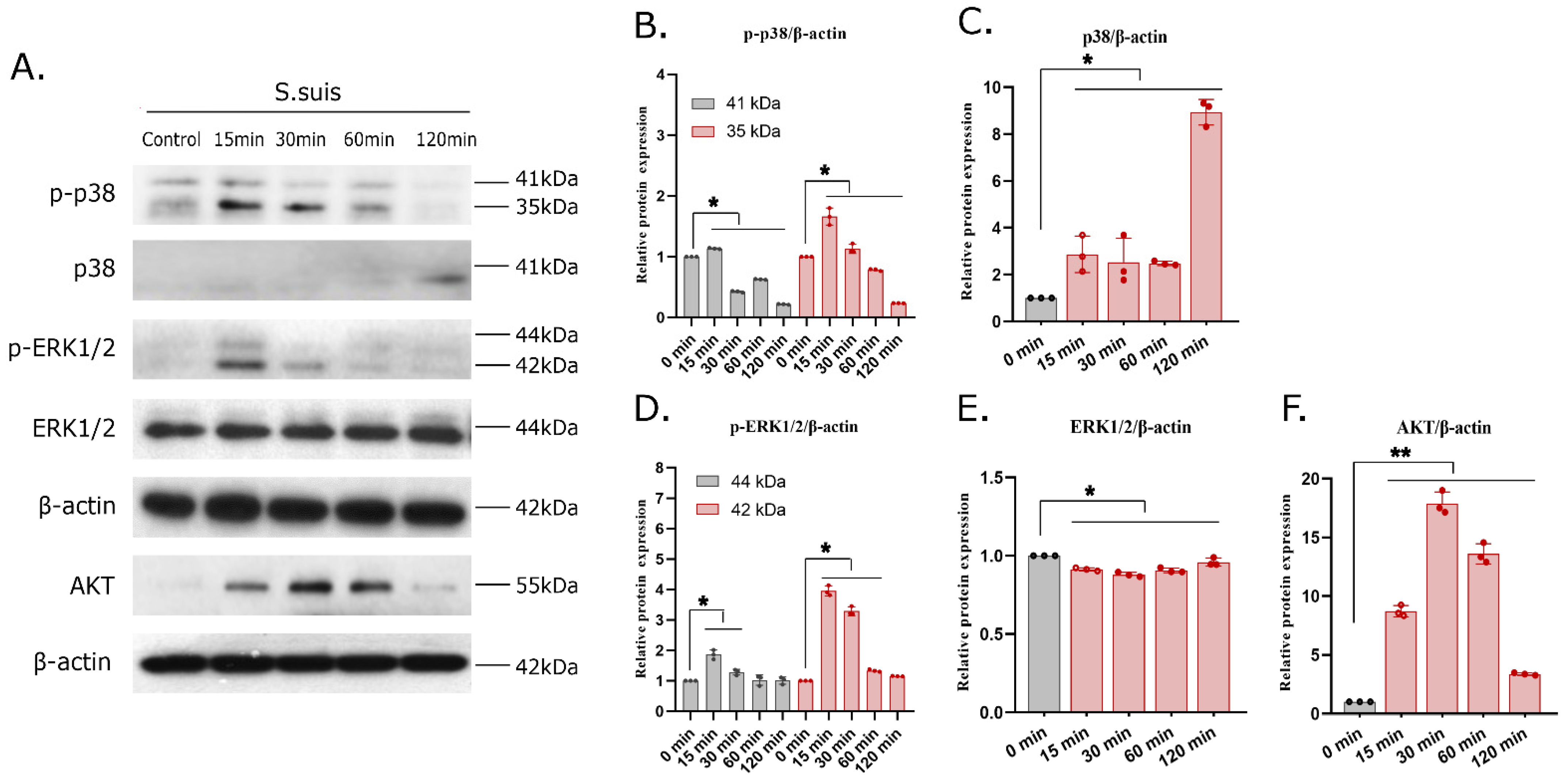
2.9. Western Blotting

iPAMs were cultured in 6-well plates at a density of 2 × 10⁶ cells/well in RPMI 1640 supplemented with 10% FBS overnight. Cells were then infected with S. suis 700794 (MOI = 1) for 0, 15, 30, 60 or 120 min, and subsequently, cell samples were lysed in Radio Immunoprecipitation Assay (RIPA) lysis buffer (Beyotime, Shanghai, China) on ice for 10 min, containing the protease inhibitors 0.1 mM phenylmethylsulfonyl fluoride (PMSF) and Complete Protease Inhibitor (EDTA-free; Merck-Millipore, Darmstadt, Germany). BCA assay was used to determine the protein concentration of the samples. Equal amounts of protein were separated by 12.5% (w/v) sodium dodecyl sulfate-polyacrylamide gel electrophoresis and Spectra Multicolor Protein (Thermo Scientific, Waltham, MA, USA) as Ladder, then transferred to polyvinylidene fluoride (PVDF) membranes (MerckMillipore, Darmstadt, Germany). After blocking with 5% dry milk dissolved in PBS at room temperature for 1 h, membranes were incubated for 1 h at room temperature, targeting MAPK and AKT signaling pathways, including rabbit anti-AKT, p-38 and phospho-p38, ERK1/2 and phospho-ERK1/2, JNK1/2/3 and phospho-JNK1/2/3 (1:1000, Abcam) and anti-β-actin (1:200,000; ABclonal). After washing, HRP-conjugated secondary antibodies were added at 1:10,000 dilution for 1 h at room temperature, followed by washing with PBST buffer. HRP signals were detected with ECL-Supersensitive Luminescence Liquid (PerkinElmer Life Sciences, Fremont, CA, USA), followed by exposure to X-ray film. The Image v1.8.0 software (National Institutes of Health, Bethesda, MD, USA) was utilized for the quantification of the relative integral density of the target protein and the internal control. The relative integral density of the control was then normalized to enable a relative quantitative comparison.

2.10. Statistical Analysis

Data analysis was conducted using GraphPad Prism software (version 8.0.1; GraphPad Software Inc., La Jolla, CA, USA) and is expressed as the mean ± standard deviation (SD) of at least three replicates. The unpaired Student’s t-test was performed to calculate the p-values, with thresholds for statistical significance set at <0.05 (*), <0.01 (**) and <0.001 (***).

3. Results

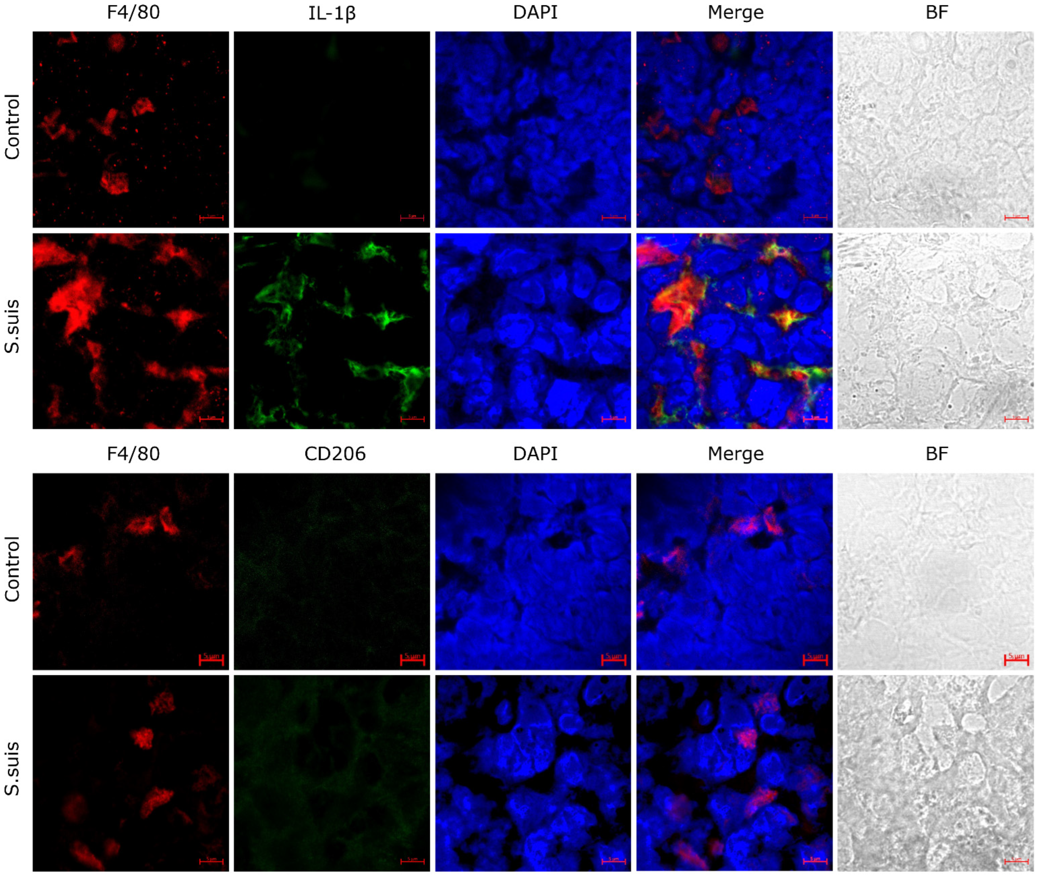
3.1. S. suis Promotes M1 Macrophage Activation in Atrophied Thymus of Infected Mice

Macrophages regulate inflammatory responses by polarizing into different subtypes. To evaluate the subtypes of activated macrophages in the thymus of infected mice at 2 dpi, the expression levels of macrophage polarization markers IL-1β (M1) and CD206 (M2) were detected. The results showed that staining for the macrophage-specific antibody (F4/80) co-localized with IL-1β-labeled M1 macrophages, and expression of IL-1β in the infected thymus was increased (Figure 1). However, the expression of CD206 was almost absent, indicating that S. suis infection induced proinflammatory signals in the thymus of mice at the early stages of infection.

3.2. S. suis Primes Pyroptosis in Atrophied Thymus of Infected Mice

In order to further investigate pyroptosis in the S. suis-infected thymus of mice, the expression of GSDMD-N was evaluated in atrophied thymus tissue (Figure 2). The results showed that the expression of GSDMD-N in the thymus was clearly enhanced in mice at 2 days after infection by S. suis. Moreover, some signals from a macrophage-specific antibody (F4/80) co-localized with GSDMD-N fluorescence signals, which suggested that macrophages present pyroptosis in the thymus. These data indicate that S. suis could induce pyroptosis in the atrophied thymus of S. suis-infected mice and that the pyroptotic cells are macrophages.

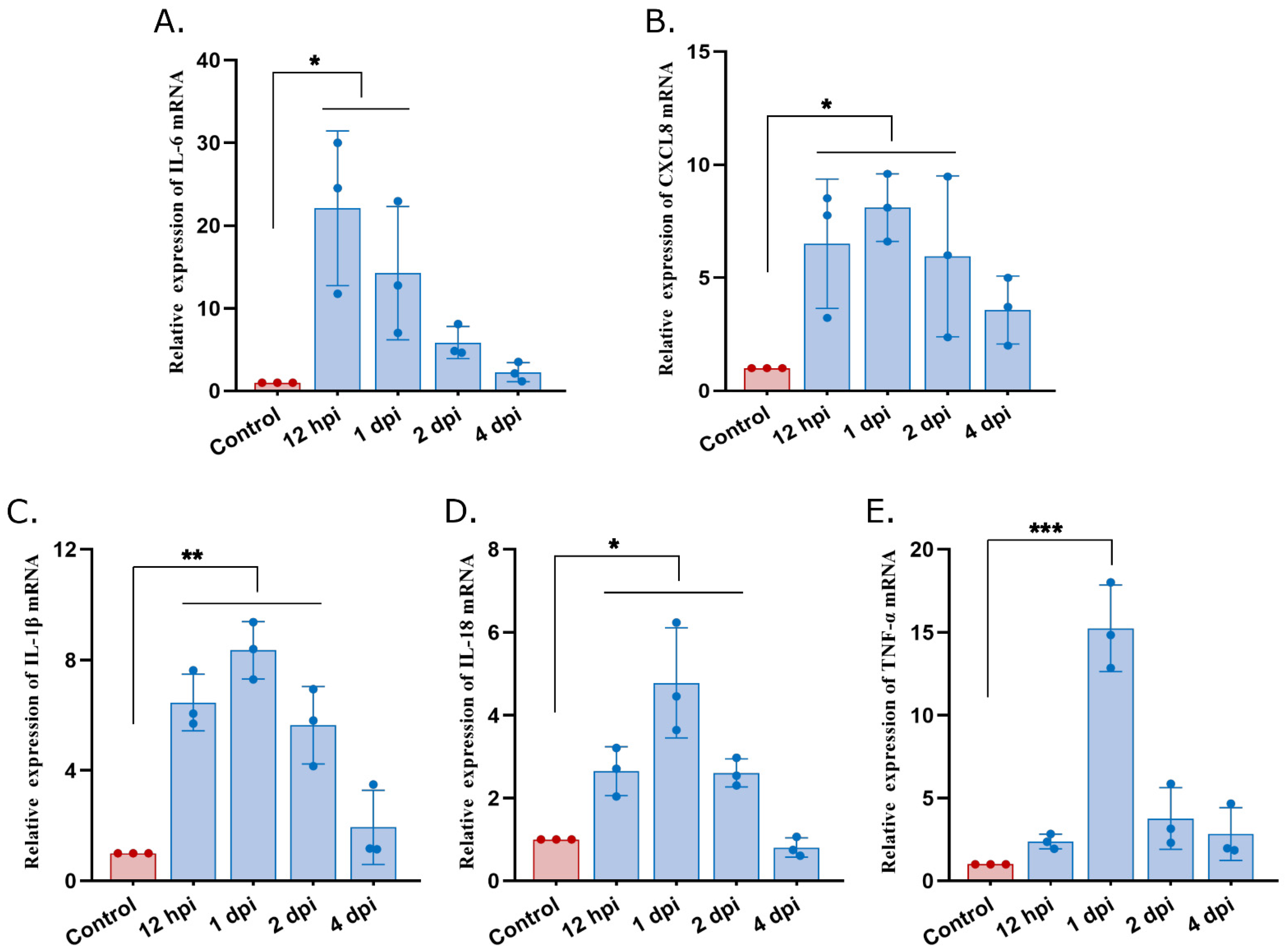
3.3. High Expression of Proinflammatory Mediators in Atrophied Thymus of S. suis-Infected Mice

To understand the immune response triggered by S. suis in the thymus, the mRNA expression of several inflammatory mediators was measured. The results showed that S. suis induced significant expression of many inflammatory mediators. The expression of IL-6 was significantly increased at 12 hpi and 1 dpi and reached a peak at 12 hpi (Figure 3A) compared with the control group. The expression of CXCL8, IL-1β and IL-18 were significantly increased at 12 hpi, 1 dpi and 2 dpi, and reached a peak at 1 dpi compared with the control group (Figure 3B–D). Meanwhile, TNF-α was significantly increased and reached a peak at 1 dpi compared with the control group (Figure 3E). These data suggest that pro-inflammatory responses in the thymus were activated at the early stages of S. suis infection.

3.4. S. suis-Induced LDH Positively Correlates with the Infection Time In Vitro

To determine cytotoxicity in vitro, LDH release from infected primary PAMs was measured at 0, 1, 2, 3, 8, 12 and 24 hpi. The results showed that S. suis infection induced significant LDH activity in primary PAMs from 1 hpi to 24 hpi and positively correlated with the infection time (Figure 4A). TEM further confirmed that S. suis infection induced cytotoxicity and that the cytotoxicity of S. suis in primary PAM cells was time-dependent, with many vacuoles present at 8 hpi but not evident at 1 hpi (Figure 4B). These data demonstrated that the S. suis-treated primary PAMs released LDH and showed morphologic signs of cytotoxicity.

3.5. S. suis Induces Pyroptosis Both in iPAMs and Primary PAMs In Vitro

Live cell imaging was used to further confirm S. suis-induced pyroptosis in both iPAMs and primary PAMs in vitro. Primary PAM or iPAM cells were infected with S. suis 700794 (MOI = 1). At the morphological level, infected iPAMs exhibited increased pyroptotic membrane ballooning and took up propidium iodide (PI) red after S. suis infection for 8 h, compared with uninfected cells (Figure 5A; also see video, additional file 1). Primary PAMs infected with S. suis presented similar features after S. suis infection for 13 h, including cell swelling and large bubbles blowing from the plasma membrane. Subsequently, the cells were lysed and cellular contents were rapidly released (Figure 5B; also see video, additional file 2). Therefore, we concluded that S. suis induces pyroptosis in both iPAMs and primary PAMs in vitro.

3.6. S. suis Infection Actives the MAPK and AKT Signaling Pathways in PAMs

To investigate the effect of the classical inflammation-related MAPK signaling pathway on the S. suis-induced inflammatory response in PAMs, iPAMs were infected with S. suis 700794 (MOI = 1) and collected at 0, 15, 30, 60 or 120 min after infection for Western blot analysis. The results showed that S. suis upregulated p38 and ERK phosphorylation in infected iPAMs compared with the control group. As shown in Figure 6A,B, the levels of p38 and ERK phosphorylation increased significantly, peaked at 15 min after infection and finally returned to control levels, while JNK phosphorylation had no significant change, which indicated that S. suis regulated the inflammatory response by activating the p38 and ERK pathways rather than the JNK pathway. Also, the AKT protein increased from 15 min to 120 min after infection and reached a peak at 30 min after infection (Figure 6A,F), which indicated that S. suis infection activated the AKT pathway. Altogether, the MAPK and AKT signaling pathways are involved in proinflammatory responses in S. suis-infected PAMs.

4. Discussion

The thymus is an important central immune organ in mammals [27], and we previously showed that S. suis infection results in significant necrosis and reduced lymphocytes within the thymus tissue as early as 1 dpi [10]. Since pyroptosis presents characteristics very similar to those of necrosis, it was unknown whether pyroptosis was present in macrophages within the thymus tissue after S. suis infection. Here, pyroptosis on macrophages in the atrophied thymus of S. suis-infected mice was identified with confocal microscopy, and pyroptosis in primary PAMs and iPAMs after S. suis-infection has also been demonstrated in vitro by live cell imaging.
Pyroptosis is a form of gasdermin-mediated programmed necrosis and a common mechanism of innate immune effects, which stimulates inflammatory responses [28]. Pyroptosis enhances immune defense function by destroying the protective intracellular replication niche of pathogens [29]. Moderate pyroptosis is a host defense against infectious pathogens, but hyperactive pyroptosis may be associated with intemperate inflammatory responses and serious tissue damage [20]. This study confirmed that pyroptosis happened in macrophages in the atrophied thymus of S. suis-infected mice, which indicated the presence of pyroptosis in the necrosis cells of the atrophied thymus found in a previous study. Meanwhile, we also confirmed pyroptosis in macrophages following S. suis infection in vitro, with cell swelling and large bubbles blowing from the plasma membrane and increased dying cells (PI+), resulting in the release of IL-1β and IL-18. In this study, the mRNA expression levels of IL-1β and IL-18 indeed increased significantly in the atrophied thymus, in which the existence of pyroptosis was further confirmed. In addition, released LDH was used to measure cytotoxicity and would also be released by cells undergoing pyroptosis in response to inflammasome activation. This study has confirmed that S. suis is capable of inducing LDH release from primary PAMs in vitro, and this effect is observed to increase with prolonged culture time, which is associated with pyroptosis and consistent with previous studies [30].
Macrophages are an important part of the thymus cells and regulate inflammatory responses, and can differentiate into specific phenotypes through polarization, which permits specific biological functions in response to microenvironmental stimuli [31]. The fluorescence imaging showed S. suis infection activated primarily M1-type macrophages as early as 1 dpi in S. suis-infected thymus. The activation of the M1 macrophage occurs in an inflammation-dominated environment, in turn promoting the production of a large number of proinflammatory cytokines, including IL-1, IL-6, IL-18, tumor necrosis factor-α (TNF-α) and other chemokines [32]. S. suis infection in a previous study induced an abnormal serum cytokine dynamic, including the levels of serum IL-2, IL-4, IL-6 and TNF-β, which increased significantly [10]. Also, the proinflammatory mediators (IL-1β, IL-6, IL-18, TNF-α and CXCL8) were found to be highly expressed in the thymus tissue from S. suis-infected mice in this study. MAPK signaling can regulate inflammatory responses, its related signaling kinases including JNK, ERK and p38 [33]. The up-regulating of the p38 and ERK signaling pathways can inhibit M2-type polarization and promote M1-type polarization of macrophages [34]. The results of this study showed exactly that the p38 and ERK signaling pathways were up-regulated and M1 macrophage polarization occurred in S. suis-infected thymus tissue, which indicated that the polarization of macrophages is closely related to the activation of the p38 and ERK signaling pathways in MAPK signaling pathways.

5. Conclusions

This study demonstrated that S. suis could activate M1 polarization, the MAPK signaling pathway and prime pyroptosis in macrophages, as well as produce inflammatory mediators and induce programmed cell death, which contribute to the reduction of lymphocytes in the atrophied thymus of infected mice. However, the underlying mechanism of pyroptosis in macrophages is undetermined and warrants further investigation.

Author Contributions

S.W. and X.C. conceived the idea. S.W. and S.L. designed the experiments and wrote the paper. S.L., T.C., K.G. and Y.-B.Y. conducted the experiments. S.L., T.C., Y.-B.Y., B.Q. and S.W. analyzed the data. C.W., T.A. and X.C. contributed reagents and materials. All authors have read and agreed to the published version of the manuscript.

Funding

This research was supported by the National Natural Science Foundation of China (32273018), Heilongjiang Provincial Natural Science Foundation of China (LH2023C048) and the Central Public-interest Scientific Institution Basal Research Fund (No. 1610302022006).

Data Availability Statement

The original contributions presented in the study are included in the article, further inquiries can be directed to the corresponding authors.

Conflicts of Interest

The authors declare no conflict of interest.

References

  1. Huang, W.; Wang, M.; Hao, H.; Yang, R.; Xie, J.; Su, J.; Lin, M.; Cui, Y.; Jiang, Y. Genomic epidemiological investigation of a Streptococcus suis outbreak in Guangxi, China, 2016. Infect. Genet. Evol. 2019, 68, 249–252. [Google Scholar] [CrossRef] [PubMed]
  2. Goyette-Desjardins, G.; Auger, J.P.; Xu, J.; Segura, M.; Gottschalk, M. Streptococcus suis, an important pig pathogen and emerging zoonotic agent-an update on the worldwide distribution based on serotyping and sequence typing. Emerg. Microbes Infect. 2014, 3, e45. [Google Scholar] [CrossRef] [PubMed]
  3. López-Martínez, M.J.; Ornelas, M.A.S.; Amarie, R.E.; Manzanilla, E.G.; Martínez-Subiela, S.; Tecles, F.; Tvarijonaviciute, A.; Escribano, D.; González-Bulnes, A.; Cerón, J.J.; et al. Changes in salivary biomarkers of stress, inflammation, redox status, and muscle damage due to Streptococcus suis infection in pigs. BMC Vet. Res. 2023, 19, 100. [Google Scholar] [CrossRef] [PubMed]
  4. Haas, B.; Grenier, D. Understanding the virulence of Streptococcus suis: A veterinary, medical, and economic challenge. Med. Mal. Infect. 2018, 48, 159–166. [Google Scholar] [CrossRef] [PubMed]
  5. Lacouture, S.; Okura, M.; Takamatsu, D.; Corsaut, L.; Gottschalk, M. Development of a mismatch amplification mutation assay to correctly serotype isolates of Streptococcus suis serotypes 1, 2, 1/2, and 14. J. Vet. Diagn. Investig. 2020, 32, 490–494. [Google Scholar] [CrossRef]
  6. Newton, K.; Strasser, A.; Kayagaki, N.; Dixit, V.M. Cell death. Cell 2024, 187, 235–256. [Google Scholar] [CrossRef]
  7. Nghia, H.D.; Hoa, N.T.; Linh, L.D.; Campbell, J.; Diep, T.S.; Chau, N.V.; Mai, N.T.; Hien, T.T.; Spratt, B.; Farrar, J.; et al. Human case of Streptococcus suis serotype 16 infection. Emerg. Infect. Dis. 2008, 14, 155–157. [Google Scholar] [CrossRef]
  8. Kerdsin, A.; Akeda, Y.; Takeuchi, D.; Dejsirilert, S.; Gottschalk, M.; Oishi, K. Genotypic diversity of Streptococcus suis strains isolated from humans in Thailand. Eur. J. Clin. Microbiol. Infect. Dis. 2018, 37, 917–925. [Google Scholar] [CrossRef]
  9. Lun, Z.R.; Wang, Q.P.; Chen, X.G.; Li, A.X.; Zhu, X.Q. Streptococcus suis: An emerging zoonotic pathogen. The Lancet. Infect. Dis. 2007, 7, 201–209. [Google Scholar] [CrossRef]
  10. Wang, S.; Lyu, C.; Duan, G.; Meng, F.; Yang, Y.; Yu, Y.; He, X.; Wang, Z.; Gottschalk, M.; Li, G.; et al. Streptococcus suis Serotype 2 Infection Causes Host Immunomodulation through Induction of Thymic Atrophy. Infect. Immun. 2020, 88, e00950-19. [Google Scholar] [CrossRef]
  11. Gottschalk, M.; Segura, M.; Xu, J. Streptococcus suis infections in humans: The Chinese experience and the situation in North America. Anim. Health Res. Rev. 2007, 8, 29–45. [Google Scholar] [CrossRef] [PubMed]
  12. Navegantes, K.C.; de Souza Gomes, R.; Pereira, P.A.T.; Czaikoski, P.G.; Azevedo, C.H.M.; Monteiro, M.C. Immune modulation of some autoimmune diseases: The critical role of macrophages and neutrophils in the innate and adaptive immunity. J. Transl. Med. 2017, 15, 36. [Google Scholar] [CrossRef] [PubMed]
  13. Guerriero, J.L. Macrophages: Their Untold Story in T Cell Activation and Function. Int. Rev. Cell Mol. Biol. 2019, 342, 73–93. [Google Scholar] [PubMed]
  14. Hulsmans, M.; Sager, H.B.; Roh, J.D.; Valero-Muñoz, M.; Houstis, N.E.; Iwamoto, Y.; Sun, Y.; Wilson, R.M.; Wojtkiewicz, G.; Tricot, B.; et al. Cardiac macrophages promote diastolic dysfunction. J. Exp. Med. 2018, 215, 423–440. [Google Scholar] [CrossRef] [PubMed]
  15. Murray, P.J. Macrophage Polarization. Annu. Rev. Physiol. 2017, 79, 541–566. [Google Scholar] [CrossRef]
  16. Gordon, S.; Martinez, F.O. Alternative activation of macrophages: Mechanism and functions. Immunity 2010, 32, 593–604. [Google Scholar] [CrossRef]
  17. Rigamonti, E.; Zordan, P.; Sciorati, C.; Rovere-Querini, P.; Brunelli, S. Macrophage plasticity in skeletal muscle repair. BioMed Res. Int. 2014, 2014, 560629. [Google Scholar] [CrossRef]
  18. Place, D.E.; Lee, S.; Kanneganti, T.D. PANoptosis in microbial infection. Curr. Opin. Microbiol. 2021, 59, 42–49. [Google Scholar] [CrossRef]
  19. Jiang, W.; Deng, Z.; Dai, X.; Zhao, W. PANoptosis: A New Insight Into Oral Infectious Diseases. Front. Immunol. 2021, 12, 789610. [Google Scholar] [CrossRef]
  20. Rao, Z.; Zhu, Y.; Yang, P.; Chen, Z.; Xia, Y.; Qiao, C.; Liu, W.; Deng, H.; Li, J.; Ning, P.; et al. Pyroptosis in inflammatory diseases and cancer. Theranostics 2022, 12, 4310–4329. [Google Scholar] [CrossRef]
  21. Cui, H.; Zhang, L. Key Components of Inflammasome and Pyroptosis Pathways Are Deficient in Canines and Felines, Possibly Affecting Their Response to SARS-CoV-2 Infection. Front. Immunol. 2020, 11, 592622. [Google Scholar] [CrossRef] [PubMed]
  22. Shi, J.; Zhao, Y.; Wang, K.; Shi, X.; Wang, Y.; Huang, H.; Zhuang, Y.; Cai, T.; Wang, F.; Shao, F. Cleavage of GSDMD by inflammatory caspases determines pyroptotic cell death. Nature 2015, 526, 660–665. [Google Scholar] [CrossRef] [PubMed]
  23. Li, S.; Wang, C.; Tang, Y.D.; Qin, L.; Chen, T.; Wang, S.; Bai, Y.; Cai, X.; Wang, S. Interaction between Porcine Alveolar Macrophage-Tang Cells and Streptococcus suis Strains of Different Virulence: Phagocytosis and Apoptosis. Microorganisms 2023, 11, 160. [Google Scholar] [CrossRef] [PubMed]
  24. Done, S.H.; Paton, D.J. Porcine reproductive and respiratory syndrome: Clinical disease, pathology and immunosuppression. Vet. Rec. 1995, 136, 32–35. [Google Scholar] [CrossRef]
  25. Imori, P.F.; Passaglia, J.; Souza, R.A.; Rocha, L.B.; Falcao, J.P. Virulence-related genes, adhesion and invasion of some Yersinia enterocolitica-like strains suggests its pathogenic potential. Microb. Pathog. 2017, 104, 72–77. [Google Scholar] [CrossRef]
  26. Li, H.; Yang, D.H.; Zhang, Y.; Zheng, F.; Gao, F.; Sun, J.; Shi, G. Geniposide suppresses NLRP3 inflammasome-mediated pyroptosis via the AMPK signaling pathway to mitigate myocardial ischemia/reperfusion injury. Chin. Med. 2022, 17, 73. [Google Scholar] [CrossRef]
  27. Savino, W. The thymus is a common target organ in infectious diseases. PLoS Pathog. 2006, 2, e62. [Google Scholar] [CrossRef]
  28. Jorgensen, I.; Miao, E.A. Pyroptotic cell death defends against intracellular pathogens. Immunol. Rev. 2015, 265, 130–142. [Google Scholar] [CrossRef]
  29. Miao, E.A.; Leaf, I.A.; Treuting, P.M.; Mao, D.P.; Dors, M.; Sarkar, A.; Warren, S.E.; Wewers, M.D.; Aderem, A. Caspase-1-induced pyroptosis is an innate immune effector mechanism against intracellular bacteria. Nat. Immunol. 2010, 11, 1136–1142. [Google Scholar] [CrossRef]
  30. Segura, M.; Gottschalk, M. Streptococcus suis interactions with the murine macrophage cell line J774: Adhesion and cytotoxicity. Infect. Immun. 2002, 70, 4312–4322. [Google Scholar] [CrossRef]
  31. Sica, A.; Mantovani, A. Macrophage plasticity and polarization: In vivo veritas. J. Clin. Investig. 2012, 122, 787–795. [Google Scholar] [CrossRef] [PubMed]
  32. Atri, C.; Guerfali, F.Z.; Laouini, D. Role of Human Macrophage Polarization in Inflammation during Infectious Diseases. Int. J. Mol. Sci. 2018, 19, 1801. [Google Scholar] [CrossRef] [PubMed]
  33. Arthur, J.S.; Ley, S.C. Mitogen-activated protein kinases in innate immunity. Nat. Rev. Immunol. 2013, 13, 679–692. [Google Scholar] [CrossRef] [PubMed]
  34. Fang, J.; Ou, Q.; Wu, B.; Li, S.; Wu, M.; Qiu, J.; Cen, N.; Hu, K.; Che, Y.; Ma, Y.; et al. TcpC Inhibits M1 but Promotes M2 Macrophage Polarization via Regulation of the MAPK/NF-κB and Akt/STAT6 Pathways in Urinary Tract Infection. Cells 2022, 11, 2674. [Google Scholar] [CrossRef]
Figure 1. Thymic macrophage polarization in S. suis infection. Using confocal laser scanning microscopy, appropriate FITC-conjugated antibodies were used to label cells in sections from thymus of infected mice; M1 macrophage marker IL-1β (green); M2 macrophage marker CD206 (green). Macrophages were stained with F4/80 antibody (red) and cell nuclei (blue) were stained with DAPI; BF: Bright-field. Scale bars, 5 μm.
Microorganisms 12 01879 g001
Figure 2. S. suis induces pyroptosis in macrophages of the thymus of infected mice. Confocal laser scanning immunostaining was performed to visualize thymic macrophages (F4/80 antibody; purple) and pyroptosis (GSDMD-N; red) in atrophied and control thymus of mice, and nuclei were stained with DAPI (blue). BF: Bright-field. Scale bars, 5 μm.
Microorganisms 12 01879 g002
Figure 3. S. suis induces pro-inflammatory mediators in thymus of infected mice. (A) Total tissue RNA was extracted at 12 hpi and 1, 2 and 4 dpi, and the levels of IL-6, (B) CXCL8, (C) IL-1β, (D) IL-18, (E) and TNF-α mRNA expression were analyzed using real-time RT-PCR. Experiments were repeated three times. Differences were analyzed using unpaired Student’s t-test; data are mean ± SD; *: p < 0.05; **: p < 0.01; ***: p < 0.001.
Microorganisms 12 01879 g003
Figure 4. S. suis-induced LDH release and damage to primary PAMs. Primary PAMs were infected with S. suis 700794 at an MOI = 1 for 1, 2, 3, 8, 12 or 24 h, with a negative control included as 0 h. (A) LDH release induced by S. suis in primary PAMs was quantified by a standard LDH-release assay. (B) Electron micrographs of infected primary PAMs showing S. suis-induced cellular damage at 8 hpi, and yellow circle showed vacuoles of PAMs. Experiments were repeated three times, and results are expressed as means ± S.D., as determined by one-way ANOVA; *: p < 0.05; scale bars, 2 μm.
Microorganisms 12 01879 g004
Figure 5. Live-cell imaging of pyroptosis after in vitro infection with S. suis. PAMs were cultured in glass-bottom cell culture dishes and treated with S. suis (MOI = 1). (A) Microscopy images of iPAMs treated with S. suis incubated in medium containing Hoechst 33342 (cell nuclei; blue) and propidium iodide (PI; red), the yellow arrows indicate pyrolyzed cells. (B) Microscopy images of primary PAMs treated with S. suis, and rectangle indicates pyrolyzed cells. Numbers at upper left indicate time post-infection (h:min:s).
Microorganisms 12 01879 g005
Figure 6. The MAPK and AKT pathways were activated by S. suis in porcine macrophages. Cells were infected with S. suis at an MOI = 1 for 15, 30, 60 and 120 min. The cell lysates were collected, and levels of proteins related to inflammatory functional indexes were determined by Western blot (A). Representative Western blots show the expression level of (B) p-p38, (C) p38, (D) p-ERK1/2, (E) ERK1/2 and (F) AKT. The mean ± SD of experiments are shown; *: p < 0.05; **: p < 0.01.
Microorganisms 12 01879 g006
Table 1. Protocols for confocal microscopy.
Cell SurfaceAntibodyDilution, Temperature, TimeFluorescent Secondary Antibody
Temperature, Time
M1 MacrophageIL-1β1:200, overnight, 4 °C Alexa Fluor 488-conjugated goat anti-mouse IgG, RT, 1 h
M2 MacrophageCD2061:100, overnight, 4 °C Alexa Fluor 488-conjugated goat anti-mouse IgG, RT, 1 h
MacrophageF4/801:50, overnight, 4 °C Alexa Fluor 647-conjugated rabbit anti-mouse F4/80, RT, 1 h
PyroptosisGSDMD-N1:50, overnight, 4 °C Alexa Fluor 568-conjugated goat anti-rabbit antibody, RT, 1 h
NucleiDAPIRT, 20 min
Table 2. Mouse primer pairs used for real-time PCR.
GeneForward Primer (5′-3′)Reverse Primer (5′-3′)
IL-6TGTGCAATGGCAATTCTGATGGTACTCCAGAAGACCAGAGGA
IL-8GGTCTTCCTGCTTGAATGGCTTGAGCGGTGTCCTGATTATCGTCCTC
IL-18GCCATGTCAGAAGACTCTTGCGTCGTACAGTGAAGTCGGCCAAAGTTGTC
IL-1βGGTGTGTGACGTTCCCATTAGACAGGGTGGGTGTGCCGTCTT
TNF-αGTCTACTGAACTTCGGGGTGATCGTGGGCTACAGGCTTGTCACTCG
β-actinGCTTCTTTGCAGCTCCTTCGTCCTTCTGACCCATTCCCACC
Disclaimer/Publisher’s Note: The statements, opinions and data contained in all publications are solely those of the individual author(s) and contributor(s) and not of MDPI and/or the editor(s). MDPI and/or the editor(s) disclaim responsibility for any injury to people or property resulting from any ideas, methods, instructions or products referred to in the content.

