Article

Coaggregation Occurs between a Piliated Unicellular Cyanobacterium, Thermosynechococcus, and a Filamentous Bacterium, Chloroflexus aggregans

Department of Biological Sciences, Tokyo Metropolitan University, 1-1 Minami-Osawa, Hachioji 192-0397, Tokyo, Japan
*
Author to whom correspondence should be addressed.
Microorganisms 2024, 12(9), 1904; https://doi.org/10.3390/microorganisms12091904
Submission received: 13 August 2024 / Revised: 4 September 2024 / Accepted: 6 September 2024 / Published: 19 September 2024
(This article belongs to the Special Issue Phototrophic Bacteria 2.0)

Abstract

:
Cyanobacteria are widely distributed in natural environments including geothermal areas. A unicellular cyanobacterium, Thermosynechococcus, in a deeply branching lineage, develops thick microbial mats with other bacteria, such as filamentous anoxygenic photosynthetic bacteria in the genus Chloroflexus, in slightly alkaline hot-spring water at ~55 °C. However, Thermosynechococcus strains do not form cell aggregates under axenic conditions, and the cells are dispersed well in the culture. In this study, Thermosynechococcus sp. NK55a and Chloroflexus aggregans NBF, isolated from Nakabusa Hot Springs (Nagano, Japan), were mixed in an inorganic medium and incubated at 50 °C under incandescent light. Small cell aggregates were detected after 4 h incubation, the size of cell aggregates increased, and densely packed cell aggregates (100–200 µm in diameter) developed. Scanning electron microscopy analysis of cell aggregates found that C. aggregans filaments were connected with Thermosynechococcus sp. cells via pili-like fibers. Co-cultivation of C. aggregans with a pili-less mutant of Thermosynechococcus sp. did not form tight cell aggregates. Cell aggregate formation was observed under illumination with 740 nm LED, which was utilized only by C. aggregans. These results suggested that Chloroflexus filaments gather together via gliding motility, and piliated cyanobacterial cells cross-link filamentous cells to form densely packed cell aggregates.

1. Introduction

Oxygenic photoautotrophic bacteria, cyanobacteria play an important role as a primary producer on Earth [1]. In natural environments, cyanobacteria form cell aggregates and biofilms [2]. Multicellular assemblage formation increases tolerance against environmental stresses, such as desiccation, osmotic pressure, and ultraviolet irradiation [3]. In cell aggregates and biofilms, cyanobacterial cells secrete extracellular polymeric substances, such as polysaccharides, proteins, and nucleic acids [2], and synthesize pili [4] to connect cells with each other. In cyanobacterial cell aggregates, coexistence of other bacteria is frequently observed [5]. The co-aggregate formation of cyanobacteria and other heterotrophic bacteria not only increases stress tolerance but also promotes their metabolic exchange (e.g., photosynthetic product and vitamins) [6,7]. However, cell-to-cell interaction between cyanobacteria and others in cell aggregates has not been studied well.
At geothermal springs, cyanobacteria-dominated biofilms are often observed under hot-spring water [8,9,10,11]. In slightly alkaline hot-spring water from 50 °C to 65 °C, a deeply branched cyanobacterial group, Thermosynechococcus (H. Katoh, S. Itoh, J.-R. Shen, and M. Ikeuchi [12]), is widely distributed as a dominant species worldwide [13,14]. Thermosynechococcus cyanobacteria have been studied in photosynthetic apparatuses [15,16,17,18,19,20,21] and their tactic behavior through type IV pili-mediated twitching motility [22]. Its CO2-fixing ability also attracts attention as a cell factory [23]. The metabolic network and material cycle in well-developed multispecies thick biofilms (~0.5 cm thickness, so-called microbial mats), dominated by Thermosynechococcus, are of great interest as a model ecosystem driven by oxygenic photosynthesis [24,25,26,27]. However, it has not been clarified yet how densely packed mats are formed and how the stratified structure of mats is developed. Unicellular Thermosynechococcus cells do not show adherability to solid surfaces and do not form cell aggregates under growing conditions, although cell aggregate formation is induced by environmental stresses, such as low temperature and blue light [28,29].
Castenholz suggested cell aggregate formation of unicellular, rod-shaped cyanobacteria with filamentous anoxygenic photosynthetic bacteria in the genus Chloroflexus in Hunter’s Hot Springs in Oregon, USA [11]. Bacteria in the genus Chloroflexus (Pierson and Castenholz [30]) have been characterized as a fast-gliding bacteria [31]. The representative species, Chloroflexus aggregans (Hanada et al. [32]), forms cell aggregates through active gliding motility on other filaments [33], but the aggregation is easily dispersed because there is no adhesive attachment between the filaments. C. aggregans was the second dominant species in the Thermosynechococcus microbial mats at Nakabusa Hot Springs (Nagano, Japan), one of several well-studied thermal springs (slightly alkaline, pH ~8.5; sulfidic, ~0.1 mM; up to 94 °C) [25,26]. It was hypothesized that C. aggregans helps form cell aggregates of Thermosynechococcus through cell-to-cell interactions.
In this study, a piliated cyanobacterium, Thermosynechococcus sp. NK55a [34], and a filamentous bacterium, C. aggregans NBF [35], isolated from Nakabusa Hot Springs, were co-cultivated under gently flowing water conditions to investigate their cell aggregate-forming ability.

2. Materials and Methods

2.1. Bacterial Strains

Thermosynechococcus sp. strain NK55a (=NBRC 108920) [34], isolated from Nakabusa Hot Springs (Nagano, Japan), was cultured at 50 °C in a 60 mL BG11 medium [36] in 100 mL glass flasks with a cellulose cap (Steristopper, Heinz Herenz, Hamburg, Germany), with shaking at 80 rpm under illumination using a fluorescent lamp (15~20 μmol/s/m2). C. aggregans NBF, isolated from Nakabusa Hot Springs [35], was anaerobically cultured at 50 °C in a heterotrophic, 1/5PE medium [32,37,38] under illumination using an incandescent lamp (15~20 μmol/s/m2). Anaerobic conditions were achieved by completely filling a screw-capped glass test tube (∅18 mm, 32 mL volume). Thermosynechococcus and Chloroflexus growth was monitored by measuring the optical density at 740 nm with UV-1800 (Shimadzu, Kyoto, Japan) and 660 nm with miniphoto 518R (Taitec, Koshigaya, Japan), respectively. The number of Thermosynechococcus cells in the culture was assessed using a counting chamber (SLGC, Saitama, Japan) with a phase-contrast microscope (AXIO Imager A2; Carl Zeiss, Oberkochen, Germany).

2.2. Disruption of the pilB Gene in Thermosynechococcus sp. NK55a

For homologous recombination, a plasmid, pUC19-∆pilB_CmR (8804 bp), harboring the upstream and downstream regions of the pilB gene (NK55_09820) in the NK55a genome [34] and chloramphenicol-resistant cassette, was constructed to transform strain NK55a. Based on the genomic information of Thermosynechococcus sp. NK55a (NC_023033), PCR primer sets (Table 1) were designed using a web tool (primer-BLAST; https://www.ncbi.nlm.nih.gov/tools/primer-blast/, accessed on 24 April 2023) to amplify the upstream (2.7 kbp) and downstream (2.6 kbp) regions of the pilB gene using NK55a genomic DNA as the template, which was extracted using a DNeasy UltraClean microbial kit (Qiagen, Venlo, The Netherlands). DNA fragments of the chloramphenicol-resistant cassette and a vector backbone were amplified from pHSG396 (Takara Bio, Kusatsu, Japan) and pUC19 (Takara Bio), respectively. PCR was performed using primerSTAR (Takara Bio) and primers listed in Table 1 according to the following protocol (32 cycles): denaturation, 95 °C for 10 s; annealing, 55 °C for 5 s; and polymerization, 72 °C for 10 s (chloramphenicol-resistant cassette) or 20 s (upstream and downstream regions of the pilB gene). DNA fragments were purified using the QIAEXII Gel Extraction kit (Qiagen) after agarose gel electrophoresis. Four fragments (vector backbone, upstream region of pilB, chloramphenicol-resistant gene cassette, and downstream region of pilB) were assembled using the HiFi DNA assembly master mix kit (New England Biolabs, Ipswich, MA, USA) according to the manufacturer’s instructions. Assembled DNA was used to transform Escherichia coli DH5α competent cells (Takara Bio) according to the manufacturer’s instructions. Construction of the obtained plasmids was confirmed by restriction enzyme digestion and DNA sequencing (BigDye Terminator kit v3.1 and SeqStudio Genetic Analyzers, Thermo Fisher Scientific, Waltham, MA, USA). The natural transformation of Thermosynechococcus sp. NK55a was performed according to Iwai et al. [39]. Mutant strains were selected by cultivation at 45 °C in BG11 agar medium containing 5 µg/mL chloramphenicol. Complete segregation via the chloramphenicol-resistant gene cassette was confirmed by DNA sequencing using the primers listed in Table S1.

2.3. Transmission Electron Microscopy (TEM) Imaging

Bacterial cells were fixed with 2% glutaraldehyde in 0.1 M of cacodylate buffer (pH 7.4) for 1 min on carbon-coated Cu grids. After that, the samples were stained with 2% phosphor tungstic acid solution (pH 7.0) for 30 s and observed with a transmission electron microscope at 100 kV (JEM 1400Plus; JEOL, Akishima, Japan) at Tokai Electron Microscopy (Nagoya, Japan).

2.4. Co-Cultivation of Thermosynechococcus sp. and C. aggregans

Cells at the late exponential growth phase (OD740 = 0.6–0.8 for Thermosynechococcus sp. and OD660 = 0.4–0.6 for C. aggregans) were harvested by centrifugation at 7600× g for 5 min and suspended together into a 5 mL BG11 medium (OD740 = 0.48 for Thermosynechococcus sp. and OD660 = 0.30 for C. aggregans). The cell mixture was added to a 25 mL glass vial, which was sealed with a butyl rubber stopper and aluminum cap. The vials were incubated at 50 °C in the light (incandescent lamp, 15–20 μmol/s/m2) using a roller apparatus at 20 rpm (VMRC-5; AS ONE, Osaka, Japan). When indicated, 630 and/or 740 nm LED arrays were used in place of incandescent lamps.

2.5. Bright-Field and Fluorescence Microscopy Imaging

Bright-field and fluorescence microscopic images of bacterial cultures were captured with a microscope (AXIO Imager A2; Carl Zeiss) equipped with a color digital camera, DP73 (Olympus, Tokyo, Japan), and CellSens standard 1.18 software (Olympus). Before fluorescence microscopic observation, cells were stained with a nucleic acid binding dye, acridine orange (0.005%, w/v). Fluorescence from acridine orange and chlorophylls were detected by 460 nm excitation and 395 to 440 nm excitation, respectively, with a long-pass emission filter.

2.6. Scanning Electron Microscopy (SEM) Imaging

Cell aggregates collected from culture solutions were fixed in 4% paraformaldehyde, 4% glutaraldehyde in phosphate buffer (pH 7.4) at 4 °C for 1 h and then 2% glutaraldehyde in phosphate buffer for one night and subsequently postfixed in 2% osmium tetraoxide in phosphate buffer at 4 °C for 2 h. The specimens were dehydrated in a graded ethanol and dried by CO2 critical point dry. Dried specimens were coated with an osmium plasma ion coater (OPC-80; Nippon Laser & Electronics, Tokyo, Japan). The observation was made using JSM-7500F at 5 kV (JEOL) at the Hanaichi UltraStructure Research Institute (Okazaki, Japan).

2.7. DNA Extraction and Quantitative PCR (qPCR)

Bacterial cells were collected from co-culture solutions, and total DNA was extracted from planktonic cells and cell aggregates using the DNeasy UltraClean microbial kit (Qiagen) and DNeasy power biofilm kit (Qiagen), respectively, according to the manufacturer’s instructions. For cell aggregates, cells were beaten with a FastPrep 24 instrument (Funakoshi, Tokyo, Japan) for 60 s at 4 m/s before DNA extraction. PCR primer sets targeting the rpoB gene were designed to distinguish Thermosynechococcus sp. NK55a (5′-CCTCCTATTTACATGACGGCT-3′ and 5′-AGATAATCTGCACTGGCGAA-3′) and C. aggregans NBF (5′-GTCAGATTCTCCGTGAGGACATC-3′ and 5′-GGTTATGTTCATCGAGCGGTGCA-3′). The StepOne Real-Time PCR system (Applied Biosystems, Foster City, CA, USA) was used with FastStart Universal SYBR Green Master (Roche, Mannheim, Germany). The reaction mixture was composed of 10 μL of FastStart SYBR Green Master mix, 0.12 μL of 50 μM primers, 8.76 μL of water, and 1 μL of DNA solution. Real-time PCR was performed using the following protocol: first denaturation, 95 °C for 10 min; denaturation and amplification, 95 °C for 15 s and 60 °C for 60 s, respectively (40 cycles). For standard curves, genomic DNA was used after the concentrations were spectrophotometrically quantified with BioSpec-nano (Shimadzu).

2.8. Total Protein Quantification after Size Fractionation

At the appropriate incubation time, the co-culture solution was filtered through nylon mesh filters (pore size, 20 μm), and the filters were washed with BG11 medium. Cells in the flow-through fraction, collected by centrifugation at 7600× g for 5 min, and cells on the filter were suspended in 0.2 mol/L of NaOH and 2% SDS solution containing zirconia silica microbeads (∅1 mm and ∅0.2 mm). The cell suspension was beaten with a FastPrep 24 instrument (Funakoshi) for 60 s at 4 m/s, incubated at 95 °C for 15 min, and centrifuged to collect supernatant. Protein concentration in the supernatant was determined with a DC protein assay kit (Bio-Rad, Hercules, CA, USA) and a spectrophotometer (Infinite 200PRO; Tecan, Seestrasse, Switzerland) using bovine serum albumin as a standard.

2.9. Statistical Analysis

A two-sample statistical test (Student’s t-test) was used to compare the amount of total cellular proteins before and after incubation. The significant difference was considered when the p value was <0.05.

3. Results

3.1. Cell Aggregate Formation in the Co-Culture of Thermosynechococcus sp. NK55a and C. aggregans NBF

Thermosynechococcus sp. NK55a and C. aggregans NBF were co-inoculated into BG11 medium in glass vials and incubated at 50 °C in incandescent light under gently rolling conditions. Periodic photographs of the glass vial and micrographs of the culture solution were taken (Figure 1a). At the beginning of the cultivation, cells in the culture were dispersed well. After 4 h cultivation, small and low dense cell aggregates (20–100 µm) appeared, and the number and size of cell aggregates increased. Finally, densely packed cell aggregates developed at 100 to 200 µm in diameter, and the color of the culture solution turned transparent. Cell aggregates formed after 12 h were hardly dispersed by vigorous vortex mixing. After size fractionation with a 20 μm pore size filter, the aggregation index was determined (Figure 1b). At 0 h incubation, the aggregation index was ~20%, resulting from capturing a part of long filamentous cells of C. aggregans on the filter. The index gradually increased during incubation and reached ~80% after 12 h incubation. No further increase in the aggregation index was observed after 12 h (80.8%, 16 h). Total cellular proteins did not significantly increase with incubation (3.02 ± 0.90 mg mL−1 at 0 h and 3.09 ± 0.53 mg mL−1 at 12 h; t = 0.92, Student’s t-test), suggesting no marked cellular growth during the incubation.
qPCR was performed to estimate the population ratio of the two species in cell aggregates. Total DNA was extracted from cell aggregates formed by 12 h incubation. The copy number of the rpoB gene was quantified by qPCR to calculate the genome copies of each species; (4.70 ± 0.74) × 108 and (8.33 ± 1.25) × 108 genome copies in cell aggregates per one vial were detected for Thermosynechococcus sp. NK55a and C. aggregans NBF, respectively. As observed in many cyanobacteria [40], polyploidy was reported for Thermosynechococcus sp. [41]. The genome copy number was estimated to be four to five copies per cell for the axenic culture of Thermosynechococcus sp. NK55a by qPCR and microscopic cell counting.

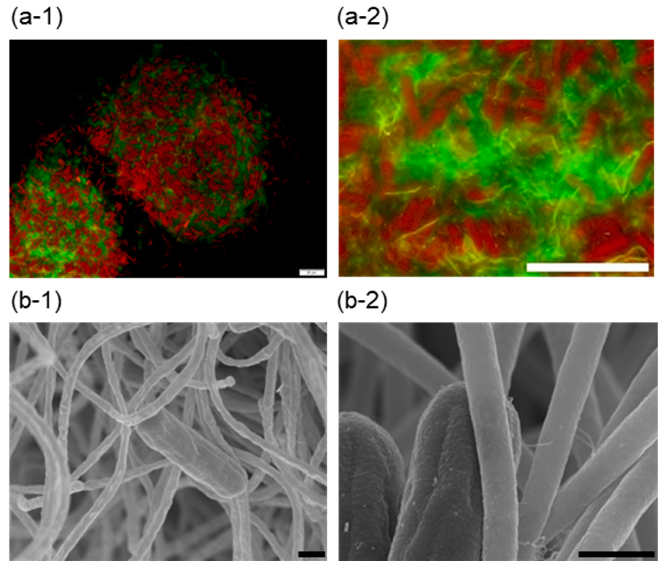
3.2. Morphology of Co-Aggregates

Cell aggregates were subjected to fluorescence microscopic analysis after staining with acridine orange (Figure 2a-1,a-2). Unicellular rod cells are reddish, indicating chlorophyll a-containing cyanobacteria. Filamentous cells were detected with a greenish color by acridine orange. The microscopic image indicated that unicellular cyanobacterial cells and filamentous cells formed cell aggregate in a mosaic manner. Kawano et al. reported that extracellular cellulose production in Thermosynechococcus sp. RKN was induced under low-temperature illuminated conditions to form cell aggregates, which were dispersed by cellulase treatment [29]. Co-aggregates formed by Thermosynechococcus sp. NK55a and C. aggregans NBF were not dispersed by cellulase treatment (Figure S1).
Cell aggregates formed after 12 h incubation were subjected to SEM analysis. Cyanobacterial cells were detected for bridging to filamentous cells (Figure 2b-1). Focusing on the interface between unicellular cells and filamentous cells, thin fibers (20–30 nm in diameter) were observed (Figure 2b-2). The fibers seemed to be type IV pili, which were identified at both poles of Thermosynechococcus cell [22].

3.3. Co-Cultivation of the pilB Mutant of Thermosynechococcus sp. and C. aggregans NBF

SEM analysis of cell aggregates (Figure 2b-2) expected that pili from Thermosynechococcus sp. contributed to cell aggregate formation with C. aggregans. The pili-less mutant of Thermosynechococcus sp. NK55a was constructed to evaluate the effects on cell aggregation. The pilB gene encodes cytoplasmic ATPase, essential to assemble pilin monomers [42,43]. It was reported that disruption of the pilB gene (tll0122) in Thermosynechococcus sp. resulted in lacking pilus filaments and motility [22]. TEM analysis of the ∆pilB mutant strain of Thermosynechococcus sp. NK55a confirmed the lack of fibers on the cell surface, although cells of the wild-type strain possessed a few fibers near the cell pole (Figure 3a). Co-culture of the ∆pilB mutant of Thermosynechococcus sp. NK55a and C. aggregans NBF was conducted as carried out for the wild-type strain in Figure 1. The aggregation index after 12 h incubation for the mutant strain was ~30% (Figure 3b), and no marked cell aggregate formation was visually observed (Figure 3c). Micro cell aggregates were microscopically detected, but cell aggregates were easily dispersed by hand shaking the glass vials.

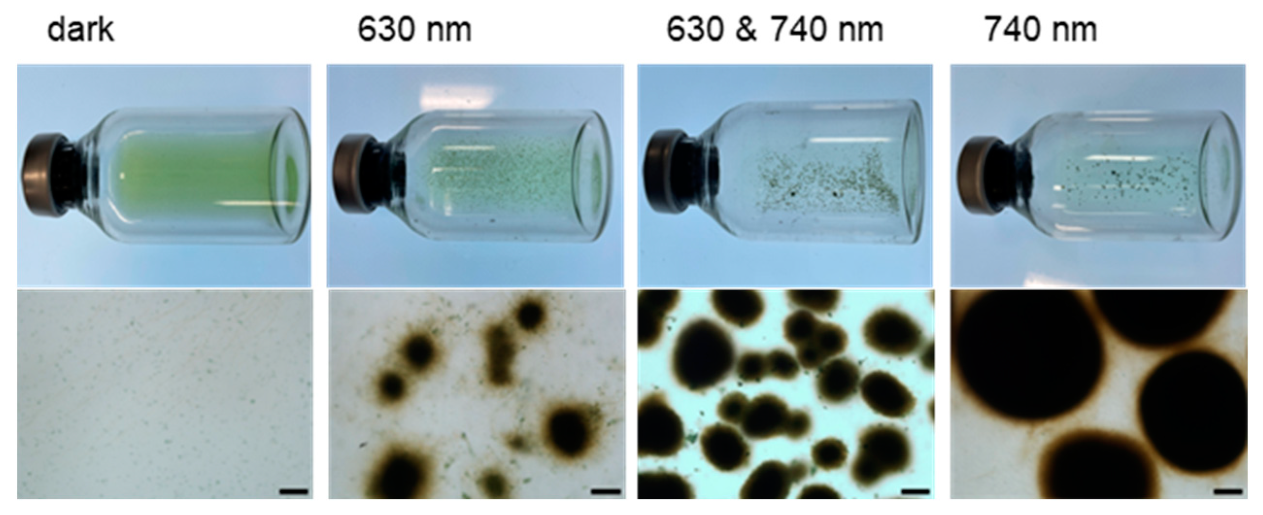
3.4. Effects of Illumination Wavelength on Cell Aggregate Formation

Co-cultivation of Thermosynechococcus sp. NK55a (wild-type) and C. aggregans NBF in the dark resulted in no marked cell aggregate formation (Figure 4). The effects of illumination wavelength on cell aggregate formation by the co-culture were examined. Cells of these phototrophs, Thermosynechococcus sp. and C. aggregans, are known to show the absorption peaks at ~630 nm and ~740 nm, respectively [24]. After 12 h incubation under illumination at 630 nm and 740 nm LED arrays, tight cell aggregates 100 to 200 µm in diameter were formed, and the color of the culture solution was transparent (Figure 4). Cell aggregate formation was similarly observed after incubation at 740 nm illumination (Figure 4). Under only 630 nm illumination, cell aggregates were formed (50 to 200 µm in diameter) (Figure 4), but cell aggregates were loosely packed and easily dispersed by hand shaking the vials.

4. Discussion

In this study, the co-culture of a piliated unicellular cyanobacterium Thermosynechococcus sp. with a filamentous gliding bacterium C. aggregans formed tightly packed cell aggregates (Figure 1). Tight cell aggregate formation was observed under 740 nm LED arrays (Figure 4), indicating that cyanobacterial active photosynthesis was not necessary, but photosynthetic activity of C. aggregans was required. It is likely that ATP produced through cyclic photophosphorylation was mainly utilized for the gliding motility of C. aggregans [33] because electron and carbon sources for C. aggregans were limited in the culture. Based on the results, this study proposed cell aggregate formation steps as follows (Figure 5): piliated cyanobacterial cells attach to Chloroflexus filamentous cells. Chloroflexus filaments attached to cyanobacterial cells gather together through gliding motility, and motility draws filamentous cells close to each other. The proximity enables cyanobacterial cells to bridge filamentous cells. The gliding motility of Chloroflexus filaments and cross-linking via cyanobacterial cells results in tightly and densely packed cell aggregate formation.
Co-culture of the ∆pilB mutant of Thermosynechococcus sp. with Chloroflexus did not form tight cell aggregates (Figure 3b,c). The pilB gene encodes a key unit required for pili assembly [44,45]. As shown by TEM images (Figure 3a), pili and pili-like structures were not detected at the cell surface of the mutant strain. In cyanobacteria species, pili which is clearly detectable by TEM is called as “thick pili” (5 to 8 nm width and ≥1 µm length) [46,47]. Thick pili are the machinery for twitching motility in unicellular cyanobacteria [44,46] and adhere to the solid surface. In Thermosynechococcus, a few pili were detected near the cell poles in strain NK55a (Figure 3a), as reported for another strain of Thermosynechococcus [22]. To the authors’ knowledge, no report has detected cell-to-cell adhesion via thick pili. Pili detected on cyanobacterial cells by SEM analysis in the co-culture (Figure 2b-2) could be thick pili and likely worked to connect filamentous cells. The number of pili detected by the SEM analysis was limited. This may be due to the detachment of a part of pili during cell treatment for SEM analysis, as reported previously [48].
In several cyanobacterial species, the existence of so-called “thin pili” was suggested [22,49,50]. The structure and function of thin pili remain unclear in most cyanobacterial species [3]. In Synechocystis sp., the disruption of a gene encoding thin pili structural protein reduced the floc-forming ability [51,52]. Genome analyses of Thermosynechococcus suggested that it harbors multiple copies of the gene encoding the structural protein of pili. ∆pilB mutant cells of Thermosynechococcus sp. could also lose other types of pili than thick pili. However, it was hard to identify thin pili by SEM observation and distinguish genes for thin pili from the genes for thick pili.
PilB for type IV pilus assembly shows homology with a secretion ATPase required for a type II secretion system [53,54]. In Synechococcus elongatus PCC7942 and Synechocystis PCC 6803, it was suggested that PilB also works for secretion of extracellular substances [3]. However, the deficiency of PilB did not markedly change the cell morphology and adhesive properties of Thermosynechococcus sp. under axenic conditions.
Cell aggregate formation by extracellular polymeric substances has not been reported for Chloroflexus [30,32,55,56,57]. Non-adhesive cell aggregate formation of C. aggregans via gliding motility was described in 1995 [32], but the gliding machinery of C. aggregans has not been identified yet [58]. Adhesive cell aggregate formation with Thermosynechococcus detected in this study probably required the gliding motility of C. aggregans as described above. This idea was supported by the observation of no cell aggregate formation in the co-culture of Thermosynechococcus with a slow gliding strain, Chloroflexus aurantiacus J-10-fl (Figure S2), whose gliding rate was ~1/100 slower than that of C. aggregans [30,32,33]. The gliding motility of C. aggregans could be affected by interspecies interactions [33,35]. Hanada et al. suggested that cAMP provided by cyanobacteria possibly promotes the gliding motility of C. aggregans [33].
This study found that Thermosynechococcus unicellular cyanobacteria formed densely packed cell aggregates by the co-cultivation with filamentous bacteria in Chloroflexus. This study indicated that Chloroflexus active gliding movement entangled piliated cyanobacterial cells in the filament matrix, increased cell density, and promoted cross-linking for cell aggregate formation. This resulted in the stimulation of interspecies interactions, such as the supply of fixed carbons by cyanobacteria to the heterotrophic growth of Chloroflexus and the removal of excess O2 by Chloroflexus for cyanobacterial growth. Immobilization of densely packed microbial cells will facilitate the application in bioproduction [59,60,61].

Supplementary Materials

The following supporting information can be downloaded at: https://www.mdpi.com/article/10.3390/microorganisms12091904/s1, Table S1: DNA sequencing primers to confirm proper segregation of the pilB gene in Thermosynechococcus sp. NK55a via chloramphenicol-resistant gene cassette; Figure S1: Bright-field microscopic images of cell aggregates after cellulase treatment; Figure S2: Co-cultivation of Thermosynechococcus sp. NK55a and Chloroflexus aurantiacus J-10-fl under incandescent light.

Author Contributions

M.K. and S.H. designed the study. M.K. performed the experiments and analyzed the data. M.K. drafted the manuscript. M.K. and S.H. wrote, revised, and approved the final version. All authors have read and agreed to the published version of the manuscript.

Funding

This work was partially supported by JSPS KAKENHI Grant Number JP21K06338 to S.H. and the Institute of Fermentation, Osaka (IFO) to S.H.

Data Availability Statement

The original contributions presented in the study are included in the article/Supplementary Material, further inquiries can be directed to the corresponding author.

Acknowledgments

We are grateful to the late Richard W. Castenholz for his inspiration to us in studying cyanobacterial ecology in hot springs. We acknowledge useful discussion with Shigeki Ehira and Rei Narikawa. We thank Gen Enomoto and Kaisei Maeda for their advice on genetic engineering of Thermosynechococcus. We also thank Daisuke Nakane for the critical suggestions about microscopic imaging.

Conflicts of Interest

The authors declare no conflicts of interest.

References

  1. Whitton, B.A.; Potts, M. Introduction to the Cyanobacteria. In Ecology of Cyanobacteria II; Whitton, B., Ed.; Springer: Dordrecht, The Netherlands, 2012; pp. 1–13. [Google Scholar] [CrossRef]
  2. Rossi, F.; De Philippis, R. Role of cyanobacterial exopolysaccharides in phototrophic biofilms and in complex microbial mats. Life 2015, 5, 1218–1238. [Google Scholar] [CrossRef] [PubMed]
  3. Schuergers, N.; Wilde, A. Appendages of the cyanobacterial cell. Life 2015, 5, 700–715. [Google Scholar] [CrossRef] [PubMed]
  4. Conradi, F.D.; Mullineaux, C.W.; Wilde, A. The Role of the Cyanobacterial Type IV Pilus Machinery in Finding and Maintaining a Favourable Environment. Life 2020, 10, 252. [Google Scholar] [CrossRef]
  5. Bharti, A.; Velmourougane, K.; Prasanna, R. Phototrophic biofilms: Diversity, ecology and applications. J. Appl. Phycol. 2017, 29, 2729–2744. [Google Scholar] [CrossRef]
  6. Bernstein, H.C.; McClure, R.S.; Thiel, V.; Sadler, N.C.; Kim, Y.M.; Chrisler, W.B.; Hill, E.A.; Bryant, D.A.; Romine, M.F.; Jansson, J.K.; et al. Indirect Interspecies Regulation: Transcriptional and Physiological Responses of a Cyanobacterium to Heterotrophic Partnership. mSystems 2017, 2, e00181-16. [Google Scholar] [CrossRef]
  7. McClure, R.S.; Overall, C.C.; Hill, E.A.; Song, H.S.; Charania, M.; Bernstein, H.C.; McDermott, J.E.; Beliaev, A.S. Species-specific transcriptomic network inference of interspecies interactions. ISME J. 2018, 12, 2011–2023. [Google Scholar] [CrossRef]
  8. Jing, H.; Lacap, D.C.; Lau, C.Y.; Pointing, S.B. Community phylogenetic diversity of cyanobacterial mats associated with geothermal springs along a tropical intertidal gradient. Extremophiles 2006, 10, 159–163. [Google Scholar] [CrossRef]
  9. Klatt, C.G.; Wood, J.M.; Rusch, D.B.; Bateson, M.M.; Hamamura, N.; Heidelberg, J.F.; Grossman, A.R.; Bhaya, D.; Cohan, F.M.; Kuhl, M.; et al. Community ecology of hot spring cyanobacterial mats: Predominant populations and their functional potential. ISME J. 2011, 5, 1262–1278. [Google Scholar] [CrossRef] [PubMed]
  10. Castenholz, R.W. Thermophilic Blue-Green Algae and the Thermal Environment. Bacteriol. Rev. 1969, 33, 476–504. [Google Scholar] [CrossRef]
  11. Castenholz, R.W. Portrait of a Geothermal Spring, Hunter’s Hot Springs, Oregon. Life 2015, 5, 332–347. [Google Scholar] [CrossRef]
  12. Katoh, H.; Itoh, S.; Shen, J.R.; Ikeuchi, M. Functional analysis of psbV and a novel c-type cytochrome gene psbV2 of the thermophilic cyanobacterium Thermosynechococcus elongatus strain BP-1. Plant Cell Physiol. 2001, 42, 599–607. [Google Scholar] [CrossRef] [PubMed]
  13. Prondzinsky, P.; Berkemer, S.J.; Ward, L.M.; McGlynn, S.E. The Thermosynechococcus Genus: Wide Environmental Distribution, but a Highly Conserved Genomic Core. Microbes Environ. 2021, 36, ME20138. [Google Scholar] [CrossRef] [PubMed]
  14. Papke, R.T.; Ramsing, N.B.; Bateson, M.M.; Ward, D.M. Geographical isolation in hot spring cyanobacteria. Environ. Microbiol. 2003, 5, 650–659. [Google Scholar] [CrossRef]
  15. Kamiya, N.; Shen, J.-R. Crystal structure of oxygen-evolving photosystem II from Thermosynechococcus vulcanus at 3.7-Å resolution. Proc. Natl. Acad. Sci. USA 2003, 100, 98–103. [Google Scholar] [CrossRef]
  16. Enomoto, G.; Ni-Ni-Win; Narikawa, R.; Ikeuchi, M. Three cyanobacteriochromes work together to form a light color-sensitive input system for c-di-GMP signaling of cell aggregation. Proc. Natl. Acad. Sci. USA 2015, 112, 8082–8087. [Google Scholar] [CrossRef] [PubMed]
  17. Hirose, Y.; Rockwell, N.C.; Nishiyama, K.; Narikawa, R.; Ukaji, Y.; Inomata, K.; Lagarias, J.C.; Ikeuchi, M. Green/red cyanobacteriochromes regulate complementary chromatic acclimation via a protochromic photocycle. Proc. Natl. Acad. Sci. USA 2013, 110, 4974–4979. [Google Scholar] [CrossRef]
  18. Loll, B.; Kern, J.; Zouni, A.; Saenger, W.; Biesiadka, J.; Irrgang, K. The antenna system of Photosystem II from Thermosynechococcus elongatus at 3.2 Å resolution. Photosynth. Res. 2005, 86, 175–184. [Google Scholar] [CrossRef]
  19. Çoruh, O.; Frank, A.; Tanaka, H.; Kawamoto, A.; El-Mohsnawy, E.; Kato, T.; Namba, K.; Gerle, C.; Nowaczyk, M.M.; Kurisu, G. Cryo-EM structure of a functional monomeric Photosystem I from Thermosynechococcus elongatus reveals red chlorophyll cluster. Commun. Biol. 2021, 4, 304. [Google Scholar] [CrossRef]
  20. Huang, G.; Xiao, Y.; Pi, X.; Zhao, L.; Zhu, Q.; Wang, W.; Kuang, T.; Han, G.; Sui, S.F.; Shen, J.R. Structural insights into a dimeric Psb27-photosystem II complex from a cyanobacterium Thermosynechococcus vulcanus. Proc. Natl. Acad. Sci. USA 2021, 118, e2018053118. [Google Scholar] [CrossRef]
  21. Kawakami, K.; Nagao, R.; Tahara, Y.O.; Hamaguchi, T.; Suzuki, T.; Dohmae, N.; Kosumi, D.; Shen, J.R.; Miyata, M.; Yonekura, K.; et al. Structural implications for a phycobilisome complex from the thermophilic cyanobacterium Thermosynechococcus vulcanus. Biochim. Biophys. Acta Bioenerg. 2021, 1862, 148458. [Google Scholar] [CrossRef]
  22. Nakane, D.; Enomoto, G.; Bahre, H.; Hirose, Y.; Wilde, A.; Nishizaka, T. Thermosynechococcus switches the direction of phototaxis by a c-di-GMP-dependent process with high spatial resolution. eLife 2022, 11, e73405. [Google Scholar] [CrossRef] [PubMed]
  23. Liang, Y.; Tang, J.; Luo, Y.; Kaczmarek, M.; Li, X.; Daroch, M. Thermosynechococcus as a thermophilic photosynthetic microbial cell factory for CO2 utilisation. Bioresour. Technol. 2019, 278, 255–265. [Google Scholar] [CrossRef] [PubMed]
  24. Nishida, A.; Thiel, V.; Nakagawa, M.; Ayukawa, S.; Yamamura, M. Effect of light wavelength on hot spring microbial mat biodiversity. PLoS ONE 2018, 13, e0191650. [Google Scholar] [CrossRef] [PubMed]
  25. Kawai, S.; Martinez, J.N.; Lichtenberg, M.; Trampe, E.; Kuhl, M.; Tank, M.; Haruta, S.; Nishihara, A.; Hanada, S.; Thiel, V. In-Situ Metatranscriptomic Analyses Reveal the Metabolic Flexibility of the Thermophilic Anoxygenic Photosynthetic Bacterium Chloroflexus aggregans in a Hot Spring Cyanobacteria-Dominated Microbial Mat. Microorganisms 2021, 9, 652. [Google Scholar] [CrossRef]
  26. Martinez, J.N.; Nishihara, A.; Lichtenberg, M.; Trampe, E.; Kawai, S.; Tank, M.; Kühl, M.; Hanada, S.; Thiel, V. Vertical Distribution and Diversity of Phototrophic Bacteria within a Hot Spring Microbial Mat (Nakabusa Hot Springs, Japan). Microbes Environ. 2019, 34, 374–387. [Google Scholar] [CrossRef]
  27. Haruta, S. 6 Thermophilic photosynthesis-based microbial communities—Energy production and conversion. In Biotechnological Applications of Extremophilic Microorganisms; Lee, N.M., Ed.; De Gruyter: Berlin, Germany, 2020; pp. 153–162. [Google Scholar]
  28. Enomoto, G.; Okuda, Y.; Ikeuchi, M. Tlr1612 is the major repressor of cell aggregation in the light-color-dependent c-di-GMP signaling network of Thermosynechococcus vulcanus. Sci. Rep. 2018, 8, 5338. [Google Scholar] [CrossRef]
  29. Kawano, Y.; Saotome, T.; Ochiai, Y.; Katayama, M.; Narikawa, R.; Ikeuchi, M. Cellulose Accumulation and a Cellulose Synthase Gene are Responsible for Cell Aggregation in the Cyanobacterium Thermosynechococcus vulcanus RKN. Plant Cell Physiol. 2011, 52, 957–966. [Google Scholar] [CrossRef]
  30. Pierson, B.K.; Castenholz, R.W. A phototrophic gliding filamentous bacterium of hot springs, Chloroflexus aurantiacus, gen. and sp. nov. Arch. Microbiol. 1974, 100, 5–24. [Google Scholar] [CrossRef]
  31. Hanada, S. The Phylum Chloroflexi, the Family Chloroflexaceae, and the Related Phototrophic Families Oscillochloridaceae and Roseiflexaceae. In The Prokaryotes: Other Major Lineages of Bacteria and the Archaea; Rosenberg, E., DeLong, E.F., Lory, S., Stackebrandt, E., Thompson, F., Eds.; Springer: Berlin/Heidelberg, Germany, 2014; pp. 515–532. [Google Scholar]
  32. Hanada, S.; Hiraishi, A.; Shimada, K.; Matsuura, K. Chloroflexus aggregans sp. nov., a filamentous phototrophic bacterium which forms dense cell aggregates by active gliding movement. Int. J. Syst. Bacteriol. 1995, 45, 676–681. [Google Scholar] [CrossRef]
  33. Hanada, S.; Shimada, K.; Matsuura, K. Active and energy-dependent rapid formation of cell aggregates in the thermophilic photosynthetic bacterium Chloroflexus aggregans. FEMS Microbiol. Lett. 2002, 208, 275–279. [Google Scholar] [CrossRef]
  34. Stolyar, S.; Liu, Z.; Thiel, V.; Tomsho, L.P.; Pinel, N.; Nelson, W.C.; Lindemann, S.R.; Romine, M.F.; Haruta, S.; Schuster, S.C.; et al. Genome Sequence of the Thermophilic Cyanobacterium Thermosynechococcus sp. Strain NK55a. Genome Announc. 2014, 2, e01060-13. [Google Scholar] [CrossRef] [PubMed]
  35. Morohoshi, S.; Matsuura, K.; Haruta, S. Secreted protease mediates interspecies interaction and promotes cell aggregation of the photosynthetic bacterium Chloroflexus aggregans. FEMS Microbiol. Lett. 2015, 362, 1–5. [Google Scholar] [CrossRef] [PubMed]
  36. Rippka, R.; Deruelles, J.; Waterbury, J.; Herdman, M.; Stanier, R. Generic Assignments, Strain Histories and Properties of Pure Cultures of Cyanobacteria. Microbiology 1979, 111, 1–61. [Google Scholar] [CrossRef]
  37. Muramatsu, S.; Hirose, S.; Iino, T.; Ohkuma, M.; Hanada, S.; Haruta, S. Neotabrizicola shimadae gen. nov., sp. nov., an aerobic anoxygenic phototrophic bacterium harbouring photosynthetic genes in the family Rhodobacteraceae, isolated from a terrestrial hot spring. Antonie van Leeuwenhoek 2022, 115, 731–740. [Google Scholar] [CrossRef]
  38. Haruta, S.; Kakuhama, H.; Fukushima, S.; Morohoshi, S. Motility assay of Chloroflexus. In Bacterial and Archaeal Motility (Springer Protocols, Methods in Molecular Biology); Minamino, T., Miyata, M., Namba, K., Eds.; Humana: New York, NY, USA, 2023; pp. 383–390. [Google Scholar]
  39. Iwai, M.; Katoh, H.; Katayama, M.; Ikeuchi, M. Improved genetic transformation of the thermophilic cyanobacterium, Thermosynechococcus elongatus BP-1. Plant Cell Physiol. 2004, 45, 171–175. [Google Scholar] [CrossRef] [PubMed]
  40. Griese, M.; Lange, C.; Soppa, J. Ploidy in cyanobacteria. FEMS Microbiol. Lett. 2011, 323, 124–131. [Google Scholar] [CrossRef]
  41. Riaz, S.; Xiao, M.; Chen, P.; Li, M.; Cui, Y.; Daroch, M. The Genome Copy Number of the Thermophilic Cyanobacterium Thermosynechococcus elongatus E542 Is Controlled by Growth Phase and Nutrient Availability. Appl. Environ. Microbiol. 2021, 87, e02993-20. [Google Scholar] [CrossRef]
  42. Piepenbrink, K.; Sundberg, E. Motility and adhesion through type IV pili in Gram-positive bacteria. Biochem. Soc. Trans. 2016, 44, 1659–1666. [Google Scholar] [CrossRef]
  43. Craig, L.; Forest, K.T.; Maier, B. Type IV pili: Dynamics, biophysics and functional consequences. Nat. Rev. Microbiol. 2019, 17, 429–440. [Google Scholar] [CrossRef]
  44. Yoshihara, S.; Ikeuchi, M. Phototactic motility in the unicellular cyanobacterium Synechocystis sp. PCC 6803. Photochem. Photobiol. Sci. 2004, 3, 512–518. [Google Scholar] [CrossRef]
  45. Jakovljevic, V.; Leonardy, S.; Hoppert, M.; Sogaard-Andersen, L. PilB and PilT are ATPases acting antagonistically in type IV pilus function in Myxococcus xanthus. J. Bacteriol. 2008, 190, 2411–2421. [Google Scholar] [CrossRef] [PubMed]
  46. Bhaya, D.; Bianco, N.R.; Bryant, D.; Grossman, A. Type IV pilus biogenesis and motility in the cyanobacterium Synechocystis sp. PCC6803. Mol. Microbiol. 2000, 37, 941–951. [Google Scholar] [CrossRef] [PubMed]
  47. Yoshihara, S.; Geng, X.; Okamoto, S.; Yura, K.; Murata, T.; Go, M.; Ohmori, M.; Ikeuchi, M. Mutational Analysis of Genes Involved in Pilus Structure, Motility and Transformation Competency in the Unicellular Motile Cyanobacterium Synechocystis sp. PCC 6803. Plant Cell Physiol. 2001, 42, 63–73. [Google Scholar] [CrossRef] [PubMed]
  48. Lee, E.; Moon, Y.J.; Oh, H.W.; Kim, S.; Chung, Y.; Kweon, H.; Kim, Y. Observations of the Cyanobacteria Synechocystis sp. PCC 6803 using Cryo-Methods and Cryo-SEM. Korean J. Microsc. 2009, 39, 65–72. [Google Scholar]
  49. Nagar, E.; Zilberman, S.; Sendersky, E.; Simkovsky, R.; Shimoni, E.; Gershtein, D.; Herzberg, M.; Golden, S.; Schwarz, R. Type 4 pili are dispensable for biofilm development in the cyanobacterium Synechococcus elongatus. Environ. Microbiol. 2017, 19, 2862–2872. [Google Scholar] [CrossRef]
  50. Nakasugi, K.; Neilan, B.A. Identification of pilus-like structures and genes in Microcystis aeruginosa PCC7806. Appl. Environ. Microbiol. 2005, 71, 7621–7625. [Google Scholar] [CrossRef]
  51. Allen, R.; Rittmann, B.E.; Curtiss, R. Axenic Biofilm Formation and Aggregation by Synechocystis sp. Strain PCC 6803 Are Induced by Changes in Nutrient Concentration and Require Cell Surface Structures. Appl. Environ. Microbiol. 2019, 85, e02192-18. [Google Scholar] [CrossRef]
  52. Conradi, F.D.; Zhou, R.Q.; Oeser, S.; Schuergers, N.; Wilde, A.; Mullineaux, C.W. Factors Controlling Floc Formation and Structure in the Cyanobacterium Synechocystis sp. Strain PCC 6803. J. Bacteriol. 2019, 201, e00344-19. [Google Scholar] [CrossRef]
  53. Peabody, C.R.; Chung, Y.J.; Yen, M.R.; Vidal-Ingigliardi, D.; Pugsley, A.P.; Saier, M.H. Type II protein secretion and its relationship to bacterial type IV pili and archaeal flagella. Microbiology 2003, 149, 3051–3072. [Google Scholar] [CrossRef]
  54. Black, W.P.; Wang, L.; Jing, X.; Saldaña, R.C.; Li, F.; Scharf, B.E.; Schubot, F.D.; Yang, Z. The type IV pilus assembly ATPase PilB functions as a signaling protein to regulate exopolysaccharide production in Myxococcus xanthus. Sci. Rep. 2017, 7, 7263. [Google Scholar] [CrossRef]
  55. Meissner, J.; Krauss, J.; Jürgens, U.; Weckesser, J. Absence of a Characteristic Cell Wall Lipopolysaccharide in the Phototrophic Bacterium Chloroflexus aurantiacus. J. Bacteriol. 1988, 170, 3213–3216. [Google Scholar] [CrossRef] [PubMed]
  56. Gaisin, V.A.; Kalashnikov, A.M.; Grouzdev, D.S.; Sukhacheva, M.V.; Kuznetsov, B.B.; Gorlenko, V.M. Chloroflexus islandicus sp. nov., a thermophilic filamentous anoxygenic phototrophic bacterium from a geyser. Int. J. Syst. Evol. Microbiol. 2017, 67, 1381–1386. [Google Scholar] [CrossRef] [PubMed]
  57. Gaisin, V.A.; Kooger, R.; Grouzdev, D.S.; Gorlenko, V.M.; Pilhofer, M. Cryo-Electron Tomography Reveals the Complex Ultrastructural Organization of Multicellular Filamentous Chloroflexota (Chloroflexi) Bacteria. Front. Microbiol. 2020, 11, 1373. [Google Scholar] [CrossRef] [PubMed]
  58. Miyata, M.; Robinson, R.C.; Uyeda, T.Q.P.; Fukumori, Y.; Fukushima, S.I.; Haruta, S.; Homma, M.; Inaba, K.; Ito, M.; Kaito, C.; et al. Tree of motility—A proposed history of motility systems in the tree of life. Genes Cells 2020, 25, 6–21. [Google Scholar] [CrossRef]
  59. Kamagata, Y. Recent biofilm studies open a new door in microbial ecology. Microbes Environ. 2020, 35, ME3501rh. [Google Scholar] [CrossRef]
  60. Mittermeier, F.; Bäumler, M.; Arulrajah, P.; García Lima, J.J.; Hauke, S.; Stock, A.; Weuster-Botz, D. Artificial microbial consortia for bioproduction processes. Eng. Life Sci. 2022, 23, e2100152. [Google Scholar] [CrossRef]
  61. Li, Z.; Wang, X.; Wang, J.; Yuan, X.; Jiang, X.; Wang, Y.; Zhong, C.; Xu, D.; Gu, T.; Wang, F. Bacterial biofilms as platforms engineered for diverse applications. Biotechnol. Adv. 2022, 57, 107932. [Google Scholar] [CrossRef]
Figure 1. Co-cultivation of Thermosynechococcus sp. NK55a and C. aggregans NBF under incandescent light. Thermosynechococcus sp. NK55a and C. aggregans NBF were co-inoculated into 5 mL of BG11 medium in a 25 mL glass vial and cultivated at 50 °C in incandescent light. (a) Photographs of the glass vial (top) and bright-field micrographs of the culture solution (bottom; bars, 100 μm) after 0 h, 4 h, 8 h, and 12 h incubation. (b) The cellular protein amount was determined for the filtrate and residue after size fractionation by filtration (pore size, 20 μm), and the percentage of the protein amount of residue on the filter in the total amount (filtrate and residue) was calculated as the aggregation index. Each bar indicates the average of three independent cultivations with three replicates. Error bars indicate standard deviations.
Microorganisms 12 01904 g001
Figure 2. Microscopic observation of cell aggregates in the co-culture. (a-1,a-2) Fluorescence micrographs of cell aggregates after 16 h incubation with acridine orange staining. Reddish and greenish colors were detected by 460 nm excitation and 395 to 440 nm excitation, respectively, with a long-pass emission filter. Bars, 20 µm. (b-1,b-2) SEM images of cell aggregates after 12 h incubation. Bars, 1 µm.
Microorganisms 12 01904 g002
Figure 3. Analyses of the ΔpilB mutant strain of Thermosynechococcus sp. NK55a. (a) TEM images of cells of the ΔpilB mutant strain of Thermosynechococcus sp. NK55a (ΔpilB mutant) and the wild-type strain (Wild type). Top, bars, 500 nm; bottom, close-up view of the area surrounded by a dotted square in the top images, bars, 100 nm. (b) The ΔpilB mutant strain and C. aggregans NBF were co-cultivated, and aggregation indexes just before cultivation and after 12 h cultivation were determined as carried out in Figure 1. Each bar indicates the average of three independent cultivations with three replicates. Error bars indicate standard deviations. (c) Photographs of the glass vial (top) and bright-field micrographs of the culture solution (bottom: bars, 100 μm) after 12 h co-cultivation of the ΔpilB mutant strain and C. aggregans NBF.
Microorganisms 12 01904 g003
Figure 4. Co-cultivation of Thermosynechococcus sp. NK55a and C. aggregans NBF in the dark and LED arrays. Thermosynechococcus sp. NK55a and C. aggregans NBF were co-cultivated for 12 h as in Figure 1 but under different illumination conditions: dark, 630 nm LED array, 630 nm and 740 nm LED arrays, and 740 nm LED array. Top, photographs of the glass vials; bottom, bright-field micrographs of the culture solution. Bars, 100 μm.
Microorganisms 12 01904 g004
Figure 5. Schematic drawing of cell aggregate formation steps (from left to right) by C. aggregans (thin filaments) with Thermosynechococcus sp. (piliated rod-shaped cell). Thermosynechococcus cells attach to C. aggregans filaments via pili. The gliding motility of C. aggregans hauls in its filamentous cells. C. aggregans filaments are cross-linked by Thermosynechococcus cells to form firmly packed cell aggregates. Arrows indicate the direction of the gliding motility of C. aggregans filaments.
Microorganisms 12 01904 g005
Table 1. PCR primers for construction of pUC19-ΔpilB_CmR.
Product (Size)NameSequence (5′–3′)
pUC19 vector backbone (2684 bp)pUC19_1R1GGCTTCTTCCCTAGAGTGCAAGCTTGGCGTAA
pUC19_8F1CTGTTTTGGCTGACTACCGAGCTCGAATTCAC
Chloramphenicol-resistant cassette of pHSG398 (1093 bp)CmR_4F1TCGATCCCGAACTACGGAAGATCACTTCGCAG
CmR_5R1AAGTCTACACGTCCTCACATTAATTGCGTTGC
Upstream region of pilB (2531 bp)up NKpilB_2F1CCAAGCTTGCACTCTAGGGAAGAAGCCAGCG
up NKpilB_3R1GTGATCTTCCGTAGTTCGGGATCGAGGCAATC
Downstream region of pilB (2607 bp)dw NKpilB_6F1GCAACGCAATTAATGTGAGGACGTGTAGACTTC
dw NKpilB_7R1TTCGAGCTCGGTAGTCAGCCAAAACAGCGATCC
The underlined sequence is specific to the target gene.
Disclaimer/Publisher’s Note: The statements, opinions and data contained in all publications are solely those of the individual author(s) and contributor(s) and not of MDPI and/or the editor(s). MDPI and/or the editor(s) disclaim responsibility for any injury to people or property resulting from any ideas, methods, instructions or products referred to in the content.

