Article

Genomic and Proteomic Analyses of Bacterial Communities of Ixodes scapularis Ticks from Broome County, New York

1
First-year Research Immersion, Binghamton University, Binghamton, NY 13902, USA
2
Department of Anthropology, Binghamton University, Binghamton, NY 13902, USA
3
Tick-borne Disease Center, Binghamton University, Binghamton, NY 13902, USA
4
Department of Pharmaceutical Sciences, School of Pharmacy and Pharmaceutical Sciences, Binghamton University, Binghamton, NY 13902, USA
5
Department of Microbiology and Immunology, Upstate Medical University, Syracuse, NY 13210, USA
*
Author to whom correspondence should be addressed.
Microorganisms 2025, 13(2), 258; https://doi.org/10.3390/microorganisms13020258
Submission received: 31 December 2024 / Revised: 14 January 2025 / Accepted: 22 January 2025 / Published: 24 January 2025
(This article belongs to the Special Issue Advanced Research on Ticks and Tick-Borne Diseases: 2nd Edition)

Abstract

:
The microbial communities of Ixodes scapularis, the primary vector of Lyme disease in North America, exhibit regional variations that may affect pathogen transmission and vector competence. We analyzed bacterial communities in I. scapularis ticks collected from Broome County, New York, using 16S rRNA gene sequencing (18 ticks) as well as mass spectrometry-based proteomics (36 ticks). According to the 16S rRNA analysis, the endosymbiont Rickettsia buchneri was the most abundant species, with significantly higher (p = 0.0011) abundance in females (54.76%) compared to males (31.15%). We detected Borreliella burgdorferi in 44.44% of ticks and Anaplasma phagocytophilum in two nymphs but in high relative abundances (12.73% and 46.46%). Male ticks exhibited higher bacterial diversity, although the community composition showed no significant clustering by sex or life stage. Co-occurrence analysis revealed negative associations between R. buchneri and Pseudomonas (p = 0.0245), but no associations with B. burgdorferi. Proteomic analysis identified 12 R. buchneri-specific proteins, additionally detecting the protozoan pathogen Babesia microti in 18.18% of females. These findings provide the first comprehensive characterization of I. scapularis microbiomes in the Southern Tier region of New York and suggest broader distribution of R. buchneri across tick life stages than previously recognized, with potential implications for pathogen transmission dynamics.

1. Introduction

Ixodes scapularis, commonly known as the blacklegged tick or deer tick, is an important vector of human pathogens in the United States, responsible for transmitting a wide array of tick-borne diseases (TBDs) [1,2]. Lyme disease, caused by the bacterium Borreliella (Borrelia) burgdorferi [3], is the most prevalent of these diseases, with approximately 500,000 cases reported annually [4]. The population density of I. scapularis has been rising in endemic areas, especially in the Northeastern and Mid-Atlantic USA. This increase is driven by several factors, particularly climate change, which is modifying the habitats and extending the geographic range of these ticks [5,6]. Other contributing factors include changes in land use, growing deer populations, particularly white-tailed deer (Odocoileus virginianus), and various ecological shifts [7,8,9,10]. As a result, I. scapularis ticks and the pathogens they carry are expanding into new regions of the USA and Canada, presenting a serious threat to public health [11,12].
Along with B. burgdorferi, I. scapularis may carry and transmit a range of less common human pathogens. In New York State, I. scapularis ticks have been shown to harbor at least four more tick-borne pathogens (TBPs) [13,14]: Anaplasma phagocytophilum, the bacterial agent of anaplasmosis [15], Babesia microti, a protozoan blood parasite [16], as well as less common Borrelia miyamotoi [17] and Powassan Virus [18]. Ixodes scapularis is also capable of transmitting other pathogens, including the bacteria causing ehrlichiosis (Ehrlichia muris eauclairensis and Ehrlichia chaffeensis) [19], as well as Borreliella mayonii, a Lyme disease-causing bacteria of the upper Midwestern USA [20]. Furthermore, co-infection of I. scapularis with tick-borne pathogens (TBPs) has been increasingly reported, and in rare cases, four pathogens have been detected in a single tick [21]. The increasing occurrence of TBP co-infections further complicates the diagnosis and treatment of TBDs [22].
The coexistence of TBPs with various other microorganisms within ticks is well documented [23,24]. These microbiota can affect the tick vector in multiple ways, including influencing nutritional adaptation, growth, reproduction, and defense against environmental stress [25,26]. Furthermore, variations in microbiomes, particularly of midgut and salivary glands, may affect I. scapularis physiology and the immune response, consequently altering the tick’s “vector competence”. For instance, it has been suggested that the presence of Pseudomonas spp., such as antibiotic-producing P. fluorescens, can limit the ability of Ixodes ticks to transmit Lyme disease by inhibiting Borrelia spp. [27,28,29]. Rickettsia buchneri is a key endosymbiont in the biology of Ixodes scapularis, providing essential nutrients such as B vitamins, which are crucial for the tick’s development and reproduction [27]. It enhances the tick’s defense against pathogens by producing antimicrobial compounds that suppress competing bacteria [30]. Rickettsia buchneri also contributes to the tick’s overall fitness by modulating the immune response and processes like autophagy [31], creating an environment that supports the persistence of tick-borne pathogens and impacting disease transmission dynamics. In addition to R. buchneri, associations between B. burgdorferi and a number of other bacteria [27,28,32,33] and viruses [34] of I. scapularis have also been proposed. Overall, there is significant potential for further research on tick microbiomes, which could provide deeper insights into the interactions between ticks, their microbiota, and the pathogens they transmit. Alongside 16S rRNA gene sequencing and other genetic methods for identifying microbial species in ticks [35], mass spectrometry-based proteomics offers a supplementary and more specific method for pathogen identification [36,37,38].
In this study, we examine the whole-body microbiomes of I. scapularis ticks from Broome County, located in the Southern Tier region of New York State. We analyzed the ticks’ whole-body microbiomes in order to gain a comprehensive overview of the microbial diversity present across all organs in I. scapularis ticks. We utilized a combination of 16S rRNA gene sequencing and mass spectrometry-based proteomics, with a subset of ticks screened by both methods. By doing so, we aim to provide the first report of the microbial compositions of female, male, and nymphal I. scapularis ticks from an unstudied region of New York. Furthermore, we examine possible associations between certain bacterial taxa and TBPs, as suggested by previous studies. Lastly, we evaluate the comparative ability of genetic and proteomic approaches in identifying the microbial compositions of I. scapularis ticks.

2. Materials and Methods

2.1. Tick Collection

Ixodes scapularis ticks were collected by dragging white corduroy fabric (1 m2) along bushes in trails in Chenango Valley State Park (42.21° N, −75.83° W) and Aqua-Terra State Park (42.03° N, −75.94° W) located in Broome County, New York. Latched ticks were carefully picked from the fabric using tweezers and were immediately placed in 1.5 mL polypropylene Eppendorf tubes containing 0.5 mL of 75% ethanol. The collected ticks were stored at −20 °C in 75% ethanol until processing (DNA or protein extraction).

2.2. Tick Processing for Genomic Microbiome Screening

Prior to DNA extraction, the ticks were pulverized using Qiagen TissueLyser LT and steel beads (Qiagen, Alameda, CA, USA). A single 5 mm bead was used for adult ticks, and three or four 3.2 mm beads were used for nymphal ticks. The tubes containing ticks and steel beads were agitated at 50 Hz for 2 min, or until the tick was thoroughly pulverized. DNA extraction was performed using the DNeasy Blood and Tissue Kit (Qiagen, Hilden, Germany). Pulverized ticks were incubated at 56 °C with 200 μL Buffer ATL and 20 μL Proteinase K for 18 to 24 h. The rest of the extraction process was conducted according to the manufacturer’s protocol. The quality and quantity of DNA in the extracted samples were assessed using a NanoDrop One spectrophotometer (Thermo Scientific, Waltham, MA, USA).
The DNA samples along with two negative DNA extraction controls (blanks) were processed for targeted 16S sequencing by a commercial provider (Microbiome Sequencing Services, Zymo Research, Irvine, CA, USA) using the Quick-16S™ Primer Set V3–V4 and NGS Library Prep Kit (Zymo Research, Irvine, CA, USA). This primer set contains two forward (341f: CCTACGGGDGGCWGCAG and CCTAYGGGGYGCWGCAG) and one reverse (806r: GACTACNVGGGTMTCTAATCC) primer. DNA libraries were cleaned up with Select-a-Size DNA Clean & Concentrator™ (Zymo Research, Irvine, CA, USA). Quantification was performed using a Qubit 2.0 fluorometer and the Invitrogen Qubit 1X dsDNA High-Sensitivity Assay (Thermo Fisher Scientific, Waltham, WA, USA). Libraries were pooled in equimolar amounts. A positive control (ZymoBIOMICS Microbial Community Standard) and a blank library preparation control were included. Sequencing was performed on the Illumina NextSeq 2000™ platform with the P1 600-Cycle Reagent Kit (Illumina, CA, USA). Unique amplicon sequences were inferred from the raw reads using the Dada2 pipeline [39]. The Dada2 pipeline was also used to remove chimeric sequences and potential sequencing errors. Taxonomy assignment of amplicon sequence variants (ASVs) was performed using Uclust from Qiime v.1.9.1. The analyses of within-sample (alpha) diversity and between-sample (beta) diversity of the microbial communities were performed with Qiime v.1.9.1 [40].
Data visualization was conducted in Rstudio v.4.4.1 [41] using the R package ‘ggplot2’ v.3.5.1 [42]. Principal component analysis (PCoA) was performed using the R package ‘vegan’ v.2.6.8 [43]. The co-occurrence of each pair of bacterial species was calculated using the ‘cooccur’ package v.1.3 [44]. Species with a relative abundance over 0.1% were assigned a value of “1”, and species with an abundance below 0.1% were assigned a value of “0”. This package uses presence–absence data points and calculates the probability that one site (an individual tick) contains both species 1 and 2, and whether they occur more or less frequently than expected. Positive or negative co-occurrence can be determined, given as “p_gt” and “p_lt”. These values are p-values to identify whether co-occurring species are significantly different.

2.3. Tick Processing for Proteomic Screening

To extract the total proteins from ticks, the excess ethanol was removed from each tube and the ticks were washed twice with ethanol, then once with PBS buffer (0.1 M, pH 7.4) to remove any outside contaminants. Each tick was then placed on absorbent towel paper, the legs and the gnathosoma or “head” were removed using a razor blade, and the remaining body was split horizontally into two pieces using the razor blade and transferred to a new polypropylene Eppendorf tube containing 50 μL of ice-cold RIPA buffer plus 1× protease inhibitor cocktail (Halt Protease Inhibitor Cocktail, Thermo Fisher Scientific). To extract the total proteins, handheld pellet pestles were used for 30 s while keeping the tubes on ice. The tubes were then vigorously vortexed for a few seconds and centrifuged at 16,000 rpm for 5 min. The supernatants were transferred into new 1.5 mL Eppendorf tubes, which were then placed on ice. The protein concentration in each sample was assessed using the bicinchoninic acid (BCA) assay (Thermo Fisher Scientific, Waltham, MA, USA). Aliquots containing 20 μg of proteins were taken from each sample, mixed with 1 μL of 200 mM DTT, and incubated for 15 min at 70 °C. Then, 5 μL of lithium dodecyl sulfate (LDS) sample buffer was added and the sample was vortexed and placed on a heating block at 95 °C for 5 min. The samples were then loaded onto 4–12% Bis-Tris polyacrylamide gel for electrophoresis (20 to 200 kDa range) and separation was performed at 150 V until the blue line ran to the bottom of the gel. The gels were subsequently rinsed with water and subjected to staining for a duration of 4 h with G250 Coomassie stain (Bio-Rad). The gel lanes containing the fractionated proteins were sliced into 2 to 3 mm sections using a razor blade, and each section was then transferred to a clean Eppendorf tube, where cysteine residues underwent reduction and alkylation before being processed for in-gel digestion according to a standardized protocol for Gel-LC–MS/MS [45]. The resulting peptides from each sample were analyzed using LC-MS/MS in data-dependent acquisition mode, and the raw mass spectra files were then collected and processed for protein identification, as previously described [36].

3. Results

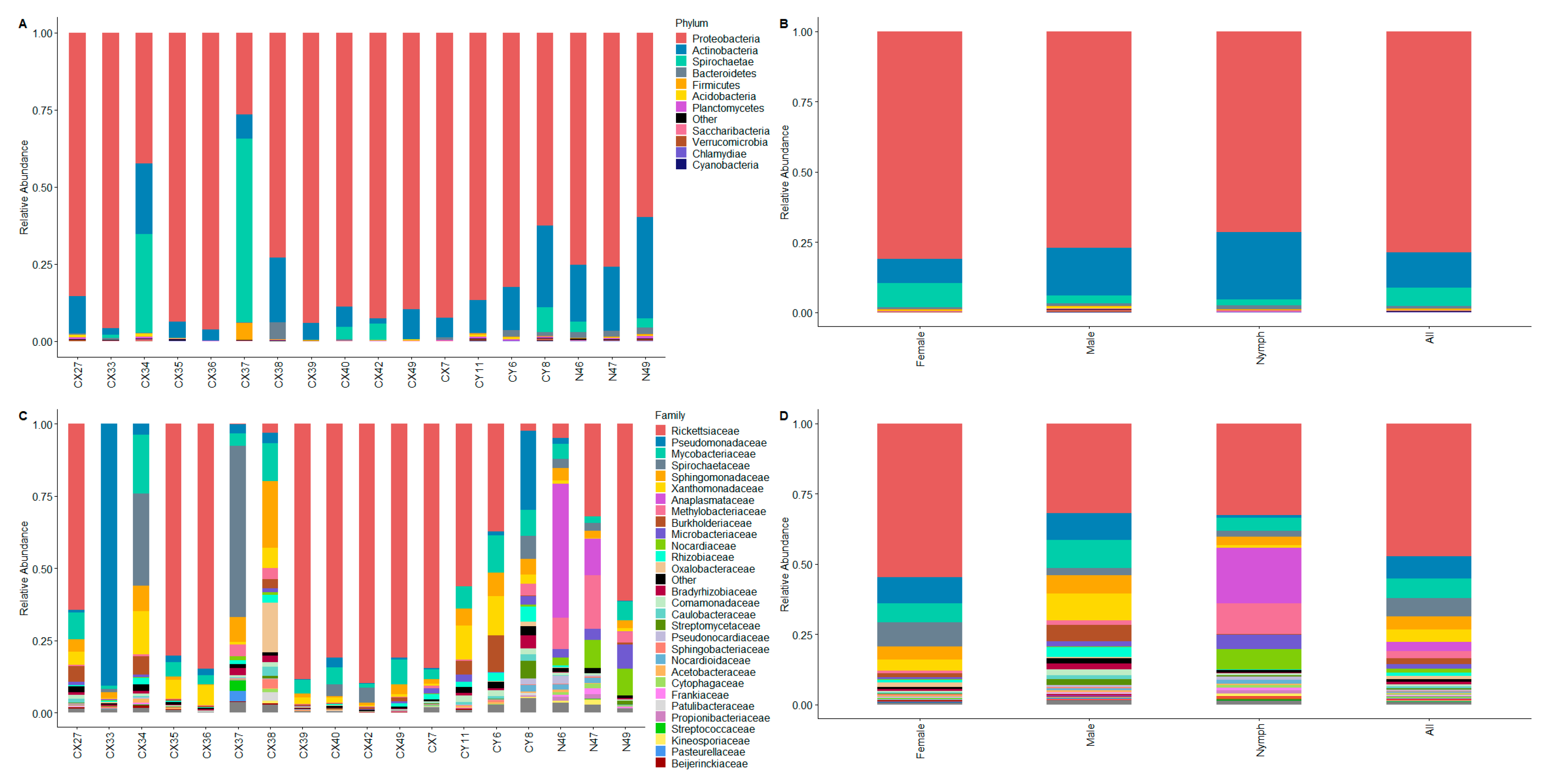
3.1. Composition of Ixodes scapularis Whole-Body Microbiomes

We identified the microbial composition of 18 I. scapularis ticks (12 females, 3 males, and 3 nymphs) collected in Broome County, NY, using DNA extracted from the ticks’ whole bodies. The tick samples yielded an average of 833,650 raw reads, with a minimum of 602,710 reads per tick. After quality and size filtration, there were 326,664 reads per tick on average, with a minimum of 242,408 reads. These reads represented 114 to 347 unique 16S rRNA gene sequences per tick (mean = 191.72) (Table S1). Alpha diversity, as a measure of diversity within a sample, was calculated using the Shannon index. While male ticks exhibited a higher diversity than female ticks (Figure 1), this difference was not statistically significant (p = 0.07). A comparison based on B. burgdorferi infection also did not show a significant difference between Borreliella-positive (n = 8) and Borreliella-negative (n = 10) ticks; however, Borreliella-positive ticks had a larger range of diversity compared to Borreliella-free ticks.
The most abundant bacterial phyla in the total sample of 18 ticks were Proteobacteria (78.65%), Actinobacteria (12.64%), and Spirochaetes (6.42%), followed by Bacteroidetes (0.90%) and Firmicutes (0.44%) (Figure 2A). One notable difference in the relative abundances of bacterial phyla between male, female, and nymphal ticks (Figure 2B) was a much lower abundance of Actinobacteria in female ticks (8.69%) compared to males (16.95%) and nymphs (24.12%). Instead, female ticks harbored Spirochaetes at a rate (8.48%) several times higher than that of male (2.66%) and nymphal ticks (1.97%). Spirochaetes were particularly abundant in the case of two female ticks (CX34 and CX37), comprising over 31% and 59% of their entire microbial communities.
The overabundance of the phylum Proteobacteria was mainly due to the high prevalence of the family Rickettsiaceae, accounting for an average of 47.29% of the amplicon sequence variants (ASVs) across the ticks. The other proteobacteria families that were abundant in our sample were Pseudomonadaceae (7.93%), Sphingomonadaceae (4.71%), Xanthomonadaceae (4.35%), and Anaplasmataceae (3.32%). Within the phylum Actinobacteria, Mycobacteriaceae was the most common family, accounting for an average of 6.90% of the bacterial communities across the sample. The phylum Spirochaetaceae comprised a single family, Spirochaetaceae (6.42%), which, along with Anaplasmataceae, represented the two tick-borne pathogen species detected in our sample (see next section). Together, these seven families accounted for about 90% of the total microbial population in our sample of 18 ticks. All other bacterial families had mean relative abundances below 3% (Figure 2C). The differences in the composition of the microbiomes between male, female, and nymphal ticks (Figure 2D) were more apparent on the family level compared to the phylum level. Most notably, Rickettsiaceae contributed to more than half of the microbiome in female ticks, compared to under a third of the total ASVs among male ticks and nymphs. The male ticks were distinguished from female ticks and nymphs by higher prevalences of Xanthomonadaceae, Burkholderiaceae, and Rhizobiaceae. In nymphs, on the other hand, neither of these three families reached 1% of the total microbiome. Conversely, the microbial communities in the whole bodies of nymphs contained relatively high loads of Methylobacteriaceae (10.98%), Nocardiaceae (7.18%), and Microbacteriaceae (5.11%), all of which were rare (relative abundance <1%) in adult ticks.
Out of the 60 most common genera accounting for 96.01% of the ASVs in our sample, a total of 42 genera had relative abundances of over 1% in at least one of the 18 ticks included in this study (Table S2). Two of these genera, Pseudomonas and Bradyrhizobium, also appeared in one of the extraction controls we analyzed along with our tick samples. Our analysis of the ASVs identified in the extraction controls supported an absence of cross contamination (See Supplementary File S1).
Identification of the microbiomes at the species level confirmed the dominant presence of the I. scapularis endosymbiont Rickettsia buchneri (Table S3). Present in 14 of the 18 ticks (77.77%), in relative abundances as high as 89.8%, R. buchneri was by far the most prevalent bacterial species (Figure 3). The two tick-borne pathogens detected in our sample, B. burgdorferi and A. phagocytophilum, were the second and fifth most abundant species, with average relative abundances of 6.42% and 3.29%, respectively. A Pseudomonas species detected in only two ticks was ranked as the third most abundant species (5.06%) due to it comprising over 88% of the microbial community in a single tick. The next two most abundant species were detected in the majority of the ticks: Luteibacter rhizovicinus (average abundance = 3.74%) comprised over 1% of the microbiome in 55.5% of ticks and Mycolicibacterium madagascariense (average abundance = 2.08%) was detected in all ticks, with relative abundances surpassing 1% in 61.1% of ticks.
The rest of the bacterial species presenting with relative abundances of over 1% in more than one tick belonged to the following genera: Mycobacterium, Sphingomonas, Methylobacterium, Williamsia, Rhizobium, Paraburkholderia, Curtobacterium, and Bradyrhizobium. All of these genera have been frequently reported in I. scapularis ticks and often compose the “core microbiome” of I. scapularis [28,46,47]. One notable observation was the high abundance of Williamsia maris in the tested nymphal ticks (average = 5.84%). This otherwise environmental actinomycete has been previously reported in I. scapularis [48,49].

3.1.1. Beta Diversity

We used principal coordinate analysis (PCoA) to visualize the weighted and unweighted UniFrac distances of bacterial communities according to tick life stage and the presence of Borreliella. The PCoA plot of the weighted UniFrac distances did not reveal a distinct clustering of ticks according to sex and life stage. When considering the Borreliella infectivity of ticks, this plot revealed a grouping of Borreliella-negative ticks regardless of sex and life stage (Figure 4A). It should be noted that two Borreliella-positive female ticks were also present in this cluster. Three other Borreliella-positive female ticks were the outliers in this plot. The PCoA plot of the bacterial communities according to the unweighted UniFrac distances did not show a separation of ticks by sex, life stage, or Borreliella infectivity (Figure 4B).

3.1.2. Detection of Endosymbiont Rickettsia in Male Ixodes scapularis Ticks

In our study, Rickettsia buchneri was identified as the most abundant bacterial species in female I. scapularis ticks. In female ticks, R. buchneri was detected with an average relative abundance of 54.76% and comprised over 80% of the microbiome in 7 of the 14 female ticks. The endosymbiont Rickettsia was present in all three nymphs, with an average abundance of 32.71%. Interestingly, R. buchneri was detected in two of the three male ticks, with an average relative abundance of 31.15%. We conducted a co-occurrence test [44] to identify pairs of species for which the co-occurrence was significantly different than that expected by chance. A significant negative co-occurrence was detected for R. buchneri and Pseudomonas fluorescens-yamanorum (p = 0.02451). We did not identify a significant positive co-occurrence between R. buchneri and other species present in the microbiomes (Table S4).

3.1.3. Tick-Borne Pathogens and Ixodes scapularis Microbiome

As anticipated, Lyme disease-causing B. burgdorferi was the most prevalent human pathogen (44.44%) detected in our sample. The relative abundance of B. burgdorferi in ticks ranged widely, from 1% to over 59% (Table S3). Borreliella burgdorferi had a positive co-occurrence with two Methylobacterium species (p-values = 0.02288 and 0.00654) and one Actinomycetospora species (p = 0.02288) (Table S4).
A. phagocytophilum was the other human pathogen present in the sample. While A. phagocytophilum was detected only in two nymphal ticks, our recent study showed no significant difference in A. phagocytophilum infectivity between nymphs and adults, with a prevalence of 7.31% [50]. Therefore, we attributed the absence of A. phagocytophilum in our 15 adult ticks to the small sample size. In the case of the two A. phagocytophilum-positive nymphal ticks, the anaplasmosis bacteria was highly abundant (12.73% and 46.46%), making it the second most abundant species after R. buchneri (Table S3). Anaplasma phagocytophilum had a negative co-occurrence with Luteibacter rhizovicinus (p = 0.01961). Otherwise, no significant co-occurrence was detected with other species.

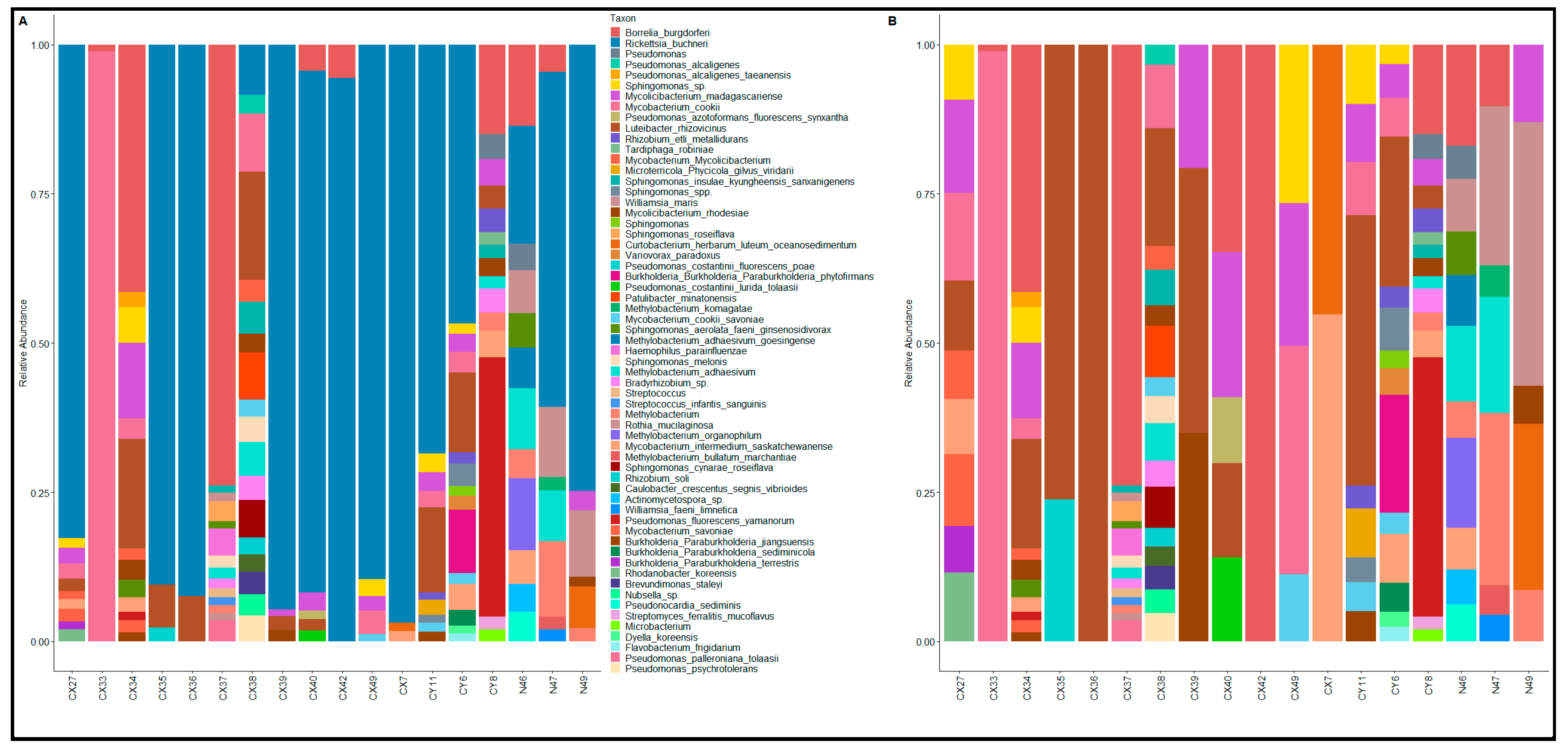
3.2. Proteome Profiling of Single Ticks and Microorganism Identification

An additional, separate set of 36 ticks (22 females, 14 males) were processed for proteome profiling, as described in the Methods section. On average, 3077 proteins were identified in each tick, of which 1.2–2% belonged to various microorganisms. Table 1 shows the prevalence of the microorganisms of interest in ticks along with the list of the specific proteins representing each microorganism. Human pathogens were detected in female ticks only. The specificity of the proteins was verified manually through Uniprot BLAST using the identified peptide sequences. More than 80% of the female ticks carried R. buchneri, as identified by 12 unique proteins. The majority of these proteins were outer membrane proteins. Other intracellular proteins were also mapped to R. buchneri; however, they were excluded due to their sequences being homologous with those of other bacterial species. Borreliella burgdorferi was detected only in 3 of the 22 analyzed female ticks. This pathogen was identified by three unique proteins, including outer surface protein A (OspA), a flagellar filament 41 kDa core protein, and the integral outer membrane protein P66. Finally, 4 of the 22 female ticks carried the human parasite Babesia microti, which was identified by an uncharacterized protein according to the Uniprot knowledge database.

4. Discussion

4.1. Endosymbiont Rickettsia

The microbial composition of I. scapularis ticks collected in Broome County, New York, was generally consistent with previous reports from the Northeastern USA and Southeastern Canada. As with previous studies [34,51,52,53], R. buchneri, an endosymbiont of I. scapularis, was the most abundant bacterial species, comprising over half of the bacterial communities in female ticks. Tick endosymbionts are intracellular microorganisms that play crucial roles in their hosts’ biology, including nutrient provision, antimicrobial defense, and the modulation of the tick’s metabolic activities [54,55]. Rickettsia buchneri is known to synthesize essential nutrients like folate, which is important for tick development and reproduction [27]. In addition, R. buchneri may exert antimicrobial effects through its antibiotic clusters, contributing to the tick’s ability to resist infection by competing with pathogenic bacteria [30]. Recently, R. buchneri has been shown to regulate tick autophagy, which benefits its intracellular infection and replication [31].
R. buchneri is predominantly found in the ovaries and the developing oocytes of female ticks and is transmitted transovarially to offspring. However, limited studies have indicated the presence of R. buchneri in female salivary glands and male ticks [56,57]. In our study, we detected R. buchneri in two of three male ticks, with relative abundances of 37.21% and 56.23%, which were higher than previously reported values for male ticks [34,47,51,52,53]. Although the sample size for male and nymphal ticks in our study was small, the observed prevalence is noteworthy and supports the role of R. buchneri as a primary symbiotic bacterium throughout the tick life cycle. Our results revealed a 1.76-fold higher abundance of R. buchneri in females compared to males, which was comparable to the 2.2-fold difference reported for fed female and male ticks [57]. The higher abundance in female ticks could be due to the higher nutritional and reproductive demands of females, which might require greater symbiont abundance.
It should be noted that while the most abundant R. buchneri sequence in our data was 100% identical to the R. buchneri reference sequence [58], it was also identical to the reference sequence of R. monacensis, a closely related Rickettsia species isolated from I. ricinus ticks in Europe [59]. These two species are highly similar genetically, with their 16S genes being identical at the V3—V4 region. Nevertheless, we used the R. buchneri designation based on the findings of previous I. scapularis studies confirming the presence of this species in North America instead of the European R. monacensis.
As for interactions between the endosymbiont R. buchneri and other members of the I. scapularis microbial community, we observed a significant, negative co-occurrence with Pseudomonas fluorescens-yamanorum (these species cannot be distinguished using 16S V3—V4 sequences, but P. yamanorum has been rarely reported from North America [60,61], making it far less likely than P. fluorescens). In addition, the absence of R. buchneri in the male tick was associated with higher relative abundances of other genera, including Streptomyces, Methylobacterium, and Bradyrhizobium. On the other hand, the R. buchneri-positive male ticks were distinguished by 6-fold higher relative abundances of Luteibacter species (11.67% and 12.01%) compared to the R. buchneri-negative male tick (2.07%).

4.2. Microbiome-TBPs Interactions

Previous studies have reported associations between B. burgdorferi and a number of other bacteria, including a negative co-occurrence with R. buchneri [62,63]. A negative correlation may hint at adversarial interactions, such as the production of antimicrobial agents (e.g., by antibiotic-producing P. fluorescens), as well as competition. In our sample, while B. burgdorferi and R. buchneri demonstrated a negative co-occurrence, the association was not significant (p = 0.06863). That said, in the case of the three male ticks, there was a perfect negative correlation between B. burgdorferi and R. buchneri, suggesting a potential competitive interaction or a role of R. buchneri in modulating pathogen presence in male I. scapularis. We did not observe a negative co-occurrence between B. burgdorferi and Pseudomonas species, as previously suggested [27,28,29]. Instead, we report positive co-occurrences of B. burgdorferi with three environment-associated species (Methylobacterium and Actinomycetospora). Similar associations with other environmental bacteria have been reported previously [27,28].
In the case of A. phagocytophilum, a significant negative co-occurrence was observed with Luteibacter rhizovicinus, an environmental bacteria commonly identified in Ixodes ticks. We did not observe associations between A. phagocytophilum and Rickettsia (negative) or Pseudomonas (positive), as previously suggested [64]. Considering the potential role of A. phagocytophilum in enhancing tolerance to freezing [26], the high abundance of this bacteria in two nymphal ticks (46.46% and 12.73%) of Broome County, New York, might reflect the enhanced colonization of A. phagocytophilum in ticks living in the cold environment.

4.3. “Core Microbiome” of I. scapularis

In addition to R. buchneri, the core microbiome of Broome County I. scapularis ticks contained many of the same genera reported from other I. scapularis populations (Pseudomonas, Mycobacterium, Sphingomonas, Methylobacterium, Williamsia, Rhizobium, Luteibacter, Paraburkholderia, Curtobacterium, and Bradyrhizobium). We also detected Bacillus, Staphylococcus, and Streptococcus species, although with lower incidence and abundance compared to previous studies [28,46,65]. Luteibacter is a known member of the microbial community of I. scapularis [28,48,66] as well as other Ixodes species [32,67,68,69] and Dermacentor [70] ticks. In addition to L. rhizovicinus, 0.13% of the total sequences in our sample were 99.06% identical to those of both L. anthropi and L. yeojuensis, two species with indistinguishable 16S V3—V4 sequences. These species also have been detected in a previous study of the I. scapularis microbiome that reported a positive association between the presence of this genus and Borreliella-positivity in ticks [48]. As for M. madagascariense, we were able to confirm the specificity of a 462 bp sequence that was 100% identical only to this species, with 13 additional sequences showing the highest identity to M. madagascariense, at 99.8%. A second Mycolicibacterium species, M. rhodesiae (average abundance = 0.72%), was also detected in all ticks, with its relative abundance exceeding 1% in five ticks. We did not find a record of these two species in previous I. scapularis studies. However, M. madagascariense has been identified in I. ricinus ticks collected in Denmark [71]. This species is known to be present in soil and plants [72,73] and has also been detected in connection to Ophiocordyceps highlandensis, a fungus that grows on larvae of Scarabaeoidea beetles [74].
At just under 8%, Pseudomonas was the second most abundant genus in our sample, although excluding the single female tick (CX33) with an extreme abundance of Pseudomonas would reduce that value to 2.90%. This was comparable to the average abundance of Pseudomonas (3.5%) reported from I. scapularis collected in Vermont [28]. On average, Pseudomonas spp. were more abundant in the male ticks (9.58%) compared to females (8.60%, 1.77% when excluding CX33) and nymphs (0.80%). While this is consistent with some previous studies of I. scapularis from the Northeastern USA that report a much higher abundance of Pseudomonas in males (23%) compared to females (>1%) [47], a study of female ticks from New York identified Pseudomonas as the most abundant genus in this group (38.1%) [51]. Nevertheless, these studies agree on the higher prevalence and abundance of Pseudomonas in male I. scapularis from the Northeastern USA compared to the Southern USA (0.1–2.7% in Oklahoma and 1% in Texas) [47,51]. Other important observations were the high variability in Pseudomonas abundance within male, female, and nymphal groups and the negative co-occurrence of Pseudomonas fluorescens-yamanorum with R. buchneri. This was consistent with previous observations that, when R. buchneri is not the main component of the I. scapularis microbial community, Pseudomonas is likely to be the most abundant bacteria [29].
When analyzing microbial communities on a species level, the limitations of V3–V4 sequencing should be noted. Checking the sequences assigned as “Pseudomonas palleroniana-tolaasii” using BLAST (blastn), we were not able to verify their specific assignment to either of these two species. In fact, the assigned sequences were also 100% identical to a few members of the Pseudomonas fluorescens group, as well as some environmental Pseudomonas species previously reported from ticks [67]. Due to the 16S genes of all these species being identical in the V3–V4 region, a species-level assignment was impossible in this case. A similar observation was made in the case of Methylobacterium species identified in this study. This is a reminder of the limitations of partial 16S rRNA analysis in identifying bacterial species within certain taxa [75,76]. In such cases, the species-level assignment is somewhat speculative, and therefore, a genus-level analysis may be more informative. That said, a majority of the tentatively assigned Pseudomonas species in our sample belonged to the P. fluorescens group. P. fluorescens has been previously reported in I. scapularis ticks from New York and Vermont with relative abundances of 8% and <1%, respectively [28,51]. In both studies, non-fluorescens Pseudomonas species (P. lurida in NY and P. putida in VA) had a several-fold higher abundance than P. fluorescens. We detected P. lurida and P. putida in our sample in low abundances (<1%).
Overall, many members of the I. scapularis core microbiome were environment-associated bacteria largely reported in soil, water, and plant samples [48,49,65]. As discussed elsewhere [66], it is likely that the geographic and local variations in I. scapularis microbiomes reflect, to some extent, the differences in environmental bacteria present at the tick collection sites. In addition to regional differences, within the Northeastern USA, the microbial composition of I. scapularis seems to be determined by the specific location of sampling rather than sex [77].
As detailed in our review of the extraction controls (Supplementary File S1), we did not detect signs of DNA contamination in our samples. The bacterial taxa commonly known to be of human origin (members of skin and oral microbiome), e.g., Staphylococcus, Streptococcus, Propionibacterium, Corynebacterium, Acinetobacter, and Cutibacterium, were virtually absent in the microbiome results. The only exception was a female tick (bur) that contained Haemophilus and Streptococcus (3.60%). Haemophilus and Streptococcus species have been previously reported in I. scapularis ticks [49,53]; however, they are likely of a human origin.

4.4. The Proteome Signature of Tick-Borne Pathogens in Ixodes scapularis Ticks

In agreement with the 16S rRNA analysis, R. buchneri was by far the most abundant and frequently identified bacteria of interest in the proteomes of female ticks. Although the proteomic technique was less sensitive than 16S rRNA gene sequencing, it provided better specificity in the case of identified proteins. For example, the outer membrane protein B (ompB) was identified by 12 unique peptides that were mapped to the ompB protein belonging to the three Rickettsia species, including R. buchneri (Rickettsia tamurae subsp buchneri), Rickettsia rickettsii (strain Iowa), and Rickettsia monacensis. However, according to the Uniprot knowledge database, 2 out of the 12 identified peptides for ompB (AVDNVAYGIWTK, SSDENYKETSTTVANK) were unique to R. buchneri. OmpB is a large outer surface protein that is believed to play a role in the adhesion of Rickettsia species to host cells [78], and its methylation is believed to be implicated in the virulence of certain Rickettsia species, such as R. prowazekii and R. typhi [79]. However, R. buchneri is considered non-pathogenic to humans and other vertebrates, and it was shown to even inhibit the growth of other pathogenic Rickettsia in ticks [30]. Another Rickettsia protein, Porin domain-containing protein, was detected in ticks by 13 peptides, including two (NTVDSVSLATEYK and YTLLDGQYMR) that were unique to R. buchneri according to the Uniprot knowledge database. A few other proteins, such as adhesin and cell surface antigen Sca1, also generated tryptic peptide sequences unique to R. buchneri. Collectively, these data verified that the Rickettsia bacteria detected in I. scapularis female ticks collected from Broome County was the endosymbiont R. buchneri. Although R. buchneri was identified in male ticks in our 16S rRNA analysis, proteomic analysis failed to detect this species in male ticks, probably due to their inherent low abundance in male ticks. Additionally, potential transcriptional or translational regulation may limit the protein expression of R. buchneri in males, even when its genomic presence is detected.
Lyme disease-causing B. burgdorferi was identified by three specific proteins (OspA, Fla, and P66), which were detected in only 14% of the collected female ticks from Broome County in 2022. Interestingly, 18% of the female ticks gathered from Broome County in 2022 tested positive for Babesia microti, a protozoan parasite responsible for babesiosis, which presents with flu-like symptoms and anemia. The babesiosis condition is increasingly prevalent in New York State [80,81].

5. Conclusions

The application of 16S rRNA gene sequencing along with proteomics facilitated the precise and sensitive identification of tick-borne pathogens in individual ticks. Overall, our analysis of ticks collected in Broome County, New York, further supports the diversity of microbial communities present in I. scapularis ticks of the Northeastern USA. The observed prevalence and abundance of R. buchneri in male I. scapularis ticks, demonstrated using 16S rRNA gene sequencing, a technique that is more sensitive than proteomics, suggest a broader impact of this endosymbiont on the biology of male ticks than previously recognized. Our future studies will aim to confirm these findings with larger sample sizes and an analysis of specific tick organs (e.g., midgut and salivary glands) instead of the whole body. Given that R. buchneri is primarily associated with female ticks, investigating how this endosymbiont may influence male physiology, stress tolerance, and reproductive fitness can inform innovative strategies for tick and tick-borne disease control.
The presence of tick-borne pathogens in high proportions in a limited number of ticks indicates that certain ticks may serve as significant reservoirs for these pathogens in New York. This can have impacts on tick-borne disease transmission, as ticks with higher pathogen loads may be more likely to transmit diseases to hosts. Therefore, conducting spatial and temporal screening of ticks in high-risk locations is a highly valuable public health measure, as it can enhance awareness regarding the true risks associated with tick-borne diseases in the area. Tick screening can also assist healthcare providers in delivering personalized treatment based on the specific causative agents of diseases. Furthermore, proteomics offers a new avenue for diagnosis of tick-borne pathogens in clinical settings.

Supplementary Materials

The following supporting information can be downloaded at: https://www.mdpi.com/article/10.3390/microorganisms13020258/s1, File S1: Analysis of DNA extraction controls; Table S1: Summary of 16S rRNA gene amplicon reads per tick; Table S2: Relative abundance (%) of 16S rRNA gene amplicon sequences per tick by genera. “CX” and “CY” indicate female and male ticks, respectively; Table S3: Relative abundance (frequencies) of the most common species per tick; Table S4: Results of the co-occurrence analysis of pairs of bacterial species present in the sample of 18 ticks. Only co-occurrent species with statistical significance (p < 0.05) are shown. References [82,83] are cited in the Supplementary Materials.

Author Contributions

Conceptualization, M.S-P. and Y.H.; methodology, M.S.-P., E.H.C., J.M., E.D., B.P., M.C., M.N., P.P. and Y.H.; data curation, M.S.-P., A.F. and Y.F.; visualization, A.F. and Y.F.; writing—original draft preparation, M.S.-P., X.-R.W. and Y.H.; writing—review and editing, M.S.-P., X.-R.W., A.F. and Y.F.; project administration, M.S.-P. and Y.H. All authors have read and agreed to the published version of the manuscript.

Funding

This research was supported by the Binghamton University’s Organized Research Centers Award to the Tick-borne Disease Center at Binghamton University.

Institutional Review Board Statement

Not applicable.

Informed Consent Statement

Not applicable.

Data Availability Statement

The original contributions presented in this study are included in the article/Supplementary Material. Further inquiries can be directed to the corresponding author.

Conflicts of Interest

The authors declare no conflicts of interest.

Abbreviations

The following abbreviations are used in this manuscript:
TBDTick-borne Disease
TBPTick-borne Pathogen
SubspSubspecies
Spp.Species

References

  1. CDC. Blacklegged Tick Surveillance. Ticks. Available online: https://www.cdc.gov/ticks/data-research/facts-stats/blacklegged-tick-surveillance.html (accessed on 29 December 2024).
  2. Eisen, L. Tick Species Infesting Humans in the United States. Ticks Tick-Borne Dis. 2022, 13, 102025. [Google Scholar] [CrossRef] [PubMed]
  3. Barbour, A.G.; Qiu, W. Borreliella. In Bergey’s Manual of Systematics of Archaea and Bacteria; John Wiley & Sons: Hoboken, NJ, USA, 2019; pp. 1–22. [Google Scholar] [CrossRef]
  4. CDC. Lyme Disease Surveillance and Data. Lyme Disease. Available online: https://www.cdc.gov/lyme/data-research/facts-stats/index.html (accessed on 30 December 2024).
  5. Eisen, R.J.; Eisen, L. Evaluation of the Association between Climate Warming and the Spread and Proliferation of Ixodes scapularis in Northern States in the Eastern United States. Ticks Tick-Borne Dis. 2024, 15, 102286. [Google Scholar] [CrossRef] [PubMed]
  6. Sonenshine, D.E. Range Expansion of Tick Disease Vectors in North America: Implications for Spread of Tick-Borne Disease. Int. J. Environ. Res. Public. Health 2018, 15, 478. [Google Scholar] [CrossRef]
  7. Diuk-Wasser, M.A.; VanAcker, M.C.; Fernandez, M.P. Impact of Land Use Changes and Habitat Fragmentation on the Eco-Epidemiology of Tick-Borne Diseases. J. Med. Entomol. 2021, 58, 1546–1564. [Google Scholar] [CrossRef]
  8. Clow, K.M.; Ogden, N.H.; Lindsay, L.R.; Michel, P.; Pearl, D.L.; Jardine, C.M. The Influence of Abiotic and Biotic Factors on the Invasion of Ixodes scapularis in Ontario, Canada. Ticks Tick-Borne Dis. 2017, 8, 554–563. [Google Scholar] [CrossRef]
  9. Martin, A.M.; Buttke, D.; Raphael, J.; Taylor, K.; Maes, S.; Parise, C.M.; Ginsberg, H.S.; Cross, P.C. Deer Management Generally Reduces Densities of Nymphal Ixodes scapularis, but Not Prevalence of Infection with Borrelia Burgdorferi Sensu Stricto. Ticks Tick-Borne Dis. 2023, 14, 102202. [Google Scholar] [CrossRef]
  10. Deshpande, G.; Beetch, J.E.; Heller, J.G.; Naqvi, O.H.; Kuhn, K.G. Assessing the Influence of Climate Change and Environmental Factors on the Top Tick-Borne Diseases in the United States: A Systematic Review. Microorganisms 2024, 12, 50. [Google Scholar] [CrossRef]
  11. Eisen, L.; Eisen, R.J. Changes in the Geographic Distribution of the Blacklegged Tick, Ixodes scapularis, in the United States. Ticks Tick-Borne Dis. 2023, 14, 102233. [Google Scholar] [CrossRef]
  12. Ripoche, M.; Irace-Cima, A.; Adam-Poupart, A.; Baron, G.; Bouchard, C.; Carignan, A.; Milord, F.; Ouhoummane, N.; Pilon, P.A.; Thivierge, K.; et al. Current and Future Burden from Lyme Disease in Québec as a Result of Climate Change. Can. Commun. Dis. Rep. 2023, 49, 446–456. [Google Scholar] [CrossRef]
  13. Hart, C.E.; Bhaskar, J.R.; Reynolds, E.; Hermance, M.; Earl, M.; Mahoney, M.; Martinez, A.; Petzlova, I.; Esterly, A.T.; Thangamani, S. Community Engaged Tick Surveillance and tickMAP as a Public Health Tool to Track the Emergence of Ticks and Tick-Borne Diseases in New York. PLoS Glob. Public Health 2022, 2, e0000215. [Google Scholar] [CrossRef]
  14. Lyme Disease and Other Diseases Carried by Ticks. Available online: https://www.health.ny.gov/diseases/communicable/lyme/ (accessed on 30 December 2024).
  15. Courtney, J.W.; Dryden, R.L.; Wyleto, P.; Schneider, B.S.; Massung, R.F. Characterization of Anaplasma Phagocytophila and Borrelia Burgdorferi Genotypes in Ixodes scapularis Ticks from Pennsylvania. Ann. N. Y. Acad. Sci. 2003, 990, 131–133. [Google Scholar] [CrossRef] [PubMed]
  16. Lobo, C.A.; Singh, M.; Rodriguez, M. Human Babesiosis: Recent Advances and Future Challenges. Curr. Opin. Hematol. 2020, 27, 399–405. [Google Scholar] [CrossRef] [PubMed]
  17. Molloy, P.J.; Telford, S.R.; Chowdri, H.R.; Lepore, T.J.; Gugliotta, J.L.; Weeks, K.E.; Hewins, M.E.; Goethert, H.K.; Berardi, V.P. Borrelia Miyamotoi Disease in the Northeastern United States: A Case Series. Ann. Intern. Med. 2015, 163, 91–98. [Google Scholar] [CrossRef]
  18. Piantadosi, A.; Solomon, I.H. Powassan Virus Encephalitis. Infect. Dis. Clin. North Am. 2022, 36, 671–688. [Google Scholar] [CrossRef]
  19. Xu, G.; Foster, E.; Ribbe, F.; Hojgaard, A.; Eisen, R.J.; Paull, S.; Rich, S.M. Detection of Ehrlichia Muris Eauclairensis in Blacklegged Ticks (Ixodes scapularis) and White-Footed Mice (Peromyscus leucopus) in Massachusetts. Vector Borne Zoonotic Dis. 2023, 23, 311–315. [Google Scholar] [CrossRef]
  20. Pritt, B.S.; Mead, P.S.; Johnson, D.K.H.; Neitzel, D.F.; Respicio-Kingry, L.B.; Davis, J.P.; Schiffman, E.; Sloan, L.M.; Schriefer, M.E.; Replogle, A.J.; et al. Identification of a Novel Pathogenic Borrelia Species Causing Lyme Borreliosis with Unusually High Spirochaetaemia: A Descriptive Study. Lancet Infect. Dis. 2016, 16, 556–564. [Google Scholar] [CrossRef]
  21. Milholland, M.T.; Xu, G.; Rich, S.M.; Machtinger, E.T.; Mullinax, J.M.; Li, A.Y. Pathogen Coinfections Harbored by Adult Ixodes scapularis from White-Tailed Deer Compared with Questing Adults Across Sites in Maryland, USA. Vector Borne Zoonotic Dis. Larchmt. N 2021, 21, 86–91. [Google Scholar] [CrossRef]
  22. Sanchez-Vicente, S.; Tokarz, R. Tick-Borne Co-Infections: Challenges in Molecular and Serologic Diagnoses. Pathog. Basel Switz. 2023, 12, 1371. [Google Scholar] [CrossRef]
  23. Bonnet, S.I.; Binetruy, F.; Hernández-Jarguín, A.M.; Duron, O. The Tick Microbiome: Why Non-Pathogenic Microorganisms Matter in Tick Biology and Pathogen Transmission. Front. Cell. Infect. Microbiol. 2017, 7, 236. [Google Scholar] [CrossRef] [PubMed]
  24. Duron, O.; Morel, O.; Noël, V.; Buysse, M.; Binetruy, F.; Lancelot, R.; Loire, E.; Ménard, C.; Bouchez, O.; Vavre, F.; et al. Tick-Bacteria Mutualism Depends on B Vitamin Synthesis Pathways. Curr. Biol. CB 2018, 28, 1896–1902.e5. [Google Scholar] [CrossRef]
  25. Wu-Chuang, A.; Hodžić, A.; Mateos-Hernández, L.; Estrada-Peña, A.; Obregon, D.; Cabezas-Cruz, A. Current Debates and Advances in Tick Microbiome Research. Curr. Res. Parasitol. Vector-Borne Dis. 2021, 1, 100036. [Google Scholar] [CrossRef] [PubMed]
  26. Neelakanta, G.; Sultana, H.; Fish, D.; Anderson, J.F.; Fikrig, E. Anaplasma phagocytophilum Induces Ixodes scapularis Ticks to Express an Antifreeze Glycoprotein Gene That Enhances Their Survival in the Cold. J. Clin. Invest. 2010, 120, 3179–3190. [Google Scholar] [CrossRef]
  27. Ross, B.D.; Hayes, B.; Radey, M.C.; Lee, X.; Josek, T.; Bjork, J.; Neitzel, D.; Paskewitz, S.; Chou, S.; Mougous, J.D. Ixodes scapularis Does Not Harbor a Stable Midgut Microbiome. ISME J. 2018, 12, 2596–2607. [Google Scholar] [CrossRef]
  28. Landesman, W.J.; Mulder, K.; Fredericks, L.P.; Allan, B.F. Cross-Kingdom Analysis of Nymphal-Stage Ixodes scapularis Microbial Communities in Relation to Borrelia Burgdorferi Infection and Load. FEMS Microbiol. Ecol. 2019, 95, fiz167. [Google Scholar] [CrossRef]
  29. Sperling, J.L.H.; Fitzgerald, D.; Sperling, F.A.H.; Magor, K.E. Microbiome Composition and Borrelia Detection in Ixodes scapularis Ticks at the Northwestern Edge of Their Range. Trop. Med. Infect. Dis. 2020, 5, 173. [Google Scholar] [CrossRef]
  30. Cull, B.; Burkhardt, N.Y.; Wang, X.-R.; Thorpe, C.J.; Oliver, J.D.; Kurtti, T.J.; Munderloh, U.G. The Ixodes scapularis Symbiont Rickettsia buchneri Inhibits Growth of Pathogenic Rickettsiaceae in Tick Cells: Implications for Vector Competence. Front. Vet. Sci. 2021, 8, 748427. [Google Scholar] [CrossRef]
  31. Wang, X.-R.; Cull, B.; Oliver, J.D.; Kurtti, T.J.; Munderloh, U.G. The Role of Autophagy in Tick-Endosymbiont Interactions: Insights from Ixodes scapularis and Rickettsia buchneri. Microbiol. Spectr. 2024, 12, e0108623. [Google Scholar] [CrossRef]
  32. Estrada-Peña, A.; Cabezas-Cruz, A.; Obregón, D. Behind Taxonomic Variability: The Functional Redundancy in the Tick Microbiome. Microorganisms 2020, 8, 1829. [Google Scholar] [CrossRef]
  33. Narasimhan, S.; Rajeevan, N.; Liu, L.; Zhao, Y.O.; Heisig, J.; Pan, J.; Eppler-Epstein, R.; Deponte, K.; Fish, D.; Fikrig, E. Gut Microbiota of the Tick Vector Ixodes scapularis Modulate Colonization of the Lyme Disease Spirochete. Cell Host Microbe 2014, 15, 58–71. [Google Scholar] [CrossRef]
  34. Cross, S.T.; Kapuscinski, M.L.; Perino, J.; Maertens, B.L.; Weger-Lucarelli, J.; Ebel, G.D.; Stenglein, M.D. Co-Infection Patterns in Individual Ixodes scapularis Ticks Reveal Associations between Viral, Eukaryotic and Bacterial Microorganisms. Viruses 2018, 10, 388. [Google Scholar] [CrossRef]
  35. Krupa, E.; Dziedziech, A.; Paul, R.; Bonnet, S. Update on Tick-Borne Pathogens Detection Methods Within Ticks. Curr. Res. Parasitol. Vector Borne Dis. 2024, 6, 100199. [Google Scholar] [CrossRef] [PubMed]
  36. Smith, H.R.; Canessa, E.H.; Roy, R.; Spathis, R.; Pour, M.S.; Hathout, Y. A Single Tick Screening for Infectious Pathogens Using Targeted Mass Spectrometry. Anal. Bioanal. Chem. 2022, 414, 3791–3802. [Google Scholar] [CrossRef] [PubMed]
  37. Yssouf, A.; Almeras, L.; Terras, J.; Socolovschi, C.; Raoult, D.; Parola, P. Detection of Rickettsia Spp in Ticks by MALDI-TOF MS. PLoS Negl. Trop. Dis. 2015, 9, e0003473. [Google Scholar] [CrossRef] [PubMed]
  38. Fotso Fotso, A.; Mediannikov, O.; Diatta, G.; Almeras, L.; Flaudrops, C.; Parola, P.; Drancourt, M. MALDI-TOF Mass Spectrometry Detection of Pathogens in Vectors: The Borrelia Crocidurae/Ornithodoros Sonrai Paradigm. PLoS Negl. Trop. Dis. 2014, 8, e2984. [Google Scholar] [CrossRef] [PubMed]
  39. Callahan, B.J.; McMurdie, P.J.; Rosen, M.J.; Han, A.W.; Johnson, A.J.A.; Holmes, S.P. DADA2: High-Resolution Sample Inference from Illumina Amplicon Data. Nat. Methods 2016, 13, 581–583. [Google Scholar] [CrossRef] [PubMed]
  40. Caporaso, J.G.; Kuczynski, J.; Stombaugh, J.; Bittinger, K.; Bushman, F.D.; Costello, E.K.; Fierer, N.; Peña, A.G.; Goodrich, J.K.; Gordon, J.I.; et al. QIIME Allows Analysis of High-Throughput Community Sequencing Data. Nat. Methods 2010, 7, 335–336. [Google Scholar] [CrossRef] [PubMed]
  41. R: The R Project for Statistical Computing. Available online: https://www.r-project.org/ (accessed on 30 December 2024).
  42. Wickham, H. Use R! In Ggplot2; Springer International Publishing: Cham, Switzerland, 2016. [Google Scholar] [CrossRef]
  43. Oksanen, J.; Simpson, G.L.; Blanchet, F.G.; Kindt, R.; Legendre, P.; Minchin, P.R.; O’Hara, R.B.; Solymos, P.; Stevens, M.H.H.; Szoecs, E.; et al. Vegan: Community Ecology Package. 2024. Available online: https://cran.r-project.org/web/packages/vegan/index.html (accessed on 30 December 2024).
  44. Griffith, D.M.; Veech, J.A.; Marsh, C.J. Cooccur: Probabilistic Species Co-Occurrence Analysis in R. J. Stat. Softw. 2016, 69, 1–17. [Google Scholar] [CrossRef]
  45. Dzieciatkowska, M.; Hill, R.; Hansen, K.C. GeLC-MS/MS Analysis of Complex Protein Mixtures. Methods Mol. Biol. Clifton NJ 2014, 1156, 53–66. [Google Scholar] [CrossRef]
  46. Paulson, A.R.; Lougheed, S.C.; Huang, D.; Colautti, R.I. Multiomics Reveals Symbionts, Pathogens, and Tissue-Specific Microbiome of Blacklegged Ticks (Ixodes scapularis) from a Lyme Disease Hot Spot in Southeastern Ontario, Canada. Microbiol. Spectr. 2023, 11, e0140423. [Google Scholar] [CrossRef]
  47. Thapa, S.; Zhang, Y.; Allen, M.S. Bacterial Microbiomes of Ixodes scapularis Ticks Collected from Massachusetts and Texas, USA. BMC Microbiol. 2019, 19, 138. [Google Scholar] [CrossRef]
  48. Wiesinger, A.; Wenderlein, J.; Ulrich, S.; Hiereth, S.; Chitimia-Dobler, L.; Straubinger, R.K. Revealing the Tick Microbiome: Insights into Midgut and Salivary Gland Microbiota of Female Ixodes Ricinus Ticks. Int. J. Mol. Sci. 2023, 24, 1100. [Google Scholar] [CrossRef] [PubMed]
  49. Rynkiewicz, E.C.; Hemmerich, C.; Rusch, D.B.; Fuqua, C.; Clay, K. Concordance of Bacterial Communities of Two Tick Species and Blood of Their Shared Rodent Host. Mol. Ecol. 2015, 24, 2566–2579. [Google Scholar] [CrossRef] [PubMed]
  50. Atkins, J.; Lam, V.; Glickman, L.; Hirsch, J.; Tampori, J.; Johnson, R.; Macher, J.; Garruto, R.; Shamoon-Pour, M. Prevalence of Human Pathogens in Ixodes scapularis Ticks of Southern Tier, NY. In Proceedings of the the Human Biology Association (HBA) 49th Annual Meetings, Los Angeles, CA, USA, 20–22 March 2024. [Google Scholar]
  51. Kumar, D.; Downs, L.P.; Adegoke, A.; Machtinger, E.; Oggenfuss, K.; Ostfeld, R.S.; Embers, M.; Karim, S. An Exploratory Study on the Microbiome of Northern and Southern Populations of Ixodes scapularis Ticks Predicts Changes and Unique Bacterial Interactions. Pathogens 2022, 11, 130. [Google Scholar] [CrossRef]
  52. Noden, B.H.; Gilliland, M.; Propst, J.; Slater, K.; Karpathy, S.E.; Paddock, C.D. Rickettsia Tillamookensis (Rickettsiales: Rickettsiaceae) in Ixodes scapularis (Acari: Ixodidae) in Oklahoma. J. Med. Entomol. 2024, 61, 257–260. [Google Scholar] [CrossRef]
  53. Van Treuren, W.; Ponnusamy, L.; Brinkerhoff, R.J.; Gonzalez, A.; Parobek, C.M.; Juliano, J.J.; Andreadis, T.G.; Falco, R.C.; Ziegler, L.B.; Hathaway, N.; et al. Variation in the Microbiota of Ixodes Ticks with Regard to Geography, Species, and Sex. Appl. Environ. Microbiol. 2015, 81, 6200–6209. [Google Scholar] [CrossRef]
  54. Kolo, A.O.; Raghavan, R. Impact of Endosymbionts on Tick Physiology and Fitness. Parasitology 2023, 150, 859–865. [Google Scholar] [CrossRef]
  55. Hussain, S.; Perveen, N.; Hussain, A.; Song, B.; Aziz, M.U.; Zeb, J.; Li, J.; George, D.; Cabezas-Cruz, A.; Sparagano, O. The Symbiotic Continuum Within Ticks: Opportunities for Disease Control. Front. Microbiol. 2022, 13, 854803. [Google Scholar] [CrossRef]
  56. Al-Khafaji, A.M.; Armstrong, S.D.; Varotto Boccazzi, I.; Gaiarsa, S.; Sinha, A.; Li, Z.; Sassera, D.; Carlow, C.K.S.; Epis, S.; Makepeace, B.L. Rickettsia buchneri, Symbiont of the Deer Tick Ixodes scapularis, Can Colonise the Salivary Glands of Its Host. Ticks Tick-Borne Dis. 2020, 11, 101299. [Google Scholar] [CrossRef]
  57. Hagen, R.; Verhoeve, V.I.; Gillespie, J.J.; Driscoll, T.P. Conjugative Transposons and Their Cargo Genes Vary across Natural Populations of Rickettsia buchneri Infecting the Tick Ixodes scapularis. Genome Biol. Evol. 2018, 10, 3218–3229. [Google Scholar] [CrossRef]
  58. Kurtti, T.J.; Felsheim, R.F.; Burkhardt, N.Y.; Oliver, J.D.; Heu, C.C.; Munderloh, U.G. Rickettsia buchneri Sp. Nov., a Rickettsial Endosymbiont of the Blacklegged Tick Ixodes scapularis. Int. J. Syst. Evol. Microbiol. 2015, 65, 965–970. [Google Scholar] [CrossRef]
  59. Simser, J.A.; Palmer, A.T.; Fingerle, V.; Wilske, B.; Kurtti, T.J.; Munderloh, U.G. Rickettsia Monacensis Sp. Nov., a Spotted Fever Group Rickettsia, from Ticks (Ixodes ricinus) Collected in a European City Park. Appl. Environ. Microbiol. 2002, 68, 4559–4566. [Google Scholar] [CrossRef] [PubMed]
  60. Ramírez-Mendoza, R.; Ángeles-Argáiz, R.; Lozano Aguirre-Beltrán, L.F.; Almaraz-Suárez, J.J.; Hernández-Oaxaca, D.; Ortiz-Lopez, I.; Perez-Moreno, J. Whole-Genome Sequence of Pseudomonas yamanorum OLsAu1 Isolated from the Edible Wild Ectomycorrhizal Mushroom lactarius Sp. Section Deliciosi. Microbiol. Resour. Announc. 2023, 12, e00843-23. [Google Scholar] [CrossRef] [PubMed]
  61. Arnau, V.G.; Sánchez, L.A.; Delgado, O.D. Pseudomonas yamanorum Sp. Nov., a Psychrotolerant Bacterium Isolated from a Subantarctic Environment. Int. J. Syst. Evol. Microbiol. 2015, 65, 424–431. [Google Scholar] [CrossRef]
  62. Steiner, F.E.; Pinger, R.R.; Vann, C.N.; Grindle, N.; Civitello, D.; Clay, K.; Fuqua, C. Infection and Co-Infection Rates of Anaplasma phagocytophilum Variants, Babesia Spp., Borrelia burgdorferi, and the Rickettsial Endosymbiont in Ixodes scapularis (Acari: Ixodidae) from Sites in Indiana, Maine, Pennsylvania, and Wisconsin. J. Med. Entomol. 2008, 45, 289–297. [Google Scholar] [CrossRef] [PubMed]
  63. Fountain-Jones, N.M.; Khoo, B.S.; Rau, A.; Berman, J.D.; Burton, E.N.; Oliver, J.D. Positive Associations Matter: Microbial Relationships Drive Tick Microbiome Composition. Mol. Ecol. 2023, 32, 4078–4092. [Google Scholar] [CrossRef]
  64. Abraham, N.M.; Liu, L.; Jutras, B.L.; Yadav, A.K.; Narasimhan, S.; Gopalakrishnan, V.; Ansari, J.M.; Jefferson, K.K.; Cava, F.; Jacobs-Wagner, C.; et al. Pathogen-Mediated Manipulation of Arthropod Microbiota to Promote Infection. Proc. Natl. Acad. Sci. USA 2017, 114, E781–E790. [Google Scholar] [CrossRef]
  65. Mota, T.F.; Fukutani, E.R.; Martins, K.A.; Salgado, V.R.; Andrade, B.B.; Fraga, D.B.M.; Queiroz, A.T.L. Another Tick Bites the Dust: Exploring the Association of Microbial Composition with a Broad Transmission Competence of Tick Vector Species. Microbiol. Spectr. 2023, 11, e0215623. [Google Scholar] [CrossRef]
  66. Brinkerhoff, R.J.; Clark, C.; Ocasio, K.; Gauthier, D.T.; Hynes, W.L. Factors Affecting the Microbiome of Ixodes scapularis and Amblyomma Americanum. PLoS ONE 2020, 15, e0232398. [Google Scholar] [CrossRef]
  67. Tveten, A.-K.; Riborg, A.; Vadseth, H.T. DGGE Identification of Microorganisms Associated with Borrelia Burgdorferi Sensu Lato- or Anaplasma phagocytophilum-Infected Ixodes Ricinus Ticks from Northwest Norway. Int. J. Microbiol. 2013, 2013, 805456. [Google Scholar] [CrossRef]
  68. Portillo, A.; Palomar, A.M.; de Toro, M.; Santibáñez, S.; Santibáñez, P.; Oteo, J.A. Exploring the Bacteriome in Anthropophilic Ticks: To Investigate the Vectors for Diagnosis. PLoS ONE 2019, 14, e0213384. [Google Scholar] [CrossRef]
  69. Swei, A.; Kwan, J.Y. Tick Microbiome and Pathogen Acquisition Altered by Host Blood Meal. ISME J. 2017, 11, 813–816. [Google Scholar] [CrossRef]
  70. Dunaj, J.; Drewnowska, J.; Moniuszko-Malinowska, A.; Swiecicka, I.; Pancewicz, S. First Metagenomic Report of Borrelia Americana and Borrelia Carolinensis in Poland–a Preliminary Study. Ann. Agric. Environ. Med. AAEM 2021, 28, 49–55. [Google Scholar] [CrossRef]
  71. Petersen, A.; Rosenstierne, M.W.; Rasmussen, M.; Fuursted, K.; Nielsen, H.V.; O’Brien Andersen, L.; Bødker, R.; Fomsgaard, A. Field Samplings of Ixodes ricinus Ticks from a Tick-Borne Encephalitis Virus Micro-Focus in Northern Zealand, Denmark. Ticks Tick-Borne Dis. 2019, 10, 1028–1032. [Google Scholar] [CrossRef]
  72. Kazda, J.; Müller, H.-J.; Stackebrandt, E.; Daffe, M.; Müller, K.; Pitulle, C. Mycobacterium madagascariense Sp. Nov. Int. J. Syst. Evol. Microbiol. 1992, 42, 524–528. [Google Scholar] [CrossRef]
  73. Uskoković, V.; Wu, V.M. Altering Microbiomes with Hydroxyapatite Nanoparticles: A Metagenomic Analysis. Mater. Basel Switz. 2022, 15, 5824. [Google Scholar] [CrossRef]
  74. Li, C.; Tang, D.; Wang, Y.; Fan, Q.; Zhang, X.; Cui, X.; Yu, H. Endogenous Bacteria Inhabiting the Ophiocordyceps Highlandensis during Fruiting Body Development. BMC Microbiol. 2021, 21, 178. [Google Scholar] [CrossRef]
  75. Laudadio, I.; Fulci, V.; Palone, F.; Stronati, L.; Cucchiara, S.; Carissimi, C. Quantitative Assessment of Shotgun Metagenomics and 16S rDNA Amplicon Sequencing in the Study of Human Gut Microbiome. Omics, J. Integr. Biol. 2018, 22, 248–254. [Google Scholar] [CrossRef]
  76. Chakravorty, S.; Helb, D.; Burday, M.; Connell, N.; Alland, D. A Detailed Analysis of 16S Ribosomal RNA Gene Segments for the Diagnosis of Pathogenic Bacteria. J. Microbiol. Methods 2007, 69, 330–339. [Google Scholar] [CrossRef]
  77. Sakamoto, J.M.; Silva Diaz, G.E.; Wagner, E.A. Bacterial Communities of Ixodes scapularis from Central Pennsylvania, USA. Insects 2020, 11, 718. [Google Scholar] [CrossRef]
  78. Uchiyama, T. Tropism and Pathogenicity of Rickettsiae. Front. Microbiol. 2012, 3, 230. [Google Scholar] [CrossRef]
  79. Yang, D.C.H.; Abeykoon, A.H.; Choi, B.-E.; Ching, W.-M.; Chock, P.B. Outer Membrane Protein OmpB Methylation May Mediate Bacterial Virulence. Trends Biochem. Sci. 2017, 42, 936–945. [Google Scholar] [CrossRef]
  80. O’Connor, C.; Prusinski, M.A.; Jiang, S.; Russell, A.; White, J.; Falco, R.; Kokas, J.; Vinci, V.; Gall, W.; Tober, K.; et al. A Comparative Spatial and Climate Analysis of Human Granulocytic Anaplasmosis and Human Babesiosis in New York State (2013–2018). J. Med. Entomol. 2021, 58, 2453–2466. [Google Scholar] [CrossRef] [PubMed]
  81. Kogut, S.J.; Thill, C.D.; Prusinski, M.A.; Lee, J.-H.; Backerson, P.B.; Coleman, J.L.; Anand, M.; White, D.J. Babesia Microti, Upstate New York. Emerg. Infect. Dis. 2005, 11, 476–478. [Google Scholar] [CrossRef] [PubMed]
  82. Maldonado-Ruiz, L.P.; Neupane, S.; Park, Y.; Zurek, L. The bacterial community of the lone star tick (Amblyomma americanum). Parasites Vectors 2021, 14, 1–10. [Google Scholar] [CrossRef] [PubMed]
  83. Ponnusamy, L.; Gonzalez, A.; Van Treuren, W.; Weiss, S.; Parobek, C.M.; Juliano, J.J.; Knight, R.; Roe, R.M.; Apperson, C.S.; Meshnick, S.R. Diversity of rickettsiales in the microbiome of the lone star tick, Amblyomma Americanum. Appl. Environ. Microbiol. 2014, 80, 354–359. [Google Scholar] [CrossRef]
Figure 1. Boxplot of alpha diversity measured with the Shannon diversity index showing differences among tick groups.
Microorganisms 13 00258 g001
Figure 2. Microbial composition analysis of tick samples. (A) Relative abundance at the phylum level for 18 individual ticks. (B) Relative abundance at the phylum level for female, male, and nymph ticks grouped together. (C) Relative abundance at the family level for 18 individual ticks. (D) Relative abundance at the family level for female, male, and nymph ticks grouped together.
Microorganisms 13 00258 g002
Figure 3. Microbial composition of 18 ticks at the species level. (A) Relative abundance of bacterial species across all 18 individual ticks. (B) Relative abundance of bacterial species across all 18 ticks after excluding Rickettsia buchneri to highlight the diversity of other species.
Microorganisms 13 00258 g003
Figure 4. Principal coordinate analysis (PCoA) using (A) weighted UniFrac distances and (B) unweighted UniFrac distances measuring Borreliella positive (red) and negative (blue) samples.
Microorganisms 13 00258 g004
Table 1. Prevalences of pathogens and R. buchneri in female and male I. scapularis ticks collected in Broome County, NY, according to the proteomic analysis. Among the 22 female ticks, 15 (68.18%) carried R. buchneri, 3 (13.64%) carried B. burgdorferi, and 4 (18.18%) carried the parasite B. microti. No pathogens were detected in the 14 male ticks.
MicroorganismIdentified ProteinNumber of Positive Ticks
Female (n = 22)Male (n = 14)
Rickettsia
buchneri
Adhesin (A0A8E1C0T9)100
Adhesin A0A8E1C0I9)80
Outer membrane protein B (A0A8E0WKQ0)120
Cell surface antigen Sca1 (A0A8E0WLZ4) 40
Porin domain-containing protein (A0A8E0WLM2)60
Peptidoglycan-associated
lipoprotein (A0A8E1C013)
30
Parvulin-like PPIase (A0A8E0WMR4) 40
Spore protein SP21 (A0A8E1BZB2)70
Hsp20/alpha crystallin family
protein (A0A8E0WKN8)
100
Biotin synthase (A0A8E1BZ92)20
Transposase (A0A8E1BZI6)30
Putative adhesin (A0A0B7J122)50
Total number of ticks with R. buchneri proteins150
Borreliella
burgdorferi
Outer surface protein A (P0CL66)20
Flagellar filament 41 kDa core protein (P11089)30
Integral outer membrane protein P66 (A0A0H3C4D7)10
Total number of ticks with B. burgdorferi proteins30
Babesia microtiUncharacterized protein (A0A1N6LY41)
[KLVSDYCSLGR]
40
Uncharacterized protein (A0A1N6LY97)
[TFGNFSLFEK]
10
Total number of ticks with B. microti proteins40
Disclaimer/Publisher’s Note: The statements, opinions and data contained in all publications are solely those of the individual author(s) and contributor(s) and not of MDPI and/or the editor(s). MDPI and/or the editor(s) disclaim responsibility for any injury to people or property resulting from any ideas, methods, instructions or products referred to in the content.

