Article

The Isolation and Identification of Pseudoalteromonas sp. H27, a Bacterial Strain Pathogenic to Crassostrea gigas

1
College of Marine Science and Environment, Dalian Ocean University, Dalian 116023, China
2
College of Fisheries and Life Science, Dalian Ocean University, Dalian 116023, China
3
Engineering Research Center of Shellfish Culture and Breeding, Dalian Ocean University, Dalian 116023, China
*
Authors to whom correspondence should be addressed.
These authors contributed equally to this work.
Microorganisms 2025, 13(2), 296; https://doi.org/10.3390/microorganisms13020296
Submission received: 18 December 2024 / Revised: 25 January 2025 / Accepted: 28 January 2025 / Published: 30 January 2025
(This article belongs to the Special Issue Infectious Diseases in Aquaculture)

Abstract

:
Bacterial infection is frequently observed in disease outbreaks of aquatic animals, making it of significance to isolate and identify the bacterial pathogens. In this study, diseased individuals of Crassostrea gigas were sampled from the nearshore area in Zhanjiang, Guangdong in May 2023. Culturable bacteria were isolated from the diseased tissue and a pathogenic strain labeled as H27 was screened through a hemolysis test and bacterial challenge experiments. Morphological characterization, 16S rRNA gene sequence-based molecular identification and biochemical tests showed that strain H27 belonged to the genus of Pseudoalteromonas, a dominant genus in the diseased tissue of C. gigas revealed by bacterial community structure analysis. The clinical signs originally observed in naturally diseased C. gigas were reproduced in strain H27-challenged adults, both with the red mantle and adductor. Histopathological analysis was further performed on the diseased tissues of the latter, which showed a significantly increased accumulation of pigment granules in the cytoplasm of the diseased mantle as well as enlarged muscle fiber distances in the diseased adductor. In addition, strain H27 was re-isolated from tissues of the moribund C. gigas after bacterial challenge, indicating the fulfillment of Koch’s postulate. Our results help to enrich the knowledge of C. gigas diseases, possibly contributing to disease prevention and control.

1. Introduction

Oysters are traditionally cultured shellfish with a huge production and high economic value. According to the statistics released by the FAO (Food and Agriculture Organization of the United Nations) in 2024, global production of oyster aquaculture reached 58.5 million tonnes in 2021, accounting for 16.9% of total aquaculture production. The production value of maricultured oysters reached USD 631.5 million, accounting for 5.4% of the total production value of aquaculture [1]. In China, the culture area of oysters in 2023 reached 276,816 hectares, with a 17.82% increment in comparison with the previous year [2]. Crassostrea gigas, also known as the Pacific Oyster, belongs to Mollusca, Bivalvia, Ostreidam, Ostreidae and Crassostrea [3]. It is widely cultured in China due to its strong reproductive capacity, fast growth rate and high nutritional value [4]. However, cultured C. gigas have been suffering from frequent diseases in recent years, caused by both abiotic factors and biotic factors. Abnormal climate and the deterioration of water quality are major abiotic factors, whereas pathogenic viruses, bacteria, fungi and parasites are common biotic factors. Bacterial diseases often occur in outbreaks with high mortality. Vibrio anguillarum, V. splendidus and Pseudomonas fluorescens have been reported to be bacterial pathogens of C. gigas [5,6]. Human pathogenic Vibrio species have been associated with gastroenteritis (V. parahaemolyticus and V. cholerae non-O1/O139) after the consumption of contaminated food [7]. According to regulations governing the sanitary control of shellfish, all bacterial disease outbreaks must undergo epidemiological studies to ensure that precise measures can be taken to prevent further infections [8]. Arcobacter, Marinobacterium, Marinomonas, Vibrio and Pseudoalteromonas are associated with Pacific Oyster Mortality Syndrome (POMS) and are collectively termed as the “POMS pathobiota” [9]. Nevertheless, it remains unknown whether other lethal bacterial flora are involved and how these bacterial communities interact with each other [10]. Functional complementarity could partly explain it, which states that competition for limited resources among the pathobiota allows for efficient utilization of diverse resources in oysters, making the coexistence of bacteria with distinct metabolic characteristics possible [11].
Bacteria of the Pseudoalteromonas genus are aerobic or facultatively anaerobic, tending to move through a single sheathless polar flagella [12]. They are widely distributed in marine environments [13,14]. Previous findings have shown that members of Pseudoalteromonas could secrete extracellular enzymes leading to algae lysis [15,16] or produce toxins lethal to Penaeus japonicus [17] and Cancer pagurus [18]. Pseudoalteromonas could also directly infect marine organisms, causing green spot disease [19,20] and yellow spot disease [21] in Pyropia yezoensis, red spot disease [22] and spot-wounded disease [23] in Laminaria japonica, stomach ulcer disease [24] and skin ulcerative syndrome in Apostichopus japonicas [25,26,27], syndrome disease in Strongylocentrotus intermedius [28], etc. In terms of the Pseudoalteromonas-conferred infection in Acanthopagrus schlegel [29], Epinephelus septemfasciatus Thunberg [30], Cynoglossus semilaevis Günther [31] and Dicentrarchus labrax L. [32], epidermis and fin ulceration are frequently observed, with death occurring occasionally. In the case of C. gigas mass death in summer, bacteria of the Pseudoalteromonas genus were often isolated as the dominant species from the diseased tissues [33,34]. Nevertheless, it is rarely reported that Pseudoalteromonas can directly cause diseases in C. gigas.
In May 2023, there was a disease outbreak in C. gigas cultured in the coastal areas of Zhanjiang, Guangdong, China, with red tissues observed in the diseased individuals. In this study, the diseased C. gigas were used as the experimental material and pathological bacteria were isolated from the diseased tissues. Morphological characterization, 16S rRNA gene sequence analysis, biochemical tests and bacterial challenge experiments showed that a bacterial strain, H27, was pathogenic to C. gigas and belonged to the genus of Pseudoalteromonas.

2. Materials and Methods

2.1. Sampling of Crassostrea gigas

The infected individuals of C. gigas were collected from the aquaculture area located in Zhanjiang, Guangdong Province, China in May 2023, where the mass infection of C. gigas was observed. Healthy individuals of C. gigas cultured in the adjacent area were collected as well. The collected oysters were transported on ice to Dalian Ocean University (Dalian, Liaoning Province, China) via flight (<24 h). The healthy C. gigas larvae and adults used for the bacterial challenge experiment were collected from Shicheng Island, Zhuanghe, Dalian, and were temporarily cultured at Zhangzidao Co., Ltd. in Dalian, China; these were of the same species as the diseased C. gigas. The larvae retained on the 100 μm sieve were rinsed with sterile seawater with a salinity of 28 to 30 psu and a temperature of 21 °C to 23 °C. Then, the larvae were placed in a container filled with sterile seawater for temporary culture. Both the larvae and the adults were temporarily cultured for 7 days at room temperature (21 °C to 23 °C), during which they were fed with microalgae three times a day, with continuous areation and a daily water change. They were inspected once a day and dead individuals were removed.

2.2. Analysis of the Bacterial Community Structure in the Soft Tissue of C. gigas

To analyze the bacterial community structure, soft tissues of the diseased C. gigas and the healthy C. gigas samples were collected using a sterile scalpel. The total DNA of the tissues was extracted using the CTAB method [35]. The V4 region of the bacterial 16S rRNA gene was amplified using 515F (5′-GTGCCAGCMGCCGCGGTAA-3′) and 806R (5′-GGACTACHVGGGTWTCTAAT-3′) primers. The amplicons were sequenced using the Illumina NovaSeq PE250 high-throughput sequencing platform (BGI). Mothur package (v. 1.31.2) [36] was used to select the unique tag. The standard of the sequence similarity was set at 97% to classify the operational taxonomy units (OTUs). QIIME (v. 1.8.0) was used to analyze α-diversity (Shannon index and Chao1 index) and Pheatmap (R software v.4.0.3) was used to analyze β-diversity.

2.3. Isolation of Culturable Bacteria from Diseased Tissue of C. gigas

The infected individuals of C. gigas were dissected using a sterile scalpel and red-colored tissue was identified as the diseased tissue. A sterile pestle was used to homogenize the tissue in seawater. The samples were plated on Zobell Marine Broth 2216 (2216E) (Qingdao Haibo Biotechnology Co., Ltd., Qingdao, China) agar plates (pH 7.6 ± 0.2) and then aerobically incubated at 30 °C in a constant temperature incubator. The dominant colonies with the same morphology were picked and purified via plate streaking at least three times.

2.4. Hemolysis Test

The dominant bacterial strain isolated from the diseased tissues of C. gigas was inoculated into Zobell Marine Broth 2216 liquid medium (pH 7.6 ± 0.2) (Qingdao Haibo Biotechnology Co., Ltd., Qingdao, China) and cultured at 30 °C, 120 rpm for 24 h. The composition of the 2216E medium includes 5.0 g/L of peptones, 1.0 g/L of yeast extract, 0.1 g/L of ferric citrate, 19.45 g/L of sodium chloride, 5.98 g/L of magnesium chloride, 3.24 g/L of sodium sulfate, 1.8 g/L of calcium chloride, 0.55 g/L of potassium chloride, 0.16 g/L of sodium carbonate, 0.08 g/L of potassium bromide, 0.034 g/L of strontium chloride, 0.022 g/L of boric acid, 0.004 g/L of sodium silicate, 0.0024 g/L of sodium fluoride, 0.0016 g/L of ammonium nitrate and 0.008 g/L of disodium phosphate. A total of 2.5 μL of the liquid culture was spotted on a Columbia blood agar plate (Qingdao Hi-Tech Industrial Park Hope Bio-Technology Co., Ltd., Qingdao, China). When the liquid culture was completely absorbed, the plate was incubated upside down at 30° C in a constant temperature incubator for one week and examined every 24 h to observe whether there was a β-hemolysis ring.

2.5. Identification of Bacterial Strains

2.5.1. Morphological Characterization

The dominant bacterial strain was inoculated onto a 2216E agar plate via plate streaking and then incubated upside down at 30 °C for 24 h. The morphological characteristics of the colonies were observed. Furthermore, the pure culture was taken and plated onto sterile double distilled water on a clean glass slide and subjected to Gram staining using a Gram staining kit (G-clone, cat. NO.RS3140, Beijing, China) following the instructions provided by the manufacturer. The morphology of the bacterial cells was observed under a microscope using an oil immersion lens.

2.5.2. Molecular Identification Based on the Full-Length Sequence of 16S rRNA Gene

For molecular identification of the bacterial strain, the total DNA was extracted from the pure culture using a bacterial DNA extraction kit according to the manufacturer’s instructions (Tiangen, Beijing, China). The molecular identification of the tested strain was performed based on the full-length sequence of the 16S rRNA gene. The full length of the 16S rRNA gene was amplified using the universal primer pair 27F (5′-AGAGTTTGATCCTGGCTCAG-3′) and 1492R (5′-GGTTACCTTGTTACGACTT-3′) [37]. The PCR products were sent to Beijing Genomics Institute (Beijing, China) for Sanger sequencing and the gene sequence was subject to a BLAST search against the gene database in the NCBI (National Center for Biotechnology Information) website to identify the strain to the genus level. The Neighbor-Joining method of MEGA 11.0 was used to construct the phylogenetic tree according to the 16S rRNA gene sequences from different species in the same genus.

2.5.3. Metabolic Profiling and Antibiotics Sensitivity Analysis

The bacterial strain was metabolically profiled using a Biolog Gen III Microplate (Guangzhou Hua Yue Instrument Co., Ltd., Guangzhou, China) as previously described [38]. The wells were compared to Biolog’s species database library using Biolog’s Microbial Identification Systems software (GEN III Retrospect 2.0 Data Management Software). When cultured for 16–24 h, if the similarity (SIM) value is ≥0.50, the system automatically provides the identification results to the species level.
The antibiotic sensitivity of the bacterial strain was determined using the Kirby–Bauer disk diffusion method. The strain was inoculated into 2216E liquid medium and shaking cultured at 30 °C at 120 rpm for 24 h. The liquid culture was adjusted to a final concentration of 1 × 10⁷ CFU/mL using fresh 2216E liquid medium and 100 μL of the diluted liquid culture was plated onto 2216E agar plates. Test papers (purchased from Hangzhou Binhe Microbiology Reagent Co., Hangzhou, China) containing different antibiotics were placed on the above-mentioned agar plates plated with the liquid culture. The agar plates were incubated at 30 °C for 24 h and the diameters of the inhibition zones which appeared were measured using Vernier calipers. The strain’s sensitivity to different antibiotics was described as sensitive (S), moderately sensitive (M) or resistant (R) according to the Clinical and Laboratory Standards Institute (CLSI) guidelines.

2.6. Bacterial Challenge Experiment

For the bacterial strain subject to bacterial challenge experiments, the larval C. gigas were involved using the dipping method (Figure 1). The larvae were first suspended in sterile seawater and then randomly divided into four groups with a density of 4–6 larvae/mL. Among the four groups, one was set as the control group and three were set as the treatment groups. For each group, three individual replicates were set. The larvae were cultured in filtered and sterilized sea water at 21–23 °C with continuous areation and fed with microalgae three times a day. A complete water refill was carried out once a day, right before adding the bacteria culture. For the three treatment groups, a liquid culture of the bacteria was added to final concentrations of 1 × 10⁶ CFU/mL, 1 × 10⁷ CFU/mL and 1 × 10⁸ CFU/mL [39,40], respectively. The larvae were observed and counted under a dissecting microscope every 24 h during the 5 days experiment period.

2.7. Histopathological Observation

To observe the pathological changes in the tissues of adult C. gigas caused by the pathogenic strain, a bacterial challenge experiment using the injection method was conducted. Individual adults were divided into two groups, the control group and the treatment group, with 6 individuals in each group. For the control group, 0.1 mL of sterile seawater was injected into the mantle using a sterile injector. For the treatment group, 0.1 mL of sterile seawater containing 1.94 × 10⁵ CFU/g of bacteria was injected into the mantle using a sterile injector. The moribund individuals of challenged C. gigas were dissected and the tissues were collected for subsequent histopathological observation. The healthy C. gigas in the control group were retained for daily observation and normal culturing. For histopathological analysis of adult C. gigas tissues, the control samples were taken on day 7 after injection with sterile seawater. The control group and the challenge group both included samples of adductor muscle, mantle, digestive gland, gills and gonads. For both the healthy C. gigas from the control group and the moribund C. gigas from the challenge group, the samples were collected and fixed with 4% polyformaldehyde solutions. The fixed specimens were then subjected to paraffin embedding, hematoxylin and eosin (H&E) staining, neutral resin sealing and sectioning into 4 μm thick sections. The sections were observed under an optical microscope (Olympus, Tokyo, Japan) to identify the histopathological changes of tissues from the challenge group as compared with the control group.

2.8. Data Processing and Analysis

The survival rate of the larvae was calculated using the following formula: survival rate = number of survived larvae/initial number of larvae × 100%. Data were analyzed using one-way analysis of variance (ANOVA) in SPSS software (version 26). Significance was assessed using the Duncan multiple range test, and difference was considered significant at p < 0.01.

3. Results

3.1. Clinical Signs of Naturally Diseased C. gigas

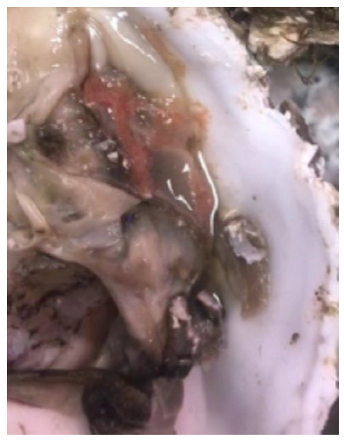
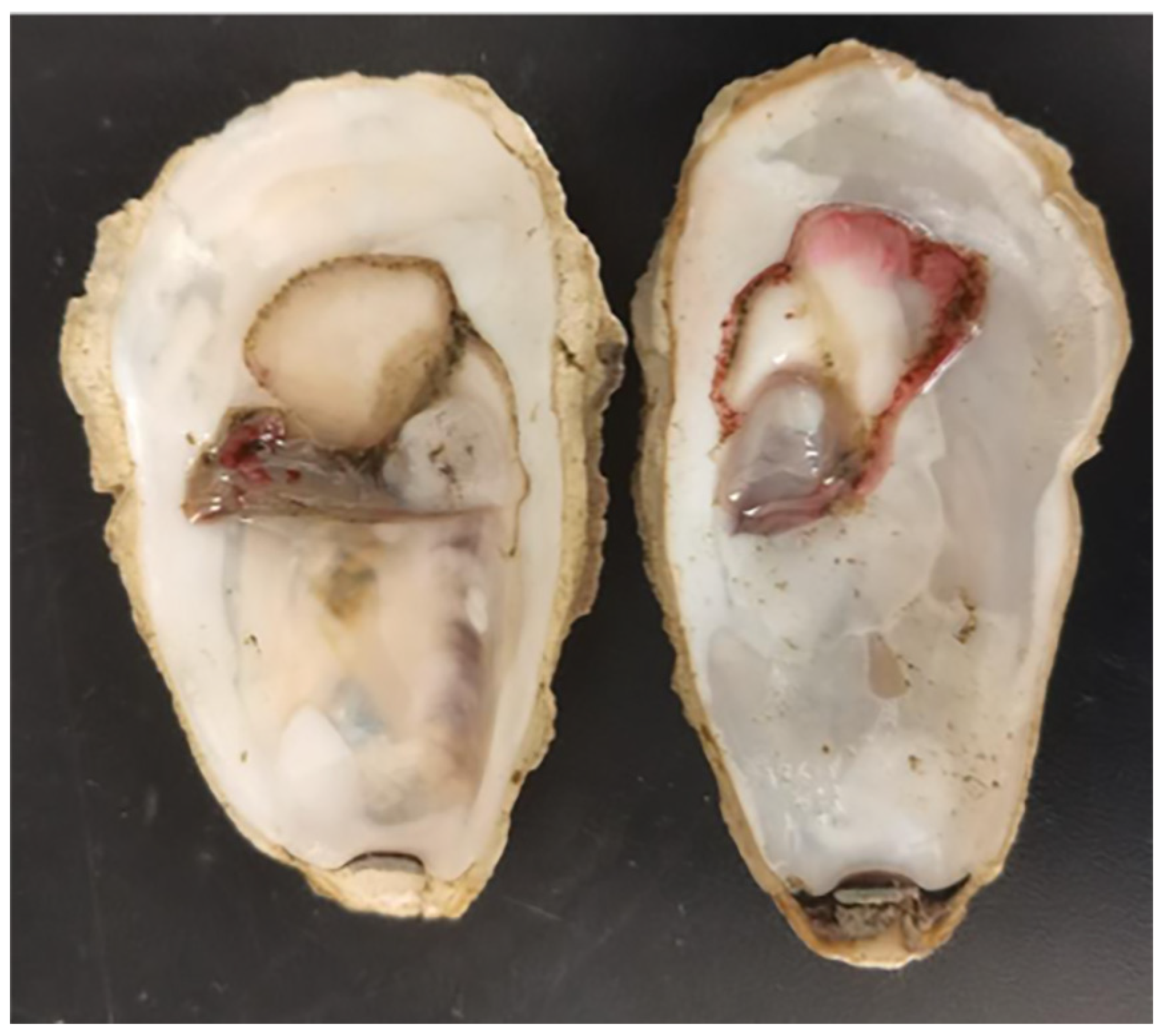
The diseased C. gigas showed decreased activity, with an average weight of 44.0 ± 7.0 g. They were dissected and the soft tissues were observed, as shown in Figure 2. The mantle of the diseased C. gigas was atrophied and turned red, with weakened adductor muscle contraction and a delay in response to external stimuli.

3.2. Bacterial Community Structure Analysis of the Soft Tissues in C. gigas

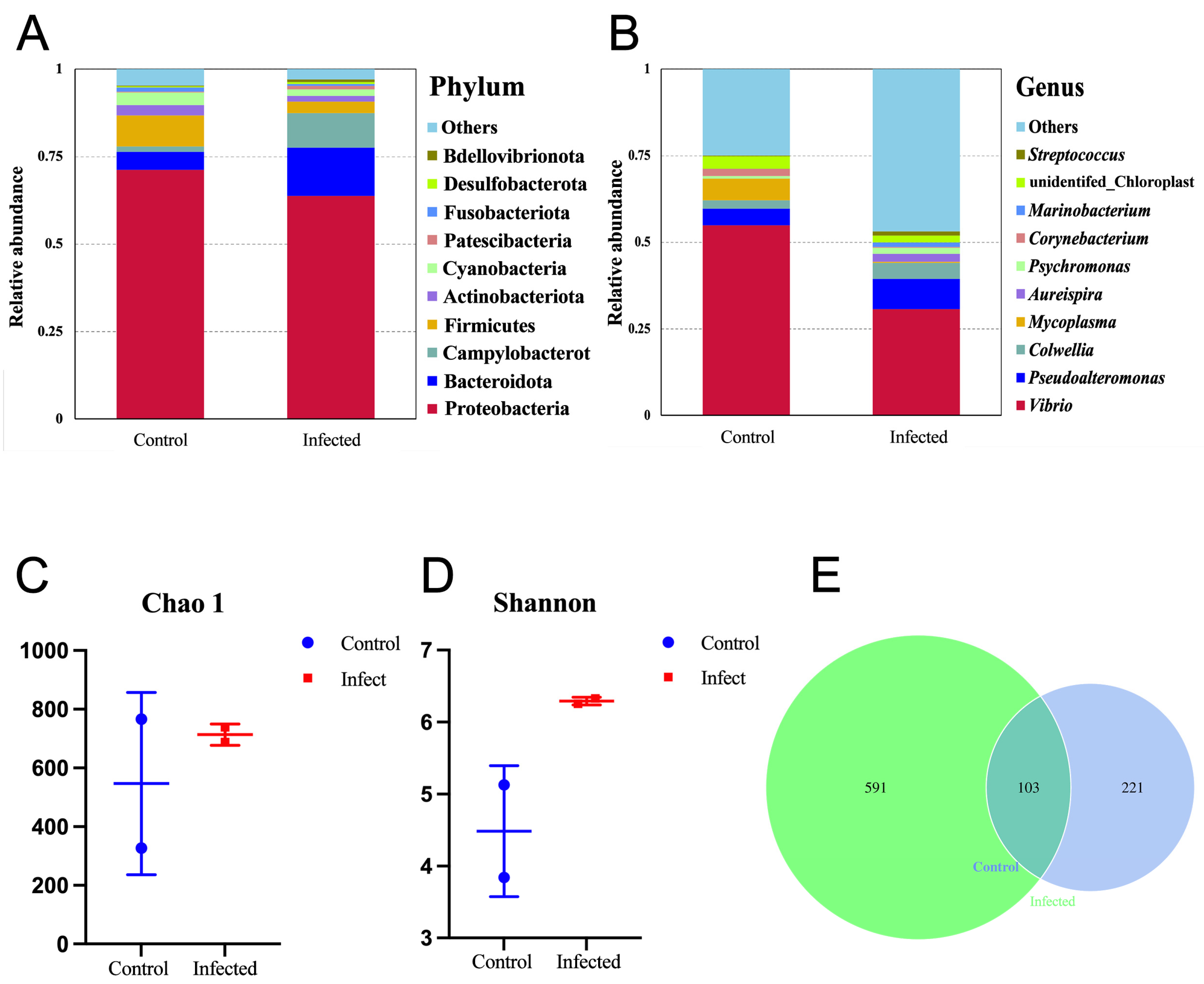
Taxonomic compositions at the phylum level of the bacterial communities are shown in Figure 3A. For both the healthy and the infected C. gigas, Proteobacteria held the overwhelming predominance, with a relative abundance above 60%. However, the community structures of these two groups differed from each other in terms of the other dominant phyla. In the healthy C. gigas, the other dominant phyla in a descending order were Firmicutes, Bacteroidota, Cyanobacteria, Actinobacteriota, etc. In the infected C. gigas, the other dominant phyla in a descending order were Bacteroidota, Campylobacterota, Firmicutes, Actinobacteriota, Cyanobacteria, etc.
Taxonomic compositions at the genus level of the bacterial communities are shown in Figure 3B. Compared with the phylum level, the difference in the bacterial community structures of the two groups at the genus level was more significant. For both groups, Vibrio was the most predominant genus, with a relative abundance of 54.9% in the healthy C. gigas and a relative abundance of 30.6% in the infected C. gigas. In the healthy C. gigas, the other dominant genera in a descending order were Mycoplasma (6.3%), Pseudoalteromonas (4.8%), Colwellia (2.4%), etc. However, in the infected C. gigas, the other dominant genera in a descending order were Pseudoalteromonas (8.7%), Colwellia (4.6%), Aureispira (2.3%), etc. The abundance of Pseudoalteromonas in the infected C. gigas increased as compared with the healthy C. gigas.
Analysis of the α-diversity was performed and the results are shown in Figure 3C,D. The Chao1 and Shannon indexes of infected C. gigas were higher than those of healthy C. gigas, yet the difference was not significant (p > 0.05). In terms of the number of OTUs, 694 were detected from the infected group and 324 were detected from the healthy group (Figure 3E). In addition, the two groups shared 103 identical OTUs.

3.3. Isolation of Culturable Bacteria from Diseased C. gigas Tissues

For isolating culturable bacteria from diseased C. gigas tissues, the dominant colonies with the same morphology and color were picked and purified at least three times. The isolated strain was named as H27.

3.4. Hemolytic Detection of Strain H27

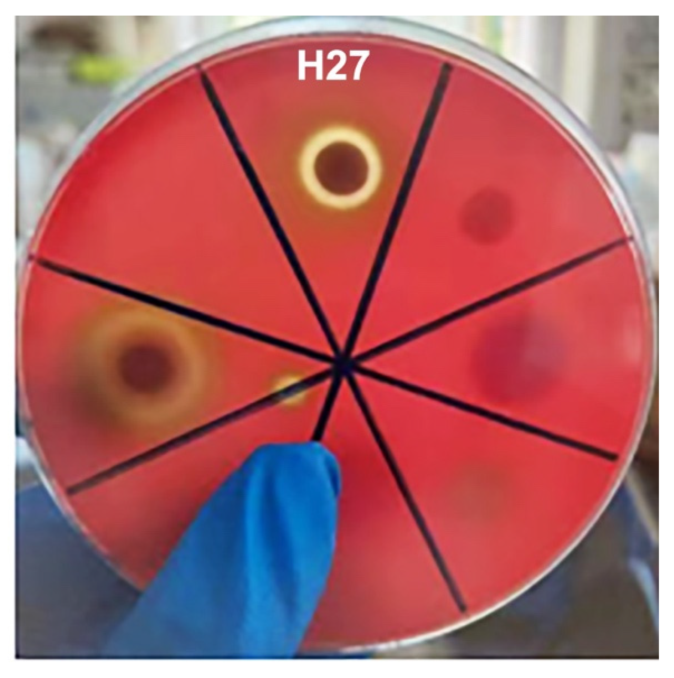
A hemolytic test showed that strain H27 produced a clear transparent ring around the colony (Figure 4), suggesting that it could produce a β-hemolysis ring.

3.5. Morphological Characterization and Molecular Identification of Strain H27

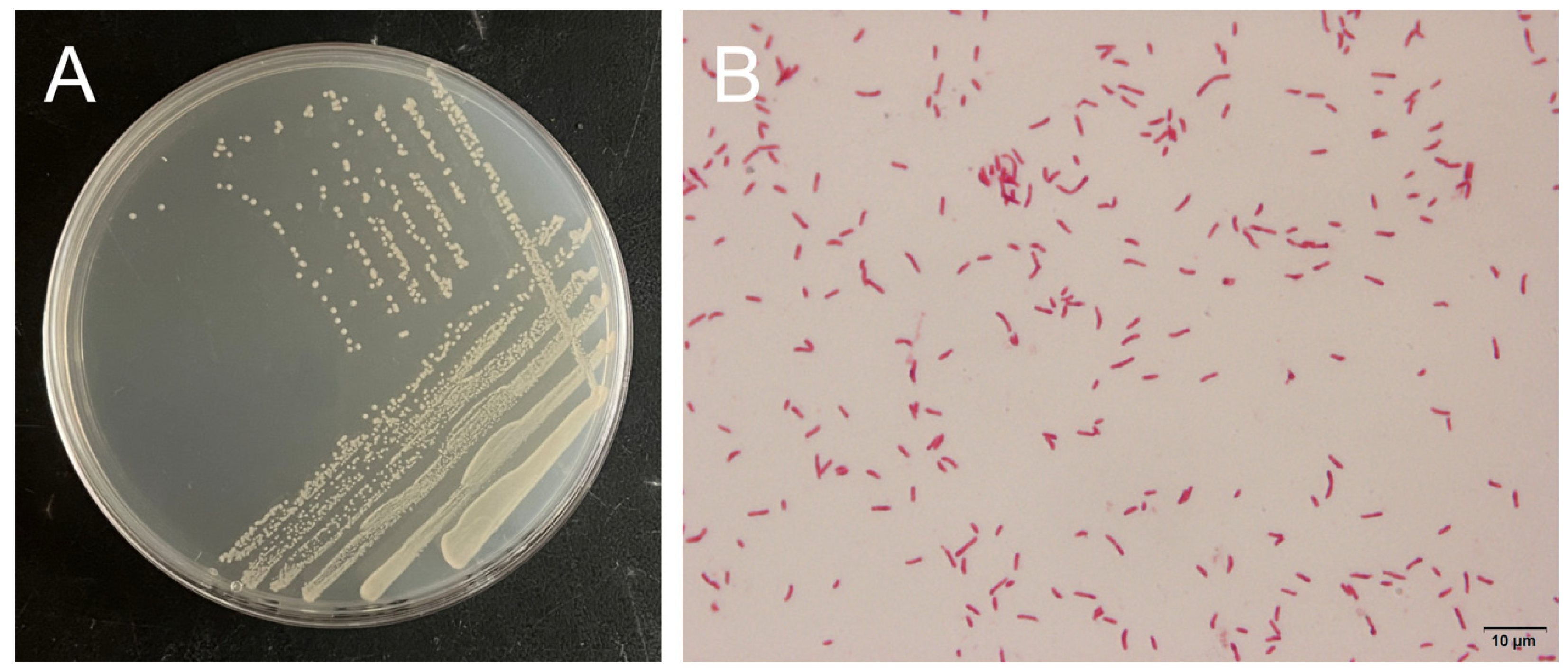
Colonies of strain H27 on 2216E agar plate had a round shape and a smooth surface with an opaque light-yellow color, as shown in Figure 5A. Under the microscope, the Gram-stained cells of strain H27 were red colored and had a single/double rod shape, suggesting that it was a Gram negative rod bacterium (Figure 5B).
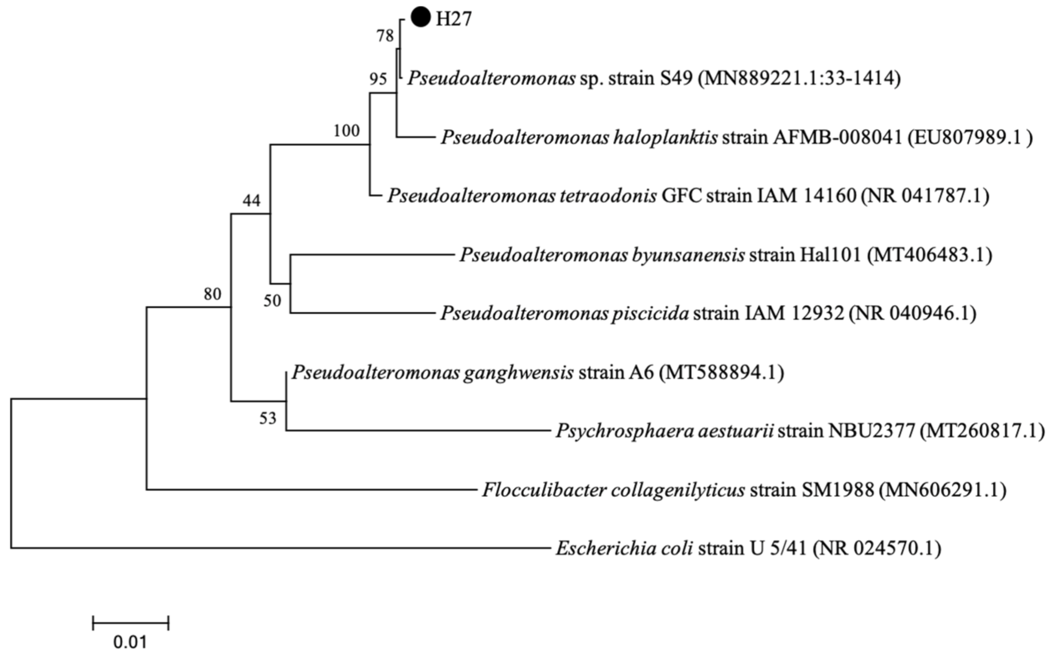
The results showed that strain H27 was most similar to Pseudoalteromonas sp. strain S49, with 99.93% of identity under 100% of coverage. As shown in Figure 6, strain H27 (PQ113772) clustered with P. sp. strain S49 and formed an individual branch. The results of morphological characterization and molecular identification both implied that strain H27 belonged to the genus of Pseudoalteromonas.

3.6. Metabolic Profiling and Antibiotic Sensitivity Analysis of Strain H27

The metabolic profiles of strain H27 using a Biolog Gen III Microplate are shown in Table 1. The results showed that strain H27 could grow at pH 6, 1–8% NaCl. The carbon sources it could utilize included dextrin, D-maltose, L-arginine, L-serine, N-acetyl-D-glucosamine, α-D-glucose, D-fructose, D-fructose-6-PO4, acetic acid, propionic acid, L-alanine, L-glutamic acid, inosine, D-galactose, L-malic acid, D-mannitol, D-glucose-6-PO4, D-galacturonic acid, L-galactonic acid lactone, D-gluconic acid, D-glucuronic acid, gelatin, glucuronamide, N-acetyl-β-D-mannosamine, N-acetyl neuraminic acid, acetoacetic acid and glycyl-L-prolineas. Additionally, it could grow in the presence of vancomycin, tetrazolium violet, 1% sodium lactate, fusidicacid, potassium tellurite and aztreonam. A search against the Biolog Gen III database revealed that strain H27 was most similar to Neisseria flava, Streptococcus halichoeri, Virgibacillus halodenitrificans and Tsukamurella paurometabola, with an SIM (similarity) value of 13.5%, 9.4%, 7.6% and 5.9%, respectively.
The results of antibiotic sensitivity analysis are shown in Table 2. Strain H27 was sensitive to ceftriaxone, amoxicillin, streptomycin, gentamicin, clarithromycin, azithromycin, norfloxacin, ofloxacin, cotrimoxazole, chloramphenicol, florfenicol and polymyxin, and resistant to cefalexin, penicillin, kanamycin, tetracycline and furazolidone.

3.7. Bacterial Challenge Experiment of Larval C. gigas Using Strain H27

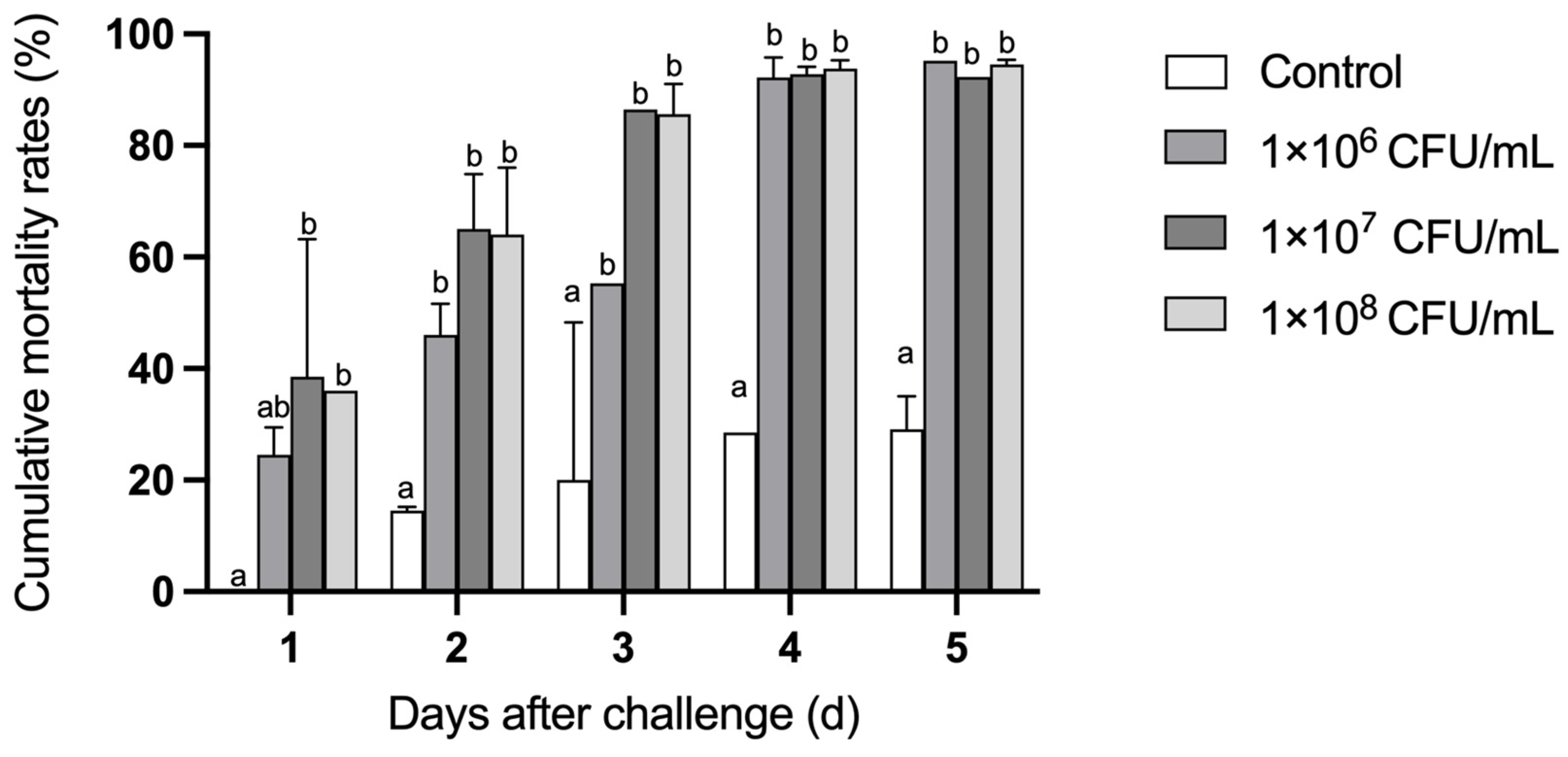
As shown in Figure 7, the mortality of the C. gigas larvae challenged with strain H27 was significantly higher than that of the larvae in the control group in the 5-day-experimental period (p < 0.01). In terms of the final mortality of the larvae challenged with strain H27 at the concentrations of 1 × 10⁶ CFU/mL, 1 × 10⁷ CFU/mL and 1 × 10⁸ CFU/mL, there was no significant difference between these three groups. After 1 day of the bacterial challenge experiment, mass death was observed in both of the two groups treated with higher concentrations of strain H27, with a mortality of nearly 40%. After 3 days of the bacterial challenge experiment, the mortality of both groups of larvae treated with higher concentrations of strain H27 was over 80%. In terms of the survival rate of the larvae challenged with strain H27 at the concentration of 1 × 10⁶ CFU/mL, it maintained a gradual decline in the first three days after infection. However, starting from day 4, the survival rate of the larvae dramatically decreased to a level which was comparable to that of the other two groups challenged with higher concentrations of strain H27. When observed under the microscope, the challenged larvae in the three treatment groups displayed weakened vitality and reddened tissues, as shown in Figure 8. Strain H27 was re-isolated from tissues of the moribund C. gigas larvae 3 days after the bacterial challenge, indicating its fulfillment of Koch’s postulate.

3.8. Histopathological Analysis of Strain H27-Challenged C. gigas Adults

As shown in Figure 9, the moribund C. gigas displayed a reddened mantle. Additionally, the infected C. gigas showed decreased feeding ability.
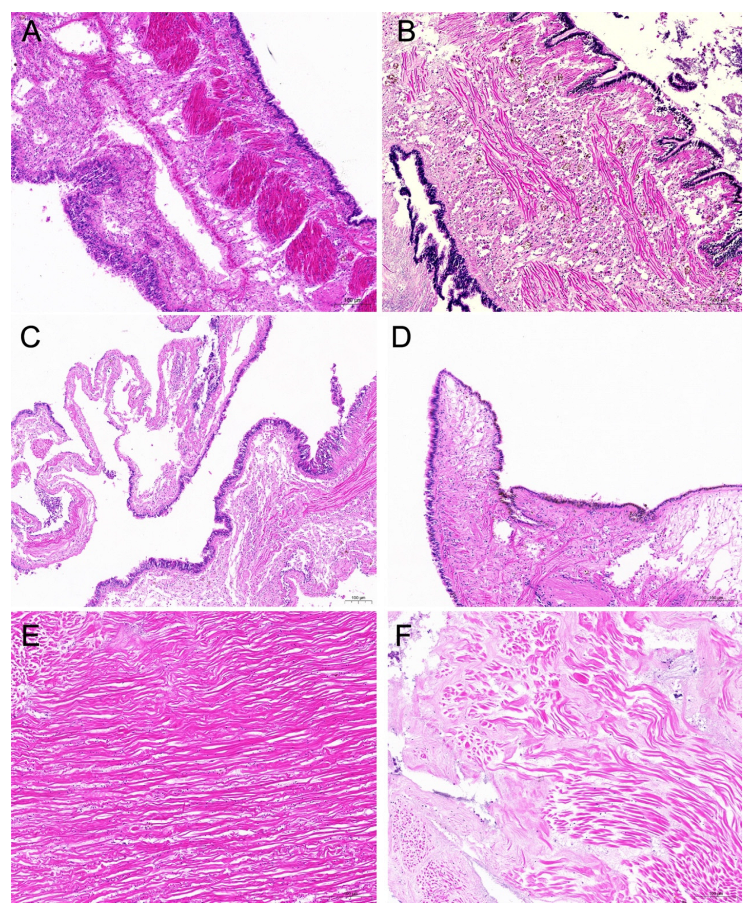
Histopathological analysis showed lesions in the mantle and adductor muscle of the diseased C. gigas (Figure 10). In the samples of mantle tissue, a large number of yellowish-brown pigment granules were observed in the cytoplasm of the mantle. The connective tissue was broken and the epithelial cells were necrotic and scattered (Figure 10B,D). Compared with the control group, the structure of the adductor muscle fibers of the diseased C. gigas was scattered and deformed, and the distance between the muscle fibers became larger (Figure 10F).

4. Discussion

Crassostrea gigas is a bivalve mollusk that inhabits estuaries and coasts [41]. As a filter feeder, it tends to accommodate a large number of microorganisms from the aquatic environment, including pathogenic bacteria. These pathogenic bacteria are mainly opportunistic and under suitable environmental conditions, they can quickly proliferate and then invade C. gigas, especially the larvae, leading to disease outbreak and even mass death. Therefore, the isolation and identification of pathogenic bacterial strains is of great significance to prevent and control C. gigas diseases. Its biochemical properties were in accordance with previously reported strains of the genus Pseudoalteromonas [11,31]. However, its metabolic profiles were not completely identical to any of the reported Pseudoalteromonas strains, which could be due to species-specific differences. The Biolog Microbial Identification System can automatically identify bacterial strains to the species level, given a SIM value of ≥50%. The highest SIM value generated by aligning the Biolog Gen III profiles of strain H27 to the existing database was as low as 13.5%, which was insufficient to support a species-level identification. The limited use of the Biolog Gen III database in accurate identification of environmental and marine bacteria has been reported in a previous study, where 24 out of 64 bacterial strains isolated from saline environments were identified to the species level [41]. In addition, due to variations in laboratory cultivation conditions for bacteria, the individual experiments may not be repeatable [42]. Therefore, in this study, strain H27 was only identified to the genus level through 16S rRNA gene sequencing and it was found to be most similar to Pseudoalteromonas sp. S49 (GenBank accession no.MN889221.1).
According to previous findings, bacterial pathogens of bivalve larvae mainly include the genera of Vibrio, Pseudomonas, Alteromonas, Moraxella and Aeromonas [43,44]. The genus Pseudoalteromonas was first proposed in 1995 [12]. Being a newly established marine bacterial genus, it is rarely directly connected with bivalve diseases. However, members of the Pseudoalteromonas genus could act as pathogens to other marine organisms. P. agarivorans NW4327 was reported to be pathogenic to sponge (Rhopaloeides odorabile) [45]. P. shioyasakiensis caused high mortality of juvenile abalone [46]. P. piscicida was isolated from fish suffering from Favobacteriosis [29]. There is also indirect evidence which connects Pseudoalteromonas with the outbreak of mariculture diseases. In 2015, Pseudoalteromonas was isolated as the dominant genus from diseased Argopecten purpuratus tissues in a scallop larvae hatchery located in Chile, accounting for 22.6% of total bacterial genera [47]. During mass death events of oysters, Pseudoalteromonas is also often present as a dominant genus in diseased oysters [33,34,48]. Consistent with previous findings, in this study, Pseudoalteromonas was the second most abundant genus in infected C. gigas, with a relatively higher abundance as compared with that of healthy C. gigas (with a concurrent upregulation of Pseudoalteromonas from 4.8% to 8.7%). Additionally, bacterial community analysis revealed a downregulation in the relative abundance of Vibrio from 54.9% in the healthy group to 30.6% in the infected group. This might be attributed to the colonization of opportunistic pathogenic Pseudoalteromonas. Previous findings have shown that species of Pseudoalteromonas showed moderate virulence to C. gigas, leading to a mortality of 30–40% [48]. When challenged with P. shioyasakiensis at a bacterial dose of 1 × 10⁴ CFU/mL, all Haliotis discus hannai died within 3 days [46]. In comparison with previous reports, the virulence of P. sp. H27 could possibly be at an intermediate level.
When inoculating strain H27 back to healthy C. gigas larvae and adults, it caused death and disease, reproducing the clinical signs originally observed. Upon histopathologic analysis, a large number of brown granules in the cytoplasm were observed in the mantle samples of challenged C. gigas. In addition, foci of tissue lysis were observed in the adductor muscle samples of challenged C. gigas. It has been established that brown cell proliferation and foci of tissue lysis are viewed as physiological, perhaps immunological coping mechanisms of bivalve mollusks against environmental stresses, infection with major pathogens, or concurrent secondary infections [49]. In a C. gigas disease caused by ostreid herpesvirus-1, these two clinical signs were observed [50]. In this study, brown cell proliferation and foci of tissue lysis were both observed in strain H27-challenged C. gigas, suggesting that strain H27 could be the causative agent of C. gigas disease.
With an objective of preventing and controlling the disease caused by this bacterium, the antibiotic sensitivity of P. sp. H27 was examined. Ceftriaxone, amoxicillin, streptomycin, gentamicin, clarithromycin, azithromycin, norfloxacin, ofloxacin, cotrimoxazole, chloramphenicol, florfenicol and polymyxin B could inhibit its growth, whereas cefalexin, penicillin, kanamycin, tetracycline and furazolidone could not. Our findings are roughly in accordance with existing reports and could provide reference for C. gigas disease control [17,29,30,51]. Therefore, for treatments using antimicrobial agents, drug sensitivity analysis of pathogenic bacterial strains should be taken into consideration. Antibiotics such as ceftriaxone, amoxicillin and streptomycin can be used to control Pseudoalteromonas infections. Moreover, to avoid drug resistance, bacterial infections should be prevented using probiotics. Additionally, monitoring environmental conditions and water quality may help to prevent and control diseases caused by Pseudoalteromonas.
Pseudoalteromonas is widely distributed in marine environments. As reported, some species produce extracellular enzymes, including agarase, β-galactosidase, α-amylase and pectin lyase [52]. Some produce antimicrobial metabolites (alkaloids, polyketides and peptides) and form a microbial barrier to protect the host from pathogen invasion, sometimes favoring the growth of bivalves [53]. Our results are the opposite to exisiting reports. Strain H27 was isolated from diseased C. gigas and was pathogenic to C. gigas, which not only demonstrated the functional versatility of the genus Pseudoalteromonas, but also enriched the knowledge on Pseudoalteromonas of marine environment origin. Environment greatly influences the physiological and biochemical characteristics of microorganisms. Previous findings have shown that Pseudoalteromonas was an opportunistic pathogen and was more likely to cause host diseases under favorable environmental conditions [46], such as high temperature and heavy rainfall [41,54]. The climatological data showed that in May 2023, Zhanjiang experienced relatively higher temperature and stronger rainfall, which might favor the propagation and pathogenicity of P. sp. H27. Both the virulence of bacteria and the immune responses of the host are largely affected by changes in physical environmental parameters, though the underlying mechanisms remain unclear [55]. In the future, further studies are required to elucidate the complex interactions among hosts, pathogens and the environment.

5. Conclusions

In this study, Pseudoalteromonas sp. H27, a bacterial strain pathogenic to C. gigas, was isolated from the diseased tissue of C. gigas, which could reproduce the originally observed clinical signs when challenging adult C. gigas. However, the specific pathogenesis has not yet been elucidated, which requires further research. This study provides valuable information for the infection of Pseudoalteromonas in C. gigas. From the perspective of the aquaculture industry, reducing pathogen loads and cultivating healthier C. gigas are of paramount importance. Maintaining optimal water quality, adjusting water temperatures and administering appropriate antibiotics to inhibit the growth of bacterial pathogens are feasible and cost-effective disease mitigation strategies. These strategies might contribute to the prevention and control of related diseases of C. gigas.

Author Contributions

Conceptualization, L.F., Z.H. and H.Q.; methodology, H.Q.; validation, J.J., Z.J. and J.W.; formal analysis, S.X.; investigation, H.Q. and R.C.; resources, B.W.; data curation, H.Q.; writing—original draft preparation, H.Q. and J.J.; writing—review and editing, L.F. and H.Q.; visualization, H.Q., J.J. and S.X.; supervision, L.F.; project administration, Z.H.; funding acquisition, Z.H. and L.F. All authors have read and agreed to the published version of the manuscript.

Funding

This research was supported by a grant from the Key R&D program of Shandong Province (2023LZGCQY001), the Applied Fundamental Research Program of Liaoning Province (2022JH2/101300141), the Outstanding Young Scientific and Technological Talents Foundation of Dalian (2021RJ09), Liaoning Revitalization Talents Program (XLYC1807271), the Central Government Subsidy Project for Liaoning Fisheries (2024) and funds earmarked for Modern Agroindustry Technology Research System (CARS-49).

Institutional Review Board Statement

The animal study protocol was approved by the Experimental Animals Ethical Review Committee of Dalian Ocean University (protocol code: DLOU2023012 and date of approval: 26 December 2023).

Informed Consent Statement

Not applicable.

Data Availability Statement

The original contributions presented in this study are included in the article. Further inquiries can be directed to the corresponding authors.

Conflicts of Interest

The authors declare no conflict of interest.

References

  1. FAO. Fishery and Aquaculture Statistics—Yearbook 2021. In FAO Yearbook of Fishery and Aquaculture Statistics; FAO: Rome, Italy, 2024. [Google Scholar]
  2. Wang, D.; Gao, D. China Fishery Statistics Yearbook, 2024 ed; China Agriculture Press: Beijing, China, 2024. (In Chinese) [Google Scholar]
  3. Yang, J.; Wang, W.; Li, B.; Feng, Y.; Sun, G.; Li, Z.; Xu, X. Crassostrea gigas “Luyi No.1”. China Fish. Mon. 2021, 6, 87–94. (In Chinese) [Google Scholar]
  4. Chen, H.; Cai, J. The nutritional and medicinal value of oysters and their development and utilization. J. Chin. Med. Mater. 2005, 3, 172–174. (In Chinese) [Google Scholar] [CrossRef]
  5. Schets, F.M.; Van Den Berg, H.H.J.L.; Rutjes, S.A.; Husman, A.M.D.R. Pathogenic Vibrio species in dutch shellfish destined for direct human consumption. J. Food Prot. 2010, 73, 734–738. [Google Scholar] [CrossRef] [PubMed]
  6. Shan, C.; Li, M.; Liu, Z.; Xu, R.; Qiao, F.; Du, Z.Y.; Zhang, M.L. Pediococcus pentosaceus Enhances Host Resistance Against Pathogen by Increasing IL-1β Production: Understanding Probiotic Effectiveness and Administration Duration. Front. Immunol. 2021, 12, 766401. [Google Scholar] [CrossRef] [PubMed]
  7. Rippey, S.R. Infectious diseases associated with molluscan shellfish consumption. Clin. Microbiol. Rev. 1994, 7, 419–425. [Google Scholar] [CrossRef] [PubMed]
  8. Takahashi, K.G.; Nakamura, A.; Mori, K. Inhibitory Effects of Ovoglobulins on Bacillary Necrosis in Larvae of the Pacific Oyster, Crassostrea gigas. J. Invertebr. Pathol. 2000, 75, 212–217. [Google Scholar] [CrossRef] [PubMed]
  9. Clerissi, C.; Luo, X.; Lucasson, A.; Mortaza, S.; de Lorgeril, J.; Toulza, E.; Petton, B.; Escoubas, J.-M.; Dégremont, L.; Gueguen, Y.; et al. A core of functional complementary bacteria infects oysters in Pacific Oyster Mortality Syndrome. Anim. Microbiome 2023, 5, 26. [Google Scholar] [CrossRef] [PubMed]
  10. Petton, B.; Destoumieux-Garzón, D.; Pernet, F.; Toulza, E.; de Lorgeril, J.; Degremont, L.; Mitta, G. The Pacific Oyster Mortality Syndrome, a Polymicrobial and Multifactorial Disease: State of Knowledge and Future Directions. Front. Immunol. 2021, 12, 630343. [Google Scholar] [CrossRef]
  11. Destoumieux-Garzón, D.; Montagnani, C.; Dantan, L.; Nicolas, N.d.S.; Travers, M.-A.; Duperret, L.; Charrière, G.M.; Toulza, E.; Mitta, G.; Cosseau, C.; et al. Cross-talk and mutual shaping between the immune system and the microbiota during an oyster’s life. Philosophical Transactions of the Royal Society of London. Ser. B Biol. Sci. 2024, 379, 20230065. [Google Scholar]
  12. Gauthier, G.; Gauthier, M.; Christen, R. Phylogenetic analysis of the genera Alteromonas, Shewanella, and Moritella using genes coding for small-subunit rRNA sequences and division of the genus Alteromonas into two genera, Alteromonas (emended) and Pseudoalteromonas gen. nov., and proposal of twelve new species combinations. Int. J. Syst. Bacteriol. 1995, 45, 755–761. [Google Scholar]
  13. Ding, Y. Study on Quorum Sensing Properties and Molecular Regulation of Pseudoalteromonas spp. Master’s Thesis, Zhejiang University of Technology, Hangzhou, China, 2019. Available online: https://kns.cnki.net/kcms2/article/abstract?v=o5eMcsLgsI4yf7Hg35q_ASzoh8fSwzLe-4_XkLJ2e0up7qZDNT3jljeOFUhYXnHZ2OUk7w3K8uDK1G0_Duf0GzvZd5KFEMV6WJloiVkx4geynAF653FwtBUR8p_EoLG8zKHi8ecoKPyADDRzFpyg-A==&uniplatform=NZKPT&language=CHS (accessed on 7 February 2023). (In Chinese).
  14. Rong, J. Genome Sequencing and Comparative Genomics of the Marine Bacterial Genus Pseudoalteromonas. Ph.D. Thesis, Shandong University, Jinan, China, 2016. Available online: https://kns.cnki.net/kcms2/article/abstract?v=5DzVwdTmeh-zNQMeQT8CrRKbRxxsiuuHFmkM5oC60pdA0Ca49D1jTHqIY_4_4LnJVo_qSDCFXlgyA6naGlZcnG4uod-S3dLlU5_MRffB04gGf74_o0lXHcrb1HgVxc4KX5bmy0NIIkNy3qCFwx4cKw==&uniplatform=NZKPT&language=CHS (accessed on 13 November 2023). (In Chinese).
  15. Lovejoy, C.; Bowman, J.P.; Hallegraeff, G.M. Algicidal effects of a novel marine Pseudoalteromonas isolate (class Proteobacteria, gamma subdivision) on harmful algal bloom species of the genera Chattonella, Gymnodinium, and Heterosigma. Appl. Environ. Microbiol. 1998, 64, 2806–2813. [Google Scholar] [CrossRef] [PubMed]
  16. Huang, Z.; Li, C.; Yu, X.; Liao, W.; Sun, J.; Peng, M. Isolation of a Strain of Pseudoalteromonas sp. and the Study on the Characteristics of its Algicidal Effect. Ocean Technol. 2008, 3, 56–60. (In Chinese) [Google Scholar]
  17. Bai, X.; Zhang, X.; Bi, K.; Fan, X.; Yan, B.; Qin, G. Isolation and identification of a Pseudoalteromonas. Jiangsu Agric. Sci. 2013, 41, 211–216. (In Chinese) [Google Scholar] [CrossRef]
  18. Costa-Ramos, C.; Rowley, A.F. Effect of extracellular products of Pseudoalteromonas atlantica on the edible crab Cancer pagurus. Appl. Environ. Microbiol. 2004, 70, 729–735. [Google Scholar] [CrossRef] [PubMed]
  19. Li, J.; Mou, Z.; Yang, H.; Mao, Y.; Yan, Y.; Mo, Z. Isolation and Identification the Pathogen of Pyropia yezoensis Green Spot Disease. Prog. Fish. Sci. 2019, 40, 140–146. (In Chinese) [Google Scholar] [CrossRef]
  20. Yan, Y.; Ma, J.; Xu, P.; Sun, Q.; Wang, H. Pseudoalteromonas citrea, the causative agent of green-spot disease of Porphyrae yezoensis. J. Fish. Sci. China 2002, 4, 353–358. (In Chinese) [Google Scholar]
  21. Wang, H.; Li, X.; Xia, Y.; Yan, B. Isolation, identification and biological pathogen of yellow spot disease in conchocelis of Porphyra yezoensis. Mar. Environ. Sci. 2011, 30, 361–364+408. (In Chinese) [Google Scholar]
  22. Sawabe, T.; Makino, H.; Tatsumi, M.; Nakano, K.; Tajima, K.; Iqbal, M.M.; Yumoto, I.; Ezura, Y.; Christen, R. Pseudoalteromonas bacteriolytica sp. nov., a marine bacterium that is the causative agent of red spot disease of Laminaria japonica. Int. J. Syst. Bacteriol. 1998, 48 Pt 3, 769–774. [Google Scholar] [CrossRef]
  23. Sawabe, T.; Tanaka, R.; Iqbal, M.M.; Tajima, K.; Ezura, Y.; Ivanova, E.P.; Christen, R. Assignment of Alteromonas elyakovii KMM 162T and five strains isolated from spot-wounded fronds of Laminaria japonica to Pseudoalteromonas elyakovii comb. nov. and the extended description of the species. Int. J. Syst. Evol. Microbiol. 2000, 50 Pt 1, 265–271. [Google Scholar] [CrossRef]
  24. Sun, S. Study on the Major Bacterial Diseases of Cultured Sea Cucumber (Aposticchopus japonicus) During Larval Stages. Master’s Thesis, Ocean University of China, Qingdao, China, 2007. Available online: https://kns.cnki.net/kcms2/article/abstract?v=2Wn7gbiy3W8QCqFgVqp86NcPeUDo7KYacuCZ1Tu0QxGctshe7JI8bl5zEU7hqa1CGZMz8UPoVz4qV5BCAMO14ksxg3a-jNGmJ1dlnoh-r6Tv-EWCfvJr2VYhvv8UmlF-pmYAL3dfyOVjiNPfSo4x0g==&uniplatform=NZKPT&language=CHS (accessed on 1 December 2023). (In Chinese).
  25. Liu, H.; Zheng, F.; Sun, X.; Hong, X.; Dong, S.; Wang, B.; Tang, X.; Wang, Y. Identification of the pathogens associated with skin ulceration and peristome tumescence in cultured sea cucumbers Apostichopus japonicus (Selenka). J. Invertebr. Pathol. 2010, 105, 236–242. [Google Scholar] [CrossRef] [PubMed]
  26. Xie, J.; Wang, Y.; Zhang, Z.; Rong, X. Rapid detection of two pathogens of skin ulcer syndrome in cultured Apostichopus japonicus by Dot-ELISA. Mar. Sci.-Qingdao-Chin. Ed. 2007, 8, 59–64. (In Chinese) [Google Scholar]
  27. Fang, B. Epidemiological and Aetiological Studies on the Skin Ulcerative Syndrome of Cultured Sea Cucumber Apostichopus japonicus and Analysis of the Pathogen’s Reservoir. Master’s Thesis, Ocean University of China, Qingdao, China, 2007. Available online: https://kns.cnki.net/kcms2/article/abstract?v=2Wn7gbiy3W9ksoBcCPSspMXml3DpWEKx40GVbgOnAfOp2eUcILaUaa3RH3cSHP2dro-QiVSYmd-ngYQEcZtiYP3S7cKnwPIXG5olGyY-mARcuEh_lpePJeZ59K-B31TV590WaG0UVRHRvYQ8nltkAQ==&uniplatform=NZKPT&language=CHS (accessed on 1 December 2023). (In Chinese).
  28. Wang, Y.; Feng, N.; Li, Q.; Ding, J.; Zhan, Y.; Chang, Y. Isolation and characterization of bacteria associated with a syndrome disease of sea urchin Strongylocentrotus intermedius in North China. Aquac. Res. 2013, 44, 691–700. [Google Scholar] [CrossRef]
  29. Qiao, Y.; Shen, H.; Wan, X.; Qi, Y.; Wang, L. The identification of a pathogenic Pseudoalteromonas that causes disease in black seabream and the analysis of its virulence genes. Jiangsu Agric. Sci. 2015, 43, 229–233. (In Chinese) [Google Scholar] [CrossRef]
  30. Qiao, Q.; Han, N.; Chen, C.; Yang, Z.; Shi, C.; Huang, J.; Li, Q. Isolation and identification of pathogenic Pseudoalteromonas sp. and antibiotic sensitivity research. J. Anhui Agric. Sci. 2010, 38, 14224–14226+14232. (In Chinese) [Google Scholar] [CrossRef]
  31. Bao, Y.; Shang, Y.; Wang, H.; Yuan, Z. Isolation and Identification of Pathogenic Pseudoalteromonas from Cynoglossus semilaevis Günther and Its Drug Sensitivity Analysis. Anhui Agric. Sci. 2020, 48, 102–105+123. (In Chinese) [Google Scholar]
  32. Pujalte, M.J.; Sitjà-Bobadilla, A.; Macián, M.C.; Álvarez-Pellitero, P.; Garay, E. Occurrence and virulence of Pseudoalteromonas spp. in cultured gilthead sea bream (Sparus aurata L.) and European sea bass (Dicentrarchus labrax L.). Molecular and phenotypic characterisation of P. undina strain U58. Aquaculture 2007, 271, 47–53. [Google Scholar] [CrossRef]
  33. Worden, P.J.; Bogema, D.R.; Micallef, M.L.; Go, J.; Deutscher, A.T.; Labbate, M.; Green, T.J.; King, W.L.; Liu, M.; Seymour, J.R.; et al. Phylogenomic diversity of Vibrio species and other Gammaproteobacteria isolated from Pacific oysters (Crassostrea gigas) during a summer mortality outbreak. Microb. Genom. 2022, 8, mgen000883. [Google Scholar] [CrossRef] [PubMed]
  34. Zhang, X.; Huang, B.-W.; Zheng, Y.-D.; Xin, L.S.; Chen, W.B.; Yu, T.; Li, C.; Wang, C.M.; Bai, C.M. Identification and Characterization of Infectious Pathogens Associated with Mass Mortalities of Pacific Oyster (Crassostrea gigas) Cultured in Northern China. Biology 2023, 12, 759. [Google Scholar] [CrossRef] [PubMed]
  35. Del Sal, G.; Manfioletti, G.; Schneider, C. The CTAB-DNA precipitation method: A common mini-scale preparation of template DNA from phagemids, phages or plasmids suitable for sequencing. BioTechniques 1989, 7, 514–520. [Google Scholar]
  36. Schloss, P.D.; Westcott, S.L.; Ryabin, T.; Ryabin, T.; Hall, J.R.; Hartmann, M.; Hollister, E.B.; Lesniewski, R.A.; Oakley, B.B.; Parks, D.H.; et al. Introducing mothur: Open-Source, Platform-Independent, Community-Supported Software for Describing and Comparing Microbial Communities. Appl. Environ. Microbiol. 2009, 75, 7537–7541. [Google Scholar] [CrossRef]
  37. Jensen, S.; Bergh, O.; Enger, O.; Hjeltnes, B. Use of PCR-RFLP for genotyping 16S rRNA and characterizing bacteria cultured from halibut fry. Can. J. Microbiol. 2002, 48, 379–386. [Google Scholar] [CrossRef]
  38. Cheng, C.; Yang, M.; Li, J.; Yao, S.; Hu, H. Biolog Microbial Identification System—Study on the Operating Regulation of Bacteria Identification. Food Ferment. Ind. 2006, 32, 50. (In Chinese) [Google Scholar]
  39. Wendling, C.C.; Batista, F.M.; Wegner, K.M. Persistence, Seasonal Dynamics and Pathogenic Potential of Vibrio Communities from Pacific Oyster Hemolymph. PLoS ONE 2014, 9, e94256. [Google Scholar] [CrossRef]
  40. Ushijima, B.; Saw, J.H.; Videau, P.; Häse, C.C. Comparison of Vibrio coralliilyticus virulence in Pacific oyster larvae and corals. Microbiology 2022, 168, 001169. [Google Scholar] [CrossRef] [PubMed]
  41. King, W.L.; Jenkins, C.; Seymour, J.R.; Labbate, M. Oyster disease in a changing environment: Decrypting the link between pathogen, microbiome and environment. Mar. Environ. Res. 2019, 143, 124–140. [Google Scholar] [CrossRef] [PubMed]
  42. Nagaraj, V.; Skillman, L.; Li, D.; Xie, Z.; Ho, G. Culturable bacteria from a full-scale desalination plant: Identification methods, bacterial diversity and selection of models based on membrane-biofilm community. Desalination 2019, 457, 103–114. [Google Scholar] [CrossRef]
  43. Canesi, L.; Pruzzo, C. Chapter 6—Specificity of Innate Immunity in Bivalves: A Lesson From Bacteria. In Lessons in Immunity; Ballarin, L., Cammarata, M., Eds.; Academic Press: Cambridge, MA, USA, 2016; pp. 79–91. [Google Scholar]
  44. Paillard, C.; Le Roux, F.; Borrego, J.J. Bacterial disease in marine bivalves, a review of recent studies: Trends and evolution. Aquat. Living Resour. 2004, 17, 477–498. [Google Scholar] [CrossRef]
  45. Choudhury, J.D.; Pramanik, A.; Webster, N.S.; Llewellyn, L.E.; Gachhui, R.; Mukherjee, J. The Pathogen of the Great Barrier Reef Sponge Rhopaloeides odorabile Is a New Strain of Pseudoalteromonas agarivorans Containing Abundant and Diverse Virulence-Related Genes. Mar. Biotechnol. 2015, 17, 463–478. [Google Scholar] [CrossRef] [PubMed]
  46. Li, M.; Wu, W.; You, W.; Huang, S.; Huang, M.; Luo, X.; Lu, Y.; Ke, C.; Xie, Q. A novel screening method for the detection of Pseudoalteromonas shioyasakiensis, an emerging opportunistic pathogen that caused the mass mortality of juvenile Pacific abalone (Haliotis discus hannai) during a record-breaking heat wave. Aquaculture 2021, 545, 737191. [Google Scholar] [CrossRef]
  47. Miranda, C.D.; Rojas, R.; Geisse, J.; Romero, J.; González-Rocha, G. Scallop larvae hatcheries as source of bacteria carrying genes encoding for non-enzymatic phenicol resistance. Mar. Pollut. Bull. 2015, 95, 173–182. [Google Scholar] [CrossRef] [PubMed]
  48. Garnier, M.; Labreuche, Y.; Garcia, C.; Robert, M.; Nicolas, J.-L. Evidence for the involvement of pathogenic bacteria in summer mortalities of the Pacific oyster Crassostrea gigas. Microb. Ecol. 2007, 53, 187–196. [Google Scholar] [CrossRef] [PubMed]
  49. Zaroogian, G.; Yevich, P. The nature and function of the brown cell in Crassostrea virginica. Mar. Environ. Res. 1994, 37, 355–373. [Google Scholar] [CrossRef]
  50. Bueno, R.; Perrott, M.; Dunowska, M.; Brosnahan, C.; Johnston, C. In situ hybridization and histopathological observations during ostreid herpesvirus-1-associated mortalities in Pacific oysters Crassostrea gigas. Dis. Aquat. Org. 2016, 122, 43–55. [Google Scholar] [CrossRef]
  51. Ivanova, E.P.; Sawabe, T.; Lysenko, A.M.; Gorshkova, N.M.; Hayashi, K.; Zhukova, N.V.; Nicolau, D.V.; Christen, R.; Mikhailov, V.V. Pseudoalteromonas translucida sp. nov. and Pseudoalteromonas paragorgicola sp. nov., and emended description of the genus. Int. J. Syst. Evol. Microbiol. 2002, 52 Pt 5, 1759–1766. [Google Scholar] [CrossRef] [PubMed]
  52. Xi, Y.; Zhu, D.; Liu, H.; Han, Z. Advances on Pseudoalteromonas species and their extracellular bioactive compounds. Microbiology 2005, 3, 108–112. (In Chinese) [Google Scholar] [CrossRef]
  53. Offret, C.; Desriac, F.; Le Chevalier, P.; Mounier, J.; Jégou, C.; Fleury, Y. Spotlight on Antimicrobial Metabolites from the Marine Bacteria Pseudoalteromonas: Chemodiversity and Ecological Significance. Mar. Drugs 2016, 14, 129. [Google Scholar] [CrossRef] [PubMed]
  54. Go, J.; Deutscher, A.T.; Spiers, Z.B.; Dahle, K.; Kirkland, P.D.; Jenkins, C. Mass mortalities of unknown aetiology in Pacific oysters Crassostrea gigas in Port Stephens, New South Wales, Australia. Dis. Aquat. Org. 2017, 125, 227–242. [Google Scholar] [CrossRef] [PubMed]
  55. Larsen, A.K.; Nymo, I.H.; Sørensen, K.K.; Seppola, M.; Rødven, R.; de Bagüés, M.P.J.; Al Dahouk, S.; Godfroid, J. Concomitant Temperature Stress and Immune Activation may Increase Mortality Despite Efficient Clearance of an Intracellular Bacterial Infection in Atlantic Cod. Front. Microbiol. 2018, 9, 2963. [Google Scholar] [CrossRef]
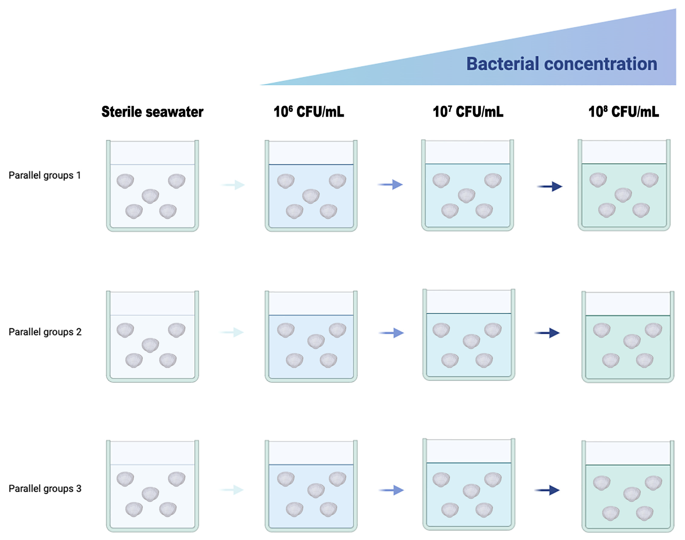
Figure 1. Scheme of the bacterial challenge experiment using larval C. gigas.
Microorganisms 13 00296 g001
Figure 2. The clinical signs of the diseased C. gigas.
Microorganisms 13 00296 g002
Figure 3. Diversity analysis of bacteria in C. gigas soft tissues. (A) The phylum level. (B) The genus level. (C) Chao 1. (D) Shannon. (E) OTU abundance Venn diagram analysis of healthy and diseased C. gigas.
Microorganisms 13 00296 g003
Figure 4. Hemolysis test of culturable bacteria. Strain H27 produced a clear transparent ring around the colony.
Microorganisms 13 00296 g004
Figure 5. Morphological characteristics of strain H27. (A) Colonies of strain H27 were round, smooth and light yellow on 2216E agar plate. (B) Microscopic image of Gram-stained strain H27 was red (magnification: 100×).
Microorganisms 13 00296 g005
Figure 6. Phylogenetic tree of strain H27 based on 16S rRNA gene sequences. According to the bootstrapping data (1000 replications), values at the node indicate the percentage of trees which fall into this grouping. The scale bar indicates the number of substitutions per site. The phylogenetic tree showed that strain H27 was clustered with P. sp. strain S49 (MN889221.1).
Microorganisms 13 00296 g006
Figure 7. The survival rate of C. gigas larvae infected with strain H27 (p < 0.01). C. gigas larvae were challenged using the dipping method with sterile seawater (control group) and different concentrations of strain H27 (1 × 10⁶ CFU/mL, 1 × 10⁷ CFU/mL, 1 × 10⁸ CFU/mL). Survival rates were recorded daily for 5 days. Different alphabets on top of the columns mean significant difference.
Microorganisms 13 00296 g007
Figure 8. Microscopic image of C. gigas larvae challenged with strain H27 via immersion on day 2 (magnification: 100×). Reddened tissues are indicated using red arrows.
Microorganisms 13 00296 g008
Figure 9. Adult C. gigas challenged with strain H27 via injection. It displayed a reddened mantle.
Microorganisms 13 00296 g009
Figure 10. Histopathological analysis of the mantle (AD) and adductor muscles (E,F) of adult C. gigas. (A,C,E): tissues of C. gigas in the control group. (B,D,F): tissues of C. gigas challenged with strain H27. A large number of yellowish-brown pigment granules were observed in the cytoplasm of the mantle. The connective tissue was broken and the epithelial cells were necrotic and scattered.
Microorganisms 13 00296 g010
Table 1. Metabolic profiles of strain H27 using Biolog Gen III Microplate.
Reaction ItemResult Reaction ItemResult
A1Negative Control-E1Gelatin+
A2Dextrin+E2Glycyl-L-Proline+
A3D-Maltose+E3L-Alanine+
A4D-Trehalose-E4L-Arginine+
A5D-Cellobiose-E5L-Aspartic Acid-
A6Gentiobiose-E6L-Glutamic Acid+
A7Sucrose-E7L-Histidine-
A8D-Turanose-E8L-Pyroglutamic Acid-
A9Stachyose-E9L-Serine+
A10Positive Control+E10Lincomycin-
A11pH 6+E11Guanidine HCl-
A12pH 5-E12Niaproof4-
B1D-Raffinose-F1Pectin-
B2α-D-Lactose-F2D-Galacturonic Acid+
B3D-Melibiose-F3L-Galactonic Acid Lactone+
B4β-Methyl-D-Glucoside-F4D-Gluconic Acid+
B5D-Salicin-F5D-Glucuronic Acid +
B6N-Acetyl-D-Glucosamine+F6Glucuronamide+
B7N-Acetyl-β-D-Mannosamine+F7Mucic Acid-
B8N-Acetyl-D-Galactosamine-F8Quinic Acid-
B9N-Acetyl Neuraminic Acid+F9D-Saccharic Acid-
B101%NaCl+F10Vancomycin+
B114%NaCl+F11Tetrezolium Violet+
B128%NaCl+F12Tetrezolium Blue-
C1α-D-Glucose+G1p-Hydroxy-Phenylacetic Acid-
C2D-Mannose-G2Methyl Pyruvate-
C3D-Fructose+G3D-Lactic Acid Methyl Ester-
C4D-Galactose+G4L-Lactic Acid-
C53-Methyl Glucose-G5Citric Acid-
C6D-Fucose-G6α-Keto-Glutaric Acid-
C7L-Fucose-G7D-Malic Acid-
C8L-Rhamnose-G8L-Malic Acid+
C9Inosine+G9Bromo-Succinic Acid-
C101% Sodium Lactate+G10Nalidixic Acid-
C11Fusidic Acid+G11Lithium Chloride-
C12D-Serine-G12Potassium Tellurite+
D1D-Sorbitol-H1Tween 40-
D2D-Mannitol+H2γ-Amino-Butyric Acid-
D3D-Arabitol-H3α-Hydroxy-Butyric Acid-
D4myo-Inositol-H4β-Hydroxy-D,L-butyric Acid-
D5Glycerol-H5α-Keto-Butyric Acid-
D6D-Glucose-6-PO4+H6Acetoacetic Acid+
D7D-Fructose-6-PO4+H7Propionic Acid+
D8D-Aapartic Acid-H8Acetic Acid+
D9D-Serine-H9Formic Acid-
D10Troleandomycin-H10Aztreonam+
D11Rifamycin SV-H11Sodium Butyrate-
D12Minocycline-H12Sodium Bromate-
Notes: “+” means positive and “-” means negative. Negative control: A1; positive control: A10; carbon source utilization: 1–9; chemical sensitivity: D,E,F,G,H10–12 and environmental factors: A,B,C10–12.
Table 2. Drug sensitivity test results of strain H27.
Type of AntibioticName of AntibioticInhibitory Zone Diameter (mm)Inhibitory Zone Diameter of H27 (mm)Sensitivity
RIS
β-lactamsCefalexin≤1415–17≥180R
Ceftriaxone≤1320–22≥2325.73 ± 0.40S
Penicillin≤19 ≥2013.23 ± 0.45R
Amoxicillin≤1314–17≥1820.90 ± 1.32S
AminoglycosidesNeomycin≤1213–16≥1715.43 ± 2.72I
Kanamycin≤1314–17≥187.83 ± 0.90R
Streptomycin≤1112–14≥1516.53 ± 0.32S
Gentamicin≤1213–14≥1531.23 ± 1.50S
MacrolidesErythromycin≤1314–22≥2318.20 ± 1.15I
Clarithromycin≤1314–17≥1818.73 ± 0.67S
Azithromycin≤1314–17≥1821.63 ± 2.87S
QuinolonesNorfloxacin≤1213–16≥1724.93 ± 0.58S
Ofloxacin≤1213–15≥1621.13 ± 0.12S
SulfonamidesCotrimoxazole≤1011–15≥1619.53 ± 1.26S
TetracyclinesTetracyclines≤1415–18≥197.87 ± 0.50R
ChloramphenicolsChloromycetin≤1213–17≥1826.63 ± 2.33S
Florfenicol≤1213–17≥1825.90 ± 0.44S
PolypeptidesPolymyxinB≤89–11≥1217.73 ± 1.14S
NitrofuransFurazolidone≤1415–16≥1712.93 ± 2.58R
Notes: “R” represents resistant; “I” represents intermediate; “S” represents sensitive.
Disclaimer/Publisher’s Note: The statements, opinions and data contained in all publications are solely those of the individual author(s) and contributor(s) and not of MDPI and/or the editor(s). MDPI and/or the editor(s) disclaim responsibility for any injury to people or property resulting from any ideas, methods, instructions or products referred to in the content.