Article

Genomic Characterization of Enterococcus casseliflavus Isolated from Beef Cows and Calves

1
Department of Biochemistry and Medical Genetics, University of Manitoba, 745 Bannatyne Avenue, Winnipeg, MB R3E 3P5, Canada
2
Lethbridge Research and Development Centre, Agriculture and Agri-Food Canada, Lethbridge, AB T1J 4B1, Canada
3
Large Animal Clinical Sciences, Western College of Veterinary Medicine, 52 Campus Dr., University of Saskatchewan, Saskatoon, SK S7N 5B4, Canada
4
National Microbiology Laboratory, Public Health Agency of Canada, Government of Canada, 1015 Arlington Street, Winnipeg, MB R3E 3R2, Canada
*
Author to whom correspondence should be addressed.
These authors contributed equally to this work.
Microorganisms 2025, 13(4), 907; https://doi.org/10.3390/microorganisms13040907
Submission received: 26 March 2025 / Revised: 4 April 2025 / Accepted: 7 April 2025 / Published: 15 April 2025

Abstract

:
Enterococcus species are used as One Health indicators of antimicrobial resistance (AMR) in humans, animals, and the environment. A surveillance study in beef cows and calves isolated Enterococcus casseliflavus along with E. faecium, E. faecalis, and E. hirae. Given the high prevalence of E. casseliflavus, we elected to characterize this species to better understand its role in the antimicrobial resistance of enterococci in cows and calves. Almost 12% of E. casseliflavus isolates exhibited multidrug resistance with the majority being resistant to lincomycin (99%), followed by quinupristin–dalfopristin (34%), ciprofloxacin (9.6%), tylosin (4.5%), erythromycin (2.7%), tetracycline (1.8%), tigecycline (1.5%), daptomycin (0.6%), streptomycin (0.3%), and kanamycin (0.3%). All E. casseliflavus were susceptible to chloramphenicol, penicillin, streptomycin, nitrofurantoin, gentamicin, and linezolid. Whole genome antimicrobial resistance gene profiling identified vanC-type intrinsic vancomycin resistance genes in all E. casseliflavus, with the vanC4XYT gene cluster being dominant (67%) followed by vanC2XYT (31%) and vanC3XYT (1.5%). Resistance genes for erythromycin (ermB) and tetracycline (tetM) were rarely identified (2.1% and 1.2%, respectively) within E. casseliflavus genomes. No resistance genes were identified to explain either the quinupristin–dalfopristin or ciprofloxacin resistance in these isolates. A core genome phylogenetic tree revealed two clades that exhibited no distinct association with the age of the host, time of sample collection, or the farm sampled. The open nature of the E. casseliflavus pan-genome highlighted its intraspecies diversity. These findings suggest that E. casseliflavus is likely a low-risk species in terms of contributing to antimicrobial resistance in the cow–calf sector.

1. Introduction

The prevalence of antimicrobial resistance (AMR) in the agriculture sector potentially represents both a food safety and public health concern [1,2]. In North America, the beef production system consists of cow–calf operations, backgrounding and finishing feedlots, and meat processing plants. Most cow–calf producers raise cow–calf pairs on pasture, with the calves being weaned and sold to feedlots in the fall. Consequently, the cow–calf sector relies primarily on forages as feed, whereas feedlots include more cereal grain within total mixed diets. Most genomic studies on AMR have focused on more intensively managed swine, dairy, and feedlot cattle, with comparatively few studies of the less intensively managed cow–calf sector.
Enterococcus spp. are abundant in the gastrointestinal tract of warm-blooded animals, including humans and livestock. Excreta from these hosts enable enterococci to enter soil, surface, and groundwater, which can serve as indicators of faecal contamination [3,4]. The widespread presence of enterococci in humans, animals, and the environment also makes them useful One Health indicators of AMR [5,6]. Enterococci can also thrive in harsh environments owing to their resistance to disinfectants, intrinsic antimicrobial resistance, and genome plasticity [7].
Enterococci have shown remarkable adaptability, acquiring resistance to nearly all clinically employed antimicrobials. For example, resistance to chloramphenicol, erythromycin, and tetracyclines arose soon after these antimicrobials were available for empirical treatment in clinical settings [3]. This adaptability highlights the critical need for ongoing surveillance and the development of novel strategies for the effective management of enterococcal infections. For this purpose, Enterococcus spp. can be used to study the correlation between the prevalence of antimicrobial resistance genes (ARGs) and AMR from a One Health perspective. For instance, in beef production systems, Enterococcus species have been used as indicators of the prevalence of macrolide and tetracycline resistance as both antimicrobial classes are used both prophylactically and therapeutically in beef cattle.
Both E. faecium and E. faecalis are often associated with nosocomial infections in immunocompromised individuals [8]. Occasionally, other species like E. casseliflavus, E. hirae, E. avium, E. durans, E. gallinarum, and E. raffinosus have also been implicated in opportunistic infections in humans [9]. There is even evidence that this host–species relationship may be specific to the stage of beef cattle production, with E. hirae being predominant in feedlot cattle [5,10,11] and E. casseliflavus in cows and calves [12]. Still, the drivers that promote E. casseliflavus in the cow–calf sector are unknown. This study addresses this gap by conducting a phenotypic and genotypic characterization of E. casseliflavus isolated from cows and calves.

2. Methodology

2.1. Sample Collection and Processing

This genomic study is based on 331 E. casseliflavus isolates recovered from faecal samples collected from 39 cow–calf herds in the spring and fall of 2010 across four provinces: Alberta (n = 20, 51%), Saskatchewan (n = 9, 23%), British Columbia (n = 5, 13%), and Manitoba (n = 5, 13%); [12]. This project was approved by the University of Saskatchewan’s Animal Research Ethics Board (AREB) under the animal use protocol # 2014003 and complies with Canadian Council on Animal Care (CCAC) guidelines, the University of Saskatchewan Animal Care and Use Procedures, and the Tri-Council MOU—Schedule 3: Ethical Review of Research Involving Animals.
Enterococcus species isolates were recovered from pre-enriched faecal samples (incubated in 1% buffered peptone water for 1 h) onto mEnterococcus agar as described previously [12]. Briefly, presumptive Enterococcus colonies were sub-cultured on Columbia agar with 5% sheep blood. Isolates from colonies were initially identified as Enterococcus using matrix-assisted laser desorption ionization time-of-flight mass spectrometry (MALDI-TOF MS; Bruker Daltonik, Bremen, Germany; [13]).

2.2. Whole Genome Sequencing

The identity of the 331 E. casseliflavus isolates reported in this analysis was confirmed using whole genome short-read sequencing using an Illumina NovaSeq 6000, generating 2 × 250 base-paired end reads. Genomic DNA from bacterial isolates was extracted using an Applied Biosystems Magmax DNA Multi-Sample Ultra 2.0 kit (Thermo Fisher Scientific, Waltham, MA, USA) with MagMAX™ Cell and tissue DNA extraction buffer (Thermo Fisher Scientific) in a KingFisher™ Flex 96 deep-well magnetic particle processor (Thermo Fisher Scientific). Wash plates (wash I solution: 1000 μL per sample; wash II, plate 1: 1000 μL of 80% ethanol per sample; wash II, plate 2: 500 μL of 80% ethanol per sample) and DNA binding bead mix (400 μL Lysis binding mix + 40 μL of binding beads per sample) were prepared as per the manufacturer’s protocol. Samples were eluted in 300 μL of 10 mM Tris-HCl PH 8.0 +0.1 mM EDTA. For each isolate, a 10 μL loop of culture was suspended in a 1.5 mL microcentrifuge tube with 200 μL of a Gram-positive cocktail mix [175 μL of PBS PH:7.4, 20 μL 100 mg/mL lysozyme; (Sigma Aldrich, St. Louis, MO, USA), 5 μL 25 KU/mL Mutanolysin (Sigma Aldrich)]. The suspended cells were then incubated at 37 °C for 90 min. Tubes were centrifuged, and the pellet was resuspended in 560 μL of lysis mix [500 μL MagMAX™ Cell and Tissue DNA extraction buffer, 20 μL enhancer solution, 40 μL proteinase K, 10 μL PureLink RNase A, (Invitrogen, Waltham, MA, USA)] and mixed by repeatedly pipetting until there were no visible particles. The lysate was transferred to a Kingfisher 96 deep-well plate, and 440 μL of DNA binding bead mix was added to each well with lysate and mixed on a plate shaker at 400 rpm for 5 min. The MMX_Ultra2_Cell_Tissue_96_V2_Flex program was selected in the Pharma KingFisher™ Flex 96 Deep-Well Magnetic Particle Processor (Applied Biosystems™, Waltham, MA, USA), and the plates were loaded as per program specifications. After the completion of the program, elution plates were sealed using Thermo Fisher Scientific sealing tape and incubated at 65 °C for 15 min, followed by mixing on a plate shaker for 15 min. After centrifugation, the elution plate was placed on a magnetic rack to separate the residual magnetic beads left over from the extracted DNA. Once the beads were separated, 100 μL of eluate was transferred to an Eppendorf twin.tec 96-well plate full skirt (Eppendorf, Mississauga, ON, Canada) and sealed using Life Technologies MicroAmp® clear adhesive film (Thermo Fisher Scientific). The remaining DNA was aliquoted into labelled 1.5 mL centrifuge tubes for storage. The eluted DNA from the plate was then quantified using a Qubit fluorometer, and the A260/280 and A260/230 ratios were measured using a Nanodrop spectrophotometer to ensure that quality metrics (DNA yield < 150 ng/μL, A260/280 = 1.8–2.0, A260/230 = 2.0) for sequencing were met. Barcoded sequencing libraries were prepared for each genomic DNA and sequenced on an Illumina NovaSeq 6000 SP platform PE250 to generate 2 × 250 bp paired-end reads. All genomic sequencing was performed at Genome Quebec sequencing services (Montreal, PQ, Canada).

2.3. Genomic Analysis of Enterococcus casseliflavus

De-multiplexed sequence reads were de novo-assembled using the Unicycler pipeline v0.4.8.0 (https://github.com/rrwick/Unicycler, accessed 10 July 2024) to generate bacterial genomes. Illumina adapters were removed from reads using Trimmomatic v0.36.5. Genome assemblies were evaluated by QUAST v5.0.2, followed by gene annotation using Prokka v1.14.6 [13].
Assembled genomes were tested for the presence of antimicrobial resistance genes, plasmids, and sequence types using the Staramr tool [14]. All genomes were also screened for the presence of virulence genes against VirulenceFinder database [15] using ABRicate tool v 0.9.8 [16].
Comparative genomic analysis was conducted using the Roary v3.12.0 pipeline with default parameters [17]. All assembled E. casseliflavus genomes (n = 331) were subjected to phylogenomic analysis. A core genome-based neighbour-joining phylogenomic tree was constructed using core gene alignment from Roary with Geneious Tree Builder v10.2.6. The generated phylogenomic tree and associated metadata were visualized using the Interactive Tree Of Life (iTOL) v 6 tool [18]. The pan-genome composed of 331 E. casseliflavus isolates was reconstructed and annotated for core and accessory genes using Prokka. The phandango interactive viewer tool was used to visualize pan-genome data generated from Roary [19]. A gene absence and presence matrix file was utilized to create a heat map based on the number of genes present or absent in each isolate, and a Newick-formatted tree file of accessory genomes generated by Roary was used to plot a relatedness dendrogram of the accessory genes present in all isolates.
A pan-genome plot was generated using the ggplot2 package of R Studio (v 2024.09.0 + 375) (R Studio Inc., Boston, MA, USA). The plot was constructed using Roary to generate output files estimating the number of conserved and total genes. The number of conserved genes determined the size of the core genome in a dataset. The number of total genes in a dataset consisted of both the core and accessory genomes, creating a curve that described the pan-genome. Pan-genome size was calculated using the Heap law equation, n = κNγ [20]. Additionally, to characterize the accessory genome, a plot was constructed to describe the genes associated with clusters of isolates as well as genes that were unique to a specific isolate.

2.4. Antimicrobial Susceptibility Testing

With the objective of identifying correlations between AMR genotype and phenotype, the 331 E. casseliflavus isolates were tested for phenotypic resistance using broth microdilution. Minimal inhibitory concentrations (MICs) were determined according to Clinical and Laboratory Standards Institute (CLSI) guidelines using NARMS CMV3AGPF Sensititre plates recommended for Gram-positive bacteria (Thermo Fisher Scientific, Waltham, MA, USA). All isolates were tested for susceptibility to 16 antimicrobials with serial dilutions across the following specified concentration ranges (μg/mL): chloramphenicol (2–32), ciprofloxacin (0.12–4), daptomycin (0.25–16), erythromycin (0.25–8), gentamicin (128–1024), kanamycin (128–1024), lincomycin (1–8), linezolid (0.5–8), nitrofurantoin (2–64), penicillin (0.25–16), quinupristin–dalfopristin (0.5–32), streptomycin (512–2048), tetracycline (l–32), tigecycline (0.015–0.5), tylosin (0.25–32), and vancomycin (8–32). Detailed methods for susceptibility testing have previously been reported [12].

3. Results

3.1. Recovery of Enterococcus casseliflavus

E. casseliflavus was consistently recovered as one of the prominent species in cows and calves sampled in the fall and spring (Figure 1; [12]). This species accounted for 42%, 36%, and 37% of the isolates from spring cows, fall calves, and fall cows. E. casseliflavus was the second most frequently recovered species from spring calves (23%), with E. faecalis (24%) being slightly more prevalent.

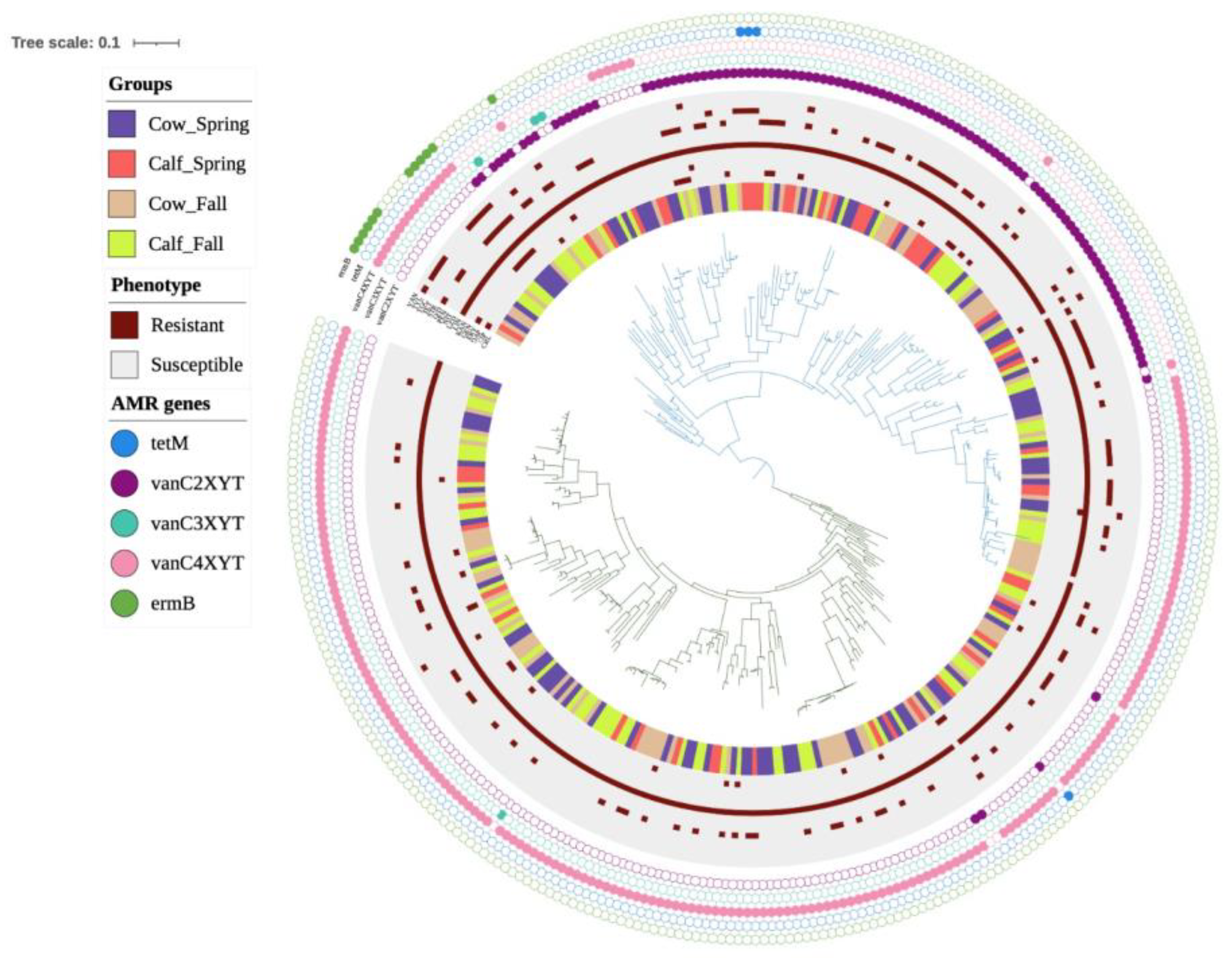
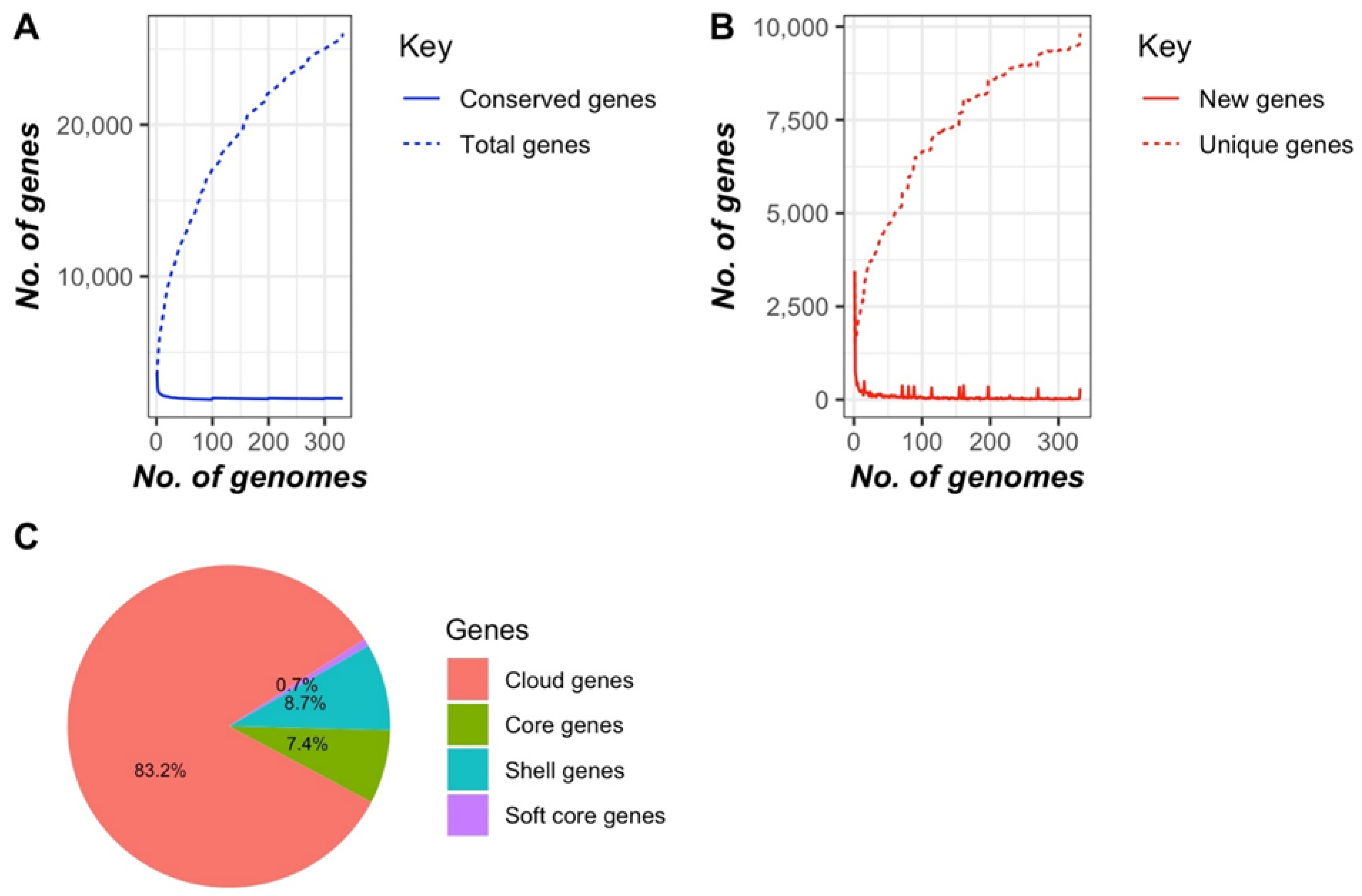
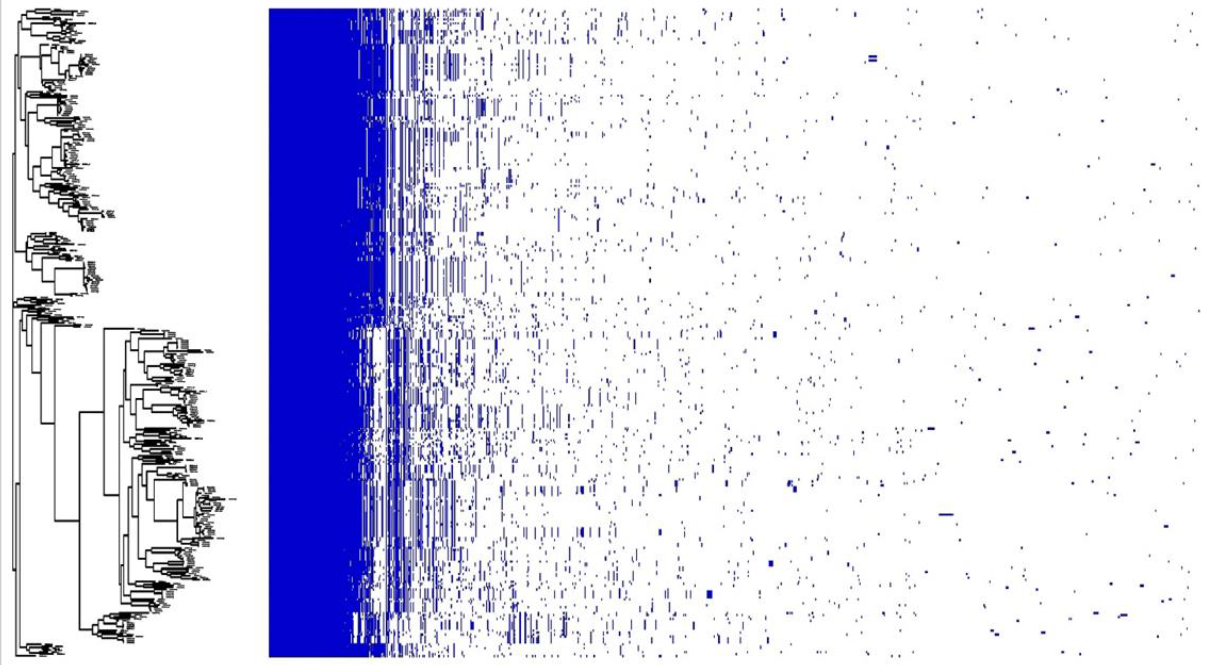
3.2. Genomic Characterization of E. casseliflavus

E. casseliflavus genomes ranged from 3,384,635 bp to 4,082,546 bp, with an average GC content of 42.4% (Table 1). The vancomycin vanC-type locus (vanC-vanXY-vanT) was present in all E. casseliflavus genomes, with vanC4XYT found in 67% (222/331) of the genomes. Other vanC variants, including vanC2XYT (31.4%, 104/331) and vanC3XYT (1.5%, 5/331), were also identified. All vanC resistance determinants were upstream from vanR and vanS, constituents of the two-component regulatory system for vancomycin resistance. The erythromycin resistance gene, ermB, was found in only 7 of 331 (2.1%) E. casseliflavus genomes. Mutations in the 23S RNA gene conferring erythromycin resistance were not found. Additionally, no known mutations in the parC or gyrA genes linked to ciprofloxacin resistance were identified in any of the E. casseliflavus genomes. Furthermore, no genes associated with quinupristin–dalfopristin, such as lsa [21] or vatE [22], were identified. The screening of genomes against the Virulence Finder database (VFDB) identified the ecbA/fss3 adhesion gene in 9% (30/331) of the E. casseliflavus genomes.
Plasmid profiling identified the presence of rep1, repUS1, repUS7, and repUS58 plasmids [23]. The repUS1 plasmid was found in 23% of genomes (77/331) from cows and calves in both seasons. In contrast, rep1 and repUS7 were found only once in the genome of an isolate recovered from a fall calf, with the repUS58 plasmid being associated with the genome of a single isolate obtained from a spring calf. No antimicrobial resistance genes were found on any of the identified plasmids.
The core genome-based phylogenomic tree formed two major clades (Figure 2). No segregation/clustering was observed based on the season or farm sampled or whether they originated from cows or calves. The nature of the E. casseliflavus pan-genome was assessed using Heap’s law (n = κNγ; [20]), where the model estimates the number of new gene clusters observed as genomes are randomly ordered. The decay parameter γ determines the openness of the pan-genome: if γ > 1.0, the pan-genome is considered closed, whereas if γ < 1.0, it is open. The analysis revealed an open pan-genome with a γ value of 0.3. Further analysis with Roary identified 1963 core genes (99 to 100% of strains), 184 soft core genes (95 to 99% of strains), 2243 shell genes (15 to 95% of strains), and 18,626 cloud genes (0 to 15% of strains) (Figure 3). The phylogenetic tree showed two distinct clusters, but this distinction did not appear to be related to the time, source or location of isolate collection (Figure 4).

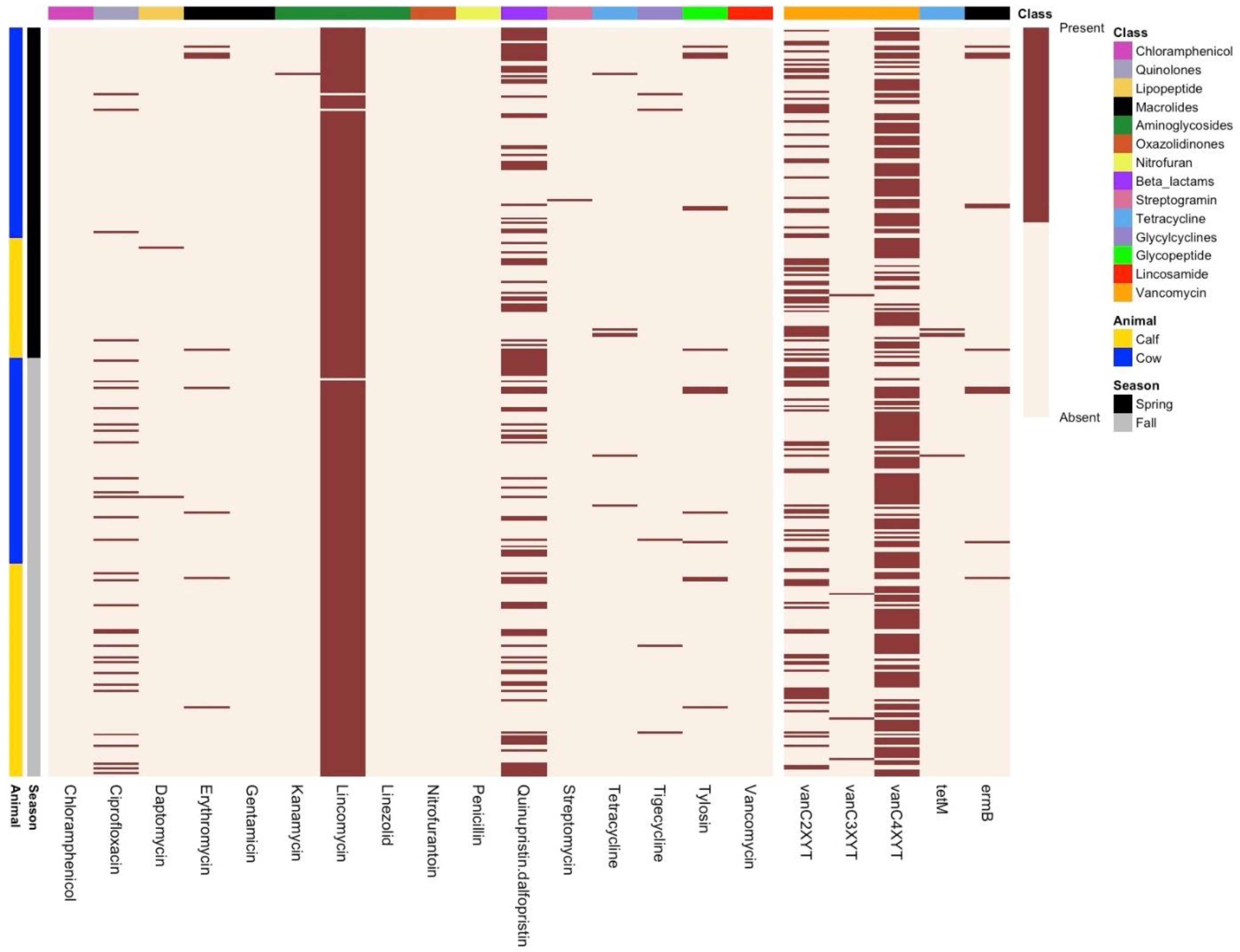
3.3. Phenotypic Resistance Profiling

A total of 331 E. casseliflavus isolates were tested for antimicrobial susceptibility against 16 different antimicrobials to investigate the correlation between phenotypic and genotypic AMR profiles (Figure 5). Across all isolates, 12% were multidrug-resistant (resistant to ≥3 antimicrobials). Most isolates were resistant to lincomycin (99%, 328/331), followed by quinupristin–dalfopristin (34%, 114/331), ciprofloxacin (9.6%, 32/331), tylosin (4.5%, 15/331), erythromycin (2.7%, 9/331), tetracycline (1.8%, 6/331), tigecycline (1.5%, 5/331), daptomycin (0.6%, 2/331), streptomycin (0.3%, 1/331), and kanamycin (0.3%, 1/331). None of the isolates exhibited resistance to chloramphenicol, penicillin, nitrofurantoin, gentamicin, or linezolid. Most isolates appeared susceptible to vancomycin with MIC values of ≤2 µg/mL (7.5%) and 4 µg/mL (65.5%), while 27% of the isolates exhibited intermediate resistance (MIC: 8 µg/mL).
Ciprofloxacin-resistant E. casseliflavus were more prevalent in cows (13.1%) and calves (17.0%) in the fall than the spring (3.2%—cows; 1.8%—calves). Daptomycin resistance was only found in a single isolate from spring cows (1.0%, 1/93) and a single isolate from fall cows (1.0%, 1/91). Likewise, streptomycin- and kanamycin-resistant isolates were rare in spring cows and calves (streptomycin, cows, 1.0%, 1/93; kanamycin calves 1.8%, 1/53). There were no differences between quinupristin–dalfopristin resistance in isolates collected from cows and calves or collected in the spring and fall.

4. Discussion

Members of the genus Enterococcus have been extensively used as indicator species of faecal contamination and in antimicrobial resistance surveillance studies [24,25,26]. Despite the ubiquitous presence of Enterococcus spp. across all sectors, including humans, animals, and the environment, previous studies have indicated niche specificity among Enterococcus species. Among all Enterococcus species, E. faecium and E. faecalis are frequently associated with nosocomial infections in humans [6,8,27], as well as being members of the normal gut microbiota of humans [28]. In contrast, E. hirae exists predominantly in feedlot cattle and swine [6,11]. Previous studies collecting isolates from cows and calves showed an increased prevalence of E. casseliflavus compared to feedlot cattle [12].
Originally isolated from a variety of plants [29], E. casseliflavus has since been recovered from animal faeces and environmental samples collected in multiple studies [30]. E. casseliflavus demonstrates unique ecological and functional roles, such as degrading organic pollutants like decabromodiphenyl ether (BDE-209), a common brominated flame retardant [31], and mediating the detoxification of the insecticide chlorantraniliprole in the agricultural pest Spodoptera frugiperda [32]. In aquaculture, E. casseliflavus acts as a probiotic in rainbow trout, enhancing gut health and disease resistance through immunomodulation [33]. Despite its predominant role as a plant and animal commensal, there are some reports of E. casseliflavus causing clinical infections in humans [10]. Compared to clinically significant Enterococcus spp. like E. faecium and E. faecalis, the prevalence of E. casseliflavus in clinical settings (>1.3% cases) is exceedingly low [34] and has only been rarely associated with bacteremia in humans [35,36]. In dairies, E. casseliflavus has been associated with subclinical mastitis in sheep and goats [37]. Despite the importance of E. casseliflavus from a One Health perspective, knowledge of its genomic structure remains limited. This study aims to bridge this gap.
Surveillance studies conducted in cow–calf herds in both Canada and the United States found an increased prevalence of E. casseliflavus compared to feedlot systems [12]. In contrast to intensive grain-based feedlot cattle production, cow–calf production uses more forages, where animals depend on grazing supplemented with hay and silage as the primary sources of feed. Since E. casseliflavus is known as a plant-associated enterococcal species, the higher prevalence of E. casseliflavus in cow–calf systems may be a reflection of frequent inoculation with E. casseliflavus from forages. In feedlot cattle, E. hirae predominates owing to the selective pressures associated with the feeding of grain-based diets [11].
There is a notable difference in antimicrobial usage in cow–calf vs. feedlot production systems. In cow–calf operations, the overall frequency of antimicrobial use is comparatively low, and antimicrobials are primarily used to treat disease in individual animals [12], whereas in feedlots, antimicrobials are frequently administered to prevent disease in the herd, either via injection or inclusion in feed [12,38,39]. In surveillance studies, enterococci serve as an indicator of antimicrobial resistance, and a previously high prevalence of tetracycline and macrolide resistance genes was found in Enterococcus hirae, the dominant species in feedlot cattle [6,11,40]. In contrast, the tetracycline resistance gene tetM and the macrolide resistance gene ermB were rarely detected in E. casseliflavus. This suggests that the low prevalence of tetracycline and macrolide resistance in cows and calves is a reflection of the limited use of these antimicrobials in this sector. Furthermore, it also strengthens the hypothesis that the prophylactic and metaphylactic use of tetracyclines and macrolides in feedlots, along with other environmental factors, promotes the prevalence of E. hirae in feedlot cattle. E. hirae has likely thrived in feedlot systems due to its acquisition of resistance to the antimicrobials commonly used in feedlot cattle. Moreover, E. hirae appears to have unique genes associated with vitamin production, cellulose, and pectin degradation, traits which may also support its adaptation to the bovine digestive tract.
Enterococcus casseliflavus typically exhibits low-level vancomycin resistance, with MICs ranging from 2 to 32 µg/mL, while remaining susceptible to teicoplanin (≤1 mg/L) [9,41]. This resistance is attributed to the intrinsic presence of the chromosomally encoded vanC gene cluster, a trait that is also present in E. flavescens and E. gallinarum [42]. The vanC cluster comprises five genes, vanC, vanXYc, vanT, vanR, and vanS [43], with vanC, vanXYc, and vanT being essential for resistance. VanC is a ligase that synthesizes d-Ala-d-Ser for alternative cell wall precursors, VanT supplies d-Ser as a membrane-bound serine racemase, and VanXYc combines the functions of the VanX and VanY enzymes to cleave d-Ala-d-Ala precursors. The remaining genes, vanR and vanS, form a two-component regulatory system. Unlike high-level glycopeptide resistance phenotypes (VanA, VanB, and VanD), which utilize d-Lac precursors, the VanC, VanE, and VanG phenotypes result from the replacement of d-Ala with d-Ser [44]. Although the resistance level conferred by the vanC cluster is lower than that of other vancomycin-resistant enterococci (VRE), this resistance could limit treatment options and complicate clinical management should E. casseliflavus play a more prominent role in nosocomial infections [7]. Due to this moderate resistance, the Clinical and Laboratory Standards Institute (CLSI) classifies Enterococcus species with vancomycin MICs below 4 mg/L as susceptible. However, vancomycin is not actively recommended for the treatment of E. casseliflavus bacteremia [9]. The accurate identification of E. casseliflavus is thus critically important for effective disease treatment, and misidentification can lead to inappropriate therapeutic strategies and potentially poor patient outcomes. From an AMU perspective, the misuse of vancomycin for treating E. casseliflavus infections may also expose other bacterial species to vancomycin, potentially resulting in the proliferation of vancomycin-resistant strains as result of selective pressure [35]. Variants of the vanC cluster [45,46] were found in all E. casseliflavus genomes in this study and harboured all five genes, vanC, vanXY, vanT, vanR, and vanS. Compared to vanA and vanB, which are often associated with mobile genetic elements, the vanC cluster was chromosomally encoded. This likely limits the transfer potential of vanC to other enterococcal species [47], an outcome that may contribute to the rarity or absence of vancomycin resistance in enterococci species isolated from feedlot cattle [6,11].
Enterococci are intrinsically tolerant to all beta-lactams and resistant to clinically achievable levels of aminoglycosides, lincosamides, and trimethoprim–sulfamethoxazole [21]. E. casseliflavus is a member of the E. gallinarum group, which exhibit intrinsic resistance to several antimicrobials. Nearly 98% of E. casseliflavus isolates were phenotypically resistant to lincomycin. Previously, lincomycin resistance was observed in 81% of E. casseliflavus isolates recovered from vegetables [48].
Virginiamycin, a streptogramin class of antibiotics, has been extensively used in human medicine and livestock, primarily as a growth promoter and to prevent liver abscesses. However, its use was discontinued in Europe in 1999 due to concerns over cross-resistance against the medically important antimicrobial combination quinupristin–dalfopristin (Q/D), used to treat vancomycin- and linezolid-resistant E. faecium infections [49,50]. A reduction in the use of Q/D in humans has led to a resurgence of the use of virginiamycin in livestock. Our study identified Q/D resistance in 34% of Enterococcus casseliflavus isolates, consistent with the findings from CIPARS, which reported a significant rise in streptogramin resistance among Enterococcus species, increasing from 15% to 35% since 2019. However, virginiamycin is not labelled for use in cow–calf herds as liver abscesses are not a common issue with grazing or high-forage-based diets, and its use was not reported in the herds participating in the present study [12]. Resistance to streptogramins is associated with antibiotic inactivation mediated by acetyltransferase encoded by vatD, vatE, and vgbA [51,52]. Despite the recovery of Q/D-resistant E. casseliflavus, no genes previously associated with Q/D resistance were detected in the E. casseliflavus genomes, raising the possibility that unknown ARGs or mutation(s) may be conferring this resistance. Further research to define the genetic factors responsible for Q/D resistance in E. casseliflavus is warranted.
Ciprofloxacin is a broad-spectrum fluoroquinolone that inhibits topoisomerases and thereby prevents DNA replication. In enterococci, ciprofloxacin resistance is associated with parC and gyrA gene mutations. In a previous study investigating ciprofloxacin resistance in E. casseliflavus, isolates with mutations in parC and gyrA did not exhibit phenotypic resistance. However, mutations in codon 80 in ParC (Ser to Leu) and in codon 87 in GyrA (Glu to Gly) resulted in E. casseliflavus isolates exceeding the MIC cut-off value (>8 µg/mL) for ciprofloxacin resistance [53]. In this study, ciprofloxacin resistance isolates had an MIC <8 µg/mL, and mutations in parC or gyrA were not identified. This agrees with previous observations that E. casseliflavus is less sensitive to ciprofloxacin as compared to other enterococcal species [53]. Unexplainably, we did find that E. casseliflavus recovered from cows and calves in the fall exhibited greater ciprofloxacin resistance than those isolated in the spring. This result was especially surprising given the infrequent use of fluoroquinolones within cow–calf herds [38].
Virulence factors in Enterococcus spp., apart from the clinically significant E. faecium and E. faecalis, are generally rare or absent. However, in E. casseliflavus, the fibrinogen-binding MSCRAMM gene (fss3) was identified. This gene belongs to a family encoding Microbial Surface Components Recognizing Adhesive Matrix Molecules (MSCRAMMs), which play a crucial role in adhesion, biofilm formation, and pathogenicity in enterococci. Previously, fss3 was also detected in E. hirae from bovine faeces and E. lacertideformus from wild Asian house geckos [54]. Surveillance studies have reported the presence of fss3 in clinical E. faecium and E. faecalis isolates but not in those isolated from beef cattle [6]. Comparative analyses suggests that virulence factors including fss3 are part of evolutionary adaptations that enable Enterococcus species to thrive in diverse environments and hosts [54,55].
Like other enterococcal species, the pan-genome of E. casseliflavus was open [56], with the accessory genome accounting for 81% of the genes in the pan-genome. A plot of unique versus new gene families showed that unique gene families are increasing within the pan-genome compared to new families. This suggests that E. casseliflavus possesses a diverse range of gene families. Its larger genome size (~3.7 Mb) compared to the opportunistic human pathogens E. faecalis (~2.7 Mb) and E. faecium (~2.7 Mb) and cattle-associated species E. hirae (~2.9 Mb) may be attributed to its environmental versatility and motility-related genes. E. casseliflavus is often found in diverse habitats such as plants, soil, and water, which likely requires a broader genetic repertoire to adapt to these environments. This broader adaptability involves additional genes for survival, motility, and metabolic versatility, reflecting its environmental and evolutionary distinctions [57]. The core genome-based phylogenetic tree formed two main clades with no segregation based on animal type, seasons, or farm location. The selective factors that contribute to the separation of these two clades remain elusive.
To summarize, E. casseliflavus is a prominent enterococcal species within the cow–calf sector, exhibiting notable genomic diversity among isolates. Consequently, it may be a more reliable indicator of AMR in cow herds than E. hirae, which tends to be the predominant species in feedlot cattle. Both phenotypic and genomic characterization suggest low-level resistance to key antimicrobials, including tetracycline and macrolides, which are routinely used in both human and animal medicine. While phenotypic studies identified moderate levels of resistance to quinupristin–dalfopristin in isolates from cows and calves across all time points and higher than expected levels of ciprofloxacin resistance in isolates from cows and calves in the fall, no genomic determinants were identified to account for these findings. Vancomycin resistance in E. casseliflavus was associated with a chromosomally located intrinsic resistance gene cluster, suggesting a limited role for this species in the spread of vancomycin resistance among enterococci in this sector.

Author Contributions

Concept and design of this study—C.W.; Sample and metadata collection—C.W. and J.F.; DNA isolation—C.W. and J.F; Assembly and analysis workflows—R.Z. and G.V.D.; Data analysis—R.Z. and S.-e.-Z.Z.; Interpretation of data—S.-e.-Z.Z., R.Z, C.W. and T.A.M.; Bioinformatics—S.-e.-Z.Z. and R.Z.; Manuscript draft preparation—S.-e.-Z.Z.; Manuscript review and editing—A.Z., R.Z., G.V.D., C.W., J.F. and T.A.M.; Funding acquisition—A.Z., C.W. and T.A.M. All authors have read and agreed to the published version of the manuscript.

Funding

The authors are grateful to the Genomics Research and Development Initiative AMR (GRDI-AMR) of the Government of Canada and the Beef Cattle Research Council (ANH.04.21) and NSERC for their financial support.

Institutional Review Board Statement

This project was approved by the University of Saskatchewan’s Animal Research Ethics Board (AREB) under the animal use protocol # 2014003 2024-03-01 and complies with Canadian Council on Animal Care (CCAC) guidelines, the University of Saskatchewan Animal Care and Use Procedures, and the Tri-Council MOU—Schedule 3: Ethical Review of Research Involving Animals.

Data Availability Statement

All Illumina sequence read data from the current study were deposited in the NCBI database as Short Read Archive (SRA) under BioProject ID PRJNA1191879.

Conflicts of Interest

The authors declare no conflicts of interest.

References

  1. Cameron, A.; McAllister, T.A. Antimicrobial usage and resistance in beef production. J. Anim. Sci. Biotechnol. 2016, 7, 68. [Google Scholar] [CrossRef] [PubMed]
  2. Manyi-Loh, C.; Mamphweli, S.; Meyer, E.; Okoh, A. Antibiotic Use in Agriculture and Its Consequential Resistance in Environmental Sources: Potential Public Health Implications. Molecules 2018, 4, 795. [Google Scholar] [CrossRef]
  3. Gilmore, M.S.; Clewell, D.B.; Ike, Y.; Shankar, N. Enterococci. In Enterococci: From Commensals to Leading Causes of Drug Resistant Infection; Gilmore, M.S., Clewell, D.B., Ike, Y., Shankar, N., Eds.; Massachusetts Eye and Ear Infirmary: Boston, MA, USA, 2014. [Google Scholar]
  4. Wheeler, A.L.; Hartel, P.G.; Godfrey, D.G.; Hill, J.L.; Segars, W.I. Potential of Enterococcus faecalis as a human fecal indicator for microbial source tracking. J. Environ. Qual. 2002, 31, 1286–1293. [Google Scholar] [CrossRef] [PubMed]
  5. Byappanahalli, M.N.; Nevers, M.B.; Korajkic, A.; Staley, Z.R.; Harwood, V.J. Enterococci in the environment. Microbiol. Mol. Biol. Rev. 2012, 76, 685–706. [Google Scholar] [CrossRef]
  6. Zaheer, R.; Cook, S.R.; Barbieri, R.; Goji, N.; Cameron, A.; Petkau, A.; Polo, R.O.; Tymensen, L.; Stamm, C.; Song, J.; et al. Surveillance of Enterococcus spp. reveals distinct species and antimicrobial resistance diversity across a One-Health continuum. Sci. Rep. 2020, 10, 3937. [Google Scholar] [CrossRef]
  7. García-Solache, M.; Rice, L.B. The Enterococcus: A Model of Adaptability to Its Environment. Clin. Microbiol. Rev. 2019, 32, e00058-18. [Google Scholar] [CrossRef]
  8. Fiore, E.; Van Tyne, D.; Gilmore, M.S. Pathogenicity of Enterococci. Microbiol. Spectr. 2019, 7. [Google Scholar] [CrossRef]
  9. Yoshino, Y. Enterococcus casseliflavus Infection: A Review of Clinical Features and Treatment. Infect. Drug Resist. 2023, 16, 363–368. [Google Scholar] [CrossRef] [PubMed]
  10. Jackson, C.R.; Lombard, J.E.; Dargatz, D.A.; Fedorka-Cray, P.J. Prevalence, species distribution and antimicrobial resistance of enterococci isolated from US dairy cattle. Lett. Appl. Microbiol. 2010, 52, 41–48. [Google Scholar] [CrossRef]
  11. Messele, Y.E.; Hasoon, M.F.; Trott, D.J.; Veltman, T.; McMeniman, J.P.; Kidd, S.P.; Low, W.Y.; Petrovski, K.R. Longitudinal Analysis of Antimicrobial Resistance among Enterococcus Species Isolated from Australian Beef Cattle Faeces at Feedlot Entry and Exit. Animals 2022, 12, 2690. [Google Scholar] [CrossRef]
  12. Fossen, J.D.; Campbell, J.R.; Gow, S.P.; Erickson, N.; Waldner, C.L. Antimicrobial resistance in Enterococcus isolated from western Canadian cow-calf herds. BMC Vet-Res. 2024, 20, 6. [Google Scholar] [CrossRef] [PubMed]
  13. Seemann, T. Prokka: Rapid prokaryotic genome annotation. Bioinformatics 2014, 30, 2068–2069. [Google Scholar] [CrossRef] [PubMed]
  14. Bharat, A.; Petkau, A.; Avery, B.P.; Chen, J.C.; Folster, J.P.; Carson, C.A.; Kearney, A.; Nadon, C.; Mabon, P.; Thiessen, J.; et al. Correlation between Phenotypic and In Silico Detection of Antimicrobial Resistance in Salmonella enterica in Canada Using Staramr. Microorganisms 2022, 10, 292. [Google Scholar] [CrossRef]
  15. Liu, B.; Zheng, D.; Jin, Q.; Chen, L.; Yang, J. VFDB 2019: A comparative pathogenomic platform with an interactive web interface. Nucleic Acids Res. 2019, 47, D687–D692. [Google Scholar] [CrossRef] [PubMed]
  16. Seemann, T. Abricate, Github. Available online: https://github.com/tseemann/abricate (accessed on 25 March 2025).
  17. Page, A.J.; Cummins, C.A.; Hunt, M.; Wong, V.K.; Reuter, S.; Holden, M.T.G.; Fookes, M.; Falush, D.; Keane, J.A.; Parkhill, J. Roary: Rapid large-scale prokaryote pan genome analysis. Bioinformatics 2015, 31, 3691–3693. [Google Scholar] [CrossRef]
  18. Letunic, I.; Bork, P. Interactive Tree of Life (iTOL) v6: Recent updates to the phylogenetic tree display and annotation tool. Nucleic Acids Res. 2024, 52, W78–W82. [Google Scholar] [CrossRef]
  19. Hadfield, J.; Croucher, N.J.; Goater, R.J.; Abudahab, K.; Aanensen, D.M.; Harris, S.R. Phandango: An interactive viewer for bacterial population genomics. Bioinformatics 2018, 34, 292–293. [Google Scholar] [CrossRef]
  20. Tettelin, H.; Riley, D.; Cattuto, C.; Medini, D. Comparative genomics: The bacterial pan-genome. Curr. Opin. Microbiol. 2008, 11, 472–477. [Google Scholar] [CrossRef]
  21. Kristich, C.J.; Rice, L.B.; Arias, C.A. Enterococcal Infection-Treatment and Antibiotic Resistance. In Enterococci: From Commensals to Leading Causes of Drug Resistant Infection; Gilmore, M.S., Clewell, D.B., Ike, Y., Shankar, N., Eds.; Massachusetts Eye and Ear Infirmary: Boston, MA, USA, 2014. [Google Scholar]
  22. Soltani, M.; Beighton, D.; Philpott-Howard, J.; Woodford, N. Mechanisms of resistance to quinupristin-dalfopristin among isolates of Enterococcus faecium from animals, raw meat, and hospital patients in Western Europe. Antimicrob. Agents Chemother. 2000, 44, 433–436. [Google Scholar] [CrossRef]
  23. Carattoli, A.; Zankari, E.; Garcìa-Fernandez, A.; Voldby Larsen, M.; Lund, O.; Villa, L.; Moller Aarestrup, F.; Hasman, H. In silico detection and typing of plasmids using PlasmidFinder and plasmid multilocus sequence typing. Antimicrob. Agents Chemother. 2014, 58, 3895–3903. [Google Scholar] [CrossRef]
  24. Himanshu, R.; Prudencio, C.; da Costa, A.C.; Leal, E.; Chang, C.-M.; Pandey, R.P. Systematic Surveillance and Meta-Analysis of Antimicrobial Resistance and Food Sources from China and the USA. Antibiotics 2022, 11, 1471. [Google Scholar] [CrossRef]
  25. Marques, J.M.; Coelho, M.; Santana, A.R.; Pinto, D.; Semedo-Lemsaddek, T. Dissemination of Enterococcal Genetic Lineages: A One Health Perspective. Antibiotics 2023, 12, 1140. [Google Scholar] [CrossRef]
  26. Osman, M.; Altier, C.; Cazer, C. Antimicrobial resistance among canine enterococci in the northeastern United States, 2007–2020. Front. Microbiol. 2022, 13, 1025242. [Google Scholar] [CrossRef]
  27. Prieto, A.M.G.; van Schaik, W.; Rogers, M.R.C.; Coque, T.M.; Baquero, F.; Corander, J.; Willems, R.J.L. Global Emergence and Dissemination of Enterococci as Nosocomial Pathogens: Attack of the Clones? Front. Microbiol. 2016, 7, 788. [Google Scholar] [CrossRef]
  28. Ramos, S.; Silva, V.; Dapkevicius, M.d.L.E.; Igrejas, G.; Poeta, P. Enterococci, from Harmless Bacteria to a Pathogen. Microorganisms 2020, 8, 1118. [Google Scholar] [CrossRef]
  29. Mundt, J.O.; Graham, W.F. Streptococcus faecium var. casseliflavus, nov. var. J. Bacteriol. 1968, 95, 2005–2009. [Google Scholar] [CrossRef]
  30. Badgley, B.D.; Thomas, F.I.M.; Harwood, V.J. The effects of submerged aquatic vegetation on the persistence of environmental populations of Enterococcus spp. Environ. Microbiol. 2010, 12, 1271–1281. [Google Scholar] [CrossRef] [PubMed]
  31. Tang, S.; Yin, H.; Chen, S.; Peng, H.; Chang, J.; Liu, Z.; Dang, Z. Aerobic degradation of BDE-209 by Enterococcus casseliflavus: Isolation, identification and cell changes during degradation process. J. Hazard. Mater. 2016, 308, 335–342. [Google Scholar] [CrossRef] [PubMed]
  32. Zhang, Y.; Ju, F. Uninheritable but Widespread Bacterial Symbiont Enterococcus casseliflavus Mediates Detoxification of the Insecticide Chlorantraniliprole in the Agricultural Invasive Pest Spodoptera frugiperda. J. Agric. Food Chem. 2024, 72, 18365–18377. [Google Scholar] [CrossRef]
  33. Safari, R.; Adel, M.; Lazado, C.C.; Caipang, C.M.; Dadar, M. Host-derived probiotics Enterococcus casseliflavus improves resistance against Streptococcus iniae infection in rainbow trout (Oncorhynchus mykiss) via immunomodulation. Fish Shellfish. Immunol. 2016, 52, 198–205. [Google Scholar] [CrossRef]
  34. Liu, Y.; Liu, K.; Lai, J.; Wu, C.; Shen, J.; Wang, Y. Prevalence and antimicrobial resistance of Enterococcus species of food animal origin from Beijing and Shandong Province, China. J. Appl. Microbiol. 2013, 114, 555–563. [Google Scholar] [CrossRef] [PubMed]
  35. Tan, C.-K.; Lai, C.-C.; Wang, J.-Y.; Lin, S.-H.; Liao, C.-H.; Huang, Y.-T.; Wang, C.-Y.; Lin, H.-I.; Hsueh, P.-R. Bacteremia caused by non-faecalis and non-faecium enterococcus species at a Medical center in Taiwan, 2000 to 2008. J. Infect. 2010, 61, 34–43. [Google Scholar] [CrossRef] [PubMed]
  36. Toc, D.A.; Pandrea, S.L.; Botan, A.; Mihaila, R.M.; Costache, C.A.; Colosi, I.A.; Junie, L.M. Enterococcus raffinosus, Enterococcus durans and Enterococcus avium isolated from a tertiary care hospital in Romania—Retrospective study and brief review. Biology 2022, 11, 598. [Google Scholar] [CrossRef]
  37. El-Zamkan, M.A.; Mohamed, H.M.A. Antimicrobial resistance, virulence genes and biofilm formation in Enterococcus species isolated from milk of sheep and goat with subclinical mastitis. PLoS ONE 2021, 16, e0259584. [Google Scholar] [CrossRef]
  38. Fossen, J.D.; Campbell, J.R.; Gow, S.P.; Erickson, N.; Waldner, C.L. Antimicrobial Use in Canadian Cow–Calf Herds. Vet. Sci. 2023, 10, 366. [Google Scholar] [CrossRef] [PubMed]
  39. Brault, S.A.; Hannon, S.J.; Gow, S.P.; Warr, B.N.; Withell, J.; Song, J.; Williams, C.M.; Otto, S.; Booker, C.W.; Morley, P.S. Antimicrobial Use on 36 Beef Feedlots in Western Canada: 2008–2012. Front. Vet. Sci. 2019, 6, 329. [Google Scholar] [CrossRef]
  40. Zaheer, R.; Cook, S.R.; Klima, C.L.; Stanford, K.; Alexander, T.; Topp, E.; Read, R.R.; McAllister, T.A. Effect of subtherapeutic vs. therapeutic administration of macrolides on antimicrobial resistance in Mannheimia haemolytica and enterococci isolated from beef cattle. Front. Microbiol. 2013, 4, 133. [Google Scholar] [CrossRef]
  41. Courvalin, P. Vancomycin resistance in gram-positive cocci. Clin. Infect. Dis. 2006, 42 (Suppl. S1), S25–S34. [Google Scholar] [CrossRef]
  42. Navarro, F.; Courvalin, P. Analysis of genes encoding D-alanine-D-alanine ligase-related enzymes in Enterococcus casseliflavus and Enterococcus flavescens. Antimicrob. Agents Chemother. 1994, 38, 1788–1793. [Google Scholar] [CrossRef]
  43. Arias, C.A.; Courvalin, P.; Reynolds, P.E. vanC cluster of vancomycin-resistant Enterococcus gallinarum BM4174. Antimicrob. Agents Chemother. 2000, 44, 1660–1666. [Google Scholar] [CrossRef]
  44. Dutta, I.; Reynolds, P.E. Biochemical and genetic characterization of the vanC-2 vancomycin resistance gene cluster of Enterococcus casseliflavus ATCC 25788. Antimicrob. Agents Chemother. 2002, 46, 3125–3132. [Google Scholar] [CrossRef]
  45. Clark, N.C.; Teixeira, L.M.; Facklam, R.R.; Tenover, F.C. Detection and differentiation of vanC-1, vanC-2, and vanC-3 glycopeptide resistance genes in enterococci. J. Clin. Microbiol. 1998, 36, 2294–2297. [Google Scholar] [CrossRef] [PubMed]
  46. Dutta, I.; Reynolds, P.E. The vanC-3 vancomycin resistance gene cluster of Enterococcus flavescens CCM 439. J. Antimicrob. Chemother. 2003, 51, 703–706. [Google Scholar] [CrossRef]
  47. Flipse, J.; von Wintersdorff, C.J.H.; van Niekerk, J.M.; Jamin, C.; van Tiel, F.H.; Hasman, H.; van Alphen, L.B. Appearance of vanD-positive Enterococcus faecium in a tertiary hospital in the Netherlands: Prevalence of vanC and vanD in hospitalized patients. Sci. Rep. 2019, 9, 6949. [Google Scholar] [CrossRef] [PubMed]
  48. McGowan, L.L.; Jackson, C.R.; Barrett, J.B.; Hiott, L.M.; Fedorka-Cray, P.J. Prevalence and antimicrobial resistance of enterococci isolated from retail fruits, vegetables, and meats. J. Food Prot. 2006, 69, 2976–2982. [Google Scholar] [CrossRef] [PubMed]
  49. Bryson, H.M.; Spencer, C.M. Quinupristin-dalfopristin. Drugs 1996, 52, 406–415. [Google Scholar] [CrossRef]
  50. Menzies-Gow, N.; Young, N. Antibiotic resistance in faecal bacteria isolated from horses receiving virginiamycin for the prevention of pasture-associated laminitis. Vet. Microbiol. 2011, 152, 424–428. [Google Scholar] [CrossRef]
  51. Schwarz, S.; Shen, J.; Kadlec, K.; Wang, Y.; Michael, G.B.; Feßler, A.T.; Vester, B. Lincosamides, Streptogramins, Phenicols, and Pleuromutilins: Mode of Action and Mechanisms of Resistance. Cold Spring Harb. Perspect. Med. 2016, 6, a027037. [Google Scholar] [CrossRef]
  52. Simjee, S.; White, D.G.; Wagner, D.D.; Meng, J.; Qaiyumi, S.; Zhao, S.; McDermott, P.F. Identification of vat (E) in Enterococcus faecalis isolates from retail poultry and its transferability to Enterococcus faecium. Antimicrob. Agents Chemother. 2002, 46, 3823–3828. [Google Scholar] [CrossRef]
  53. Petersen, A.; Jensen, L.B. Analysis of gyrA and parC mutations in enterococci from environmental samples with reduced susceptibility to ciprofloxacin. FEMS Microbiol. Lett. 2004, 231, 73–76. [Google Scholar] [CrossRef]
  54. Agius, J.E.; Phalen, D.N.; Rose, K.; Eden, J.-S. Genomic Insights Into the Pathogenicity of a Novel Biofilm-Forming Enterococcus sp. Bacteria (Enterococcus lacertideformus) Identified in Reptiles. Front. Microbiol. 2021, 12, 635208. [Google Scholar] [CrossRef] [PubMed]
  55. Lee, T.; Pang, S.; Abraham, S.; Coombs, G.W. Molecular characterization and evolution of the first outbreak of vancomycin-resistant Enterococcus faecium in Western Australia. Int. J. Antimicrob. Agents 2019, 53, 814–819. [Google Scholar] [CrossRef] [PubMed]
  56. Zhong, Z.; Zhang, W.; Song, Y.; Liu, W.; Xu, H.; Xi, X.; Menghe, B.; Zhang, H.; Sun, Z. Comparative genomic analysis of the genus Enterococcus. Microbiol. Res. 2017, 196, 95–105. [Google Scholar] [CrossRef]
  57. Palmer, K.L.; Godfrey, P.; Griggs, A.; Kos, V.N.; Zucker, J.; Desjardins, C.; Cerqueira, G.; Gevers, D.; Walker, S.; Wortman, J.; et al. Comparative Genomics of Enterococci: Variation in Enterococcus faecalis, clade structure in E. faecium, and defining characteristics of E. gallinarum and E. casseliflavus. mBio 2012, 3, e00318-11. [Google Scholar] [CrossRef] [PubMed]
Figure 1. The prevalence of Enterococcus species isolated from 1505 cows and calves (743 cows, 762 calves) originating from 39 herds in the spring and fall of 2021 [12].
Microorganisms 13 00907 g001
Figure 2. Core genome neighbour-joining phylogenomic tree of Enterococcus casseliflavus genomes (n = 331) isolated from cows and calves in spring or fall. E. casseliflavus EC20 (GenBank accession # CP004856.1) was used as reference genome.
Microorganisms 13 00907 g002
Figure 3. Pan-genome of Enterococcus casseliflavus (n = 331) (A) Conserved and total genes, (B) New and unique genes and (C) Overall pan-genome, Development of pan- and core genomes was based on conserved genes and presence of new/unique genes, illustrating the open nature of pan-genome.
Microorganisms 13 00907 g003
Figure 4. Phylogenetic tree generated based on presence and type of accessory genes in isolates and corresponding heat map representing absence or presence of genes in isolates.
Microorganisms 13 00907 g004
Figure 5. Antimicrobial resistance profiles of Enterococcus casseliflavus isolated and confirmed by whole genome sequencing from faeces collected from cows and calves in spring or fall (n = 331).
Microorganisms 13 00907 g005
Table 1. Assembly statistics of 331 E. casseliflavus genomes isolated from cattle 1.
Assembly StatsAverage Value
N50 contig length (bp)406,085
Number of contigs35 (range: 8–114)
Number of contigs >= 1 kb28
Number of contigs in N504
Genome size (bp)3,674,039
GC content of contigs42.4%
1 Sequence data for bacterial isolates are available at NCBI via BioProject PRJNA1191879.
Disclaimer/Publisher’s Note: The statements, opinions and data contained in all publications are solely those of the individual author(s) and contributor(s) and not of MDPI and/or the editor(s). MDPI and/or the editor(s) disclaim responsibility for any injury to people or property resulting from any ideas, methods, instructions or products referred to in the content.

