Article

Inactivation of Genes Encoding MutL and MutS Proteins Influences Adhesion and Biofilm Formation by Neisseria gonorrhoeae

by
Jagoda Płaczkiewicz
1,
Monika Adamczyk-Popławska
1,
Robert Lasek
1,
Pawel Bącal
2,3 and
Agnieszka Kwiatek
1,*
1
Institute of Microbiology, Faculty of Biology, University of Warsaw, Miecznikowa 1, 02-096 Warsaw, Poland
2
Faculty of Chemistry, University of Warsaw, Pasteura 1, 02-093 Warsaw, Poland
3
Nalecz Institute of Biocybernetics and Biomedical Engineering, Polish Academy of Sciences, 02-109 Warsaw, Poland
*
Author to whom correspondence should be addressed.
Microorganisms 2019, 7(12), 647; https://doi.org/10.3390/microorganisms7120647
Submission received: 23 October 2019 / Revised: 29 November 2019 / Accepted: 2 December 2019 / Published: 4 December 2019
(This article belongs to the Section Molecular Microbiology and Immunology)

Abstract

:
Neisseria gonorrhoeae is an etiological agent of gonorrhea, which remains a global health problem. This bacterium possesses MutL and MutS DNA repair proteins encoded by mutL and mutS genes, whose inactivation causes a mutator phenotype. We have demonstrated the differential gene expression in N. gonorrhoeae mutL and mutS mutants using DNA microarrays. A subset of differentially expressed genes encodes proteins that can influence adhesion and biofilm formation. Compared to the wild-type strain, N. gonorrhoeae mutL and mutS mutants formed denser biofilms with increased biofilm-associated biomass on the abiotic surface. The N. gonorrhoeae mutS::km, but not the mutL mutant, was also more adherent and invasive to human epithelial cells. Further, during infection of epithelial cells with N. gonorrhoeae mutS::km, the expression of some bacterial genes encoding proteins that can influence gonococcal adhesion was changed compared with their expression in cells infected with the wild-type gonococcus, as well as of human genes’ encoding receptors utilized by N. gonorrhoeae (CD46, CEACAM 1, HSPG 2). Thus, deficiency in the mutS gene resulting in increased mutation frequency in singular organisms can be beneficial in populations because these mutants can be a source of features linked to microbial fitness.

1. Introduction

Neisseria gonorrhoeae (gonococcus) is a human-specific, gram-negative diplococcus that causes the sexually transmitted disease gonorrhea. Gonococcal infections remain a global health issue, as highlighted by the World Health Organization, which has recently classified N. gonorrhoeae as a “priority pathogen” due to spreading multidrug resistance and increasing infection incidence [1,2]. Additionally, gonococcal infection may increase the risk of human immunodeficiency virus transmission [3,4,5]. During infection, N. gonorrhoeae interacts with host epithelial cells using multistep adhesion as the key stage of infection, allowing biofilm formation, which is an important virulence factor [6,7].
DNA repair systems are crucial in all living organisms. MutL and MutS proteins are conserved DNA repair molecules present at all domains of life. MutL is a “molecular matchmaker”, which mediates the protein–protein interactions during mismatch recognition and strand removal, and MutS recognizes unpaired and mispaired bases and small insertion/deletion loops in duplex DNA [8]. In N. gonorrhoeae, inactivation of mutL and mutS genes leads to an overall increase in mutability. Thus far, it has been demonstrated that gonococcal MutL and MutS proteins remove mononucleotide mismatches and insertion-deletion loops, indicating their engagement in the methyl-directed mismatch repair system (MMR) and affect the activity of Vsr endonucleases, implying a role in the Very Short Patch repair system [9,10]. Furthermore, in addition to the engagement of MutL and MutS proteins in DNA repair systems, they limit the frequency of RecA-dependent gene conversion events at the pilE gene, and MutS protein prevents recombination between partially homologous sequences [9,11].
There are data indicating that besides maintaining the integrity of the genome, DNA repair systems can also play broader functions affecting the intraspecific variation of strains, host defense avoidance mechanisms, improved adaption to changing environmental conditions, and virulence [12,13,14]. In Neisseria meningitidis, in addition to repairing DNA damage [15], the MMR system plays an important role in infection, as it was demonstrated that epidemic N. meningitidis serogroup A isolates were found to be strains with an increased frequency of mutations caused by defects in the MMR [16]. Further, the meningococcal mutS mutant can be regarded as more pathogenic, because it has higher ability to escape from the bactericidal activity of monoclonal antibodies [17]. Whereas for Escherichia coli, Salmonella spp., or Pseudomonas aeruginosa, the occurrence of mutator strains in population was demonstrated [18,19,20].
To date, there are no data on the influence of disruption of mutL or mutS genes on processes other than DNA repair in N. gonorrhoeae. Therefore, the goal of this work was to evaluate the influence of the disruption of mutL and mutS genes on processes connected with gonococcal infection, specifically on biofilm formation and interactions with human cells, detailed on adhesion and invasiveness to epithelial cells. Our results indicated that N. gonorrhoeae with a disrupted mutS gene formed denser biofilms and was more adherent and invasive to human epithelial cells compared to the wild-type strain.

2. Materials and Methods

2.1. Bacterial Strains and Growth Conditions

N. gonorrhoeae FA1090 and its derivatives were used in the study. N. gonorrhoeae FA1090 (ATCC® 700825) was originally isolated from the endocervix of a woman with a probable disseminated gonococcal infection in 1981 [21]. The strain is well-characterized, serum-resistant, streptomycin-resistant, and proline-requiring. N. gonorrhoeae strain FA1090 is extensively used in studies of gonococcal pathogenesis, as it is the first gonococcal strain for which the entire genome sequence was determined (NC_002946.2).
N. gonorrhoeae FA1090 and its derivatives were grown at 37 °C on a GC agar base (Difco, Detroit, MI, USA) supplemented with 1% Kellogg’s supplement in 5% CO2, and depending on the experiment, with or without 1% hemoglobin or cultivated in GC broth supplemented with 1% Kellogg’s supplement and 0.043% NaHCO3 [22]. When needed, media were supplemented with 30 μg/mL kanamycin or/and 2 μg/mL chloramphenicol.
For all experiments, gonococci exhibiting the same piliation and opaque phenotype were used as was determined by microscopy using stereo dissecting microscope (PZO Mikroskopy, Warsaw, Poland) observation in accordance with the principles described by Dillard [22].
Each set of experiments has always included: N. gonorrhoeae wild-type, N. gonorrhoeae mutL::km, and N. gonorrhoeae mutS::km mutants, complementation gonococci: N. gonorrhoeae mutL::km + mutLwt and N. gonorrhoeae mutS::km + mutSwt.

2.2. Cell Lines and Culture Conditions

The human epithelial cell line HEC-1-B is one of the models for infection of human tissue culture cells by N. gonorrhoeae [23]. The use of HEC-1-B cells for the study of gonococcal infection of epithelial cells has already been described [24,25,26,27,28]. The epithelial cell line HEC-1-B (ATCC: HTB113) was grown in DMEM medium (Biowest, Nuaillé, France) supplemented with 10% fetal bovine serum (FBS) (Biowest) (v/v), 2 mM l-glutamine, 1 mM sodium pyruvate and, when necessary, with 100 µg/mL penicillin/streptomycin (Biological Industries, Cromwell, CT, USA) or 2.5 µg/mL fungizone (Biological Industries) at 37 °C and 5% CO2. Confluence was determined by visual examination.

2.3. Construction of Neisseria gonorrhoeae (N. gonorrhoeae) Mutants

Construction of N. gonorrhoeae mutL::km and N. gonorrhoeae mutS::km mutants along with the obtained complementation strains N. gonorrhoeae mutL::km + mutLwt and N. gonorrhoeae mutS::km + mutSwt, has been previously detailed described in Adamczyk-Popławska et al. [10].

2.4. Field Emission Scanning Electron Microscopy (FE SEM)

Field emission scanning electron microscopy (FE SEM) (Carl Zeiss, Oberkochen, Germany) was performed as described previously with slight modifications [27,28,29]. First, bacterial suspensions (107 cells/mL) were made (in GC broth to study biofilm on abiotic surface or in DMEM to visualization of N. gonorrhoeae attached to human epithelial cells) and then were added: (i) to coverslips placed in Petri dishes, and bacteria were cultivated for 24 h at 37 °C in 5% CO2 for study of N. gonorrhoeae biofilm formation on abiotic surface, or (ii) to human epithelial cells (m.o.i 1:100) seeded on coverslips for 4 h at 37 °C in 5% CO2 for study of N. gonorrhoeae attached to epithelial cells. Visualization of biofilms formed on abiotic surface or of N. gonorrhoeae attached to epithelial cells was performed as described in Kwiatek et al. [27,28].

2.5. Scanning Confocal Laser Microscopy (SCLM)

Scanning confocal laser microscopy (SCLM) was performed as described previously with slight modifications [27,28,29]. First, bacterial suspensions (107 cells/mL) were made (in GC broth to study biofilm on abiotic surface or in DMEM to visualization of N. gonorrhoeae attached to human epithelial cells). Then: (i) bacterial suspensions were added to glass bottom microwell dishes (MatTek Corporation, Ashland, MA, USA) and bacterial growth was continued at 37 °C in 5% CO2 for 24 h for study of N. gonorrhoeae biofilm formation on abiotic surface, or (ii) to human epithelial cells (m.o.i 1:100) seeded on coverslips for 4 h at 37 °C in 5% CO2 for study of N. gonorrhoeae attached to epithelial cells. Next, N. gonorrhoeae biofilm formed on abiotic surface was washed with 10 mM MgSO4, stained for 30 min with acridine orange solution (10 µg/mL in 10 mM MgSO4) and rinsed with 10 mM MgSO4. Whereas, to investigate N. gonorrhoeae attached to human cells, after 4 h infection, epithelial cells were stained with a CellTracker Orange CMTMR (1:5000 dilution) (Thermo Fisher Scientific, Waltham, MA, USA) and N. gonorrhoeae was detected with Anti-Neisseria gonorrhoeae antibody (1:200) (Abcam, Cambridge, UK) as primary antibodies and Goat Anti-Mouse IgG H and L (1:500) conjugated with Alexa Fluor® 488 (green-fluorescent dye) (Abcam) as secondary antibodies.
SCLM was performed using a Nikon Eclipse Ti (A1) microscope (Nikon, Tokyo, Japan) equipped with a ×60, 1.4 NA oil immersion phase-contrast lens and an argon laser with a maximum emission line at 565 nm for CellTracker Orange CMTMR and at 519 nm for Alexa Fluor® 488. Horizontal optical thin sections were collected at 0.21 µm intervals from the outer surface of the biofilm or epithelial cells to the bottom of the glass plate. NIS-ELEMENTS interactive software was used for capture and three-dimensional (3D) reconstructions of images.

2.6. The Crystal Violet Assay

The crystal violet assay was performed as described previously [27,28,29,30] with slight modifications. Briefly, gonococcal cell suspensions (107 cells/mL in GC broth) were added into a 96-well microtiter plate, and bacteria were grown without shaking for 24 h at 37 °C in 5% CO2. Then, the amounts of planktonic cells in each well was assessed by measuring OD600 followed by aspiration and removal of planktonic cells. The remaining attached bacteria were fixed with ethanol, air-dried, and stained with 0.8% crystal violet dye. Excess stain was rinsed off, samples were air-dried, and the crystal violet dye bound to the adherent cells was resolubilized with 96% ethanol. The amount of adhered bacterial cells stained by crystal violet was measured at 570 nm using an automated Sunrise microplate reader and Magellan software (Magellan 7.2, Tecan, Männedorf, Switzerland).

2.7. RNA Extraction

RNA was extracted from (i) human epithelial cells not infected by N. gonorrhoeae, (ii) human epithelial cells infected by N. gonorrhoeae as done in the adhesion assay (4 h infection), (iii) N. gonorrhoeae cultivated on a GC agar base for 24 h, and (iv) N. gonorrhoeae infecting human epithelial cells (4 h infection). Total RNA from the above samples was extracted using a High Pure RNA Isolation Kit (Roche, Basel, Switzerland). RNA from N. gonorrhoeae infecting human epithelial cells was separated from mixed samples containing human and bacterial RNA using a MICROBEnrich™ Kit (Thermo Fisher Scientific). Contaminating DNA was removed using DNA-free™, DNase Treatment and Removal (Thermo Fisher Scientific). RNA quality was measured using a 2100 Bioanalyzer (Agilent Technologies, Santa Clara, CA, USA), and RNA with a RIN (RNA integrity number) >8.0 was used.

2.8. Microarray Experiments

Detailed protocols and specifications for DNA microarray analysis and array analysis are described in Kwiatek et al. [27,28]. Briefly, for the microarray experiments, an Agilent custom-designated 60-mer oligonucleotide array (Agilent-034141) was used. RNA was extracted according to the protocol described above. Further manipulations with RNA (amplification and labeling whole transcripts with Cy3 and Cy5) were performed using the Low Input Quick Amp Labeling WT Kit (Agilent Technologies). The Cy3 dye was used to label the wild-type cRNA, and the Cy5 dye was used to label the cRNA of the N. gonorrhoeae mutants. To monitor the microarray workflow, as an internal control, the RNA Spike in Kit for Two Color v4.0 (Agilent Technologies) was used. Equal amounts of Cy5- and Cy3-labeled cRNAs were hybridized onto the microarray for 17 h at 65 °C using the Gene Expression Hybridization Kit (Agilent Technologies) followed by microarray washing with the Gene Expression Wash Buffer Kit (Agilent Technologies). Microarray slides were scanned with an Agilent G2565CA Microarray Scanner System (Agilent Technologies).

2.9. Array Analysis

Array analysis was performed using GeneSpring version 12.5 (Agilent Technologies). Normalized data were filtered on genes flagged as present or marginal, with the resulting gene list used for further gene expression analysis and clustering (Student’s t-test against zero, asymptotic p value computation, and Benjamini-Hochberg FDR multiple testing correction). Changes were expressed as the ratio of gene expression of the wild-type FA1090 strain over the expression of the same gene for the mutant. Genes with differential expression of ≥1.5-fold and a p value < 0.05 were selected. Supporting microarray data for all studied gonococcal mutants have been deposited in the NCBI’s Gene Expression Omnibus and are accessible through the GEO Series accession number GSE61789.

2.10. RT-qPCR

RT-qPCR experiments were performed according to the Minimum Information for Publication of Quantitative Real-Time PCR Experiments guidelines [31]. Bacterial cDNAs were obtained by reverse transcription of 10 µg of total RNA using the Maxima First Strand cDNA Synthesis Kit for RT-qPCR and human cDNAs using the RevertAid First Strand cDNA Synthesis Kit (Thermo Fisher Scientific). RT-qPCR using 1× HOT FIREPol® EvaGreen® qPCR Mix Plus (ROX) (Solis BioDyne, Tartu, Estonia) was performed on a LightCycler®96 system (Roche, Basel, Switzerland) (95 °C for 900 s, 40 cycles of 95 °C for 15 s, 60 °C for 30 s, and 72 °C for 20 s). Oligonucleotide primer pairs (HPLC purified) specific for each bacterial gene of interest were designed using PrimerQuest software (Primer 3 2.2.3, Cambridge, MA, USA) hosted by Integrated DNA Technologies and purchased from Sigma-Aldrich (Saint Louis, MO, USA). Oligonucleotide primer pairs specific for each human gene of interest were obtained from Bio-Rad (Hercules, CA, USA) or Thermo Fisher Scientific (Table S1). Relative quantification of gene expression was performed using the comparative ΔΔCt (threshold cycle) method. The relative amount of target cDNA was normalized using the 16S rRNA gene as an internal reference standard for bacterial gene expression and both the Hypoxanthine phosphoribosyltransferase 1 (HPRT 1) gene and Beta-2-microglobulin gene for human gene expression.

2.11. Adhesion Assay

The adhesion assay was performed as described previously [27,28,32]. Briefly, confluent epithelial cells were infected by N. gonorrhoeae (m.o.i. 1:100, bacterial suspension (107 cells/mL) made in DMEM) for 4 h at 37 °C in 5% CO2. Next, one set of the N. gonorrhoeae-infected epithelial cells was washed with PBS, lysed by 0.5% saponin, and lysates were plated on GC agar base with 1% hemoglobin to enumeration of CFU of cell-associated gonococci. At the same time, another set of the N. gonorrhoeae-infected epithelial cells wasn’t rinsed with PBS, but only culture medium from the above cells was removed, following this, epithelial cells were lysed by 0.5% saponin and plated on GC agar base with 1% hemoglobin to enumeration of CFU. The sum of CFU from the medium supernatant and from the cell lysates constitutes total CFU. The adhesion index was calculated by dividing the number of cell-associated CFU by the number of total CFU.

2.12. Invasion Assay

An invasion assay was performed as described previously with slight modifications [27,28,32]. Briefly, confluent epithelial cells were infected by N. gonorrhoeae (m.o.i. 1:100, bacterial suspension (107 cells/mL) made in DMEM) for 4 h at 37 °C in 5% CO2, as for adhesion assays. Then, a set of the N. gonorrhoeae-infected epithelial cells was incubated with 0.2 mg/mL gentamicin for 1 h at 37 °C to kill extracellular gonococci and further lysed by 1% saponin to release intracellular gonococci. The lysates were plated on a GC agar base with 1% hemoglobin to enable enumeration of intracellular (gentamicin-resistant (GmR)) N. gonorrhoeae. At the same time, another set of the N. gonorrhoeae-infected epithelial cells was processed for calculation of N. gonorrhoeae total CFU, as was described for the adhesion assay. The invasion index was calculated by dividing the number of GmR CFU by total CFU.

2.13. Computer Analysis, In Silico Analysis of Metabolic Pathways, and Determination of Clusters of Orthologous Groups (COG) Category

DNA and protein sequences were compared with GenBank on the BLAST server hosted by the National Center for Biotechnology Information (www.ncbi.nlm.nih.gov/blast), KEGG (Kyoto Encyclopedia of Genes and Genomes), and UniProt databases. Clusters of Orthologous Groups (COGs) were assigned with the NCBI Conserved Domains server (http://www.ncbi.nlm.nih.gov/Structure/cdd/wrpsb.cgi) [33,34].
Comstat2 software (http://www.comstat.dk, Lyngby, Denmark) was used for an analysis of image stacks of biofilms recorded by SCLM [35], which allowed for determination of biofilm biomass, roughness coefficient, and maximum thickness. According to this software, biofilm biomass is defined as volume by surface area of the image (μm3/μm2) and represents the volume of the biofilm in the image (volume/area or μm3/μm2). Roughness coefficient (R*a) is a measure for the variability in the height of the biofilm. In Comstat2, the number of thickness measurements equals the number of pixels in the substratum, as the thickness is determined for each pixel in the observed area.

2.14. Chemicals and Kits

Chemicals were purchased from Sigma-Aldrich unless otherwise noted. All methods performed with described kits were performed according to the manufacturer’s recommendations.

3. Results and Discussion

3.1. The Genes Encoding Proteins that can Influence Adhesion and Biofilm Formation Are Differentially Expressed in N. gonorrhoeae mutL::km and N. gonorrhoeae mutS::km Mutants Compared to the Wild-Type Strain

To identify processes, other than DNA repair, in which gonococcal MutL and MutS proteins may be involved, an analysis of global transcriptomes of gonococcal mutL and mutS mutants and the parental wild-type N. gonorrhoeae FA1090 cultivated on abiotic surfaces was performed using DNA expression microarrays (followed by RT-qPCR in selected cases). A total of 79 genes in the N. gonorrhoeae mutL::km mutant and 42 genes in the N. gonorrhoeae mutS::km mutant had a 1.5-fold cutoff threshold for differential expression compared to the parental wild-type strain. In the N. gonorrhoeae mutL::km and N. gonorrhoeae mutS::km mutants, 46 and 31 genes were upregulated respectively, and 33 and 11 were downregulated (Figure 1 and Table S2).
Overall, upregulated genes in the N. gonorrhoeae mutS::km mutant encode proteins belonging to protein Cluster of Orthologous Groups (COG) categories including: cellular processes and signaling, metabolism, and poorly characterized or proteins without known domains, whereas proteins encoded by downregulated genes belonged only to cellular processes and signaling, metabolism, and poorly characterized. In turn, in the N. gonorrhoeae mutL::km mutant, both upregulated and downregulated genes encode proteins belonging to COG categories such as cellular processes and signaling, metabolism, information storage and processing, and poorly characterized or proteins without known domains (Figure 1 and Table S2).
Based on the results of global transcriptomic analysis, we focused on selected genes encoding proteins that can influence adhesion and biofilm formation (Figure S1 and Table S2). The DNA microarray analysis indicated that in both the N. gonorrhoeae mutL::km and N. gonorrhoeae mutS::km mutants, there was an increase in the expression of ngo1393 and ngo1585 genes. These genes form a part of the mafA-mafB-mafI gene clusters located in so-called maf genomic islands (MGIs), which constitute approximately 2% of the genome of pathogenic Neisseria species [36]. The genome of N. gonorrhoeae FA1090 contains five genomic islands of this type (MGI-1–MGI-5). The ngo1393 gene is associated with ngo1392 gene and the ngo1585 gene with the ngo1584 gene. The genes ngo1393ngo1392 and ngo1584ngo1585 are located within MGI-5FA1090 and MGI-1FA1090, respectively. The two regions share a high level of nucleotide sequence similarity (Figure S2). In particular, the nucleotide sequences of ngo1393 and ngo1584 genes are 100% identical. Whereas, the ngo1392 and ngo1585 genes exhibit 99% similarity of the 1487-nt-long 5′-terminal part of their 1818-nt-long coding sequences. The gene expression analysis shows that the expression of the ngo1393 gene was increased 2.70-fold and 8.14-fold in N. gonorrhoeae mutL::km and N. gonorrhoeae mutS::km respectively, compared to the parental strain (p = 1.34 × 10⁻⁴, 2.06 × 10⁻⁵, respectively). Whereas, the ngo1585 gene was 2.70-fold and 4.90-fold overexpressed in N. gonorrhoeae mutL::km and N. gonorrhoeae mutS::km respectively, compared to the parental strain (p = 2.86 × 10⁻⁴, 6.37 × 10⁻⁶, respectively). The ngo1392 and ngo1585 genes encode MafB proteins whose homologue was demonstrated to function as a secreted polymorphic toxin with EndoU ribonuclease activity in N. meningitidis NEM8013 [36]. According to Jamet and Nassif, Jamet et al., and Arenas et al., meningococcal MafB toxins could be engaged in competition between different microorganisms occupying the same ecological niche [36,37,38]. Thus, our results demonstrating the increased expression of the ngo1585 gene in gonococcal mutL and mutS mutants could imply that the gonococcal mutator phenotype ascribed to deficiency in mutL or mutS genes may be favored in interbacterial competition between microorganisms sharing the same environment. In turn, the products of the ngo1393 and ngo1584 genes are predicted to encode MafA proteins, which have been shown to function as glycolipid-binding adhesins [39] and have been found in the outer membrane and outer membrane vesicles (OMVs) [40]. Consequently, it has been suggested that the delivery of MafB toxins to bacterial or eukaryotic cells may be an OMVs-dependent process [36].
In addition to the ngo1393-ngo1392 and ngo1584-ngo1585 genes, in N. gonorrhoeae mutL and mutS mutants, several other genes encoding proteins that can influence adhesion, intraspecies communication, or biofilm formation were differentially expressed. In N. gonorrhoeae mutL::km, these genes are ngo0206, ngo1535, and ngo1822 (secY), and in N. gonorrhoeae mutS::km, they are ngo0626 and ngo0834 (Figure S1 and Table S2).
The ngo0206 gene, which expression was increased by 2.17-fold (p = 2.95 × 10⁻⁵) encodes a polyamine ABC transporter substrate-binding protein that according to the KEGG database, belongs to the quorum sensing system. So far, there are no data indicating that N. gonorrhoeae has a quorum sensing system. The only data about this system coded by Neisseria spp. concern LuxS (AI-2), encoded by N. meningitidis, which is thought to be a metabolic by-product with a yet undefined role in quorum sensing [41]. However, we cannot rule out that the product of the ngo0206 gene in N. gonorrhoeae is engaged in intraspecies communication that allows bacteria to communicate about cell density. The ngo1535 (murD) gene, whose expression was increased 2.45-fold (p = 3.97 × 10⁻⁴) in the gonococcal mutL mutant compared to the parental strain (Table S2), encodes MurD, a UDP-N-acetylmuramoyl-l-alanyl-d-glutamate synthetase (EC 6.3.2.9) involved in peptidoglycan biosynthesis during cell wall formation. Falsetta et al. demonstrated that this gene was differentially expressed in biofilm cells compared to planktonic cells [42]. In turn, the ngo1822 (secY) gene encodes the preprotein translocase SecY belonging to the COG0201:SecY category, which is involved in intracellular trafficking, secretion, and vesicular transport. Deficiency in biofilm formation by Streptococcus pneumoniae TIGR4 with a disrupted secA gene encoding preprotein translocase [43] could indirectly suggest that the 1.80-fold (p = 2.22 × 10⁻⁴) increased expression of the ngo1822 (secY) gene in the mutL gonococcal mutant may influence biofilm formation by N. gonorrhoeae mutL::km.
The ngo0834 gene, whose expression is increased 4.72-fold (p = 1.86 × 10⁻⁶) in N. gonorrhoeae mutS::km compared to expression in the parental strain (Table S2), encodes a membrane protein that is a component of curli, which are biologically important cell surface fibers associated with biofilm formation and composition, adhesion to host cells and colonization, as was demonstrated for Bacteroidetes and Proteobacteria, e.g., for E. coli, Salmonella sp., and Caulobacter crescentus [44,45,46,47,48,49,50,51,52]. The ngo0626 gene, overexpressed 1.89-fold in N. gonorrhoeae mutS::km (p = 3.36 × 10⁻⁶) encodes lytic transglycosylase (LT), and recently, it was demonstrated that in addition to the role of LTs in peptidoglycan turnover, these enzymes facilitate pathogenesis in N. gonorrhoeae [53,54]. Moreover, in Salmonella enterica serovar Typhimurium, in addition to engagement in cell wall turnover, LTs participate in signaling pathways that link cell wall turnover to biofilm formation [55].
Thus, the differential expression of the above genes can be relevant for adhesion and biofilm formation by mutL and mutS mutants, since in addition to pilus type IV and Opa proteins, N. gonorrhoeae also has other surface adhesins [39]. It is also worth mentioning that identification of COGs was possible for differentially expressed genes of only 66 of 79 proteins and 31 of 42 proteins for the N. gonorrhoeae mutL::km and N. gonorrhoeae mutS::km mutants, respectively. The remaining proteins encoded by genes with changed expression did not exhibit any known conserved domains; therefore, we cannot rule out that among these proteins, there are also those that participate in adhesion and biofilm formation.
In Neisseria spp., many genes undergo phase variation, and meningococcal MMR proteins are related to this phenomenon [15,56]. Therefore, to study whether the differential expression demonstrated for genes in N. gonorrhoeae mutL and mutS mutants might result from phase variation, we examined upstream and within sequences of differentially expressed genes with respect to the occurrence of homo- and hetero-polymeric tracts, which could be phase variable. Our in silico analysis ruled out the presence of such repetitions upstream and within sequences of most differentially expressed genes, with the exception of ngo0207 and ngo0721 in the N. gonorrhoeae mutL mutant. A more detailed analysis of the upstream sequences of differentially expressed genes, specifically gonococcal maf genes, identified the presence of a common nucleotide motif resembling the NadR minimal binding sequence (5′-AATCCGTTCAACATCAAACAA-3′). Further, we discovered similar sequences upstream of other differentially expressed genes in gonococcal mutL and mutS mutants (Figure S2). Thus, the observed differential expression of MGI-associated genes may be dependent on the activity of the NadR regulon in the analyzed mutants. It is worth noting that gonococcal MafA proteins NGO1393 and NGO1584 exhibit 97% identity to the product of the mafA1 gene (AAF62309.1) of N. meningitidis MC58, whose expression was shown to be regulated by the action of the transcriptional regulator NadR [57].

3.2. Inactivation of MutL or MutS Genes Changes the Formation and Structure of the Biofilm by N. gonorrhoeae on Abiotic Surfaces

Considering the differential expression of certain genes encoding proteins that can influence adhesion and biofilm formation, the biofilm-forming ability by N. gonorrhoeae mutL::km and N. gonorrhoeae mutS::km mutants on abiotic surface was determined and compared to those formed by the wild-type strain using the crystal violet assay, SCLM, and FE SEM. Both the wild-type strain and mutants adhered to the abiotic surface and generated visible biofilms.
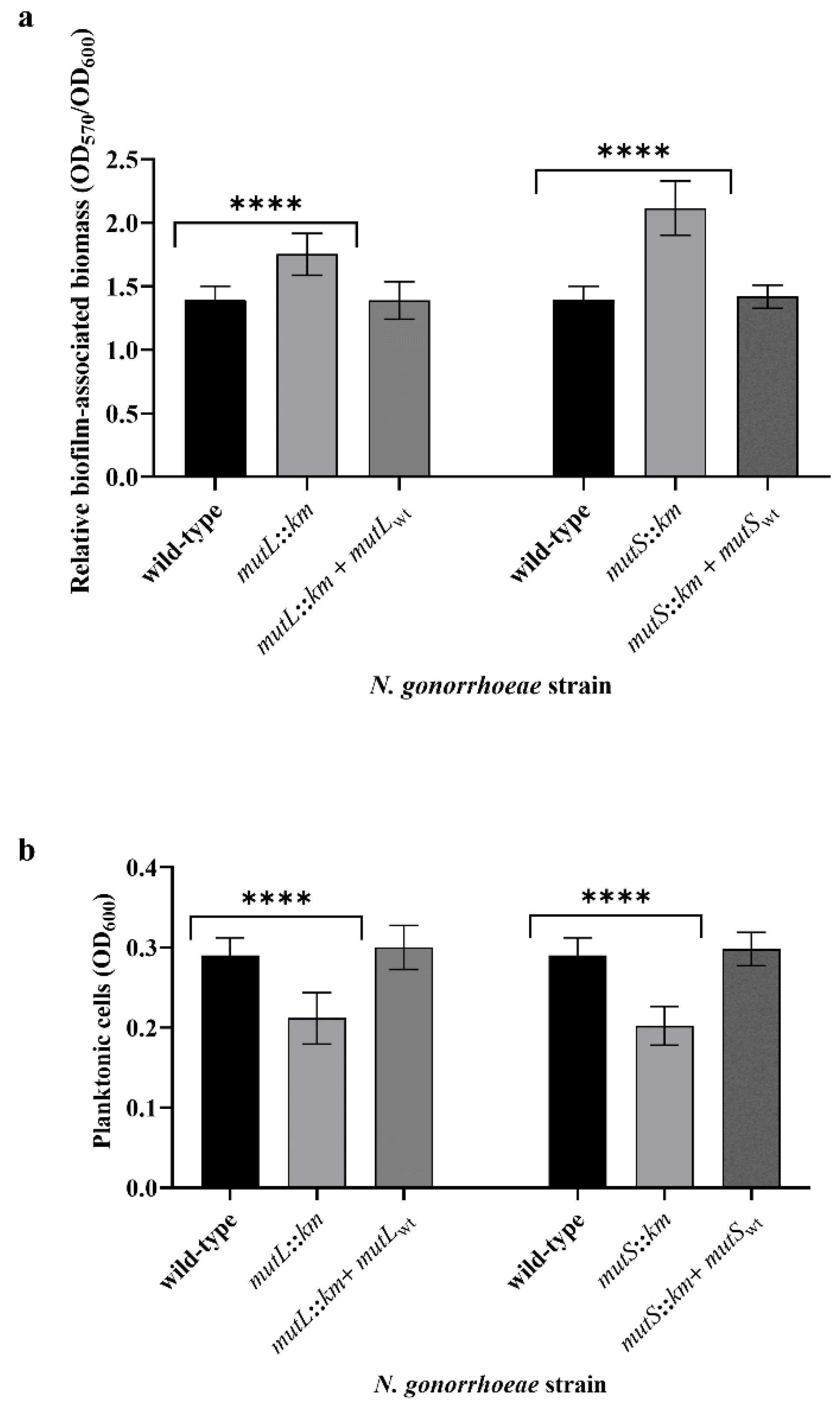
We have observed that the mutL and mutS mutants produced more biofilm-associated biomass, 1.26-fold (p = 4.42 × 10⁻⁶) and 1.52-fold (p = 1.12 × 10⁻⁸) respectively, than did the wild-type parental strain, as was determined by the crystal violet assay. Simultaneously, the increase in the number of cells forming the biofilm was accompanied by a decrease in the number of cells in the planktonic stage, suggesting that mutL and mutS mutants have a greater capability to form biofilms than wild-type parental strains (Figure 2).
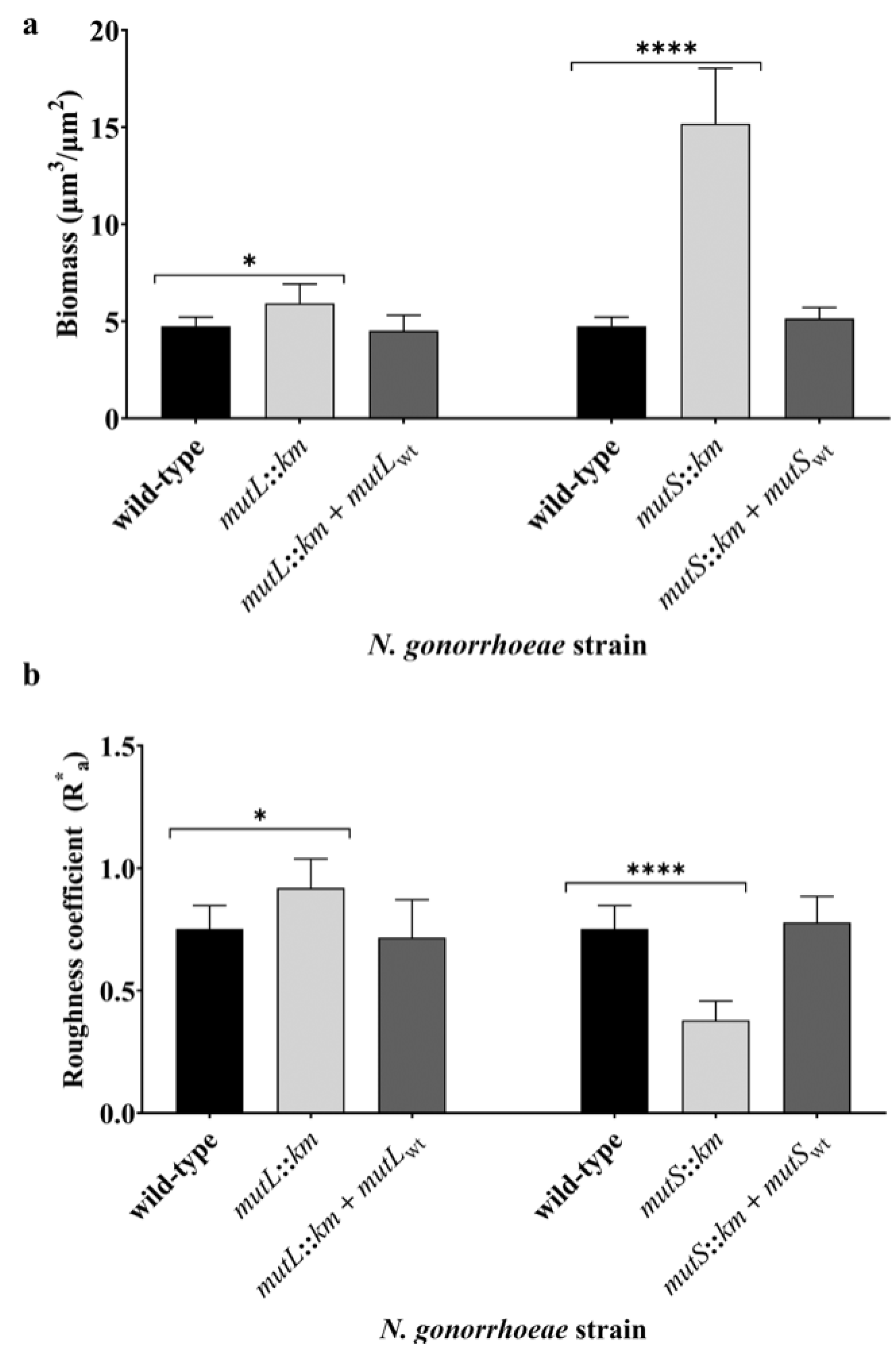
The comparison of the three-dimensional structure of live biofilm revealed that the N. gonorrhoeae mutS::km mutant forms more condensed biofilms than those observed for the wild-type strain, as demonstrated by SCLM and quantified using a Comstat2 software (Figure 3 and Figure S3). The average biomass of biofilm formed by N. gonorrhoeae mutS::km mutant was 3.19-fold higher than the biomass of the wild-type strain biofilm (15.18 ± 2.85 µm3/µm2 versus 4.75 ± 0.47 µm3/µm2; p = 2.32 × 10⁻⁴). N. gonorrhoeae mutL::km also formed a biofilm whose average biomass was higher than the biomass of the biofilm created by the wild-type strain. However, the increase was only 1.25-fold (average biomass 5.93 ± 0.99 µm3/µm2 versus 4.75 ± 0.47 µm3/µm2; p = 3.26 × 10⁻²) (Figure 3). The roughness coefficient (R*a), which is an indicator of biofilm heterogeneity reflecting diversity in thickness and biomass through a biofilm, was significantly different both for N. gonorrhoeae mutS::km biofilm and for N. gonorrhoeae mutL::km biofilm compared to that of the wild-type strain (R*a 0.37 ± 0.078 and 0.92 ± 0.12 versus 0.75 ± 0.095, respectively; p = 2.81 × 10⁻⁵ and 2.42 × 10⁻², respectively). Furthermore, biofilm-forming cells of N. gonorrhoeae mutL::km were arranged in cell clusters and formed characteristic roughness (Figure S3). It could indicate that the structure of biofilms formed by the N. gonorrhoeae mutL::km and N. gonorrhoeae mutS::km mutants differs from that of the wild-type strain biofilm. However, the maximum thickness of biofilms formed by N. gonorrhoeae mutL::km, and N. gonorrhoeae mutS::km and wild-type strain equaled 27.16 ± 4.27 µm (data not presented).
Accordingly, the higher biofilm-forming cell density and more condensed biofilms formed by N. gonorrhoeae mutL::km and N. gonorrhoeae mutS::km mutants compared to the wild-type strain were also visible when FE SEM were used (Figures S4 and S5).
It is also noteworthy that, although both mutants produced more biofilm-associated biomass, gonococcal mutS mutant formed biofilm with higher average biomass than the mutL mutant. Based on our results, we can suppose that it may be related to different gene expression in gonococcal mutL and mutS mutants. 79 genes in the N. gonorrhoeae mutL::km mutant and 42 genes in the N. gonorrhoeae mutS::km mutant were differentially expressed compared to the parental wild-type strain. An additional analysis revealed that N. gonorrhoeae mutL::km and N. gonorrhoeae mutS::km mutants share only five differentially expressed genes. Among them, there are ngo1393 and ngo1585 genes which encode proteins that may be engaged in interactions between gonococci or with their host. However, it should be noticed that the expression of these genes was significantly increased in the mutS mutant compared to the mutL mutant (Table S2). Moreover, 16 and 12 genes, in the mutL and mutS mutants respectively, encode proteins that were not determined to any COG domain. Therefore, we cannot rule out that some of these genes encode proteins that may also participate in biofilm formation or these involved in the interactions with human host. Nevertheless, further examination is required in order to determine the precise functions of proteins encoded by these genes.
Introduction in cis of wild-type copies of mutL or mutS genes in the genome of appropriate N. gonorrhoeae mutants resulted in restoration of the level of adherence, biofilm formation, and phenotype of biofilms comparable to wild-type gonococcus strains.

3.3. N. gonorrhoeae mutS::km but not N. gonorrhoeae mutL::km Is Characterized by Increased Adhesion and Invasiveness to Human Epithelial Cells Compared to the Wild-Type Strain

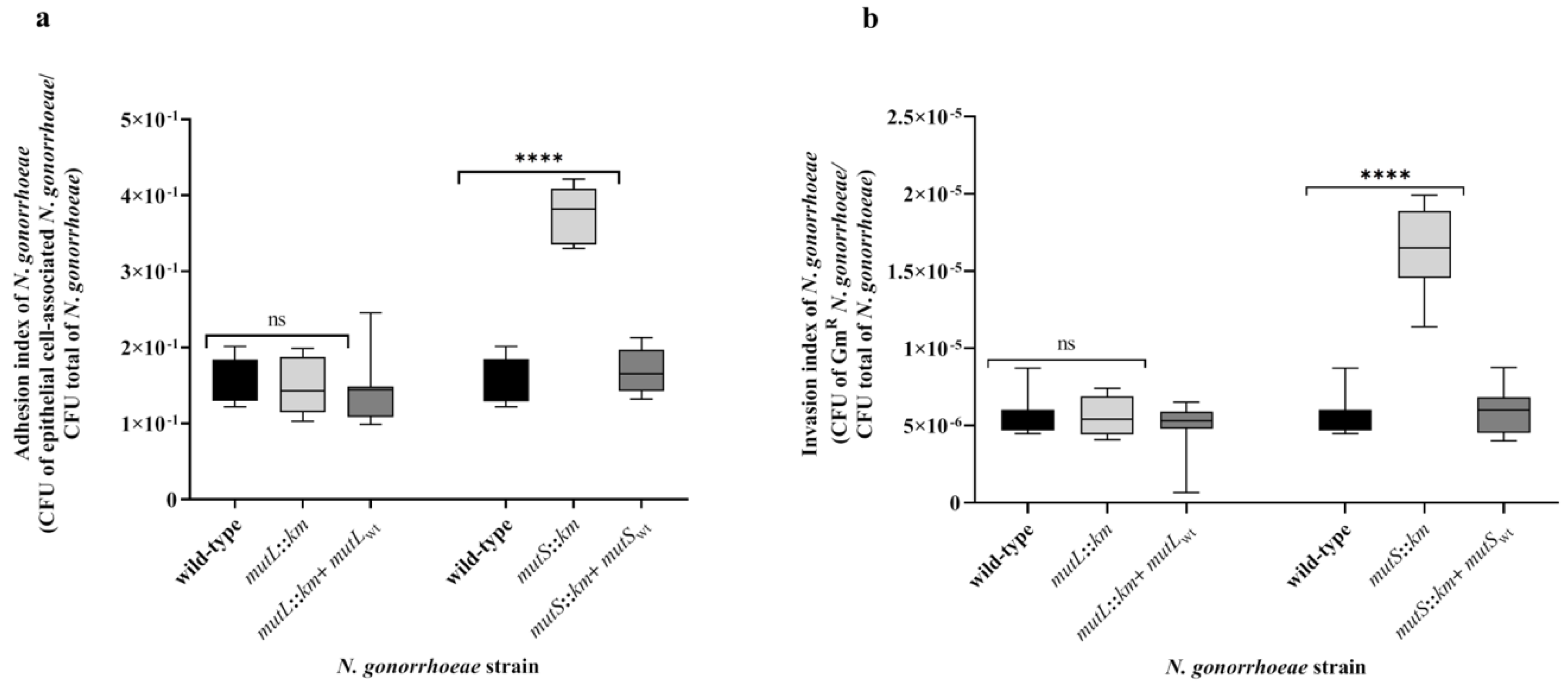
As shown above, the N. gonorrhoeae mutL::km and N. gonorrhoeae mutS::km mutants have increased adhesion and biofilm formation ability on abiotic surfaces. Therefore, the adhesion and invasiveness of these mutants to human epithelial cells was studied by determining adhesion and invasion indexes. A comparison of the adhesion indexes of N. gonorrhoeae mutS::km and the wild-type strain showed that the mutant has 2.42-fold higher adhesion to human epithelial cells than wild-type strain (adhesion index 3.77 × 10⁻¹ ± 3.59 × 10⁻² versus 1.56 × 10⁻¹ ± 2.85 × 10⁻²; p = 1.34 × 10⁻¹³) (Figure 4). Whereas, the adhesion index of N. gonorrhoeae mutL::km did not significantly differ compared to those of the wild-type strain (adhesion index 1.20 × 10⁻¹ ± 2.23 × 10⁻² versus 1.56 × 10⁻¹ ± 2.85 × 10⁻²; p = 6.05 × 10⁻¹) (Figure 4).
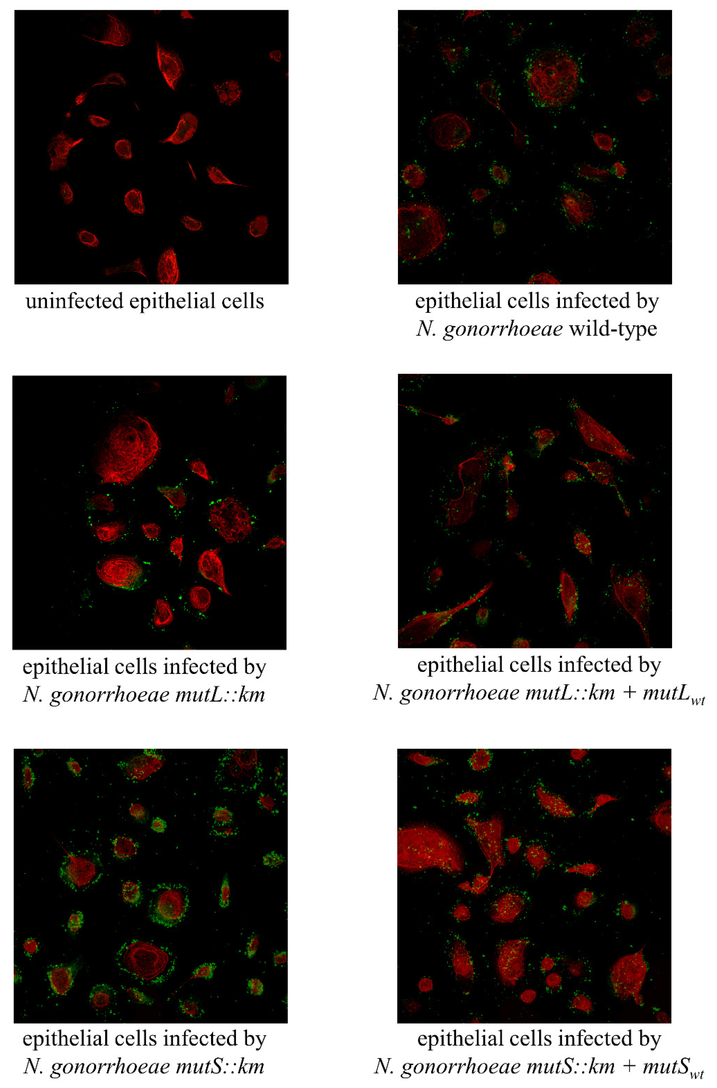
Microscopy visualization of epithelial cells infected by the N. gonorrhoeae mutL::km, N. gonorrhoeae mutS::km, and wild-type strains confirmed the presence of diplococci and small microcolonies of each strain on the surface of human epithelial cells (Figure 5 and Figure S6). Furthermore, analogously, as was determined by comparison of the adhesion indexes, an increased amount of N. gonorrhoeae mutS::km attached to human epithelial cells was detected compared to the wild-type strain, whereas there were no differences in the amount of bacteria attached to human cells between N. gonorrhoeae mutL::km and the wild-type strain.
The results presented in Figure 4 indicate that N. gonorrhoeae mutS::km is characterized not only by increased adhesion but also by increased invasiveness, as demonstrated by a 3.04-fold increase in the invasion index of the mutant compared to the wild-type strain (invasion index 1.65 × 10⁻⁵ ± 2.59 × 10⁻⁶ versus 5.43 × 10⁻⁶ ± 1.19 × 10⁻⁶; p = 6.62 × 10⁻¹⁰). This effect was not observed for N. gonorrhoeae mutL::km, whose invasiveness was not significantly different from that of the wild-type strain (invasion index 5.60 × 10⁻⁶ ± 1.25 × 10⁻⁶ versus 5.43 × 10⁻⁶ ± 1.19 × 10⁻⁶; p = 7.32 × 10⁻¹) (Figure 4).
The adhesion and invasiveness of N. gonorrhoeae complementation mutants to epithelial cells were not significantly different from that of the wild-type strain. Adhesion indexes for these gonococci were as follows: 1.33 × 10⁻¹ ± 2.63 × 10⁻² and 1.69 × 10⁻¹ ± 2.88 × 10⁻² for N. gonorrhoeae mutL::km + mutLwt and N. gonorrhoeae mutS::km + mutSwt respectively, versus 1.56 × 10⁻¹ ± 2.85 × 10⁻² for wild-type strain (p = 5.80 × 10⁻² and p = 2.68 × 10⁻¹, respectively). Invasion indexes for these gonococci were as follows: 4.96 × 10⁻⁶ ± 1.56 × 10⁻⁶ and 5.86 × 10⁻⁶ ± 1.37 × 10⁻⁶ for N. gonorrhoeae mutL::km + mutLwt and N. gonorrhoeae mutS::km + mutSwt respectively, versus 5.43 × 10⁻⁶ ± 1.19 × 10⁻⁶ wild-type strain (p = 4.17 × 10⁻¹, p = 4.22 × 10⁻¹, respectively).

3.4. N. gonorrhoeae mutS::km Has an Altered Expression of Genes Encoding Proteins that can Influence Adhesion to Human Epithelial Cells

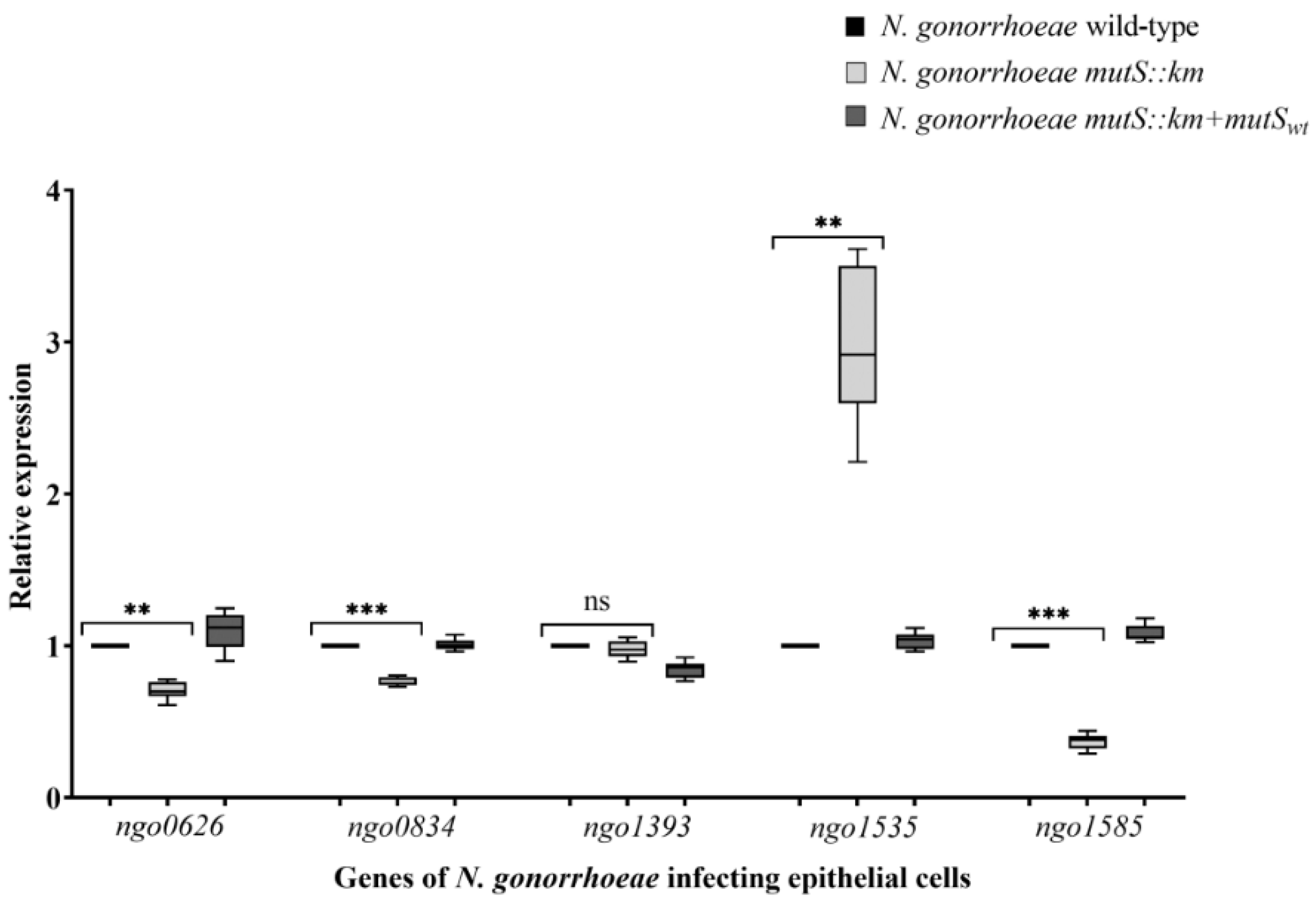
The N. gonorrhoeae mutS::km exhibits increased adhesion to human epithelial cells and differential expression of genes encoding proteins that can participate in adhesion and biofilm formation during growth on the abiotic surface. Accordingly, the expression of ngo0626, ngo0834, ngo1393, and ngo1585 genes during the growth of N. gonorrhoeae mutS::km on human epithelial cells was evaluated. In contrast to the upregulation of these genes in the mutant grown on the abiotic surface, the expression of ngo0626, ngo0834, and ngo1585 genes in the mutant cultivated on epithelial cells was decreased in comparison to the wild-type strain also cultivated on epithelial cells (1.42-, 1.3-, and 2.7-fold, respectively; p = 2.47 × 10⁻², 4.63 × 10⁻³ and 4.75 × 10⁻³, respectively). Whereas, the expression of the ngo1393 gene in the mutant was similar to that in the wild-type strain (p = 3.36 × 10⁻¹). Interestingly, during infection of epithelial cells with N. gonorrhoeae mutS::km, the expression of the ngo1535 gene was increased 3-fold compared to that in the wild-type strain infecting epithelial cells (p = 2.38 × 10⁻⁴) (Figure 6).
The upregulation of the ngo1535 gene in the mutant compared to the parental strain can explain the increased adhesion of N. gonorrhoeae mutS::km to epithelial cells, because this gene is downregulated in the planktonic growth of the wild-type strain [42]. Differences in the expression of the above genes in the mutant grown on abiotic and biotic surfaces could imply that there is a different molecular basis for increased adhesion and biofilm formation by the N. gonorrhoeae mutS::km mutant on these surfaces. The upregulation of the ngo1535 gene appears to be particularly important for the growth of the N. gonorrhoeae mutS::km mutant on epithelial cells since the expression of this gene was decreased 1.4-fold when the mutant was grown on an abiotic surface but increased 3-fold when gonococcus was cultivated on epithelial cells.

3.5. During Infection of Human Epithelial Cells with N. gonorrhoeae mutS::km, the Expression of Some Host Cell-Surface Receptors is Increased

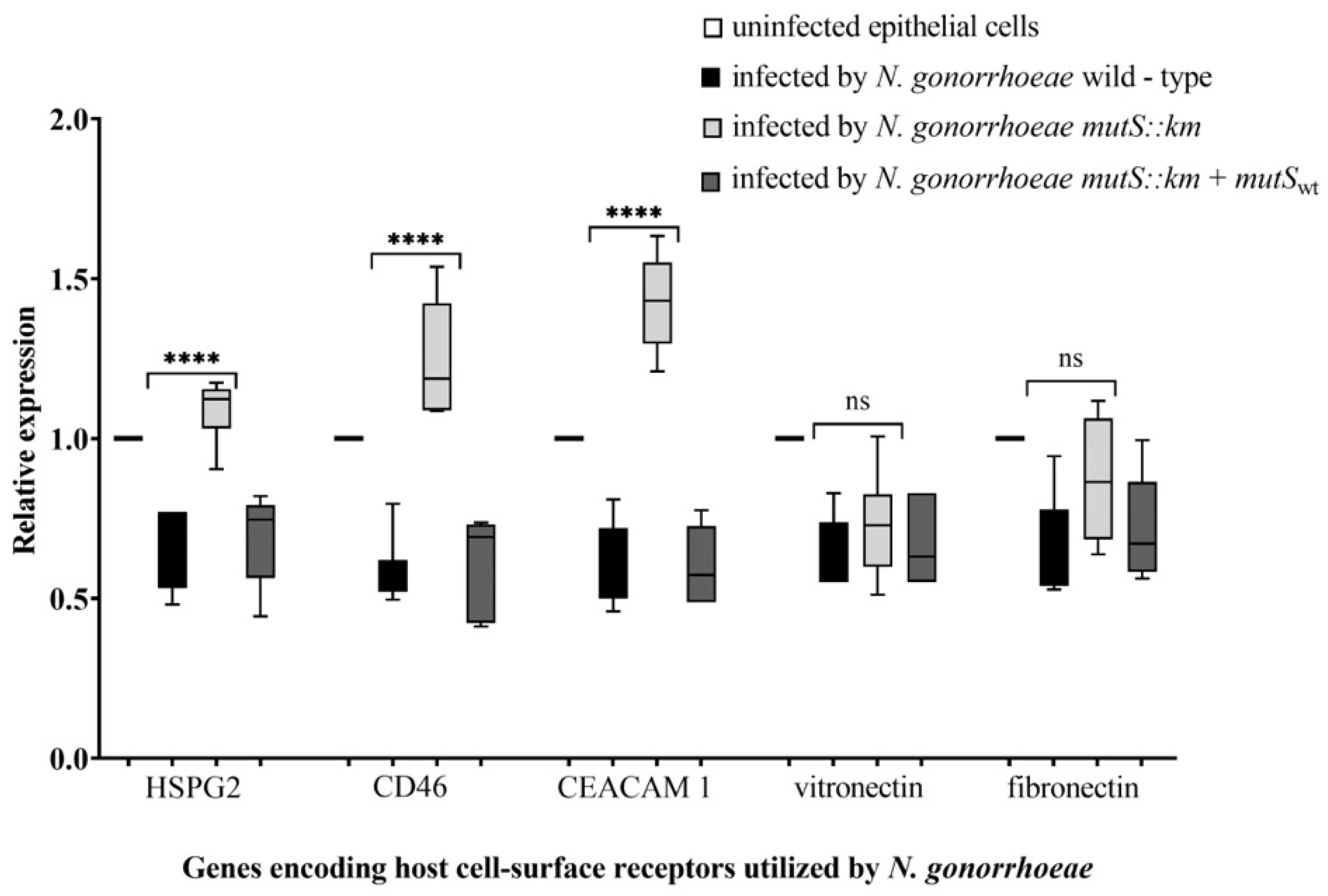
The network of interactions between N. gonorrhoeae and the host involves not only bacterial proteins but also host cell-surface receptors. Accordingly, the expression of genes encoding host cell-surface receptors for N. gonorrhoeae [58,59], such as the complement regulator CD46, heparin sulfate proteoglycan 2 (HSPG 2), carcinoembryonic antigen-related cell adhesion molecule 1 (CEACAM 1) receptor, fibronectin, and vitronectin, in epithelial cells infected with the N. gonorrhoeae mutS::km mutant was estimated and compared with the expression in epithelial cells infected with wild-type N. gonorrhoeae.
The expression of genes encoding host-cell surface receptors such as CD46, HSPG 2, and CEACAM 1 was increased 2.12-, 1.67- and 2.37-fold (p = 4.19 × 10⁻⁵, 1.39 × 10⁻⁵ and 2.41 × 10⁻⁵, respectively) in cells infected with the mutant compared to those infected with the wild-type strain (Figure 7) after 4 h of infection. In contrast, the expression of genes encoding fibronectin and vitronectin was at the same level in epithelial cells infected with both the N. gonorrhoeae mutS::km mutant and the parental strain (p = 2.96 × 10⁻¹ and 3.16 × 10⁻¹, respectively). Since CD46, HSPG 2, and CEACAM 1 are utilized by gonococci to attach and adhere to host cells, the increased expression of their genes could support the increased adhesion of the mutant to host cells.

4. Conclusions

The mutator phenotype ascribed to deficiency of the mutS gene results in not only increased mutation rates but also increased biofilm formation, adhesion, and invasiveness. Thus, although the mutator phenotype results in increased mutation frequency in singular organisms, mutator strains in the population can be beneficial because they can be a source of features linked to microbial fitness to host or environmental conditions [60]. We also hypothesize that deficiency of the mutS gene may also impact on virulence of this strain; however, understanding the molecular basis of this process requires further analysis, including determination of the functions of numerous unknown bacterial genes whose expression is affected by mutS and mutL genes deletion, in order to clarify the role of their products in gonococcal physiology.

Supplementary Materials

The following are available online at https://www.mdpi.com/2076-2607/7/12/647/s1.

Author Contributions

J.P. part of the RT-qPCR analysis, SCLM, and FE SEM analysis, and adhesion and invasiveness analysis; M.A.-P. microarray analysis and part of RT-qPCR analysis; R.L. in silico analysis of the upstream and within sequences of differentially expressed genes; P.B. FE SEM analysis; A.K. SCLM analysis, part of RT-qPCR analysis, research concept, manuscript writing, and figure preparation.

Funding

This research was funded by the National Science Centre and the Ministry of Science and Higher Education in Poland (grant nos. UMO-2014/15/B/NZ6/02514 and NN301 165135). Scanning electron microscopy was carried out with the use of CePT infrastructure financed by the European Union—The European Regional Development Fund within the Operational Programme “Innovative economy” for 2007–2013.

Conflicts of Interest

The authors declare no conflict of interest.

References

  1. Unemo, M.; Fasth, O.; Fredlund, H.; Limnios, A.; Tapsall, J. Phenotypic and genetic characterization of the 2008 WHO Neisseria gonorrhoeae reference strain panel intended for global quality assurance and quality control of gonococcal antimicrobial resistance surveillance for public health purposes. J. Antimicrob. Chemother. 2009, 63, 1142–1151. [Google Scholar] [CrossRef] [PubMed]
  2. World Health Organization. Global Priority List of Antibiotic-Resistant Bacteria to Guide Research, Discovery, and Development of New Antibiotics; World Health Organization: Geneva, Switzerland, 2017; pp. 1–7. [Google Scholar]
  3. Jarvis, G.A.; Chang, T.L. Modulation of HIV transmission by Neisseria gonorrhoeae: Molecular and immunological aspects. Curr. HIV Res. 2012, 10, 211–217. [Google Scholar] [CrossRef] [PubMed] [Green Version]
  4. Ding, J.; Rapista, A.; Teleshova, N.; Mosoyan, G.; Jarvis, G.A.; Klotman, M.E.; Chang, T.L. Neisseria gonorrhoeae enhances HIV-1 infection of primary resting CD4+ T cells through TLR2 activation. J. Immunol. 2010, 184, 2814–2824. [Google Scholar] [CrossRef] [PubMed] [Green Version]
  5. Chen, A.; Boulton, I.C.; Pongoski, J.; Cochrane, A.; Gray-Owen, S.D. Induction of HIV-1 long terminal repeat-mediated transcription by Neisseria gonorrhoeae. AIDS 2003, 17, 625–628. [Google Scholar] [CrossRef] [PubMed]
  6. Greiner, L.L.; Edwards, J.L.; Shao, J.; Rabinak, C.; Entz, D.; Apicella, M.A. Biofilm Formation by Neisseria gonorrhoeae. Infect. Immun. 2005, 73, 1964–1970. [Google Scholar] [CrossRef] [PubMed] [Green Version]
  7. Steichen, C.T.; Shao, J.Q.; Ketterer, M.R.; Apicella, M.A. Gonococcal cervicitis: A role for biofilm in pathogenesis. J. Infect. Dis. 2008, 198, 1856–1861. [Google Scholar] [CrossRef] [Green Version]
  8. Reyes, G.X.; Schmidt, T.T.; Kolodner, R.D.; Hombauer, H. New insights into the mechanism of DNA mismatch repair. Chromosoma 2015, 124, 443–462. [Google Scholar] [CrossRef] [Green Version]
  9. Criss, A.K.; Bonney, K.M.; Chang, R.A.; Duffin, P.M.; LeCuyer, B.E.; Seifert, H.S. Mismatch correction modulates mutation frequency and pilus phase and antigenic variation in Neisseria gonorrhoeae. J. Bacteriol. 2010, 192, 316–325. [Google Scholar] [CrossRef] [Green Version]
  10. Adamczyk-Popławska, M.; Bandyra, K.; Kwiatek, A. Activity of Vsr endonucleases encoded by Neisseria gonorrhoeae FA1090 is influenced by MutL and MutS proteins. BMC Microbiol. 2018, 18, 95. [Google Scholar] [CrossRef]
  11. Rotman, E.; Seifert, H.S. Neisseria gonorrhoeae MutS affects pilin antigenic variation through mismatch correction and not by pilE guanine quartet binding. J. Bacteriol. 2015, 197, 1828–1838. [Google Scholar] [CrossRef] [Green Version]
  12. Ambur, O.H.; Davidsen, T.; Frye, S.A.; Balasingham, S.V.; Lagesen, K.; Rognes, T.; Tønjum, T. Genome dynamics in major bacterial pathogens. FEMS Microbiol. Rev. 2009, 33, 453–470. [Google Scholar] [CrossRef] [PubMed] [Green Version]
  13. Kline, K.A.; Sechman, E.V.; Skaar, E.P.; Seifert, H.S. Recombination, repair and replication in the pathogenic Neisseriae: The 3 R’s of molecular genetics of two human-specific bacterial pathogens. Mol. Microbiol. 2003, 50, 3–13. [Google Scholar] [CrossRef] [PubMed]
  14. Eisen, J.A.; Hanawalt, P.C. A phylogenomic study of DNA repair genes, proteins, and processes. Mutat. Res. 1999, 435, 171–213. [Google Scholar] [CrossRef] [Green Version]
  15. Richardson, A.R.; Stojiljkovic, I. Mismatch repair and the regulation of phase variation in Neisseria meningitidis. Mol. Microbiol. 2001, 40, 645–655. [Google Scholar] [CrossRef]
  16. Richardson, A.R.; Yu, Z.; Popovic, T.; Stojiljkovic, I. Mutator clones of Neisseria meningitidis in epidemic serogroup a disease. Proc. Natl. Acad. Sci. USA 2002, 99, 6103–6107. [Google Scholar] [CrossRef] [Green Version]
  17. Bayliss, C.D.; Hoe, J.C.; Makepeace, K.; Martin, P.; Hood, D.W.; Moxon, E.R. Neisseria meningitidis escape from the bactericidal activity of a monoclonal antibody is mediated by phase variation of lgtG and enhanced by a mutator phenotype. Infect. Immun. 2008, 76, 5038–5048. [Google Scholar] [CrossRef] [Green Version]
  18. LeClerc, J.E.; Li, B.; Payne, W.L.; Cebula, T.A. High mutation frequencies among Escherichia coli and Salmonella pathogens. Science 1996, 274, 1208–1211. [Google Scholar] [CrossRef]
  19. LeClerc, J.E.; Payne, W.L.; Kupchella, E.; Cebula, T.A. Detection of mutator subpopulations in Salmonella typhimurium LT2 by reversion of his alleles. Mutat. Res. 1998, 400, 89–97. [Google Scholar] [CrossRef]
  20. Oliver, A.; Baquero, F.; Blázquez, J. The mismatch repair system (mutS, mutL and uvrD genes) in Pseudomonas aeruginosa: Molecular characterization of naturally occurring mutants. Mol. Microbiol. 2002, 43, 1641–1650. [Google Scholar] [CrossRef]
  21. Nachamkin, I.; Cannon, J.G.; Mittler, R.S. Monoclonal antibodies against Neisseria gonorrhoeae: Production of antibodies directed against a strain-specific cell surface antigen. Infect. Immun. 1981, 32, 641–648. [Google Scholar]
  22. Dillard, J.P. Genetic Manipulation of Neisseria gonorrhoeae. Curr. Protoc. Microbiol. 2011, 23, Unit4A.2.1–Unit4A.2.24. [Google Scholar] [CrossRef] [Green Version]
  23. Shaw, J.H.; Falkow, S. Model for invasion of human tissue culture cells by Neisseria gonorrhoeae. Infect. Immun. 1988, 56, 1625–1632. [Google Scholar] [PubMed]
  24. Ilver, D.; Källström, H.; Normark, S.; Jonsson, A.B. Transcellular passage of Neisseria gonorrhoeae involves pilus phase variation. Infect. Immun. 1998, 66, 469–473. [Google Scholar] [PubMed]
  25. Merz, A.J.; So, M. Attachment of piliated, Opa- and Opc- gonococci and meningococci to epithelial cells elicits cortical actin rearrangements and clustering of tyrosine-phosphorylated proteins. Infect. Immun. 1997, 65, 4341–4349. [Google Scholar]
  26. Naumann, M.; Wessler, S.; Bartsch, C.; Wieland, B.; Meyer, T.F. Neisseria gonorrhoeae epithelial cell interaction leads to the activation of the transcription factors nuclear factor kappaB and activator protein 1 and the induction of inflammatory cytokines. J. Exp. Med. 1997, 186, 247–258. [Google Scholar] [CrossRef]
  27. Kwiatek, A.; Bacal, P.; Wasiluk, A.; Trybunko, A.; Adamczyk-Poplawska, M. The dam replacing gene product enhances Neisseria gonorrhoeae FA1090 viability and biofilm formation. Front. Microbiol. 2014, 5, 712. [Google Scholar] [CrossRef] [Green Version]
  28. Kwiatek, A.; Mrozek, A.; Bacal, P.; Piekarowicz, A.; Adamczyk-Popławska, M. Type III Methyltransferase M.NgoAX from Neisseria gonorrhoeae FA1090 Regulates Biofilm Formation and Interactions with Human Cells. Front. Microbiol. 2015, 6, 1426. [Google Scholar] [CrossRef] [Green Version]
  29. Bhoopalan, S.V.; Piekarowicz, A.; Lenz, J.D.; Dillard, J.P.; Stein, D.C. nagZ Triggers Gonococcal Biofilm Disassembly. Sci. Rep. 2016, 6, 22372. [Google Scholar] [CrossRef] [Green Version]
  30. Stepanovic, S.; Vukovic, D.; Dakic, I.; Savic, B.; Svabic-Vlahovic, M. A modified microtiter-plate test for quantification of staphylococcal biofilm formation. J. Microbiol. Methods 2000, 40, 175–179. [Google Scholar] [CrossRef]
  31. Bustin, S.A.; Benes, V.; Garson, J.A.; Hellemans, J.; Huggett, J.; Kubista, M.; Mueller, R.; Nolan, T.; Pfaffl, M.W.; Shipley, G.L.; et al. The MIQE guidelines: Minimum information for publication of quantitative real-time PCR experiments. Clin. Chem. 2009, 55, 611–622. [Google Scholar] [CrossRef] [Green Version]
  32. Hopper, S.; Wilbur, J.S.; Vasquez, B.L.; Larson, J.; Clary, S.; Mehr, I.J.; Seifert, H.S.; So, M. Isolation of Neisseria gonorrhoeae mutants that show enhanced trafficking across polarized T84 epithelial monolayers. Infect. Immun. 2000, 68, 896–905. [Google Scholar] [CrossRef] [PubMed] [Green Version]
  33. Tatusov, R.L.; Galperin, M.Y.; Natale, D.A.; Koonin, E.V. The COG database: A tool for genome-scale analysis of protein functions and evolution. Nucleic Acids Res. 2000, 28, 33–36. [Google Scholar] [CrossRef] [PubMed] [Green Version]
  34. Natale, D.A.; Galperin, M.Y.; Tatusov, R.L.; Koonin, E.V. Using the COG database to improve gene recognition in complete genomes. Genetica 2000, 108, 9–17. [Google Scholar] [CrossRef] [PubMed]
  35. Heydorn, A.; Nielsen, A.T.; Hentzer, M.; Sternberg, C.; Givskov, M.; Ersbøll, B.K.; Molin, S. Quantification of biofilm structures by the novel computer program COMSTAT. Microbiology 2000, 146, 2395–2407. [Google Scholar] [CrossRef] [PubMed] [Green Version]
  36. Jamet, A.; Jousset, A.B.; Euphrasie, D.; Mukorako, P.; Boucharlat, A.; Ducousso, A.; Charbit, A.; Nassif, X. A new family of secreted toxins in pathogenic Neisseria species. PLoS Pathog. 2015, 11, e1004592. [Google Scholar] [CrossRef] [PubMed] [Green Version]
  37. Jamet, A.; Nassif, X. Characterization of the Maf family of polymorphic toxins in pathogenic. Microb. Cell 2015, 2, 88–90. [Google Scholar] [CrossRef] [PubMed] [Green Version]
  38. Arenas, J.; de Maat, V.; Catón, L.; Krekorian, M.; Herrero, J.C.; Ferrara, F.; Tommassen, J. Fratricide activity of MafB protein of N. meningitidis strain B16B6. BMC Microbiol. 2015, 15, 156. [Google Scholar] [CrossRef] [Green Version]
  39. Paruchuri, D.K.; Seifert, H.S.; Ajioka, R.S.; Karlsson, K.A.; So, M. Identification and characterization of a Neisseria gonorrhoeae gene encoding a glycolipid-binding adhesin. Proc. Natl. Acad. Sci. USA 1990, 87, 333–337. [Google Scholar] [CrossRef] [Green Version]
  40. Zielke, R.A.; Wierzbicki, I.H.; Weber, J.V.; Gafken, P.R.; Sikora, A.E. Quantitative proteomics of the Neisseria gonorrhoeae cell envelope and membrane vesicles for the discovery of potential therapeutic targets. Mol. Cell Proteom. 2014, 13, 1299–1317. [Google Scholar] [CrossRef] [Green Version]
  41. Dove, J.E.; Yasukawa, K.; Tinsley, C.R.; Nassif, X. Production of the signalling molecule, autoinducer-2, by Neisseria meningitidis: Lack of evidence for a concerted transcriptional response. Microbiology 2003, 149, 1859–1869. [Google Scholar] [CrossRef]
  42. Falsetta, M.L.; Bair, T.B.; Ku, S.C.; Vanden Hoven, R.N.; Steichen, C.T.; McEwan, A.G.; Jennings, M.P.; Apicella, M.A. Transcriptional profiling identifies the metabolic phenotype of gonococcal biofilms. Infect. Immun. 2009, 77, 3522–3532. [Google Scholar] [CrossRef] [PubMed] [Green Version]
  43. Bandara, M.; Skehel, J.M.; Kadioglu, A.; Collinson, I.; Nobbs, A.H.; Blocker, A.J.; Jenkinson, H.F. The accessory Sec system (SecY2A2) in Streptococcus pneumoniae is involved in export of pneumolysin toxin, adhesion and biofilm formation. Microbes. Infect. 2017, 19, 402–412. [Google Scholar] [CrossRef] [PubMed] [Green Version]
  44. Robinson, L.S.; Ashman, E.M.; Hultgren, S.J.; Chapman, M.R. Secretion of curli fibre subunits is mediated by the outer membrane-localized CsgG protein. Mol. Microbiol. 2006, 59, 870–881. [Google Scholar] [CrossRef] [PubMed] [Green Version]
  45. Cao, B.; Zhao, Y.; Kou, Y.; Ni, D.; Zhang, X.C.; Huang, Y. Structure of the nonameric bacterial amyloid secretion channel. Proc. Natl. Acad. Sci. USA 2014, 111, E5439–E5444. [Google Scholar] [CrossRef] [Green Version]
  46. Goyal, P.; Krasteva, P.V.; Van Gerven, N.; Gubellini, F.; Van den Broeck, I.; Troupiotis-Tsaïlaki, A.; Jonckheere, W.; Péhau-Arnaudet, G.; Pinkner, J.S.; Chapman, M.R.; et al. Structural and mechanistic insights into the bacterial amyloid secretion channel CsgG. Nature 2014, 516, 250–253. [Google Scholar] [CrossRef] [Green Version]
  47. Berne, C.; Ducret, A.; Hardy, G.G.; Brun, Y.V. Adhesins Involved in Attachment to Abiotic Surfaces by Gram-Negative Bacteria. Microbiol. Spectr. 2015, 3. [Google Scholar] [CrossRef] [Green Version]
  48. Chagnot, C.; Zorgani, M.A.; Astruc, T.; Desvaux, M. Proteinaceous determinants of surface colonization in bacteria: Bacterial adhesion and biofilm formation from a protein secretion perspective. Front. Microbiol. 2013, 4, 303. [Google Scholar] [CrossRef] [Green Version]
  49. Fronzes, R.; Remaut, H.; Waksman, G. Architectures and biogenesis of non-flagellar protein appendages in Gram-negative bacteria. EMBO J. 2008, 27, 2271–2280. [Google Scholar] [CrossRef] [Green Version]
  50. Kikuchi, T.; Mizunoe, Y.; Takade, A.; Naito, S.; Yoshida, S. Curli fibers are required for development of biofilm architecture in Escherichia coli K-12 and enhance bacterial adherence to human uroepithelial cells. Microbiol. Immunol. 2005, 49, 875–884. [Google Scholar] [CrossRef]
  51. Loferer, H.; Hammar, M.; Normark, S. Availability of the fibre subunit CsgA and the nucleator protein CsgB during assembly of fibronectin-binding curli is limited by the intracellular concentration of the novel lipoprotein CsgG. Mol. Microbiol. 1997, 26, 11–23. [Google Scholar] [CrossRef] [Green Version]
  52. Kurtz, H.D.; Smith, J. Analysis of a Caulobacter crescentus gene cluster involved in attachment of the holdfast to the cell. J. Bacteriol. 1992, 174, 687–694. [Google Scholar] [CrossRef] [PubMed] [Green Version]
  53. Ragland, S.A.; Schaub, R.E.; Hackett, K.T.; Dillard, J.P.; Criss, A.K. Two lytic transglycosylases in Neisseria gonorrhoeae impart resistance to killing by lysozyme and human neutrophils. Cell. Microbiol. 2017, 19. [Google Scholar] [CrossRef] [PubMed] [Green Version]
  54. Knilans, K.J.; Hackett, K.T.; Anderson, J.E.; Weng, C.; Dillard, J.P.; Duncan, J.A. Neisseria gonorrhoeae Lytic Transglycosylases LtgA and LtgD Reduce Host Innate Immune Signaling through TLR2 and NOD2. ACS Infect. Dis. 2017, 3, 624–633. [Google Scholar] [CrossRef] [PubMed]
  55. Monteiro, C.; Fang, X.; Ahmad, I.; Gomelsky, M.; Römling, U. Regulation of biofilm components in Salmonella enterica serovar Typhimurium by lytic transglycosylases involved in cell wall turnover. J. Bacteriol. 2011, 193, 6443–6451. [Google Scholar] [CrossRef] [Green Version]
  56. Martin, P.; Sun, L.; Hood, D.W.; Moxon, E.R. Involvement of genes of genome maintenance in the regulation of phase variation frequencies in Neisseria meningitidis. Microbiology 2004, 150, 3001–3012. [Google Scholar] [CrossRef] [Green Version]
  57. Fagnocchi, L.; Pigozzi, E.; Scarlato, V.; Delany, I. In the NadR regulon, adhesins and diverse meningococcal functions are regulated in response to signals in human saliva. J. Bacteriol. 2012, 194, 460–474. [Google Scholar] [CrossRef] [Green Version]
  58. Nassif, X.; Pujol, C.; Morand, P.; Eugène, E. Interactions of pathogenic Neisseria with host cells. Is it possible to assemble the puzzle? Mol. Microbiol. 1999, 32, 1124–1132. [Google Scholar] [CrossRef]
  59. Dehio, C.; Gray-Owen, S.D.; Meyer, T.F. Host cell invasion by pathogenic Neisseriae. In Subcellular Biochemistry; Springer: Boston, MA, USA, 2000; pp. 61–96. [Google Scholar]
  60. Schofield, M.J.; Hsieh, P. DNA mismatch repair: Molecular mechanisms and biological function. Annu. Rev. Microbiol. 2003, 57, 579–608. [Google Scholar] [CrossRef]
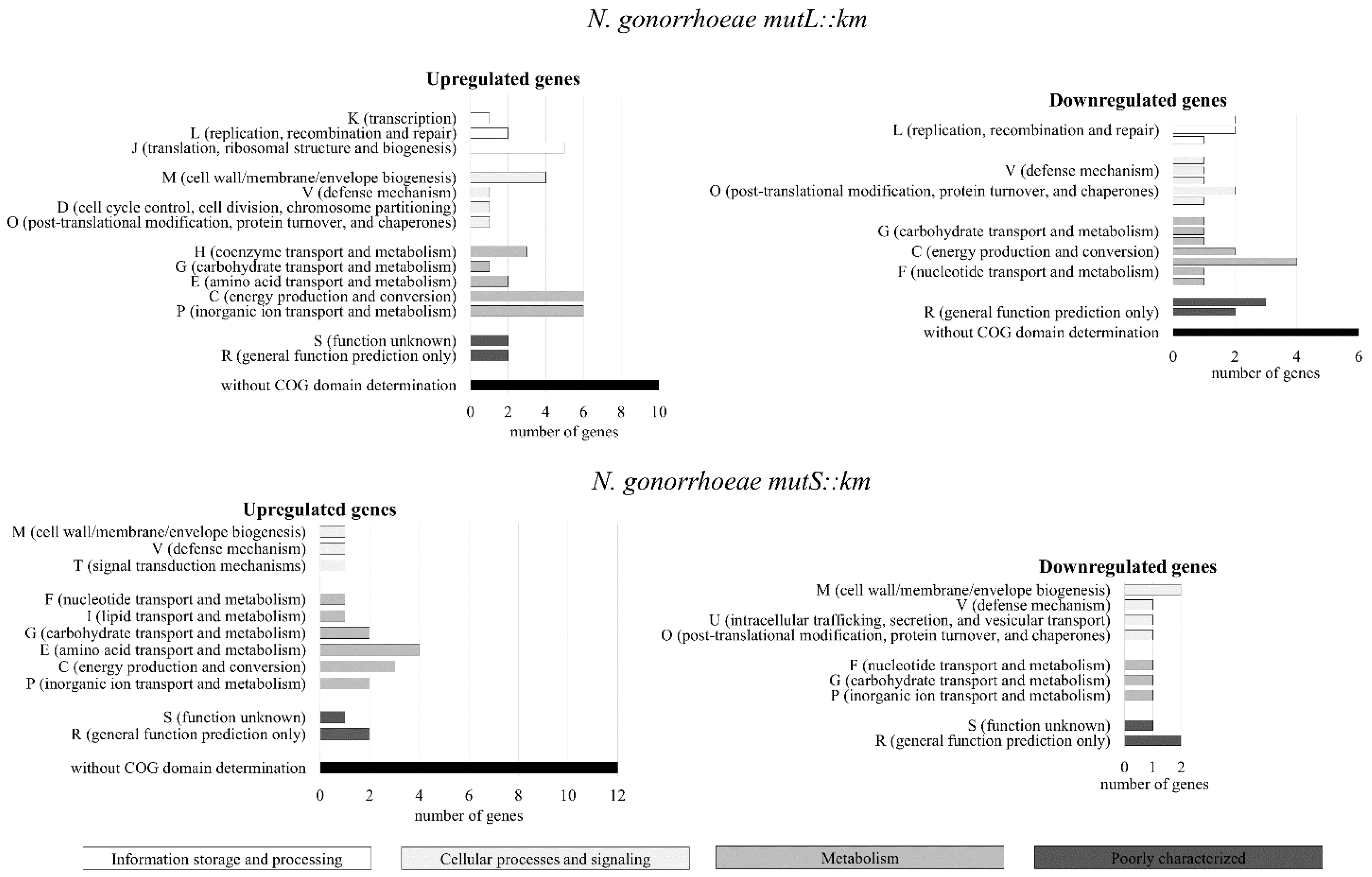
Figure 1. Differential gene expression in Neisseria gonorrhoeae (N. gonorrhoeae) mutL and mutS mutants determined by DNA microarray method. Classification of the Cluster of Orthologous Groups (COGs) by functional categories of proteins encoded by differentially expressed genes in N. gonorrhoeae mutL and mutS mutants cultivated on abiotic surface. The graphs show functional COGs categories. The color of the functional category corresponds to the main category indicated in rectangles.
Microorganisms 07 00647 g001
Figure 2. Effects of the inactivation of mutL and mutS genes on biofilm formation by N. gonorrhoeae on abiotic surfaces determined by the crystal violet assay. (a) The relative biofilm-associated biomass after 24 h. (b) The amount of gonococcal planktonic cells after 24 h. The asterisks indicate the statistically significant differences between mutants with a disrupted mutL or mutS gene (N. gonorrhoeae mutL::km and N. gonorrhoeae mutS::km, respectively), complementation strains (N. gonorrhoeae mutL::km + mutLwt and N. gonorrhoeae mutS::km + mutSwt) and the wild-type strain, as were calculated using two-way analysis of variance (ANOVA) followed by Bonferroni post-tests. Data represent mean values (± standard deviation (SD)) from three independent experiments performed in triplicate. **** asterisks indicate p values < 0.0001.
Microorganisms 07 00647 g002
Figure 3. The structure and density of live biofilms formed by gonococci with a disrupted mutL or mutS gene (N. gonorrhoeae mutL::km and N. gonorrhoeae mutS::km, respectively), complementation strains (N. gonorrhoeae mutL::km + mutLwt and N. gonorrhoeae mutS::km + mutSwt), and the wild-type strain on abiotic surfaces assayed by scanning confocal laser microscopy (SCLM). Analysis of biofilms properties: (a) biomass of biofilm (µm3/µm2) and (b) roughness coefficient (R*a) determined by Comstat2 software. The statistically significant differences were calculated using two-way ANOVA followed by Bonferroni post-tests. Data represent mean values (± SD) from three independent experiments performed in triplicate. * and **** asterisks indicate p value < 0.05, and < 0.0001, respectively.
Microorganisms 07 00647 g003
Figure 4. Adhesion and invasiveness of N. gonorrhoeae mutL::km and N. gonorrhoeae mutS::km to human epithelial cells. (a) The adhesion of N. gonorrhoeae mutL::km and N. gonorrhoeae mutS::km in comparison to complementation and the wild-type strains. (b) The invasiveness of N. gonorrhoeae mutL::km and N. gonorrhoeae mutS::km in comparison to complementation and the wild-type strains. The asterisks indicate statistically significant differences between mutants with a disrupted mutL or mutS gene (N. gonorrhoeae mutL::km and N. gonorrhoeae mutS::km, respectively), complementation strains (N. gonorrhoeae mutL::km + mutLwt and N. gonorrhoeae mutS::km + mutSwt), and the wild-type strain, as calculated using two-way ANOVA followed by Bonferroni post-tests. Data represent mean values (± SD) from three independent experiments performed in triplicate. **** asterisks indicate p values < 0.0001, ns—not statistically significant.
Microorganisms 07 00647 g004
Figure 5. Microscopy visualization of epithelial cells infected by N. gonorrhoeae mutL::km and N. gonorrhoeae mutS::km assayed by SCLM (magnification: 40×). N. gonorrhoeae was added to the epithelial cells and cultivated for 4 h, followed by which, the human cells were stained with CellTracker Orange CMTMR, and N. gonorrhoeae were detected by Anti-Neisseria gonorrhoeae antibody as the primary antibody and Goat Anti-Rabbit IgG H and L conjugated with Alexa Fluor® 488 (green-fluorescent dye) as the secondary antibody. All experiments were performed in triplicate, and representative images are shown.
Microorganisms 07 00647 g005
Figure 6. The expression of gonococcal ngo0626, ngo0834, ngo1393, ngo1535, and ngo1585 genes during growth of N. gonorrhoeae mutS::km on human epithelial cells. The expression of the genes was evaluated by RT-qPCR using RNA extracted from bacterial cells infecting human epithelial cells. The relative quantitation data analysis was performed using the comparative quantification method ΔΔCt with 16S rRNA as the endogenous reference. The statistically significant differences between N. gonorrhoeae mutS::km, the complementation strain (N. gonorrhoeae mutS::km + mutSwt), and the wild-type strain were calculated using two-way ANOVA followed by Bonferroni post-tests. Data on gene expression in N. gonorrhoeae mutS::km and N. gonorrhoeae mutS::km + mutSwt were compared to data on gene expression in the wild-type strain, for which the value of 1 was assumed. Data represent mean values (± SD) from three independent experiments performed in triplicate. **, *** asterisks indicate p values < 0.01 and < 0.0001 respectively, ns—not statistically significant.
Microorganisms 07 00647 g006
Figure 7. The expression of human genes encoding complement regulator CD46, heparin sulfate proteoglycan 2 (HSPG 2), carcinoembryonic antigen-related cell adhesion molecule 1 (CEACAM 1) receptor, fibronectin, and vitronectin in epithelial cells infected with N. gonorrhoeae mutS::km in comparison to those infected with wild-type N. gonorrhoeae. The expression of the genes was evaluated by RT-qPCR using RNA extracted from human epithelial cells infected 4 h with N. gonorrhoeae. The relative quantitation data analysis was performed using the comparative quantification method ΔΔCt with HPRT 1 (Hypoxanthine-guanine phosphoribosyltransferase 1) and Beta-2-microglobulin as endogenous references. The statistically significant differences in gene expression caused by N. gonorrhoeae mutS::km infection in comparison to infection caused by the complementation strain (N. gonorrhoeae mutS::km + mutSwt) and wild-type strain were calculated using two-way ANOVA followed by Bonferroni post-tests. Data were normalized to data from uninfected epithelial cells, for which the value of 1 was assumed. Data represent mean values (± SD) from three independent experiments performed in triplicate. **** asterisks indicate p values < 0.0001, ns—not statistically significant.
Microorganisms 07 00647 g007