Article

Real-Time PCR for Molecular Detection of Zoonotic and Non-Zoonotic Giardia spp. in Wild Rodents

1
Unit 16 Mycotic and Parasitic Agents and Mycobacteria, Department of Infectious Diseases, Robert Koch-Institute, 13353 Berlin, Germany
2
Centre for Infection Medicine, Institute of Immunology, Freie Universität Berlin, 14163 Berlin, Germany
*
Author to whom correspondence should be addressed.
Microorganisms 2021, 9(8), 1610; https://doi.org/10.3390/microorganisms9081610
Submission received: 17 June 2021 / Revised: 6 July 2021 / Accepted: 7 July 2021 / Published: 28 July 2021

Abstract

:
Giardiasis in humans is a gastrointestinal disease transmitted by the potentially zoonotic Giardia duodenalis genotypes (assemblages) A and B. Small wild rodents such as mice and voles are discussed as potential reservoirs for G. duodenalis but are predominantly populated by the two rodent species Giardia microti and Giardia muris. Currently, the detection of zoonotic and non-zoonotic Giardia species and genotypes in these animals relies on cumbersome PCR and sequencing approaches of genetic marker genes. This hampers the risk assessment of potential zoonotic Giardia transmissions by these animals. Here, we provide a workflow based on newly developed real-time PCR schemes targeting the small ribosomal RNA multi-copy gene locus to distinguish G. muris, G. microti and G. duodenalis infections. For the identification of potentially zoonotic G. duodenalis assemblage types A and B, an established protocol targeting the single-copy gene 4E1-HP was used. The assays were specific for the distinct Giardia species or genotypes and revealed an analytical sensitivity of approximately one or below genome equivalent for the multi-copy gene and of about 10 genome equivalents for the single-copy gene. Retesting a biobank of small rodent samples confirmed the specificity. It further identified the underlying Giardia species in four out of 11 samples that could not be typed before by PCR and sequencing. The newly developed workflow has the potential to facilitate the detection of potentially zoonotic and non-zoonotic Giardia species in wild rodents.

1. Introduction

Giardia spp. are flagellated protozoan parasites of vertebrates and a frequent cause of gastrointestinal disease in wild and domestic animals and humans [1]. Giardia duodenalis is the most widespread species in mammals and subdivided into eight genetic subgroups (assemblages A-H) [1]. These subgroups show distinct host ranges: Assemblages A and B are found in humans and a wide range of other mammals, including rodents, and are considered potentially zoonotic [2]; assemblages C/D are found in canids; assemblage E in hoofed animals; assemblage F in cats; assemblage G in rodents (in particular rats) and assemblage H in pinnipeds [1]. Small rodents can furthermore be infected with the distinct species Giardia muris and Giardia microti [1,3,4]. For both G. microti and, in particular, for G. muris, distinct morphological characteristics in comparison to G. duodenalis have been described [5].
The zoonotic G. duodenalis assemblages A and B are genetically very distinct, and each group can be further subclassified based on genetic information determined by multi-locus sequence typing (MLST) [6]. Within these subgroups, assemblage AI forms a pan-global, almost clonal subpopulation that is found in a wide range of vertebrates, including humans, whereas assemblage AII seems very genetically diverse and is almost exclusively found in humans [1]. The subclassification of assemblage B is less well-defined—in particular, when classical MLST procedures are used. Additionally, assemblage B has a higher degree of allelic sequence heterogeneity (ASH) in the tetraploid genome that further hampers isolate typing [7].
Rodents are frequently parasitized by Giardia spp., and the potential zoonotic risk has been highlighted by several studies [8,9,10,11,12,13]. For example, in North American wildlife, the prevalence of Giardia-positive beavers (Castor canadensis) has been estimated at 13–30%, and zoonotic transmission has been implicated by several studies [14,15,16]. A recent molecular characterization of the genomic sequences of historic samples of water, animals and humans from Canada gave further evidence that beavers can be infected with zoonotic G. duodenalis assemblages A and B [16]. Another rodent species that has been implicated with zoonotic transmissions is the pet chinchilla Chinchilla lanigera, where mainly G. duodenalis assemblage B-type infections have been recognized [10,17,18].
The roles of zoonotic transmission by smaller rodents commonly found in natural and human habitats, such as mice, voles and rats, are not well-resolved. The few studies providing molecular typing data revealed that these genera are mainly infected by G. muris, G. microti and/or G. duodenalis assemblage G, respectively [4,19,20,21,22,23]. However, experimental infections in mice and gerbils with zoonotic assemblages are possible [24,25,26,27], and in some rare cases, these were also described in nature in these animals [4,28,29].
Studies reporting the Giardia prevalence in wild rodents often lack species information or genotyping data, so that the zoonotic risk cannot be estimated from those studies. One likely reason for the lacking data is that Giardia species determination and assemblage typing mainly relays on a cumbersome test principle, including amplification by the (nested) PCR and sequencing of genetic marker genes, such as small subunit rRNA gene (SSU), triosephosphate isomerase (TPI), glutamate dehydrogenase (GDH) and beta giardin (BG) [1,2,3,4,6,21,30,31,32,33,34]. However, these assays are not equally reliable for all species at all marker genes, and genomic information is not readily available for all species to elaborate more adequate procedures. Currently, the only available reliable genomic marker to determine all Giardia species is the SSU locus.
The aim of the study was to provide a new workflow based on the available and newly developed real-time PCR protocols to determine the G. muris, G. microti and zoonotic G. duodenalis assemblage types in rodent samples. These protocols will help in future studies to better determine the potential risk for zoonotic Giardia transmission from wild rodents.

2. Materials and Methods

2.1. Sample Material and Reference Sequences

Trophozoites of G. duodenalis assemblages A (isolate WB6, ATCC 50803) and B (isolate GS, ATCC 50581) were derived from axenic in vitro cultures using standard culture procedures [35]. Cysts of G. muris (Thompson isolate) were commercially obtained (Waterborne Inc, New Orleans, LA, USA). Fecal samples of G. muris and G. duodenalis-infected mice were obtained from experimental laboratory infections done in a different context. These procedures were approved by the local authorities, the Landesamt für Gesundheit und Soziales (G0277-17, G0207/19).
DNA samples of wild rodents are described elsewhere [4]. DNA of Balamuthia mandrillaris, Toxoplasma gondii and Leishmania donovani were previously extracted from routine in vitro cultures and available from archived materials. DNA of Entamoeba histolytica axenic cultures was a kind gift from Prof. Iris Bruchhaus (Bernhard Nocht Institute, Hamburg, Germany).
The following reference sequences (GenBank accession numbers) of the SSU gene locus were used to design Giardia species-specific oligonucleotides: G. muris (X65063 and AF113895), G. microti (AF006676 and AF006676), G. duodenalis assemblage A (M54878 and AF199446), G. duodenalis assemblage B (AF199447 and AF3898), G. duodenalis assemblage C (AF199449), G. duodenalis assemblage D (AF199443), G. duodenalis assemblage E (AF199448), G. duodenalis assemblage F (AF199444) and G. duodenalis assemblage G (AF199450). These references were supplemented with the sequence information of G. muris and G. microti derived from a screen in wild rodents (KY114167-KY114486) [4]. The aligned sequence fragment comprised approximately 250 bp, as determined by a Giardia-specific nested PCR and sequencing approach, as described earlier [4,30,36]. Currently, this sequence fragment is the one with the largest sequence collection of different Giardia isolates in the public databases, including all three mammalian Giardia species and assemblage types. In addition, due to its comparably high copy number in the genome, the ribosomal gene loci has been widely used for genotyping and phylogenetic approaches, as it promises supposedly higher PCR detection sensitivity [37]. The design of primers and probes to distinguish G. muris, G. microti and G. duodenalis was done using the Geneious software tool (Biomatters, Auckland, New Zealand).
For determination of the G. duodenalis assemblage types A and B, we used a previously described protocol that detects the single-copy target gene 4E1-HP [38].

2.2. DNA Extraction and Real-Time PCR

DNA extraction of samples was done with the QIAamp Fast DNA Stool Mini Kit (Qiagen, Hilden, Germany) according to the manufacturer’s protocols. Real-time PCR assays were standardized and performed using single PCR approach as the multiplexing reduced assay performance (data not shown). Conditions were as follows using the 2X Maxima Probe/ROX qPCR Master Mix (Thermo Fisher, Schwerte, Germany) in a 25 µL PCR reaction: 12.5 µL reaction buffer, 1 µL of each forward and reverse primer (from 3.3-µM stock), 0.5 µL of probe (from 3.3-µM stock), 1–5 µL DNA template and add water for the final volume. Reactions were started at 95 °C for 10 min, followed by 45 cycles of 95 °C denaturation for 15 s, 60 °C annealing for 30 s and 72 °C extension for 30 s. Real-time PCR and analysis was performed using a CFX Maestro PCR machine and the respective software tools (both from Bio-Rad, Feldkirchen, Germany). Sequences of the primers and probes used in the study are presented in Table 1. All real-time PCR assays for specificity and analytical sensitivity were repeated in 3 separate experiments for each run in duplicates. For specificity testing, 2µL of 50 pg/µL DNA was used in the PCR. For sensitivity testing, a serial dilution of DNA was performed, ranging from 50 pg/µL to 0.5 fg/µL, and 2µL of diluted DNA was used in the PCR assay. Primary samples were tested once in duplicates, and the results were presented as mean cq values. Samples were only considered PCR-positive when both duplicates gave an amplification signal. The inhibition of PCR was controlled using an internal amplification control and was not found to be an issue [4]. Data of the sensitivity testing were analyzed using Prism (GraphPad, San Diego, CA, USA), and plots were presented as mean cq values (n = 3) with 95% confidence intervals.

3. Results

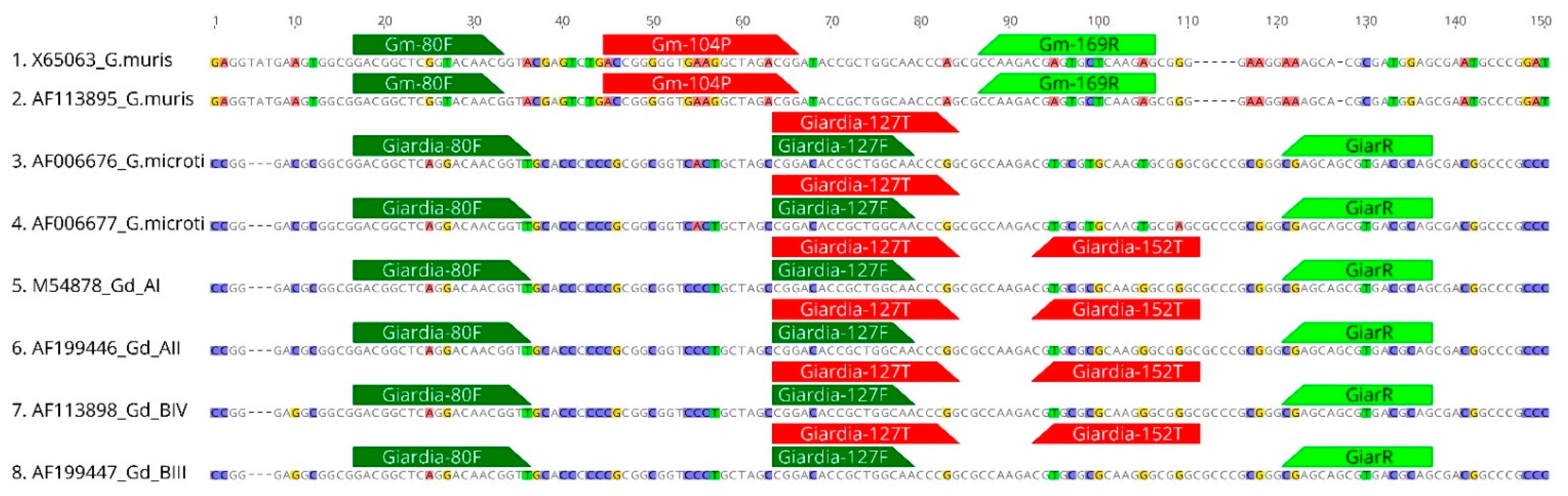
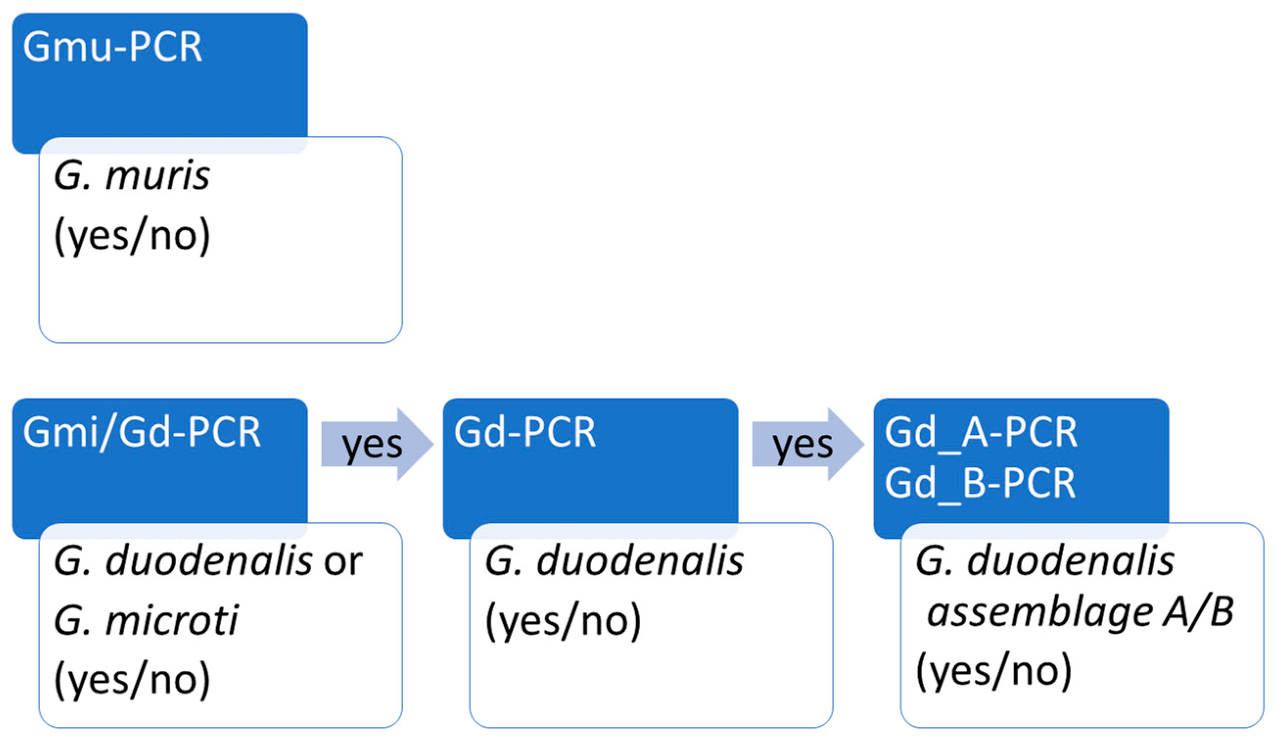
3.1. Development of Real Time PCR Workflow to Distinguish G. muris, G. microti and G. duodenalis

An approximately 250 bp SSU genome fragment (see Methods for details) was used for the primer design. At this locus, a well-established and robust real-time PCR to diagnose G. duodenalis in humans has been previously described [39]. Furthermore, significant differences at this locus sequence between G. muris on the one hand and G. duodenalis and G. microti on the other hand allowed for the design of a G. muris-specific primer and probe combination (Figure 1 and Table 1). In contrast, G. duodenalis and G. microti sequences showed high overall similarities, so that no G. microti-specific real time PCR could be designed (Figure 1). The data analysis included 104 previously identified G. microti sequences ([4], not shown), which revealed a high variability at the 3’-end of the sequence fragment, and this region was therefore not reliable for primer design.
Instead, an alternative approach was chosen to distinguish G. microti from G. duodenalis: First, sequences were evaluated for highly conserved regions shared by both G. duodenalis and G. microti, and a PCR was designed to distinguish these two species from G. muris. Secondly, the sequences were evaluated for regions that distinguish G. duodenalis from G. microti to design a G. duodenalis-specific PCR probe (Table 1 and Figure 1). In combination, these two PCR approaches allow the distinction between G. microti-positive and G. duodenalis-negative samples.
To further identify and distinguish zoonotic assemblages A and B, we used a real-time PCR assay developed by Vanni et al. 2012 [40] and modified by Pijnacker et al. 2016 [38]. Using these five PCR approaches allowed us to identify all three Giardia species and zoonotic assemblage types (see the workflow in Figure 2).

3.2. Specificity of Real-Time PCR Assays

After the primer and probe selection, we tested the specificity of the single PCR assays. We therefore used DNA from various sources, including fecal DNA samples of infected rodents and the DNA of axenic cultures or purified cysts where available. We confirmed the expected specificity of all real-time PCRs, as shown in Table 2.

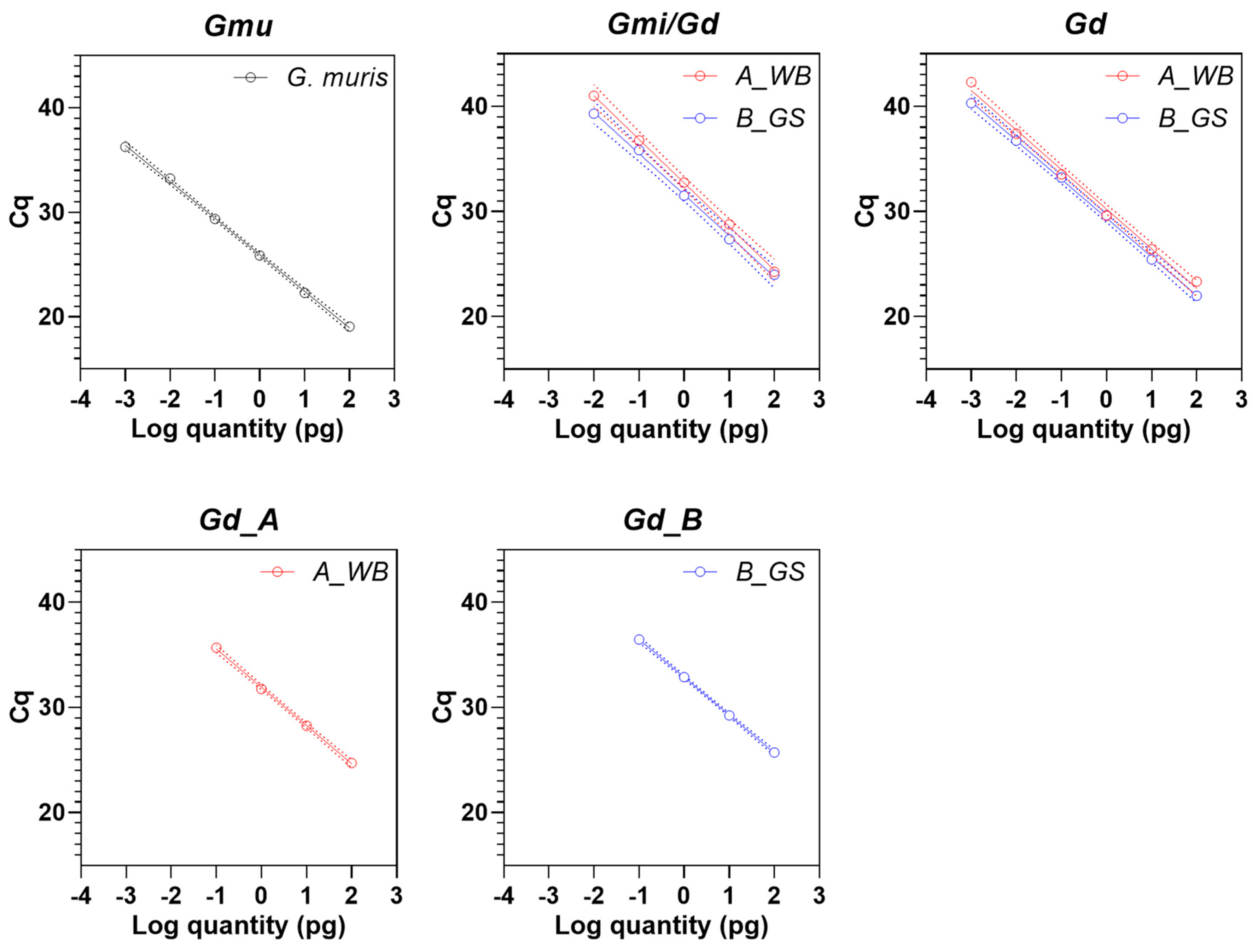
3.3. Analytical Sensitivity of Real-Time PCR Assays

To test the analytical sensitivity of the PCR assays, we used extracted DNA from axenically cultured G. duodenalis trophozoites or from purified G. muris cysts (Figure 3). We were not able to test the sensitivity of G. microti, as no adequate material was readily available for this Giardia species.
The assay designed for the detection of G. muris (Gmu PCR) indicated a high sensitivity reporting 1 fg DNA with a cq value of 36.3 ± 0.65. This equals approximately 0.1 genome equivalents for this species (estimated genome size of G. muris is 9.8 MB [41]).
The DNA of G. duodenalis assemblages A and B were equally well-detected in the G. duodenalis/G. microti (Gmi/Gd PCR) approach, with a sensitivity of 10 fg (equals about 0.7 genome equivalents, cq value 41.0 ± 1.52 for assemblage A and 39.3 ± 0.95 for assemblage B) and, in the G. duodenalis-only (Gd PCR) approach, with a sensitivity of about 1 fg DNA (42.3 ± 1.39 cq values for assemblage A and 40.3 ± 1.27 cq values for assemblage B), which equals about 0.07 genome equivalents (estimated genome size of G. duodenalis assemblage A is 12.6 MB [42]), respectively. The sensitivity of the former PCR was reproducibly lower and did not reliably detect the 1 fg DNA sample dilution.
Sensitivity of the G. duodenalis assemblage A- and B-specific PCR was also similar for both assemblage types, but only about 100 fg DNA (cq value 35.7 ± 0.29 for assemblage A and 36.5 ± 0.26 for assemblage B) or approximately 7 genome equivalents per PCR reaction were detected. The lower sensitivity was expected due to the single-copy target versus multi-copy target in the former PCRs.

3.4. Reanalysis of Known Giardia Positive Wild Rodent Samples Confirms Applicability of PCR Workflow for Detection of G. muris, G. microti and Zoonotic G. duodenalis Assemblages

To test the applicability of the new PCR approach, we reanalyzed 38 Giardia-positive samples from a previous project [4] (Table 3). These samples consisted of samples tested positive for G. muris, G. microti or G. duodenalis, respectively, as previously determined by nested PCR and the sequencing of a fragment at the SSU locus [4]. We also included samples for which the Giardia species could not be determined in the previous study.
All nine G. muris samples were confirmed by our new G. muris-specific real-time PCR. In addition, the new method revealed an underlying coinfection by a putatively non-zoonotic genotype of G. duodenalis in one sample (Isolate 301, Table 3, negative in A- and B-specific PCR). As highlighted above, the lack of a direct G. microti-specific PCR (as we were not able to appropriately design one) does not allow to formally exclude an additional underlying G. microti infection. However, in this case, a lower cq value in the Gd/Gmi combi-PCR compared to the Gd-only PCR would be expected, which was not the case.
Of the 14 G. microti samples, 12 were confirmed by our new PCR workflow. Unexpectedly, two samples exhibited a signal in the G. muris-specific PCR with relatively low cq values of 31, but no signal was recorded in the Gd/Gmi-PCR. We can only speculate on the reason and favor the possibility of an underlying double infection with a lower G. microti load that may have been amplified in the previous nested PCR approach. The nested PCR tends to amplify G. microti (and G. duodenalis) sequences with higher sensitivity than that of G. muris (see also below).
Two of the four G. duodenalis samples were also confirmed by our assay; however, two samples were not confirmed, likely due to lower sensitivity of the real-time assay compared to the nested PCR approach used in the previous study (see, also, Discussion below). One sample was further confirmed as a zoonotic assemblage type, whereas the other sample (exhibiting a high cq value in the two Gd PCR assays) was not confirmed. Again, the reason is most likely lower sensitivity of the assemblage-specific PCR that targets single-copy genes in comparison to the PCR at the multi-copy SSU locus.
Four of the 11 samples that could not be typed in our previous study displayed a positive signal in the G. muris real-time PCR, which indicates a possible underestimation of the G. muris prevalence in the previous work [4]. One sample also showed a signal in the Gd-specific PCR; however, as the Gmi/Gd PCR was negative, we do not consider this result as reliable.

4. Discussion

Here, we provided a real-time PCR workflow for the detection of G. muris, G. microti and G. duodenalis infections in small rodents, including the detection of zoonotic G. duodenalis assemblage types A and B. The workflow provided an improved time-to-results procedure for the analysis of rodent fecal samples to determine the Giardia species and for an assessment of the potential zoonotic risk.
Two rodent species are well-described to harbor the potential zoonotic G. duodenalis assemblages A and B, and the zoonotic transmission has been described [16,43]. One is the North American beaver, and a recent study provided the first in-depth molecular analysis based on whole-genome sequencing of historical outbreak samples from various water, beaver and human sources and clearly highlighted the presence of zoonotic assemblages in the water and beaver samples [16]. The sequenced samples were mostly derived from in vivo propagation in gerbils and by in vitro cultivation. This could lead to a potential bias in the propagated assemblages, and beavers may also carry other assemblages or Giardia species. Pet chinchillas harbor zoonotic G. duodenalis (mostly assemblage B) and non-zoonotic assemblages C, D and E [10,17,18,43,44,45].
For ubiquitous small mammals such as mice and voles, the potential relevance of zoonotic transmission is largely unknown. Most of the larger screens for zoonotic pathogens that included Giardia in their analyses were based on the microscopic detection of cysts and rarely included molecular tools to determine the Giardia species or assemblage types. The potential relevance for zoonotic transmissions was nevertheless often discussed [13,46]. Here, we provided a new approach that could be used in future studies to facilitate the determination of Giardia species and may help to assess the potential relevance of rodent species for zoonotic transmissions of Giardia infections.
The results presented here indicated that our workflow was sensitive and robust for such purposes. However, our method also had clear limitations. The most prominent limitation was that G. microti cannot be detected directly. We based the design of the primers and probes on previously acquired sequence information at the SSU locus of 106 unique G. microti and 10 unique G. muris sequences [4]. To our knowledge, this currently reflects the largest sequence collection available for these species. For G. microti, the sequence analysis revealed a broad variety of genotypes even at the usually highly conserved SSU gene locus [4]. Due to the sequence variety and shortage of further sequence information, we lacked the appropriate oligonucleotides for a direct G. microti-specific PCR and needed to rely on PCR signals obtained with primers detecting G. microti/G. duodenalis or solely G. duodenalis, respectively. Notably, a previously described real-time PCR for human diagnostic purposes [39] partially detected some, but not all, G. micoti genotypes (data not shown), hence providing little benefit to the current study.
The analysis revealed different sensitivities for the various PCR approaches, which was important for the interpretation of the results. However, all PCRs detected one or less genome equivalents at the SSU locus and about 10 genome equivalents at the 4E1-HP locus. This was in good concordance with previously published results using these loci [47,48]. It is unclear why G. microti/G. duodenalis-specific real-time PCR is less sensitive than G. duodenalis PCR in particular, because the same region is covered. Possibly, conformational hindrance at the gene locus may influence the binding affinity of the primer and probe. For the G. duodenalis assemblage-specific PCR, the lower sensitivity was likely due to the single-copy target gene used for detection. The lower sensitivity compared to real-time PCR at the SSU locus has been also described previously [38].
Whether the low-level detection of pathogens in real-time PCR assays, as indicated by high cq values, are indeed relevant for zoonotic transmissions should be carefully evaluated in future screening studies. For example, in our original study, an analysis of 358 SSU sequences derived from wild rodents revealed only five G. duodenalis-positive samples, and these samples were not typable at the single locus genes TPI, GDH and BG. Four of these samples were included in the present study and revealed either very low cq values for G. duodenalis qPCR or G. duodenalis was undetectable with the presented PCR methods. Only one sample revealed detectable amounts of G. duodenalis assemblage B DNA, confirming our previous typing results. Overall, this implied a very low zoonotic risk in the studied German rodent population.
In conclusion, we provided a processive and robust workflow for the detection of all Giardia species populating small rodents, including the zoonotic G. duodenalis assemblage types. This approach could be used in future studies to estimate the possible relevance for zoonotic transmissions by these animals.

Author Contributions

Conceptualization, C.K. and T.A.; methodology, C.K.; investigation, C.K., E.R. and P.G.-H.; resources, S.R.; writing—original draft preparation, C.K.; writing—review and editing, C.K., S.R. and T.A.; visualization, C.K. and supervision, C.K. All authors have read and agreed to the published version of the manuscript.

Funding

This research received no external funding.

Institutional Review Board Statement

Samples used in this study were partially obtained from experimental laboratory infections done in a different context. These procedures were conducted according to the guidelines of the Declaration of Helsinki and approved by the local authorities, the Landesamt für Gesundheit und Soziales, Berlin, Germany (G0277-17, G0207/19).

Data Availability Statement

Data is contained within the article.

Conflicts of Interest

The authors declare no conflict of interest.

References

  1. Cacciò, S.M.; Lalle, M.; Svärd, S. Host specificity in the Giardia duodenalis species complex. Infect. Genet. Evol. 2018, 66, 335–345. [Google Scholar] [CrossRef]
  2. Ryan, U.; Cacciò, S.M. Zoonotic potential of Giardia. Int. J. Parasitol. 2013, 43, 943–956. [Google Scholar] [CrossRef]
  3. Monis, P.; Caccio, S.M.; Thompson, R.A. Variation in Giardia: Towards a taxonomic revision of the genus. Trends Parasitol. 2009, 25, 93–100. [Google Scholar] [CrossRef]
  4. Helmy, Y.A.; Spierling, N.G.; Schmidt, S.; Rosenfeld, U.M.; Reil, D.; Imholt, C.; Jacob, J.; Ulrich, R.G.; Aebischer, T.; Klotz, C. Occurrence and distribution of Giardia species in wild rodents in Germany. Parasites Vectors 2018, 11, 213. [Google Scholar] [CrossRef] [PubMed] [Green Version]
  5. Thompson, A.R.C.; Monis, P.T. Taxonomy of giardia species. In Giardia: A Model Organism; Luján, H.D., Svärd, S., Eds.; Springer: Wien, Austria; New York, NY, USA, 2011; pp. 3–15. [Google Scholar]
  6. Feng, Y.; Xiao, L. Zoonotic potential and molecular epidemiology of giardia species and giardiasis. Clin. Microbiol. Rev. 2011, 24, 110–140. [Google Scholar] [CrossRef] [Green Version]
  7. Woschke, A.; Faber, M.; Stark, K.; Holtfreter, M.; Mockenhaupt, F.; Richter, J.; Regnath, T.; Sobottka, I.; Reiter-Owona, I.; Diefenbach, A.; et al. Suitability of current typing procedures to identify epidemiologically linked human Giardia duodenalis isolates. PLoS Negl. Trop. Dis. 2021, 15, e0009277. [Google Scholar] [CrossRef] [PubMed]
  8. Fayer, R.; Santín, M.; Trout, J.M.; DeStefano, S.; Koenen, K.; Kaur, T. Prevalence of Microsporidia, Cryptosporidium spp., and Giardia spp. in beavers (Castor canadensis) in Massachusetts. J. Zoo Wildl. Med. 2006, 37, 492–497. [Google Scholar] [CrossRef] [PubMed]
  9. Galán-Puchades, M.; Trelis, M.; Sáez-Durán, S.; Cifre, S.; Gosálvez, C.; Sanxis-Furió, J.; Pascual, J.; Bueno-Marí, R.; Franco, S.; Peracho, V.; et al. One health approach to zoonotic parasites: Molecular detection of intestinal protozoans in an urban population of Norway rats, Rattus norvegicus, in Barcelona, Spain. Pathogens 2021, 10, 311. [Google Scholar] [CrossRef]
  10. Lecova, L.; Hammerbauerova, I.; Tumova, P.; Nohynkova, E. Companion animals as a potential source of Giardia intestinalis infection in humans in the Czech Republic—A pilot study. Vet Parasitol. Reg. Stud. Rep. 2020, 21, 100431. [Google Scholar] [CrossRef] [PubMed]
  11. Meerburg, B.G.; Singleton, G.; Kijlstra, A. Rodent-borne diseases and their risks for public health. Crit. Rev. Microbiol. 2009, 35, 221–270. [Google Scholar] [CrossRef]
  12. Padovan, D. Infectious Diseases of Wild Rodents; Corvus Publishing Company: Anacortes, WA, USA, 2006; 348p. [Google Scholar]
  13. Perec-Matysiak, A.; Buńkowska-Gawlik, K.; Zaleśny, G.; Hildebrand, J. Small rodents as reservoirs of Cryptosporidium spp. and Giardia spp. in south-western Poland. Ann. Agric. Environ. Med. 2015, 22, 1–5. [Google Scholar] [CrossRef]
  14. Erlandsen, S.L.; Sherlock, L.A.; Bemrick, W.J.; Ghobrial, H.; Jakubowski, W. Prevalence of Giardia spp. in beaver and muskrat populations in northeastern states and Minnesota: Detection of intestinal trophozoites at necropsy provides greater sensitivity than detection of cysts in fecal samples. Appl. Environ. Microbiol. 1990, 56, 31–36. [Google Scholar] [CrossRef] [PubMed] [Green Version]
  15. Heitman, T.L.; Frederick, L.M.; Viste, J.R.; Guselle, N.J.; Morgan, U.M.; Thompson, R.C.; Olson, M.E. Prevalence of Giardia and Cryptosporidium and characterization of Cryptosporidium spp. isolated from wildlife, human, and agricultural sources in the North Saskatchewan River Basin in Alberta, Canada. Can. J. Microbiol. 2002, 48, 530–541. [Google Scholar] [CrossRef]
  16. Tsui, C.K.; Miller, R.; Uyaguari-Diaz, M.; Tang, P.; Chauve, C.; Hsiao, W.; Isaac-Renton, J.; Prystajecky, N. Beaver fever: Whole-genome characterization of waterborne outbreak and sporadic isolates to study the zoonotic transmission of giardiasis. mSphere 2018, 3, e00090-18. [Google Scholar] [CrossRef] [Green Version]
  17. Gherman, C.M.; Kalmár, Z.; Györke, A.; Mircean, V. Occurrence of Giardia duodenalis assemblages in farmed long-tailed chinchillas Chinchilla lanigera (Rodentia) from Romania. Parasites Vectors 2018, 11, 86. [Google Scholar] [CrossRef] [Green Version]
  18. Levecke, B.; Meulemans, L.; Dalemans, T.; Casaert, S.; Claerebout, E.; Geurden, T. Mixed Giardia duodenalis assemblage A, B, C and E infections in pet chinchillas (Chinchilla lanigera) in Flanders (Belgium). Vet. Parasitol. 2011, 177, 166–170. [Google Scholar] [CrossRef]
  19. Backhans, A.; Jacobson, M.; Hansson, I.; Lebbad, M.; Lambertz, S.T.; Gammelgård, E.; Saager, M.; Akande, O.; Fellström, C. Occurrence of pathogens in wild rodents caught on Swedish pig and chicken farms. Epidemiol. Infect. 2012, 141, 1885–1891. [Google Scholar] [CrossRef]
  20. Fernandez-Alvarez, A.; Martin-Alonso, A.; Abreu-Acosta, N.; Feliu, C.; Hugot, J.-P.; Valladares, B.; Foronda, P. Identification of a novel assemblage G subgenotype and a zoonotic assemblage B in rodent isolates of Giardia duodenalis in the Canary Islands, Spain. Parasitology 2013, 141, 206–215. [Google Scholar]
  21. Sulaiman, I.M.; Fayer, R.; Bern, C.; Gilman, R.H.; Trout, J.M.; Schantz, P.M.; Das, P.; Lal, A.A.; Xiao, L. Triosephosphate isomerase gene characterization and potential zoonotic transmission of Giardia duodenalis. Emerg. Infect. Dis. 2003, 9, 1444–1452. [Google Scholar] [CrossRef] [PubMed]
  22. Van Keulen, H.; Feely, D.E.; Macechko, P.T.; Jarroll, E.L.; Erlandsen, S.L. The sequence of Giardia small subunit rRNA shows that voles and muskrats are parasitized by a unique species Giardia microti. J. Parasitol. 1998, 84, 294–300. [Google Scholar] [CrossRef] [PubMed]
  23. Zhao, Z.; Wang, R.; Zhao, W.; Qi, M.; Zhao, J.; Zhang, L.; Li, J.; Liu, A. Genotyping and subtyping of Giardia and Cryptosporidium isolates from commensal rodents in China. Parasitology 2015, 142, 800–806. [Google Scholar] [CrossRef]
  24. Araujo, N.S.; Mundim, M.J.; Gomes, M.A.; Amorim, R.M.; Viana, J.C.; Queiroz, R.P.; Rossi, M.A.; Cury, M.C. Giardia duodenalis: Pathological alterations in gerbils, Meriones unguiculatus, infected with different dosages of trophozoites. Exp. Parasitol. 2008, 118, 449–457. [Google Scholar] [CrossRef]
  25. Bartelt, L.A.; Roche, J.; Kolling, G.; Bolick, D.; Noronha, F.; Naylor, C.; Hoffman, P.; Warren, C.; Singer, S.; Guerrant, R. Persistent G. lamblia impairs growth in a murine malnutrition model. J. Clin. Investig. 2013, 123, 2672–2684. [Google Scholar] [CrossRef] [PubMed] [Green Version]
  26. Fink, M.Y.; Shapiro, D.; Singer, S.M. Giardia lamblia: Laboratory maintenance, lifecycle induction, and infection of murine models. Curr. Protoc. Microbiol. 2020, 57, e102. [Google Scholar] [CrossRef]
  27. Pham, J.K.; Nosala, C.; Scott, E.Y.; Nguyen, K.F.; Hagen, K.D.; Starcevich, H.N.; Dawson, S.C. Transcriptomic profiling of high-density Giardia foci encysting in the murine proximal intestine. Front. Cell Infect. Microbiol. 2017, 7, 227. [Google Scholar] [CrossRef] [Green Version]
  28. Bajer, A. Between-year variation and spatial dynamics of Cryptosporidium spp. and Giardia spp. infections in naturally infected rodent populations. Parasitology 2008, 135, 1629–1649. [Google Scholar] [CrossRef] [PubMed]
  29. Caccio, S.M.; Beck, R.; Almeida, A.; Bajer, A.; Pozio, E. Identification of Giardia species and Giardia duodenalis assemblages by sequence analysis of the 5.8S rDNA gene and internal transcribed spacers. Parasitology 2010, 137, 919–925. [Google Scholar] [CrossRef] [Green Version]
  30. Hopkins, R.M.; Meloni, B.P.; Groth, D.M.; Wetherall, J.D.; Reynoldson, J.A.; Thompson, R.C. Ribosomal RNA sequencing reveals differences between the genotypes of Giardia isolates recovered from humans and dogs living in the same locality. J. Parasitol. 1997, 83, 44–51. [Google Scholar] [CrossRef] [Green Version]
  31. Lalle, M.; Pozio, E.; Capelli, G.; Bruschi, F.; Crotti, D.; Caccio, S.M. Genetic heterogeneity at the beta-giardin locus among human and animal isolates of Giardiaduodenalis and identification of potentially zoonotic subgenotypes. Int. J. Parasitol. 2005, 35, 207–213. [Google Scholar] [CrossRef]
  32. Read, C.; Walters, J.; Robertson, I.D.; Thompson, R.C. Correlation between genotype of Giardia duodenalis and diarrhoea. Int. J. Parasitol. 2002, 32, 229–231. [Google Scholar] [CrossRef]
  33. Sprong, H.; Caccio, S.M.; van der Giessen, J.W.; Zoopnet Network and Partners. Identification of zoonotic genotypes of Giardia duodenalis. PLoS Negl. Trop. Dis. 2009, 3, e558. [Google Scholar] [CrossRef] [PubMed] [Green Version]
  34. Thompson, R.C.; Ash, A. Molecular epidemiology of Giardia and Cryptosporidium infections. Infect. Genet. Evol. 2016, 315–323. [Google Scholar] [CrossRef] [PubMed] [Green Version]
  35. Keister, D.B. Axenic culture of Giardia lamblia in TYI-S-33 medium supplemented with bile. Trans. R Soc. Trop. Med. Hyg. 1983, 77, 487–488. [Google Scholar] [CrossRef]
  36. Appelbee, A.J.; Frederick, L.M.; Heitman, T.L.; Olson, M.E. Prevalence and genotyping of Giardia duodenalis from beef calves in Alberta, Canada. Vet. Parasitol. 2003, 112, 289–294. [Google Scholar] [CrossRef]
  37. Minetti, C.; Lamden, K.; Durband, C.; Cheesbrough, J.; Fox, A.; Wastling, J.M. Determination of Giardia duodenalis assemblages and multi-locus genotypes in patients with sporadic giardiasis from England. Parasites Vectors 2015, 8, 444. [Google Scholar] [CrossRef] [PubMed] [Green Version]
  38. Pijnacker, R.; Mughini-Gras, L.; Heusinkveld, M.; Roelfsema, J.; van Pelt, W.; Kortbeek, T. Different risk factors for infection with Giardia lamblia assemblages A and B in children attending day-care centres. Eur. J. Clin. Microbiol. Infect. Dis. 2016, 35, 2005–2013. [Google Scholar] [CrossRef] [PubMed]
  39. Verweij, J.J.; Blange, R.A.; Templeton, K.; Schinkel, J.; Brienen, E.A.; van Rooyen, M.A.; van Lieshout, L.; Polderman, A.M. Simultaneous detection of Entamoeba histolytica, Giardia lamblia, and Cryptosporidium parvum in fecal samples by using multiplex real-time PCR. J. Clin. Microbiol. 2004, 42, 1220–1223. [Google Scholar] [CrossRef] [Green Version]
  40. Vanni, I.; Caccio, S.M.; van Lith, L.; Lebbad, M.; Svard, S.G.; Pozio, E.; Tosini, F. Detection of Giardia duodenalis assemblages A and B in human feces by simple, assemblage-specific PCR assays. PLoS Negl. Trop. Dis. 2012, 6, e1776. [Google Scholar] [CrossRef]
  41. Xu, F.; Jimenez-Gonzalez, A.; Einarsson, E.; Astvaldsson, A.; Peirasmaki, D.; Eckmann, L.; Andersson, J.O.; Svard, S.G.; Jerlstrom-Hultqvist, J. The compact genome of Giardia muris reveals important steps in the evolution of intestinal protozoan parasites. Microb. Genom. 2020, 6, e000402. [Google Scholar]
  42. Xu, F.; Jex, A.; Svard, S.G. A chromosome-scale reference genome for Giardia intestinalis WB. Sci. Data 2020, 7, 38. [Google Scholar] [CrossRef] [Green Version]
  43. Tumova, P.; Mazanek, L.; Lecova, L.; Dluhosova, J.; Typovska, H.; Kotrasova, V.; Tichackova, V.; Nohynkova, E. A natural zoonotic giardiasis: Infection of a child via Giardia cysts in pet chinchilla droppings. Parasitol. Int. 2018, 67, 759–762. [Google Scholar] [CrossRef]
  44. Pantchev, N.; Broglia, A.; Paoletti, B.; Vrhovec, M.G.; Bertram, A.; Nockler, K.; Caccio, S.M. Occurrence and molecular typing of Giardia isolates in pet rabbits, chinchillas, guinea pigs and ferrets collected in Europe during 2006–2012. Vet. Rec. 2014, 175, 18. [Google Scholar] [CrossRef] [Green Version]
  45. Soares, R.M.; de Souza, S.L.; Silveira, L.H.; Funada, M.R.; Richtzenhain, L.J.; Gennari, S.M. Genotyping of potentially zoonotic Giardia duodenalis from exotic and wild animals kept in captivity in Brazil. Vet. Parasitol. 2011, 180, 344–348. [Google Scholar] [CrossRef] [PubMed]
  46. Kilonzo, C.; Li, X.; Vivas, E.J.; Jay-Russell, M.T.; Fernandez, K.L.; Atwill, E.R. Fecal shedding of zoonotic food-borne pathogens by wild rodents in a major agricultural region of the central California coast. Appl. Environ. Microbiol. 2013, 79, 6337–6344. [Google Scholar] [CrossRef] [PubMed] [Green Version]
  47. Uiterwijk, M.; Nijsse, R.; Kooyman, F.N.J.; Wagenaar, J.A.; Mughini-Gras, L.; Koop, G.; Ploeger, H.W. Comparing four diagnostic tests for Giardia duodenalis in dogs using latent class analysis. Parasites Vectors 2018, 11, 439. [Google Scholar] [CrossRef] [PubMed]
  48. Van Lith, L.; Soba, B.; Vizcaino, V.V.; Svard, S.; Sprong, H.; Tosini, F.; Pozio, E.; Caccio, S.M. A real-time assemblage-specific PCR assay for the detection of Giardia duodenalis assemblages A, B and E in fecal samples. Vet. Parasitol. 2015, 211, 28–34. [Google Scholar] [CrossRef] [PubMed]
Figure 1. Alignment of the partial SSU gene locus of selected Giardia reference sequences illustrating the position of the primers and probes of the newly developed real-time PCR approaches. Forward primers in dark green, reverse primers in light green and probes in red.
Microorganisms 09 01610 g001
Figure 2. Workflow of the real-time PCR approach used to determine G. muris, G. microti and zoonotic G duodenalis in rodent samples.
Microorganisms 09 01610 g002
Figure 3. Analytical sensitivity of the real-time PCR assays used in the study. The G. muris-specific PCR (Gmu) was done with DNA from purified cysts. G. microti/G. duodenalis PCR (Gmi/Gd), G. duodenalis-specific PCR (Gd), G. duodenalis assem-blage A-specific PCR (Gd_A) and G. duodenalis assemblage B-specific PCR (Gd_B) was done using the DNA of axenic trophozoite cultures of assemblage A isolate WB (A_WB) and of assemblage B isolate GS (B_GS).
Microorganisms 09 01610 g003
Table 1. Primers used in the study.
Target Species Specificity (Abbreviation of qPCR Assay) and OligonucleotidesOligonucleotide SequenceReference
G. muris (Gmu)
Gm80F5‘-GACGGCTCGGTACAACG-3‘This study
Gm169R5‘-CTCTTGAGCACTCGTCTTGG-3‘This study
Gm104PFAM-5‘-ACCGGGGGTGAAGGCTAGACGG-3‘-BHQ1This study
G. microti/G. duodenalis (Gmi/Gd)
Giardia-80F5‘-GACGGCTCAGGACAACGGTT-3‘[39]
GiaR5‘-CTGCGTCACGCTGCTCG-3‘[32]
Giardia-127TTexasRed-5’-CGGACACCGCTGGCAACCCGG-3’-BHQ2This study
G. duodenalis (Gd)
Giardia-127F5‘-CGGACACCGCTGGCAA-3‘This study
GiaR5‘-CTGCGTCACGCTGCTCG-3‘[32]
Giardia-152THEX-5‘-GCCCGCCCTTGCGCGCACG-3‘-BHQ2This study
G. duodenalis Assemblage A (Gd_A)
4E1-HP-Af5’-AAAGAGATAGTTCGCGATGTC-3’[38,40]
4E1-HP-Ar5’-ATTAACAAACAGGGAGACGTATG-3’[38,40]
4E1-HP-AtpVIC-5’-aggcacacggtttacaccg-3’-BHQ1[38]
G. duodenalis Assemblage B (Gd_B)
4E1-HP-Bf5’-GAAGTCATCTCTGGGGCAAG-3’[38,40]
4E1-HP-Br5’-GAAGTCTAGATAAACGTGTCGG-3’[38,40]
4E1-HP-BtpTexasRed-5’-TACACTGTTCGTATGACCACTGTCGATA-3’-BHQ2[38]
Table 2. Specifics of the qPCR-assays.
Sample SpeciesMaterial for DNA ExtractionPCR-Assay
GmuGmi/GdGdGd_AGd_B
G. murisFeces of lab mouse 1+
G. murisPurified cysts+
G. duodenalis A 2 (WB6)In vitro culture+++
G. duodenalis B (GS) 2In vitro culture+++
G. duodenalis B (GS) 2Feces of lab mouse 1+++
G. microtiFeces of wild bank vole 1+
Balamutia mandrilarisIn vitro culture
Entamoeba histolyticaIn vitro culture
Toxoplasma gondiiIn vitro culture
Leishmania donovaniIn vitro culture
1 Lab mice were experimentally infected with the respective Giardia species, and G. microti infection of the wild bank vole was previously confirmed by PCR and sequencing [4]. 2 G. duodenalis A (WB6): assemblage A, isolate WB6 and G. duodenalis B (GS): assemblage B, isolate GS.
Table 3. Analysis of Giardia-positive wild rodent samples to determine the underlying Giardia species. Reanalysis of selected Giardia-positive samples from reference [4] by the established qPCR workflow.
Rodent SpeciesIsolate # [4]Previous Result [4]qPCR-Assay (Cq-Value)
GmuGmi/GdGdGd_AGd_B
Microtus agrestis118G. muris27.6
Apodemus agrarius220G. muris35.0
Apodemus agrarius243G. muris36.0
Apodemus agrarius301G. muris36.141.337.1
Apodemus agrarius311G. muris32.5
Myodes glareolus328G. muris36.8
Myodes glareolus334G. muris27.9
Apodemus flavicollis385G. muris35.5
Apodemus flavicollis511G. muris30.1
Microtus arvalis443G. microti 36.9
Myodes glareolus451G. microti 36.6
Myodes glareolus516G. microti 34.1
Microtus arvalis603G. microti 34.8
Microtus arvalis495G. microti 34.3
Microtus arvalis496G. microti 30.9
Myodes glareolus502G. microti 36.5
Myodes glareolus508G. microti 29.1
Apodemus flavicollis518G. microti31.4
Apodemus flavicollis524G. microti31.1
Myodes glareolus559G. microti 32.9
Myodes glareolus561G. microti 29.7
Microtus arvalis566G. microti 31.9
Microtus arvalis568G. microti 31.9
Myodes glareolus041G. duodenalis Ass. A
Myodes glareolus056G. duodenalis Ass. A 40.239.6
Apodemus sp.207G. duodenalis Ass. A
Myodes glareolus340G. duodenalis Ass. B 33.030.2 34.5
Apodemus sylvaticus305Unknown 1
Apodemus flavicollis348Unknown 1 41.5
Apodemus flavicollis376Unknown 1
Apodemus sp.400Unknown 1
Apodemus flavicollis520Unknown 1
Apodemus agrarius576Unknown 132.2
Myodes glareolus554Unknown 1
Myodes glareolus555Unknown 131.1
Microtus arvalis567Unknown 135.8
Microtus arvalis569Unknown 1
Myodes glareolus591Unknown 136.2
1 Previous nested PCR approach at the SSU locus revealed no typable results [4].
Publisher’s Note: MDPI stays neutral with regard to jurisdictional claims in published maps and institutional affiliations.

