Article

The miR-214-5p/Lactoferrin/miR-224-5p/ADAM17 Axis Is Involved in Goat Mammary Epithelial Cells’ Immune Regulation

1
College of Animal Science and Technology, Northwest A&F University, Yangling 712100, China
2
Yunnan Academy of Animal Husbandry and Veterinary Sciences, Kunming 650224, China
*
Authors to whom correspondence should be addressed.
These authors contributed equally to this work.
Animals 2023, 13(18), 2835; https://doi.org/10.3390/ani13182835
Submission received: 10 July 2023 / Revised: 19 August 2023 / Accepted: 31 August 2023 / Published: 6 September 2023

Abstract

:

Simple Summary

Lactoferrin (LF) is considered to be an important active protein in goat milk, and it plays an important role in immunity. In this study, we tried to find factors affecting the expression of lactoferrin. Furthermore, we further explored the anti-inflammatory effect of LF and related molecular mechanisms in order to further understand how lactoferrin affects the immune system and provide a reference for further study and application of lactoferrin in the future.

Abstract

Lactoferrin (LF) is believed to be an important active protein in goat milk, which plays an anti-inflammatory role. Although LF has been reported to be associated with body health, its exact underlying mechanism remains unclear. Here, we aimed to elucidate the mechanism of this anti-inflammatory effect of LF in vitro. We first identified that miR-214-5p inhibited the expression of LF mRNA and protein in cells through the 3′UTR of LF mRNA. We next identified the alterations in miRNA following LF overexpression in goat mammary epithelial cells (GEMCs). Overexpression of LF significantly increased (p < 0.05) miR-224-5p expression. We further revealed that transcriptional activation of ADAM17, TNF-α, IL-1β, and IL-6 was efficiently decreased (p < 0.05) in GMECs treated by miR-224-5p mimic. Conversely, knockdown of miR-224-5p increased (p < 0.05) ADAM17, TNF-α, IL-1β, and IL-6 expression. Additionally, TNF-α, IL-1β, and IL-6 expression levels were dramatically decreased in GMECs after administration of siADAM17. Herein, we indicate that the miR-214-5p/LF/miR-224-5p/ADAM17 axis is involved in the immune regulation of GEMCs.

1. Introduction

Mastitis is a common disease in livestock breeding, which can lead to decreased milk yield, decreased milk quality and huge economic losses. Therefore, how to control the occurrence of mastitis has become the focus of attention in animal husbandry production. But there are relatively few studies on dairy goats. In goat mammary epithelial cells, IL-17A was found to promote the secretion of a variety of pro-inflammatory cytokines, chemokines, and defensive proteins. Results indicate that IL-17A may be involved in the occurrence of mastitis in dairy goats and plays a certain regulatory role in the mammary gland immune defense of goats [1]. Studies show that the expression level of antibacterial peptide S100A7 mRNA in the mammary cells of goats has a good correlation with mastitis, which could reflect the severity of mastitis in a certain range. The expression of antibacterial peptide S100A7 in goat mammary epithelial cells can be induced by LPS. LPS may be involved in regulating the expression and secretion of antimicrobial peptide S100A7 through the TLR4/NF-κB signaling pathway [2]. At this time, lactoferrin, which has good anti-inflammatory and immunomodulatory effects and other biologically active functions, has become the object of our study.
Lactoferrin (LF) is mainly present in mammalian milk, and its content is high in colostrum and low in mature milk [3,4,5]. As a widely distributed protein in biological secretions, LF plays an important role in innate immunity. A large number of studies have shown that LF has strong anti-inflammatory activity, which can not only regulate the inflammatory response of epithelial cells infected with intracellular bacteria [6] but also reduce the inflammatory response triggered by the involvement of TLR [7,8]. The mRNA expression levels of IL-6, IL-8, and TNF-α in Caco-2 cells infected with E. coli HB101 were decreased after bLF (Bovine Lactoferrin) treatment [9]. Experiments have been conducted to treat mice-fed bacterial lipopolysaccharide (LPS) with bLF before inducing systemic inflammation. The results showed that the levels of TNFα, IL-6, and IL-10 in mice significantly decreased after pre-administration of LF but no significant changes were found in the above inflammatory factors after feeding with LPS. This suggests that LF can counteract the inflammatory response induced by LPS [10]. It was also found that LF ingested by newborns can promote the establishment of the natural immune system and enhance the function of the immune system [11].
MicroRNAs (miRNAs) are small noncoding RNAs (~22 nucleotides in length) that modulate gene expression by targeting 3′-untranslated regions (3′UTRs) of mRNA transcripts [8]. Early trials found that miR-214 inhibits the expression of LF and promotes apoptosis in human mammary epithelial cells by binding to LF mRNA [12]. miRNAs can regulate the post-transcriptional silencing of target genes [13]. A single miRNA can simultaneously target hundreds of mRNAs and affect the expression of multiple genes involved in related functional interactions. And this gives us a starting point. We can explore the specific regulatory pathway of LF in the immune response of dairy goat mammary epithelial cells by looking for the upstream and downstream miRNAs and the genes regulated by those miRNAs. ADAM17 is one of the downstream genes that is associated with immunity. In cardiovascular disease, miR-26a-5p reduces apoptosis and the release of inflammatory cytokines, such as TNF-α, IL-1β, and IL-6, by targeting ADAM17 [14]. miR-708-3p reduces inflammatory responses and damage to cardiomyocytes after acute myocardial infarction by negatively regulating ADAM17 [15].
Therefore, in the present study, we focused on identifying upstream and downstream differential miRNAs of LF related to immune regulation. Based on this, we further investigated the potential molecular mechanism of LF involvement in the immune response of mammary epithelial cells in dairy goats. This study provides a theoretical basis for an LF regulatory mechanism in enhancing immune ability, paving the way for improving production efficiency in goats.

2. Materials and Methods

2.1. Ethics Statement

All animal experiments were carried out under the agreement of the Institutional Animal Use and Care Committee (Northwest A&F University, Yangling, China; permit number DK2022008; 28 February 2022). All surgeries were performed so as to minimize suffering. All animals in the present study received humane care according to the Guide for the Care and Use of Experimental Animals of the National Institutes of Health.

2.2. Mammary Tissue Collection and Cell Culture

The GMECs we used were isolated from mammary tissue washed with D-Hank’s solution preserved in our laboratory. The mammary tissue was collected from three individual 3-year-old dairy goats in their peak lactation period. The tissue sections were cut into cubes of about 1 mm3 and cultured in a petri dish at 37 °C in 5% CO2. The medium was configured according to the formula of our laboratory [16]. The culture medium was changed every 2 days until epithelial cells separated from the tissue block. Cells were subsequently digested from the tissue block with 0.25% trypsin–ethylene diamine tetraacetic acid (EDTA) solution. The differential adhesion method was used to purify GMECs by removing fibroblasts, which adhered to culture dishes 30–40 min faster than GMECs. The suspended GMECs and culture medium were transferred to new culture dishes. The purity of the GMECs after threefold purification was >99%. The GMECs obtained from a five-passage purification process were used in subsequent experiments. The culture medium contained basal DMEM/F12 (SH30023, HyClone, Logan, UT, USA) medium, 1 μg/mL hydrocortisone (H0888, Sigma, St. Louis, MO, USA), 5 μg/mL insulin (16634, Sigma), 10 ng/mL epidermal growth factor (PHG0311, Invitrogen, Carlsbad, CA, USA), 100 U/mL penicillin/streptomycin (080092569, Harbin Pharmaceutical Group, Harbin, China), and 10% fetal bovine serum (10099141, Gibco, Gaithersburg, MD, USA).

2.3. MiRNA Sequencing

A mirVana miRNA isolation kit (Thermo Fisher Ambion, Austin, TX, USA) was used to isolate small RNA from GMECs cultured in culture medium containing basal medium for 24 h. Illumina ‘s TruSeq small RNA sample preparation kit A was then used to prepare a small RNA barcode library. Additional size selection of miRNA barcodes with a band size of 147 bp was performed on a 6% TBE PAGE gel to reduce tRNA contamination. An Agilent 2100 bioanalyzer was used to determine the mass and concentration of RNA. We used the Illumina MiSeq system at Stanford’s Functional Genomics Center (SFGC) to perform sequencing of barcoded samples, which were pooled at equal molar ratios. R1 single-end sequencing files were processed in Galaxy (https://usegalaxy.org, 9 July 2023) using tools for quality control (groomer), and adaptor clipping and miRanalyzer (https://bioinfo5.ugr.es/miRanalyzer, 9 July 2023) were used to assess miRNA expression. The Norm-Expressed-Mapped data from the miRanalyzer mature unique reads were used for the scatterplot and differential expression analyses. We used half of the minimum value of the condition to replace the zero value for miRNAs with a zero value in only one condition.

2.4. Vector Construction

Based on goat LF (NC_030829.1) and the ADAM17 (XM_005687094.3) CDS we designed the clone primers. All plasmids were confirmed through sequencing before a pcDNA3.1 vector was used to clone the coding sequence of goat LF/ADAM17 between the HindIII (Takara) and XhoI (Takara) restriction sites to produce pcDNA3.1-LF and pcDNA3.1-ADAM17, respectively.

2.5. GMECs Cell Transfection

In the first experiment, GMECs were seeded in 48-well plates until 80% confluence before applying treatments. miR-214-5p were predicted to target LF 3′UTR using the TargetScan database. To overexpress miRNA, we used Lipofectamine RNAiMAX transfection reagent (13778150, Invitrogen), and, for miRNA knockdown, the inhibitor NC, miR-214-5p inhibitor, miR-214-5p mimic, and mimic NC (100 nM, RiboBio, Guangzhou, China) were separately transfected into cells. A mimic NC and an inhibitor NC were used as controls, respectively.
In the second experiment, the pcDNA3.1-LF recombinant vector was used to overexpress the LF gene in GMECs. A pcDNA3.1 vector was used as the control.
In the third experiment, GMECs were seeded in 48-well plates until 80% confluence before applying treatments. The inhibitor NC, miR-224-5p inhibitor, miR-224-5p mimic, and mimic NC (100 nM, RiboBio) were separately transfected into cells. A mimic NC and inhibitor NC were used as controls, respectively.
In the fourth experiment, small interfering RNA (siADAM17, GenePharma, Shanghai, China) was used to knock down ADAM17. The siADAM17 sequences are shown in Table 1. siNRA NCs were used as the control. The negative control siRNA (siNC) sequences were as follows: sense: 5′-UUCUCCGAACGUGUCACGUTT-3′, antisense: 5′-ACGUGACACGUUCGGAGAATT-3′. In addition, the pcDNA3.1-ADAM17 recombinant vector was used to overexpress the ADAM17 gene in GMECs. A pcDNA3.1 vector was used as the control.
In the last experiment, for co-treatment assays of miR-224-5p, cells were transfected with a total of 100 inhibitors or 100 nM siRNAs in each group (the same proportions of the two ingredients).
Lipofectamine 2000 and Lipofectamine RNAiMAX were used for the transfection of pcDNA3.1-LF/ADAM17, miRNA mimic and inhibitors (100 nM), and siRNA (100 nM) into GMECs, respectively. After 48 h from the protein extraction and RNA transfection, the GMECs were harvested. A mimic NC, inhibitor NC, and siRNA NC pcDNA3.1 were used as controls. All experiments were carried out in triplicate and repeated three times.

2.6. RNA Extraction and Quantitative Real-Time PCR (qRT-PCR)

Before applying treatments, GMECs were cultured until 80% confluence in 12-well plates. RNAiso Plus (9109, Takara) was used to extract the total RNA from each sample. The quality of RNA was detected with a NanoDrop 2000 spectrophotometer (ThermoScientific, Waltham, MA, USA). The cDNA was synthesized with a high-capacity cDNA reverse transcription kit (Applied Biosystems, Foster City, CA, USA). The miRcute cDNA First-Strand kit (KR211, Tiangen, Beijing, China) was used to synthesize the mature miRNAs’ first-strand cDNA for quantitative analysis of miRNAs. A MiRcute miRNA q-PCR kit (FP411, Tiangen) was used to amplify the reaction products, and 5S rRNA was used as an internal control for miRNAs. The primers for qRT-PCR of genes are shown in Table 1. The SYBR Green kit (RR820A, TB Green II, PerfectReal Time; Takara) was used to perform quantitative real-time PCR (qRT-PCR). As for the internal control genes, we chose ubiquitously expressed transcript (UXT) and ribosomal protein S9 (RPS9) for their stable expression during the different lactation stages of dairy goats. The relative expression values were normalized to the level of UXT and RPS9 and calculated by the 2−ΔΔCt method. Each sample was analyzed in three biological replicates.

2.7. Western Blot

Before applying treatments, GMECs were cultured until 80% confluence in 6-well plates. Radio immunoprecipitation assay (RIPA) buffer (R0010, Solarbio, Beijing, China) containing protease and phosphatase inhibitor cocktail tablets (04693132001 and 04906845001, Roche, Basel, Switzerland) was used to lyse GMECs before cellular protein was harvested. A BCA protein assay kit (23227, Thermo Scientific, Waltham, MA, USA) was used to examine the protein concentration. Sodium dodecyl sulfate–polyacrylamide gel electrophoresis (SDS-PAGE) was used to separate the protein. Transfer of the separated protein onto a PVDF membrane (Roche) was performed by Trans-Blot SD semidry transfer cell (Bio-Rad, Hercules, CA, USA). A quantity of 5% skim milk (232100, BD Biosciences, Franklin Lakes, NJ, USA) was used to block membranes for 2 h. After membranes were incubated, the secondary antibody horseradish peroxidase HRP-conjugated goat anti-rabbit-IgG (CW0103, CW Biotech; Beijing, China; 1:5000) was used for primary antibodies’ LTF. The antibody HRP-conjugated goat anti-mouse-IgG (CW0102, CW Biotech;1:5000) was used for primary antibody antisera preservation in this laboratory. The primary antibody was incubated at 4 °C for 12 h and the secondary antibody was incubated at 37 °C for 2 h. Enhanced chemiluminescence (ECL) Western blot system (1705061, Bio-Rad) was applied to measure signals. Densitometry using ImageJ software (http://imagej.nih.gov/ij/, 9 July 2023) was used to quantify the intensity of indicated bands.

2.8. Luciferase Assays

psi-CHECK2 was used as the luciferase vector. The GMECs were inoculated into 24-well plates. When the cell growth fusion degree reached about 70%, the miRNA mimics were co-transfected with the luciferase vector. First, the luciferase plasmid 0.5 ug was transfected into each well, and then the transfection was mixed well, left standing for 20 min, evenly added to each well, and shaken well. The transfection concentration of miR-214-5p mimic/NC was 50 nM, and an equal volume of Lipofectamine RNAiMAX transfection reagent was added. DMEM was then supplemented to 50 μL. The miR-214-5p inhibitor/NC transfection concentration was 100 nM, and DMEM was added to a 50 μL volume transfection reagent. The transfection compound was blown and mixed well, left for 20 min, and then uniformly added into each well and mixed by shaking. We repeated with three holes for each addition. A 24-well plate was prepared with luciferase vector and transfected, and the transfection concentrations of miR-214-5p mimic and mimic were divided into three gradient levels of 50 nM, 100 nM, and 150 nM. The transfections were performed using normal controls as control groups. Using the constructed dual-luciferase vector as a template, two pairs of mutant primers were designed for PCR amplification. The site sequence that binds to the miR-214-5p seed region in the LF 3’UTR was successfully mutated. The mutant sequence was digested and ligated with the psi-CHECK2 vector to construct psi-MUT-LF. The assay was repeated three times, adding three repeat holes to each hole. Transfection lasted for 48 h, and the dual luciferase activity was detected by enzyme labeling and the Promega reporting system.

2.9. Statistical Analysis

All experiments were repeated with at least three biological replicates. SPSS 20.0 (IBM, Chicago, IL, USA) was used to perform the statistical analysis. When only two groups were compared, the data were analyzed using Student’s t-test (two-tailed). p-values < 0.05 were considered statistically significant (* p < 0.05, ** p < 0.01). Values are presented as mean ± standard error of the mean (SEM).

3. Results

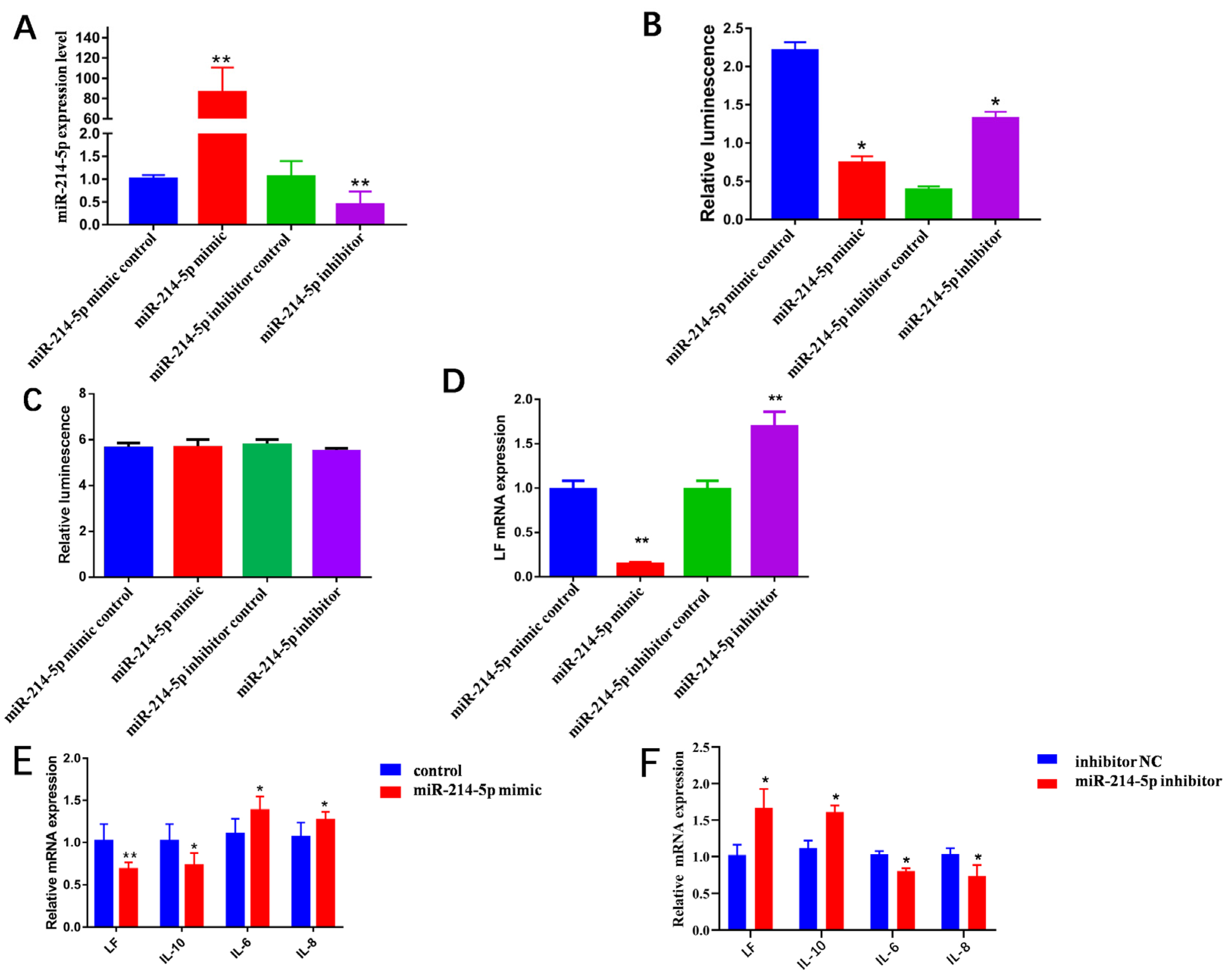
3.1. miR-214-5p Target 3′UTR of LF mRNA

The synthetic miRNA mimic/control and its inhibitor/control were diluted according to standards and transfected into GMECs, respectively. The transfected cell miRNA was extracted for quantitative detection. Results showed that the level of miR-214-5p in the cells transfected with miR-214-5p mimic increased 87 times (p < 0.01, Figure 1A) compared with the miR-214-5p mimic control group. More critically, compared with the miR-214-5p inhibitor control group, the miR-214-5p inhibitor attenuated miR-214-5p expression (p < 0.01, Figure 1A). Subsequently, compared with the miR-214-5p mimic control group, the activity of dual luciferase in cells transfected with miR-214-5p mimic was significantly decreased (p < 0.05, Figure 1B), as the miR-214-5p inhibitor had the opposite effect (p < 0.05, Figure 1B).
To target the relationship between miR-214-5p and LF, psi-MUT-LF was respectively co-transfected with miR-214-5p mimic/control or miR-214-5p inhibitor/control in GMECs. The results of applying luciferase assays after transfection with miR-214-5p mimic and inhibitor showed no significant change compared with the control (p > 0.05, Figure 1C). This indicates that miR-214-5p is bound to the 3′UTR of LF mRNA. After transfection of the miR-214-5p mimic, the intracellular LF mRNA level was significantly decreased (p < 0.01, Figure 1D). In contrast, LF expression increased in the miR-214-5p inhibitor group (p < 0.01, Figure 1D). These results indicate that miR-214-5p can inhibit the expression of LF mRNA and protein in cells.

3.2. miR-214-5p Decreases the Expression of Inflammatory Factors

The miR-214-5p mimic/inhibitor and its respective control were transfected into GMECs to quantitatively detect the expression of related inflammatory factors in cells. The results show that the levels of inflammatory factors IL-6 and IL-8 were significantly increased in cells transfected with miR-214-5p mimic (p < 0.05) and the levels of anti-inflammatory factor IL-10 were significantly decreased (p < 0.05, Figure 1E). The levels of proinflammatory cytokines IL-6 and IL-8 were significantly decreased in miR-214-5p-inhibitor-transfected cells (p < 0.05) and the levels of anti-inflammatory factor IL-10 were significantly increased (p < 0.05, Figure 1F).

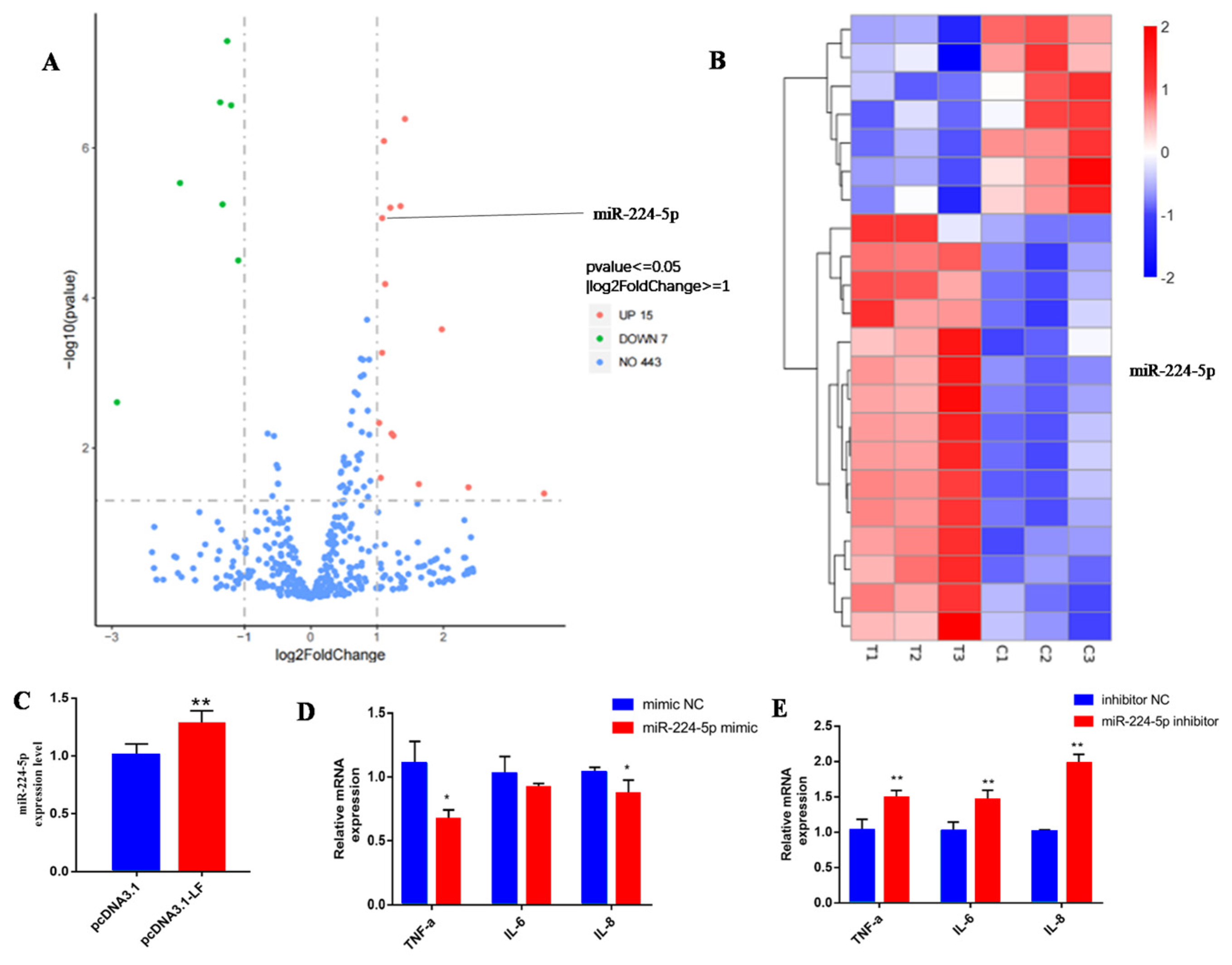
3.3. LF Decreases the Expression of Inflammatory Factors

The overexpression efficiency of LF was tested, and the results show that the expression of LF was 78 times greater than in the control group (Figure 2A). We then examined the expressions of inflammatory factors in LF-overexpressing GMECs. The results show that, in LF-overexpressing GMECs, the levels of proinflammatory factors IL-6 and IL-8 were greatly decreased, and the level of anti-inflammatory factor IL-10 was significantly increased (p < 0.05, Figure 2B).

3.4. LF Attenuates the Inflammatory Response via Targeting miR-224-5p

We next further elucidate the molecular mechanisms underlying LF-induced inflammatory response through miRNA sequencing. miRNA sequencing was performed after LF overexpression. According to statistical analysis with R software, 22 differentially expressed miRNAs were identified, including 15 up-regulated miRNAs and 7 down-regulated miRNAs. Among these differential miRNAs, miR-224-5p is related to immune regulation (Figure 3A,B). Subsequently, we verified that miR-224-5p was up-regulated in the pcDNA3.1-LF transfected GMECs through qRT-PCR methods (Figure 3C). To determine whether miR-224-5p could affect the expression of inflammation factors, the miR-224-5p mimic and inhibitor were transfected into GMECs. The results show that the mRNA levels of TNF-α and IL-8 were greatly decreased in GMECs treated with miR-224-5p mimic (p < 0.05) (Figure 3D) and the mRNA levels of TNF-α, IL-6, and IL-8 were significantly increased in GMECs treated by miR-224-5p (p < 0.01, Figure 3E). These results indicate that LF decreases the expression of inflammation factors via targeting of miR-224-5p.

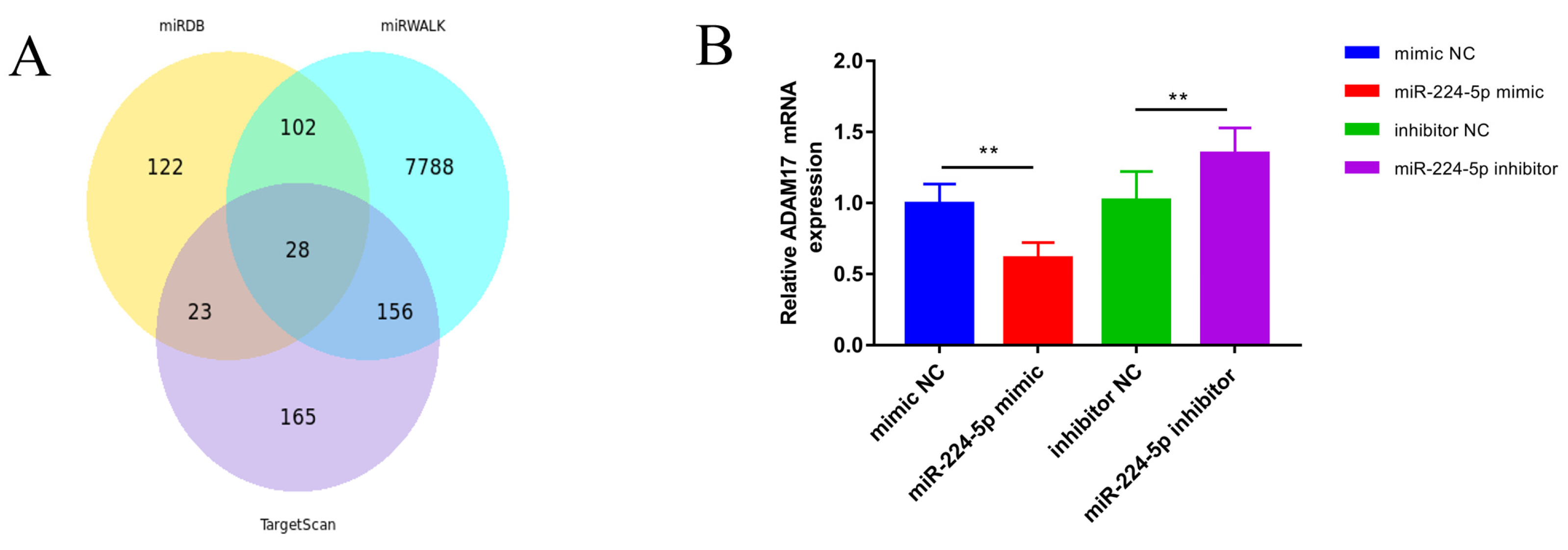
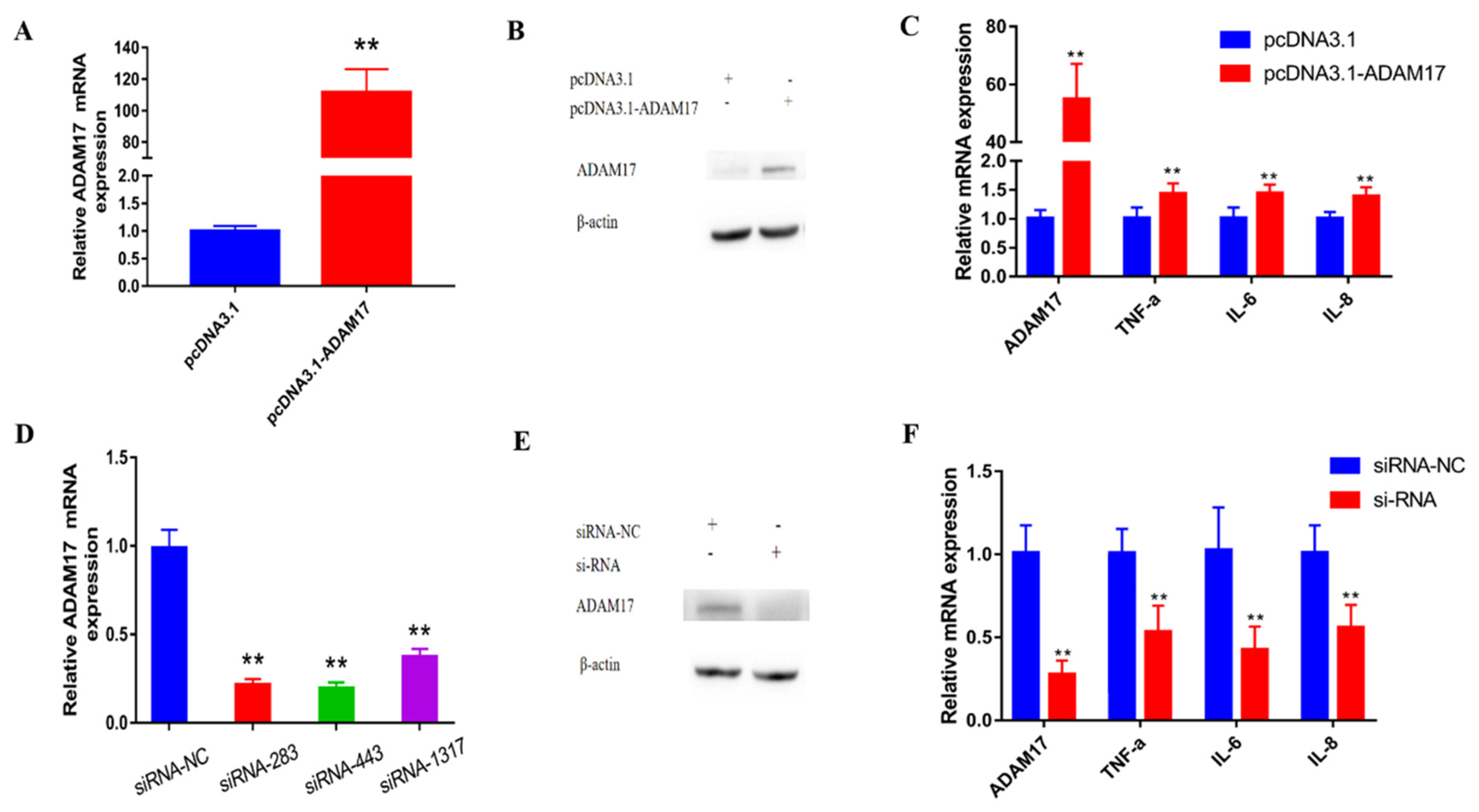
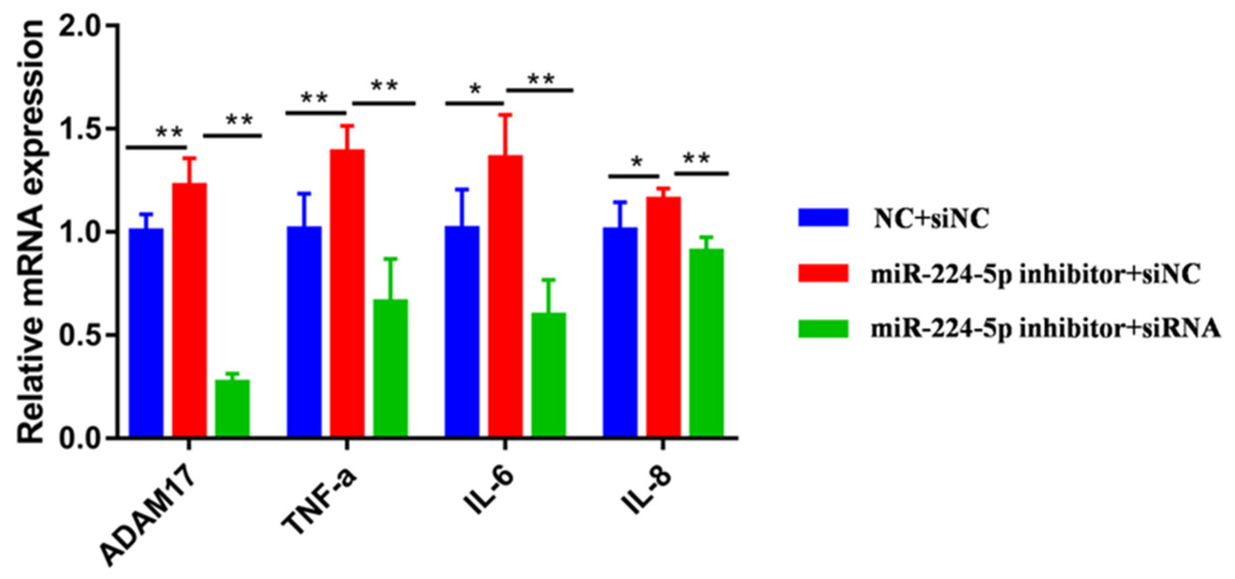
3.5. miR-224-5p Attenuates the Inflammatory Response via Targeting of ADAM17

The target genes of miR-224-5p were identified as a total of 28 target genes through prediction (Figure 4A). Among these genes, ADAM17 was found because it is related to immune regulation. miR-224-5p mimic/inhibitor and their negative controls were transfected into GMECs. Results showed that the expression of the ADAM17 gene was significantly decreased in GMECs treated with miR-224-5p mimic (p < 0.01, Figure 4B). Expression of the ADAM17 gene was significantly increased in GMECs treated by miR-224-5p inhibitor (p < 0.01, Figure 4B), showing that miR-224-5p can negatively affect ADAM17.
The mRNA and protein levels of the ADAM17 gene in the PCDNA3.1-ADAM17 treatment group increased significantly (p < 0.01, Figure 5A,B). Three different siRNA of ADAM17 could each significantly reduce the mRNA expression level of the ADAM17 gene compared to the NC group (p < 0.01, Figure 5D); the results show that transfection of siRNA-ADAM17-443 will significantly reduce the expression of ADAM17 mRNA and protein compared with the siRNA-NC group (p < 0.05, Figure 5E). At the same time, overexpression of ADAM17 significantly increased the mRNA expression levels of IL-6 (p < 0.01, Figure 5C), IL-8 (p < 0.01, Figure 5C), and TNF-α (p < 0.01, Figure 5C). In contrast, interference with the expression of ADAM17 significantly reduced the mRNA expression levels of TNF-α, IL-6, and IL-8 (p < 0.01, Figure 5F). Meanwhile, compared with the miR-224-5p inhibitor negative control and siRNA-NC groups, the expression of ADAM17 and TNF-α genes in the miR-224-5p inhibitor and siRNA-NC groups were significantly increased (p < 0.01, Figure 6). The mRNA expressions of IL-6 and IL-8 were also increased (p < 0.05, Figure 6). Next, compared with the miR-224-5p inhibitor and siRNA-NC groups, the mRNA expressions of ADAM17, TNF-α, IL-6 and IL-8 in the miR-224-5p inhibitor and the siRNA group were significantly decreased (p < 0.01). Taken together, the data we have suggest that miR-224-5p down-regulates inflammatory response via targeting of ADAM17.

4. Discussion

Excess inflammation of the breast may contribute to pathological development of mastitis disease, which in turn causes adverse consequences for lactation performance and the physical health of dairy goats. In this study, we found that LF inhibited inflammatory response in GEMCs [7,8]. Furthermore, we propose that the miR-214-5p/LF/miR-224-5p/ADAM17 axis participates in the immune regulation of GEMCs.
It was found that LF gene expression was species-specific. In the primary structure, the homology of the LF gene between humans and bovines was 66%. There was also a big difference between human and mouse LF, and LF expression also had space–time specificity. In the mouse uterus, the LF gene was only expressed at 1–8 days of pregnancy [17]. Studies have found that expression of the LF gene in the uterus is the result of the combined action of estrogen and progesterone, and changes specifically with animal estrus [18]. The concentration of LF in human milk is high, especially in colostrum, which can reach up to 6~14 mg/mL. The concentration of LF in milk decreases with the prolongation of lactation to about 1 mg/mL. LF concentration in milk is relatively low, only 100~600 μg/mL [19]. The average concentration of LF in the milk samples of goats was 10–28 μg/mL at the peak and middle stages of lactation, while it increased by 3.2 times up to 107 ± 19 μg/mL at the end of lactation [20]. It was reported that LF was able to play an anti-inflammatory role. For example, a previous study indicated that LF may be involved in the transcriptional regulation of certain genes in the host inflammatory response, thereby inhibiting pro-inflammatory cytokines as transcription factors and regulators of the inflammatory process [21]. LF has strong anti-inflammatory activity and can regulate the inflammatory response of epithelial cells [9,22,23]. Consistent with this, our study also demonstrated that LF overexpression suppressed inflammatory response with decreasing IL-6 and IL-8, as well as increasing IL-10 mRNA levels. We predicted that miR-214-5p is targeted at the 3′UTR of LF mRNA through miRanda, Targetscan, DIANATOOLS, and other databases. Furthermore, a previous study has shown that miR-214 inhibits the expression of LF and promotes cell apoptosis in human mammary epithelial cells [24]. It has been perviously suggested that MiR-214 can also regulate the expression levels of intracellular inflammatory cytokines IL-6 and IL-1β in bovine mammary epithelial cells by targeting NFATc3 and TRAF3 [25]. In addition, miR-214-5p has been shown to inhibit inflammatory response by respectively targeting HOXA13 [26], PAK4 [27], and SIRT2 [28]. In the present study, the levels of inflammatory factors IL-6 and IL-8 were significantly increased in cells after overexpression of miR-214-5p. In parallel, the level of anti-inflammatory factor IL-10 was significantly reduced. In contrast, inhibiting miR-214-5p can result in the opposite results in GMECs. Collectively, these data suggested that miR-214-5p directly targets LF and plays an important regulatory role in mammary immunity and health in goats.
To further confirm the improved regulatory network behind LF’s action, we mapped the LF regulatory gene network using miRNA-seq. By integrated analysis of these sequenced data, we found 22 differentially miRNAs directly regulated by LF overexpression. Among these differential miRNAs, miR-224-5p was reported to be associated with immune regulation. In mouse bronchial epithelial cells, overexpression of miR-224 can inhibit the expression of TLR2, significantly reduce the expression of IL-4, IL-5, and IL-17, and significantly increase the expression of IL-10, thereby inhibiting the inflammation of bronchial epithelial cells [29]. In this study, LF overexpression enhanced miR-224-5p mRNA level, which might be associated with improved anti-inflammatory capability. In line with our findings, it has been reported that the expression levels of IL-6 and TNF-α in the LPS+ mimic NC group were significantly higher than those in the LPS+ miR-224 mimic group. The expression levels of IL-6 and TNF-α in the LPS+ miR-224 inhibitor group were also significantly increased compared with the LPS+ inhibitor NC group [30]. Altogether, LF exhibited an anti-inflammatory effect in GMECs, partly via targeting inhibition of miR-224-5p.
Next, we investigated the regulation of miR-224-5p on inflammatory factors in GMECs and tried to find target genes of miR-224-5p. Utilizing target gene prediction online software, the unknown target gene ADAM17 of miR-224-5p was screened. Of note, the ADAM17 gene was one of the candidate target genes for miR-224-5p and may be related to immune regulation. ADAM17 played an important role in the regulation of inflammation, which was supported by previous findings showing that ADAM17 expression in endothelial cells affected renal inflammation and modulated renal function and histology in an obese prediabetic mouse model [31]. Furthermore, miR-224 can inhibit the growth and invasion of oral squamous cell carcinoma (OSCC) cells by targeting ADAM17 expression [32]. In this work, miR-224-5p up-regulation contributed to anti-inflammation ability by decreasing the expression of the ADAM17 gene in GMECs. Furthermore, after knocking down ADAM17, the expression of TNF-α, IL-6, and IL-8 in cells decreased. Co-transfection assays showed that miR-224-5p down-regulates the expression of goat inflammatory response via targeting ADAM17. These data demonstrate that ADAM17 is required for miR-224-5p in inhibiting inflammatory response. Altogether, this study demonstrates that the miR-214-5p/LF/miR-224-5p/ADAM17 axis can effectively regulate the expression of cytokines related to cellular inflammation.

5. Conclusions

This study has demonstrated that miR-214-5p/LF target miR-224-5p/ADAM17 and negatively affect the immune factors’ levels. Furthermore, miR-224-5p regulates transcription of the IL-6, IL-8, and TNF-α genes through ADAM17 expression. In summary, this study contributes to the exploration of the anti-inflammatory effects and molecular mechanisms of LF, providing a theoretical reference for the prevention and cure of inflammation by manipulating bioactive milk proteins in ruminants.

Author Contributions

H.S. designed and wrote the manuscript. S.P., Y.S., X.Z. and Y.Z. performed the experiments. S.P., K.S., Y.Y., Y.S., H.S., W.L. and Y.J. analyzed the data and revised the manuscript. All authors have read and agreed to the published version of the manuscript.

Funding

This research was funded by the Key Science and Technology Special Program of Yunnan Province (No. 202102AE090039), the Yunnan Science and Technology Talents and Platform Program (202205AD160003), and the National Natural Science Foundation of China (32272828).

Institutional Review Board Statement

Not applicable.

Informed Consent Statement

Not applicable.

Data Availability Statement

Data is contained within the article.

Conflicts of Interest

The authors declare no conflict of interest.

References

  1. Gao, Y.; Tong, J.Z.; Gu, K.; Gao, F.; Ma, W.T.; Chen, D.K. Effects of IL-17A on the secretion of immune defense factors in Goat mammary epithelial cells. Chin. J. Vet. Med. 2020, 40, 2205–2213. [Google Scholar]
  2. Yan, Y.T. Study on the Relationship between Antibacterial Peptide S100A7 and Mastitis in Goat Milk Cell; Northwest Agriculture and Forestry University of Science and Technology: Xianyang, China, 2021. [Google Scholar]
  3. Rachman, A.B.; Maheswari, R.R.A.; Bachroem, M.S. Composition and isolation of lactoferrin from colostrum and milk of various goat breeds. Procedia Food Sci. 2015, 3, 200–210. [Google Scholar] [CrossRef]
  4. Legrand, D.; Elass, E.; Carpentier, M.; Mazurier, J. Lactoferrin: A modulator of immune and inflammatory responses. Cell. Mol. Life. Sci. 2005, 62, 2549–2559. [Google Scholar] [CrossRef] [PubMed]
  5. Teng, C.T. Lactoferrin: The path from protein to gene. Biometals 2010, 23, 359–364. [Google Scholar] [CrossRef] [PubMed]
  6. Sessa, R.; Di Pietro, M.; Filardo, S.; Bressan, A.; Rosa, L.; Cutone, A.; Frioni, A.; Berlutti, F.; Paesano, R.; Valenti, P. Effect of bovine lactoferrin on Chlamydia trachomatis infection and inflammation. Biochem. Cell Biol. 2016, 95, 34–40. [Google Scholar] [CrossRef]
  7. Puddu, P.; Valenti, P.; Gessani, S. Immunomodulatory Effects of Lactoferrin On Antigen Presenting Cells. Biochimie 2009, 91, 11–18. [Google Scholar] [CrossRef]
  8. Puddu, P.; Latorre, D.; Carollo, M.; Catizone, A.; Ricci, G.; Valenti, P.; Gessani, S. Bovine Lactoferrin Counteracts Toll-Like Receptor Mediated Activation Signals in Antigen Presenting Cells. PLoS ONE 2011, 6, 22504. [Google Scholar] [CrossRef] [PubMed]
  9. Berlutti, F.; Schippa, S.; Morea, C.; Sarli, S.; Perfetto, B.; Donnarumma, G.; Valenti, P. Lactoferrin downregulates pro-inflammatory cytokines upexpressed in intestinal epithelial cells infected with invasive or noninvasive Escherichia coli strains. Biochem. Cell Biol. 2006, 84, 351–357. [Google Scholar] [CrossRef] [PubMed]
  10. Kruzel, M.L.; Harari, Y.; Mailman, D.; Actor, J.K.; Zimecki, M. Differential effects of prophylactic, concurrent and therapeutic lactoferrin treatment on LPS-induced inflammatory responses in mice. Clin. Exp. Immunol. 2002, 130, 25–31. [Google Scholar] [CrossRef] [PubMed]
  11. Friedman, P.C.; Farh, K.K.; Burge, C.B.; Bartel, D.P. Most mammalian mRNAs are conserved targets of microRNAs. Genome Res. 2008, 19, 92–105. [Google Scholar] [CrossRef] [PubMed]
  12. Siqueiros-Cendón, T.; Arévalo-Gallegos, S.; Iglesias-Figueroa, B.F.; García-Montoya, I.A.; Salazar-Martínez, J.; Rascón-Cruz, Q. Immunomodulatory effects of lactoferrin. Acta Pharmacol. Sin. 2014, 35, 557–566. [Google Scholar] [CrossRef] [PubMed]
  13. Bartel, D.P. MicroRNAs: Genomics, biogenesis, mechanism, and functions. Cell 2004, 116, 281–297. [Google Scholar] [CrossRef] [PubMed]
  14. Wen, X.; Yin, Y.; Li, X.; He, T.; Wang, P.; Song, M.; Gao, J. Effect of miR-26a-5p targeting ADAM17 gene on apoptosis, inflammatory factors and oxidative stress response of myocardial cells in hypoxic model. J. Bioenerg. Biomembr. 2020, 52, 83–92. [Google Scholar] [CrossRef]
  15. Qu, Y.; Zhang, J.; Zhang, J.; Xiao, W. MiR-708-3p Alleviates Inflammation and Myocardial Injury After Myocardial Infarction by Suppressing ADAM17 Expression. Inflammation 2021, 44, 1083–1095. [Google Scholar] [CrossRef] [PubMed]
  16. Hasmall, S. Downregulation of Lactoferrin by PPARalpha Ligands: Role in Perturbation of Hepatocyte Proliferation and Apoptosis. Toxicol. Sci. 2002, 68, 304–313. [Google Scholar] [CrossRef] [PubMed]
  17. Chen, L.J.; Li, J.; Meng, S. Research progress on the molecular mechanism of lactoferrin regulating the expression of NF-κB in inflammatory response. Shandong Pharm. 2016, 56, 106–109. [Google Scholar]
  18. Mcmaster, M.T.; Dey, S.K.; Andrews, G.K. Association of monocytes and neutrophils with early events of blastocyst implantation in mice. Reproduction 1993, 99, 561–569. [Google Scholar] [CrossRef] [PubMed]
  19. Shigeta, H.; Newbold, R.R.; McLachlan, J.A.; Teng, C. Estrogenic effect on the expression of estrogen receptor, COUP-TF, and lactoferrin mRNA in developing mouse tissues. Mol. Reprod. Dev. 2015, 45, 21–30. [Google Scholar] [CrossRef]
  20. Hertaviani, R.F. Nutritional Ingredients and Lactoferrin Concentration in Dairy Goat Milk from Etawah Grades and Jawarandu Breed. Ipb 2009. Available online: https://repository.ipb.ac.id/handle/123456789/59954 (accessed on 9 July 2023).
  21. Paesano, R.; Natalizi, T.; Berlutti, F.; Valenti, P. Body iron delocalization: The serious drawback in iron disorders in both developing and developed countries. Pathog. Glob. Health 2012, 106, 200–216. [Google Scholar] [CrossRef] [PubMed]
  22. Frioni, A.; Conte, M.P.; Cutone, A.; Longhi, C.; Musci, G.; di Patti, M.C.; Natalizi, T.; Marazzato, M.; Lepanto, M.S.; Puddu, P.; et al. Lactoferrin differently modulates the inflammatory response in epithelial models mimicking human inflammatory and infectious diseases. Biometals 2014, 27, 843–856. [Google Scholar] [CrossRef] [PubMed]
  23. Valenti, P.; Catizone, A.; Pantanella, F.; Frioni, A.; Natalizi, T.; Tendini, M.; Berlutti, F. Lactoferrin decreases inflammatory response by cystic fibrosis bronchial cells invaded with Burkholderia cenocepacia iron-modulated biofilm. Int. J. Immunopathol. Pharmacol. 2011, 24, 1057–1068. [Google Scholar] [CrossRef] [PubMed]
  24. Black, R.A.; Rauch, C.T.; Kozlosky, C.J.; Peschon, J.J.; Slack, J.L.; WoLFson, M.F.; Castner, B.J.; Stocking, K.L.; Reddy, P.; Srinivasan, S.; et al. A metalloproteinase disintegrin that releases tumour-necrosis factor-alpha from cells. Nature 1997, 385, 729–733. [Google Scholar] [CrossRef]
  25. Liao, Y.; Du, X.; Lonnerdal, B. miR-214 Regulates Lactoferrin Expression and Pro-Apoptotic Function in Mammary Epithelial Cells. J. Nutr. 2010, 140, 1552–1556. [Google Scholar] [CrossRef] [PubMed]
  26. Song, N.; Wang, X.; Gui, L.; Raza, S.H.A.; Luoreng, Z.; Zan, L. MicroRNA-214 regulates immunity-related genes in bovine mammary epithelial cells by targeting NFATc3 and TRAF3. Mol. Cell. Probes 2017, 35, 27–33. [Google Scholar] [CrossRef] [PubMed]
  27. Peng, J.; Peng, J.Q.; Qin, P.; Luo, X.R.; Zhang, M. miR-214-5p inhibits high glucose-induced inflammation and fibrosis in glomerular mesangial cells by regulating HOXA13. Chin. J. Gerontol. 2021, 41, 1719–1723. [Google Scholar]
  28. Yang, X.Y.; Guan, X.N.; Zhao, W.S.; Liu, J.; Zhao, L.; Yang, D. The regulatory effect of miR-214-5p targeting PAK4 on myocardial injury and immune response in rats with ischemia-reperfusion. Chin. J. Physiol. 2020, 36, 394–400. [Google Scholar]
  29. Li, P.; Wang, J.; Guo, F.; Zheng, B.; Zhang, X. A novel inhibitory role of microRNA-224 in particulate matter 2.5-induced asthmatic mice by inhibiting TLR2. J. Cell. Mol. Med. 2020, 24, 3040–3052. [Google Scholar] [CrossRef] [PubMed]
  30. Hiss, S.; Meyer, T.; Sauerwein, H. Lactoferrin concentrations in goat milk throughout lactation. Small Rumin. Res. 2008, 79, 87–90. [Google Scholar] [CrossRef]
  31. Palau, V.; Jarrín, J.; Villanueva, S.; Benito, D.; Márquez, E.; Rodríguez, E.; Soler, M.J.; Oliveras, A.; Gimeno, J.; Sans, L.; et al. Endothelial ADAM17 Expression in the Progression of Kidney Injury in an Obese Mouse Model of Pre-Diabetes. Int. J. Mol. Sci. 2021, 23, 221. [Google Scholar] [CrossRef] [PubMed]
  32. Lu, Y.; Huang, W.; Chen, H.; Wei, H.; Luo, A.; Xia, G.; Deng, X.; Zhang, G. MicroRNA-224, negatively regulated by c-jun, inhibits growth and epithelial-to-mesenchymal transition phenotype via targeting ADAM17 in oral squamous cell carcinoma. J. Cell. Mol. Med. 2019, 23, 4913–4920. [Google Scholar] [CrossRef] [PubMed]
Figure 1. The effects of miR-214 on LF and inflammatory factors. (A) Quantitative detection of miR-214 from the transfection efficiency of miRNA mimic after the miR-214-5p mimic/control and its inhibitor/control synthesized by the company were diluted according to standards and transfected into GMECs and the cell miRNA was extracted according to the instructions of the kit. (B) The constructed LF dual-luciferase vector was co-transfected with miR-214-5p mimic/control and miR-214-5p inhibitor/control into GMECs. After 48 h, the cells were collected to verify the target relationship between miR-214-5p and LF by detecting the dual luciferase activity. (C) The constructed mutant double-fluorescent plasmid was co-transfected into GMECs with 50 nM miR-214-5p mimic/control and 100 nM miR-214-5p inhibitor/control, respectively. After 48 h of culture, the cells were collected to detect the luciferase activity to detect the binding site of LF and miR-214-5p. (D) GMECs were cultured in 12-well plates and 6-well plates, miR-214-5p mimic (50 nM)/inhibitor (100 nM) transfection was performed when the confluence degree of cells reached about 70%, and the control group was transfected with the control. After 24 h of culture, 12-well cells were collected. The total RNA of the cells was extracted by the Trizol method, and RNA inversion was performed. Quantitative fluorescence PCR was performed to detect LF mRNA levels after overexpression of miR-214-5p. (E) After the cells were transfected with miR-214 mimic, the expression levels of inflammatory factors and LF in cells were quantitatively detected. (F) After the cells were transfected with miR-214 inhibitor, the expression levels of inflammatory factors and LF in cells were quantitatively detected. * Significant difference (p < 0.05). ** Extremely significant difference (p < 0.01).
Animals 13 02835 g001
Figure 2. LF regulates the expression of cellular inflammatory factors. (A) The LF overexpression vector was transfected into GMECs, and the cells were collected after 24 h of culture to quantitatively measure the mRNA level of LF. (B) All GMECs were transfected with the LF overexpression vector, and the expression of inflammatory factors was quantitatively detected. * Significant difference (p < 0.05). ** Extremely significant difference (p < 0.01).
Animals 13 02835 g002
Figure 3. The differential miRNA miR-224-5p was screened through overexpression of LF and its effects on cellular inflammatory factors. (A) The LF overexpression vector was transfected into GMECs, and a volcano map of miRNA differential expression was obtained by high-throughput sequencing analysis. (B) The LF overexpression vector was transfected into GMECs, and a heat map of miRNA differential expression was obtained by high-throughput sequencing analysis. (C) Validation of miR-224-5p fluorescence quantitative PCR results. (D) GMECs were cultured until cell density reached 60–80%; miR-224-5p mimic NC, mimic, inhibitor NC, and inhibitor were diluted in DMEM/F12, mixed with transfection reagent, and added to the cell culture medium to treat the cells. The effect of up-regulated miR-224-5p on inflammation factors was then analyzed. (E) GMECs were cultured until cell density reached 60–80%; miR-224-5p mimic NC, mimic, inhibitor NC, and inhibitor were diluted in DMEM/F12, mixed with transfection reagent, and added to the cell culture medium to treat the cells. The effect of down-regulated miR-224-5p on inflammation factors was then analyzed. * Significant difference (p < 0.05). ** Extremely significant difference (p < 0.01).
Animals 13 02835 g003
Figure 4. Verification of the target relationship between miR-224-5p and ADAM17. (A) Venn diagram of miR-224-5p target gene prediction by miRDB, miRWALK, and TargetScan. (B) GMECs were cultured until the cell density reached 60–80%; miR-224-5p mimic NC, mimic, inhibitor NC, and inhibitor were diluted in DMEM/F12, mixed with transfection reagent, and added to the cell culture medium to treat the GMECs. The effect of miR-224-5p on the mRNA level for the candidate target gene ADAM17 was then analyzed. ** Extremely significant difference (p < 0.01).
Animals 13 02835 g004
Figure 5. ADAM17 regulates the expression of cellular inflammatory factors. (A) The pcDNA3.1 high-purity plasmid and pcDNA3.1-ADAM17 high-purity plasmid were transfected into 60–80% mammary epithelial cells, respectively, and the expression of ADAM17 mRNA was detected after the overexpression. (B) The pcDNA3.1 empty vector and pcDNA3.1-ADAM17 vector were transfected into cells for 48 h, and the total protein was extracted for Western blotting to detect the change in ADAM17 gene protein level. (C) After overexpression of ADAM17, RNA was extracted from the cells after 24 h of culture, and the mRNA expression levels of ADAM17, TNF-α, IL-6, and IL-8 were detected by fluorescence quantitative technology to show the effects of ADAM17 overexpression on inflammation factors. (D) After completion of siRNA synthesis, the samples were centrifuged and diluted with RNA-free water and stored at −20 °C. GMECs were cultured, and, when the cell density reached 60% to 80%, siRNAs were diluted in DMEM/F12 and transfected at a final concentration of 100 nM. After the three interfering RNAs were transfected into cells, the interference efficiency on the mRNA level of ADAM17 was detected. (E) The interference efficiency of ADAM17 at the protein level was detected after transfecting three interfering RNAs into cells as described above. (F) After interference on ADAM17, RNA was extracted from the cells after 24 h of culture, and the mRNA expression levels of ADAM17, TNF-α, IL-6 and IL-8 were detected by fluorescence quantitative technology to show the effects of ADAM17 interference on inflammation factors. ** Extremely significant difference (p < 0.01).
Animals 13 02835 g005
Figure 6. Effects of miR-224-5p inhibitor and siRNA co-transfection on ADAM17 and inflammation factors. * Significant difference (p < 0.05). ** Extremely significant difference (p < 0.01).
Animals 13 02835 g006
Table 1. Primers Used for Quantitative Real-Time PCR of Genes.
GenePrimer Sequences (5′~3′)
LFF: TCTGGCTGCCCCGAGGAAAAACGTT
R: TCCTGCAGGCACTTGAAGGCACCAG
miR-214-5pF: ACAGCAGGCACAGACAGGCAGT
R: TCTCAGAAGCTAAACAGGGTCG
ADAM17F: GGCGCGGGAGGAATAAGAAG
R: TGTCCAGGAAATCAGAGAGCC
IL6F: AGATATACCTGGACTTCCT
R: TGTTCTGATACTGCTCTG
IL8F: AAGCTGGCTGTTGCTCTCTTG
R: TGTTCTGATACTGCTCTG
IL10F: CATGGGCCTGACATCAAGGA
R: CTCTTGTTTTCGCAGGGCAG
TNF-αF: TGGTTCAGACACTCAGGT
R: CGCTGATGTTGGCTACAA
siADAM17F: CCGCUUUGGAGACUAAUUATT
R: UAAUUAGUCUCCAAAGCGGTT
MRPL39F: AGGTTCTCTTTTGTTGGCATCC
R: TTGGTCAGAGCCCCAGAAGT
UXTF: TGTGGCCCTTGGATATGGTT
R: GTGTCTGGGACCACTGTGTCAA
Disclaimer/Publisher’s Note: The statements, opinions and data contained in all publications are solely those of the individual author(s) and contributor(s) and not of MDPI and/or the editor(s). MDPI and/or the editor(s) disclaim responsibility for any injury to people or property resulting from any ideas, methods, instructions or products referred to in the content.

