Article

Lactobacillus delbrueckii Ameliorated Blood Lipids via Intestinal Microbiota Modulation and Fecal Bile Acid Excretion in a Ningxiang Pig Model

1
Key Laboratory of Agro-Ecological Processes in Subtropical Region, Institute of Subtropical Agriculture, Chinese Academy of Sciences, Changsha 410125, China
2
Hunan Co-Innovation Center of Safety Animal Production, College of Animal Science and Technology, Hunan Agricultural University, Changsha 410128, China
3
Academician Workstation, Changsha Medical University, Changsha 410219, China
*
Author to whom correspondence should be addressed.
Animals 2024, 14(12), 1801; https://doi.org/10.3390/ani14121801
Submission received: 26 April 2024 / Revised: 7 June 2024 / Accepted: 10 June 2024 / Published: 17 June 2024

Abstract

:

Simple Summary

Ningxiang pigs are popular for their high meat quality and unique flavor, but they contain a low lean percentage. Long-term excessive intake of pork with high fat content and its products might be associated with dyslipidemia. Lactobacillus delbrueckii has been proven to regulate lipid metabolism and improve blood lipids in our previous studies; however, the underlying mechanism remains unclear. In the present study, we investigated the effects of L. delbrueckii on gut microbiota and body lipid metabolism and found that L. delbrueckii regulated hepatic and tissular fat metabolism via gut microbiota modulation and fecal TBA excretion to reduce blood lipid levels in Ningxiang pigs.

Abstract

Lactobacillus delbrueckii intervention can regulate body lipid metabolism, but the underlying mechanism remains unclear. Our study investigated the effects of L. delbrueckii on serum lipid levels, tissular fat metabolism and deposition, bile acid metabolism, and gut microbiota in Ningxiang pigs. Ninety-six pigs were divided into two groups and fed basal diets containing either 0 (CON) or 0.1% L. delbrueckii (LD) for 60 days. Dietary L. delbrueckii promoted fecal total bile acid (TBA) excretion and increased hepatic enzyme activities related to cholesterol and bile synthesis but decreased hepatic and serum lipid concentrations. L. delbrueckii downregulated gene expression associated with fatty acid synthesis but upregulated gene expression related to lipolysis and β-fatty acid oxidation in liver and subcutaneous fat. L. delbrueckii elevated gut Lactobacillus abundance and colonic short-chain fatty acid (SCFA)-producing bacteria but declined the abundance of some pathogenic bacteria. These findings demonstrated that L. delbrueckii modulated intestinal microbiota composition and facilitated fecal TBA excretion to regulate hepatic fat metabolism, which resulted in less lipid deposition in the liver and reduced levels of serum lipids.

1. Introduction

The blood lipid levels in the Chinese population have gradually increased over the past three decades [1]. Hyperlipidemia is characterized by high levels of TC, LDL-C, and TG but low levels of HDL-C in the blood [2,3]. A report from the 2016 Chinese guidelines for the management of dyslipidemia in adults showed that the average contents of serum TG and TC were 1.38 mmol/L and 4.5 mmol/L, and the prevalence of hypertriglyceridemia and hypercholesterolemia were 13.1% and 4.9% in adults, respectively [4]. Dyslipidemia is a major causative factor for atherosclerotic cardiovascular disease (ASCVD) [5], which has become the leading cause of death worldwide [6]. An epidemiologic survey revealed that the prevalence of dyslipidemia reached up to 34.0% in Chinese individuals [7]. Eating habits and lifestyle are closely associated with human health and disease. Chronic excessive intake of energy, fat, and cholesterol and a lack of physical exercise easily result in excess weight, obesity, diabetes, and ASCVDs, usually accompanied by dyslipidemia [8,9].
Pork is one of the most important animal foods for Chinese people, accounting for over 60% of national meat consumption and contributing to approximately 50% of global pork output [9]. However, long-term excessive intake of pork with high fat content and its products might be associated with dyslipidemia due to overconsumption of energy and fat, resulting in hyperlipidemia, cardiovascular disease, and type 2 diabetes, usually characterized by high levels of blood total cholesterol and triglyceride [9,10]. Therefore, clarifying the mechanisms of fat metabolism in pigs and developing targeted regulatory strategies for lipid metabolism have promising implications for pork production, which might help control dyslipidemia via dietary interventions. Additionally, given the high similarity in anatomy, physiology, polyphagy, habits, and metabolism, swine may be a good model for studying human nutrient metabolism and health [11].
Ningxiang pigs, a local fat-type breed with less than 35% lean percent, are popular for their tender and succulent meat, unique flavor, higher intramuscular fat (IMF) content, and high-quality unsaturated fatty acid content compared to imported commercial pigs (lean type) [12,13]. Lean and fat pigs with distinct genetic characteristics, as indicated by great differences in fat deposition, are valuable models for exploring the mechanisms of fat metabolism [14]. The lipid-lowering effects of Lactobacillus delbrueckii in lean pigs have been confirmed in our previous studies [3,9,15]; however, the role of the strain in lard-type pigs is unclear. In this study, we investigated the effects of L. delbrueckii on intestinal microbiota composition, hepatic fat metabolism, tissue fat deposition, and blood lipid levels in Ningxiang pigs.

2. Materials and Methods

2.1. Bacterial Strain

Lactobacillus delbrueckii (Storage No.: M207096) was acquired from the microbiology functional laboratory at the College of Animal Science and Technology at the Hunan Agricultural University (Changsha, China). The strain was activated in MRS (de Man, Rogosa, and Sharpe) medium and sent to the PERFLY-BIO (Changsha, China) for scale-up production, and the detected viable counts of final products reached 5 × 10¹¹ CFU/g.

2.2. Animals, Diets and Experimental Design

Ninety-six healthy Ningxiang pigs with similar age or initial body weight (110 ± 2 d, 33.28 ± 1.15 kg) were randomly assigned to 2 groups with 6 pens, and each pen had 8 pigs (the same ratio of female to male). Pigs were provided basal diets supplemented with either 0 (CON) or 0.1% L. delbrueckii (5 × 10¹⁰ CFU/g, LD) for 60 days. The basal diets (Table 1) were formulated according to the standard of NY/T 65-2004 to meet or exceed the requirement for 30–50 kg and 50–80 kg pigs, respectively [16]. All pigs were fed their respective diets twice daily (8:00 a.m. and 15:00 p.m.) ad libitum, and water was available all the time. Pigs in each pen were weighed together at the start and end of each period, and the daily feed consumption per pen was recorded. Fecal samples from each pen were collected for 3 consecutive days before the conclusion of the feeding experiment. On day 61, one pig per pen (3 male and 3 female pigs per group) with similar body weight was selected to collect anterior venous blood and then slaughtered by electric shock to isolate the gut, liver, subcutaneous fat, and Longissimus dorsi.

2.3. Sample Collection and Preparation

Serum was obtained, aliquoted, and stored at −20 °C for lipid analysis. Digesta (in the ileum and colon) and tissues (in the liver, Longissimus dorsi, and subcutaneous fat) were quickly removed, snap-frozen in liquid nitrogen, and stored at −80 °C for microbiota composition, lipid, enzyme activity, and gene mRNA expression measurements.

2.4. Determination of Lipid Contents in Serum and Tissue

The concentrations of serum triglyceride (TG), total cholesterol (TC), low-density lipoprotein cholesterol (LDL-C), and high-density lipoprotein cholesterol (HDL-C) were determined by the BS 200 automatic blood biochemical analyzer (Mindray, Shenzhen, China) with corresponding kits.
The total protein contents (g protein/L) in the liver, Longissimus dorsi, and subcutaneous fat were quantified using BCA protein assay reagent kits (Nanjing Jiancheng Bioengineering Institute, Nanjing, China). About 100 mg of selected tissues were homogenized with 1 mL of a chloroform/methanol solution (2:1, v/v). The homogenate was centrifuged at 3000 r/min for 10 min at 4 °C to extract tissue lipids. The contents of TC (mmol/g.prot), TG (mmol/g.prot), and TBA (μmol/g·protein) in the selected tissue were measured by corresponding commercial kits (Nanjing Jianchen Bioengineering Institute, Nanjing, Jiangsu, China).

2.5. Determination of Enzyme Activity Related to Fat Metabolism

Hepatic total protein contents (g protein/L) were quantified using a BCA protein assay reagent kit (Nanjing Jiancheng Bioengineering Institute, Nanjing, Jiangsu, China). The contents of hepatic fatty acid synthetase (FAS, U/g.prot), adipose triglyceride lipase (ATGL, U/g.prot), β-hydroxy-β-methylglutarate monoyl coenzyme A reductase (HMGR, U/g.prot), and cholesterol-7α hydroxylase (CYP7A1, U/g.prot) were detected using corresponding commercial ELISA kits (Jiangsu Meimian Industrial Co., Ltd., Yancheng, Jiangsu, China).

2.6. RT-PCR to Determine the mRNA Expression of Genes Related to Fat Metabolism

The total RNA of tissues in the liver and subcutaneous fat was isolated and reverse-transcribed to cDNA as previously described [9]. The two-step qRT-PCR reactions were performed in triplicate on 96-well plates using a 7500 real-time PCR system (Applied Biosystems, Foster, CA, USA) with the SYBR® Premix Ex TaqTM (TaKaRa Biotechnology (Dalian), China). The primer sequences (Table 2) for FAS, lipoprotein lipase (LPL), carnitine palmitoyltransferase (CPT1), peroxidase proliferative receptor γ (PPARγ), and glyceraldehyde-3-phosphate dehydrogenase (GAPDH) were synthesized by Sangon Biotech (Shanghai, China). Target gene expression was calculated using the 2−ΔΔt method relative to GAPDH gene amplification.

2.7. Fecal TC and TBA Content Determination

Fecal lipids were extracted as described above for TC analysis. Fecal TBA was extracted according to our previous description [3]. Briefly, 1 g of frozen fecal sample was dissolved in 40 mL of methanol. After 4 min of sonication and 1 h of shock, the mixture was centrifuged at 10,000× g for 10 min to collect the supernatants. Total TC (mmol/L) and TBA (μmol/L) concentrations in the supernatants were determined using a commercial kit purchased from the Nanjing Jianchen Bioengineering Institute. Finally, fecal TC and TBA contents (mg/g) were obtained by formula conversion.

2.8. Intestinal Bacterial Composition and Structure

Microbiota composition and structure in ileal and colonic digesta were analyzed via 16S rDNA sequencing, as described in our previous study [3]. Briefly, total DNA was extracted and purified from ileal and colonic digesta samples (n = 4 pigs/group) using a TIANamp Stool DNA kit (Tiangen Biotech (Beijing) Co., Ltd., China). DNA quality and quantity were evaluated by gel electrophoresis and a NanoDrop ND-1000 spectrophotometer (Thermo Fisher Scientific, Waltham, MA, USA), respectively. Finally, eight acceptable DNA samples were delivered to LC-Bio (Hangzhou, China) for 16S rDNA sequencing.
The V3-V4 hypervariable region of the bacterial 16S rDNA gene was amplified with barcoded universal primers (341F-806R). Purified amplicons were sequenced on the Illumina HiSeq platform (Illumina, San Diego, CA, USA) according to the standard procedures in LC-Bio (Hangzhou, China). Sequences with 97% similarity were assigned to the same operational taxonomic units (OTUs). An OTU table was further generated to record the abundance of each OTU in each sample, and a profiling histogram was made using R software (v3.1.1) to represent the relative abundance of taxonomic groups from phylum to species. A Venn diagram was generated to visualize the occurrence of shared and unique OTUs among groups.

2.9. Analysis of SCFA Composition in Gut Contents

The concentrations of acetic acid, propionic acid, and butyric acid in ileal and colonic digesta were determined using an Agilent 7890A gas chromatographer (Agilent Technologies Inc., Palo Alto, CA, USA) according to our previous method [9,17].

2.10. Statistics Analysis

All results were expressed as mean ± SD. Statistical analysis was conducted by the two-tailed unpaired Student’s t-test of SPSS 17.0 (SPSS Inc., Chicago, IL, USA), with an individual pig as an experimental unit. The Ribosomal Database Project (RDP, V.11.3), Greengenes (V.13_8), and NCBI 16SMicrobial were used for data analysis, comparison, and annotation of 16S rDNA sequences. Shapiro–Wilk’s test was used to check the normal distribution of the data. The Kruskal test was used for post hoc comparisons of taxonomies. Pearson correlation analysis between the differential microbiota and measured SCFAs was conducted by GraphPad Prism 7.0 (GraphPad Software Inc., San Diego, CA, USA). For all tests, p < 0.05 was considered a significant difference, while 0.05 < p < 0.10 was considered a tendency.

3. Results

3.1. L. delbrueckii did Not Affect the Growth Performance

Compared with the CON group, Dietary L. delbrueckii did not affect the average daily weight (ADG, 643 g vs. 658 g) or average daily feed intake (ADFI, 2382 g vs. 2389 g) of Ningxaing pigs.

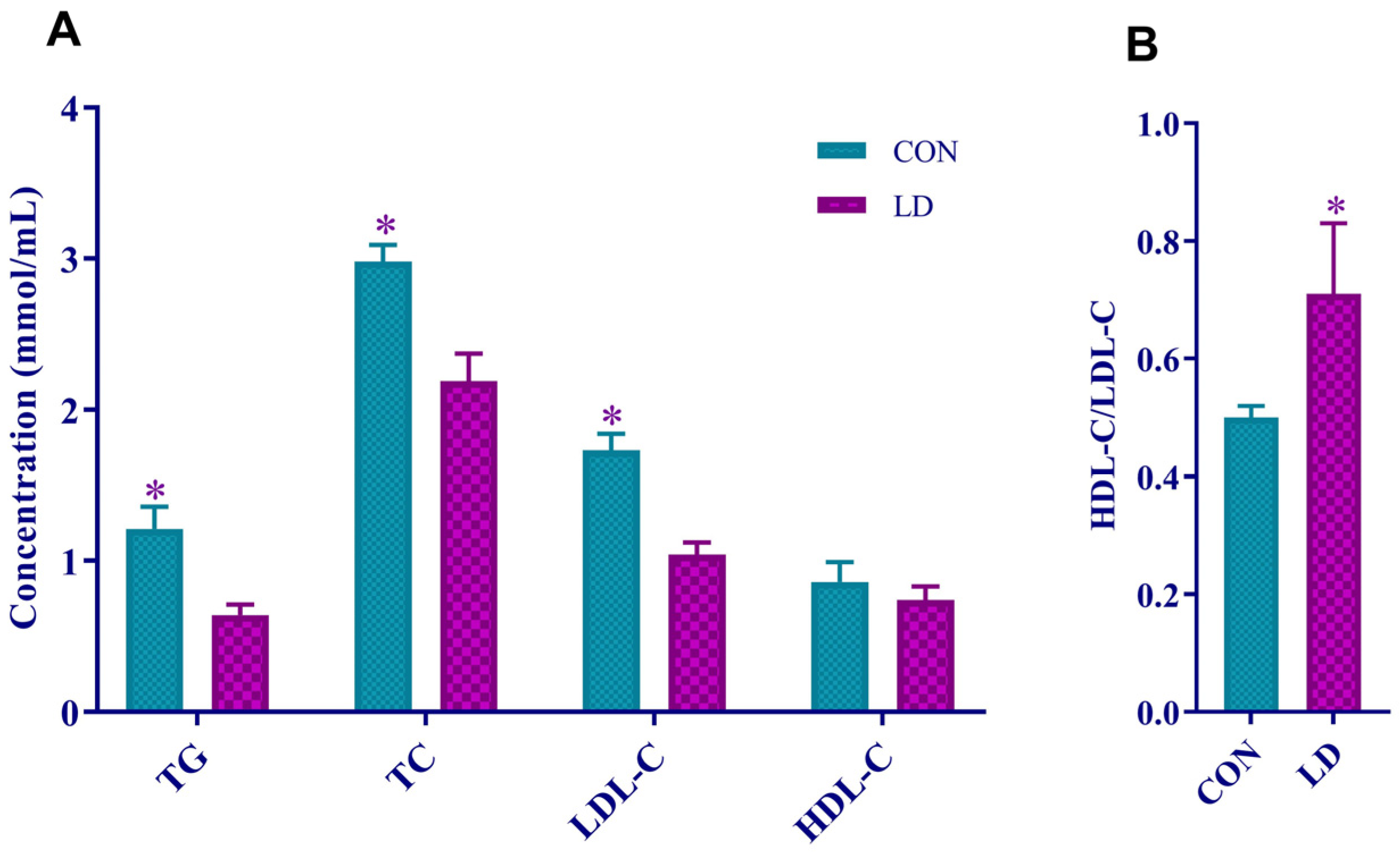
3.2. L. delbrueckii Reduced Serum Lipid Levels

Serum TG, TC, and LDL-C contents in LD were significantly decreased (Figure 1A) relative to those in CON. However, the serum HDL-C/LDL-C in LD was notably increased (Figure 1B) compared with CON.

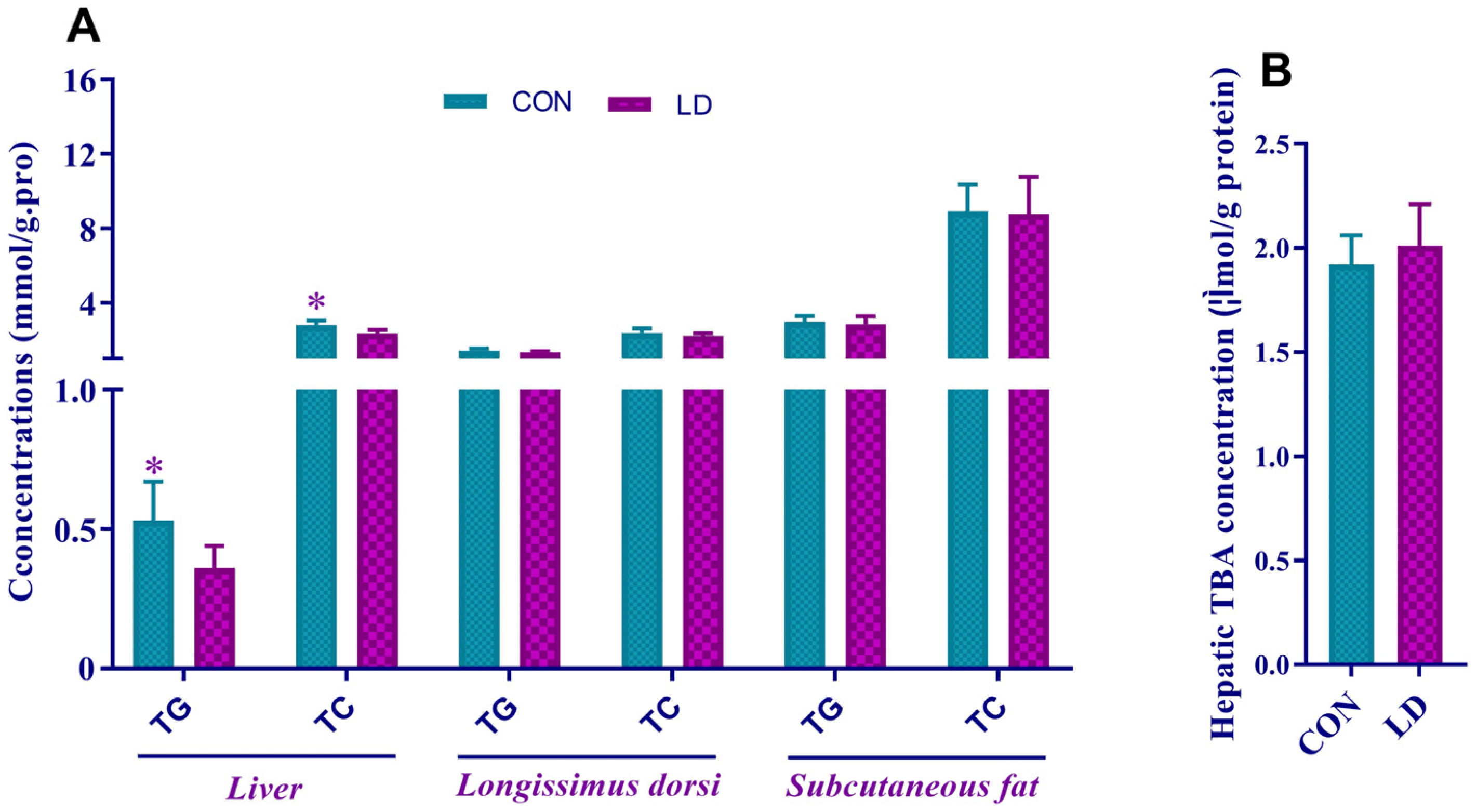
3.3. L. delbrueckii Altered Lipid Contents in Liver

Dietary L. delbrueckii remarkably reduced the hepatic concentrations of TG and TC (Figure 2A) but did not change the TG and TC contents in Longissimus dorsi and subcutaneous fat (Figure 2A) or hepatic TBA content (Figure 2B).

3.4. L. delbrueckii Affected Hepatic Cholesterol and Bile Acid Metabolism

Compared with CON, hepatic HMGR activity tended to elevate in LD (Figure 3A, p = 0.092). The increased activity of hepatic CYP7A1 appeared in LD (Figure 3A), but no differences in the hepatic activities of FAS and ATGL were observed between the two groups (Figure 3B,C).

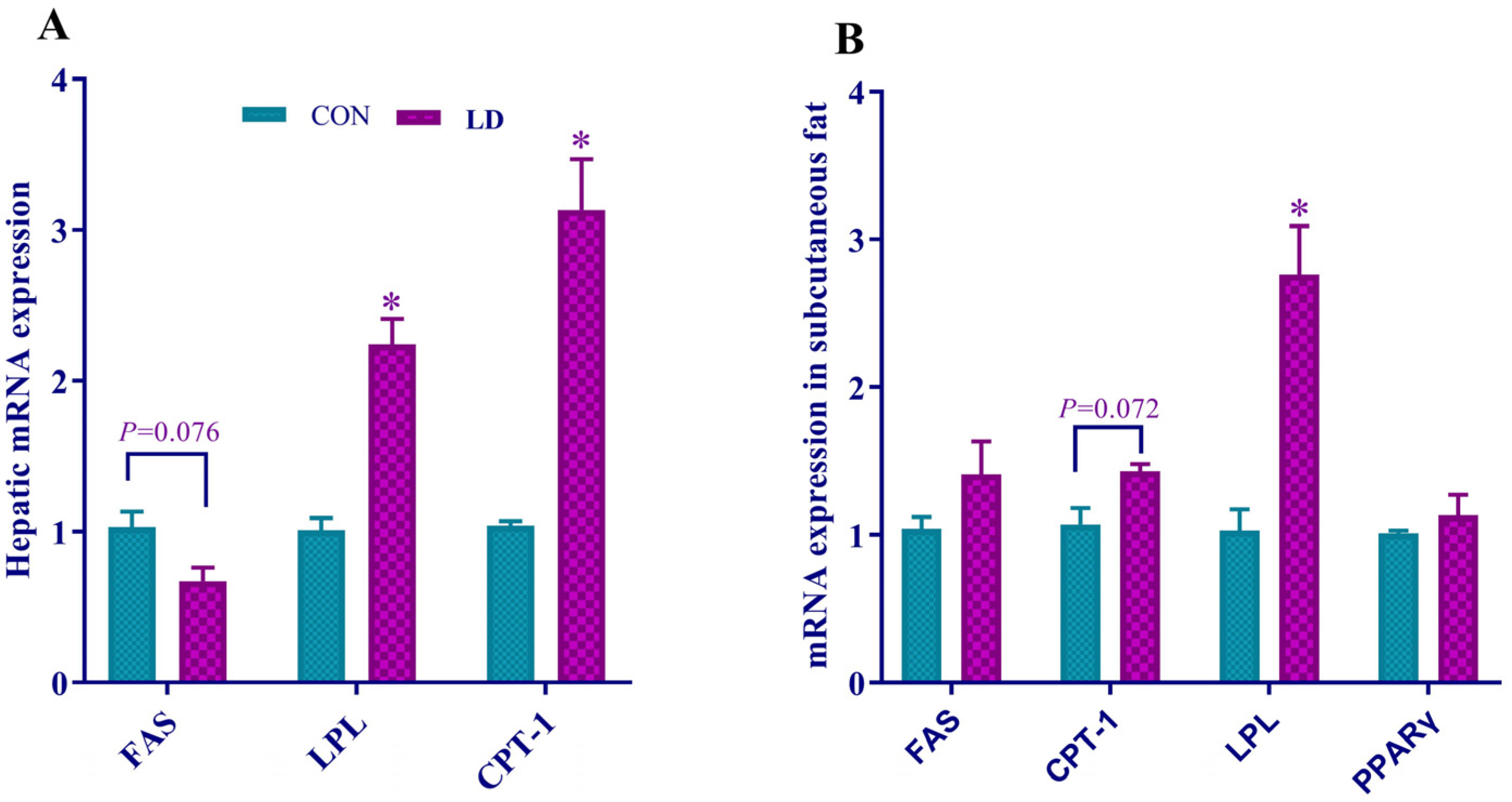
3.5. L. delbrueckii Regulated Gene Expression Related to Fatty Acid Synthesis, Lipolysis and β-oxidation in Liver and Subcutaneous Fat

Hepatic mRNA expression of FAS tended to downregulate (p = 0.076), but LPL and CPT-1 were significantly upregulated by LD (Figure 4A). The upward trend of CPT-1 (p = 0.072) and increased expression of LPL in subcutaneous fat were found in LD (Figure 4B).

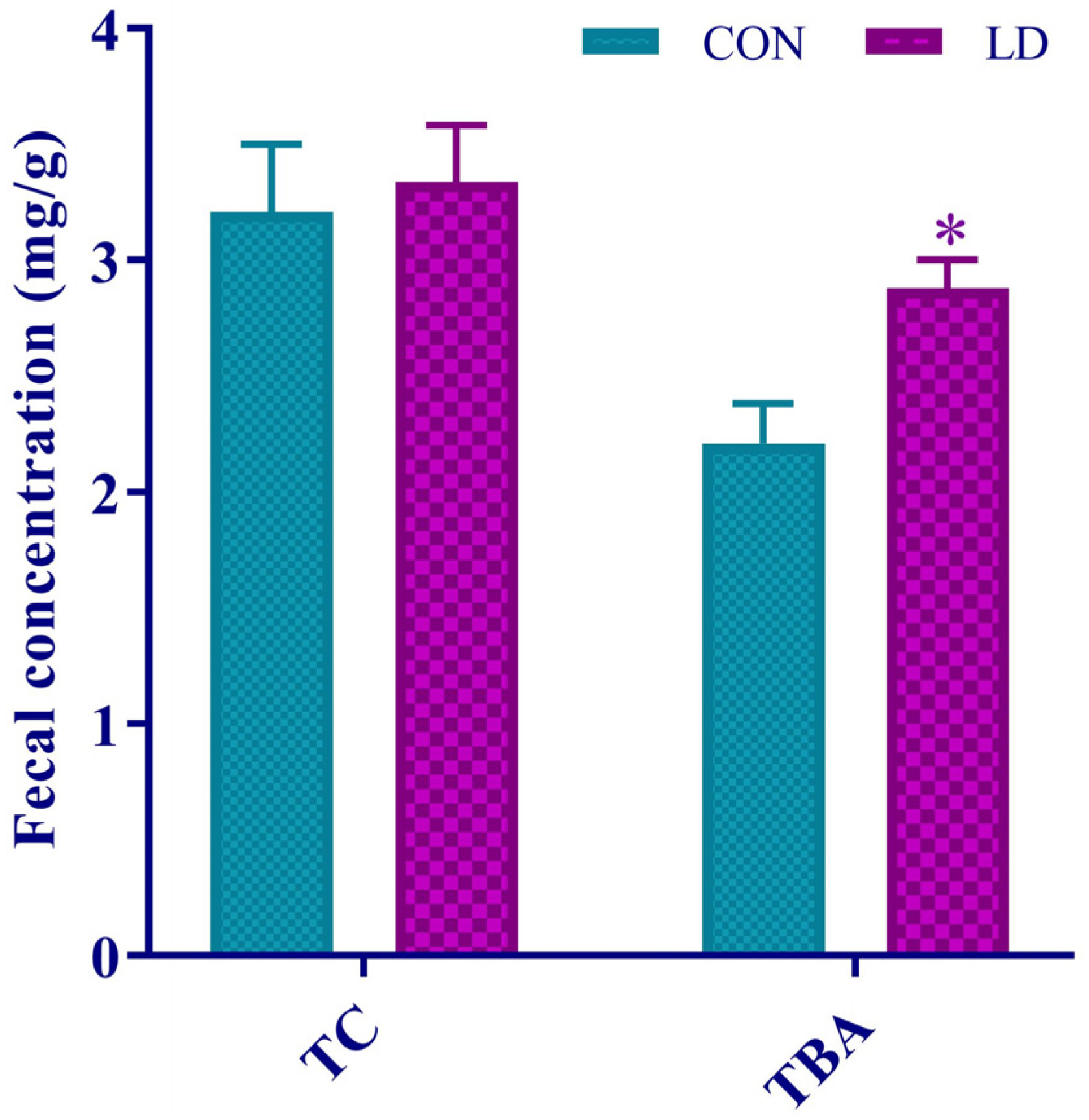
3.6. L. delbrueckii Facilitated Fecal TBA Excretion

Dietary L. delbrueckii notably increased fecal TBA concentrations but did not affect fecal TC contents (Figure 5).

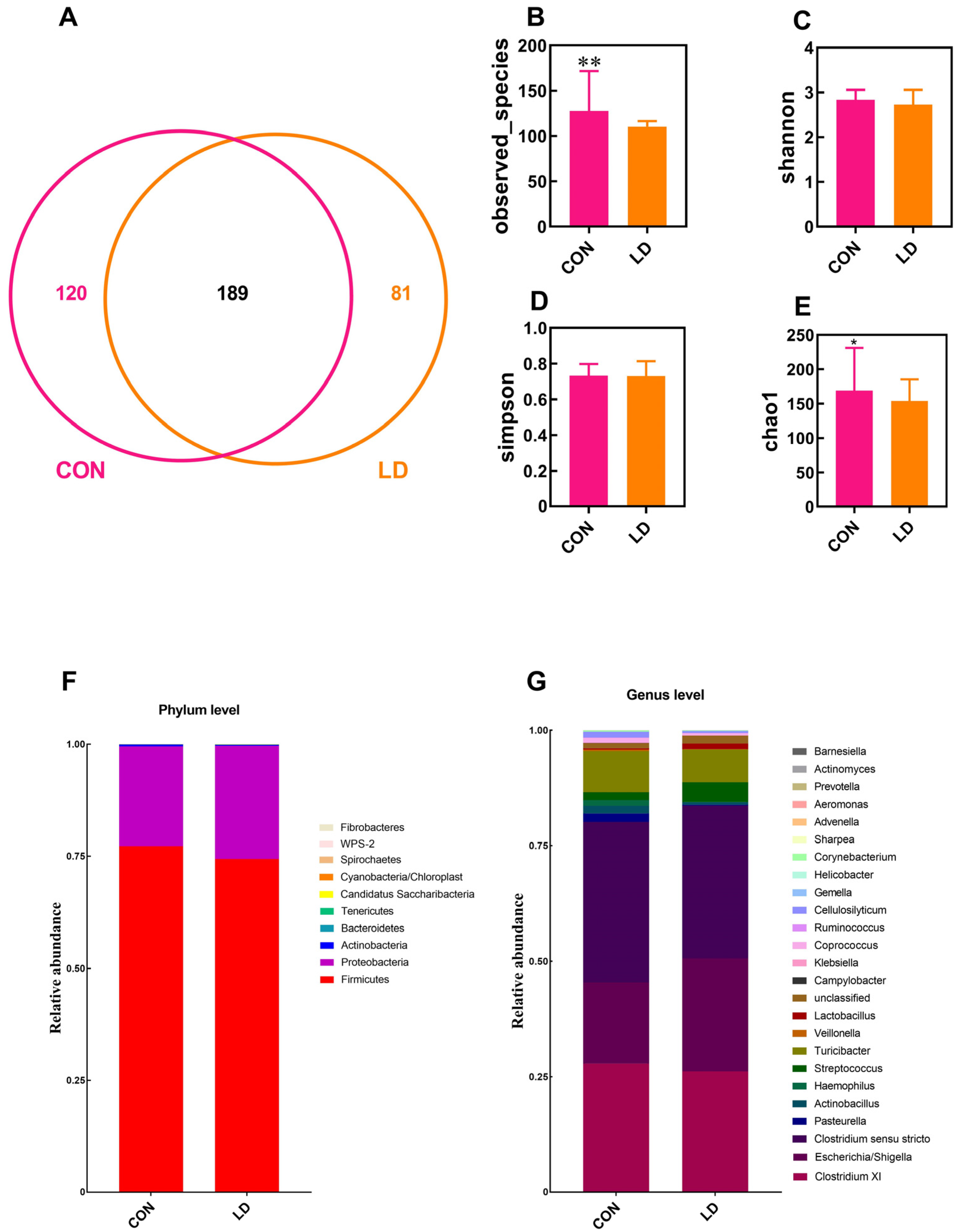
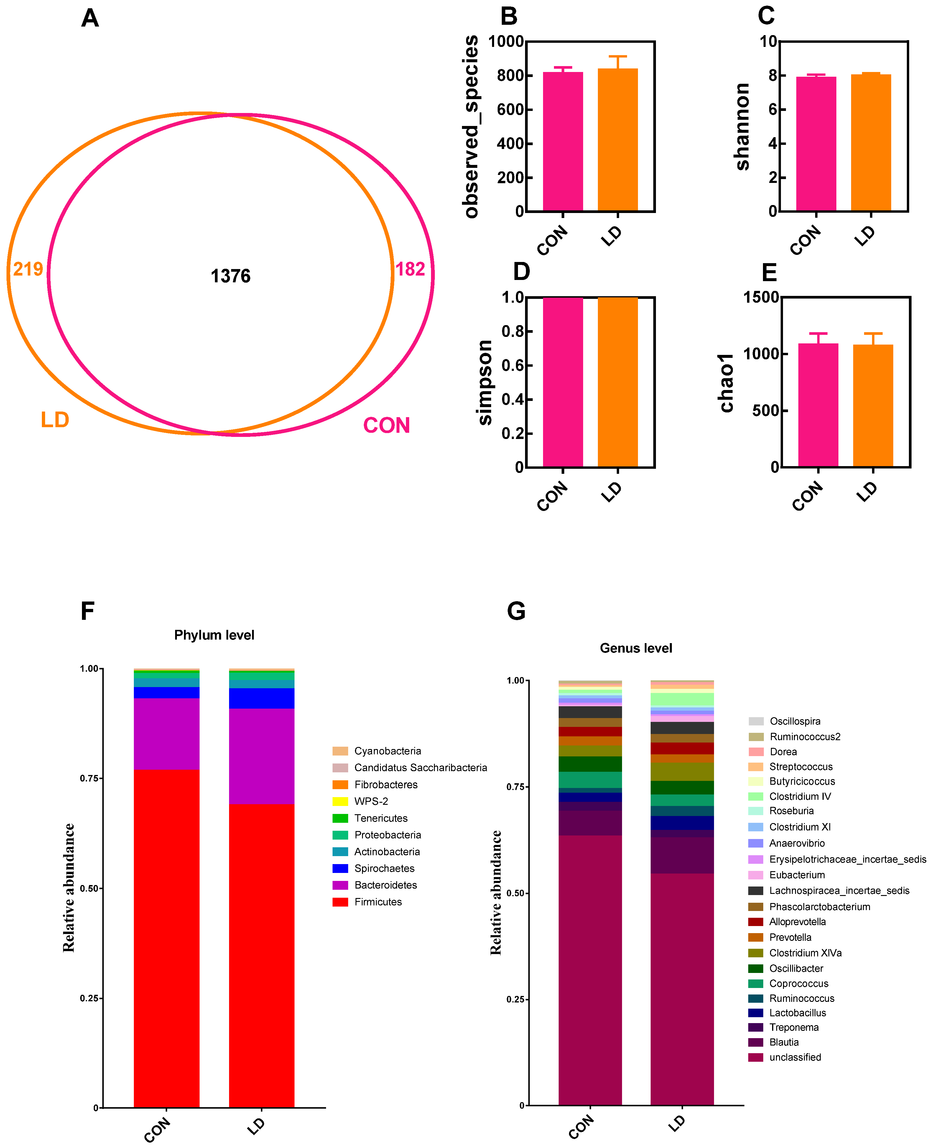
3.7. L. delbrueckii Altered Intestinal Microbiota Structure

The microbiota structure in the ileal digesta was analyzed by 16S rDNA sequencing (Figure 6). The Venn diagram identified 189 shared OTUs between the two groups, and 120 and 81 unique OTUs were observed in the CON and LD groups, respectively (Figure 6A). The α-diversity analysis showed that the observed species (Figure 6B) and Chao1 (Figure 6E) indices were markedly reduced in LD in contrast to CON. The dominant phyla present in ileal digesta were Firmicutes and Proteobacteria, accounting for more than 99% of the total OTUs (Figure 6F and Table S1), and dietary treatment did not affect ileal bacterial taxa at the phylum level. At the genus level, the relative abundances of Pasteurella, Actinobacillus, Haemophilus, Streptococcus, Veillonella, and Cellulosilyticum were significantly decreased (Figure 6G and Table S1), whereas the relative abundance of Lactobacillus was markedly increased.
The microbiota analysis of colonic digesta is presented in Figure 7. The Venn diagram displayed 182 unique OTUs in CON, 219 unique OTUs in LD, and 1376 shared OTUs in the two groups (Figure 7A). The α-diversity indices of colonic bacteria were not different between the two groups (Figure 7B–E). At the phylum level, Firmicutes, Bacteroidetes, Spirochaetes, and Actinobacteria were the four dominant bacterial populations, accounting for more than 98% of total OTUs (Figure 7F and Table S2). The Tenericutes and WPS-2 abundances in LD were significantly reduced relative to those in CON. At the genus level, compared with CON, LD notably increased Butyricicoccus abundance but decreased Anaerovibrio abundance (Figure 7G and Table S2). The abundances of Lactobacillus (p = 0.065), Streptococcus (p = 0.073), and Doreaabundance (p = 0.087) tended to increase, while the abundances of Eubacterium (p = 0.096), Clostridium IV (p = 0.056), and Ruminococcus2 (p = 0.065) had a downtrend in LD.

3.8. L. delbrueckii Changes Intestinal SCFA Profiles

Compared with the CON group, the ileal contents of acetic acid decreased, and the total SCFAs tended to decline in the LD group (Table 3). Higher butyric acid contents were observed in the LD group than in the CON group, and the colonic concentrations of propionic acid and total SCFAs tended to increase in the LD group relative to the CON group.

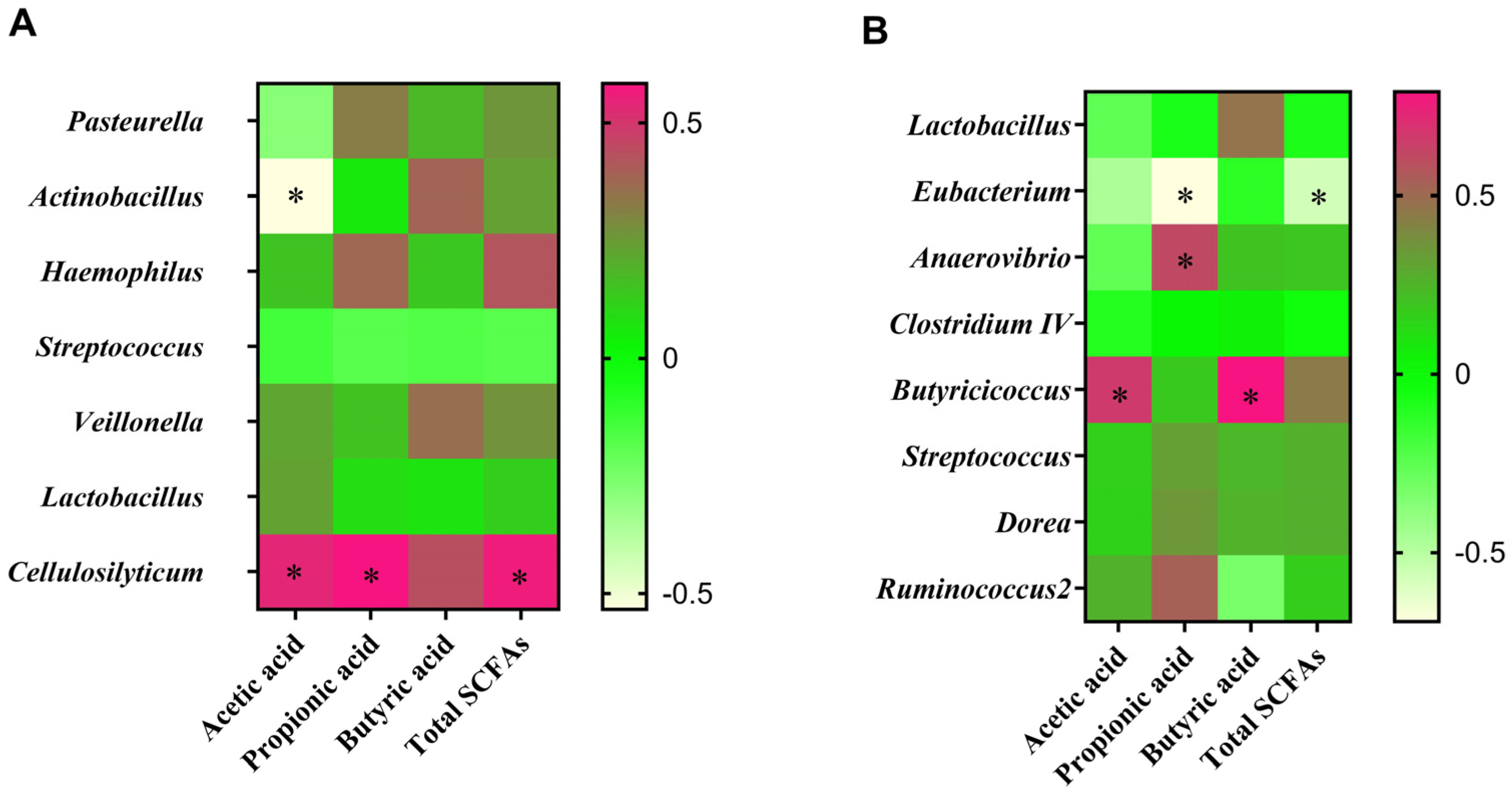
3.9. Correlation between Intestinal Microbiota and SCFA Contents

The Pasteurella abundance had a negative correlation with acetic acid content in the ileal digesta (Figure 8A), whereas the Cellulosilyticum abundance was positively associated with the concentrations of acetic acid, propionic acid, and total SCFAs. There was a positive relationship between colonic Anaerovibrio and propionic acid (Figure 8B), while a negative correlation was observed between Eubacterium and propionic acid or total SCFAs. Meanwhile, Butyricicoccus had a positive association with acetic or butyric acid.

4. Discussion

Blood lipids are closely associated with human health. Long-term high TG, TC, and LDL-C levels, along with low HDL-C levels, characterize hyperlipidemia, inducing cardiovascular disease (CVD) [3,18]. Probiotic intervention is a potential strategy for the prevention and treatment of hyperlipidemia. Lactobacillus, a common probiotic, has been extensively studied and used in humans and animals. Some reviews on the lipid control of Lactobacillus demonstrated that the consumption of Lactobacillus exerted beneficial effects on the improvement of serum lipids [19,20]. In this study, dietary L. delbrueckii lowered the serum TG, TC, and LDL-C concentrations in Ningxiang pigs, coinciding with our previous results [3,9,15]. Our recent findings indicated that the serum lipid-lowering effects of L. delbrueckii in pigs were primarily mediated by its bile salt hydrolase (BSH) activity, intestinal microbiota modulation, and SCFA-mediated fat metabolism [3,9].
The swine intestine hosts a vast, complex, and dynamic microbial community, exhibiting a vital role in host metabolism and health [21,22]. The marked shifts in the relative abundance of gut microbiota from the phylum to the genus level are closely bound up with various host physiological statuses [21]. Pasteurella, Actinobacillus, Haemophilus, and Streptococcus are prevalent bacterial pathogens associated with respiratory tract infections in the swine industry [23,24,25], resulting in inflammation and impaired growth performance [26]. Veillonella is tightly linked to intestinal dysbiosis and disease status, leading to inflammation [27]. Interestingly, we found that the ileal abundances of Pasteurella, Actinobacillus, Haemophilus, Streptococcus, and Veillonella were lowered by L. delbrueckii addition. Additionally, consistent with our previous studies [3,9], greater ileal Lactobacillus abundance appeared in the LD group, possibly a cause of the reduction in pathogenic bacteria due to competitive inhibition. These findings suggested that dietary L. delbrueckii maintained and improved foregut health by lowering the abundance of some pathogenic bacteria, which provided an explanation for the growth promotion of Ningxiang pigs in previous experiments [28]. The abundance of Cellulosilyticum was positively related to pig growth due to the characteristics of cellulose and starch decomposition [29,30]. Surprisingly, the ileal abundance of Cellulosilyticum was decreased by L. delbrueckii, likely due to fewer substrates in the ileum for the strain to use because of the increased foregut digestibility. Additionally, the reduced concentrations of ileal acetic acid and total SCFAs in the study also indicated that ileal fermentation was weakened due to a lack of substrates.
The hindgut is the main site for fermentation, and undigested nutrients in the distal ileum will flow into the large bowel for microbial fermentation [17,31]. Most SCFAs are generated in colon fermentation, and more than 95% of them are rapidly absorbed and transported into the blood and subsequently taken up and metabolized by body organs as substrates or signal molecules, participating in energy and lipid metabolism [9]. Anaerovibrio, Butyricicoccus, Eubacterium, Clostridium IV, and Ruminococcus2 are SCFA-producing bacteria. We observed that dietary L. delbrueckii increased colonic butyric acid and SCFA contents but reduced colonic propionic acid. Butyric acid is a preferred energy substrate for colonocytes and is almost fully used onsite, whereas acetate and propionate move to the liver via the portal vein for cholesterol and fatty acid synthesis and gluconeogenesis. We also found that dietary L. delbrueckii elevated Butyricicoccus, Lactobacillus, Streptococcus, and Dorea abundance but declined Anaerovibrio, Eubacterium, Clostridium IV, and Ruminococcus2 abundance. Butyricicoccus is a common butyrate-producing bacteria [32], and Anaerovibrio is known to produce propionate as a major fermentation product [33]. Eubacterium was positively linked with visceral fat mass in people [34]. Clostridium IV and Ruminococcus2 are closely related to obesity caused by excessive calorie intake and blood glucose [35,36]. Additionally, we found that the Firmicutes abundance and F/B ratio were numerically increased, while the abundance of Bacteroidetes was numerically reduced by L. delbrueckii in the colon, which was negatively associated with obesity [9]. The above results indicated that L. delbrueckii affects host fat and energy metabolism by altering gut microbiota composition and metabolites.
The liver is the center of lipid synthesis and metabolism in the body, playing a key role in maintaining the dynamic balance of blood lipids. HMGR and CYP7A1 are key enzymes for cholesterol and bile acid synthesis in the liver, respectively [37]. Our results showed that L. delbrueckii increased hepatic HMGR and CYP7A1 activities, suggesting that the synthesis of cholesterol and bile acid might be enhanced. However, the hepatic TC content was reduced in the LD group, possibly due to cholesterol conversion to bile acids. Cholesterol is a precursor to bile acid synthesis, and increased bile acid synthesis requires more cholesterol, which in turn enhances cholesterol synthesis [3,38]. Meanwhile, dietary L. delbrueckii accelerated fecal bile acid excretion, suggesting that fewer bile acids returned to the liver via enterohepatic circulation. As a result, hepatic bile acid synthesis was boosted to compensate for the loss of enterohepatic circulation. This result also provides an explanation for the enhanced hepatic HMGR and CYP7A1 activities in Ningxiang pigs. FAS and ATGL are key enzymes in fatty acid synthesis and decomposition, respectively [39]. Dietary L. delbrueckii did not affect the activities of FAS and ATGL in the liver but downregulated hepatic FAS expression and upregulated hepatic CPT-1 and LPL expression. FAS is responsible for the de novo synthesis of fatty acids. Although the mRNA expression of FAS was downregulated, its enzyme activity did not change, likely not affecting fatty acid synthesis. Curiously, hepatic TC concentrations were reduced by L. delbrueckii, which might be due to enhanced lipolysis. LPL is in charge of lipolysis, and CPT-1 is a key enzyme in fatty acid β-oxidation [40]. We observed that CPT-1 and LPL expression were increased in the liver and subcutaneous fat, suggesting that L. delbrueckii supplementation might promote lipolysis, which adds new evidence for the lipid control of Lactobacillus.

5. Conclusions

Lactobacillus delbrueckii regulates hepatic and tissular fat metabolism via intestinal microbiota modulation and fecal bile acid excretion to reduce serum TC and TG levels in Ningxiang pigs (Figure S1).

Supplementary Materials

The following supporting information can be downloaded at: https://www.mdpi.com/article/10.3390/ani14121801/s1, Figure S1: Underlying mechanism by which L. delbrueckii lowers blood lipid levels in Ningxiang pigs; Table S1: Microbiota composition at phylum and genus level in ileal digesta; Table S2: Microbiota composition at phylum and genus level in colonic digesta.

Author Contributions

Conceptualization, data curation, and supervision, G.H., R.L. and X.H.; investigation and methodology, G.H., L.W., R.L. and F.C.; resources and software, R.L. and J.Y.; writing and editing, G.H. and R.L.; funding acquisition, Y.Y., R.L. and X.H. All authors have read and agreed to the published version of the manuscript.

Funding

This work was jointly supported by the National Key Research and Development Program (2021YFD1300201, 2021YFD1301004), the National Natural Science Foundation of China (U20A2055, 32172761), the Natural Science Foundation of Hunan Province (2022JJ40532), the Key Science and Technology Special Program of Yunnan Province (202202AE090032), and the Open Fund of Key Laboratory of Agro-ecological Processes in Subtropical Region, Chinese Academy of Sciences (ISA2021103, ISA2023201).

Institutional Review Board Statement

All animal work was approved by the Animal Care and Use Committee (Approval No.: IACUC#201302) in the Institute of Subtropical Agriculture, Chinese Academy of Sciences (Changsha, China).

Informed Consent Statement

Not applicable.

Data Availability Statement

The data presented in this study are available in this article.

Acknowledgments

The authors are grateful for help with microbiota sequencing and data processing from the workers at LC-Bio (Hangzhou, China).

Conflicts of Interest

The authors declare no conflicts of interest.

References

  1. Tang, F.; Wang, J.; Nicholas, S.; Qian, D.; Liu, R. A cohort study on risk factors of high-density lipoprotein cholesterol hypoli pidemia among urban chinese adults. Lipids Health Dis. 2021, 20, 20. [Google Scholar] [CrossRef] [PubMed]
  2. Jia, X.; Xu, W.; Zhang, L.; Li, X.; Wang, R.; Wu, S. Impact of gut microbiota and microbiota-related metabolites on hyper lipidemia. Front. Cell Infect. Microbiol. 2021, 11, 634780. [Google Scholar] [CrossRef]
  3. Hou, G.; Peng, W.; Wei, L.; Li, R.; Yuan, Y.; Huang, X.; Yin, Y. Lactobacillus delbrueckii interfere with bile acid enterohepatic circulation to regulate cholesterol metabolism of growing-finishing pigs via its bile salt hydrolase activity. Front. Nutr. 2020, 7, 617676. [Google Scholar] [CrossRef] [PubMed]
  4. Hu, D.Y. New guidelines and evidence for the prevention and treatment of dyslipidemia and atherosclerotic cardiovascular disease in China. Chronic Dis. Transl. Med. 2016, 3, 73–74. [Google Scholar] [CrossRef] [PubMed]
  5. Walldius, G.; de Faire, U.; Alfredsson, L.; Leander, K.; Westerholm, P.; Malmström, H.; Ivert, T.; Hammar, N. Long-term risk of a major cardiovascular event by apob, apoa-1, and the Apob/Apoa-1 ratio-experience from the swedish amoris cohort: A cohort study. PLoS Med. 2021, 18, e1003853. [Google Scholar] [CrossRef] [PubMed]
  6. Borrelli, M.A.; Turnquist, H.R.; Little, S.R. Biologics and their delivery systems: Trends in myocardial infarction. Adv. Drug Deliv. Rev. 2021, 173, 181–215. [Google Scholar] [CrossRef]
  7. Zhang, H.; Xu, Z.; Zhao, H.; Wang, X.; Pang, J.; Li, Q.; Yang, Y.; Ling, W. Anthocyanin supplementation improves anti-oxidative and anti-inflammatory capacity in a dose-response manner in subjects with dyslipidemia. Redox Biol. 2020, 32, 101474. [Google Scholar] [CrossRef]
  8. Meeran, M.F.N.; Azimullah, S.; Al Ahbabi, M.M.; Jha, N.K.; Lakshmanan, V.K.; Goyal, S.N.; Ojha, S. Nootkatone, A dietary fragrant bioactive compound, attenuates dyslipidemia and intramyocardial lipid accumulation and favorably alters lipid metabolism in a rat model of myocardial injury: An in vivo and in vitro study. Molecules 2020, 25, 5656. [Google Scholar] [CrossRef] [PubMed]
  9. Hou, G.F.; Yin, J.; Wei, L.K.; Li, R.; Peng, W.; Yuan, Y.; Huang, X.G.; Yin, Y.L. Lactobacillus delbrueckii might lower serum triglyceride levels via colonic microbiota modulation and scfa-mediated fat metabolism in parenteral tissues of growing-finishing pigs. Front. Vet. Sci. 2022, 9, 982349. [Google Scholar] [CrossRef]
  10. Ma, H.M.; Jiang, J.; He, J.; Liu, H.F.; Han, L.L.; Gong, Y.; Li, B.; Yu, Z.G.; Tang, S.G.; Zhang, Y.B.; et al. Long-read assembly of the chinese indigenous ningxiang pig genome and identification of genetic variations in fat metabolism among different breeds. Mol. Ecol. Resour. 2022, 22, 1508–1520. [Google Scholar] [CrossRef]
  11. Olayanju, A.; Jones, L.; Greco, K.; Goldring, C.E.; Ansari, T. Application of porcine gastrointestinal organoid units as a poten tial in vitro tool for drug discovery and development. J. Appl. Toxicol. 2019, 39, 4–15. [Google Scholar] [CrossRef] [PubMed]
  12. He, J.; Wu, X.L.; Zeng, Q.H.; Li, H.; Ma, H.M.; Jiang, J.; Rosa, G.J.M.; Gianola, D.; Tait, R.G.; Bauck, S. Genomic mating as sustainable breeding for chinese indigenous ningxiang pigs. PLoS ONE. 2020, 15, e0236629. [Google Scholar] [CrossRef] [PubMed]
  13. Jiang, Q.M.; Li, C.Y.; Yu, Y.N.; Xing, Y.T.; Xiao, D.F.; Zhang, B. Comparison of fatty acid profile of three adipose tissues in ningxiang pigs. Anim. Nutr. 2018, 4, 256–259. [Google Scholar] [CrossRef] [PubMed]
  14. Nakajima, I.; Kojima, M.; Oe, M.; Ojima, K.; Muroya, S.; Chikuni, K. Comparing pig breeds with genetically low and high backfat thickness: Differences in expression of adiponectin, its receptor, and blood metabolites. Domest. Anim. Endocrin. 2019, 68, 54–63. [Google Scholar] [CrossRef] [PubMed]
  15. Li, R.; Hou, G.; Wei, L.; Peng, W.; Pan, J.; Huang, X. Effects of lactobacillus delbrueckii on serum lipid indexes, enzyme activities and genes mRNA expression related to cholesterol metabolism and fat deposition of finishing pigs. Chin. J. Anim. Nutr. 2017, 29, 3184–3192. (in Chinese). [Google Scholar]
  16. Li, D.F.; Wang, K.L.; Qiao, S.Y.; Jia, G.; Jiang, Z.Y.; Chen, Z.L.; Lin, Y.C.; Wu, D.; Zhu, X.M.; Xiong, B.H.; et al. Feeding Standard of Swine, the Agricultural Industry Standard of the People’s Republic of China (NY/T 65-2004); The National Standards of the People’s Republic of China: Beijing, china, 2004. (in Chinese) [Google Scholar]
  17. Li, R.; Chang, L.; Hou, G.; Song, Z.; Fan, Z.; He, X.; Hou, D.X. Colonic microbiota and metabolites response to different dietary protein sources in a piglet model. Front. Nutr. 2019, 6, 151. [Google Scholar] [CrossRef] [PubMed]
  18. Shao, Y.; Huo, D.; Peng, Q.; Pan, Y.; Jiang, S.; Liu, B.; Zhang, J. Lactobacillus Plantarum HNU082-derived improvements in the intestinal microbiome prevent the development of hyperlipidaemia. Food Funct. 2017, 8, 4508–4516. [Google Scholar] [CrossRef] [PubMed]
  19. Wu, Y.; Zhang, Q.; Ren, Y.; Ruan, Z. Effect of Probiotic lactobacillus on lipid profile: A systematic review and meta-analysis of randomized, controlled trials. PLoS ONE 2017, 12, e0178868. [Google Scholar] [CrossRef]
  20. Khare, A.; Gaur, S. Cholesterol-lowering effects of lactobacillus species. Curr. Microbiol. 2020, 77, 638–644. [Google Scholar] [CrossRef]
  21. Xie, C.; Teng, J.; Wang, X.; Xu, B.; Niu, Y.; Ma, L.; Yan, X. Multi-omics analysis reveals gut microbiota-induced intramuscular fat deposition via regulating expression of lipogenesis-associated genes. Anim. Nutr. 2022, 9, 84–99. [Google Scholar] [CrossRef]
  22. Wang, H.; Xu, R.; Zhang, H.; Su, Y.; Zhu, W. Swine gut microbiota and its interaction with host nutrient metabolism. Anim. Nutr. 2020, 6, 410–420. [Google Scholar] [CrossRef]
  23. Chen, J.; Li, F.; Yang, W.; Jiang, S.; Li, Y. Supplementation with exogenous catalase from penicillium notatum in the diet amliorates lipopolysaccharide-induced intestinal oxidative damage through affecting intestinal antioxidant capacity and microbiota in weaned pigs. Microbiol. Spectr. 2021, 9, e0065421. [Google Scholar] [CrossRef] [PubMed]
  24. Yao, X.; Song, Q.; Zhu, W.; Wei, J.; Shao, D.; Liu, K.; Li, Z.; Qiu, Y.; Ma, Z.; Xia, L.; et al. Characterization of small plasmids carrying florfenicol resistance gene flor in actinobacillus pleuropneumoniae and pasteurella multocida isolates from swine in china. Front. Vet. Sci. 2023, 10, 1084491. [Google Scholar] [CrossRef] [PubMed]
  25. Sohn, J.; Li, L.; Zhang, L.; Genco, R.J.; Falkner, K.L.; Tettelin, H.; Rowsam, A.M.; Smiraglia, D.J.; Novak, J.M.; Diaz, P.I.; et al. Periodontal disease is associated with increased gut colonization of pathogenic haemophilusparainfluenzae in patients with crohn’s disease. Cell Rep. 2023, 42, 112120. [Google Scholar] [CrossRef]
  26. Helm, E.T.; Outhouse, A.C.; Schwartz, K.J.; Dekkers, J.C.M.; Lonergan, S.M.; Rauw, W.M.; Gabler, N.K. Impact of myco plasma hyopneumoniae and lawsonia intracellularis on the performance of pigs divergently selected for feed efficiency. J. Anim. Sci. 2018, 96, 462–472. [Google Scholar] [CrossRef] [PubMed]
  27. Ye, S.; Wang, L.; Li, S.; Ding, Q.; Wang, Y.; Wan, X.; Ji, X.; Lou, Y.; Li, X. The correlation between dysfunctional intestinal flora and pathology feature of patients with pulmonary tuberculosis. Front. Cell Infect. Microbiol. 2022, 12, 1090889. [Google Scholar] [CrossRef] [PubMed]
  28. Wei, L.; Li, R.; Liu, M.; Wang, H.; Hou, S.; Hou, G.; Huang, X. Effects of lactobacillus delbrueckii on performance, carcass traits and meat quality of Ningxiang pigs. Chin. J. Anim. Nutr. 2017, 29, 4562–4569. (in Chinese). [Google Scholar]
  29. Torres-Pitarch, A.; Gardiner, G.E.; Cormican, P.; Rea, M.; Crispie, F.; O’Doherty, J.V.; Cozannet, P.; Ryan, T.; Lawlor, P.G. Effect of cereal soaking and carbohydrase supplementation on growth, nutrient digestibility and intestinal microbiota in liquid-fed grow-finishing pigs. Sci. Rep. 2020, 10, 1023. [Google Scholar] [CrossRef] [PubMed]
  30. Chen, X.; An, M.; Zhang, W.; Li, K.; Kulyar, M.F.; Duan, K.; Zhou, H.; Wu, Y.; Wan, X.; Li, J.; et al. Integrated bacteria-fungi diversity analysis reveals the gut microbial changes in buffalo with mastitis. Front. Vet. Sci. 2022, 9, 918541. [Google Scholar] [CrossRef]
  31. Li, R.; Hou, G.; Jiang, X.; Song, Z.; Fan, Z.; Hou, D.X.; He, X. Different dietary protein sources in low protein diets regulate colonic microbiota and barrier function in a piglet model. Food Funct. 2019, 10, 6417–6428. [Google Scholar] [CrossRef]
  32. Wang, G.; Wang, X.; Ma, Y.; Cai, S.; Yang, L.; Fan, Y.; Zeng, X.; Qiao, S. Lactobacillus reuteri improves the development and maturation of fecal microbiota in piglets through mother-to-infant microbe and metabolite vertical transmission. Microbiome 2022, 10, 211. [Google Scholar] [CrossRef]
  33. Gao, Q.; Sun, G.; Duan, J.; Luo, C.; Yangji, C.; Zhong, R.; Chen, L.; Zhu, Y.; Wangdui, B.; Zhang, H. Alterations in gut microbiota improve scfa production and fiber utilization in tibetan pigs fed alfalfa diet. Front. Microbiol. 2022, 13, 969524. [Google Scholar] [CrossRef]
  34. Zhu, B.; Zhai, Y.; Ji, M.; Wei, Y.; Wu, J.; Xue, W.; Tao, W.W.; Wu, H. Alismaorientalis beverage treats atherosclerosis by regulating gut microbiota in ApoE(−/−) mice. Front. Pharmacol. 2020, 11, 570555. [Google Scholar] [CrossRef]
  35. Jin, H.; Leng, Q.; Zhang, C.; Zhu, Y.; Wang, J. P-cymene prevent high-fat diet-associated colorectal cancer by improving the structure of intestinal flora. J. Cancer 2021, 12, 4355–4361. [Google Scholar] [CrossRef]
  36. Lu, K.; He, Y.; Wu, C.; Bao, J. Moderate hyperglycemia-preventive effect and mechanism of action of periplaneta americana oligosaccharides in streptozotocin-induced diabetic mice. Nutrients 2022, 14, 4620. [Google Scholar] [CrossRef]
  37. Kenny, D.J.; Plichta, D.R.; Shungin, D.; Koppel, N.; Hall, A.B.; Fu, B.; Vasan, R.S.; Shaw, S.Y.; Vlamakis, H.; Balskus, E.P.; et al. Cholesterol metabolism by uncultured human gut bacteria influences host cholesterol level. Cell Host Microbe 2020, 28, 245–257.e6. [Google Scholar] [CrossRef]
  38. Wang, Y.; Gunewardena, S.; Li, F.; Matye, D.J.; Chen, C.; Chao, X.; Jung, T.; Zhang, Y.; Czerwinski, M.; Ni, H.M.; et al. An FGF15/19-TFEB regulatory loop controls hepatic cholesterol and bile acid homeostasis. Nat. Commun. 2020, 11, 3612. [Google Scholar] [CrossRef]
  39. Zechner, R.; Zimmermann, R.; Eichmann, T.O.; Kohlwein, S.D.; Haemmerle, G.; Lass, A.; Madeo, F. FAT SIGNALS—Lipases and lipolysis in lipid metabolism and signaling. Cell Metab. 2012, 15, 279–291. [Google Scholar] [CrossRef]
  40. Cani, P.D.; Van Hul, M.; Lefort, C.; Depommier, C.; Rastelli, M.; Everard, A. Microbial regulation of organismal energy ho meostasis. Nat. Metab. 2019, 1, 34–46. [Google Scholar] [CrossRef]
Figure 1. L. delbrueckii reduced serum lipid levels. Serum levels of TG, TC, LDL-C, and HDL-C (A) and the ratio of HDL-C to LDL-C (B) in Ningxaing pigs. Values in columns are shown as the mean ± SD, * p < 0.05. TG = triglyceride; TC = total cholesterol; LDL-C = low-density lipoprotein cholesterol; HDL-C = high-density lipoprotein cholesterol; CON = basal diets; LD = basal diets + 0.1% L. delbrueckii.
Animals 14 01801 g001
Figure 2. L. delbrueckii altered lipid contents in the liver but not in other selected tissues. Concentrations of TG and TC (A) and TBA (B) in the selected tissues of Ningxiang pigs. Values in columns are shown as the mean ± SD, * p < 0.05. TG = triglyceride; TC = total cholesterol; TBA = total bile acid; CON = basal diets; LD = basal diets + 0.1% L. delbrueckii.
Animals 14 01801 g002
Figure 3. L. delbrueckii affected hepatic enzyme activity related to cholesterol and bile acid metabolism. Hepatic activities of HMGR and CYP7A1 (A), FAS (B), and ATGL (C) in Ningxiang pigs. Values in columns are shown as the mean ± SD, * p < 0.05. HMGR = β-hydroxy-β-methylglutarate monoyl coenzyme A reductase; CYP7A1 = cholesterol-7α hydroxylase; FAS = fatty acid synthetase; ATGL = adipose triglyceride lipase; CON = basal diets; LD = basal diets + 0.1% L. delbrueckii.
Animals 14 01801 g003
Figure 4. L. delbrueckii regulates gene expression related to fatty acid synthesis, lipolysis, and β-oxidation in selected tissues. Gene expression is related to fat metabolism in the liver (A) and subcutaneous fat (B) in Ningxiang pigs. Values in columns are shown as the mean ± SD, * p < 0.05. FAS = fatty acid synthetase; LPL = lipoprotein lipase; CPT-1 = carnitine palmitoyltransferase; PPARγ = peroxidase proliferative receptor γ; CON = basal diets; LD = basal diets + 0.1% L. delbrueckii.
Animals 14 01801 g004
Figure 5. L. delbrueckii facilitated fecal TBA excretion. Fecal concentrations of TC and TBA in Ningxiang pigs. Values in columns are shown as the mean ± SD, * p < 0.05. TC = total cholesterol; TBA = total bile acid; CON = basal diets; LD = basal diets + 0.1% L. delbrueckii.
Animals 14 01801 g005
Figure 6. Ileal microbiota composition. Venn picture (A), α-diversity (BE), and relative abundance at the phylum (F) and genus (G) levels of ileal microbiota in Ningxiang pigs. Values in columns are shown as the mean ± SD, * p < 0.05, ** p < 0.01. CON = basal diets; LD = basal diets + 0.1% L. delbrueckii.
Animals 14 01801 g006
Figure 7. Colonic microbiota composition. Venn picture (A), α-diversity (BE), and relative abundance at the phylum (F) and genus (G) levels of the colonic microbiota in Ningxiang pigs. Values in columns are shown as the mean ± SD, CON = basal diets; LD = basal diets + 0.1% L. delbrueckii.
Animals 14 01801 g007
Figure 8. Correlation between gut microbiota composition and SCFA profiles. Pearson correlation analysis between the differential microbiota and measured SCFAs in the ileum (A) and colon (B). Red represents a positive correlation, while light yellow indicates a negative correlation. The asterisk (*) shows a significant difference (p < 0.05). CON = basal diets; LD = basal diets + 0.1% L. delbrueckii.
Animals 14 01801 g008
Table 1. Ingredient composition and nutrient levels of the basal diet (air-dry basis, %).
Items30–50 kg50–80 kg
Ingredients
Corn51.30 45.50
Soybean meal (CP, 43%)12.70 8.20
Paddy rice15.70 23.50
Rice bran meal18.00 20.50
Limestone0.70 0.70
CaHPO40.80 0.80
NaCl0.30 0.30
Premix 10.50 0.50
Total100.00 100.00
Calculated nutritional levels
Digestible energy, DE MJ/kg13.07 12.64
Crude protein, CP13.51 12.01
Standardized ileal digestible lysine, SID Lys0.53 0.43
Standardized ileal digestible methionine, SID Met0.22 0.20
Standardized ileal digestible threonine, SID Thr0.40 0.34
Standardized ileal digestible tryptophan, SID Trp0.11 0.09
Calcium, Ca0.57 0.56
Available phosphorus, AP0.42 0.44
Analyzed nutritional levels
Gross energy, GE MJ/kg14.8714.31
Crude protein, CP13.7212.27
Ether extract, EE 2.422.35
1 The premix provided the following per kg of diet: vitamin A, 4 060 IU; vitamin D3, 2 030 IU; vitamin E, 40.6 IU; vitamin K3, 4.0 mg; vitamin B12, 20.0 μg; vitamin B1, 2.0 mg; vitamin B2, 8.0 mg; pantothenic acid, 15.2 mg; niacin, 20.3 mg; choline chloride, 609 mg; Mn (manganese oxide), 61.8 mg; Fe (ferrous sulfate), 109.2 mg; Zn (zinc oxide), 104 mg; Cu (copper sulfate), 22 mg; I (potassium iodide) 0.3 mg; Se (sodium selenite), 0.31 mg.
Table 2. Primers for Target Genes.
GeneAccession NumberSequences (5′-3′)Product Size (pb)
CPT-1NM_001129805.1F: GACAAGTCCTTCACCCTCATCGC117
R: GGGTTTGGTTTGCCCAGACAG
PPARγNM_214379.1F: ACTTTATGGAGCCCAAGTTCG108
R: GCAGCAAATTGTCTTGAATGTCC
LPLNM_214286.1F: CACATTCACCAGAGGGTC126
R: TCATGGGAGCACTTCACG
FASNM_213839.1F: TTTTCCCTGGCACTGGCTACCTG81
R: TGCAGCGTCACGTCCTCAAACAC
GAPDHNM_001206359.1F: ATGGTGAAGGTCGGAGTGAAC235
R: CTCGCTCCTGGAAGATGGT
CPT-1 = Carnitine palmitoyltransferase; PPARγ = Peroxidase proliferative receptor γ; LPL = Lipoprotein lipase; FAS = Fatty acid synthetase; GAPDH = glyceraldehyde-3-phosphate dehydrogenase.
Table 3. Short-chain fatty acid (SCFA) profiles in the gut contents of Ningxiang pigs (mg/g).
ItemsCONLDp-Value
Ileal digesta
Acetic acid0.671 ± 0.165 a0.374 ± 0.198 b0.018
Propionic acid0.039 ± 0.0290.060 ± 0.0430.329
Butyric acid0.067 ± 0.0600.074 ± 0.0570.837
Total SCFAs0.775 ± 0.2320.506 ± 0.2000.058
Colonic digesta
Acetic acid0.509 ± 0.1770.680 ± 0.1490.101
Propionic acid0.532 ± 0.0980.319 ± 0.2500.080
Butyric acid0.239 ± 0.060 a0.597 ± 0.238 b0.045
Total SCFAs1.280 ± 0.2591.610 ± 0.5810.063
CON = basal diets; LD = basal diets + 0.1% L. delbrueckii. Letters a and b in the rows describe significant differences between treatments at p < 0.05.
Disclaimer/Publisher’s Note: The statements, opinions and data contained in all publications are solely those of the individual author(s) and contributor(s) and not of MDPI and/or the editor(s). MDPI and/or the editor(s) disclaim responsibility for any injury to people or property resulting from any ideas, methods, instructions or products referred to in the content.

