Article

Distribution of Bovine Mastitis Pathogens in Quarter Milk Samples from Bavaria, Southern Germany, between 2014 and 2023—A Retrospective Study

by
Verena Bechtold
1,2,*,
Wolfram Petzl
2,
Reglindis Huber-Schlenstedt
1 and
Ulrike S. Sorge
1
1
Department of Udder Health and Milk Quality, Bavarian Animal Health Services, 85586 Poing, Germany
2
Clinic for Ruminants with Ambulatory and Herd Health Services, Centre for Clinical Veterinary Medicine, Ludwig Maximilians University Munich, 85764 Oberschleissheim, Germany
*
Author to whom correspondence should be addressed.
Animals 2024, 14(17), 2504; https://doi.org/10.3390/ani14172504 (registering DOI)
Submission received: 2 August 2024 / Revised: 20 August 2024 / Accepted: 27 August 2024 / Published: 29 August 2024

Abstract

:

Simple Summary

Bovine mastitis is the most common disease affecting the dairy industry and is mostly caused by intramammary infections (IMIs) due to mastitis pathogens. In this retrospective study we investigated the distribution of mastitis pathogens in all quarter milk samples (QMSs) submitted to the Bavarian Animal Health Service (TGD) in Southern Germany between 2014 and 2023. Overall, 19% of the QMSs contained mastitis pathogens and the most frequently isolated pathogens, in decreasing frequency, were non-aureus staphylococci (NAS), Staphylococcus (S.) aureus, Streptococcus (Sc.) uberis, and Sc. dysgalactiae. However, differences were found in the distribution of the mastitis pathogens depending on the mastitis status of the quarter from which the samples originated and the time of year.

Abstract

The objective of this study was to investigate the distribution of mastitis pathogens in quarter milk samples (QMSs) submitted to the laboratory of the Bavarian Animal Health Service (TGD) between 2014 and 2023 in general, in relation to the clinical status of the quarters, and to analyze seasonal differences in the detection risk. Each QMS sent to the TGD during this period was analyzed and tested using the California Mastitis Test (CMT). Depending on the result, QMSs were classified as CMT-negative, subclinical, or clinical if the milk character showed abnormalities. Mastitis pathogens were detected in 19% of the QMSs. Non-aureus staphylococci (NAS) were the most common species isolated from the culture positive samples (30%), followed by Staphylococcus (S.) aureus (19%), Streptococcus (Sc.) uberis (19%), and Sc. dysgalactiae (9%). In culture-positive QMSs from CMT-negative and subclinically affected quarters, the most frequently isolated pathogens were NAS (44% and 27%, respectively), followed by S. aureus (25% and 17%, respectively) and Sc. uberis (8% and 22%, respectively). In QMSs from clinically affected quarters, the most frequently isolated pathogens were Sc. uberis (32%), S. aureus (13%), Sc. dysgalactiae (11%), and Escherichia (E.) coli (11%). The distribution of NAS and Sc. uberis increased throughout the study period, while that of S. aureus decreased. From June to October, QMSs from subclinically affected quarters increased and environmental pathogens, such as Sc. uberis, were detected more frequently. In conclusion, this study highlights the dynamic nature of the distribution of mastitis pathogens, influenced by mastitis status and seasonal factors. Environmental pathogens still play an important role, especially in clinical mastitis and seasonal dependency, with the number of positive samples continuing to increase. It is therefore essential to continue mastitis control measures and to regularly monitor the spread of mastitis pathogens in order to track trends and adapt targeted prevention measures.

1. Introduction

Bovine mastitis is the most common disease in the dairy industry, leading to significant economic losses due to reduced milk production and quality [1]. Apart from the economic consequences, mastitis also affects animal welfare and raises public health concerns due to the increased use of antibiotics [2]. Typically, bovine mastitis is caused by pathogens like staphylococci, streptococci, or coliform species that induce intramammary infections (IMIs) [3]. These IMIs can manifest either clinically, with typical signs of inflammation, or subclinically, without visible signs [4]. The pathogens that cause mastitis are traditionally classified into contagious and environmental, or alternatively major and minor pathogens. The first classification depends on the transmission of the causative pathogens. Contagious pathogens like Staphylococcus (S.) aureus and Streptococcus (Sc.) agalactiae are thought to spread predominantly via milk droplets among cows [5]. This usually happens during milking time, where the hands of the milker, towels, or the milking machine serve as a fomite for the transmission of contagious pathogens [6]. In contrast, environmental pathogens such as Sc. uberis and coliforms as Escherichia (E.) coli are commonly found in the cow’s environment, such as their bedding or lanes. From there, they can infect the udder [7]. Major pathogens often cause clinical mastitis and can remain in the udder for an extended period, whereas minor pathogens usually cause less severe reactions [8,9]. Historically, contagious pathogens such as Sc. agalactiae and S. aureus were the primary cause of mastitis [2]. In the 1960s, a five-point plan was developed by the National Institute for Research in Dairying, which was later expanded to a ten-point plan, that included practices such as teat dipping after milking, proper maintenance of milking equipment, and treating all cows with antibiotics at dry-off [2,10]. These measures led to a significant reduction in contagious mastitis pathogens and shifted the prevalence towards environmental pathogens [11]. Besides poor hygiene, environmental factors, such as hot and humid climate, can promote the growth of environmental mastitis pathogens [1]. For example, dairy cows are affected by heat stress in the warmer months, and a high temperature–humidity index may lead to a higher shedding of mastitis-causing pathogens [12]—even in moderate climates such as Germany’s.
In Germany, environmental pathogens, Sc. uberis in particular, have the highest prevalence and are responsible for the majority of clinical mastitis cases in Northern Germany [13]. This was also reported for Bavaria, where Sc. uberis also accounts for the majority of clinical mastitis cases [14]. Bavaria is a significant dairy-producing region in Germany, housing 30% of German dairy cows. The predominant breed is Simmental cows and the average herd size is 44 cows per herd [15,16]. This differs from other parts of Germany, e.g., eastern Germany, where the predominant breed is Holstein-Friesian and the average herd size is 197 cows [17]. Regional differences of dairy production may impact the distribution of mastitis pathogens. Therefore, it is important to monitor trends of those pathogens for specific regions over a longer period. This knowledge may directly impact management practices and preventive measures on dairy farms and thus plays an essential role in improving dairy cattle health. Therefore, the objectives of this study were to investigate the distribution of mastitis pathogens in quarter milk samples (QMSs) submitted to the laboratory of the Bavarian Animal Health Service (TGD) between 2014 and 2023 (a) in general, (b) in relation to the clinical status of the quarters from which the QMS originated, and (c) to analyze possible seasonal differences in the detection of mastitis pathogens.

2. Materials and Methods

This retrospective study included all quarter milk samples (QMSs) sent to the milk quality laboratory of the Bavarian Animal Health Service (TGD) between 2014 and 2023 (n = 3,886,162). The samples used in this study were collected for herd health management and diagnostic purposes. Institutional Animal Care and Use Committee approval was therefore not necessary.
Most QMSs were taken from whole herd screenings by TGD technicians (around 83% across the years). Those herd screenings were requested by the farmer or veterinarian, and generally, TGD technicians collected aseptic QMSs from all functioning quarters of lactating cows. This was carried out to check the intramammary infection status within herds, improve udder health, or even to help avoid a milk delivery ban in rare instances. Due to the small herd size (on average around 45 cows), all lactating cows were usually sampled to identify the predominant mastitis pathogen within the herd. In some cases, e.g., larger herds or follow-up sampling, a subset of cows might have been chosen for examination. The remaining QMSs (about 17% across the years) were submitted by farmers or veterinarians for individual case screenings, such as cows with clinical mastitis or before drying off. In Germany, usually, all quarters of a cow are sampled because billing is on a per cow (not sample) basis and the sample is collected in sterile 9 mL vials with boric acid. A semiquantitative analysis of somatic cells in milk of each QMS was evaluated directly on farm by TGD technicians or upon arrival at the milk laboratory using the California Mastitis Test (CMT). Samples were classified as negative (N) if the CMT showed a negative or as subclinical (S) if the CMT showed a positive result. Clinical mastitis (C) was diagnosed if the milk showed abnormal characteristics or if the cow exhibited other signs of clinical mastitis (e.g., swollen udder). This classification was determined either by technicians during on-farm sampling or through visual examination of the milk in the laboratory.

2.1. Bacteriological Analysis

All QMSs were culturally tested in the TGD milk laboratory according to the (at the time) current guidelines of the German Veterinary Association (DVG) (e.g., [18]). Each QMS was cultured on esculin blood agar based on Columbia agar with sheep’s blood additive using an inoculum volume of 0.01 mL or 0.05 mL for QMSs from clinically affected quarters. The plates were incubated aerobically at 36 ± 2 °C. Evaluations were performed after 18–24 h and 48 h of incubation. If it was not possible to evaluate the plates twice, they were only evaluated after 36 h of incubation. The determination of a positive result depended on the respective species in accordance with DVG guidelines.
The isolates were initially differentiated based on colony morphology, Gram stain, hemolysis, and hemotoxin zones. S. aureus was identified by colony morphology and hemolysis (clear zone of β-hemolysis). Clumping factor or coagulase were assessed only in isolates that did not exhibit a clear zone of β-hemolysis to distinguish them from non-aureus staphylococci (NAS). MALDI-TOF (microflex MALDI Biotyper, reference database V.3.3.1.0., Bruker Daltonik GmbH, Leipzig, Germany) was used for strains with unclear results or for further differentiation into individual species (e.g., S. haemolyticus, S. chromogenes, S. epidermidis). NAS were rarely further differentiated. To simplify matters, the identified staphylococcal species were divided into S. aureus and NAS.
Streptococcal strains were differentiated based on several criteria, including colony morphology, hemolysis pattern, esculin hydrolysis, CAMP (Christie–Atkins–Munch–Peterson) factor, and classification into Lancefield groups. To identify the according Lancefield group, a commercial test kit was used (StreptexTM Acid Extraction KT/50TST, ZL59/R30951301, Thermo Scientific™, Waltham, MA, USA). Esculin-negative strains were further differentiated using the CAMP factor test, in which a β-hemolytic S. aureus strain was used. Sc. agalactiae was identified as esculin-negative and CAMP factor-positive, in contrast to Sc. dysgalactiae, which was identified as esculin-negative and CAMP factor-negative, belonging to Lancefield Group C. Strains of streptococci displaying significant β-hemolysis within Lancefield Group G were identified as Sc. canis. Esculin-positive streptococci were cultured on KAA-Agar (kanamycin–esculin–azide agar, Merck 1.05222.0500, Darmstadt, Germany), an in-house method with an agar selective for enterococci, and a disk test against penicillin (10 μg, Oxoid CT0043B, Thermo Scientific™, Waltham, MA, USA) and rifampicin (2 μg, Oxoid CT0078B, Thermo Scientific™, Waltham, MA, USA). Depending on the results of this method, strains could be classified into Sc. uberis, Enterococcus (E.) spp., or other esculin-positive streptococci. Using MALDI-TOF, species could be further differentiated but were here summarized as Enterococcus spp. (E. faecium, E. faecalis), Lactococcus (L.) spp. (L. lactis, L. garvieae), and other esculin-positive streptococci (if not applicable to either enterococci or lactococci). Trueperella (T.) pyogenes was identified on the basis of colony morphology, hemolysis pattern and, if necessary, microscopy.
Further differentiation by MALDI-TOF was carried out for all Gram-negative rod bacteria. Here, species were summarized as E. coli, Serratia (Se.) spp. (incl. Se. marcescens), and Gram-negative species (e.g., Pseudomonas spp., Pasteurella spp., Proteus spp., Klebsiella spp., coliforms, Raoultella spp.).
Other rarely detected species that were not applicable to the other groups were classified as “others” (e.g., Nocardia spp., Listeria spp., Mycoplasma spp.).
In the following text, the pathogens are assigned as follows: Environmental pathogens: Sc. uberis, Sc. dysgalactiae, E. coli, T. pyogenes, NAS, other esculin-positive streptococci, Serratia spp., and Gram-negative pathogens. Contagious pathogens: S. aureus, Sc. agalactiae, and Sc. canis.

2.2. Statistical Analysis
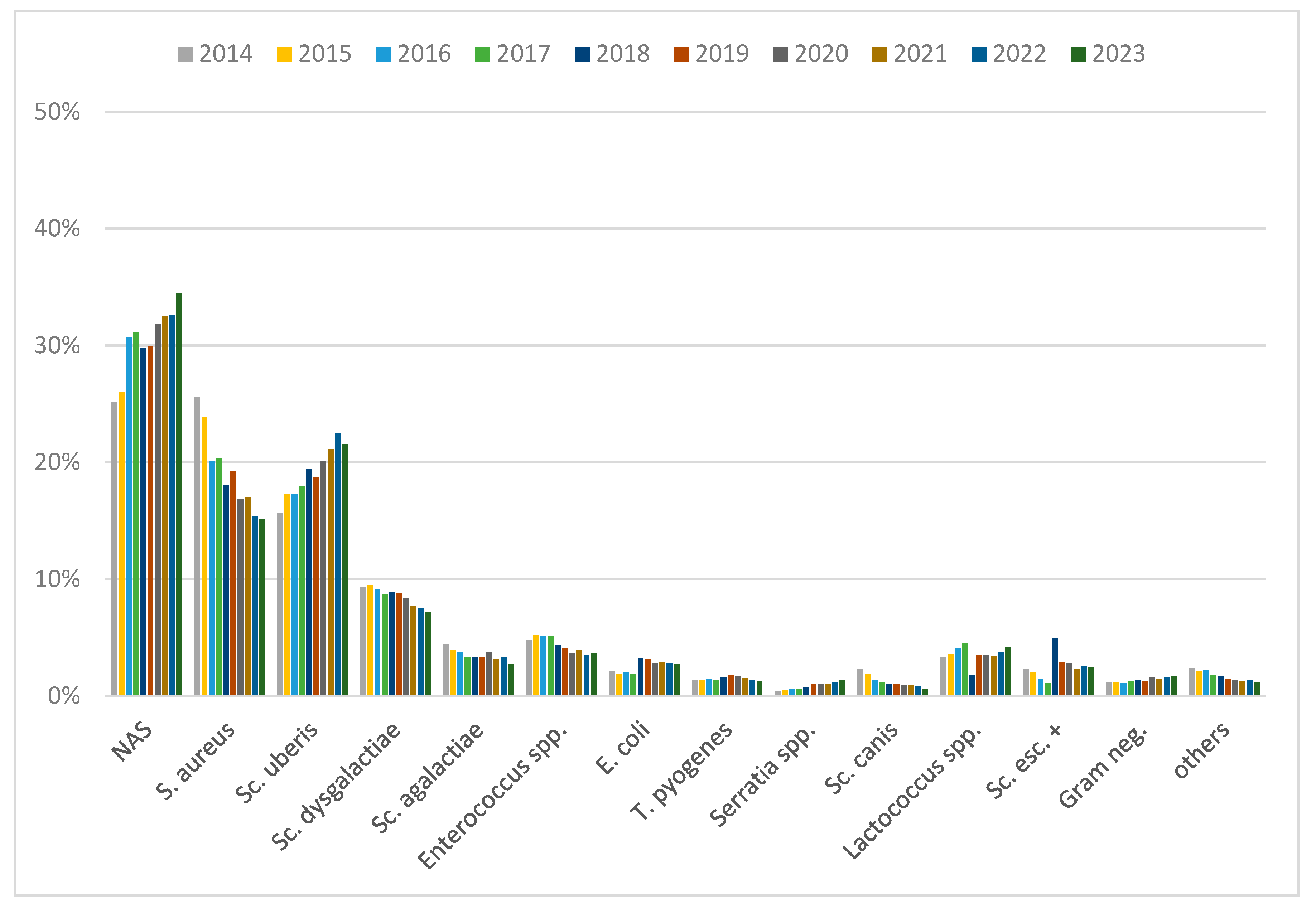
SAS 9.4 software (SAS Analytics Software Institute Inc. SAS Institute GmbH, Heidelberg, Germany) was used for the statistical analysis. PROC FREQ procedures were used to display the individual pathogens by year, month, and mastitis status. Chi-square test was used to compare prevalence of the different mastitis status, and according to sample origin (herd/individual). The Cochran–Armitage trend was used to assess a prevalence trend for each pathogen over the years and months. The graphics were created using Microsoft Excel 2010 (Microsoft Excel for Microsoft 365 MSO, Version 2308, Redmond, WA, USA) and α was set at 0.01.

3. Results

Between 2014 and 2023, 3,886,162 QMSs from 634,022 cows and 15,609 herds were analyzed. Of the total, 1.5% of QMSs were contaminated and therefore excluded. Mastitis pathogens were cultured in only 19% of the QMSs, meaning that 81% showed no growth. One pathogen was detected in 95.5% of the culture-positive QMSs and two pathogens were detected in 4.5%. Overall, 729,459 isolates of mastitis pathogens were included in this analysis.
Table 1 provides an overview of all QMSs included in this study. Of the total, 83% of QMSs came from herd samplings, while the remaining were sent in by farmers or veterinarians from individual case investigations. Herd screenings contributed 85% of the samples from CMT-negative quarters and 67% from subclinically affected quarters. In contrast, QMSs from clinically affected quarters were more likely to come from individual samplings (p < 0.01). Furthermore, there was a sharp increase in individual submissions from clinically affected quarters from an average of 40% (2014–2017) to an average of 55% (2018–2023, p < 0.01). As the majority of samples were from herd screenings, the majority of isolates were detected in samples from herd screenings. Only E. coli (68%) and T. pyogenes (57%) were found more frequently in samples from individual submissions than herd screenings (p < 0.01).
Considering culture-negative and culture-positive QMSs, the apparent prevalences of the most frequently isolated pathogens were as follows: NAS (5.7%), S. aureus (3.6%), Sc. uberis (3.6%), and Sc. dysgalactiae (1.6%).
Figure 1 shows the distribution of pathogens in culture-positive samples over the years. The most frequently isolated pathogens in culture-positive samples were NAS (30%). Though the vast majority of NAS (95%) were not further differentiated, the rest consisted of 2% S. chromogenes, 0.9% S. epidermidis, 0.8% S. haemolyticus, 0.5% S. simulans, 0.4% S. xylosus, and others less than 0.2%. The second most common pathogen in culture-positive samples was S. aureus (19%). The distribution of S. aureus isolates declined from 26% in 2014 to 15% in 2023 (p < 0.01). In contrast, NAS (25% to 34%) and Sc. uberis (16% to 22%) isolates increased over the whole study period (p < 0.01). An increase in E. coli isolates was observed from 2018 onwards (2014–2017: 2% to 2018–2023: 3%, p < 0.01).

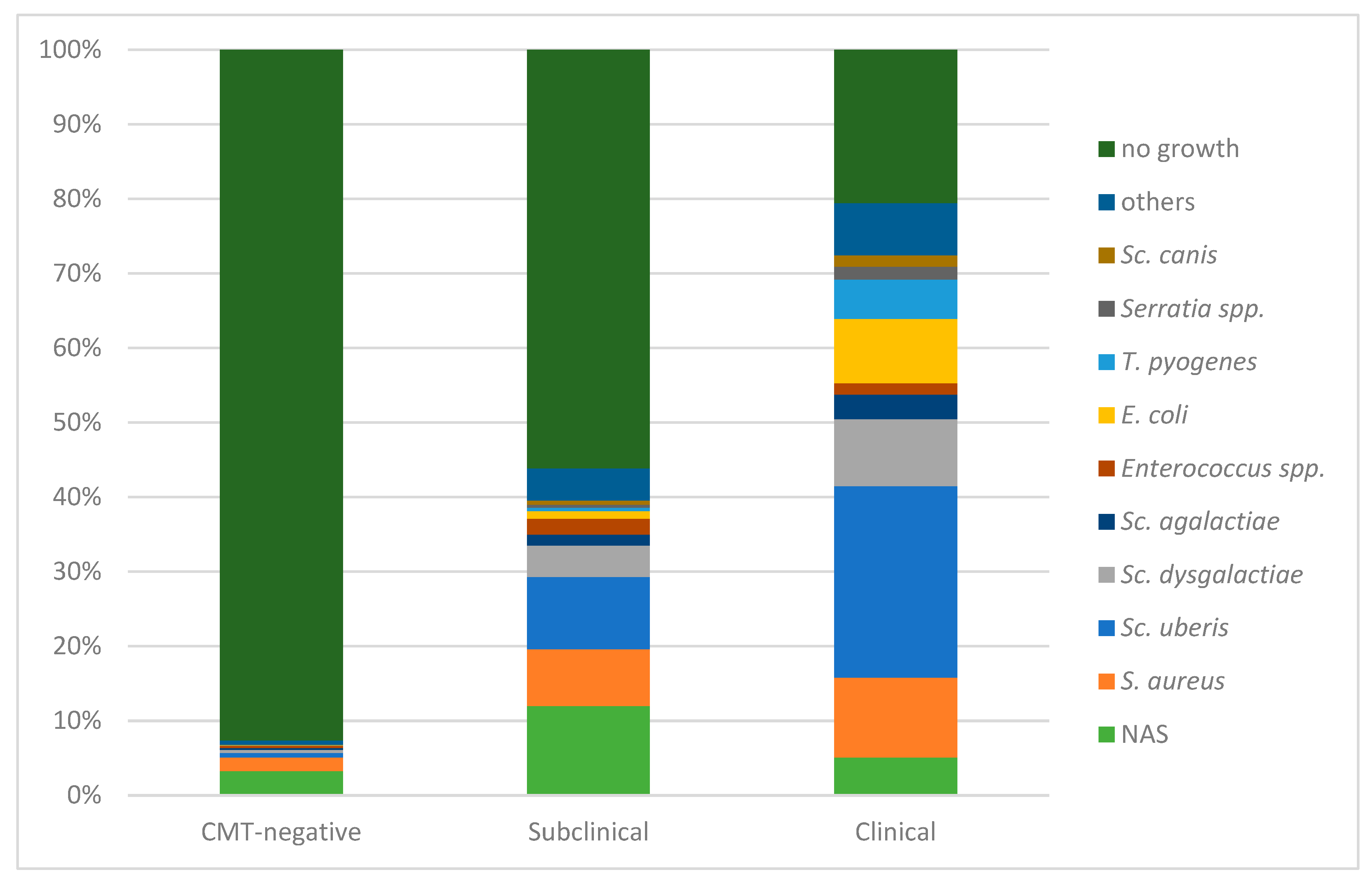
3.1. Distribution in Dependence of Mastitis Status

The distribution of mastitis pathogens shifted depending on the mastitis status of the udder (Figure 2). Thus, pathogens were more likely cultured from QMSs from clinically affected quarters (p < 0.01), compared to CMT-negative or subclinically affected quarters. When considering all QMSs, the apparent prevalences of the most commonly detected mastitis pathogens in samples from CMT-negative quarters were as follows: NAS (3%), S. aureus (2%), Sc. uberis (0.6%), and Sc. dysgalactiae (0.4%). In QMSs from subclinically affected quarters, the apparent prevalences were NAS (12%), Sc. uberis (10%), S. aureus (8%), and Sc. dysgalactiae (4%). In QMSs from clinically affected quarters, the apparent prevalences were Sc. uberis (26%), S. aureus (11%), Sc. dysgalactiae (9%), and E. coli (9%).
Figure 3A–C shows the annual distribution of mastitis pathogens from culture-positive samples for each mastitis status. While NAS were the most common pathogens in QMSs from CMT-negative (44%) and subclinically affected quarters (27%), they were only isolated in 6% of QMSs from clinically affected quarters. The distribution of NAS from CMT-negative and subclinically affected quarters increased over the study period (Figure 3A,B, p < 0.01), while no trend was observed for the distribution of NAS from clinically affected quarters (Figure 3C, p = 0.29); 43% of NAS from clinically affected quarters were undifferentiated; the differentiated NAS included S. chromogens (16%), S. epidermidis (11%), S. haemolyticus (10%), S. simluans (8%), S. xylosus (3%), S. sciuri (3%), and others at less than 2.5% each. Similarly, S. aureus was detected more frequently in QMS from CMT-negative (25%) and subclinically affected quarters (17%) than in clinically affected quarters (13%). Overall, S. aureus isolates from all three mastitis classifications showed a decline in their detection in culture-positive samples during the study period (Figure 3A–C, p < 0.01, respectively), though the distribution of S. aureus isolates from clinically affected quarters remained at a constant level from 2018 onwards (p = 0.02).
In contrast, Sc. uberis, Sc. dysgalactiae, E. coli, T. pyogenes, and Serratia spp. were more common in QMSs from clinically affected quarters (Figure 3C, p < 0.01, respectively). Sc. uberis was the most frequently isolated pathogen in QMSs from clinically affected quarters (32%) and was detected in only 8% of QMSs from culture-positive and CMT-negative quarters. Sc. dysgalactiae was the third most frequent pathogen in clinically affected QMSs (11%)—after Sc. uberis and S. aureus. E. coli had 10% higher detection in samples from clinically affected quarters compared to CMT-negative quarters (1%, p < 0.01). T. pyogenes and Serratia spp. showed a 5% and 2% higher distribution, respectively, in QMSs from clinically affected quarters than in QMSs from CMT-negative quarters (p < 0.01, respectively). Sc. uberis isolates showed an increase across all three mastitis classifications (p < 0.01, respectively), while Sc. dysgalactiae isolates showed a decrease across all three mastitis classifications (p < 0.01, respectively). E. coli showed a clear increase in culture-positive QMSs from clinically affected quarters (from 8% in 2014 to 12% in 2023, p < 0.01), with a sudden increase from 2017 (8%) to 2018 (13%, p < 0.01). T. pyogenes isolates showed no trend regardless of mastitis status (p > 0.08, respectively). In contrast, Serratia spp. showed a steady increase in QMSs from clinically affected quarters (2% to 4%, p < 0.01).

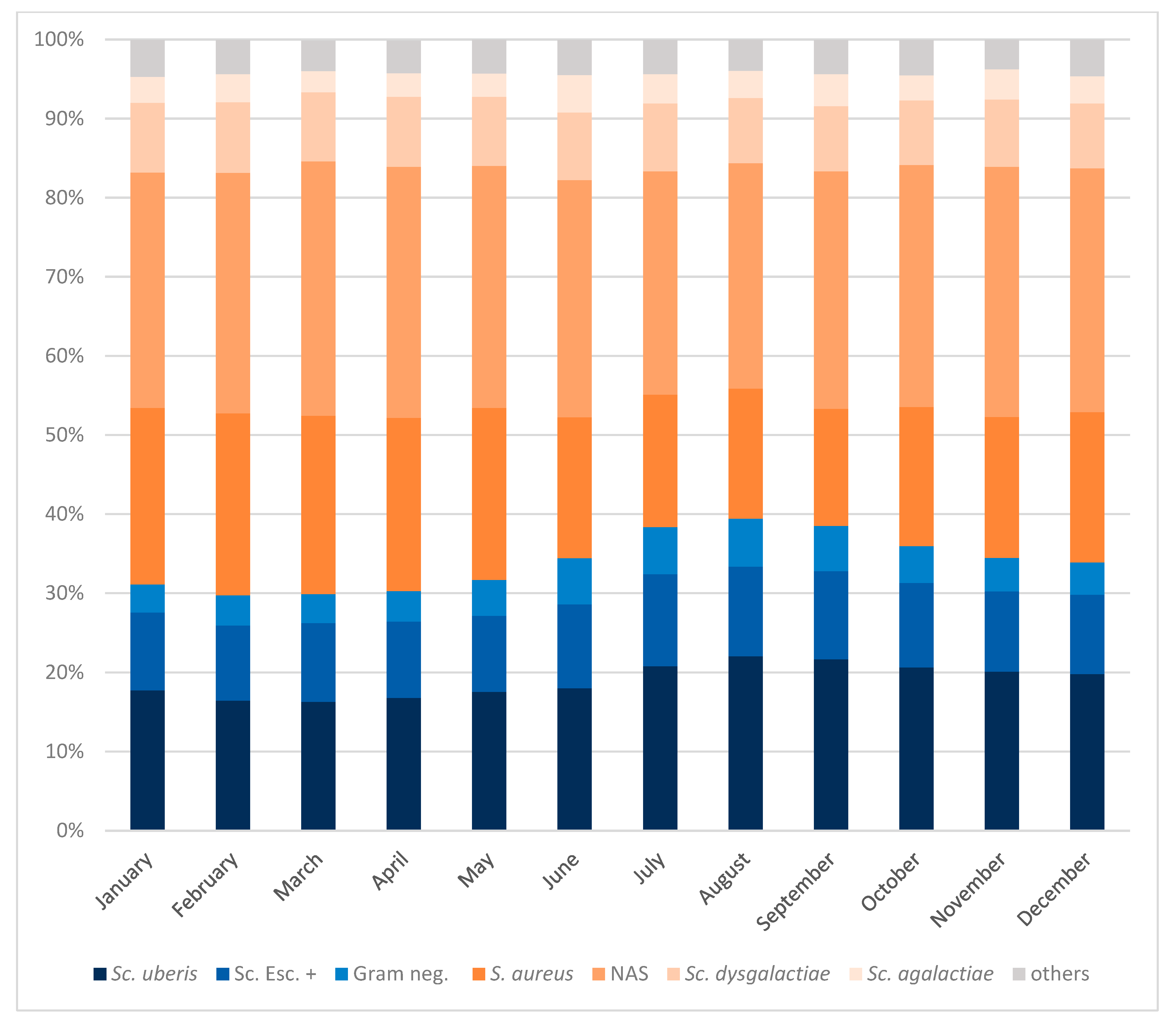
3.2. Seasonal Distribution of Mastitis Pathogens

Differences in the mastitis status of culture-positive QMSs were found depending on season. In the warmer months, from June to October, the frequency of CMT-negative samples decreased (on average from 30% to 26%, p < 0.01) and the frequency of subclinical samples increased (on average from 62% to 66%, p < 0.01). In contrast, the proportion of QMSs from clinically affected quarters remained constant at 8% across all months.
Figure 4 provides an overview of the distribution of mastitis pathogens from culture-positive samples over the months during the entire study period. The detection of environmental pathogens like Sc. uberis; other esculin-positive streptococci, including Enterococcus spp. and Lactococcus spp.; and Gram-negative pathogens, including E. coli and Serratia spp., increased during the warmer months from June to October (pooled average 37%) compared to the colder months from November to May (pooled average 32%, p < 0.01).
Sc. uberis isolates were detected at an average of 21% of culture-positive QMSs from June to October, but at an average of 18% of QMSs in the colder months of November to May. The situation was similar with other esculin-positive streptococci and Gram-negative pathogens, with an average of 2% more isolates detected in the months of June to October in each case (p < 0.01, respectively). In contrast, the detection of S. aureus fell from an average of 21% during November to May to an average of 17% during the warmer months of June to October (p < 0.01). Similarly, the detection of NAS fell from 31% to 29% during the same period (p < 0.01). Sc. dysgalactiae and Sc. agalactiae showed no differences depending on the season (on average 9% and 3%, respectively, across months).

4. Discussion

A strength of this study is that a large number of isolates from numerous herds and cows were included over a 10-year period. In addition, QMSs from different clinical scores were analyzed in a single milk quality laboratory. This made it possible to evaluate trends in a specific region. Enrollment of herds or submission of individual samples was based on voluntary submissions rather than a random sample. Therefore, statements regarding the prevalence of pathogens within herds of this region should be avoided.
The majority of samples stemmed from herd screening, where TGD technicians took aseptic QMSs from all four quarters of every lactating cow of the herd (or a subset of cows in larger herds). Consequently, most samples in this study came from CMT-negative or subclinically affected quarters. Accordingly, a higher proportion of QMSs from clinically affected quarters came from individual submissions compared to herd samples, as veterinarians and farmers are more likely to submit samples to base their therapy on laboratory results. Furthermore, we found that E. coli and T. pyogenes were the only pathogens that were each more often found in samples from individual cows. An important factor that influences the prevalence is the duration of the infection [19]. E. coli is known to cause short infections (10–30 days) and is quickly eliminated by the host’s immune response [20]. For T. pyogenes, Wente et al. [21] found an infection duration of one week to seven months. The timing of the sampling therefore plays an important role in the detection of mastitis pathogens, since sampling later during the infection likely leads to a no-growth result. As veterinarians and farmers often submitted acute mastitis cases, where the probability of detecting these pathogens is higher, these pathogens were detected more frequently in individual samples. When sampling entire herds at one time point, it is unlikely to catch clinical mastitis cases early during infection and therefore mastitis cases due to pathogens with a short infection period are more likely to end up in the “no-growth” category.
When interpreting our data, it should be taken into account that we only investigated the distribution of mastitis pathogens from QMSs sent to the TGD milk quality laboratory and the results may be biased by the high proportion of herd samplings and QMSs from CMT-negative and subclinically affected quarters. In our study, NAS were the most common pathogens in culture positive samples. Tenhagen et al. [22] found NAS to be the most common minor pathogens in QMSs from northern German dairy farms, and the German Veterinary Association observed the same Germany-wide [23]. Other studies also showed that NAS were most frequently detected in countries other than Germany, e.g., Norway [24], Slovakia [25], and Australia [26]. In addition, a study from Condas et al. [27] reported NAS to be the pathogens most frequently isolated from bovine milk worldwide. In most studies, NAS were the most frequently detected pathogens in quarters with subclinical mastitis [28]. This is consistent with our findings. However, we found NAS in 6% of QMSs from clinical mastitis, which is comparable to a study from Belgium, where they found NAS in 5% of all clinical samples [29], but different to a study from Finland, with 23.71% of NAS in clinical samples [30]. Furthermore, we found that the likelihood of NAS isolation increased throughout the study period, as also reported in Norway [24]. Zigo et al. [31] noted that this increase occurred after a reduction in main pathogens and that NAS are characterized by increased resistance to antibiotics and disinfectants, which may explain the rising distribution. Despite their surge, NAS are considered minor pathogens and therefore of lesser importance for dairy production [28].
The second most common pathogen in our study was S. aureus. We were more likely to find S. aureus than other German regions [22,23,32]. This may be explained by the sample selection (high number of QMSs from CMT-negative and subclinically affected quarters) and different herd structures in the regions. Fadlelmoula et al. [33] found that the risk of mastitis due to contagious pathogens is higher in small herds. Eastern German dairy herds are larger than Bavarian herds [22]. Furthermore, Wang et al. [34] noted that the prevalence of S. aureus varies greatly regional and worldwide. They further described that the prevalence of S. aureus has decreased over the last decade in China, which we also found in our study. Wang et al. [34] concluded that this development is due to rapid technological development and biosecurity measures taken by farms. Additionally, Munoz et al. [35] found that Bavarian farms have become larger and increased in their performance. In contrast, Acharya et al. [36] found an increasing proportion of S. aureus between 2008 and 2017 in Ontario. Similarly, Kortstegge and Krömker [32] noted an increasing risk of S. aureus in bulk tank milk with increasing herd sizes. Furthermore, Smistad et al. [24] observed a higher proportion of S. aureus (24.5%) in their study in Norway, and found S. aureus to be the most frequently detected major pathogen. They stated that the prevalence of S. aureus in Norway was relatively stable between 2000 and 2020 and explained that measures such as routine teat disinfection after milking are less consistently carried out in Norway. In addition to our results, Karell et al. [37] found an overall decreasing resistance trend in S. aureus in their study in Bavaria and concluded that this trend can be seen as a success of the measures taken in recent decades to tighten the use of antimicrobials and to control mastitis pathogens, which aimed to prevent new infections and eliminate existing infections. Therefore, the decreasing likelihood of detection and resistance of S. aureus can be seen as a success of the measures taken to combat mastitis pathogens in recent decades. Another notable observation was that S. aureus isolates were also fairly commonly detected in QMSs from CMT-negative and subclinically affected quarters. This is important, as Karell et al. [37] observed that S. aureus isolates from healthy or subclinical quarters were more likely to be in vitro resistant than isolates from clinical quarters. Woudstra et al. [9] found in their study that one S. aureus strain could cover 80% of the infections within a herd and that infected udder quarters are the main reservoir, underlining the contagious nature of this pathogen. Therefore, it could be concluded that cows with undetected infections act as a reservoir for within-herd transmission of S. aureus that also may carry virulence and resistance genes. The most important focus in preventing S. aureus infections is on reducing transmission from infected to uninfected quarters [9,38]. For this purpose, healthy cows or cows with subclinical mastitis should also be included in herd screenings in order to find potential sources of infection.
Sc. uberis was the third most common pathogen in culture positive samples in this study (19%). Its detection risk within culture-positive samples aligned with other reports from Germany (20.3%, [23]). It is noteworthy that it was the most common pathogen isolated from QMSs from clinically affected quarters (32%), which agrees with other studies from around the world [13,29,39]. In our study, Sc. uberis isolates showed an overall increase over the entire study period, which was also observed in Ontario by Acharya et al. [36] and previously by Phuektes et al. [40] for other parts of the world. Cobirka et al. [25] stated that Sc. uberis is mostly present in bedding material and that control measurements, such as post-milking teat disinfection and dry cow therapy, are far less effective against environmental pathogens, such as Sc. uberis. This may explain the increasing probability of Sc. uberis detection, since a decline in contagious pathogens (e.g., S. aureus), has been reported to go hand in hand with an increase in Gram-negative and therefore environmental pathogens [41]. In line with this, we found that the detection of other environmental pathogens such as E. coli and Serratia spp. also increased throughout the study period. Furthermore, the detection likelihood of E. coli jumped from 2018 onwards compared to previous years—especially in clinical cases. Since the number of individual submissions from clinical mastitis also increased at the same time, one can see the impact of a change in legislation in Germany in 2018 for the use of antimicrobials in veterinary medicine. It aimed to minimize the use of critically important antimicrobials (Verordnung über tierärztliche Hausapotheken, TÄHAV) [42] and included obligatory antimicrobial susceptibility testing, for example, if critically important antimicrobials were selected or antimicrobials were changed during therapy. Interestingly, this increase was especially observed for samples that tested positive for E. coli. This was also reported by Pirner et al. [43], who investigated the resistance of Gram-negative pathogens in Bavaria. One possible explanation could be that most of the pathogens detected in this study were Gram-positive pathogens. Those pathogens are more likely to cause subclinical mastitis and were commonly detected in herd screenings even before 2018 [43]. The change in legislation therefore did not impact their detection as much as it did for E. coli with its short infection duration. As QMSs now had to be sampled as soon as clinical signs appeared for treatment decisions [43], this increased the likelihood of detecting E. coli. E. coli is the most common Gram-negative pathogen to cause clinical mastitis worldwide [44]. In our study, E. coli was the most common Gram-negative pathogen in culture-positive QMSs from clinically affected quarters (11%). In Lower Saxony, Germany, Krebs et al. [45] reported in their study a much higher detection of E. coli from culture-positive clinical samples (35.2%). Other authors found E. coli to be associated with 19.8% of clinical mastitis cases in England and Wales [46], 15.5% in Belgium [29], or 27% of cases in China [47]. This underlines that the distribution of pathogens may vary depending on geographical region [48].
Besides region, a variety of factors can influence the prevalence of mastitis-causing pathogens, such as herd-size, housing system, and season [49]. Season also impacts cows in Germany, as they suffer from heat stress too [50]. The temperature–humidity index (THI) is widely used to assess heat stress, and a high THI is associated with an increased somatic cell score [51,52]. In our study, we found seasonal changes in the occurrence of QMSs from subclinically affected quarters, as they were slightly more common during the warmer months of June to October. Furthermore, we found differences in the detection of various mastitis pathogens depending on the month and therefore season. Environmental pathogens (e.g., Sc. uberis, Gram-negative as well as other esculin-positive pathogens) were more likely detected during the warmer months of June to October, while the proportion of S. aureus and NAS slightly decreased during that time. High temperatures and humidity may increase the probability of IMIs caused by environmental pathogens [53]. Other studies also reported that the prevalence of environmental mastitis due to, e.g., Sc. uberis [30,54] and E. coli, was the highest in summer and autumn [30], and the frequency of S. aureus and NAS mastitis cases was higher during winter [30,55]. All three studies noted a housing difference during the season, which may explain the observed distribution. Olde Riekerink et al. [54] stated that cows on pasture during summer are at an increased risk of environmental mastitis caused by pathogens such as Sc. uberis. The warm and humid conditions that prevail in summer, combined with the organic material on the pastures, create a favorable environment for these pathogens to thrive [54]. However, other studies reported an increased incidence of environmental mastitis or somatic cell count in hot and humid weather and explained this with immunosuppression due to heat stress and a higher pathogen load in the environment, leading to increased susceptibility to IMIs [56,57]. Furthermore, Hamel et al. [12] found higher shedding of Sc. uberis with a higher THI, which may be a possible explanation for the higher detection of Sc. uberis in summer. Other possible explanations are given by Kabelitz et al. [11], who suggest that high temperatures and thus the higher reproduction rate of pathogens in the environment could be a reason, as well as the transmission of bacteria by flies, which are particularly present in summer. In this study, however, we could only report the observed differences, as we did not have the climate or other risk factors like farming practices for all submissions. Therefore, we can only suggest explanations for the observed dynamics, and further research is needed. Nevertheless, increasing temperatures due to climate change, increasing heat stress, and therefore vulnerability to mastitis, as well as the influence on mastitis pathogens, likely bring new challenges for maintaining udder health [58].
Overall, the increase in environmental pathogens observed in this study emphasizes the need to continue and improve management practices. Effective control of environmental mastitis can be achieved by minimizing teat-end exposure to these pathogens and enhancing cow resistance to intramammary infections, e.g., through vaccination strategies [59]. Reducing the exposure of cows to environmental mastitis pathogens also involves maintaining clean and dry bedding, regular removal of manure, and avoiding overcrowding in barns and pastures [60]. In addition, season-dependent measures such as appropriate cooling can reduce heat stress for cows, thereby strengthening their immune response and reducing the likelihood of mastitis outbreaks [61].

5. Conclusions

This study underscores the dynamic nature of mastitis pathogen distribution, influenced by mastitis status and seasonal factors. Contagious pathogens such as S. aureus have decreased over the last decade, while environmental pathogens continue to play an important role for udder health in Bavaria. In addition, the study results emphasize that both healthy cows and those with subclinical mastitis can serve as a reservoir for mastitis pathogens. It is therefore essential to consider healthy cows and cows with subclinical mastitis as a reservoir for mastitis pathogens during monitoring and control efforts in order to track trends and adapt targeted prevention measures.

Author Contributions

Conceptualization, U.S.S., W.P. and V.B.; Methodology, U.S.S., R.H.-S. and V.B.; Software, U.S.S. and V.B.; Validation, U.S.S., W.P. and V.B.; Formal Analysis, U.S.S. and V.B.; Investigation, R.H.-S. and V.B.; Resources, U.S.S.; Data Curation, U.S.S. and V.B.; Writing—Original Draft Preparation, V.B.; Writing—Review and Editing, U.S.S., W.P., R.H.-S. and V.B.; Visualization, V.B.; Supervision, U.S.S.; Project Administration, U.S.S.; Funding Acquisition, U.S.S. All authors have read and agreed to the published version of the manuscript.

Funding

This study was made possible with the financial support of the Free State of Bavaria and the Bavarian Joint Founding Scheme for the Control and Eradication of Contagious Livestock Diseases (Bayerische Tierseuchenkasse, Munich, Germany).

Institutional Review Board Statement

The samples used in this retrospective study were collected for herd health management and diagnostic purposes. IACUC approval was therefore not necessary.

Informed Consent Statement

Not applicable.

Data Availability Statement

Data are contained within the article.

Conflicts of Interest

The authors declare no conflicts of interest.

References

  1. Cheng, W.N.; Han, S.G. Bovine mastitis: Risk factors, therapeutic strategies, and alternative treatments—A review. Asian-Australas. J. Anim. Sci. 2020, 33, 1699–1713. [Google Scholar] [CrossRef]
  2. Ruegg, P.L. A 100-Year Review: Mastitis detection, management, and prevention. J. Dairy Sci. 2017, 100, 10381–10397. [Google Scholar] [CrossRef]
  3. Heikkilä, A.M.; Liski, E.; Pyörälä, S.; Taponen, S. Pathogen-specific production losses in bovine mastitis. J. Dairy Sci. 2018, 101, 9493–9504. [Google Scholar] [CrossRef]
  4. Kitila, G.; Kebede, B.; Wakgari, M. Prevalence, aetiology and risk factors of mastitis of dairy cows kept under extensive management system in west Wollega, western Oromia, Ethiopia. Vet. Med. Sci. 2021, 7, 1593–1599. [Google Scholar] [CrossRef]
  5. Meçaj, R.; Muça, G.; Koleci, X.; Sulçe, M.; Turmalaj, L.; Zalla, P.; Koni, A.; Tafaj, M. Bovine environmental mastitis and their control: An overview. Int. J. Agric. Biosci. 2023, 12, 216–221. [Google Scholar] [CrossRef]
  6. Sharif, A.; Umer, M.; Muhammad, G. Mastitis control in dairy production. J. Agric. Soc. Sci 2009, 5, 102–105. [Google Scholar]
  7. Khasa, V.; Chaudhary, V.; Singh, P. Mastitis: A review on disease affecting livestock and its control. J. Entomol. Zool. Stud. 2020, 8, 1393–1395. [Google Scholar]
  8. Reyher, K.K.; Dohoo, I.R.; Scholl, D.T.; Keefe, G.P. Evaluation of minor pathogen intramammary infection, susceptibility parameters, and somatic cell counts on the development of new intramammary infections with major mastitis pathogens. J. Dairy Sci. 2012, 95, 3766–3780. [Google Scholar] [CrossRef] [PubMed]
  9. Woudstra, S.; Wente, N.; Zhang, Y.; Leimbach, S.; Gussmann, M.K.; Kirkeby, C.; Krömker, V. Strain diversity and infection durations of Staphylococcus spp. and Streptococcus spp. causing intramammary infections in dairy cows. J. Dairy Sci. 2023, 106, 4214–4231. [Google Scholar] [CrossRef]
  10. Middleton, J.R.; Saeman, A.; Fox, L.K.; Lombard, J.; Hogan, J.S.; Smith, K.L. The National Mastitis Council: A Global Organization for Mastitis Control and Milk Quality, 50 Years and Beyond. J. Mammary Gland Biol. Neoplasia 2014, 19, 241–251. [Google Scholar] [CrossRef]
  11. Kabelitz, T.; Aubry, E.; van Vorst, K.; Amon, T.; Fulde, M. The Role of Streptococcus spp. in Bovine Mastitis. Microorganisms 2021, 9, 1497. [Google Scholar] [CrossRef]
  12. Hamel, J.; Zhang, Y.; Wente, N.; Krömker, V. Heat stress and cow factors affect bacteria shedding pattern from naturally infected mammary gland quarters in dairy cattle. J. Dairy Sci. 2021, 104, 786–794. [Google Scholar] [CrossRef] [PubMed]
  13. Schmenger, A.; Krömker, V. Characterization, Cure Rates and Associated Risks of Clinical Mastitis in Northern Germany. Vet. Sci. 2020, 7, 170. [Google Scholar] [CrossRef] [PubMed]
  14. Huber-Schlenstedt, R.; Gey, A.; Schierling, K.; Sorge, U. Mastitis? TGD checkt die Erreger. Milchpur 2017, 1, 18–24. [Google Scholar]
  15. Bayerische Landesamt für Statistik. Zum Tag der Milch am 1. Juni in Bayern. Available online: https://www.statistik.bayern.de/presse/mitteilungen/2023/pm134/index.html (accessed on 20 August 2024).
  16. Bayerisches Landwirtschaftliches Wochenblatt. Bayern ist Fleckviehland. Available online: https://www.wochenblatt-dlv.de/feld-stall/tierhaltung/bayern-fleckviehland-571662 (accessed on 20 August 2024).
  17. Tergast, H.; Hansen, H.; Weber, E.-C. Steckbriefe zur Tierhaltung in Deutschland: Milchkühe; Thünen-Institut für Betriebswirtschaft: Braunschweig, Germany, 2022; pp. 1–17. [Google Scholar]
  18. DVG. Leitlinien zur Labordiagnostik der Mastitis—Probenahme und Mikrobiologische Untersuchung, 3rd ed.; DVG Service GmbH: Gießen, Germany, 2018. [Google Scholar]
  19. Zadoks, R.; Fitzpatrick, J. Changing trends in mastitis. Ir. Vet. J. 2009, 62 (Suppl. 4), S59–S70. [Google Scholar] [CrossRef] [PubMed]
  20. Goulart, D.B.; Mellata, M. Escherichia coli mastitis in dairy cattle: Etiology, diagnosis, and treatment challenges. Front. Microbiol. 2022, 13, 928346. [Google Scholar] [CrossRef]
  21. Wente, N.; Leimbach, S.; Woudstra, S.; Krömker, V. Trueperella Pyogenes—Strain Diversity and Occurrence in Dairy Herds. Pathogens 2024, 13, 534. [Google Scholar] [CrossRef]
  22. Tenhagen, B.A.; Koster, G.; Wallmann, J.; Heuwieser, W. Prevalence of mastitis pathogens and their resistance against antimicrobial agents in dairy cows in Brandenburg, Germany. J. Dairy Sci. 2006, 89, 2542–2551. [Google Scholar] [CrossRef]
  23. DVG. Zur Prävalenz von Mastitiserregern in Milchproben in Deutschland—Update 2022. Available online: https://www.dvg.de/wp-content/uploads/2024/04/24-03-19_AG-Euter_Praevalenz-Mastitserreger2022.pdf (accessed on 20 August 2024).
  24. Smistad, M.; Bakka, H.C.; Sølverød, L.; Jørgensen, H.J.; Wolff, C. Prevalence of udder pathogens in milk samples from Norwegian dairy cows recorded in a national database in 2019 and 2020. Acta Vet. Scand. 2023, 65, 19. [Google Scholar] [CrossRef]
  25. Cobirka, M.; Tancin, V.; Slama, P. Epidemiology and Classification of Mastitis. Animals 2020, 10, 2212. [Google Scholar] [CrossRef]
  26. Al-Harbi, H.; Ranjbar, S.; Moore, R.J.; Alawneh, J.I. Bacteria isolated from milk of dairy cows with and without clinical mastitis in different regions of Australia and their AMR profiles. Front. Vet. Sci. 2021, 8, 743725. [Google Scholar] [CrossRef] [PubMed]
  27. Condas, L.A.Z.; De Buck, J.; Nobrega, D.B.; Carson, D.A.; Naushad, S.; De Vliegher, S.; Zadoks, R.N.; Middleton, J.R.; Dufour, S.; Kastelic, J.P.; et al. Prevalence of non-aureus staphylococci species causing intramammary infections in Canadian dairy herds. J. Dairy Sci. 2017, 100, 5592–5612. [Google Scholar] [CrossRef]
  28. De Buck, J.; Ha, V.; Naushad, S.; Nobrega, D.B.; Luby, C.; Middleton, J.R.; De Vliegher, S.; Barkema, H.W. Non-aureus Staphylococci and Bovine Udder Health: Current Understanding and Knowledge Gaps. Front. Vet. Sci. 2021, 8, 658031. [Google Scholar] [CrossRef] [PubMed]
  29. Verbeke, J.; Piepers, S.; Supré, K.; De Vliegher, S. Pathogen-specific incidence rate of clinical mastitis in Flemish dairy herds, severity, and association with herd hygiene. J. Dairy Sci. 2014, 97, 6926–6934. [Google Scholar] [CrossRef] [PubMed]
  30. Koivula, M.; Pitkälä, A.; Pyörälä, S.; Mäntysaari, E.A. Distribution of bacteria and seasonal and regional effects in a new database for mastitis pathogens in Finland. Acta Agric. Scand. A Anim. Sci. 2007, 57, 89–96. [Google Scholar] [CrossRef]
  31. Zigo, F.; Vasil, M.; Ondrašovičová, S.; Výrostková, J.; Bujok, J.; Pecka-Kielb, E. Maintaining Optimal Mammary Gland Health and Prevention of Mastitis. Front. Vet. Sci. 2021, 8, 607311. [Google Scholar] [CrossRef]
  32. Kortstegge, J.; Krömker, V. Prevalence of Contagious Mastitis Pathogens in Bulk Tank Milk from Dairy Farms in Lower Saxony, Germany. Hygiene 2024, 4, 122–134. [Google Scholar] [CrossRef]
  33. Fadlelmoula, A.; Fahr, R.D.; Anacker, G.; Swalve, H.H. The Management Practices Associated with Prevalence and Risk Factors of Mastitis in Large Scale Dairy Farms in Thuringia-Germany 1: Environmental Factors Associated With Prevalence of mastitis. Aust. J. Basic Appl. Sci. 2007, 1, 619–624. [Google Scholar]
  34. Wang, K.; Cha, J.; Liu, K.; Deng, J.; Yang, B.; Xu, H.; Wang, J.; Zhang, L.; Gu, X.; Huang, C.; et al. The prevalence of bovine mastitis-associated Staphylococcus aureus in China and its antimicrobial resistance rate: A meta-analysis. Front. Vet. Sci. 2022, 9, 1006676. [Google Scholar] [CrossRef]
  35. Munoz, B.; Lakner, S.; Brümmer, B. Determinants of Economic Growth in Organic Farming: The Case of Bavaria and Baden-Wuerttemberg. In Proceedings of the 2011 International Congress, Zurich, Switzerland, 30 August–2 September 2011. [Google Scholar]
  36. Acharya, K.R.; Brankston, G.; Slavic, D.; Greer, A.L. Spatio-temporal variation in the prevalence of major mastitis pathogens isolated from bovine milk samples between 2008 and 2017 in Ontario, Canada. Front. Vet. Sci. 2021, 8, 742696. [Google Scholar] [CrossRef]
  37. Karell, J.; Petzl, W.; Gangl, A.; Huber-Schlenstedt, R.; Sorge, U.S. Changes in antimicrobial resistance of Staphylococcus aureus in bovine quarter milk samples from southern Germany between 2012 and 2022. J. Dairy Sci. 2024, 107, 3802–3812. [Google Scholar] [CrossRef] [PubMed]
  38. Rossi, B.F.; Bonsaglia, E.C.R.; Castilho, I.G.; Dantas, S.T.A.; Salina, A.; Langoni, H.; Pantoja, J.C.F.; Budri, P.E.; Fitzgerald-Hughes, D.; Júnior, A.F.; et al. Genotyping of long term persistent Staphylococcus aureus in bovine subclinical mastitis. Microb. Pathog. 2019, 132, 45–50. [Google Scholar] [CrossRef] [PubMed]
  39. Petrovski, K.R.; Heuer, C.; Parkinson, T.J.; Williamson, N.B. The incidence and aetiology of clinical bovine mastitis on 14 farms in Northland, New Zealand. N. Z. Vet. J. 2009, 57, 109–115. [Google Scholar] [CrossRef]
  40. Phuektes, P.; Mansell, P.D.; Dyson, R.S.; Hooper, N.D.; Dick, J.S.; Browning, G.F. Molecular epidemiology of Streptococcus uberis isolates from dairy cows with mastitis. J. Clin. Microbiol. 2001, 39, 1460–1466. [Google Scholar] [CrossRef]
  41. Schukken, Y.; Chuff, M.; Moroni, P.; Gurjar, A.; Santisteban, C.; Welcome, F.; Zadoks, R. The “Other” Gram-Negative Bacteria in Mastitis: Klebsiella, Serratia, and More. Vet. Clin. N. Am. Food Anim. Pract. 2012, 28, 239–256. [Google Scholar] [CrossRef]
  42. Bundestierärztekammer. Die neue TÄHAV ist in Kraft. Dtsch. Tierärzteblatt 2018, 66, 484–489. [Google Scholar]
  43. Pirner, L.H.; Petzl, W.; Gangl, A.; Huber-Schlenstedt, R.; Sorge, U.S. In vitro antimicrobial resistance of Escherichia coli, Serratia marcescens, Klebsiella oxytoca and Klebsiella pneumoniae on Bavarian dairy farms between 2014–2022. J. Dairy Sci. 2024, in press. [Google Scholar] [CrossRef]
  44. Campos, F.C.; Castilho, I.G.; Rossi, B.F.; Bonsaglia É, C.R.; Dantas, S.T.A.; Dias, R.C.B.; Fernandes Júnior, A.; Hernandes, R.T.; Camargo, C.H.; Ribeiro, M.G.; et al. Genetic and Antimicrobial Resistance Profiles of Mammary Pathogenic E. coli (MPEC) Isolates from Bovine Clinical Mastitis. Pathogens 2022, 11, 1435. [Google Scholar] [CrossRef] [PubMed]
  45. Krebs, I.; Zhang, Y.; Wente, N.; Leimbach, S.; Krömker, V. Severity of Clinical Mastitis and Bacterial Shedding. Pathogens 2023, 12, 1098. [Google Scholar] [CrossRef]
  46. Bradley, A.J.; Leach, K.A.; Breen, J.E.; Green, L.E.; Green, M.J. Survey of the incidence and aetiology of mastitis on dairy farms in England and Wales. Vet. Rec. 2007, 160, 253–258. [Google Scholar] [CrossRef]
  47. Xu, T.; Cao, W.; Huang, Y.; Zhao, J.; Wu, X.; Yang, Z. The Prevalence of Escherichia coli Derived from Bovine Clinical Mastitis and Distribution of Resistance to Antimicrobials in Part of Jiangsu Province, China. Agriculture 2023, 13, 90. [Google Scholar] [CrossRef]
  48. Morales-Ubaldo, A.L.; Rivero-Perez, N.; Valladares-Carranza, B.; Velázquez-Ordoñez, V.; Delgadillo-Ruiz, L.; Zaragoza-Bastida, A. Bovine mastitis, a worldwide impact disease: Prevalence, antimicrobial resistance, and viable alternative approaches. Vet. Anim. Sci. 2023, 21, 100306. [Google Scholar] [CrossRef]
  49. Tomazi, T.; Ferreira, G.C.; Orsi, A.M.; Gonçalves, J.L.; Ospina, P.A.; Nydam, D.V.; Moroni, P.; Dos Santos, M.V. Association of herd-level risk factors and incidence rate of clinical mastitis in 20 Brazilian dairy herds. Prev. Vet. Med. 2018, 161, 9–18. [Google Scholar] [CrossRef]
  50. Gorniak, T.; Meyer, U.; Südekum, K.-H.; Dänicke, S. Impact of mild heat stress on dry matter intake, milk yield and milk composition in mid-lactation Holstein dairy cows in a temperate climate. Arch. Anim. Nutr. 2014, 68, 358–369. [Google Scholar] [CrossRef]
  51. Negri, R.; dos Santos Daltro, D.; Cobuci, J.A. Heat stress effects on somatic cell score of Holstein cattle in tropical environment. Livest. Sci. 2021, 247, 104480. [Google Scholar] [CrossRef]
  52. Nasr, M.A.F.; El-Tarabany, M.S. Impact of three THI levels on somatic cell count, milk yield and composition of multiparous Holstein cows in a subtropical region. J. Therm. Biol. 2017, 64, 73–77. [Google Scholar] [CrossRef] [PubMed]
  53. Gantner, V.; Popović, V.; Steiner, Z.; Gantner, R.; Potočnik, K. The differences in subclinical mastitis prevalence and effect on milk production due to cows’ breed and breeding region. In Proceedings of the 4th International Scientific Conference “Sustainable Agriculture and Rural Development”, Belgrade, Serbia, 14–15 December 2023. [Google Scholar]
  54. Olde Riekerink, R.G.M.; Barkema, H.W.; Stryhn, H. The Effect of Season on Somatic Cell Count and the Incidence of Clinical Mastitis. J. Dairy Sci. 2007, 90, 1704–1715. [Google Scholar] [CrossRef] [PubMed]
  55. Ericsson Unnerstad, H.; Lindberg, A.; Persson Waller, K.; Ekman, T.; Artursson, K.; Nilsson-Öst, M.; Bengtsson, B. Microbial aetiology of acute clinical mastitis and agent-specific risk factors. Vet. Microbiol. 2009, 137, 90–97. [Google Scholar] [CrossRef] [PubMed]
  56. Rakib, M.R.H.; Zhou, M.; Xu, S.; Liu, Y.; Asfandyar Khan, M.; Han, B.; Gao, J. Effect of heat stress on udder health of dairy cows. J. Dairy Res. 2020, 87, 315–321. [Google Scholar] [CrossRef]
  57. Bokharaeian, M.; Toghdory, A.; Ghoorchi, T.; Ghassemi Nejad, J.; Esfahani, I.J. Quantitative Associations between Season, Month, and Temperature-Humidity Index with Milk Yield, Composition, Somatic Cell Counts, and Microbial Load: A Comprehensive Study across Ten Dairy Farms over an Annual Cycle. Animals 2023, 13, 3205. [Google Scholar] [CrossRef]
  58. Feliciano, R.J.; Boué, G.; Membré, J.-M. Overview of the Potential Impacts of Climate Change on the Microbial Safety of the Dairy Industry. Foods 2020, 9, 1794. [Google Scholar] [CrossRef] [PubMed]
  59. Smith, K.L.; Hogan, J.S. Environmental Mastitis. Vet. Clin. N. Am. Food Anim. Pract. 1993, 9, 489–498. [Google Scholar] [CrossRef] [PubMed]
  60. Hogan, J.; Smith, K.L. Managing Environmental Mastitis. Vet. Clin. N. Am. Food Anim. Pract. 2012, 28, 217–224. [Google Scholar] [CrossRef] [PubMed]
  61. Dahl, G.E. Impact and Mitigation of Heat Stress for Mastitis Control. Vet. Clin. N. Am. Food Anim. Pract. 2018, 34, 473–478. [Google Scholar] [CrossRef]
Figure 1. Distribution of mastitis pathogens in culture-positive samples per year. NAS = non-aureus staphylococci; S. = Staphylococcus; Sc. = Streptococcus; E. = Escherichia; T. = Trueperella; Sc. esc. + = other esculin-positive streptococci; Gram neg. = Gram negative pathogens.
Animals 14 02504 g001
Figure 2. Distribution of no-growth samples and mastitis pathogens in CMT-negative, subclinically affected, and clinically affected quarters. NAS = non-aureus staphylococci; Sc. = Streptococcus; S. = Staphylococcus; E. = Escherichia; T. = Trueperella; all other detected species are included in “others”.
Animals 14 02504 g002
Figure 3. Distribution of mastitis pathogens from culture-positive samples from CMT-negative (A), subclinically affected (B), and clinically affected (C) quarters per year. NAS = non-aureus staphylococci; S. = Staphylococcus; Sc. = Streptococcus; E. = Escherichia; T. = Trueperella; all other detected species are included in “others”.
Animals 14 02504 g003
Figure 4. Distribution of mastitis pathogens in seasonal comparison per month in culture-positive samples. Sc. = Streptococcus; Sc. esc. + = other esculin-positive streptococci (incl. Lactococcus spp. and Enterococcus spp.); Gram neg. = Gram-negative pathogens (incl. E. coli and Serratia spp.); S. = Staphylococcus; NAS = non-aureus staphylococci. Others: all others, incl. Sc. canis and T. pyogenes.
Animals 14 02504 g004
Table 1. All quarter milk samples (QMSs) between 2014 and 2023. The QMSs stemmed from entire or partial herd screenings (herd) or submissions from individual cows (individual).
YearQMS
(n)
Herds
(n)
Cows
(n)
Herd
(%)
Individual
(%)
CMT 1
Negative
(%)
Subclinical Mastitis (%)Clinical Mastitis (%)
All3,886,16215,609634,022831771272
2014382,752495797,364871374252
2015371,039482094,878861472272
2016389,947464699,713831772272
2017401,4534719103,335811971272
2018451,5735767116,397811971272
2019395,0164899101,980821873252
2020380,001468998,385821867312
2021388,417447999,747821869292
2022350,167377389,689821870272
2023375,797377496,993831772262
1 California Mastitis Test.
Disclaimer/Publisher’s Note: The statements, opinions and data contained in all publications are solely those of the individual author(s) and contributor(s) and not of MDPI and/or the editor(s). MDPI and/or the editor(s) disclaim responsibility for any injury to people or property resulting from any ideas, methods, instructions or products referred to in the content.

