Case Report

The Impact of a Hypoallergenic Diet on the Control of Oral Lesions in Cats: A Case Report

by
Luiza da Silva
1,
Taís Martins
1,
Mariana Yukari Hayasaki Porsani
2 and
Fabio Alves Teixeira
2,3,4,*
1
Independent Researcher, Florianópolis 88052-629, Brazil
2
NutricareVet, São Paulo 05690-080, Brazil
3
School of Veterinary Medicine and Animal Science, University of São Paulo-Brazil, São Paulo 05508-270, Brazil
4
National Association of São Paulo for Small Animal Practice-Brazil, São Paulo 03077-000, Brazil
*
Author to whom correspondence should be addressed.
Animals 2024, 14(18), 2656; https://doi.org/10.3390/ani14182656
Submission received: 31 July 2024 / Revised: 28 August 2024 / Accepted: 3 September 2024 / Published: 12 September 2024
(This article belongs to the Special Issue Stomatology of Companion Animals)

Abstract

:

Simple Summary

Feline gingivostomatitis is a common condition affecting the oral cavity of cats, characterized by severe mucosal inflammation. The etiopathogenesis remains not fully clear, but it is known to be multifactorial, involving alterations in immune response which may be related to infectious agents. Other potential triggers include food sensitivity and genetic and environmental factors. This study followed a cat refractory to conventional treatment who was placed on an elimination diet. After the implementation of the elimination diet, the cat showed remission of oral lesions. This report aims to highlight the importance of recognizing food sensitivity as a significant factor in maintaining the chronicity of the disease and how dietary management and switching to a hypoallergenic diet can aid in controlling the lesions. To the authors’ knowledge, this is the first documented case demonstrating the impact of food sensitivity on the progression of feline gingivostomatitis. The findings emphasize the importance of incorporating nutritional considerations into the comprehensive management of this challenging condition.

Abstract

Feline stomatitis or gingivostomatitis is a chronic inflammatory condition affecting approximately 0.7% of patients. The cause is multifactorial, involving infectious agents, genetic factors, and environmental influences. Therapeutic strategies include pharmacological and surgical interventions to controlling inflammation and enhancing patient quality of life. There are discussions in the literature regarding the potential involvement of adverse food reactions as a contributing factor to oral cavity lesions, without clear evidence. This case report describes the control of gingivostomatitis with a hypoallergenic diet in a cat that had oral lesions and who was refractory to conventional treatment with prednisolone and cyclosporine, even after periodontal treatment and partial tooth extraction. After 30 days of dietary change, there was complete remission of the lesions. The animal was then re-exposed to the previous food, with new lesions appearing after 7 days. Upon returning to the hypoallergenic food, there was new remission of the lesions. This report suggests that food sensitivity may play a role in the control of feline gingivostomatitis, as periods of hypoallergenic diet coincided with the remission of the condition, even without changes in medication. It reinforces the importance of investigating adverse food reactions as clinical signs in the oral cavity of cats.

1. Introduction

Stomatitis is a complex, chronic, and destructive inflammatory process that affects the epithelium and lamina propria, often extending to submucosal tissues. Feline gingivostomatitis is a condition that affects the oral cavity of cats, characterized by severe inflammation in the mucosa. The lesions are ulcerative and proliferative and are frequently located in areas near the premolar and molar teeth, but they can extend to the palate, tongue, and pharynx, and in some cases, resorptive dental lesions are observed. It is estimated that this inflammation of the oral mucosa affects 0.7% of cats that receive veterinary care [1,2,3,4,5].
The etiopathogenesis is still not fully clear, but it is known to be multifactorial, involving alterations in immune response associated with the production of T lymphocytes and immunoglobulins, which may be related to infectious agents such as viruses (feline leukemia, feline immunodeficiency, calicivirus, herpesvirus, and panleukopenia) and commensal bacteria of the oral cavity [6,7,8]. One of the main causes of the disease’s chronicity is the exposure of the oral mucosa to agents present in dental plaque, increasing the severity of lesions [4,6]. Other possible triggers are related to food sensitivity or genetic and environmental factors [8].
Clinical signs vary according to the severity of lesions in the oral mucosa, but may include halitosis, ptyalism, hyporexia, anorexia, dysphagia, and behavioral changes due to pain, such as aggression and isolation. Subsequently, the patient presents progressive weight loss and dehydration, and the patient’s body initiates a process of proteolysis and lipolysis [3,4,9].
On oral physical examination, the inflammatory process in the mucosa can be observed. Complementary exams are necessary to rule out systemic conditions and exclude differentials that cause oral ulcerations [1,2]. Histopathologically, hyperplasia of the oral epithelium with deep ulcerations and an infiltrate of inflammatory cells is evident [10].
Inflammation control is most commonly achieved when clinical and surgical treatments are combined, aiming for an improvement in quality of life rather than a cure for the disease [3,10,11,12]. Surgical treatment consists of dental prophylaxis and extractions. In partial extraction, molar and premolar teeth are removed, while in total extraction, all teeth are removed. This intervention aims to minimize the contact of the oral mucosa with dental plaque and immune stimulation [12]. Clinical treatment aims to control pain, reduce inflammation and infection, and promote immunosuppression [3].
Regardless of the cause, nutritional management in these cases is fundamental since, with oral compromise, there is a high chance that the patient will not eat adequately. Moreover, food hypersensitivity is cited [1] as a potential trigger for gingivostomatitis. However, no evidence was found in the literature that dietary change aids in controlling oral mucosa inflammation. The objective of this study is to report a case of oral inflammation control in a feline using an extruded hypoallergenic diet based on hydrolyzed protein.

2. Case Presentation

A domestic short hair feline patient, 2-year-old spayed female, living indoors, Feline Immunodeficiency Virus (FIV) and Feline Leukemia Virus (FeLV) negative, was presented with hematuria and dysuria. Despite these issues, the patient exhibited normal appetite, normal thirst, and was active. The diet consisted of a maintenance dry extruded diet with the main ingredients including chicken meat, poultry viscera meal, dehydrated egg, fish meal, isolated pork protein, potato starch, and cassava starch. On physical examination, a body condition score of 6/9 [13], lean mass score of 3/3 [14], healthy coat, and vital parameters within reference intervals were observed. However, upon examining the oral cavity, ulcerated lesions were noted in the caudal buccal mucosa, along with gingivitis on the upper premolar teeth (Figure 1).
Other conditions were ruled out since the parameters of the complete blood count, urea, creatinine, alanine aminotransferase, aspartate aminotransferase, total proteins, and albumin were all normal. Urinalysis showed struvite crystals, the urine culture and sensitivity tests were negative, and the abdominal ultrasound was suggestive of cystitis. After evaluating these results, and no signs to justify another cause of stress, we determined that the patient was likely experiencing stress-induced inflammatory cystitis due to discomfort from ulcers in the oral cavity. There are no other diagnostic indicators, such as histopathological evaluation, due to the owner’s financial issues.
At the first appointment (day 1), prednisolone (1 mg/kg/SID, orally) and amitriptyline (2.5 mg/SID, orally) were prescribed. After 8 days of clinical treatment, the patient showed complete improvement of urinary symptoms, but the oral cavity lesions remained unchanged. Starting on the tenth day, the dose of prednisolone was gradually reduced by half (0.5 mg/kg SID) for 5 days and then administered on alternate days for an additional 5 days until the medication was completely discontinued. On the tenth day of treatment, cyclosporine (7 mg/kg/SID, orally) was prescribed, and after 14 days with cyclosporine, surgical treatment was performed for oral prophylaxis and extraction of teeth with gingivitis and bone resorption (Figure 2).
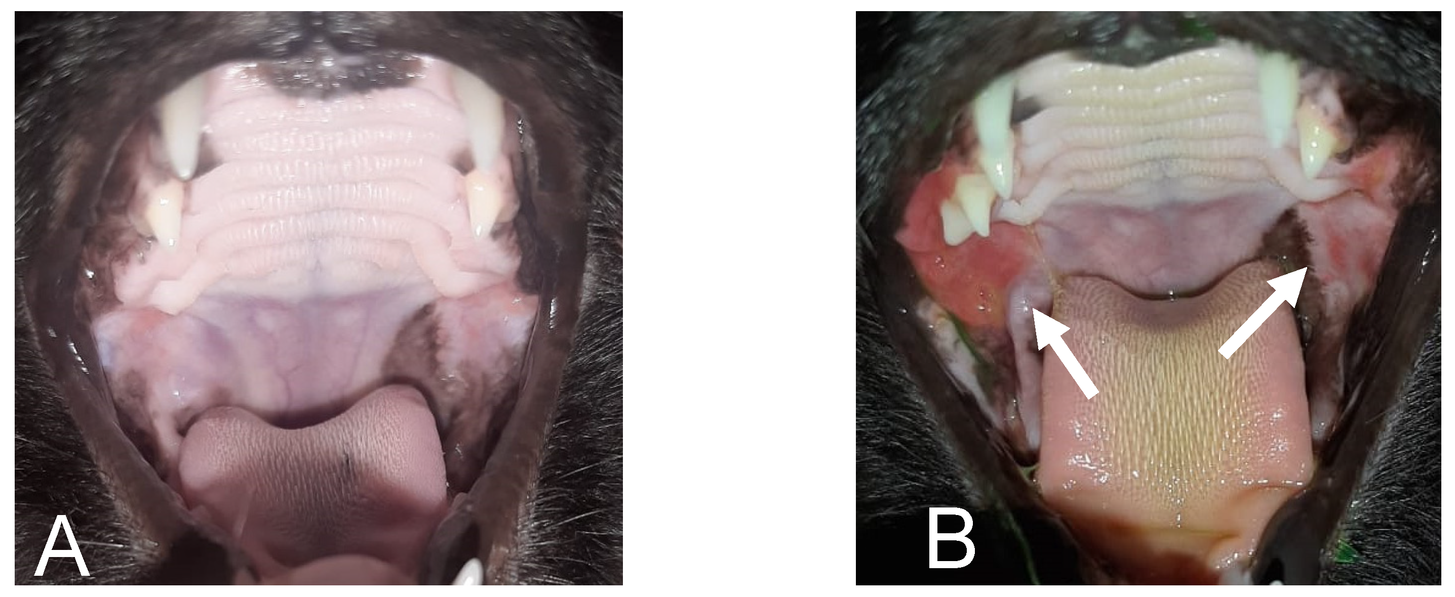
Even after 30 days following the procedure, during which three molars were removed (Figure 2) and the patient was six weeks on cyclosporine, the cat continued to have the same oral lesions. We decided to replace the previous diet with a hypoallergenic extruded dry food based on hydrolyzed soy protein and rice bran (Hypoallergenic Feline, Royal Canin), prescribed according to maintenance energy needs [NEM (kcal/day) = 75 × BW0.67, where BW = body weight [15]]. After 30 days, the patient showed complete remission of the lesions (Figure 3A), and the amitriptyline was discontinued. At this point, the cat owner, who had another cat, requested to resume the maintenance diet, because they wanted both cats to eat the same diet. Therefore, the diet re-exposure was performed, without modifying the cyclosporine regimen, using the previously administered extruded dry food. After seven days, the patient exhibited inflammation of the oral mucosa again (Figure 3B), prompting a return to the hypoallergenic diet, after which the patient showed complete improvement of the lesions within a few days.
After remission of the oral lesions, the dosage of cyclosporine was reduced. This resulted in the recurrence of the lesions, making it necessary to return to the initial dose. After 6 months, the patient remained stable with the daily administration of cyclosporine and the hypoallergenic dry diet, without access to any other diet. Additionally, it was recommended to the caretaker to strictly control the amount of food to prevent the patient from gaining weight and becoming significantly overweight, which would worsen the feline’s condition.

3. Discussion

This case demonstrates that although conventional clinical and surgical treatments were combined, as described in the literature, the patient remained refractory. Since gingivostomatitis involves alterations in the immune response, a transition to a hypoallergenic diet was implemented, resulting in complete remission of the lesions.
Initially, the patient was medicated with prednisolone to reduce the inflammatory process, as it is effective in 70 to 80% of stomatitis cases [3]. Lower urinary tract signs could be justified by stress and pain, and it was managed with amitriptyline, a tricyclic antidepressant with anti-inflammatory, analgesic, sympatholytic, antihistaminic, and anticholinergic properties [16]. On the eighth day of treatment, despite the remission of urinary signs, the oral ulcerative lesions persisted. Specific periodontal treatment was performed, which is estimated to have an 80% success rate due to a reduction in antigenic stimulation [4]. Additionally, cyclosporine was administered, which is classified as an immunosuppressant that inhibits T lymphocyte activation by reducing pro-inflammatory cytokines [9]. After 44 days of medication use, improvement was expected, especially since the surgical treatment had already been performed [12]. Cyclosporine has an average time of 2 to 6 weeks before the onset of improvement in lesion cases. However, the patient did not show improvement in the lesions, even after surgical treatment.
Some authors [1,8,17] suggest that nutritional management similar to that approached in cases of adverse food reaction is relevant in these situations; however, there are no studies or case reports in the literature that clearly demonstrate this relationship between oral lesions and food hypersensitivity. The main clinical signs of food sensitivity in felines are cutaneous and gastrointestinal [18,19], with reports of ophthalmic, respiratory, and behavioral alterations [20,21,22].
The diagnosis and treatment of adverse food reactions are based on the use of an elimination diet followed by provocative exposure [23], as in the case reported here. For cutaneous clinical signs, the recommended elimination period is up to eight weeks [24]. For gastrointestinal clinical signs, this elimination diet time is not fully established, with some authors recommending a shorter time than for dermatological changes, such as 2 to 8 weeks [22,23,25]. The recommended provocative diet period for diagnostic confirmation of an adverse food reaction is one to two weeks in felines [26]. In the case reported here, the time taken for the elimination diet to result in an improvement in clinical signs and for the provocative test to result in recurrence of the lesions was in line with the time recommended in the literature. It is important to note that provocative exposure was only possible because the patient had an excellent appetite and no refusals during the diet changes, and because of the consent of the owner.
Since there was a dietary change and the patient was maintained exclusively on a hypoallergenic diet, leading to the first complete improvement of the ulcerative lesions in the oral mucosa without changes in the medication or environmental protocols, it is possible to consider that the patient had an adverse reaction to the food, manifested by lesions in the oral cavity. We raised the hypothesis that chronic antigenic stimulation caused by some component of the diet food was a cofactor in perpetuating the inflammatory process. The diet with hydrolyzed protein may have aided this process, as hydrolysis reduces the molecular weight of the protein, decreasing the likelihood of epitope recognition by the immune system, which could potentially trigger an adverse food reaction [27].
It is an unknown variable whether the cyclosporine took time to reach its maximum effect and whether this occurred during the elimination diet. However, the patient’s re-exposure to the previous maintenance diet and the subsequent recurrence of oral mucosal lesions, followed by new remission upon returning to the hypoallergenic food, increase the evidence that diet may be an important factor in controlling this patient’s inflammatory process.
In parallel with veterinary medicine, similar conditions can be observed in humans, where recurrent aphthous stomatitis (RAS) presents a multifactorial cause and a pathophysiology similar to feline gingivostomatitis. It is linked to genetic, environmental, and immunological factors, and can also be associated with food allergy [28,29]. Treatment of RAS in humans depends on the frequency, size, and number of the ulcers, and consists of topical and systemic treatment with corticosteroids and immunosuppressants [30,31]. A possible link between food allergy and some cases of RAS has been suggested, with about 25 to 75% of human patients experiencing remission of oral cavity lesions after following an elimination diet for 1 to 2 weeks [32].
In this reported case, we decided to maintain the feline patient on a combined regiment of the hypoallergenic diet and cyclosporine, as the initial attempt to discontinue the treatments individually was unsuccessful. Studies show that some allergic conditions in pets with cutaneous manifestations can be minimized with the use of hypoallergenic food based on hydrolyzed protein, but only when combined with medication during the initial stage of the diet trial [32]. However, it is uncertain how a new attempt to suspend the medication or change the diet would fare after a long period in our case.
In the “Guideline Proposal for the Management of Chronic Gingivostomatitis in Cats” [33], the authors note that it may be interesting to make dietary changes to treat the condition; however, there are no robust studies supporting the use of hypoallergenic diets in the treatment of Chronic Gingivostomatitis in felines. Thus, studies with a higher degree of scientific evidence, assessing cats with gingivostomatitis subjected to elimination diets and provocative exposure, could reinforce the findings of this report that adverse food reactions may be associated with lesions in the oral cavity, and that the use of a hypoallergenic diet could be adjunctive to the treatment of these cases. However, in clinical practice, evaluating the diet of feline patients with gingivostomatitis can be challenging, as these animals often exhibit hyporexia [3,4,9] and may not accept dietary changes, which could leave the patient exposed to potential food antigens. This may explain why many cases of oral lesions in felines are refractory to anti-inflammatory and immunosuppressive treatment, as likely seen in this patient, who did not respond to corticosteroid therapy nor to the first monthly cycle of the most recommended immunosuppressant, cyclosporine [9], even after periodontal treatment.

4. Conclusions

The reported case highlights the possibility of an adverse food reaction being a cofactor in the presentation of oral lesions in a feline diagnosed through an elimination diet and provocative exposure, as well as highlighting the benefit of using an hypoallergenic diet based on hydrolyzed protein as an adjunct in treatment.

Author Contributions

Conceptualization, L.d.S. and F.A.T.; investigation, L.d.S. and T.M.; writing—original draft preparation, L.d.S. and F.A.T.; writing—review and editing, F.A.T. and M.Y.H.P. All authors have read and agreed to the published version of the manuscript.

Funding

This research received no external funding, except the APC that was funded by Royal Canin.

Institutional Review Board Statement

The retrospective nature of this case series involved the analysis of previously collected data and therefore did not require ethics approval.

Informed Consent Statement

Written informed consent has been obtained from the cat´s owner to publish this paper.

Data Availability Statement

The original contributions presented in the study are included in the article, further inquiries can be directed to the corresponding author.

Conflicts of Interest

The authors declare no conflicts of interest. The funder had no role in the interpretation of data; in the writing of the manuscript; or in the decision to publish the case.

References

  1. Diehl, K.; Rosychuk, R.A.W. Feline Gingivitis-Stomatitis-Pharyngitis. Vet. Clin. N. Am. Small Anim. Pract. 1993, 23, 139–153. [Google Scholar] [CrossRef]
  2. Niza, M.M.R.E.; Mestrinho, L.A.; Vilela, C.L. Gengivo-estomatite crônica felina—Um desafio clínico. Rev. Port. Ciênc. Vet. 2004, 99, 127–135. [Google Scholar]
  3. Lyon, K.F. Gingivostomatitis. Vet. Clin. N. Am. Small Anim. Pract. 2005, 35, 891–911. [Google Scholar] [CrossRef]
  4. Soltero-Rivera, M.M.; Reiter, A.M. Diseases of the Oral Cavity and Salivary Glands. In Clinical Small Animal Internal Medicine; Bruyette, D.S., Bexfield, N., Chretin, J.D., Kidd, L., Kube, S., Langston, C., Owen, T.J., Oyama, M.A., Peterson, N., Reiter, L.V., et al., Eds.; Wiley: Hoboken, NJ, USA, 2020; pp. 533–546. [Google Scholar] [CrossRef]
  5. Williams, C.A.; Aller, M.S. Gingivitis/Stomatitis in Cats. Vet. Clin. N. Am. Small Anim. Pract. 1992, 22, 1361–1383. [Google Scholar] [CrossRef]
  6. Healey, K.A.; Dawson, S.; Burrow, R.; Cripps, P.; Gaskell, C.J.; Hart, C.A.; Pinchbeck, G.L.; Radford, A.D.; Gaskell, R.M. Prevalence of Feline Chronic Gingivo-Stomatitis in First Opinion Veterinary Practice. J. Feline Med. Surg. 2007, 9, 373–381. [Google Scholar] [CrossRef]
  7. Robson, D. Review of the Properties and Mechanisms of Action of Cyclosporine with an Emphasis on Dermatological Therapy in Dogs, Cats and People. Vet. Rec. 2003, 152, 768–772. [Google Scholar] [CrossRef]
  8. Lommer, M.J. Efficacy of Cyclosporine for Chronic, Refractory Stomatitis in Cats: A Randomized, Placebo-Controlled, Double-Blinded Clinical Study. J. Vet. Dent. 2013, 30, 8–17. [Google Scholar] [CrossRef]
  9. Hennet, P. Chronic Gingivo-Stomatitis in Cats: Long-Term Follow-up of 30 Cases Treated by Dental Extractions. J. Vet. Dent. 1997, 14, 15–21. [Google Scholar] [CrossRef]
  10. Crystal, M.A. Gingivitis/stomatitis/faringitis. In El Paciente Felino: Bases del Diagnóstico y Tratamiento; Norsworthy, G.D., Crystal, M.A., Foochee, S.R., Tilley, L.P., Eds.; Inter-Médica Editorial: Buenos Aires, Argentina, 1998; pp. 228–231. [Google Scholar]
  11. Hofmann-Appollo, F.; Carvalho, V.G.G.; Gioso, M.A. Complexo Gengivite-Estomatite-Faringite Dos Felinos. Clínica Veterinária 2010, 15, 44–52. [Google Scholar]
  12. Laflamme, D.P. Development and Validation of a Body Condition Score System for Dogs: A Clinical Tool; Canine Practice: Santa Barbara, CA, USA, 1997; Volume 22, pp. 10–15. [Google Scholar]
  13. Michel, K.E.; Anderson, W.; Cupp, C.; Laflamme, D.P. Correlation of a Feline Muscle Mass Score with Body Composition Determined by Dual-Energy X-Ray Absorptiometry. Br. J. Nutr. 2011, 106 (Suppl. 1), S57–S59. [Google Scholar] [CrossRef]
  14. FEDIAF. Nutritional Guidelines for Complete and Complementary Pet Food for Cats and Dogs; European Pet Food Industry Federation: Brussels, Belgium, 2021. [Google Scholar]
  15. Westropp, J.L.; Tony Buffington, C.A. Feline Idiopathic Cystitis: Current Understanding of Pathophysiology and Management. Vet. Clin. N. Am. Small Anim. Pract. 2004, 34, 1043–1055. [Google Scholar] [CrossRef]
  16. Johnston, N. An updated approach to chronic feline gingivitis stomatitis syndrome. Vet. Pract. 2012, 44, 34–38. [Google Scholar]
  17. Carlotti, D.N.; Remy, I.; Prost, C. Food Allergy in Dogs And Cats. A Review and Report of 43 Cases. Vet. Dermatol. 1990, 1, 55–62. [Google Scholar] [CrossRef]
  18. Olivry, T.; Mueller, R.S. Critically Appraised Topic on Adverse Food Reactions of Companion Animals (7): Signalment and Cutaneous Manifestations of Dogs and Cats with Adverse Food Reactions. BMC Vet. Res. 2019, 15, 140. [Google Scholar] [CrossRef]
  19. White, S.D.; Sequoia, D. Food hypersensitivity in cats: 14 cases (1982–1987). J. Am. Vet. Med. Assoc. 1989, 194, 692–695. [Google Scholar]
  20. Hobi, S.; Linek, M.; Marignac, G.; Olivry, T.; Beco, L.; Nett, C.; Fontaine, J.; Roosje, P.; Bergvall, K.; Belova, S.; et al. Clinical Characteristics and Causes of Pruritus in Cats: A Multicentre Study on Feline Hypersensitivity- associated Dermatoses. Vet. Dermatol. 2011, 22, 406–413. [Google Scholar] [CrossRef]
  21. Mazzeranghi, F.; Zanotti, C.; Di Cerbo, A.; Verstegen, J.P.; Cocco, R.; Guidetti, G.; Canello, S. Clinical Efficacy of Nutraceutical Diet for Cats with Clinical Signs of Cutaneus Adverse Food Reaction (CAFR). Pol. J. Vet. Sci. 2017, 20, 269–276. [Google Scholar] [CrossRef]
  22. Mueller, R.S.; Olivry, T. Critically appraised topic on adverse food reactions of companion animals (4): Can we diagnose adverse food reactions in dogs and cats with in vivo or in vitro tests? BMC Vet. Res. 2017, 13, 275. [Google Scholar] [CrossRef]
  23. Olivry, T.; Mueller, R.S. Critically Appraised Topic on Adverse Food Reactions of Companion Animals (9): Time to Flare of Cutaneous Signs after a Dietary Challenge in Dogs and Cats with Food Allergies. BMC Vet. Res. 2020, 16, 158. [Google Scholar] [CrossRef]
  24. Moriello, K.A.; Kahn, C.M.; Line, S. The Merck Veterinary Manual; Merck & Co: Whitehouse Station, NJ, USA, 2010; pp. 649–852. [Google Scholar]
  25. Tiffany, S.; Parr, J.M.; Templeman, J.; Shoveller, A.K.; Manjos, R.; Yu, A.; Verbrugghe, A. Assessment of Dog Owners’ Knowledge Relating to the Diagnosis and Treatment of Canine Food Allergies. Can. Vet. J. 2019, 60, 268–274. [Google Scholar]
  26. Olivry, T.; Bizikova, P. A Systematic Review of the Evidence of Reduced Allergenicity and Clinical Benefit of Food Hydrolysates in Dogs with Cutaneous Adverse Food Reactions. Vet. Dermatol. 2010, 21, 32–41. [Google Scholar] [CrossRef]
  27. Woo, S.-B.; Sonis, S.T. Recurrent Aphthous Ulcers: A Review of Diagnosis and Treatment. J. Am. Dent. Assoc. 1996, 127, 1202–1213. [Google Scholar] [CrossRef]
  28. Zunt, S.L. Recurrent aphthous stomatitis. Dermatol. Clin. 2003, 21, 33–39. [Google Scholar] [CrossRef]
  29. Chavan, M.; Jain, H.; Diwan, N.; Khedkar, S.; Shete, A.; Durkar, S. Recurrent Aphthous Stomatitis: A Review. J. Oral Pathol. Med. 2012, 41, 577–583. [Google Scholar] [CrossRef]
  30. Eversole, L.R.; Shopper, T.P.; Chambers, D.W. Effects of Suspected Foodstuff Challenging Agents in the Etiology of Recurrent Aphthous Stomatitis. Oral Surg. Oral Med. Oral Pathol. 1982, 54, 33–38. [Google Scholar] [CrossRef]
  31. Wardhana, D.E.A. Recurrent aphthous stomatitis caused by food allergy. Acta Med. Indones. 2010, 42, 236–240. [Google Scholar] [PubMed]
  32. Miller, J.; Simpson, A.; Bloom, P.; Diesel, A.; Friedeck, A.; Paterson, T.; Wisecup, M.; Yu, C.M. AAHA Management of Allergic Skin Diseases in Dogs and Cats Guidelines. J. Am. Anim. Hosp. Assoc. 2023, 59, 255–284. [Google Scholar] [CrossRef]
  33. Camy, G.; Fahrenkrug, P.; Gracis, M.; Hennet, P.; Johnston, N.; Mihaljevic, S.; Schreyer, J. Proposed Guidelines on the Management of Feline Chronic Gingivostomatitis (FCGS) Syndrome: A Consensus Statement Consultation Version September. J. Feline Med. Surg. 2010, 20, 244–255. [Google Scholar]
Figure 1. Lesions in the oral mucosa at the first appointment (arrows).
Animals 14 02656 g001
Figure 2. (A) Radiograph of the right mandible and (B) left mandible with bone loss in the region of the distal root of the first molar (arrows). Images (C) and (D) show exodontia. Arrows indicate the exodontia.
Animals 14 02656 g002
Figure 3. (A) Remission after 30 days with the use of the hypoallergenic diet. (B) Reappearance of lesions approximately 7 days after food re-exposure. Arrows indicate the location of the lesions.
Animals 14 02656 g003
Disclaimer/Publisher’s Note: The statements, opinions and data contained in all publications are solely those of the individual author(s) and contributor(s) and not of MDPI and/or the editor(s). MDPI and/or the editor(s) disclaim responsibility for any injury to people or property resulting from any ideas, methods, instructions or products referred to in the content.