Article

Addressing Challenges in Wildlife Rehabilitation: Antimicrobial-Resistant Bacteria from Wounds and Fractures in Wild Birds

by
Esther Sánchez-Ortiz
1,2,†,
María del Mar Blanco Gutiérrez
1,†,
Cristina Calvo-Fernandez
3,4,
Aida Mencía-Gutiérrez
2,
Natalia Pastor Tiburón
2,
Alberto Alvarado Piqueras
2,
Alba Pablos-Tanarro
5 and
Bárbara Martín-Maldonado
5,*
1
Departamento de Sanidad Animal, Facultad de Veterinaria, Universidad Complutense, Avenida de Puerta de Hierro s/n, 28040 Madrid, Spain
2
Grupo de Rehabilitación de la Fauna Autóctona y su Hábitat, Calle Monte del Pilar s/n, 28220 Majadahonda, Spain
3
Research Group for Food Microbiology and Hygiene, National Food Institute, Technical University of Denmark, Henrik Dams Allé, 204, 2800 Kongens Lyngby, Denmark
4
Research Group for Foodborne Pathogens and Epidemiology, National Food Institute, Technical University of Denmark, Henrik Dams Allé, 204, 2800 Kongens Lyngby, Denmark
5
Departamento de Veterinaria, Facultad de Ciencias Biomédicas y de la Salud, Universidad Europea de Madrid, Calle Tajo s/n, 28760 Villaviciosa de Odón, Spain
*
Author to whom correspondence should be addressed.
These authors contributed equally to this work.
Animals 2024, 14(8), 1151; https://doi.org/10.3390/ani14081151
Submission received: 18 February 2024 / Revised: 3 April 2024 / Accepted: 9 April 2024 / Published: 10 April 2024

Abstract

:

Simple Summary

Wildlife rescue centers frequently admit animals with injuries and bone fractures. Open fractures are common in birds due to their anatomy, and this can lead to complications like osteomyelitis, which implies a serious bone infection and often necrosis, or death of the affected bone tissue. Antibiotic therapy is crucial, but the rise in antimicrobial-resistant isolates in wildlife raises concerns about treatment efficacy. A study focused on isolating, identifying, and assessing antimicrobial resistance in bacteria from wounds and fractures in wild birds. Among 36 isolates, Staphylococcus spp. dominated (63.8%), with 82.6% exhibiting antimicrobial resistance, particularly to clindamycin, an antimicrobial key in the treatment of infected bone fractures. This escalating resistance poses a dual threat to wildlife—therapeutic failure and the spread of resistant bacteria in ecosystems.

Abstract

Injuries and bone fractures are the most frequent causes of admission at wildlife rescue centers. Wild birds are more susceptible to open fractures due to their anatomical structure, which can lead to osteomyelitis and necrosis. Antibiotic therapy in these cases is indispensable, but the increase of antimicrobial-resistant isolates in wildlife has become a significant concern in recent years. In this context, the likelihood of antibiotic failure and death of animals with infectious issues is high. This study aimed to isolate, identify, and assess the antimicrobial resistance pattern of bacteria in wounds and open fractures in wild birds. To this end, injured birds admitted to a wildlife rescue center were sampled, and bacterial isolation and identification were performed. Then, antimicrobial susceptibility testing was assessed according to the disk diffusion method. In total, 36 isolates were obtained from 26 different birds. The genera detected were Staphylococcus spp. (63.8%), Escherichia (13.9%), Bacillus (11.1%), Streptococcus (8.3%), and Micrococcus (2.8%). Among Staphylococcus isolates, S. lentus and S. aureus were the most frequent species. Antimicrobial resistance was detected in 82.6% of the isolates, among which clindamycin resistance stood out, and 31.6% of resistant isolates were considered multidrug-resistant. Results from this study highlight the escalating scope of antimicrobial resistance in wildlife. This level of resistance poses a dual concern for wildlife: firstly, the risk of therapeutic failure in species of significant environmental value, and, secondly, the circulation of resistant bacteria in ecosystems.

1. Introduction

According to the International Union for Conservation of Nature (IUCN), 159 bird species have become extinct during the last 20 years, and now 12% of the described bird species are threatened [1]. Specifically in Europe, bird populations have declined by 600 million individuals since 1980 [2]. As species abundance has been described as a synonym of a healthy ecosystem, biodiversity conservation becomes key to ensuring global health for humans, animals, and the wild [3]. In this context, the role of wildlife rescue centers (WRCs) is essential as an ex situ action to ensure animal welfare, treating and caring for injured, sick, and stray native animals [4]. However, despite WRC efforts, the release rate ranges between 27% and 56% for wildlife admitted to WRCs, depending on the animal class and the region [5,6,7,8].
Among the causes of admission to a WRC, those linked to human activity, such as electrocutions, car or window collisions, or gunshots, are the most frequent [9,10]. In contrast, injuries due to other animals’ attacks are not so common but still are represented among traumatic causes of admission. Due to the anatomical adaptations of the bones for flight, birds are more prone to suffer open fractures with bone exposure [11,12]. In those species, the trauma category represents around 30–65% of the total admissions, and the prevalence of injuries and bone fractures in wild birds admitted to a WRC are, therefore, higher and the most common cause of morbidity and mortality [4,5,6,7]. A high percentage of wild birds admitted due to a traumatic cause, involving injuries and/or fractures, had a negative outcome, either by death or euthanasia [6]. This, coupled with the time elapsed from the trauma until the animal’s capture and arrival at the WRC, increases the risk of infection that can lead to osteomyelitis and necrosis [5,13,14,15]. The majority of avian wounds are older than 8 h and/or contaminated by the time they present for treatment [16]. The anatomical point of fracture, the extension of soft-tissue lesions, the infection and osteomyelitis, or the bone and soft-tissue necrosis are some of the criteria that help veterinarians assess euthanasia [6,17]. Unlike in mammals, osteomyelitis in birds is typically not systemic unless it involves a pneumatic bone, like the humerus or femur. This communication between the fracture and infection focus and the respiratory system can lead to fatal pneumonia [18].
The bacterial genera most frequently involved in wounds and open fractures in wild birds are mainly Staphylococcus, Enterococcus, Bacillus, Aeromonas, and other bacteria from the Enterobacteriaceae family, such as Escherichia, Enterobacter, Shigella, and Proteus [13,14]. Staphylococcus spp. is a ubiquitous bacterium in the normal microbiota of humans and animals, with the ability to cause a broad spectrum of infections [19]. Concretely, Staphylococcus aureus is one of the seven ESKAPE micro-organisms producing resistant infections in hospitals worldwide. The name of this group of highly virulent and antimicrobial-resistant bacteria (ARB) is the acronym of their seven scientific names: Enterococcus faecium, Staphylococcus aureus, Klebsiella pneumoniae, Acinetobacter baumannii, Pseudomonas aeruginosa, Enterobacter spp., and Escherichia coli [20]. The infection with S. aureus is the most common and can exacerbate osteomyelitis development through exfoliative and pore-forming toxins and superantigens that promote osteoclastogenesis and bone resorption [18]. Moreover, S. aureus has been described as one of the species with the highest rate of antimicrobial resistance (AMR) and resistant strains, which have been associated with more than 700,000 deaths worldwide in 2019 [21].
Despite the fact that wild bird rehabilitation from trauma usually entails antibiotic therapy, it must be conducted in a way so as to avoid the development and increase of new resistant bacteria. Nowadays, antimicrobial resistance (AMR) is one of the biggest health issues in the world, causing millions of human deaths yearly. Treating these bacterial infections has become arduous due to increased antimicrobial resistance [21]. In recent years, many authors have published the isolation of ARBs from wild birds, even though wildlife should not have direct contact with clinical settings [14,19,22,23]. This is due to the infinite interactions between humans and wildlife, direct or indirect, and the ecosystem’s pollution with antimicrobial residues [24,25]. Moreover, migratory birds have been suggested to have a higher probability of carrying ARBs in their microbiome and disseminating them through different regions due to the vast territory they cover [25,26]. Therefore, wild birds are crucial in disseminating ARB between environments and habitats [26].
This study aimed to isolate and characterize the bacteria in wounds and open fractures in wild birds admitted to a WRC from Spain. As a secondary objective, antimicrobial susceptibility tests were conducted to refine treatment protocols for wild birds accurately.

2. Materials and Methods

2.1. Sample Collection

From November 2018 to May 2020, wild birds admitted because of trauma at the WRC managed by Grupo de Rehabilitación para la Fauna Autóctona y su Hábitat (GREFA) were examined and sampled if their health status allowed it. Wound or open fracture samples were collected for microbiological diagnosis and antimicrobial susceptibility test with a sterile swab during their first examination. Samples were preserved in a liquid nutrient transport medium with fructose-1,6-bisphosphatase (FBP) (Oxoid®, Basingstoke, UK) and 0.5% active charcoal (Sigma-Aldrich®, Saint Louis, MO, USA) [27]. Then, samples were frozen at −20 °C until analysis at the laboratory. Handling procedures complied with Spanish legislation (Royal Decree 53/2013) [28]. Ethical review and approval were waived for this study because of the standard protocol for the sanitary status analysis of animals admitted to the GREFA Wildlife Hospital. Therefore, no extra handling of the animals was necessary to collect the samples, and no extra samples were collected outside the hospital’s standard workflow protocol.

2.2. Bacterial Culture and Isolation

At the laboratory, samples were unfrozen at room temperature (20–15 °C). After homogenization in a vortex, 100 µL of each sample were collected and transferred onto two agar media: Columbia Agar with 5% sheep blood and MacConkey (Oxoid Ltd.®, Basingstoke, UK). The blood agar culture was performed in aerobic and anaerobic conditions with AnaeroGen® (Thermofisher Scientific®, Waltham, MA, USA). All plates were incubated at 37 ± 1 °C for 24 h. Then, the macroscopic characteristics of different colonies formed on each plate were observed (size, shape, and color of the colonies; presence of alpha- or beta-hemolysis or no hemolysis on blood agar cultures; and whether there was lactose fermentation or not on MacConkey cultures) and used to sort them out. Plates without growth in the first 24 h were kept for another 24 h under the same conditions before confirming negative growth. Then, a single colony of each morphology present was selected from each plate with growth and stroked on Columbia Base (Oxoid Ltd.®, Basingstoke, UK) to obtain a monoclonal culture after 24 h of incubation at 37 ± 1 °C. A sample of each monoclonal culture was preserved in cryovials with nutritive broth and glycerol (80–20%, respectively) at −80 °C for further analysis.

2.3. Bacterial Identification

Bacterial identifications were performed by microscopical morphology, shape, and staining characteristics with Gram stain (Panreac AppliChem®, Darmstadt, Germany) and classical biochemical tests, including catalase, potassium hydroxide (KOH) (MERCK®, Darmstadt, Germany), and oxidase (MAST® ID, Merseyside, UK) [29]. Additionally, isolates whose characteristics were compatible with Staphylococcus spp. were identified at the species level using Analytical Profile Index (API) STAPH multisubstrate galleries (bioMérieux®, Marcy l’Etoile, France). In the same way, isolates compatible with E. coli were confirmed with API 20E multisubstrate galleries (bioMérieux®, Marcy l’Etoile, France). Positive controls of S. aureus (ATCC 25923) and E. coli (ATCC 4157) were included in the analysis with API STAPH and API 20E galleries, respectively.

2.4. Antimicrobial Susceptibility Test

Antimicrobial susceptibility test (AST) was performed according to the disk diffusion or Kirby–Bauer method and the European Committee on Antimicrobial Susceptibility Testing (EUCAST) guidelines [30]. Isolates were retrieved from cryovials and plated onto Columbia Agar supplemented with 5% sheep blood (Becton Dickinson GmbH, Heidelberg, Germany). A suspension of the inoculum was prepared in a sterile 0.8% saline solution to achieve a turbidity equivalent to 0.5 McFarland. Subsequently, the inoculum was transferred onto Mueller–Hinton agar (Becton, Dickinson GmbH, Heidelberg, Germany), and antimicrobial disks were placed on the surface. The selection of antimicrobials for the susceptibility test was based on the panel of antimicrobials routinely used in treating wild birds at GREFA Wildlife Hospital. According to EUCAST recommendations, the antimicrobial susceptibility analysis of staphylococci includes chloramphenicol, clindamycin, erythromycin, tetracycline, linezolid, and tedizolid [31]. However, the latter two are scarcely used in veterinary medicine, especially in wildlife, so they were not included in the test. In exchange, ampicillin, ciprofloxacin, and trimethoprim-sulfamethoxazole were added, as they are commonly used in wildlife clinical practice. In total, susceptibility to seven groups of antibiotics was analyzed: penicillins, quinolones, sulphonamides, macrolides, lincosamides, amphenicols, and tetracyclines (Table 1). After 24 h at 37 ± 1 °C, susceptibility or resistance was determined by growth inhibition diameter regarding standardized breakpoint tables from EUCAST [31]. For the antimicrobial without cut-off values in EUCAST tables (chloramphenicol), the breakpoint was established following the Clinical and Laboratory Standards Institute (CLSI) guidelines [32]. Multidrug resistance (MDR) was considered when the isolate was non-susceptible to at least one antimicrobial agent in three or more different classes of antimicrobial [33].

3. Results

Samples of 26 individuals belonging to 11 different species of birds were analyzed: black kite, booted eagle, Eurasian eagle-owl, Tawny owl, little owl, mallard, white stork, grey heron, lesser black-backed gull, common blackbird, and rock dove (Table 2).

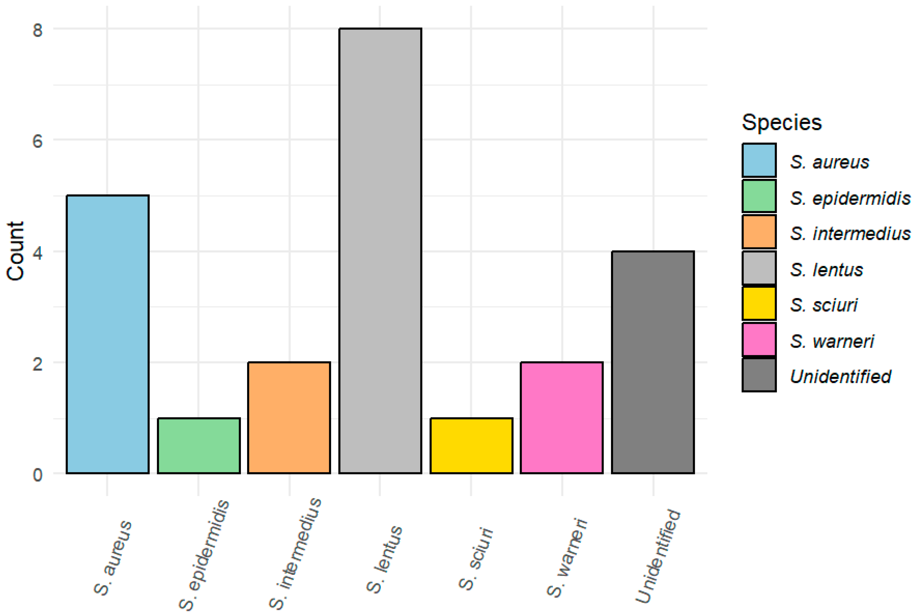
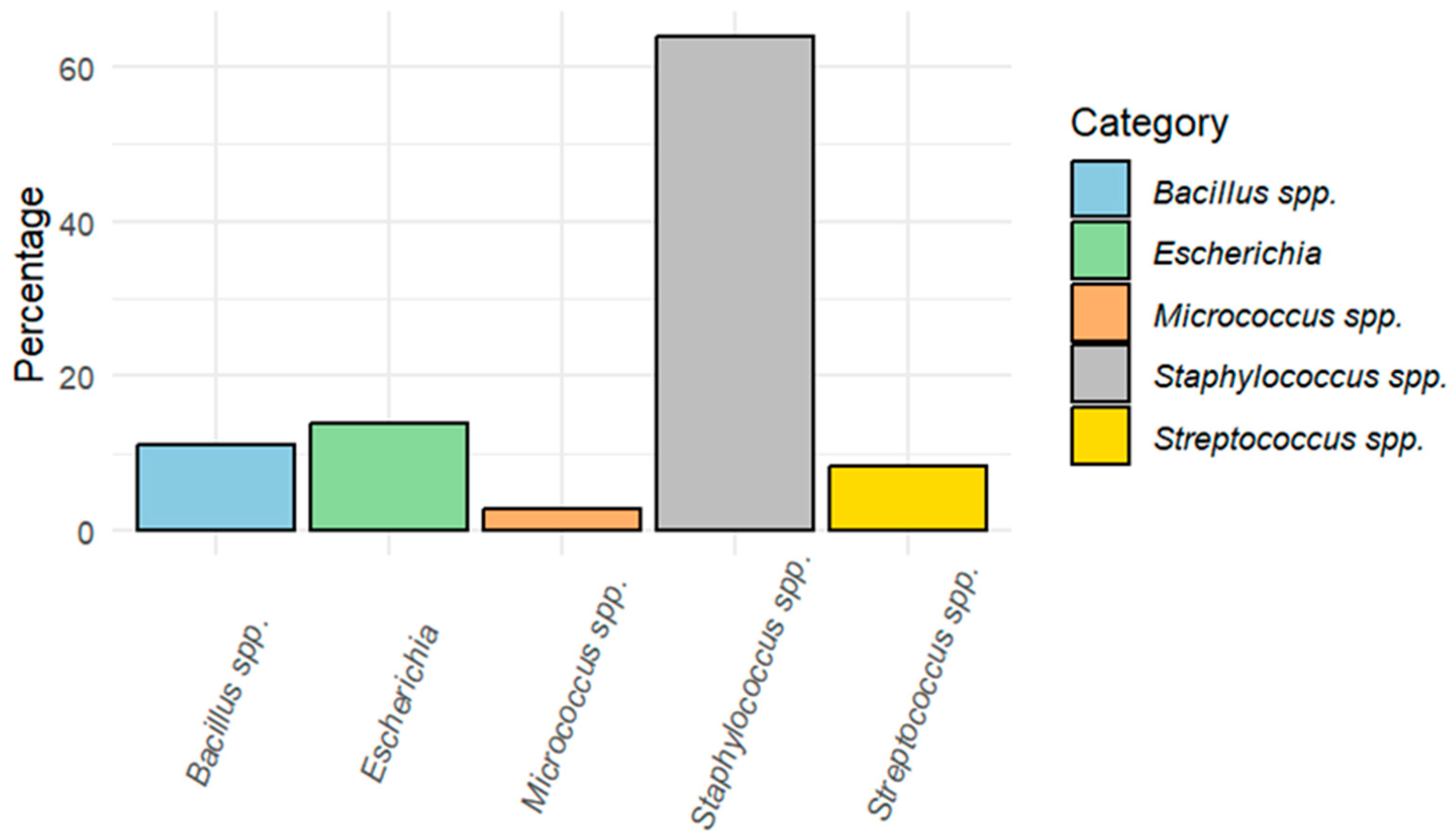
3.1. Bacterial Isolation and Identification

Positive bacterial growth was obtained in 14 of the 26 including birds (53.8%), while the other 12 birds had no bacterial growth in any of the media used. In total, 36 different colonies were isolated. After basic biochemical tests and Gram staining, they could be classified at the genus level: 63.8% (23/36) Staphylococcus spp., 13.9% (5/36) Escherichia, 11.1% (4/36) Bacillus spp., 8.3% (3/36) Streptococcus spp., and 2.8% (1/36) Micrococcus spp. (Figure 1). Moreover, Staphylococcus isolates could be identified at the species level using API STAPH® galleries, with reliability ≥ 90%: S. lentus (34.8%, 8/23), S. aureus (21.7%, 5/23), S. warneri (8.7%, 2/23), S. intermedius (8.7%, 2/23), S. epidermidis (4.3%, 1/23), and S. sciuri (4.3%, 1/23) (Figure 2). S. lentus was the species most isolated from open-wound and fracture samples. The staphylococci species could not be identified in four isolates (17.3%) (Table 2). All the Escherichia isolates belonged to the species E. coli.

3.2. Antimicrobial Susceptibility Results

As Staphylococcus was the genus most frequent and because it has been considered a sentinel for AMR surveillance, AST was performed only with Staphylococcus isolates. Of the 23 staphylococci isolates, 82.6% were resistant to at least one antimicrobial (19/23). Six isolates were susceptible to all the antimicrobials tested: two from Eurasian eagle-owls, two from white storks, one from a black kite, and the last one from a booted eagle. Still, none showed efficacy in all the isolates. The highest percentage of resistance found was to clindamycin (CMN) (52.2%, 12/23), followed in decreasing order by ampicillin (AMP), erythromycin (ERY), tetracycline (TET), and trimethoprim-sulfamethoxazole (SXT), ciprofloxacin (CIP), and, finally, chloramphenicol (CHL) (Table 3).
Overall, 31.6% of resistant isolates (6/19) were considered MDR since they were resistant to at least three different antimicrobial classes, half of them (50%, 3/6) to four antimicrobial classes. All MDR isolates were resistant to CMN combined with different antimicrobials, mainly cephalosporins, penicillins, and/or erythromycin. S. lentus was the species with a higher rate of MDR. Regarding bird species, the grey heron has the highest rate of MDR staphylococci, followed by the white stork. In contrast, lesser black-backed gulls and Eurasian eagle-owls were the only species without MDR strains.

4. Discussion

The role of WRC in treating injured wildlife could be key for the restoration of endangered populations as an ex situ tool [4]. As one of the main causes of admission of wild birds is trauma, the management of injuries and fractures becomes essential for the successful in wildlife rehabilitation, even more so in wild birds due to their anatomical adaptations to flight [11]. However, there is a lack of studies about the distribution of bacteria in injuries and open fractures in wild birds, as well as their antimicrobial resistance patterns, which is of great importance in establishing the best treatment. Moreover, the presence of resistant bacteria in wild birds could represent a potential hazard to global health in the dissemination of resistant pathogens, many with zoonotic potential, such as Escherichia coli, Salmonella enterica, or Staphylococcus aureus. During their daily movements and migration, they can transport these resistant pathogens to new areas and spread them to the environment [14,25]. The present study assessed the bacterial genera most present in open fractures and wounds in wild birds admitted at a WRC.
In this work, the genus most frequently isolated was Staphylococcus (63.8%), followed by Escherichia (13.9%), Bacillus (11.1%), Streptococcus (8.3%), and Micrococcus (2.8%). These results contrast with those published by Gambino et al. [23], where the prevalence of Staphylococcus and Streptococcus were lower (11.4% and 3.1%, respectively), and no Bacillus were detected. Staphylococcus in birds was the most frequent genus in the samples analyzed and is highly relevant in human and veterinary medicine due to its ability to cause infections in the heart, bone, skin, and other tissues [34,35]. It is one of the genera with the highest resistance to antimicrobials, which makes its treatment extremely difficult, and S. aureus is considered one of the most lethal resistant bacteria worldwide, which globally had been indirectly associated with more than 4 million deaths in 2019 [21]. In addition, due to its ubiquity and easy cultivation, it has been used in numerous studies as a sentinel bacterium to study AMR trends [36,37]. The source of these bacteria colonizing injuries from wild birds might be related to scratches or bites from other animals, especially in urban wild birds. A mixed aerobic/anaerobic population is often isolated from infected bite wounds from small animals like felids and canids, which are also known to carry bacterial groups like Staphylococcus spp. and Streptococcus spp. on their gingival tissue and teeth. This is why antimicrobial therapy is always indicated in any bird attacked by other animals [38].
Within the genus Staphylococcus, the most frequently isolated species was S. lentus (34.8%) (proposed to be reclassified as Mammallicoccus lentus by Madhaiyan et al. [39]), which agrees with a study performed on nocturnal raptors where the prevalence of that species was 29.7% [19]. It has previously been identified in the trachea of healthy wild birds as a component of the respiratory microbiota and is also found on the skin of healthy pigeons [40,41]. Moreover, S. lentus has been isolated from the skin of people working with pigeons and described as a neglected pathogen for humans, but, to the best of our knowledge, there is no scientific report about zoonotic transmission [41,42]. In the present study, S. lentus was isolated from six individuals: three of them had open fractures of pneumatic bones (humerus and femur), so the presence of S. lentus in these fractures could be related to the respiratory microbiota. Instead, S. aureus was the second species most detected from open fractures and wounds; our detection rate is moderate (21.7%), according to Silva et al. [19]. The role of S. aureus as a zoonotic pathogen with high AMR rates is well-documented in both humans and animals, even in wild birds [19,21]. In a lower percentage, two isolates of S. intermedius and S. warneri, a species found in the feathers of migratory birds without associated symptoms, were obtained [43]. As a coagulase-positive Staphylococcus (CoPS), S. intermedius had an increased pathogenicity. On the contrary, S. sciuri is the most common coagulase-negative staphylococcal (CoNS) species in healthy wild animals, including birds, but it has a high zoonotic potential [40,43,44]. Now proposed also as Mammallicoccus sciuri [39], it was one of our study’s less dominant staphylococci species. These CoNS were considered less pathogenic than S. aureus, but recent studies have demonstrated an increasing clinical impact and can act as opportunistic pathogens [45,46]. Interestingly, a recent study on wild birds from Spain reported S. sciuri as the most frequent staphylococci species [40]. In another study, Sousa et al. [47] confirmed the presence of S. sciuri in rodents, rats, and squirrels, which can be part of the birds’ prey diet and could be a source of infection. Finally, only one isolate of S. epidermidis was obtained, which is considered an essential opportunistic species in human infectious diseases, and it has been described as part of the respiratory and digestive microbiota of birds in low concentration [40,47].
The antimicrobial susceptibility test of Staphylococcus spp. isolates showed a concerning proportion of AMR isolates (82.6%) from infected open fractures and wounds from wild birds. Even though ARB should be expected to be in a lower proportion in wild birds, some species, such as white storks (Ciconia Ciconia) or Columbiformes, are considered urban birds and have closer contact with human activities and garbage, increasing the risk of acquiring ARB [25,48]. Clindamycin was the antimicrobial with the highest rate of resistance (54.5%), which agrees with previous reports from wildlife [14,19,23,37,49]. Ruiz-Ripa et al. [40] detected 90% of CMN-resistant staphylococci, highlighting a potential conflict with veterinary clinical procedures as clindamycin is widely used, especially for pododermatitis and osteomyelitis treatment in wild birds [5,14]. In this context, a recent in-vitro experiment has demonstrated that lower doses of CMN can favor osteoblast proliferation and differentiation, as well as calcium deposition [50]. Beta-lactams are also common in both human and veterinary practice as the first choice, and resistance to this antimicrobial family is frequent in staphylococci isolated from wildlife [51]. Among penicillins, the rate of resistance to AMP was high (39.1%), which agrees with the data published by some authors [23,49]. This resistance against AMP has also been reported in livestock, companion animals, and humans in similar or higher proportions. This finding, added to the critical role of wildlife in disseminating and maintaining AMR in the ecosystems, confirms the connections between humans, animals, and the environment, and the need for a One Health approach to monitoring and curbing the dissemination of AMR [52,53]. While some authors recommend using clindamycin in osteomyelitis treatment, others suggest that cephalosporins such as ceftiofur or cefotaxime could give better outcomes [14]. However, third-, fourth-, and fifth-generation cephalosporins have been classified as the highest-priority critically important antimicrobials by the World Health Organization (WHO), so their use in veterinary practice should be limited [54]. Our results suggest that clindamycin is not the best option to treat open fractures or wounds infected by Staphylococcus spp. However, in our study, no cephalosporins were included, so it is not possible to assess the resistance of our strains to this class of antimicrobials. According to the published CLSI guidelines, resistance to some cephalosporins and other beta-lactam antimicrobials can be extrapolated from the result obtained from AST against oxacillin. Therefore, despite being an antimicrobial rarely used in veterinary medicine, the inclusion of oxacillin in the AST would have been very informative [32]. On the other hand, the absence of CHL-resistant isolates found in the present study contrasts with results obtained by Sousa et al. in a study of the characterization of staphylococci isolated from the nasal cavity in wild birds (71.3%) [47]. Resistance to ERY, CIP, TET, and SXT has also been observed in lower proportions in similar studies with wild birds [14,37,40,48,51].
From the 19 strains of resistant Staphylococcus spp., 6 were considered MDR (31.6%). This percentage is close to that reported in previous studies on wild birds, which showed percentages near to 50% of MDR [14,40]. In contrast, Fernandez-Fernandez et al. [37] confirmed the absence of MDR strains in 259 staphylococci isolates from the white stork respiratory system. MDR has been widely assessed in wildlife under a public-concern approach. However, it is important to highlight that the emergence of bacteria resistant to multiple antimicrobial classes could precipitate treatment failures in these animals, many of which play pivotal ecological roles.
Finally, to obtain more precise and statistically significant results, a larger number of cases is necessary. However, this study includes a small number of cases due to the complexity of obtaining complete clinical cases and collecting samples before any veterinary treatment, always prioritizing animal welfare when working with wildlife. Therefore, the results presented in this study are simply observational, and no statistical conclusions can be drawn from them. Further research is needed to evaluate and characterize the bacterial species that may be involved in infected wounds and open fractures in wild birds, including their resistance patterns.

5. Conclusions

In conclusion, the most frequent bacteria isolated from wounds and open fractures of wild birds in our study were S. lentus, followed by S. aureus and E. coli. Among staphylococci isolates, the antimicrobial resistance proportion was concerning (82.6%); mainly clindamycin, ampicillin, and erythromycin, and 31.6% of the resistant isolates were considered MDR. Although clindamycin benefits osteoblast proliferation, its administration should be avoided in infected open fracture treatment. Our study’s outcomes emphasize the alarming expansion of antimicrobial resistance in wildlife. This heightened resistance threatens therapeutic success in species of paramount environmental importance, intensifies the spread of ARB throughout ecosystems, and focuses attention on the urgent need to incorporate wildlife into surveillance protocols for antimicrobial resistance. Despite budget limitations linked to wildlife research, incorporating new molecular techniques such as sequencing in epidemiological studies would help us to understand whether the observed resistance mechanisms are acquired and share genomic structures with those identified in bacteria from other environments.

Author Contributions

Conceptualization, B.M.-M. and M.d.M.B.G.; methodology, E.S.-O., A.P.-T., B.M.-M. and M.d.M.B.G.; formal analysis, E.S.-O. and B.M.-M.; data curation, E.S.-O., A.M.-G. and C.C.-F.; writing—original draft preparation, B.M.-M.; writing—review and editing, E.S.-O., M.d.M.B.G., C.C.-F., A.P.-T. and A.M.-G.; visualization, A.M.-G. and C.C.-F.; supervision, B.M.-M.; project administration, B.M.-M.; funding acquisition, M.d.M.B.G., A.A.P. and N.P.T. All authors have read and agreed to the published version of the manuscript.

Funding

The microbiological analysis was funded by the Ministry of Ecological Transition (MITECO) of Spain and the Complutense University of Madrid (Spain).

Institutional Review Board Statement

Ethical review and approval were waived for this study because of the standard protocol for the sanitary status analysis of animals admitted to the GREFA Wildlife Hospital. Therefore, no extra handling of the animals was necessary to collect the samples, and no extra samples were collected outside the hospital’s standard work protocol. For this reason, according to the current legislation at the time of the research (Directive 2010/63/EU), it is not mandatory to have the approval of an Ethics Committee.

Informed Consent Statement

Not applicable.

Data Availability Statement

The data presented in this study are available on request from the corresponding author.

Acknowledgments

We thank all the GREFA workers and volunteers for their technical support.

Conflicts of Interest

The authors declare no conflict of interest.

References

  1. IUCN. The IUCN Red List of Threatened Species. Version 2023-1. 2023. Available online: https://www.iucnredlist.org/ (accessed on 16 February 2024).
  2. Burns, F.; Eaton, M.A.; Burfield, I.J.; Klvaňová, A.; Šilarová, E.; Staneva, A.; Gregory, R.D. Abundance decline in the avifauna of the European Union reveals cross-continental similarities in biodiversity change. Ecol. Evol. 2021, 11, 16647–16660. [Google Scholar] [CrossRef] [PubMed]
  3. Rosenberg, K.V.; Dokter, A.M.; Blancher, P.J.; Sauer, J.R.; Smith, A.C.; Smith, P.A.; Stanton, J.C.; Panjabi, A.; Helft, L.; Parr, M.; et al. Decline of the North American avifauna. Science 2019, 366, 120–124. [Google Scholar] [CrossRef] [PubMed]
  4. Romero, F.; Espinoza, A.; Sallaberry-Pincheira, N.; Napolitano, C. A five-year retrospective study on patterns of casuistry and insights on the current status of wildlife rescue and rehabilitation centers in Chile. Rev. Chil. Hist. Nat. 2019, 92, 6. [Google Scholar] [CrossRef]
  5. Punch, P. A retrospective study of the success of medical and surgical treatment of wild Australian raptors. Aust. Veter. J. 2001, 79, 747–752. [Google Scholar] [CrossRef]
  6. Cococcetta, C.; Coutant, T.; Collarile, T.; Vetere, A.; Di Ianni, F.; Huynh, M. Causes of Raptor Admission to the Wildlife Rehabilitation Centre in Abruzzo (Central Italy) from 2005–2016. Animals 2022, 12, 1916. [Google Scholar] [CrossRef] [PubMed]
  7. Vezyrakis, A.; Bontzorlos, V.; Rallis, G.; Ganoti, M. Two decades of wildlife rehabilitation in Greece: Major threats, admission trends and treatment outcomes from a prominent rehabilitation centre. J. Nat. Conserv. 2023, 73, 126372. [Google Scholar] [CrossRef]
  8. Molina-López, R.A.; Mañosa, S.; Torres-Riera, A.; Pomarol, M.; Darwich, L. Morbidity, outcomes and cost-benefit analysis of wildlife rehabilitation in Catalonia (Spain). PLoS ONE 2017, 12, e0181331. [Google Scholar] [CrossRef] [PubMed]
  9. Molina-López, R.A.; Casal, J.; Darwich, L. Causes of Morbidity in Wild Raptor Populations Admitted at a Wildlife Rehabilitation Centre in Spain from 1995–2007: A Long Term Retrospective Study. PLoS ONE 2011, 6, e24603. [Google Scholar] [CrossRef]
  10. Durá Alemañ, C.J.; Morales-Reyes, Z.; Ayerza, P.; Bodega, D.D.L.; Aguilera-Alcalá, N.; Botella, F.; Jiménez, J.; López-Bao, J.V.; Mateo-Tomás, P.; Moleón, M.; et al. La responsabilidad por el daño ambiental generado en el caso de la lucha contra el uso del veneno en España. Actual. Jurídica Ambient. 2020, 102/2, 564–576. Available online: http://hdl.handle.net/10261/217609 (accessed on 4 November 2023).
  11. Sullivan, T.N.; Wang, B.; Espinosa, H.D.; Meyers, M.A. Extreme lightweight structures: Avian feathers and bones. Mater. Today 2017, 20, 377–391. [Google Scholar] [CrossRef]
  12. Vigneault, A.; Fitzgerald, G.; Desmarchelier, M. A retrospective study of femoral fractures in wild birds of prey: 119 cases. J. Zoo Wildl. Med. 2021, 52, 564–572. [Google Scholar] [CrossRef] [PubMed]
  13. Vergneau-Grosset, C.; Dubé, C.; Fitzgerald, G.; Lair, S. Characteristics of antebrachial fractures associated with a successful outcome among free-ranging birds of prey that received treatment in a rehabilitation program. J. Am. Veter. Med. Assoc. 2020, 256, 580–589. [Google Scholar] [CrossRef] [PubMed]
  14. Tardón, A.; Bataller, E.; Llobat, L.; Jiménez-Trigos, E. Bacteria and antibiotic resistance detection in fractures of wild birds from wildlife rehabilitation centres in Spain. Comp. Immunol. Microbiol. Infect. Dis. 2021, 74, 101575. [Google Scholar] [CrossRef] [PubMed]
  15. Bertuccelli, T.; Crosta, L.; Costa, G.L.; Schnitzer, P.; Sawmy, S.; Spadola, F. Predisposing Anatomical Factors of Humeral Fractures in Birds of Prey: A Preliminary Tomographic Comparative Study. J. Avian Med. Surg. 2021, 35, 123–134. [Google Scholar] [CrossRef] [PubMed]
  16. Cousquer, G. Wound Management in the Avian Wildlife Casualty. World Wide Wounds. November 2003b. 2003. Available online: http://www.worldwidewounds.com/2003/november/Cousquer/Avian-Wound-Management-Part-2.html (accessed on 4 November 2023).
  17. Scott, D. Natural history and medical management of raptors. In Medical Management of Wildlife Species: A Guide for Practitioners; Wiley Online Library: Hoboken, NJ, USA, 2019; pp. 215–228. [Google Scholar] [CrossRef]
  18. Hofstee, M.I.; Muthukrishnan, G.; Atkins, G.J.; Riool, M.; Thompson, K.; Morgenstern, M.; Stoddart, M.J.; Richards, R.G.; Zaat, S.A.; Moriarty, T.F. Current concepts of osteomyelitis: From pathologic mechanisms to advanced research methods. Am. J. Pathol. 2020, 190, 1151–1163. [Google Scholar] [CrossRef] [PubMed]
  19. Silva, V.; Lopes, A.F.; Soeiro, V.; Caniça, M.; Manageiro, V.; Pereira, J.E.; Maltez, L.; Capelo, J.L.; Igrejas, G.; Poeta, P. Nocturnal Birds of Prey as Carriers of Staphylococcus aureus and Other Staphylococci: Diversity, Antimicrobial Resistance and Clonal Lineages. Antibiotics 2022, 11, 240. [Google Scholar] [CrossRef] [PubMed]
  20. De Oliveira, D.M.P.; Forde, B.M.; Kidd, T.J.; Harris, P.N.A.; Schembri, M.A.; Beatson, S.A.; Paterson, D.L.; Walker, M.J. Antimicrobial Resistance in ESKAPE Pathogens. Clin. Microbiol. Rev. 2020, 33, e00181-19. [Google Scholar] [CrossRef] [PubMed]
  21. Murray, C.J.L.; Ikuta, K.S.; Sharara, F.; Swetschinski, L.; Aguilar, G.R.; Gray, A.; Han, C.; Bisignano, C.; Rao, P.; Wool, E.; et al. Global burden of bacterial antimicrobial resistance in 2019: A systematic analysis. Lancet 2022, 399, 629–655. [Google Scholar] [CrossRef] [PubMed]
  22. Martín-Maldonado, B.; Montoro-Dasi, L.; Pérez-Gracia, M.T.; Jordá, J.; Vega, S.; Marco-Jiménez, F.; Marin, C. Wild Bonelli’s eagles (Aquila fasciata) as carrier of antimicrobial resistant Salmonella and Campylobacter in Eastern Spain. Comp. Immunol. Microbiol. Infect. Dis. 2019, 67, 101372. [Google Scholar] [CrossRef]
  23. Gambino, D.; Vicari, D.; Vitale, M.; Schirò, G.; Mira, F.; La Giglia, M.; Riccardi, A.; Gentile, A.; Giardina, S.; Carrozzo, A.; et al. Study on bacteria isolates and antimicrobial resistance in wildlife in Sicily, southern Italy. Microorganisms 2021, 9, 203. [Google Scholar] [CrossRef]
  24. Arnold, K.E.; Williams, N.J.; Bennett, M. «Disperse abroad in the land»: The role of wildlife in the dissemination of antimicrobial resistance. Biol. Lett. 2016, 12, 20160137. [Google Scholar] [CrossRef] [PubMed]
  25. Martín-Maldonado, B.; Rodríguez-Alcázar, P.; Fernández-Novo, A.; González, F.; Pastor, N.; López, I.; Suárez, L.; Moraleda, V.; Aranaz, A. Urban Birds as Antimicrobial Resistance Sentinels: White Storks Showed Higher Multidrug-Resistant Escherichia coli Levels than Seagulls in Central Spain. Animals 2022, 12, 2714. [Google Scholar] [CrossRef] [PubMed]
  26. Ramey, A.M.; Ahlstrom, C.A. Antibiotic Resistant Bacteria in Wildlife: Perspectives on Trands, Acquisition and Dissemination, Data GAPS, and Future Directions. J. Wildl. Dis. 2020, 56, 1–15. [Google Scholar] [CrossRef] [PubMed]
  27. Gorman, R.; Adley, C. An evaluation of five preservation techniques and conventional freezing temperatures of −20 °C and −85 °C for long-term preservation of Campylobacter jejuni. Lett. Appl. Microbiol. 2004, 38, 306–310. [Google Scholar] [CrossRef]
  28. BOE. Boletín Oficial del Estado, Real Decreto 53/2013, de 1 de Febrero, por el que se Establecen las Normas Básicas Aplicables para la Protección de los Animales Utilizados en Experimentación y Otros Fines Científicos, Incluyendo la Docencia; BOE: Madrid, Spain, 2013; Available online: https://www.boe.es/eli/es/rd/2013/02/01/53/con (accessed on 4 November 2023).
  29. Markey, B.; Leonard, F.; Archambault, M.; Cullinane, A.; Maguire, D. Clinical Veterinary Microbiology, 2nd ed.; Mosby Elsevier Ltd.: Dublin, Ireland, 2013; ISBN 9780723432371. [Google Scholar]
  30. Matuschek, E.; Brown, D.F.J.; Kahlmeter, G. Development of the EUCAST disk diffusion antimicrobial susceptibility testing method and its implementation in routine microbiology laboratories. Clin. Microbiol. Infect. Off. Publ. Eur. Soc. Microbiol. Infect. Dis. 2014, 20, 255–266. [Google Scholar] [CrossRef] [PubMed]
  31. EUCAST. European Committee on Antimicrobial Susceptibility Testing (EUCAST), Breakpoint Tables for Interpretation of MICs and Zone Diameters, Version 11. 2021. Available online: http://www.eucast.org (accessed on 2 April 2024).
  32. CLSI. Performance Standards for Antimicrobial Susceptibility Testing, 33rd ed.; CLSI Supplement M100; Clinical and Laboratory Standards Institute: Wayne, PA, USA, 2023. [Google Scholar]
  33. Magiorakos, A.-P.; Srinivasan, A.; Carey, R.B.; Carmeli, Y.; Falagas, M.E.; Giske, C.G.; Harbarth, S.; Hindler, J.F.; Kahlmeter, G.; Olsson-Liljequist, B.; et al. Multidrug-resistant, extensively drug-resistant and pandrug-resistant bacteria: An international expert proposal for interim standard definitions for acquired resistance. Clin. Microbiol. Infect. 2012, 18, 268–281. [Google Scholar] [CrossRef] [PubMed]
  34. Tong, S.Y.C.; Chen, L.F.; Fowler, V.G., Jr. Colonization, pathogenicity, host susceptibility, and therapeutics for Staphylococcus aureus: What is the clinical relevance? Semin. Immunopathol. 2012, 34, 185–200. [Google Scholar] [CrossRef] [PubMed]
  35. Parte, A.C.; Carbasse, J.S.; Meier-Kolthoff, J.P.; Reimer, L.C.; Göker, M. List of Prokaryotic names with Standing in Nomenclature (LPSN) moves to the DSMZ. Int. J. Syst. Evol. Microbiol. 2020, 70, 5607–5612. [Google Scholar] [CrossRef] [PubMed]
  36. Kutkowska, J.; Turska-Szewczuk, A.; Kucharczyk, M.; Kucharczyk, H.; Zalewska, J.; Urbanik-Sypniewska, T. Methicillin-resistant Staphylococcus aureus and glycopeptide-resistant enterococci in fecal samples of birds from South-Eastern Poland. BMC Veter. Res. 2019, 15, 472. [Google Scholar] [CrossRef]
  37. Fernández-Fernández, R.; Abdullahi, I.N.; González-Azcona, C.; Ulloa, A.; Martínez, A.; García-Vela, S.; Höfle, U.; Zarazaga, M.; Lozano, C.; Torres, C. Detection of antimicrobial producing Staphylococcus from migratory birds: Potential role in nasotracheal microbiota modulation. Front. Microbiol. 2023, 14, 1144975. [Google Scholar] [CrossRef]
  38. Tóth, A.G.; Tóth, I.; Rózsa, B.; Dubecz, A.; Patai, Á.V.; Németh, T.; Kaplan, S.; Kovács, E.G.; Makrai, L.; Solymosi, N. Canine saliva as a possible source of antimicrobial resistance genes. Antibiotics 2022, 11, 1490. [Google Scholar] [CrossRef] [PubMed]
  39. Madhaiyan, M.; Wirth, J.S.; Saravanan, V.S. Phylogenomic analyses of the Staphylococcaceae family suggest the reclassification of five species within the genus Staphylococcus as heterotypic synonyms, the promotion of five subspecies to novel species, the taxonomic reassignment of five Staphylococcus species to Mammaliicoccus gen. nov., and the formal assignment of Nosocomiicoccus to the family Staphylococcaceae. Int. J. Syst. Evol. Microbiol. 2020, 70, 5926–5936. [Google Scholar] [CrossRef]
  40. Ruiz-Ripa, L.; Gómez, P.; Alonso, C.A.; Camacho, M.C.; Ramiro, Y.; de la Puente, J.; Fernández-Fernández, R.; Quevedo, M.; Blanco, J.M.; Báguena, G.; et al. Frequency and Characterization of Antimicrobial Resistance and Virulence Genes of Coagulase-Negative Staphylococci from Wild Birds in Spain. Detection of tst-Carrying S. sciuri Isolates. Microorganisms 2020, 8, 1317. [Google Scholar] [CrossRef] [PubMed]
  41. Szczuka, E.; Wesołowska, M.; Krawiec, A.; Kosicki, J.Z. Staphylococcal species composition in the skin microbiota of domestic pigeons (Columba livia domestica). PLoS ONE 2023, 18, e0287261. [Google Scholar] [CrossRef] [PubMed]
  42. Mazal, C.; Sieger, B. Staphylococcus lentus: The troublemaker. Int. J. Infect. Dis. 2010, 14, e397. [Google Scholar] [CrossRef]
  43. Giorgio, A.; De Bonis, S.; Balestrieri, R.; Rossi, G.; Guida, M. The isolation and identification of bacteria on feathers of migratory bird species. Microorganisms 2018, 6, 124. [Google Scholar] [CrossRef] [PubMed]
  44. Zeman, M.; Mašlaňová, I.; Indráková, A.; Šiborová, M.; Mikulášek, K.; Bendíčková, K.; Plevka, P.; Vrbovská, V.; Zdráhal, Z.; Doškař, J.; et al. Staphylococcus sciuri bacteriophages double-convert for staphylokinase and phospholipase, mediate interspecies plasmid transduction, and package mecA gene. Sci. Rep. 2017, 7, 46319. [Google Scholar] [CrossRef] [PubMed]
  45. Becker, K.; Both, A.; Weißelberg, S.; Heilmann, C.; Rohde, H. Emergence of coagulase-negative staphylococci. Expert Rev. Anti-infective Ther. 2020, 18, 349–366. [Google Scholar] [CrossRef] [PubMed]
  46. França, A.; Gaio, V.; Lopes, N.; Melo, L.D.R. Virulence factors in coagulase-negative staphylococci. Pathogens 2021, 10, 170. [Google Scholar] [CrossRef]
  47. Sousa, M.; Silva, N.; Igrejas, G.; Silva, F.; Sargo, R.; Alegria, N.; Benito, D.; Gómez, P.; Lozano, C.; Gómez-Sanz, E.; et al. Antimicrobial resistance determinants in Staphylococcus spp. recovered from birds of prey in Portugal. Veter. Microbiol. 2014, 171, 436–440. [Google Scholar] [CrossRef]
  48. Atterby, C.; Ramey, A.M.; Hall, G.G.; Järhult, J.; Börjesson, S.; Bonnedahl, J. Increased prevalence of antibiotic-resistant E. coli in gulls sampled in South central Alaska is associated with urban environments. Infect. Ecol. Epidemiol. 2016, 6, 32334. [Google Scholar] [CrossRef] [PubMed]
  49. Matias, C.A.R.; Pereira, I.A.; Rodrigues, D.P.; Siciliano, S. Staphylococcus spp. isolated from wild birds apprehended in the local illegal trade in Rio de Janeiro, Brazil, and relevance in public health. Lett. Appl. Microbiol. 2018, 67, 292–298. [Google Scholar] [CrossRef] [PubMed]
  50. Olvera-Huertas, A.; Costela-Ruiz, V.; García-Recio, E.; Melguizo-Rodríguez, L.; Illescas-Montes, R.; Reyes-Botella, C.; Manzano-Moreno, F. The Effect of Chlorhexidine, Amoxicillin, and Clindamycin on the Growth and Differentiation of Primary Human Osteoblasts. Int. J. Oral Maxillofac. Implant. 2022, 37, 283–288. [Google Scholar] [CrossRef]
  51. Martínez-Seijas, C.; Mascarós, P.; Lizana, V.; Martí-Marco, A.; Arnau-Bonachera, A.; Chillida-Martínez, E.; Cardells, J.; Selva, L.; Viana, D.; Corpa, J.M. Genomic Characterization of Staphylococcus aureus in Wildlife. Animals 2023, 13, 1064. [Google Scholar] [CrossRef] [PubMed]
  52. EFSA; ECDC. The European Union Summary Report on Antimicrobial Resistance in zoonotic and indicator bacteria from humans, animals and food in 2020/2021. EFSA J. 2023, 21, e07867. [Google Scholar] [CrossRef]
  53. Laborda, P.; Sanz-García, F.; Ochoa-Sánchez, L.E.; Gil-Gil, T.; Hernando-Amado, S.; Martínez, J.L. Wildlife and Antibiotic Resistance. Front. Cell. Infect. Microbiol. 2022, 12, 873989. [Google Scholar] [CrossRef]
  54. Lhermie, G.; La Ragione, R.M.; Weese, J.S.; Olsen, J.E.; Christensen, J.P.; Guardabassi, L. Indications for the use of highest priority critically important antimicrobials in the veterinary sector. J. Antimicrob. Chemother. 2020, 75, 1671–1680. [Google Scholar] [CrossRef]
Figure 1. The percentage of the different bacterial genera was identified after basic biochemical tests and Gram staining.
Animals 14 01151 g001
Figure 2. Staphylococcus species identified using API STAPH®(bioMérieux®, Marcy l’Etoile, France).
Animals 14 01151 g002
Table 1. Details about antimicrobial disks used in the antimicrobial susceptibility test. Bio-Rad®, Hercules, CA, USA; Oxoid®, Basingstoke, United Kingdom.
Class of AntimicrobialAntimicrobialCodeConcentration (µg)SupplierDiameter Cut-Off (mm)
PenicillinsAmpicillinAMP10Oxoid®≤18
QuinolonesCiprofloxacinCIP5Oxoid®≤17
SulphonamidesTrimethoprim-sulfamethoxazoleSXT25Bio-Rad®≤14
MacrolidesErythromycinERY15Bio-Rad®≤21
LincosamidesClindamycinCMN2Bio-Rad®≤22
AmphenicolsChloramphenicolCHL30Oxoid®≤12
TetracyclinesTetracyclineTET30Oxoid®≤22
Table 2. Taxonomic details about the individuals included in the study, population distribution, and bacteria isolated from the different lesions.
OrderFamilySpeciesNumber of BirdsLessionBacteria Isolated
Birds of prey
Accipitriformes AccipitridaeBlack kite
(Milvus migrans)
6Open fracture (n = 5)S. aureus, Bacillus spp.
Wound (n = 1)S. aureus, S. sciuri, S. warneri
Booted eagle
(Aquila pennata)
3Open fracture (n = 1)Negative culture
Wound (n = 2)S. lentus, Bacillus spp., E. coli
StrigiformesStrigidaeEurasian eagle-owl
(Bubo bubo)
1Open fracture (n = 1)S. lentus, S. intermedius, E. coli
Tawny owl
(Strix aluco)
2Open fracture (n = 1)Negative culture
Wound (n = 1)Negative culture
Little owl
(Athene noctua)
2Open fracture (n = 2)Negative culture
Aquatic birds
AnseriformesAnatidaeMallard
(Anas platyrhynchos)
2Open fracture (n = 2)Negative culture
PelecaniformesArdeidaeGrey heron
(Ardea cinerea)
1Open fracture (n = 1)S. lentus, Micrococcus spp., E. coli
CharadriiformesLaridaeLesser black-backed gull
(Larus fuscus)
2Open fracture (n = 1)Negative culture
Wound (n = 1)S. intermedius
Urban birds
CiconiformesCiconidaeWhite stork
(Ciconia ciconia)
5Open fracture (n = 1)Staphylococcus spp., S. lentus, Streptococcus spp., E. coli
Wound (n = 4)S. lentus, S. epidemidis, S. warneri, Streptococcus spp., E. coli
ColumbiformesColumbidaeRock dove
(Streptotelia decaopto)
1Open fracture (n = 1)Negative culture
PasseriformesTurdidaeCommon blackbird
(Turdus merula)
1Open fracture (n = 1)Negative culture
Table 3. Antimicrobial resistance patterns among the staphylococci isolated obtained from wild bird’s wounds or open fractures. Resistant isolates: light grey.
Bird SpeciesStaphylococcus SpeciesIDZone Diameter a (mm)
AMPCIPERYCMNCHLTETSXT
Black kite
(Milvus migrans)
S. aureus844112626303230
945112527323230
1615302422303224
1815302626323228
S. sciuri1727312818303726
S. warneri2940233333233538
Booted eagle
(Aquila pennata)
S. lentus1137253634192522
131130140251825
Eurasian eagle-owl
(Bubo bubo)
S. intermedius129422728342332
S. lentus225342930302630
Grey heron
(Ardea cinerea)
S. lentus3304011030230
3503810030250
Lesser black-backed gull
(Larus fuscus)
S. intermedius73730241192529
White stork
(Ciconia ciconia)
S. aureus202035031304334
S. epidermidis1955353028344033
S. lentus31740100302526
41437100302428
251834303626230
S. warneri3020402831202740
Staphylococcus spp.222430239252628
242222247222832
31029220222220
322525210272226
Prevalence 39.1%
(9/23)
8.7%
(2/23)
30.4%
(7/23)
52.2%
(12/23)
0%
(0/23)
13%
(3/23)
13%
(3/23)
a Antimicrobials: AMP: ampicillin, CIP: ciprofloxacin, ERY: erythromycin, CMN: clindamycin, CHL: chloramphenicol, TET: tetracycline, SXT: trimethoprim-sulfamethoxazole.
Disclaimer/Publisher’s Note: The statements, opinions and data contained in all publications are solely those of the individual author(s) and contributor(s) and not of MDPI and/or the editor(s). MDPI and/or the editor(s) disclaim responsibility for any injury to people or property resulting from any ideas, methods, instructions or products referred to in the content.

