Article

The Role of NFAT5 in Immune Response and Antioxidant Defense in the Thick-Shelled Mussel (Mytilus coruscus)

1
Zhejiang Fisheries Technical Extension Center, Hangzhou No. 181, Jingchang Road, Wuchang Street, Yuhang District, Hangzhou 310012, China
2
National Engineering Research Center for Marine Aquaculture, Zhejiang Ocean University, Zhoushan 316022, China
*
Authors to whom correspondence should be addressed.
Animals 2025, 15(5), 726; https://doi.org/10.3390/ani15050726
Submission received: 26 January 2025 / Revised: 18 February 2025 / Accepted: 28 February 2025 / Published: 4 March 2025
(This article belongs to the Section Animal Physiology)

Simple Summary

This study reveals, for the first time, the immune function of the NFAT5 gene (McNFAT5) in the thick-shelled mussel (Mytilus coruscus). The results show that McNFAT5 plays a critical role in the mussel’s defense against Vibrio alginolyticus infection with its highest expression detected in hemolymphs. The silencing McNFAT5 significantly reduced antioxidant defense capabilities, including the activities of superoxide dismutase (SOD) and Na+/K+-ATPase. This research provides valuable insights into the immune regulation and evolutionary mechanisms of bivalves.

Abstract

Nuclear Factor of Activated T Cells 5 (NFAT5) is a transcription factor that plays a pivotal role in immune regulation. While its functions have been extensively studied in mammalian immune systems, its role in marine invertebrates, particularly in bivalves, remains largely unexplored. This study provides the first characterization of the NFAT5 gene in the thick-shelled mussel (Mytilus coruscus), investigating its evolutionary characteristics and immunological functions. Using direct RNA sequencing, McNFAT5 was comprehensively analyzed, revealing its critical involvement in the innate immune response of M. coruscus to Vibrio alginolyticus challenge. Differential expression patterns of McNFAT5 were observed across various tissues with the highest expression detected in hemolymphs. The knockdown of McNFAT5 using small interfering RNA (siRNA) led to a significant reduction in the activities of superoxide dismutase (SOD), Na+/K+-ATPase, and antioxidant enzymes compared to levels observed post-infection. These findings highlight the central role of McNFAT5 in modulating antioxidant defense mechanisms. In conclusion, McNFAT5 is a key regulatory factor in the innate immune system of M. coruscus, providing valuable insights into the immune adaptive mechanisms and evolutionary mechanisms of bivalve immunity. This study contributes to a deeper understanding of the immune regulatory networks in marine invertebrates.

1. Introduction

Bivalve mollusks, such as mussels, clams, and scallops, are integral components of marine ecosystems [1]. These organisms play a crucial role not only in maintaining marine biodiversity and ecological balance but also hold significant importance in global aquaculture. Due to their high nutritional value and substantial economic potential, bivalve mollusks have become indispensable in the human food chain. However, with environmental changes and anthropogenic impacts, these mollusks are increasingly facing challenges from various pathogenic infections [2], which threaten their survival and reproduction. The invasion of pathogens not only causes significant economic losses in the aquaculture industry but also endangers the stability of marine ecosystems. Therefore, in-depth studies of the immune mechanisms in bivalve mollusks are of paramount scientific and practical value for the conservation and sustainable utilization of these precious resources.
Nuclear Factor of Activated T-cells (NFAT) is an inducible DNA-binding factor that associates with interleukin-2 (IL-2) [3]. The NFAT transcription factor family consists of five members: NFAT1 (NFATc2), NFAT2 (NFATc1), NFAT3 (NFATc4), NFAT4 (NFATc3), and NFAT5 (TonE-BP or NFATL1) [4], all of which share a highly conserved Rel homology domain (RHD). These factors regulate T-cell tolerance, cancer, adaptive immunity, and innate immunity [5,6,7]. Among these, NFAT5 stands out as the only member of the NFAT family that is not regulated by calcium ions, which distinguishes it from the others [8]. This unique characteristic is associated with NFAT5’s evolutionary divergence, making it the most evolutionarily conserved member of the NFAT family [9]. NFAT5 is found in both invertebrates and vertebrates [10], and this conservation underscores its specialized function in mediating cellular responses to osmotic stress and other environmental stimuli. NFAT5’s evolutionary lineage traces back to the RHD-containing transcription factor family, with its first known occurrence in the sea anemone Nematostella vectensis, suggesting its ancient origins [11].
Early studies primarily focused on the role of NFAT5 in mammalian cells, particularly in T-cell activation and cellular responses to osmotic stress. Through the use of NFAT5 knockout mouse models, research demonstrated that NFAT5 is essential for the survival of T cells in high-sodium environments. In the absence of NFAT5, T cells exhibited significant apoptosis and were unable to maintain normal numbers under high-sodium conditions. This suggests that NFAT5 is a key regulatory factor in maintaining T-cell homeostasis and proliferation, especially under pathological hypernatremia. The absence of NFAT5 leads to increased T-cell apoptosis and impaired proliferation [12]. Beyond T cells, NFAT5 also plays a notable role in macrophages. When Toll-like receptors (TLRs) are activated, NFAT5 is significantly upregulated, particularly in response to pathogens such as LPS (TLR4 activation), indicating that NFAT5 is a critical regulator of pro-inflammatory gene expression [13]. Additionally, NFAT5 has been found to regulate the production of cytokines during inflammatory responses. Under hyperosmotic conditions, NFAT5 functions through the Fyn and p38 signaling pathways, not only aiding in cellular adaptation to osmotic pressure but also helping orchestrate the immune system’s response to infection and inflammation by modulating the expression of pro-inflammatory cytokines such as TNF-α and IL-6 in inflammatory environments [14]. This multifaceted role of NFAT5, particularly its involvement in both osmotic stress responses and immune regulation, highlights its broader significance within the immune system. Its functions extend beyond a simple response to environmental stressors, encompassing vital regulatory mechanisms in immune cell survival, activation, and the inflammatory response. As research continues, the importance of NFAT5 in immune homeostasis and its potential as a therapeutic target in immune-related disorders, such as autoimmune diseases and chronic inflammation, becomes increasingly apparent.
In recent years, the role of NFAT5 in immune defense has garnered increasing attention, particularly in invertebrates. For example, in Branchiostoma belcheri, an NFAT gene involved in innate immunity was identified, marking the first experimental evidence of an NFAT family member participating in immune responses in an invertebrate [15]. This discovery highlights the evolutionary significance of NFAT5 in mediating immune defense across a broad range of species. Research on NFAT in invertebrates remains relatively sparse compared to the extensive studies conducted on vertebrates. Unlike vertebrates, invertebrates, including bivalves, lack T cell homologs, which suggest that NFAT5 may have a macrophage-regulation-oriented role in these organisms. Hemolymphs, the primary immune cells in bivalves, play a pivotal role in the innate immune response. This highlights the need to explore NFAT5’s function in macrophage-like cells, such as hemolymphs, which act as key mediators in pathogen recognition and clearance. Additionally, the molecular mechanisms governing immune regulation in mollusks remain largely elusive. Most molluscan cytokines are poorly characterized, and homologs of vertebrate interleukins are largely undetectable in these species. This represents a critical limitation in understanding how NFAT5 and other transcription factors function within the unique context of invertebrate immunity. Filling these knowledge gaps could provide valuable insights into the evolution of immune defense mechanisms in marine invertebrates. To address this gap, the present study represents the first attempt to clone and characterize a homolog of NFAT5 (McNFAT5) from the thick-shelled mussel (Mytilus coruscus), which is a species of significant commercial importance in China’s aquaculture industry. The genomic structure of the McNFAT5 gene was analyzed, and its role in the innate immune response of M. coruscus was investigated, specifically in response to Vibrio infections. The results of this research demonstrated that McNFAT5 plays a significant role in the innate immune system of M. coruscus by responding to Vibrio stimulation, thus highlighting its involvement in the defense against bacterial pathogens. This discovery not only sheds light on the crucial regulatory function of McNFAT5 in mussel immune responses but also provides valuable new insights into the broader immune mechanisms of marine mollusks.

2. Materials and Methods

2.1. Animals

Two hundred mussels were collected from the Shengsi Islands, Zhoushan, Zhejiang Province. After thorough cleaning, they were transferred to tanks containing artificial seawater and maintained at 25 ± 1 °C with a salinity of 25‰ (representing the total dissolved salts, not just NaCl) for a one-week acclimation period. During acclimation, they were fed spirulina powder daily, and the seawater was regularly refreshed. Mortality stayed under 5% during this period. To standardize conditions, feeding stopped a day before the experiment, and only healthy mussels were selected for further analysis.

2.2. Full-Length Cloning and Bioinformatics Analysis of McNFAT5

We obtained the full-length transcriptome of mussels using Direct RNA Sequencing (DRS) technology based on the Oxford Nanopore platform. From this dataset, we identified a Nuclear Factor of Activated T-cells (McNFAT5). First, BLASTP searches were performed using the human NFAT5 protein sequence (UniProt ID: Q9ET54) as the query against the assembled transcriptome to identify putative homologous sequences. Conserved domains specific to NFAT5, such as the Rel-like DNA-binding domain and the transcriptional activation domain, were further confirmed using the NCBI Conserved Domain Database (CDD) and SMART analysis. The open reading frame (ORF) of McNFAT5 was confirmed by sequencing PCR products amplified with two specific primer pairs. The corresponding amino acid sequence was translated, and its theoretical isoelectric point and molecular weight were estimated via ExPASY, while conserved domains were identified using SMART [16]. For phylogenetic analysis, NFAT5 sequences from invertebrates and vertebrates were obtained from the NCBI gene database. Multiple sequence alignment (MSA) was performed using MAFFT (v7.505), which is a widely recognized tool for the accurate alignment of sequences with low complexity regions. Regions with poor alignment quality or low phylogenetic informativeness were filtered using GBLOCKS to remove background noise. The filtered alignment was then used to construct a phylogenetic tree using the maximum likelihood (ML) method implemented in IQ-TREE, which employs explicit models of molecular evolution. Bootstrap analysis with 5000 replicates was conducted to assess the reliability of each node, and the final tree was presented as a phylogram with branch lengths reflecting evolutionary distances [17].

2.3. Bacterial Challenge Experiment, RNA Extraction, and cDNA Synthesis

The bacterial challenge experiment was conducted following the methods described in our previous research [18], In brief, 60 healthy mussels were randomly divided into three treatment groups, each with three replicates, with 20 mussels in each replicate. The adductor muscles were injected with Vibrio alginolyticus at a final concentration of 1.0 × 10⁷ cfu/mL with an injection volume of 100 μL. The control group was injected with an equal volume of PBS. McNFAT5 stimulation times were set at 0 h, 6 h, 12 h, 24 h and 48 h. Total RNA samples were extracted from the hemolymph using RNAiso reagent (TaKaRa, Beijing, China). Subsequently, cDNA was synthesized using the HiScript® II Q Select RT SuperMix for qPCR (+gDNA wiper) kit (Vazyme, Nanjing, China) according to the manufacturer’s instructions.

2.4. Quantitative Real-Time PCR Analysis

Quantitative real-time PCR (qRT-PCR) was performed using a 7500 Real-Time PCR System (Applied Biosystems, Waltham, MA, USA) to quantify McNFAT5 mRNA expression. The primer sequences for RT-qPCR were designed using Primer 5.0 and synthesized by Sangon Bioengineering (Shanghai, China) Co., Ltd., as detailed in Table 1, following previously reported methods. The PCR reaction was conducted in a 20 μL system, which included 0.48 μL of each primer, 10 μL of 2× TB Green Premix Ex Taq II, 2 μL of cDNA template, 0.4 μL of ROX II, and 6 μL of ddH2O. The thermocycling program consisted of an initial denaturation at 95 °C for 30 s, which was followed by 40 cycles of 95 °C for 5 s and 60 °C for 34 s. Gene expression levels were analyzed using the 2−ΔΔCt method with β-actin serving as the internal control [19].

2.5. Immunohistochemistry

Hemolymph was extracted from three mussels 24 h post-infection with V. alginolyticus and preserved in 10% formaldehyde overnight. The samples were dehydrated through a graded ethanol series, and 4 μm tissue sections were prepared using a microtome and mounted on coated slides for immunohistochemical analysis. After deparaffinization and rehydration, the slides were incubated overnight at 37 °C with anti-rWLP antibodies (1:200) in 1% BSA. Detection was performed using a peroxidase-conjugated secondary antibody for rabbit IgG, which was followed by DAB staining. Finally, the sections were examined and imaged using a DFC450C microscope (Leica, Wetzlar, Germany).

2.6. RNA Interference

The RNA interference experiment involved designing and synthesizing specific small interfering RNAs (siRNAs) targeting McNFAT5 along with a control siRNA (siNC) obtained from ShengGong (Shanghai, China) (Table 1). To ensure specificity and minimize off-target effects, the BLAST (https://blast.ncbi.nlm.nih.gov/Blast.cgi accessed on 27 February 2025) online tool was used to screen the designed siRNA sequences for potential non-specific binding with unintended genes. The siRNAs were dissolved in nuclease-free water to a final concentration of 20 mM. For the experiment, 20 mussels infected for 24 h were randomly assigned to two groups: the siNC group and the siRNA group. Each mussel received an injection of 100 μL siRNA solution into the adductor muscle. After 24 h, hemolymph samples were collected from three mussels in each group to evaluate interference efficiency at the mRNA level, using β-actin as an internal control. All samples were tested in triplicate for accuracy and reproducibility.

2.7. Measurement of Antioxidant Capacity

To evaluate the impact of exposure to V. alginolyticus on the antioxidant capacity of thick-shelled mussels, key biomarkers such as superoxide dismutase (SOD), Na+/K+-ATPase activity, and total antioxidant capacity (T-AOC) were analyzed [20]. Three mussels were randomly selected from both the control group and each Vibrio-infected group. Hemolymph samples were dissected on ice and collected for subsequent analysis. SOD, Na+/K+-ATPase, and T-AOC activities were measured using commercial detection kits (A001-3, A070-2-2, and A015-2-1) supplied by the Jiancheng Bioengineering Institute (Nanjing, China) following the manufacturer’s instructions. To ensure the reliability of the results, samples that could not be immediately processed were rapidly frozen in liquid nitrogen and stored at −80 °C with all analyses completed within two weeks. For statistical analysis, a two-tailed t-test was employed to assess significant differences between treatment groups and the control group, providing a robust evaluation of treatment effects.

2.8. Statistical Analysis

Prior to conducting ANOVA, we tested the assumptions of normality and homogeneity of variance. Normality was assessed using the Shapiro–Wilk test, and homogeneity of variance was evaluated using Levene’s test. All data met these assumptions (p > 0.05), allowing parametric analysis. One-way ANOVA was used to analyze the differences in McNFAT5 expression levels across different tissues following pathogen stimulation. Two-way ANOVA was conducted to evaluate the effects of V. alginolyticus infection and McNFAT5 knockdown on antioxidant enzyme activities, including superoxide dismutase (SOD), Na+/K+-ATPase, and total antioxidant capacity (T-AOC). Duncan’s multiple comparison test was performed for post hoc analysis. Results are presented as mean ± standard error (SE). The significance level was set at 0.05, and a p-value < 0.05 was considered statistically significant.

3. Results

3.1. McNFAT5 Molecular Characterization

The complete cDNA sequencing of McNFAT5 revealed that it consists of 1566 amino acids. Using Expasy, McNFAT5 was found to have a molecular weight of 182.44 kDa, and an isoelectric point of 9.57. SMART (V3.0) analysis predicted RHD and IPT domains in the amino acid sequence (Figure 1A). Additionally, the three-dimensional structure of the McNFAT5 protein was predicted, displaying the spatial arrangement of its structural elements (Figure 1B). Phylogenetic analysis showed that McNFAT5 clusters with the NFAT5 protein of Mytilus galloprovincialis, forming a large branch with other mollusks. The clustering of species within the same phylum in the phylogenetic tree indicates that NFAT5 is highly conserved during evolution (Figure 1C).

3.2. Differential McNFAT5 Gene Expression in Different Tissues After Pathogen Stimulation

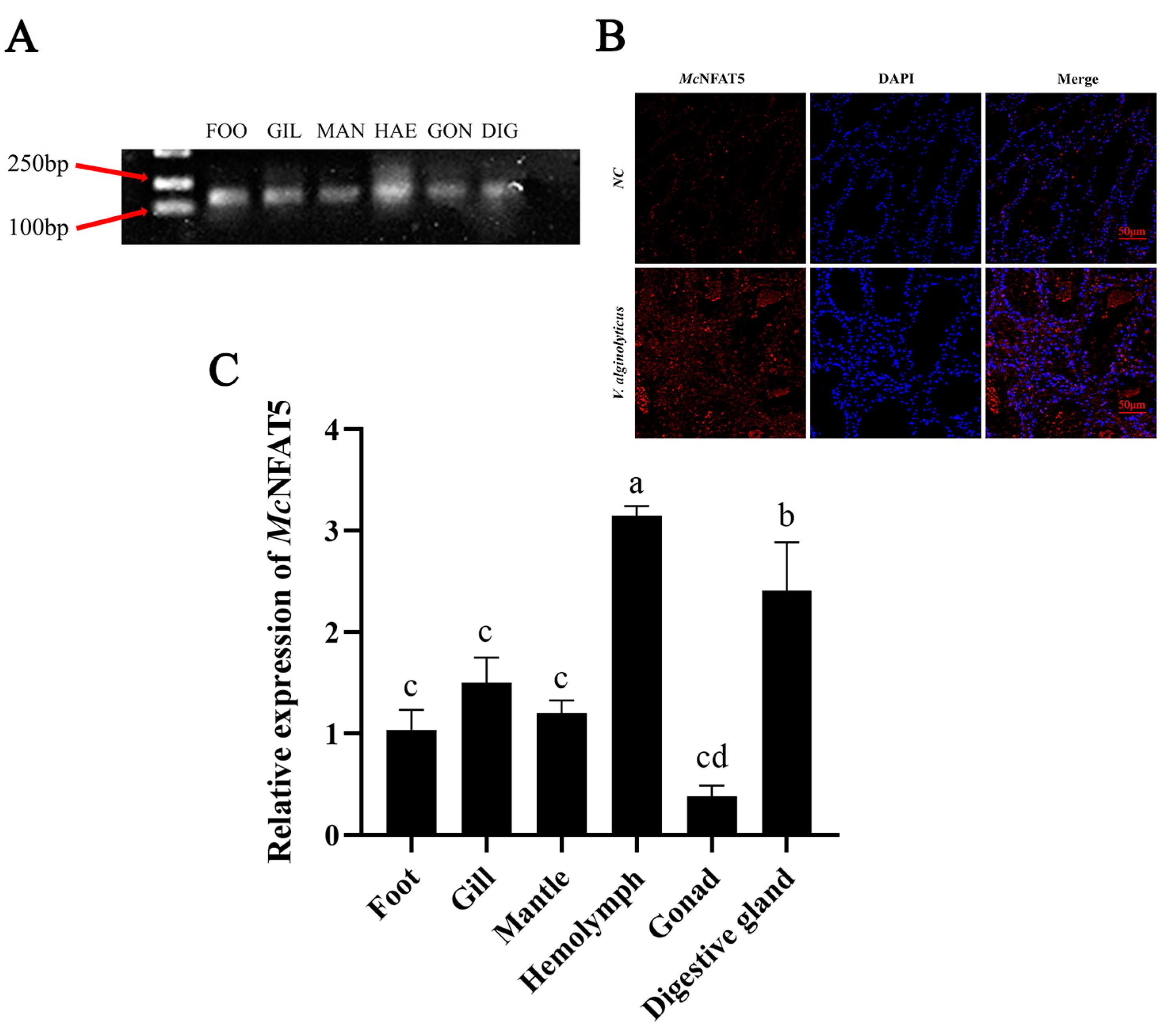
Agarose gel electrophoresis was used to validate the expression of the McNFAT5 gene in different tissues of M. coruscus. The results showed clear expression bands of McNFAT5 in all tested tissues, including foot, gill, mantle, hemolymph, gonad and digestive gland, indicating that this gene is widely expressed across various tissues (Figure 2A). After 12 h of exposure to V. alginolyticus, the expression and localization of McNFAT5 protein were assessed using immunofluorescence. DAPI, which stains the nucleus, displayed blue fluorescence. Red fluorescence represented McNFAT5. In the control group, weak red fluorescence was observed and was mainly concentrated in the cytoplasm. Following exposure to V. alginolyticus, sections of the hemolymph exhibited intense red fluorescence, which was primarily localized in the cell nucleus (Figure 2B). The spatial expression of McNFAT5 in adult M. coruscus after V. alginolyticus infection was analyzed via quantitative real-time RT-PCR using the total RNA from six tissues (foot, gill, mantle, hemolymph, gonad, and digestive gland) of 15 individuals. McNFAT5 was expressed in all tissues at varying levels, with the highest expression in the hemolymph and the digestive gland, moderate levels in the foot, gill, and mantle, and the lowest in the gonad (Figure 2C).

3.3. Temporal Expression Patterns of McNFAT5 After V. alginolyticus Stimulation

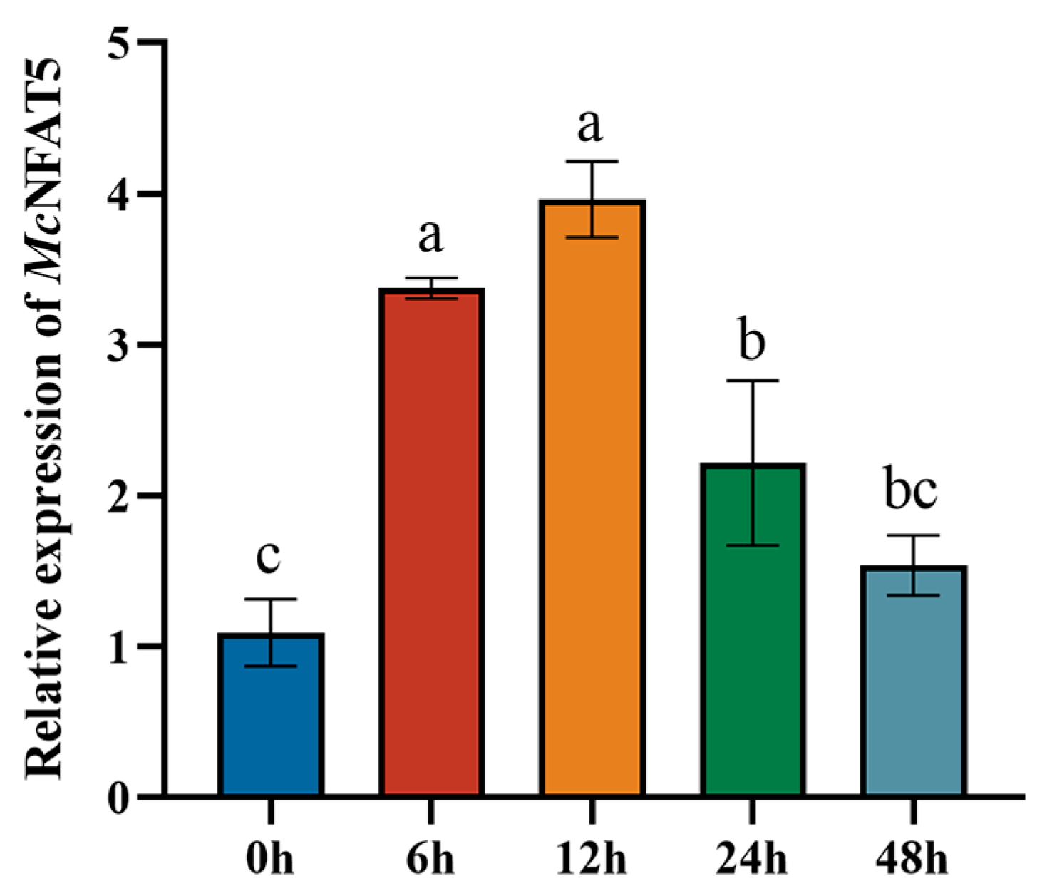
To investigate the potential biological function of McNFAT5 in the immune response of M. coruscus, the expression levels of McNFAT5 were measured at various time points following V. alginolyticus challenge. The results revealed a significant upregulation of the McNFAT5 expression at 6 h post-stimulation (p < 0.01) with a 3.8-fold increase observed at 12 h (p < 0.01). Subsequently, the expression of McNFAT5 decreased at 24 h and 48 h compared to its peak at 12 h (p < 0.01). All data are presented as mean ± SD from three independent biological replicates (Figure 3).

3.4. Effects of V. alginolyticus Infection on Antioxidants Activities

In this study, the activities of superoxide dismutase (SOD), Na+, K+-ATPase, and total antioxidant capacity (T-AOC) in hemolymphs were evaluated following V. alginolyticus infection and siNFAT5 knockdown (Figure 4A). The results demonstrated a significant increase in Na+, K+-ATPase (Figure 4B), SOD (Figure 4C), and T-AOC (Figure 4D) activities in the infection group. However, the subsequent knockdown of NFAT5 resulted in a marked reduction in the activity levels of SOD, Na+, K+-ATPase, and T-AOC.

4. Discussion

NFAT5, as a critical transcription factor, plays a central role in regulating cellular responses to hypertonic stress and immune activities. In this research, direct RNA sequencing was employed to analyze data from M. coruscus, leading to the identification of the transcription factor NFAT5. Further analysis using the SMART (version 9.0) software (https://smart.embl.de/, accessed on 27 February 2025) predicted the presence of a highly conserved Rel-homology domain (RHD) and immunoglobulin-like fold domain (IPT) in McNFAT5, which is crucial for DNA binding and transcriptional activation. This RHD domain is conserved across a wide range of species from invertebrates to mammals [21]. This domain enables NFAT5 to specifically bind to DNA target sites and regulate the transcription of genes involved in immune and osmotic stress responses [22]. Similar domains have also been identified in other marine invertebrates, such as oysters [23] and lamprey [24], indicating a conserved function of NFAT5 across species. The three-dimensional structure further reveals the position of the domains. Phylogenetic analysis further confirmed the evolutionary relationship between McNFAT5 and other molluscan NFAT5 proteins. McNFAT5 clustered closely with other molluscan NFAT5s, forming a distinct evolutionary branch within the NFAT5 family, suggesting a shared ancestral origin and conserved function in mollusks [23]. Together, the results from BLAST (https://blast.ncbi.nlm.nih.gov/Blast.cgi accessed on 27 February 2025) searches, conserved domain analysis, and phylogenetic studies provide strong evidence for the identification and functional characterization of NFAT5 in M. coruscus.
Subcellular localization can determine the specific position of NFAT5 within the cell [25], such as within the nucleus, cytoplasm, cell membrane, or specific organelles. This localization information is crucial for understanding the function of NFAT5. For example, if NFAT5 is primarily localized within the nucleus, it may be involved in the regulation of gene expression. If localized in the cytoplasm or on the cell membrane, it may participate in processes such as signal transduction or material transport. The immunohistological evaluation of NFATc1 in nearly 300 cases of lymphoma indicates that the majority of tumor lymphocytes express NFATc1 as a cytosolic component despite its absence in classical Hodgkin’s disease and plasma cell proliferations. Particularly intriguing is the discovery of NFATc1 relocating to the nucleus in a minority of lymphoid tumors, potentially reflecting the activation of the NFAT pathway [26]. Under specific physiological or pathological conditions, the subcellular localization of NFAT5 (Nuclear Factor of Activated T cells 5) shifts from the cytoplasm to the nucleus. For instance, in studies on sepsis-induced acute kidney injury, NFAT5 has been found to play a crucial role in renal collecting duct cells. When these cells are stimulated by LPS (lipopolysaccharide), Western blot analysis fails to detect an increase in NFAT5 protein expression levels within the nucleus after LPS stimulation. However, chromatin immunoprecipitation (ChIP) assays reveal that NFAT5 can bind to the promoter regions of inflammatory cytokines TNFα and MCP-1, initiating their transcription and protein synthesis. This finding suggests that under LPS stimulation, NFAT5 may translocate from the cytoplasm to the nucleus to regulate the transcription and synthesis of inflammatory cytokines. We conducted immunofluorescence assays based on studies in vertebrates, and the results showed that the localization changes of NFAT5 following bacterial infection can be observed: in uninfected cells, NFAT5 is primarily located in the cytoplasm, while after infection, NFAT5 signals gradually shift to the nucleus. This indicates that NFAT5 has been activated and is involved in the immune response.
To clarify the role of McNFAT5 in regulating innate immune responses, we analyzed its expression patterns post-Vibrio infection using qPCR. Following exposure to V. alginolyticus, McNFAT5 mRNA levels in the hemolymph showed a significant increase, indicating its essential function in initiating and modulating the immune defense against bacterial invasion. This finding is consistent with prior studies. In B. belcheri, an NFAT-like gene was identified and cloned, and its evolutionary relationship across different species was analyzed. The transcriptional activity of NFAT5 was assessed, and through bacterial infection experiments, the impact of NFAT5 expression on the immune response in B. belcheri was observed. The results indicated that the expression of NFAT5 is correlated with the intensity of the immune response [24]. In invertebrates, NFAT5 is mainly involved in the cellular response to environmental stress, such as osmotic pressure changes and pathogen invasion. Its function is not limited to cellular stress responses but also includes the regulation of immune defense mechanisms. For example, in insects, NFAT5 enhances the organism’s resistance to pathogens by regulating the expression of immune-related genes [27]. In contrast to invertebrates, NFAT5 plays a more complex and diverse role in vertebrates. Studies have shown that NFAT5 plays an important role in the immune system of vertebrates, particularly in the activation of immune cells and the regulation of immune tolerance. For example, in rheumatoid arthritis (RA) animal models, NFAT5 has been shown to regulate macrophage function and participate in the disease’s pathogenesis. Specifically, after the activation of macrophages via Toll-like receptors (TLRs), NFAT5 promotes the production of reactive oxygen species (ROS) and further activates the p38MAPK cascade. Activation of this pathway induces NFAT5 to secrete CCL2, helping macrophages resist apoptosis, thereby exacerbating the immune response in RA [28]. Upon the stimulation of macrophages by Toll-like receptors (TLRs), NFAT5 is activated through the xanthine oxidase–reactive oxygen species (ROS)–p38MAPK cascade. This protein provides apoptotic resistance to RA macrophages by inducing CCL2 secretion. Compared to normal tissues, the expression of NFAT5 is significantly elevated in hepatocellular carcinoma and lung adenocarcinoma cells [29]. The function of NFAT5 differs between invertebrates and vertebrates. Although NFAT5 is involved in regulating immune responses in both, in invertebrates, NFAT5 is more related to cellular responses to osmotic stress and pathogen invasion. In contrast, in vertebrates, NFAT5 plays a role in more complex immune regulation, including cell death, immune tolerance, and its involvement in chronic inflammation and cancer.
During the infection process of M. coruscus by V. alginolyticus, hemocytes generate a substantial amount of reactive oxygen species (ROS) to eliminate invading pathogens [30]. At this stage, the expression and activity of superoxide dismutase (SOD) are significantly elevated, mitigating excessive ROS accumulation and preventing oxidative damage to the host [31]. Hemocytes may face challenges such as osmotic imbalance and oxidative stress. The activation of NFAT5 can regulate the gene expression of Na+, K+-ATPase, aiding hemocytes in maintaining ionic homeostasis and thereby supporting their phagocytic activity and the secretion of immune factors [32]. Firstly, NFAT5 influences the activity of Na+/K+-ATPase by regulating the expression of genes related to ion homeostasis. Several studies have shown that NFAT5, by modulating the transcription of specific genes, directly or indirectly affects the function of ion pumps. These genes may be related to the subunits, regulatory factors, or accessory proteins of Na+/K+-ATPase, thereby influencing its activity. Furthermore, the regulatory role of NFAT5 may indirectly affect the activity of Na+/K+-ATPase through oxidative stress response pathways. Specifically, when cells encounter oxidative stress, NFAT5 helps maintain cellular redox balance by regulating the expression of antioxidant genes, thereby indirectly supporting the normal function of ion pumps. The increased total antioxidant capacity (T-AOC) indicates that M. coruscus has initiated a comprehensive antioxidant defense mechanism to counteract the oxidative stress induced by infection. This study demonstrates that V. alginolyticus infection causes significant damage to the hemolymph tissue of M. coruscus, as evidenced by the increased activities of superoxide dismutase (SOD) and Na+, K+-ATPase, and total antioxidant capacity (T-AOC) in the hemolymph. This enhancement in antioxidant enzyme activity indicates that the mussels have adopted an adaptive response to cope with the oxidative stress induced by bacterial infection. SOD plays a crucial role in detoxifying superoxide radicals, thereby preventing oxidative damage to cellular components [33]. The elevated levels of T-AOC further reflect the overall capacity of the hemolymph to neutralize reactive oxygen species (ROS) and maintain cellular homeostasis. Interestingly, subsequent knockdown of NFAT5 resulted in a significant decrease in the activities of SOD, Na+, K+-ATPase, and T-AOC. This finding suggests that NFAT5 is a key regulatory factor in the antioxidant defense mechanism of mussels. The transcription factor NFAT5 may promote the expression of genes encoding antioxidant enzymes and other protective proteins, thereby enhancing the organism’s ability to respond to oxidative stress.

5. Conclusions

This study explores McNFAT5’s role in immune regulation in M. coruscus, especially after V. alginolyticus infection. Through a series of experiments, we confirmed that McNFAT5, as an essential transcription factor, is significantly upregulated post-infection, leading to the enhanced expression of antioxidant enzyme activities and the strengthening of immune defense mechanisms in the hemolymph. This process aids the host in coping with oxidative stress and pathogen invasion. These findings provide valuable insights into the molecular pathways underlying immune defense in marine invertebrates, emphasizing the fundamental role of NFAT5 in immune response regulation. Moreover, the results of this study offer a crucial theoretical basis for improving disease resistance in aquaculture species and enhancing their resilience to environmental stressors and pathogens. Future research can further explore the functional roles and regulatory networks of NFAT5, enabling the optimization of disease resistance in bivalves and promoting the sustainable development of the aquaculture industry in the face of challenges posed by climate change and the spread of infectious diseases.

Author Contributions

All authors contributed to the study conception and design. Data curation, Software, Writing—original draft, preparation by Y.B. and X.S. The first draft of the manuscript was written by Y.B. Conceptualization, Methodology, Writing—review and editing by P.Q. and Y.Y. All authors have read and agreed to the published version of the manuscript.

Funding

This work was supported by the Technology Collaboration Projects of Zhejiang Provincial Department of Agriculture and Rural Affairs (2024SNJF057) and the Special Grant of Zhoushan for Breeding Aquatic Animals (2024Y001).

Institutional Review Board Statement

Not applicable.

Informed Consent Statement

Not applicable.

Data Availability Statement

No datasets were generated or analyzed during the current study.

Conflicts of Interest

The authors declare no conflicts of interest. The authors are fully responsible for the content and writing of this paper.

References

  1. Dame, R.F.; Kenneth, M.J. Ecology of Marine Bivalves: An Ecosystem Approach; Taylor & Francis: Abingdon, UK, 2011. [Google Scholar]
  2. Zannella, C.; Mosca, F.; Mariani, F.; Franci, G.; Folliero, V.; Galdiero, M.; Tiscar, P.G.; Galdiero, M. Microbial diseases of bivalve mollusks: Infections, immunology and antimicrobial defense. Mar. Drugs 2017, 15, 182. [Google Scholar] [CrossRef]
  3. Shaw, J.-P.; Utz, P.J.; Durand, D.B.; Toole, J.J.; Emmel, E.A.; Crabtree, G.R. Identification of a putative regulator of early T cell activation genes. Science 1988, 241, 202–205. [Google Scholar] [CrossRef] [PubMed]
  4. Macian, F. NFAT proteins: Key regulators of T-cell development and function. Nat. Rev. Immunol. 2005, 5, 472–484. [Google Scholar] [CrossRef]
  5. Fric, J.; Zelante, T.; Wong, A.Y.; Mertes, A.; Yu, H.-B.; Ricciardi-Castagnoli, P. NFAT control of innate immunity. J. Am. Soc. Hematol. 2012, 120, 1380–1389. [Google Scholar] [CrossRef] [PubMed]
  6. Zanoni, I.; Granucci, F. Regulation and dysregulation of innate immunity by NFAT signaling downstream of pattern recognition receptors (PRRs). Eur. J. Immunol. 2012, 42, 1924–1931. [Google Scholar] [CrossRef] [PubMed]
  7. Lee, J.-U.; Kim, L.-K.; Choi, J.-M. Revisiting the concept of targeting NFAT to control T cell immunity and autoimmune diseases. Front. Immunol. 2018, 9, 2747. [Google Scholar] [CrossRef]
  8. Hogan, P.G.; Chen, L.; Nardone, J.; Rao, A. Transcriptional regulation by calcium, calcineurin, and NFAT. Genes Dev. 2003, 17, 2205–2232. [Google Scholar] [CrossRef]
  9. Trama, J.P. Identification and Characterization of NFAT5: A Novel Rel Family Transcription Factor; University of California, San Diego: San Diego, CA, USA, 2002. [Google Scholar]
  10. Graef, I.A.; Gastier, J.M.; Francke, U.; Crabtree, G.R. Evolutionary relationships among Rel domains indicate functional diversification by recombination. Proc. Natl. Acad. Sci. USA 2001, 98, 5740–5745. [Google Scholar] [CrossRef]
  11. Sullivan, J.C.; Kalaitzidis, D.; Gilmore, T.D.; Finnerty, J.R. Rel homology domain-containing transcription factors in the cnidarian Nematostella vectensis. Dev. Genes Evol. 2007, 217, 63–72. [Google Scholar] [CrossRef]
  12. Berga-Bolanos, R.; Drews-Elger, K.; Aramburu, J.; Lopez-Rodriguez, C. NFAT5 regulates T lymphocyte homeostasis and CD24-dependent T cell expansion under pathologic hypernatremia. J. Immunol. 2010, 185, 6624–6635. [Google Scholar] [CrossRef]
  13. Tellechea, M.; Buxadé, M.; Tejedor, S.; Aramburu, J.; Lopez-Rodriguez, C. NFAT5-regulated macrophage polarization supports the proinflammatory function of macrophages and T lymphocytes. J. Immunol. 2018, 200, 305–315. [Google Scholar] [CrossRef]
  14. Ko, B.C.; Lam, A.K.; Kapus, A.; Fan, L.; Chung, S.K.; Chung, S.S. Fyn and p38 signaling are both required for maximal hypertonic activation of the osmotic response element-binding protein/tonicity-responsive enhancer-binding protein (OREBP/TonEBP). J. Biol. Chem. 2002, 277, 46085–46092. [Google Scholar] [CrossRef]
  15. Song, X.; Hu, J.; Jin, P.; Chen, L.; Ma, F. Identification and evolution of an NFAT gene involving Branchiostoma belcheri innate immunity. Genomics 2013, 102, 355–362. [Google Scholar] [CrossRef]
  16. Letunic, I.; Doerks, T.; Bork, P. SMART: Recent updates, new developments and status in 2015. Nucleic Acids Res. 2015, 43, D257–D260. [Google Scholar] [CrossRef]
  17. Kumar, S.; Stecher, G.; Tamura, K. MEGA7: Molecular evolutionary genetics analysis version 7.0 for bigger datasets. Mol. Biol. Evol. 2016, 33, 1870–1874. [Google Scholar] [CrossRef]
  18. Qi, P.; Wu, Y.; Gu, Z.; Li, H.; Li, J.; Guo, B.; Liao, Z.; Yan, X. A novel molluscan TLR molecule engaged in inflammatory response through MyD88 adapter recruitment. Dev. Comp. Immunol. 2022, 131, 104373. [Google Scholar] [CrossRef]
  19. Livak, K.J.; Schmittgen, T.D. Analysis of relative gene expression data using real-time quantitative PCR and the 2−ΔΔCT method. Methods 2001, 25, 402–408. [Google Scholar] [CrossRef]
  20. Li, Z.; Chang, X.; Hu, M.; Fang, J.K.-H.; Sokolova, I.M.; Huang, W.; Xu, E.G.; Wang, Y. Is microplastic an oxidative stressor? Evidence from a meta-analysis on bivalves. J. Hazard. Mater. 2022, 423, 127211. [Google Scholar] [CrossRef]
  21. Jia, S.; Flores-Saaib, R.n.D.; Courey, A.J. The Dorsal Rel homology domain plays an active role in transcriptional regulation. Mol. Cell. Biol. 2002, 22, 5089–5099. [Google Scholar] [CrossRef]
  22. Huang, T. Study on the Tonicity-Regulated Nucleocytoplasmic Trafficking of NT5 Using Proteomics and Functional siRNA Screen. Ph.D. Thesis, The Chinese University of Hong Kong, Hong Kong, China, 2015. [Google Scholar]
  23. Huang, X.-D.; Wei, G.-j.; Zhang, H.; He, M.-X. Nuclear factor of activated T cells (NFAT) in pearl oyster Pinctada fucata: Molecular cloning and functional characterization. Fish Shellfish. Immunol. 2015, 42, 108–113. [Google Scholar] [CrossRef]
  24. Duan, X.; Lv, M.; Liu, A.; Pang, Y.; Li, Q.; Su, P.; Gou, M. Identification and evolution of transcription factors RHR gene family (NFAT and RBPJ) involving lamprey (Lethenteron reissneri) innate immunity. Mol. Immunol. 2021, 138, 38–47. [Google Scholar] [CrossRef]
  25. Feng, Z.-P. An overview on predicting the subcellular location of a protein. Silico Biol. 2002, 2, 291–303. [Google Scholar]
  26. Marafiot, T.; Pozzobon, M.; Hansmann, M.L.; Ventura, R.; Pileri, S.A.; Roberton, H.; Gesk, S.; Gaulard, P.; Barth, T.F.; Du, M.Q. The NFATc1 transcription factor is widely expressed in white cells and translocates from the cytoplasm to the nucleus in a subset of human lymphomas. Br. J. Haematol. 2005, 128, 333–342. [Google Scholar] [CrossRef]
  27. Silva, R.C.M.C.; Gomes, F.M. Evolution of the Major Components of Innate Immunity in Animals. J. Mol. Evol. 2024, 92, 3–20. [Google Scholar] [CrossRef]
  28. Kim, N.H.; Choi, S.; Han, E.J.; Hong, B.K.; Choi, S.Y.; Kwon, H.M.; Hwang, S.Y.; Cho, C.S.; Kim, W.U. The xanthine oxidase–NFAT5 pathway regulates macrophage activation and TLR-induced inflammatory arthritis. Eur. J. Immunol. 2014, 44, 2721–2736. [Google Scholar] [CrossRef]
  29. Lee, J.H.; Suh, J.H.; Choi, S.Y.; Kang, H.J.; Lee, H.H.; Ye, B.J.; Lee, G.R.; Jung, S.W.; Kim, C.J.; Lee-Kwon, W. Tonicity-responsive enhancer-binding protein promotes hepatocellular carcinogenesis, recurrence and metastasis. Gut 2019, 68, 347–358. [Google Scholar] [CrossRef]
  30. Chaitanya, R.; Shashank, K.; Sridevi, P. Oxidative stress in invertebrate systems. Free. Radicla Dis. 2016, 19, 51–68. [Google Scholar]
  31. Sahu, P.K.; Jayalakshmi, K.; Tilgam, J.; Gupta, A.; Nagaraju, Y.; Kumar, A.; Hamid, S.; Singh, H.V.; Minkina, T.; Rajput, V.D. ROS generated from biotic stress: Effects on plants and alleviation by endophytic microbes. Front. Plant Sci. 2022, 13, 1042936. [Google Scholar] [CrossRef]
  32. Hollborn, M.; Fischer, S.; Kuhrt, H.; Wiedemann, P.; Bringmann, A.; Kohen, L. Osmotic regulation of NFAT5 expression in RPE cells: The involvement of purinergic receptor signaling. Mol. Vis. 2017, 23, 116. [Google Scholar]
  33. Ighodaro, O.; Akinloye, O.A. First line defence antioxidants-superoxide dismutase (SOD), catalase (CAT) and glutathione peroxidase (GPX): Their fundamental role in the entire antioxidant defence grid. Alex. J. Med. 2018, 54, 287–293. [Google Scholar] [CrossRef]
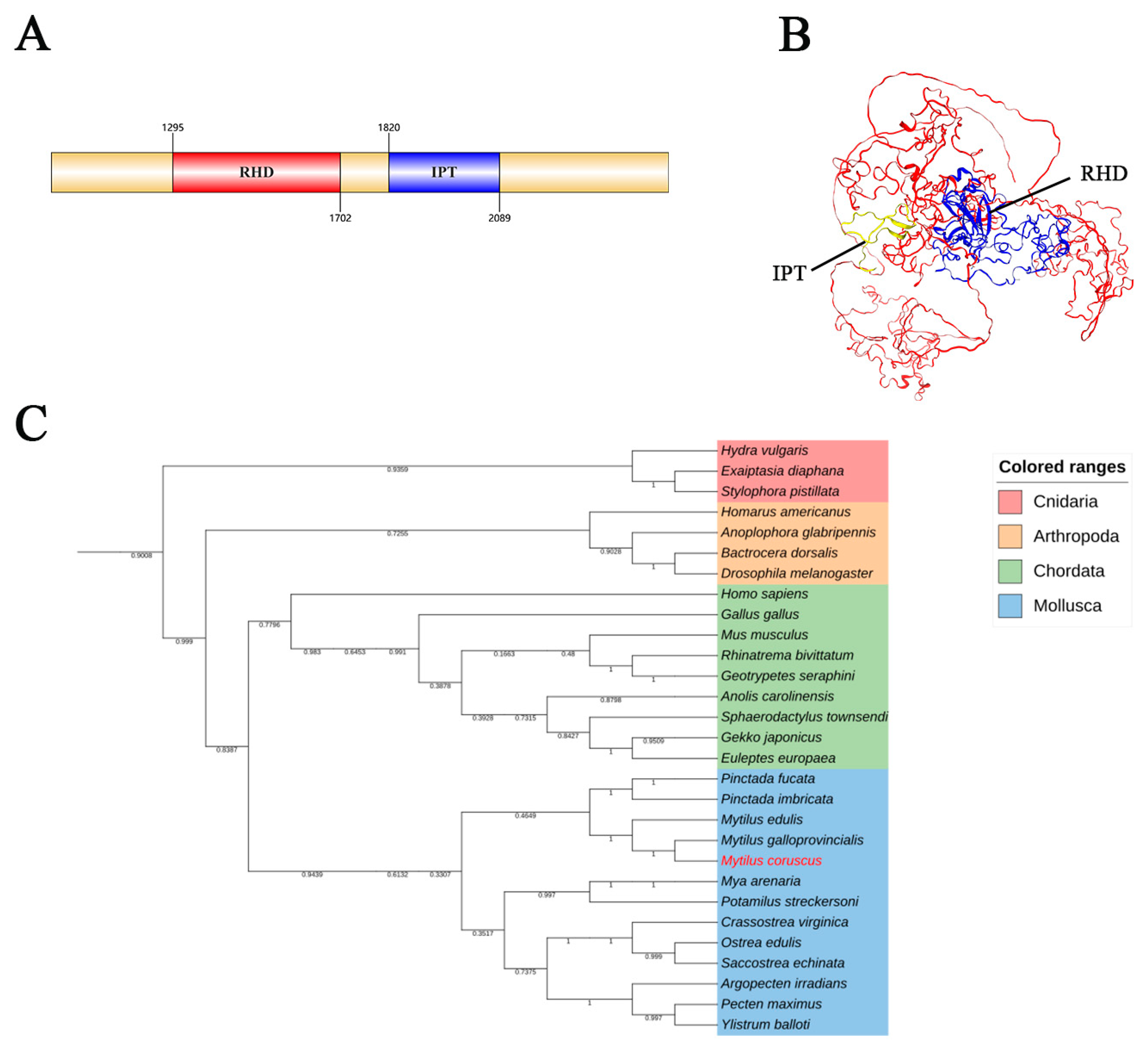
Figure 1. Molecular characterization of McNFAT5 (A) SMART-predicted schematic of McNFAT5 functional domains. (B) SWISS-MODEL-generated three-dimensional structure of McNFAT5. (C) Phylogenetic analysis of McNFAT5 with selected species using the neighbor-joining method in MEGA 7.0 with 5000 bootstrap replications. Different colors indicate distinct taxa. The species in red font, Mytilus coruscus, represents the focus of our study.
Animals 15 00726 g001
Figure 2. McNFAT5 responds to pathogen stimulation. (A) Agarose gel electrophoresis validation of McNFAT5 expression in various tissues of M. coruscus, including foot, gill, mantle, hemolymph, gonad and digestive gland. The presence of clear bands in all tested tissues confirms the expression of McNFAT5 across different tissue types. (B) Immunofluorescence showing translocation of NFAT5 to the nucleus after V. alginolyticus infection. Red: McNFAT5 protein, Blue: DAPI-stained nuclei. Scale bars = 20. Each sample was analyzed in triplicate. Vertical error bars represent the mean ± standard error (SE, n = 3). (C) The β-actin gene of M. coruscus was used as an internal reference to normalize the cDNA templates across all samples. Differential expression across the six tissues was analyzed using one-way ANOVA. Distinct letters indicate statistically significant differences (p < 0.05) in the relative expression levels of McNFAT5 mRNA.
Animals 15 00726 g002
Figure 3. Temporal expression patterns of McNFAT5 after infection. McNFAT5 mRNA expression was assessed via qRT-PCR at 6 h, 12 h, 24 h, and 48 h post-V. alginolyticus infection. Data are shown as mean ± SD (n = 3) with different letters indicating significant differences (p < 0.05).
Animals 15 00726 g003
Figure 4. The impact of NFAT5 knockdown on the activity of three key enzymes and antioxidant capacity. (A) The expression of McNFAT5 was downregulated by Si-McNFAT5. (B) Na+, K+-ATPase. (C) SOD. (D) T-AOC. The vertical bars represent the mean ± SD (n = 3). Different letters indicate significant differences (p < 0.05).
Animals 15 00726 g004
Table 1. PCR primer pairs used in the present study.
PrimerSequences (5′–3′)Usage
McNFAT5-FTCTTCTTGACCGTGCTGGACqRT–PCR
McNFAT5-RTCGTCGGACTTTTGGCACTT
McTRAF-FTGTGCCAATTCCCTGTCCTqRT–PCR
McTRAF-RGGACACTCTTTATGCAGG
McIRAK-FCCTTTTATGGCAGCAGCGTGqRT–PCR
McIRAK-RAAAATCCAGTGCCCGATGGT
Mcmyticofensin-FTGTGGCTCTAGAAGTTGCTGATGqRT–PCR
Mc myticofensin-RTCAATCTGAACCAGCCTCCAC
β-actin-FGCTACGAATTACCTGACGGACqRT–PCR
β-actin-RTTCCCAAGAAAGATGGTTGTAACAT
siNCUUCUCCGAACGUGUCACGUTTRNAi
ACGUGACACGUUCGGAGAATT
SiNFAT5GACAAUAAAUCAACUGUUATTRNAi
UAACAGUUGAUUUAUUGUCTT
Disclaimer/Publisher’s Note: The statements, opinions and data contained in all publications are solely those of the individual author(s) and contributor(s) and not of MDPI and/or the editor(s). MDPI and/or the editor(s) disclaim responsibility for any injury to people or property resulting from any ideas, methods, instructions or products referred to in the content.