1. Introduction
The electrical properties of skin and hair alongside their texture and anatomy provide information about a person’s health, efficiency of drug delivery and effects from application of cosmetic products. For these reasons, scientists from a variety of fields use non-invasive instruments to extract such information and achieve experimental results that strengthen their research. As part of this introduction, selected publications are illustrating the above points right after a summary of human skin structure and hair anatomy. Then, the use of capacitive imaging in various research fields is demonstrated by summarizing selected research in the literature.
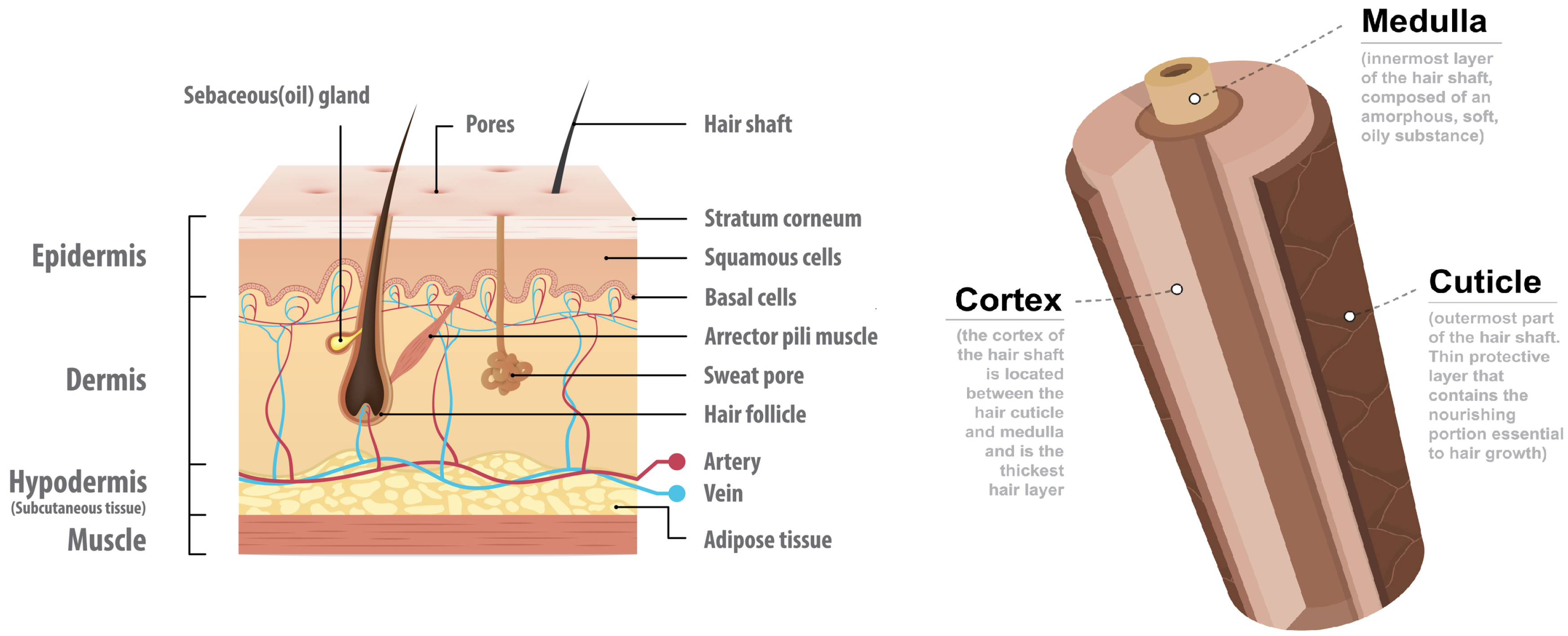
The skin is the largest human organ in terms of surface area and its thickness varies from 5 μm to 1 mm (or more) in different areas of the body [
1]. It protects internal organs from environmental influences, and it regulates the water body loss. As illustrated in
Figure 1 (left), the skin is separated into three main layers: epidermis, dermis and subcutis. Among other functions, the epidermis provides chemical and diffusion protection, the dermis protects from external mechanical forces and the subcutis connects the skin to the underlying tissue [
1]. The outermost sublayer, target of non-invasive instruments, is called stratum coreum and consists mainly of dead keratinocytes. The textural information of this layer, or skin microrelief, is affected by the internal body health, age and living habits as well as by environmental influences [
2]. The visible part of hair (
Figure 1 right) is shaped as a three-layered shaft of dead protein filament: the cuticle, cortex and medulla [
3]. The cuticle is a thin surrounding layer of roof tiles-like structures with about five degrees inclination from the hair shaft core. The cortex is the thickest layer of the hair, it consists of tightly packed keratin cells and it is responsible for the hair color as well as most of the water holding capabilities. Last, the medulla is a soft unstructured keratin that forms the core in the thicker human hair (e.g., scalp hair) [
4].
In the literature, a variety of methods have been used to extract information from the human skin surface. Zhang et al. [
7] used a capacitive imaging system to measure solvent concentration and penetration in human skin to demonstrate how such a system can support skin clinical trial studies. In the same work, skin damage by intense washing, tape stripping and SLS irritation was characterized. In the area of skin aging analysis, Corcuff et al. [
8] studied the skin furrow response during arm extension based on image analysis of negative skin replicas. They provided evidence that younger people can buffer skin strain between primary and secondary line orientations while elderly subjects tend to have furrows only in one orientation that rotate during extension. In more recent years, Zahouani et al. [
9] achieved better classification between primary and secondary skin lines by measuring their depth using three-dimensional confocal microscopy. Experimental results on 120 Caucasian women confirmed that secondary lines fade with age while the depth of primary lines increases. A different approach of evaluating skin aging was employed by [
10,
11], where the surface area of polygons shaped between skin furrow was measured and it was found to associate well with subjects’ chronological age.
As illustrated for the area of skin research, hair samples are also analyzed by scientists to detect health and cosmetic conditions. Wosu et al. [
12] reviewed 39 studies that associate hair cortisol concentrations to stress psychiatric symptoms and disorders. This approach was found to be more accurate in detecting long-term stress because cortisol concentration measurements from blood, urine and saliva samples are influenced by living habits and environment conditions. Furthermore, Kristensen et al. [
13] used hair samples from 266 women to determine that hair dyeing and frequent washing does not affect cortisol measurements. In a different scientific field, Boll et al. [
14] employed ATR FT-IR spectroscopy to differentiate between dyed and undyed hair. Such information can be used in forensic hair analysis, given their static classification was found to be
accurate in detecting whether a sample is dyed or not, but also in identifying the brand and the color of the dye. In the field of cosmetic science, Barba et al. [
15] used thermogravimetric methodology to measure the water content of hair and to assess hair damage from bleaching and straightening. They found that bleached hair shown
reduced water holding capabilities while the straighten hair
.
In this work, a capacitive imaging system is used to extract information from skin and hair samples. To the best of our knowledge, Lévêque and Querleux [
16] first used this technology for human skin characterization and surface hydration mapping in 2003. They used a fingerprint system to measure the distribution of skin surface capacitance in different body sites, to detect main microrelief orientation and density as well as to support the system’s usability in the field of skin research by performing side-by-side experiments with Corneometer CM812. Later on, Batisse et al. [
17] demonstrated advantages of such technology over other skin hydration apparatuses by pointing out the importance of visually observing the contact of a capacitive sensor with the skin during lips moisturization and volar forearm inflammation experiments. Since then, capacitive imaging systems have been used in order to examine various skin conditions e.g., mapping of psoriasis and acne lesions, assessment of sun exposure, skin aging, damage or irritation [
18,
19,
20,
21,
22]. Furthermore, it is worth mentioning successful attempts to improve the capacitive imaging technology. Bevilacqua and Gherardi [
23] achieved depth profiling up to 50 μm by fusing the image with pressure information monitored by a subsystem attached on the back of the measurement apparatus. Also, Huang et al. developed a wearable capacitive imaging system using an “ultrathin, stretchable sheets with arrays of embedded impedance sensors for precise measurement and spatially multiplexed mapping” [
24].
In the following sections, the measurement principle of capacitive imagining for non-invasive skin and hair measurements is analyzed. Then, image processing algorithms are suggested to extract quantitative values from skin and hair samples. Finally, three experiments are conducted to evaluate both the imaging system but also the integrity of the selected algorithms.
3. Results
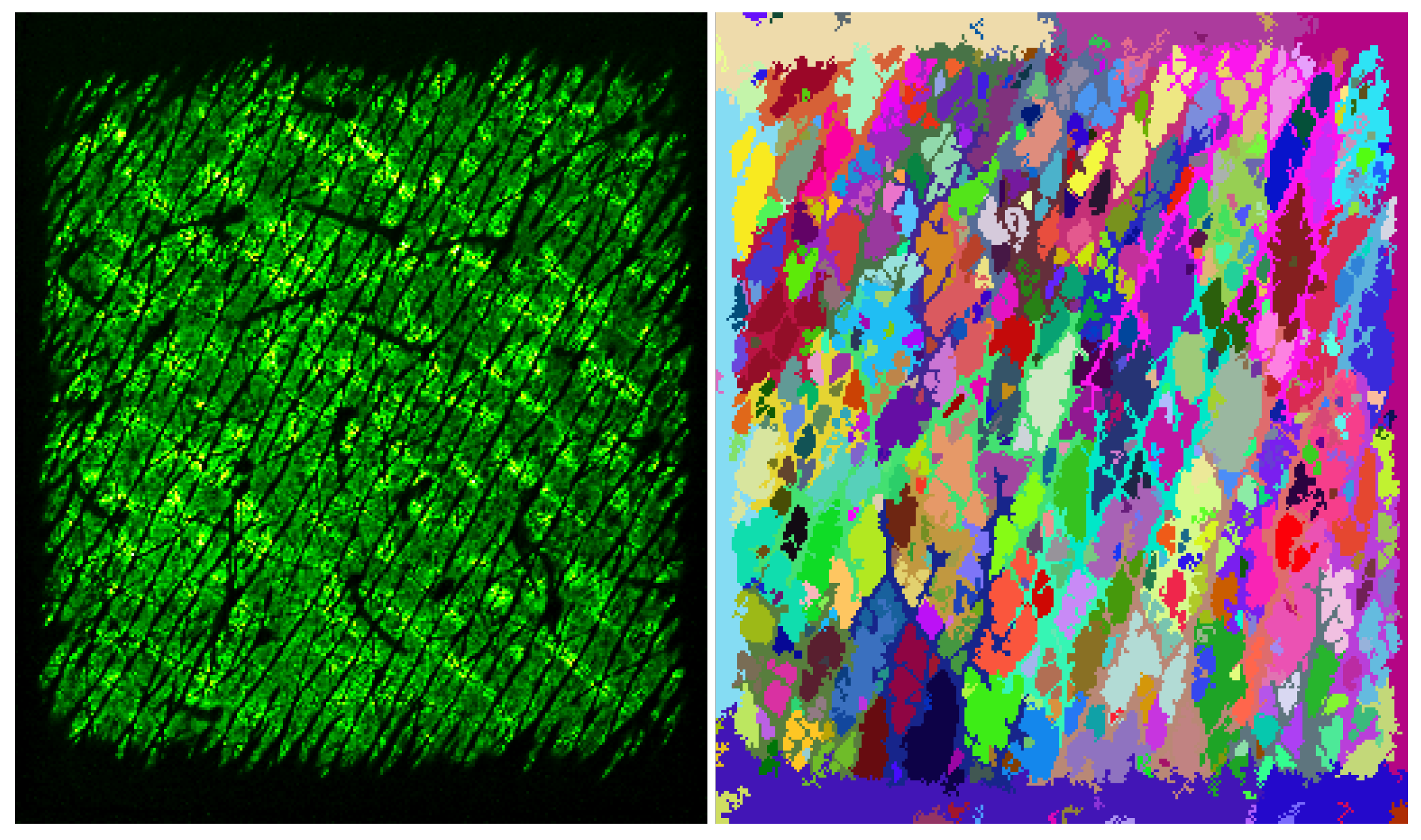
In this section, three experiments are conducted to examine the performance of the presented algorithms on capacitive images. The first experiment evaluates Vincent and Soille segmentation algorithm to automatically count the skin polygons, i.e., the skin areas shaped between wrinkles. For this purpose, capacitive images were recorded from 12 volunteers aging from 12 to 74 years old. The samples were taken from the middle volar forearm area while the arm was in resting position to reduce strain. Then, the segmentation algorithm was applied using Epsilon E100 software and the average number of polygons per square millimeter was correlated against the subjects’ age. The results in
Table 1 demonstrate that the average number of polygons per surface area decreases with age. The calculated correlation (−0.71) comes in agreement with previous studies in the literature [
10,
11].
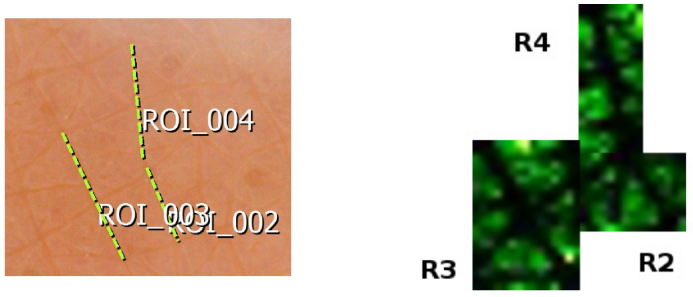
The second experiment consists of a short comparison between C-Cube, a calibrated digital spectroscope (Pixience, Toulouse, France) [
37], and Epsilon E100 in feature length estimation. The same skin area of volar forearm was captured with both instruments and three furrows were randomly selected (
Figure 7). C-Cube software provides the length measurement as a default feature by drawing the linear segment of interest on the captured frame (
Figure 7 left). Epsilon E100 does not provide such feature, so the region of interest was cropped, and our length estimation algorithm was applied (
Figure 7 right). The results in
Table 2 suggest that if there are no neighboring artifacts in capacitive images, such systems can calculate the length of a furrow with good accuracy.
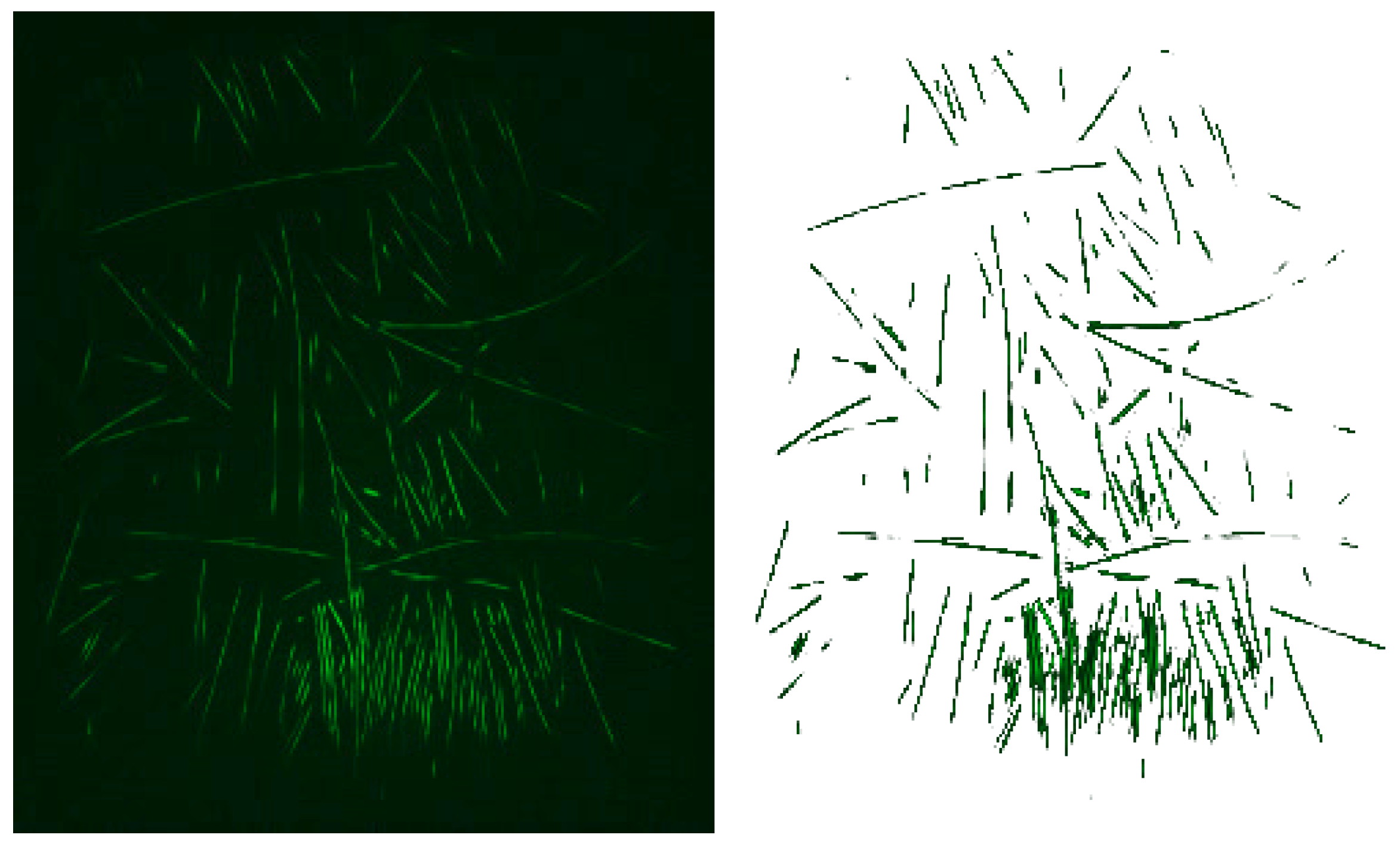
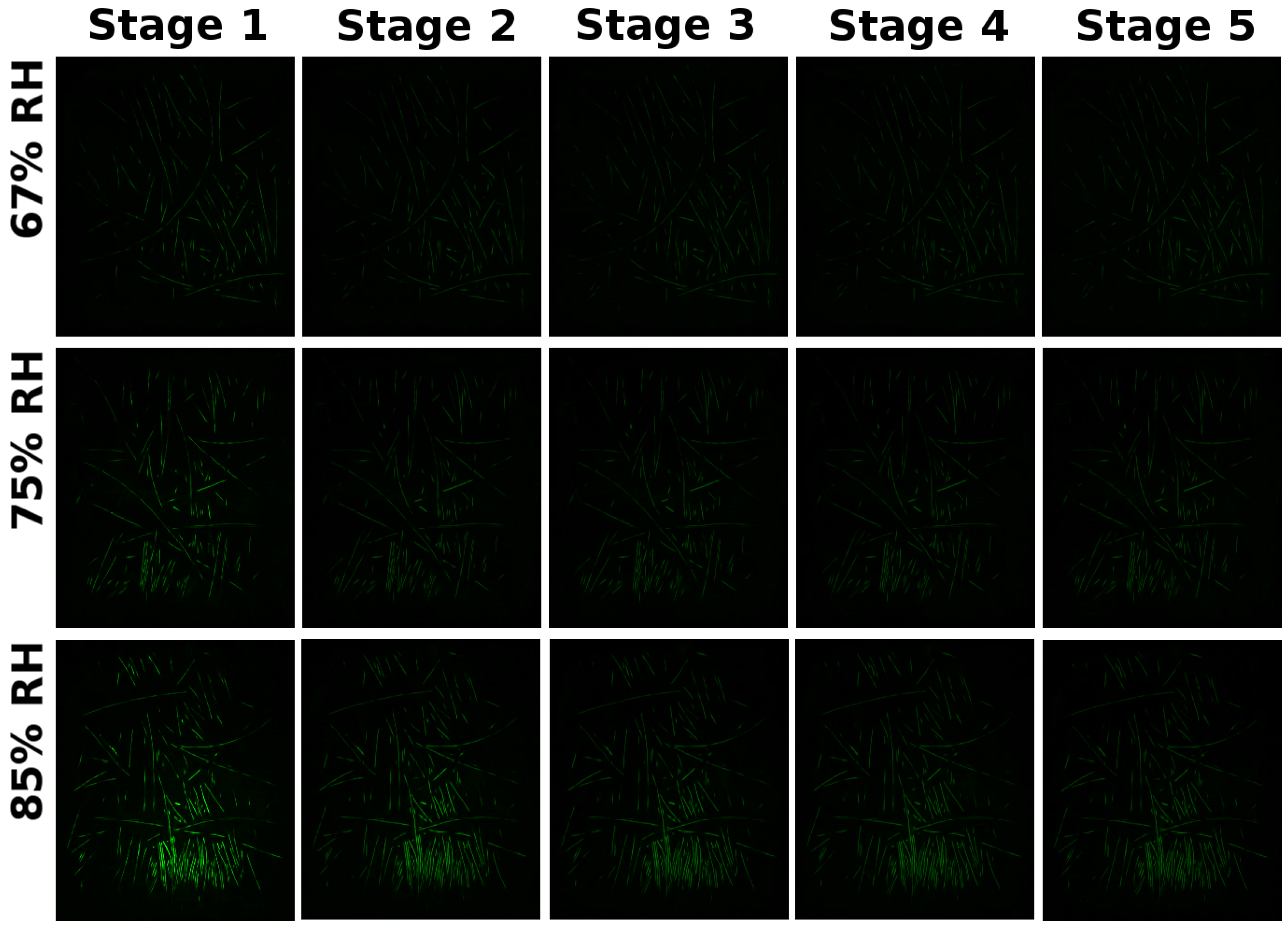
In the final experiment, the ability of capacitive imaging sensors to measure hair water content and desorption rate are examined. For this purpose, scalp hair samples from three volunteers were washed in deionized water and dried before left to acclimatize overnight in three different humidity chambers. Saturated salt solutions adjusted the relative humidity levels while both temperature and relative humidity were logged every 10 s using SHT35 by Sensirion [
38]. The selected salts are potassium nitrate (85% RH), sodium chloride (75% RH) and magnesium nitrate (67% RH). After acclimatization, the samples were moved in 21 °C &
RH conditions, side by side, and they were held against the sensor surface with a plastic plug provided by the manufacturer. The system was capturing video frames until the water loss rates reached a flat state or until the video exceeded 5.5 h. In order to target only the pixels in contact with hair, a range filter from 3.5 to 80 was applied on each frame. The selection of these limits is based on previous work with Epsilon E100 [
39]. Five video instances from the same hair sample per acclimatization chamber are shown in
Figure 8.
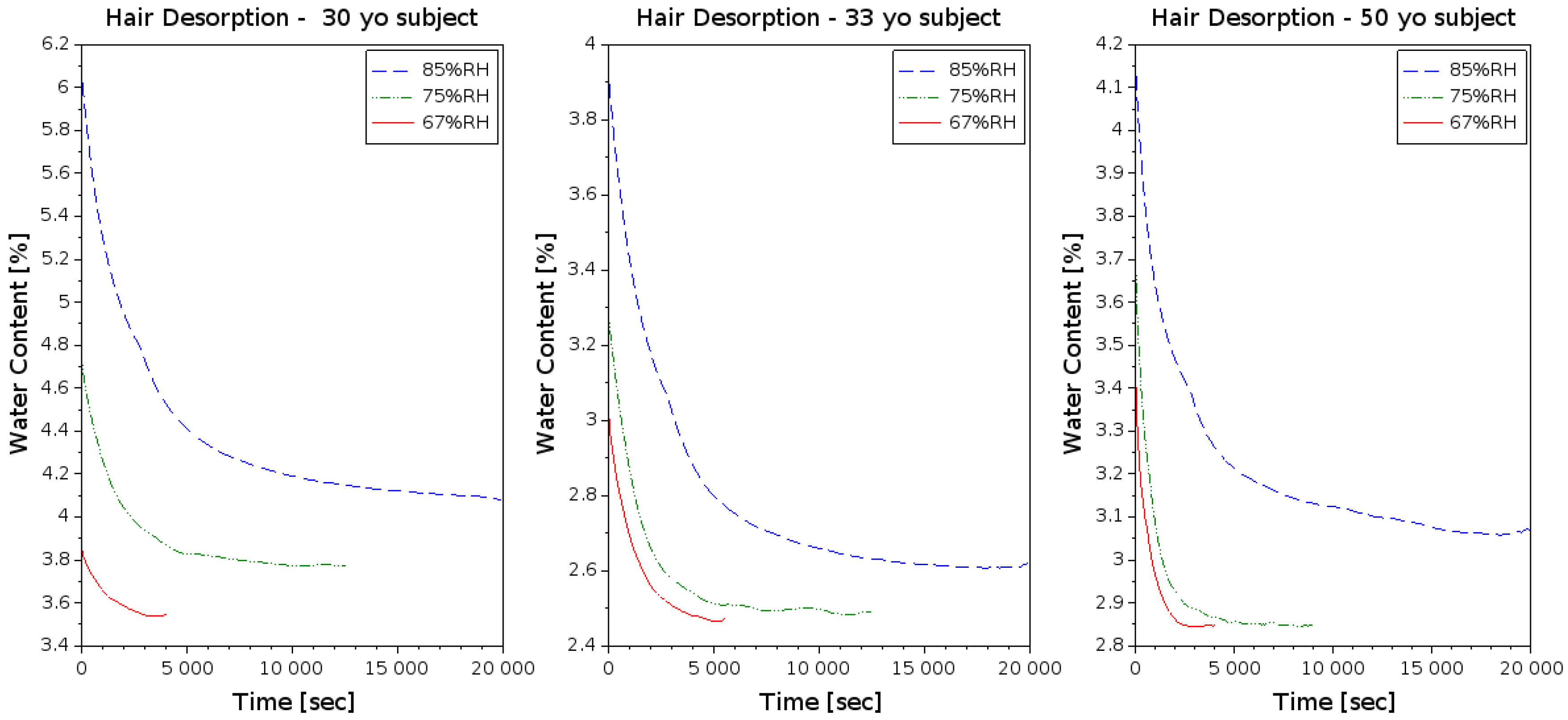
Two observations are made from the experimental results in
Figure 9 and their summary in
Table 3. First, the hair water content right after acclimatization correlates well with the relative humidity of the chamber. Second, the hair samples from younger subjects tend to hold water for a longer period of time. The latter comes in agreement with Xiao P. et al. [
40], stating that lower diffusion rates are observed in younger subjects meaning better water holding capabilities. Note that in many occasions the sample never reached the expected baseline. In those cases, the lowest water content readout was used in the calculations for
Table 3.
4. Discussion
In this study, we achieved to summarize the importance of skin and hair analysis in a variety of scientific fields, we introduced and analyzed the apparatus of capacitive imaging systems using a Maxwell-based simulation, we suggested algorithms for information extraction using such equipment and we conducted experiments to evaluate the overall system performance.
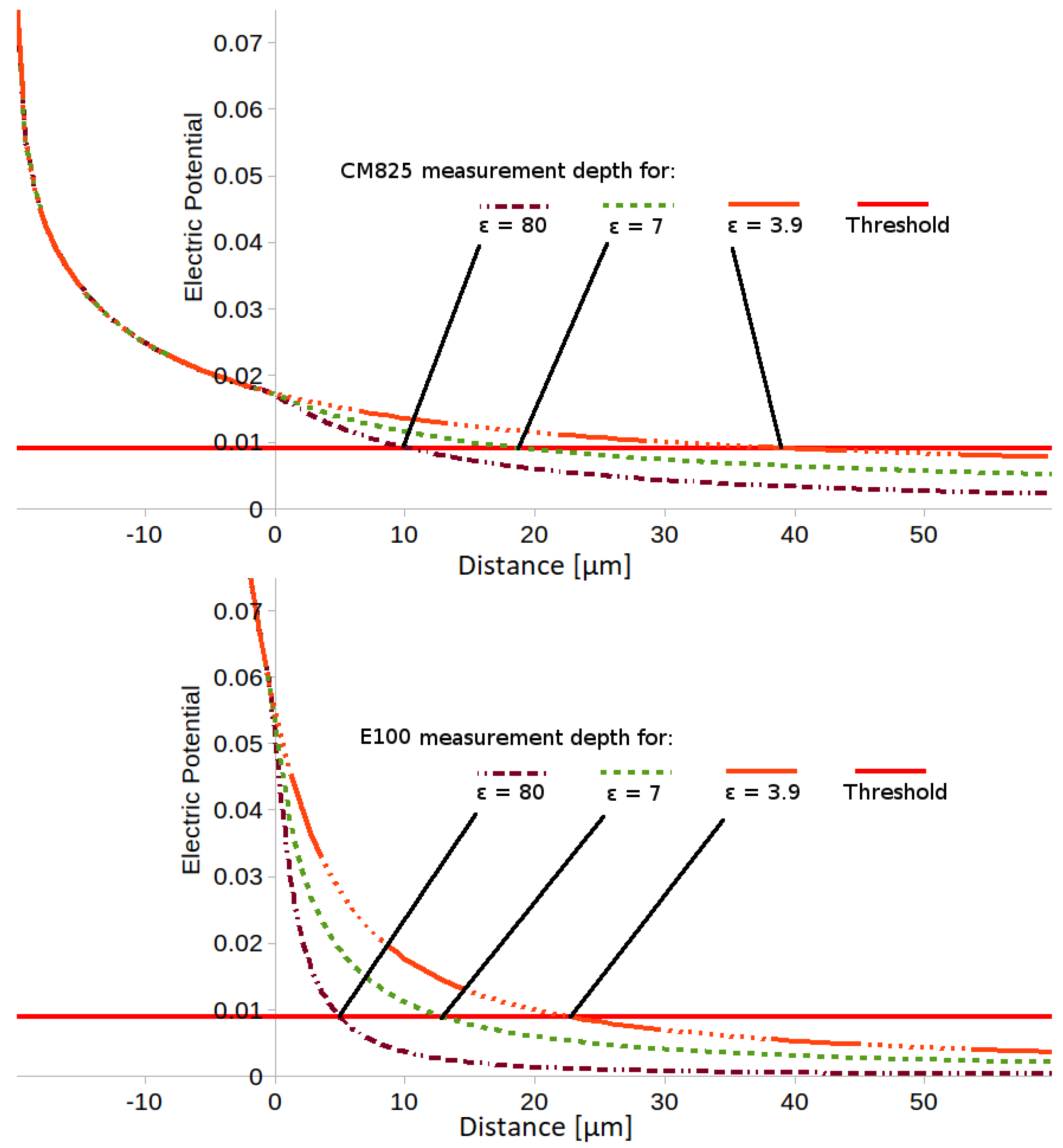
The simulation compared the penetration depth of the electric field between Corneometer C825 and Epsilon E100. The results show that both instruments have satisfactory penetration depth for skin measurements, with CM825 reaching twice the measurement depth. This implies that if stratum corneum thickness is less than 40 μm, errors should be found in a side-by-side comparative study. Such results have not been found in the literature and might be of interest to achieve in future work for validation of our simulation. Another conclusion we draw by this simulation is that both instruments have insufficient measurement depth for hair analysis. Their electric field reaches the shaft cortex and it will give some reasonable readouts, but it could not represent the absolute hair water content.
Our experiments focused on the evaluation of capacitive imaging systems in cosmetic and skin research studies. The first experiment demonstrated how capacitive images can be used to extract skin texture information. While this could be performed with any calibrated spectroscope, expanding applications of existing laboratory equipment is one of our goals. The suggested algorithm successfully detected the skin polygons and measured their average surface area. The results associate well with subjects’ age, giving −0.71 correlation with <0.0006 statistical significance. Furthermore, the achieved correlation value agrees with [
10,
11], where the correlation between age and polygons density were calculated to −0.64 and −0.65 in the dorsal hand and volar forearm correspondingly.
The second experiment used the GLCM to estimate the length of wrinkles. In order to determine the reliability of this method, we compared our results with calibrated spectroscopy for a small group of samples. The experiment was not extended further because the need to bring the capacitive sensor in contact with the sample results to skin deformation. This is enough to twist the frame and make repositioning algorithms to fail identifying the same wrinkle. Nevertheless, the same logic could be applied on objects with greater surface area (e.g., moles or scars) and track changes in their dimensions over time.
Our last experiment focused on measuring water desorption rate from scalp hair samples. The experimental results shown that the measurement apparatus is capable of differentiating desorption rates from young and elder subjects. This means that the comparative interpretation of the results between samples are in agreement with the literature [
40], indicating that such systems can be used in hair analysis studies. Unfortunately, the observed desorption rates are lower than the ones reported in similar studies using different measurement methods (e.g., DVS or thermogravimetric) and for many of our samples the expected baseline was not reached. More specifically, Xiao et al. [
40] found that it takes only 58 min for soaked hair samples to return to their baseline hydration using DVS.
To conclude, we believe that capacitive imaging sensors can be used for skin texture analysis and human skin age classification. We also believe that evidence is found for capacitive imaging application on hair water loss studies. This will require a sensor with greater penetration depth and a better sample-holding mechanism.
References


