Article

Effect of Alkaline Treatment on Characteristics of Bio-Calcium and Hydroxyapatite Powders Derived from Salmon Bone

by
Anthony Temitope Idowu
1,
Soottawat Benjakul
1,
Sittichoke Sinthusamran
1,
Thanasak Sae-leaw
1,
Nobuo Suzuki
2,
Yoichiro Kitani
2 and
Pornsatit Sookchoo
3,*
1
Department of Food Technology, Faculty of Agro-Industry, Prince of Songkla University, Hat Yai, Songkhla 90112, Thailand
2
Noto Marine Laboratory, Institute of Nature and Environmental Technology, Kanazawa University, Ogi, Noto-cho, Ishikawa 927-0553, Japan
3
Department of Material Product Technology, Faculty of Agro-Industry, Prince of Songkla University Hat Yai, Songkhla 90112, Thailand
*
Author to whom correspondence should be addressed.
Appl. Sci. 2020, 10(12), 4141; https://doi.org/10.3390/app10124141
Submission received: 11 April 2020 / Revised: 10 June 2020 / Accepted: 11 June 2020 / Published: 16 June 2020
(This article belongs to the Section Materials Science and Engineering)

Abstract

:
Alkaline treatment has been extensively implemented in the extraction process of hydroxyapatite (HAp) extraction from various kinds of bio-materials, such as animal bone and scales. The main purpose of such treatment is to remove proteinaceous substances from raw materials. The influence of the alkaline treatment that could alter not only the organic contents but also chemical composition—specifically the Ca/P mole ratios of bio-calcium, HAp, and the biphasic apatite powders derived from salmon bone, a by-product from the salmon industry—was investigated. Both HAp and biphasic apatite powders were obtained from the calcination of bio-calcium powders with and without alkaline treatment, respectively. An X-ray diffraction analysis confirmed the presence of hydroxyapatite and β-tricalcium phosphate (β-TCP) in the calcined bone powder without alkaline treatment while only a single phase of hydroxyapatite was observed in the alkaline-treated sample. Calcium and phosphorus contents were measured by an inductively coupled plasma optical emission spectrometer (ICP-OES). A variation of Ca/P ratios was observed among all samples, depending on the chemical and heat treatment conditions. Organic molecules, such as protein, fat, hydroxyproline, and TBARS, were significantly lowered in bio-calcium powders with the alkaline treatment. This work represents important research on chemical treatment prior to the raw material conversion process, which significantly influences chemical and phase compositions of the bio-calcium and hydroxyapatite powder derived from salmon bone waste.

Graphical Abstract

1. Introduction

Fish consumption has increased tremendously, and its demand by 2050 is estimated to be 9.8 billion tons [1]. During the processing of fish, more than 60% of fish mass is generated as leftovers, including viscera (liver, kidney, and roe), frames, trimmings (containing muscle, bone, and skin), heads (containing the gills), and mince [2]. Salmon (Salmo salar) constitutes a large portion of the fish globally served due to its high market demand. It is usually sold as a fillet or as whole, which often leads to the generation of frames attached with remaining meat. Consequently, a large amount of waste is generated [3]. The hydrolysis of salmon frames prepared in the mince and chunk form has been studied [3]. After hydrolysis, a high amount of fish bones remains as residues, particularly when chunk form is used. These residual bones could be used as a starting material for the production of bio-calcium for calcium supplement. Calcium is an essential element that is abundantly found in the structure of human bones and teeth. It is also involved in the numerous physio-logical activities of the human system, including maintaining nerve impulse transfer and heart rate, facilitating blood flow within capillaries, participating in blood coagulation, and modulating muscle function [4]. Deficiency of calcium is a general problem associated with reduced bone mass and osteoporosis [5]. This is due to inadequate calcium in most regular meals consumed by people. Therefore, an alternative source of calcium in necessitated.
In general, fish bones are rich in calcium and phosphorus as well as other trace elements such as Na, Mg, Fe, etc. [6]. Calcination at high temperature has been used to produce the hydroxyapatite (HAp: Ca10(PO4)6(OH)2), Ca:P = 1.67) compound from natural bio-mass, such as animal bones and shells [7]. This compound has been widely used in dental and bio-medical applications [7,8,9], as well as in catalysis, ion exchange, and heavy-metal removal sorbents [10]. HAp produced from different fish bone species resulted in a variation of chemical composition and a deviation of Ca/P ratios from 1.67. Several studies have reported the formation of other compounds in addition to HAp such as CaO and β-tricalcium phosphate (β-TCP) when Ca/P ratios are higher and lower than 1.67, respectively [10]. The HAp/β-TCP biphasic compound has been extensively studied due to its ability to promote osteoconductive property [11].
Extraction of HAp from fish and other animal bones usually involves the elimination of organic matters during the pre-treatment process. Alkaline treatment has been documented to be one of the effective means to remove proteinaceous substance from the bone matrix [3,4]. Nevertheless, no information exists regarding whether there is an influence of alkaline treatment on the Ca/P ratios of treated bones, which could determine the formation of HAp and β-TCP upon calcination. Therefore, this work aims at elucidating the effect of alkaline treatment used in the extraction process of bio-calcium and HAp from salmon bone on their characteristics, including chemical compositions, Ca/P ratios, and phase morphology.

2. Materials and Methods

2.1. Chemicals

Hydrogen peroxide, sodium hydroxide, and sodium hypochlorite were supplied from QReC (Auckland, New Zealand). Hexane was procured from LabScan (Bangkok, Thailand). In addition, 1,1,3,3-tetramethoxypropane, trichloroacetic acid, and hydrochloric acid were bought from Sigma-Aldrich Chemical Co. (St. Louis, MO, USA), while 2-Thiobarbituric acid was purchased from Fluka (Buchs, Switzerland).

2.2. Collection and Preparation of Bone from Salmon Frame

Frames of salmon (Salmo salar) of about 30–35 cm (in length) were obtained from Kingfisher Holding Ltd., Songkhla, Thailand. The frames were cut into the length of 4–5 cm using a sawing machine. The prepared frames (chunks) were subjected to hydrolysis, as detailed by Idowu et al. [3]. Bone residues obtained after hydrolysis were cleaned using a high-pressure water jet cleaner (Model Andaman 120 bar, Zinsano, Bangkok, Thailand) to remove the meat attached to the bones at a pressure of 120 bar. After cleaning, the bones were divided into two portions and kept at 4 °C before use.

2.3. Pre-Treatment of Bones

The first portion of prepared bones (50 g) were immersed in 2 M NaOH with a bone/solution ratio of 1:10 (w/v) at 50 °C up to 120 min. Continuous stirring of the mixture was done at a speed of 150 rpm using an overhead stirrer attached to a propeller (Model RW 20n, IKA-Werke GmbH & CO.KG, Staufen, Germany). At different times (0, 10, 20, 30, 40, 50, 60, 90, and 120 min), 5 mL of the solution were taken for the determination of the total soluble protein content by the biuret method [12] and hydroxyproline [13]. The time used for rendering the solution with the highest soluble protein and the lowest hydroxyproline content was selected for alkaline treatment. The second portion was not subjected to alkaline treatment. Both bones were dried separately with a laboratory scale rotary dryer (air velocity = 1.5 m/s; temp. = 50 °C; time = 2 h). Dried samples were ground using a crushing mill (Model YCM1.1E, Yor Yong Hah Heng, Bangkok, Thailand) until particle sizes of approximately 3–4 mm were obtained.

2.4. Preparation of Bio-Calcium and Calcined Bone

Both alkaline and non-alkaline treated samples were subjected to lipid removal by soaking them in hexane with a matter/solution ratio of 1:10 (w/v) at 25 °C and uninterruptedly stirred for 60 min. Bleaching was done by soaking the samples in 2.5% (v/v) sodium hypochlorite with a matter/solution ratio of 1:10 (w/v) at room temperature for 30 min with continuous stirring. Thereafter, the samples were washed with running water for 5 min and then bleached with 2.5% (v/v) hydrogen peroxide using a matter/solution ratio of 1:10 (w/v) at room temperature (28–30 °C) for 60 min. Grinding into fine particles was implemented using a planetary ball mill (Model PM 100, Retsch GmbH, Haan, Germany). The rotation mode in one direction was performed at a speed of 200 rpm for 2.5 h. The obtained powders were sieved using a sieving machine to collect particles with sizes less than 75 mm. All procedures were detailed by Benjakul et al. [14].
Bio-calcium powders obtained from alkaline and non-alkaline treated salmon bones were termed as Bio-cal-A and Bio-cal-H, respectively. Another portion of Bio-cal-A and Bio-cal-H was calcined using a muffle furnace (Model 320, P Nabertherm, Bremen, Germany) at 900 °C for 6 h and 9 h, and the resulting powders were named Cal-A (6 h), Cal-A (9 h), Cal-H (6 h), and Cal-H (9 h). All the samples were ground to obtain fine particles using the same instruments and procedures as described above. All the samples were subjected to analyses.

2.5. Characterization of Bio-Calcium and Calcined Bone

2.5.1. Chemical Composition

The moisture, protein, fat, and ash contents of all the samples were determined [15]. An inductively coupled plasma optical emission spectrometer (ICP-OES) (Model Optima 4300 DV, Perkin Elmer, Shelton, MA, USA) was used for the determination of Ca and P in all the samples as per the method of Feist and Mikula [16].

2.5.2. Thiobarbituric Acid-Reactive Substances (TBARS)

Thiobarbituric acid-reactive substances (TBARS) of the samples were determined as per the method of Benjakul et al. [14] to measure the decomposition of hydroperoxides into the secondary oxidation products. The samples (2.0 g) were homogenized with 10 mL of a solution containing 0.375% thiobarbituric acid (w/v), 15% trichloroacetic acid (w/v), and 0.25 M HCl. After being heated in a boiling water bath (95–100 °C) for 10 min to develop a pink color, the mixture was then cooled with running tap water and centrifuged at 3600× g at 25 °C for 20 min. The absorbance of the supernatant was measured at 532 nm, and 1,1,3,3-tetramethoxypropane (0–10 ppm) was used for standard preparation. The TBARS value was calculated and reported as mg malonaldehyde/kg sample.

2.5.3. Color

The color of samples was determined using a Hunter lab colorimeter (Colour Flex, Hunter Lab Inc., Reston, VA, USA). Here, the L*, a*, b*, ΔE*, and ΔC* values were recorded.

2.5.4. Mean Particle Size

The mean particle size was determined following the method of Mad-Ali et al. [17] using a laser particle size analyzer (LPSA) (Model LS 230, Beckman Coulter®, Fullerton, CA, USA). The powder sample was first dispersed in distilled water. Five successive readings were conducted. A volume-weighted mean particle diameter (d43) representing the mean diameter of a sphere with the same volume was recorded.

2.5.5. X-ray Diffraction Analysis

The phase compositions and degree of crystallinity of the samples were determined by X-ray diffraction (XRD) using an X-ray diffractometer (X’ Pert MPD, PHILIPS, Eindhoven, the Netherlands) as tailored by Benjakul et al. [14] All powder samples were measured at a 2theta angle ranging from 20 to 60 degrees, with a step size of 0.05 and with X-ray power of 40 kV and 40 mA. A peak profile matching method was employed to identify the phase compositions of each sample by matching the measured peak positions to the Joint Committee on Powder Diffraction Standards (JCPDS).

2.5.6. Scanning Electron Microscopy with Energy Dispersive X-ray Spectroscopy (SEM-EDX)

The microstructure, surface morphology, and local elemental analysis of bone powder samples before and after calcination were observed by SEM-EDX as detailed by Chuaychan et al. [18] using a field emission scanning electron microscope (FEI-XL30, FEI Company, Hillsboro, OR, USA) equipped with an electron-dispersive X-ray spectroscope (EDX). After gold coating, the surface of the specimens was observed with the secondary electron mode using an accelerating voltage of 20 kV and a magnification of 50,000 while the elemental analysis was performed at a 5 kV.

2.6. Statistical Analysis

Experiments were run in triplicate. An analysis of variance (ANOVA) was carried out. Means were compared using Duncan’s multiple range test. The Statistical Package for Social Science (SPSS 11.0 for Windows, SPSS Inc, Chicago, IL, USA) was used for statistical analysis.

3. Results and Discussions

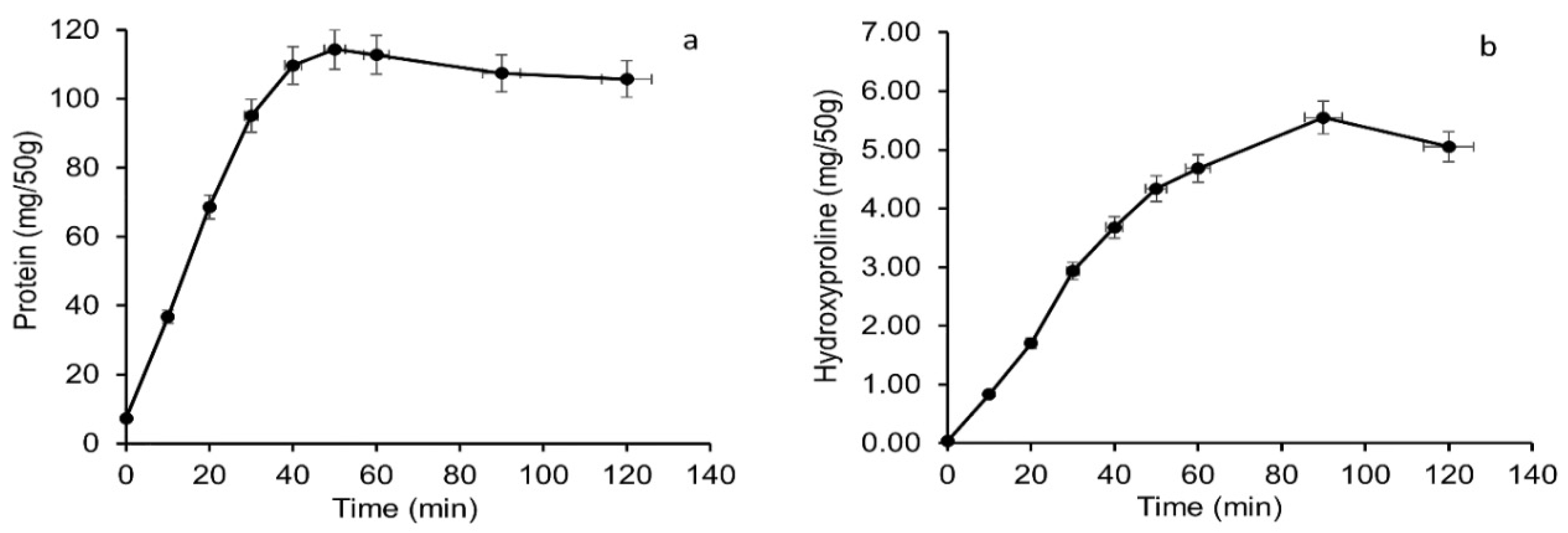
3.1. Total Soluble Protein and Hydroxyproline Content of Salmon Bone Leached out during Alkaline Treatment

The total soluble protein content liberated into alkaline solution used for the treatment of salmon bone, a leftover from the hydrolysis process, was monitored as a function of time (Figure 1a). A sharp increase in extractable protein from salmon bones was observed up to 50 min of the alkaline treatment. Thereafter, a slight decrease in protein content was noticeable up to 120 min. Alkali was able to solubilize the protein attached to the bone that remained after the enzymatic hydrolysis. This could lead to the removal of proteinaceous substances from the aforementioned bones. With continuous stirring at an operating temperature of 50 °C, proteins were likely to undergo denaturation or unfolding. This resulted in an increase in mass diffusivity, which in turn accelerated the mass transfer and solubilization of denatured proteins from the bone matrix. The result was in line with Kumoro et al. [19], who reported the positive impact of high temperature on the alkaline extraction of protein from chicken bone waste.
Alkaline solutions were reported to be effective in the removal of non-collagenous proteins from the starting materials used for collagen or gelatin production [20]. After 50 min, no further increase in the total soluble protein was found. This could be a result of less availability of soluble proteins in the bone. A further degradation of peptides to free amino acids or di-peptides could result in a lower content of proteins detected by the biuret method [21].
The content of hydroxyproline, a distinct amino acid presented in collagen, was determined during alkaline treatment as shown in Figure 1b. Hydroxyproline in the bone matrix represents collagenous proteins. Collagenous proteins were released with continuous stirring as a result of softening and rupturing of the bones induced by the alkaline condition at high temperature. The release of these collagenous proteins occurred continuously up to 100 min. A decline in hydroxyproline content was found at 120 min. In the present study, the alkaline treatment for 40 min was selected to remove non-collagenous proteins in the bone.

3.2. Chemical Compositions of Bio-Calcium and Calcined Bone from Salmon Frame with and without Alkaline Treatment

Chemical compositions of the bio-calcium and calcined bone are shown in Table 1. On a dry basis, bones that were calcined for 6 h had very low moisture content, of which those calcined for 9 h possessed no moisture as a result of the high temperature treatment on bones during calcination, which removed all the organic compounds and water molecules. For bio-calcium, Bio-cal-H had a higher moisture content than Bio-cal-A (p < 0.05). Fluids or other proteinaceous residues might bind or form a complex with water in the bone matrix as indicated by a higher moisture content. The ash content of calcined powders was higher than that in their bio-calcium counterparts (p < 0.05). During calcination, all organic components were combusted. It is noted that calcination for a longer time yielded a powder with higher ash content as a result of a more complete decomposition of the remaining organic matters. Overall, a high ash content of calcined powders (99.55–99.99%) was obtained.
The Ca/P mole ratios of Bio-cal-A and Bio-cal-H were 1.66 and 1.60, respectively. A slight increase in the Ca/P ratio of the bone sample with alkaline treatment could be due to the removal of non-collagenous phosphoproteins presented in the bone matrix [22]. Calcination at an elevated temperature for a longer time also provided the calcined bone with increases in calcium and phosphorus contents, especially for the non-alkaline treatment samples. The result suggests that the removal of non-collagenous protein with alkaline treatment could have led to a higher ash content of Bio-cal-A. The calcined bones show a similar ratio, in which Cal-A (6 h), Cal-H (6 h), Cal-A (9 h), and Cal-H (9 h) had a ratio of 1.66, 1.61, 1.66, and 1.63, respectively. Garner and Anderson [23] reported that vertebrate bone contains inorganic matter in the form of non-stoichiometric HAp crystals deposited in the matrix of cross-linked collagen fibrils. In general, pure HAp has a Ca/P mole ratio of 1.67. In the present study, the mole ratio of Ca/P from salmon bone (Table 1) was related closely to that of hydroxyapatite. Based on the Ca/P mole ratio, it could be proposed that HAp was present as the dominant constituent in the bio-calcium and calcined powders from salmon bone, a residue from the protein hydrolysis process.
Hydroxyproline was found only in Bio-cal-A and Bio-cal-H, while it was absent in the calcined bones, regardless of the alkaline pre-treatment or calcination temperature (Table 1). The presence of hydroxyproline indicated that collagenous protein is constituted in the bio-calcium, which correlated well with the protein content of bio-calcium. Nevertheless, Bio-cal-H possessed a higher hydroxyproline content than Bio-cal-A. Some collagenous proteins might be removed during the alkaline treatment of salmon bones used for Bio-cal-A production. Calcination at high temperature directly combusted these collagenous proteins in the calcined samples (p < 0.05). Therefore, hydroxyproline was absent in the calcined bones. In general, the alkaline pre-treatment of salmon bone determined the characteristics of bio-calcium, whereas calcination completely eliminated collagen from the bones.
The lipid oxidation products of bio-calcium samples Bio-cal-A and Bio-cal-H expressed as TBAR values are also shown in Table 1. Bio-cal-H had a higher TBAR value than Bio-cal-A (Table 1). The TBARS measurement is used to monitor the degradation of hydroperoxides, which leads to the formation of secondary oxidation products such as aldehydes [24]. The decrease in the TBARS value of Bio-cal-A could be as a result of alkaline treatment, which could remove pro-oxidant proteins such as hemoglobin. The result correlated well with differences in the fat content of Bio-cal-A and Bio-cal-H as highlighted in Table 1. During alkaline treatment, the bone became soft and possibly led to the leaching out of some lipids trapped in the bone matrix. Subsequent treatment with hexane could contribute to the removal of lipid and lipid oxidation products to some extent. Oxidizing agents such as NaOCl and H2O2 plausibly disintegrated the lipid oxidation product easily in Bio-cal-A than in Bio-cal-H as a result of the softness of the bones after the alkaline treatment in the former. This plausibly facilitated the reduction in TBARS values in Bio-cal-A. In calcined samples, lipids were completely removed, and the secondary oxidation products were not formed. Hence, TBAR values could not be detected for all calcined bones. Overall, various pre-treatments and calcination process of fish bones could lower the lipid oxidation in bio-calcium and calcined powder, respectively.

3.3. Color of Bio-Calcium and Calcined Powders

Bio-cal-A possessed a higher value of lightness (L* value) than Bio-cal-H and the calcined samples (p < 0.05) (Table 2). The slightly higher creamy whitish color of Bio-cal-A could be a result of the removal of meat residue as well as the blood during the alkaline treatment of salmon bone. For Bio-cal-A and Bio-cal-H, during the drying process, carbonyl compound-related products were formed via lipid oxidation and could undergo a non-enzymatic browning reaction with the proteins, peptides, and amino group of the free amino acid retained in the bone powders. This resulted in the increased yellow color obtained in the bio-calcium powders as indicated by the greater positive b* values. Both bio-calcium powders had low redness (a* value). A similar trend was reported by Benjakul et al. [25] in which bio-calcium powders from pre-cooked tongol (Thunnus tonggol) and yellowfin (Thunnus albacores) tuna bone had higher yellowness than their calcined counterpart.
For the calcined samples, an increase in lightness (L*) was observed in both Cal-A and Cal-H when calcination time increased from 6 to 9 h. Similarly, the increases in *ΔC were also observed. It should be noted that the L* value of Cal-H powders (69.49 ± 0.04) was less than that of Bio-cal-H powder (98.84 ± 0.08). The darker color could result from the dark grey color of the unburnt carbonaceous residue that may be trapped inside the porous structure of the fish bones during the calcination process. The result corresponded with the large quantity of organic constituents present in the Bio-cal-H powder as shown in Table 1. For a longer calcination period, the aforementioned carbonaceous particles could be further removed, which resulted in an increment of the L* value to 80.68 ± 0.01. On the other hand, this observation was less pronounced in Cal-A powders. The result illustrated the impact of the alkaline pre-treatment and calcination process on the color of both the resulting bio-calcium and calcined bone powders.

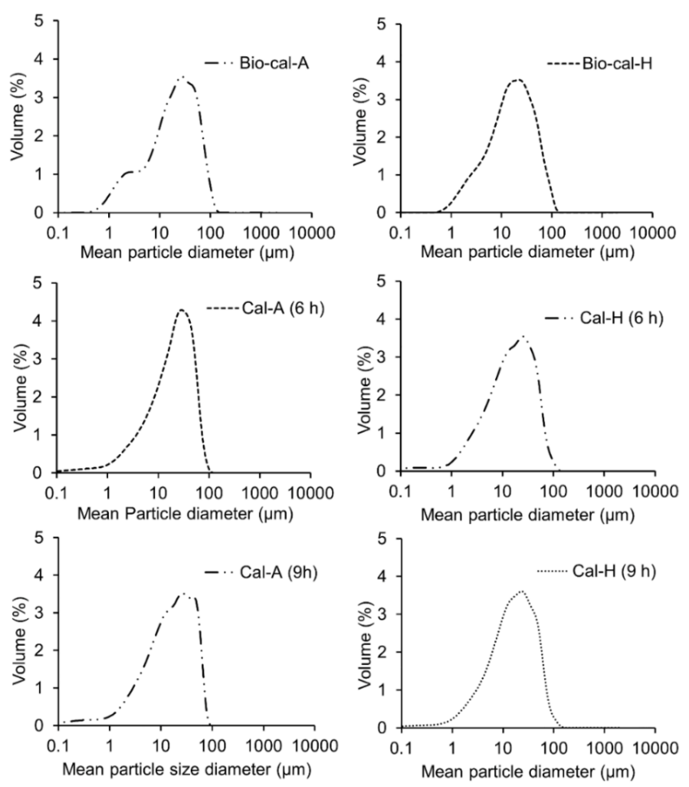
3.4. Mean Particle Size of Bio-Calcium and Calcine Bones

All samples showed differences in the mean diameter (d43), which ranged from 22.21 to 27.53 μm. Bio-cal-A showed larger particle size than Bio-cal-H (p < 0.05) (Table 2). For Cal-A, the higher calcination time resulted in the decrease in particle size. On the other hand, a slightly higher size was found for Cal-H when calcination time was increased (p < 0.05). Bio-cal-A showed a bi-modal distribution because of non-homogenous particles as indicated by the peak with the shoulder, while Bio-cal-H showed a mono-modal distribution, indicating the presence of homogeneous particles (Figure 2). The surface moisture, protein, and fat concentration are the factors affecting the stickiness and agglomeration of particles, thus influencing the particle size distribution [26]. During alkaline treatment, the collagen in the bone matrix, which is tightly linked with HAp, might be removed. This could bring about the weakened matrix, and the size could be reduced with ease.
For calcined samples, Cal-A and Cal-H had a more ordered and compact form with uniform particle size distribution as shown by mono-modal distribution. In addition, the removal of organic compounds possibly resulted in the lower particle diameters due to more compactness of HAp. Therefore, the particle size distribution was greatly affected by the alkaline pre-treatment and calcination temperatures.

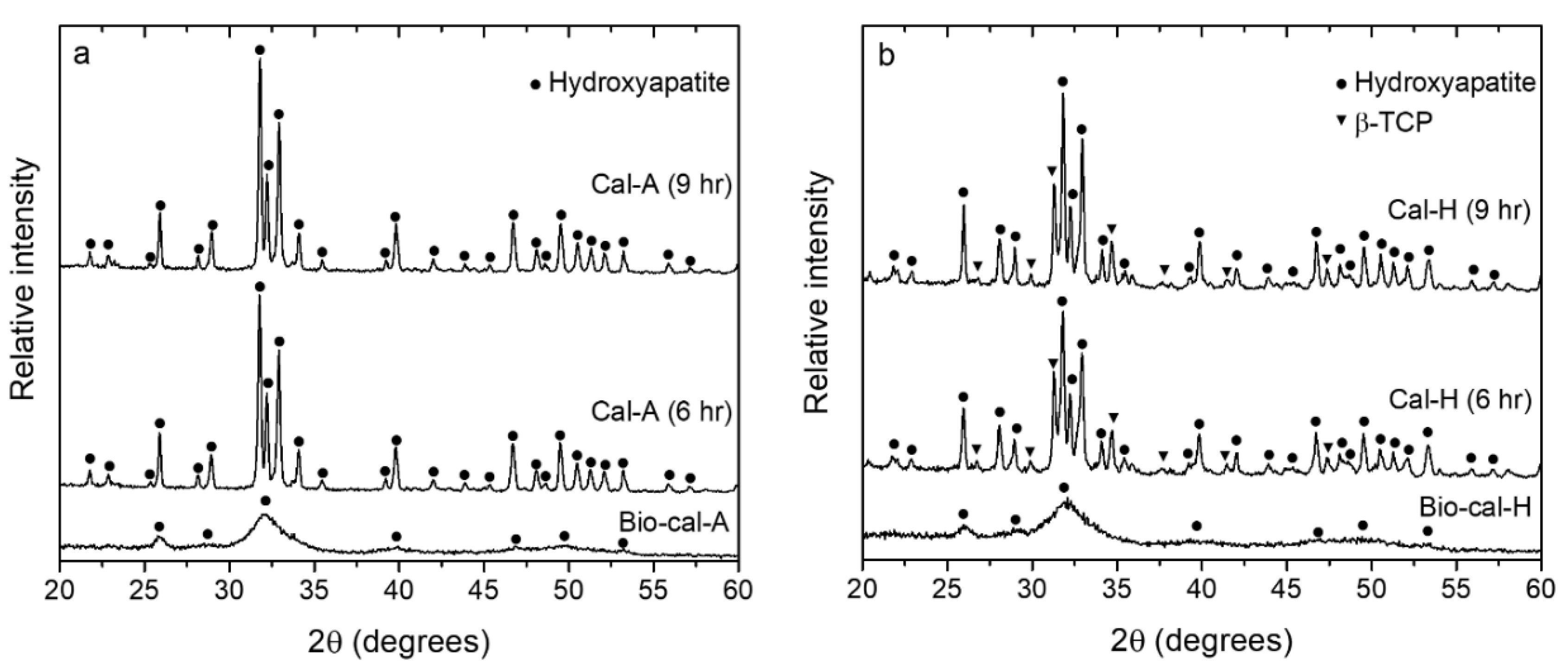
3.5. X-ray Diffraction (XRD) Patterns of Bio-Calcium and Calcine Bone Powders

Diffraction patterns of both the bio-calcium and bone powders calcined at 900 °C at 6 and 9 h showed that the HAp (Ca10(PO4)6(OH)2) phase (JCPDS:01-074-4172) was the dominant phase in all the samples (Figure 3). When calcination was implemented, the removal of organic matters and water occurred along with the agglomeration of HAp nano-crystals [27]. As a result, the diffraction peaks became more pronounced for the calcined powders than in bio-calcium powders (Figure 3a,b). It should be noted that the diffraction patterns of Cal-H powders showed a secondary phase of another apatite material in addition to the major phase of HAp (Figure 3b). According to the peak profile fitting, this minor apatite phase could be identified as β-tricalcium phosphate (β-TCP: Ca/P mole ratio = 1.5) (JCPDF: 01-073-4869). The XRD data were in good agreement with the Ca/P ratios shown in Table 1, where the β-TCP phase was only observed in non-alkaline-treated Cal-H powders which possessed a Ca/P ratio of about 1.61–1.63, less than that of stoichiometric HAp (Ca:P = 1.67) as earlier stated. On the other hand, only a single phase of HAp was observed in alkaline-treated Cal-A samples with a Ca/P of 1.66. The results are supported by numerous works that have reported the formation of β-TCP for the calcined HAp that has a Ca/P molar ratio lower than 1.67 [8,10,28].

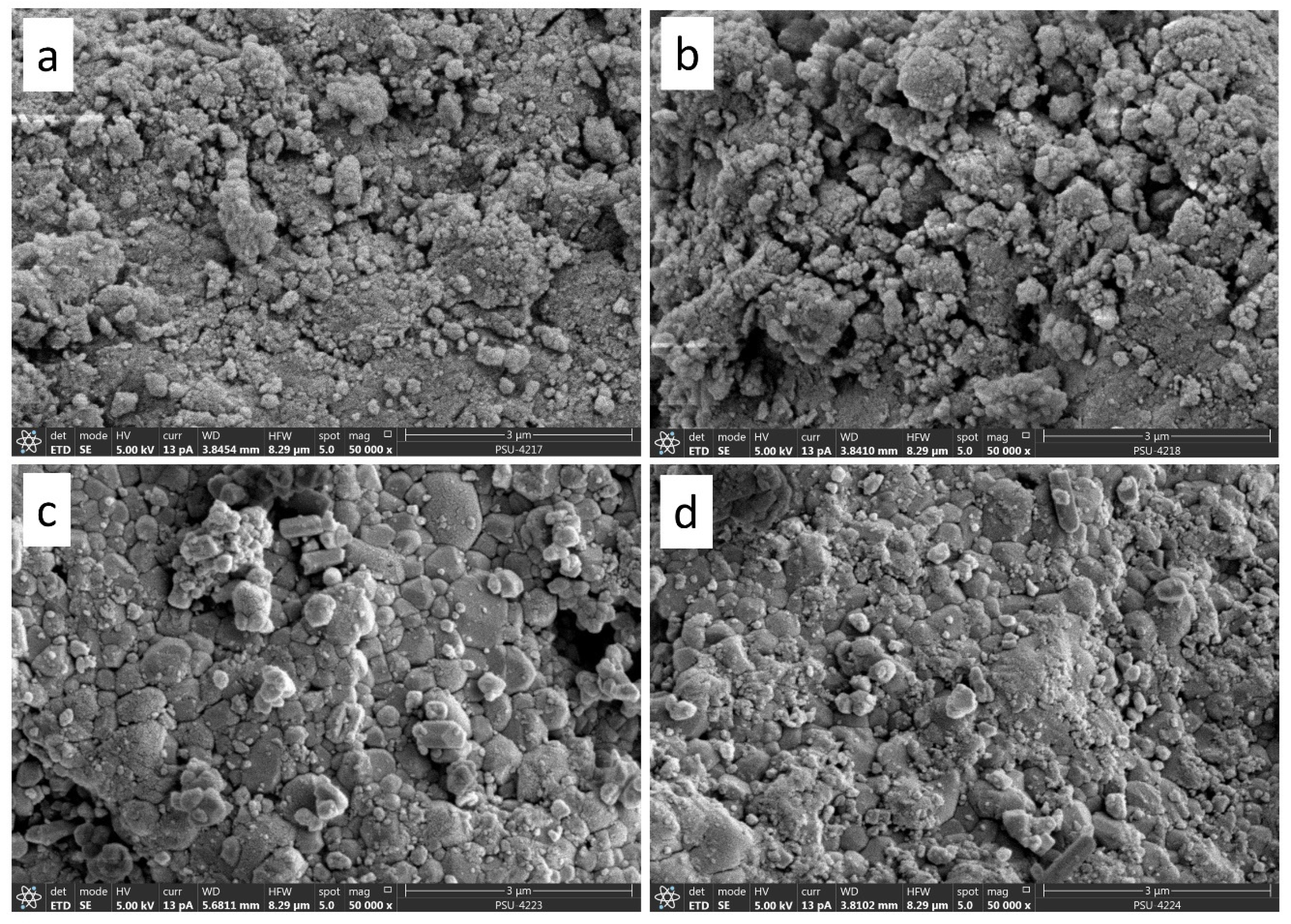
3.6. Scanning Electron Microscopy with Energy Dispersive X-ray Spectroscopy (SEM-EDX)

Figure 4a–d shows SEM images at 50,000× magnification of bio-calcium and bone powders calcined at 900 °C 6 h. Nano-particles in the range of a few tens of nano-meters that agglomerated into larger clusters were observed for both Bio-cal-A and Bio-cal-H (Figure 4a,b). After calcination of these powders, larger crystals or grains with the sizes of a few hundred nano-meters were formed (Figure 4c,d) as a result of crystal growth at an elevated temperature. The results were in agreement with XRD data that showed significant peak narrowing along with a sharp increasing in peak intensity of the calcined bone powders. The crystal sizes of samples with 9-h calcination (not shown in this article) were similar to that of the 6-h calcination samples, which were supported by their nearly identical full width at half maximum (FWHM) of diffraction patterns shown in Figure 3.
Nano-particles in the range of a few tens of nano-meters that agglomerated into larger clusters were observed for both Bio-cal-A and Bio-cal-H (Figure 4a,b). After the calcination of these powders, larger crystals or grains with the sizes of a few hundred nano-meters were formed (Figure 4c,d) as a result of crystal growth at an elevated temperature. The results were in agreement with XRD data that showed significant peak narrowing along with sharp increasing in peak intensity of the calcined bone powders. The crystal sizes of samples with 9-h calcination (not shown in this article) were similar to that of 6-h calcination samples, which were supported by their nearly identical FWHM of diffraction patterns shown in Figure 3.
A relative abundance of elements in all the samples was observed using SEM-EDX. Elements such as Ca, P, C, O, Na, and Mg were found in both the bio-calcium and calcined powders as depicted in Figure 5. It was noted that the detection limit of SEM-EDX could vary from 1–10%wt as a result of spectral resolution and difficulty in detecting low-Z elements [29]. Consequently, other trace elements could not be detected in the bio-calcium and calcined powders. Despite the instrumental limitation, the results show that the alkaline treatment of fish bones led to an increase in Ca and P contents of the bio-calcium powder. This was in line with higher contents of Ca and P determined by ICP-OES (Table 1). In addition, for all calcined samples, more pronounced and sharp peaks indicating a higher intensity of Ca and P were observed. Simultaneously, the contents of C were decreased, re-affirming the removal of organic matters such as lipids and meat proteins as a result of calcination.

4. Conclusions

The present study demonstrates the effect of alkaline treatment in the material conversion process of the salmon frame by-product to bio-calcium and HAp powders. The results showed that alkaline treatment played an important role on the characteristics of all samples. The amount of bio-molecular compounds such as fat, protein, and collagen was significantly lowered in the bio-calcium powder with alkaline treatment. On the other hand, the Ca/P molar ratio was lower in the bio-calcium sample without the treatment. Consequently, phase compositions of the calcine bone powders could be either a single-phase HAp or mixed-phase between HAp and β-TCP depending on their Ca/P ratios. In addition, salmon frame could be used as a potential source to produce high value-added compounds such as bio-calcium for calcium supplements and apatite compounds for bio-medical applications.

Author Contributions

Conceptualization, S.B.; methodology, A.T.I. and S.B.; software, S.S., T.S.-l., N.S., and Y.K.; validation, S.S., T.S.-l., N.S., and Y.K.; formal analysis, A.T.I.; investigation, A.T.I.; resources, S.B. and P.S.; data curation, P.S.; writing—original draft preparation, A.T.I.; writing—review and editing, S.B. and P.S.; visualization, S.B. and P.S.; supervision, S.B. and P.S.; project administration, S.B. and P.S.; funding acquisition, S.B. and P.S. All authors have read and agreed to the published version of the manuscript.

Funding

The authors declared that no funding was secured for this project.

Acknowledgments

This research was supported by the Higher Education Research Promotion and Thailand’s Education Hub for Southern Region of ASEAN Countries Project, the Office of the Higher Education Commission and the Graduate School, Prince of Songkla University (Grant number AGR6302013N).

Conflicts of Interest

The authors at this moment declared no conflict of interest.

References

  1. United Nations. World Population Prospects: The 2017 Revision, Key Fndings and Advance Tables; Working Paper No. ESA/P/WP/248; United Nations: New York, NY, USA, 2017. [Google Scholar]
  2. See, S.F.; Hoo, L.; Babji, A. Optimization of enzymatic hydrolysis of salmon (Salmo salar) skin by Alcalase. Int. Food Res. J. 2011, 18, 1359–1365. [Google Scholar]
  3. Sinthusamran, S.; Idowu, A.T.; Benjakul, S.; Prodpran, T.; Yesilsu, A.F.; Kishimura, H. Effect of proteases and alcohols used for debittering on characteristics and antioxidative activity of protein hydrolysate from salmon frames. J. Food Sci. Technol. 2019, 57, 473–483. [Google Scholar] [CrossRef] [PubMed]
  4. Benjakul, S.; Mad-Ali, S.; Senphan, T.; Sookchoo, P. Biocalcium powder from precooked skipjack tuna bone: Production and its characteristics. J. Food Biochem. 2017, 41, e12412. [Google Scholar] [CrossRef]
  5. Cashman, K.D. Calcium intake, calcium bioavailability and bone health. Br. J. Nutr. 2002, 87, 169–177. [Google Scholar] [CrossRef]
  6. Watanabe, T.; Kiron, V.; Satoh, S. Trace minerals in fish nutrition. Aquaculture 1997, 151, 185–207. [Google Scholar] [CrossRef]
  7. Boutinguiza, M.; Pou, J.; Comesaña, R.; Lusquiños, F.; De Carlos, A.; León, B. Biological hydroxyapatite obtained from fish bones. Mater. Sci. Eng. C 2012, 32, 478–486. [Google Scholar] [CrossRef]
  8. Piccirillo, C.; Silva, M.; Pullar, R.C.; Da Cruz, I.B.; Jorge, R.; Pintado, M.; Castro, P. Extraction and characterisation of apatite- and tricalcium phosphate-based materials from cod fish bones. Mater. Sci. Eng. C 2013, 33, 103–110. [Google Scholar] [CrossRef]
  9. Figueiredo, M.M.L.; Fernando, A.; Martins, G.; Freitas, J.; Judas, F.; Figueiredo, H. Effect of the calcination temperature on the composition and microstructure of hydroxyapatite derived from human and animal bone. Ceram. Int. 2010, 36, 2383–2393. [Google Scholar] [CrossRef]
  10. Goto, T.; Sasaki, K. Effects of trace elements in fish bones on crystal characteristics of hydroxyapatite obtained by calcination. Ceram. Int. 2014, 40, 10777–10785. [Google Scholar] [CrossRef]
  11. Cheng, L.; Ye, F.; Yang, R.; Lu, X.; Shi, Y.; Li, L.; Fan, H.; Bu, H. Osteoinduction of hydroxyapatite/β-tricalcium phosphate bioceramics in mice with a fractured fibula. Acta Biomater. 2010, 6, 1569–1574. [Google Scholar] [CrossRef]
  12. Gornall, A.G.; Bardawill, C.J.; David, M.M. Determination of serum proteins by means of the biuret reaction. J. Biol. Chem. 1949, 177, 751–766. [Google Scholar]
  13. Bergman, I.; Loxley, R. Two Improved and Simplified Methods for the Spectrophotometric Determination of Hydroxyproline. Anal. Chem. 1963, 35, 1961–1965. [Google Scholar] [CrossRef]
  14. Benjakul, S.; Mad-Ali, S.; Senphan, T.; Sookchoo, P. Characteristics of Biocalcium from Pre-cooked Skipjack Tuna Bone as Affected by Different Treatments. Waste Biomass-Valorization 2017, 9, 1369–1377. [Google Scholar] [CrossRef]
  15. Ensminger, L.G. The Association of Official Analytical Chemists. Clin. Toxicol. 1976, 9, 471. [Google Scholar] [CrossRef]
  16. Feist, B.; Mikula, B. Preconcentration of heavy metals on activated carbon and their determination in fruits by inductively coupled plasma optical emission spectrometry. Food Chem. 2014, 147, 302–306. [Google Scholar] [CrossRef]
  17. Mad-Ali, S.; Benjakul, S.; Prodpran, T.; Maqsood, S. Characteristics and gel properties of gelatin from goat skin as affected by spray drying. Dry. Technol. 2017, 35, 218–226. [Google Scholar] [CrossRef]
  18. Chuaychan, S.; Benjakul, S.; Kishimura, H. Characteristics of acid- and pepsin-soluble collagens from scale of seabass (Lates calcarifer). LWT 2015, 63, 71–76. [Google Scholar] [CrossRef]
  19. Kumoro, A.C.; Sofiah, S.; Aini, N.; Retnowati, D.S.; Budiyati, C.S. Effect of Temperature and Particle Size on the Alkaline Extraction of Protein From Chicken Bone Waste. Reaktor 2010, 13, 124. [Google Scholar] [CrossRef]
  20. Liu, D.; Wei, G.; Li, T.; Hu, J.; Lu, N.; Regenstein, J.M.; Zhou, P. Effects of alkaline pretreatments and acid extraction conditions on the acid-soluble collagen from grass carp (Ctenopharyngodon idella) skin. Food Chem. 2015, 172, 836–843. [Google Scholar] [CrossRef]
  21. Wu, H.-C.; Chen, H.-M.; Shiau, C.-Y. Free amino acids and peptides as related to antioxidant properties in protein hydrolysates of mackerel (Scomber austriasicus). Food Res. Int. 2003, 36, 949–957. [Google Scholar] [CrossRef]
  22. Al-Qtaitat, A.I.; Aldalaen, S.M. The Isolation and Characterization of Glycosylated Phosphoproteins From Herring Fish Bones. J. Biol. Chem. 2010, 285, 36170–36178. [Google Scholar]
  23. Garner, S.; Anderson, J. Skeletal Tissues and Mineralization. Diet Nutr. Bone Health 2011, 33, 49–50. [Google Scholar]
  24. Sae-Leaw, T.; Benjakul, S. Physico-chemical properties and fishy odour of gelatin from seabass (Lates calcarifer) skin stored in ice. Food Biosci. 2015, 10, 59–68. [Google Scholar] [CrossRef]
  25. Benjakul, S.; Mad-Ali, S.; Sookchoo, P. Characteristics of Biocalcium Powders from Pre-Cooked Tongol (Thunnus tonggol) and Yellowfin (Thunnus albacores) Tuna Bones. Food Biophys. 2017, 12, 412–421. [Google Scholar] [CrossRef]
  26. Pısecký, J. Handbook of milk powder manufacture. Niro A/S, Copenhagen. Proc. Eng. 1997, 3, 3–9. [Google Scholar]
  27. Londoño-Restrepo, S.M.; Jeronimo-Cruz, R.; Millán-Malo, B.M.; Rivera-Muñoz, E.M.; Rodríguez-García, M.E. Effect of the Nano Crystal Size on the X-ray Diffraction Patterns of Biogenic Hydroxyapatite from Human, Bovine, and Porcine Bones. Sci. Rep. 2019, 9, 5915. [Google Scholar] [CrossRef]
  28. Meinke, D.K.; Skinner, H.C.W.; Thomson, K.S. X-ray diffraction of the calcified tissues inPolypterus. Calcif. Tissue Int. 1979, 28, 37–42. [Google Scholar] [CrossRef]
  29. Choël, M.; Deboudt, K.; Osán, J.; Flament, P.; Van Grieken, R. Quantitative Determination of Low-ZElements in Single Atmospheric Particles on Boron Substrates by Automated Scanning Electron Microscopy−Energy-Dispersive X-ray Spectrometry. Anal. Chem. 2005, 77, 5686–5692. [Google Scholar] [CrossRef]
Figure 1. Total soluble protein (a) and hydroxyproline (b) contents as a function of time during the alkaline treatment of salmon bones. Bars represent the standard deviation (n = 3).
Applsci 10 04141 g001
Figure 2. Particle size distribution of bio-calcium and calcined bone powders from salmon bones.
Applsci 10 04141 g002
Figure 3. X-ray diffraction patterns of bio-calcium and 900 °C-calcined bone powders at 6 and 9 h with alkaline treatment (a) and without alkaline treatment (b).
Applsci 10 04141 g003
Figure 4. SEM images at 50,000× magnification for (a) Bio-cal-A, (b) Bio-cal-H, (c) Cal-A 900 °C 6 h and (d) Cal-H 900 °C 6 h.
Applsci 10 04141 g004
Figure 5. Elemental profile of bio-calcium and calcined powders from salmon bones as analyzed by SEM-EDX.
Applsci 10 04141 g005
Table 1. Chemical composition of bio-calcium and calcined powders from salmon frame.
Chemical CompositionBio-Cal-ABio-Cal-HCal-A (6 h)Cal-H (6 h)Cal-A (9 h)Cal-H (9 h)
Moisture (%)4.82 ± 0.07 d7.81 ± 0.04 e0.27 ± 0.00 b0.45 ± 0.01 c0.00 ± 0.00 a0.00 ± 0.00 a
Protein (%) *12.07 ± 0.18 b20.90 ± 0.06 c0.00 ± 0.00 a0.00 ± 0.00 a0.00 ± 0.00 a0.00 ± 0.00 a
Fat (%) *0.33 ± 0.01 b1.70 ± 0.01 c0.00 ± 0.00 a0.00 ± 0.00 a0.00 ± 0.00 a0.00 ± 0.00 a
Ash (%) *82.78 ± 0.25 b69.59 ± 0.57 a99.73 ± 0.05 d99.55 ± 0.32 c99.99 ± 0.00 f99.97 ± 0.00 e
Hydroxyproline (mg/g) *5.07 ± 0.01 b14.79 ± 0.02 c0.00 ± 0.00 a0.00 ± 0.00 a0.00 ± 0.00 a0.00 ± 0.00 a
TBARS (mg malonaldehyde/kg sample) *0.95 ± 0.00 b3.34 ± 0.01 c0.00 ± 0.00 a0.00 ± 0.00 a0.00 ± 0.00 a0.00 ± 0.00 a
Calcium (%) *30.88 ± 0.33 b27.32 ± 0.17 a36.01 ± 0.26 e31.54 ± 0.20 c38.84 ± 0.55 f34.24 ± 0.40 d
Phosphorus (%) *14.40 ± 0.39 b13.22 ± 0.48 a16.79 ± 0.25 e15.16 ± 0.32 c18.11 ± 0.35 f16.27 ± 0.45 d
Mole ratio Ca/P1.661.601.661.611.661.63
Values are presented as mean ± SD (n = 3). Different lowercase letters in the same row indicate significant difference (p < 0.05). * Dry weight basis.
Table 2. Mean particle size and color values of bio-calcium and calcined powders from salmon frame.
ParametersBio-Cal-ABio-Cal-HCal-A (6 h)Cal-H (6 h)Cal-A (9 h)Cal-H (9 h)
Mean particle size (d43, µm)26.53 ± 3.49 f24.05 ± 3.14 d25.36 ± 2.78 e22.21 ± 2.84 a22.39 ± 2.64 b23.12 ± 2.94 c
L*95.29 ± 0.08 f94.84 ± 0.08 d93.61 ± 0.01 c69.49 ± 0.04 a95.01 ± 0.01 e80.68 ± 0.01 b
A*−0.60 ± 0.02 c−0.31 ± 0.07 d−1.59 ± 0.01 b−0.23 ± 0.04 e−2.63 ± 0.01 a−0.36 ± 0.03 d
B*7.13 ± 0.02 d6.86 ± 0.14 c−0.19 ± 0.02 b−0.10 ± 0.07 b−1.67 ± 0.01 a−0.22 ± 0.01 b
ΔE*6.94 ± 0.12 d6.60 ± 0.08 c0.87 ± 0.14 a24.15 ± 0.15 f3.00 ± 0.14 b12.97 ± 0.09 e
ΔC*7.15 ± 0.03 f6.87 ± 0.14 e1.60 ± 0.01 c0.26 ± 0.06 a3.11 ± 0.00 d0.42 ± 0.01 b
Values are presented as mean ± SD (n = 3). Different lowercase letters in the same row indicate significant difference (p < 0.05).

