Article

Osteogenic Potential of Fast Set Bioceramic Cements: Molecular and In Vitro Study

1
Department of Surgical Sciences, CIR Dental School, University of Turin, Endodontics, Via Nizza 230, 10126 Torino, Italy
2
Department of Biomedical and Neuromotor Sciences, DIBINEM, University of Bologna-Alma Mater Studiorum, 40125 Bologna, Italy
3
Department of Medical Sciences, University of Turin, 10126 Turin, Italy
4
Candiolo Cancer Institute, FPO–IRCCS, Pathology Division, 10060 Candiolo, Italy
*
Author to whom correspondence should be addressed.
Appl. Sci. 2020, 10(19), 6713; https://doi.org/10.3390/app10196713
Submission received: 29 August 2020 / Revised: 18 September 2020 / Accepted: 22 September 2020 / Published: 25 September 2020
(This article belongs to the Section Applied Dentistry and Oral Sciences)

Abstract

:

Featured Application

Bioceramic materials have been recently proposed for several applications in endodontic practice. The impact of their setting time and chemical composition on biocompatibility is still controversial. This in vitro study evaluated the osteogenic potential of different bioceramic formulations.

Abstract

Recently, pre-mixed bioceramics in fast set formulations have been increasingly utilized in clinical practice as an alternative to mineral trioxide aggregate (MTA) for their shorter setting time and better handling properties. However, the impact on their osteogenic potential, due to modifications in chemical composition to promote a fast setting, is still unclear. This molecular and in vitro study compared the osteogenic potential of root repairing material putty fast set (FSP) with root-repairing material putty (RRMPU), root-repairing material paste (RRMPA), Biodentine™ and MTA. The null hypothesis tested was that there are no differences among the tricalcium silicate materials in terms of osteogenic potential. Standardized discs were cultured with MG-63 human osteoblastic-like cells to assess biocompatibility, the activity of alkaline phosphatase (ALP) and osteogenic potential. Biocompatibility was evaluated at baseline and after 24 and 48 h. Osteogenic differentiation was assessed after 15 days. Data were analyzed with one-way ANOVAs and Tukey’s post-hoc test (p < 0.05). All materials showed biocompatibility and bioactivity. ALP activity, which induces mineral nodule deposition, increased in all the cements tested, with a significant increase in RRMPU (p < 0.001) and FSP (p < 0.001) samples versus MTA. In vitro mineralization was significantly increased for RRMPU (p < 0.0001), FSP (p = 0.00012) and Biodentine™ (p < 0.0001) versus MTA. The bioceramics tested showed higher levels of biocompatibility and bioactivity than MTA; a higher capacity for mineralization was observed with RRMPU and FSP versus MTA.

1. Introduction

Endodontics involves the prevention and treatment of pulpal and periradicular diseases [1,2]. ProRoot® Mineral Trioxide Aggregate (MTA; Dentsply Maillefer, Ballaigues, Switzerland), a hydraulic silicate cement, has been perceived as the gold standard for vital pulp therapy, apexification, repair of root perforations and root-end fillings [3,4]. Mineral trioxide aggregate is biocompatible and induces the formation of new mineralized tissue [5,6]. However, disadvantages such as fluid consistency, make it difficult to handle and control, and long setting time challenges clinical handling. Biodentine™ (Septodont, Saint-Maur-des-Fosses, France) has been introduced as an alternative bioceramic material with reduced setting time and the ability to promote the formation of a dentinal bridge when used for direct pulp capping [7,8]. Other bioceramic materials incorporate calcium phosphate into tricalcium silicate cements (CPCSs) for improving bioactivity and facilitating clinical handling [9,10]. These bioceramic materials have been proposed for apexification, the repair of root defects and perforation [11]. They are also available in pre-mixed formulations and have been reported to be as biocompatible as MTA or Biodentine [12]. Endosequence BC root repair material putty (RRMPU; Brasseler, Savannah, GA, USA) and Root Repair Material Paste (RRMPA; Brasseler) are premixed bioceramics that have been reported to be biocompatible [13,14] and promote fibroblast growth [15]. These pre-mixed bioceramics are available in “regular” and “fast-set” versions, with setting times ranging from 4 h to 20 min, respectively [10]. The fast-set formulation has similar mechanical properties as MTA and a higher final hardness value than other CPCS cement formulations [16]. Biocompatibility studies on novel bioceramics have reported the absence of cytotoxicity and genotoxicity; however, their ion release profiles, mineralization capability and interactions with bone marker are yet to be determined [17]. Although fast set bioceramic cements showed similar biocompatibility and better cell adhesion capacity compared with MTA [18], their osteogenic potential remains largely unknown. Human MG-63 osteoblastic-like cells are considered a reliable in vitro model for evaluating the osteogenic potential of biomaterials by examining the expression of cytokines, bone markers and osteoblast-derived proteins [19]. Accordingly, the objective of the present study was to compare the biocompatibility and osteogenic potential of fast-set bioceramic cements with MTA and other bioceramic materials using the MG-63 human cell line model. The null hypothesis tested was that there are no differences among the tricalcium silicate materials in terms of their biocompatibility and osteogenic potential.

2. Materials and Methods

2.1. Cell Culture and Osteogenic Differentiation

The human osteosarcoma-derived MG-63 cell line (ATCC® CRL-1427, American Type Culture Collection, Mamassas, VA, USA) was grown in Dulbecco’s Modified Eagle’s Medium (MilliporeSigma, Burlington, MA, USA), with 10% fetal bovine serum (Gibco, Thermo Fisher Scientific, Waltham, MA, USA), 1% glutamine (MilliporeSigma, Burlington, MA, USA) and 1% streptomycin, penicillin and fungizone (Millipore Sigma, Burlington, MA, USA). This medium was designated as the “Normal Medium” (NM). For osteogenic differentiation, the cells were cultured in “Differentiation Medium” (DM), which was composed of NM supplemented with 50 mg/mL L-ascorbic acid and 10 mmol/L β-glycerolphosphate (all from MilliporeSigma, Burlington, MA, USA). The MG-63 cells, cultured in NM or DM, were maintained at 37 °C in a humidified 5% CO2 atmosphere. Cell growth was monitored daily with a digital inverted microscope (EVOS, Advanced Microscopy Group, Bothell, WA, USA).

2.2. Biomaterial Preparation and Eluate Collection

The study was performed using the following biomaterials: Root Repair Material Putty (RRMPU, Brasseler), Root Repair Material Paste (RRMPA, Brasseler), Fast-set Putty (FSP, Brasseler), Biodentine™ (BIO) and ProRoot® MTA (Table 1). All materials were prepared according to the manufacturer’s instructions. Discs of ~1 mm thickness and 8 mm diameter were prepared by packing the mix for pre-mixed materials into polyvinylchloride molds. The discs were stored at 37 °C and 95 ± 5% relative humidity until set. The set materials were transferred to 24-well plates and disinfected by exposure to ultraviolet light for 2 h. The NM medium was added to each well (1 mL/well) and maintained for 24 h at 37 °C in a humidified 5% CO2 atmosphere to allow any cytotoxic material to be eluted from the set cement. After 24 h, the eluate (EM) was collected and stored at −80 °C for subsequent analysis.

2.3. Cell Attachment and Biocompatibility

Cell attachment and biocompatibility were evaluated with the aforementioned digital inverted microscope by examining the morphology and adhesion of the MG-63 cells that were cultured directly on the biomaterial or in the corresponding eluate. For each material to be tested, the MG-63 cells were seeded in 24-well plates according to the following conditions:
(a)
1 × 10⁴ MG-63 cells cultured in 1 mL of NM (positive control);
(b)
1 × 10⁴ MG-63 cells cultured in 1 mL of EM, obtained, as previously described;
(c)
1 × 10⁴ MG-63 cells cultured in 1 mL of NM together with a pre-prepared bioceramic disc.
The culture plates were incubated at 37 °C in a humidified 5% CO2 atmosphere. The cells were monitored at time-zero, 24 h and 48 h with the inverted microscope. Three independent experiments were performed. The cells were observed for morphological alteration and adhesion. Evaluation was performed by two independent operators. A material was considered biocompatible if more than 50% of the cells appeared flat or partially round and were attached to the culture plate or material surface. If cells were totally round and detached from a surface, the material was considered toxic and not biocompatible. The MG-63 cells that were not adjacent to any materials were used as control [20,21,22].

2.4. Osteogenic Differentiation

The MG-63 cells were seeded in 24-well plates (1 × 10⁴ cells/well) in NM, with or without bioceramic discs. After 24 h, the NM was replaced with DM and the cells were induced to osteogenically differentiate for 15 days [23,24]. The DM was replaced every 48 h. The cells were photographed using the EVOS inverted microscope at 4X magnification. The ImageJ software (National Institute of Health, Bethesda, MD, USA) was used to measure the area of mineralized nodules. Multiple images were analyzed for each condition tested and areas were expressed as mean ± standard deviation.

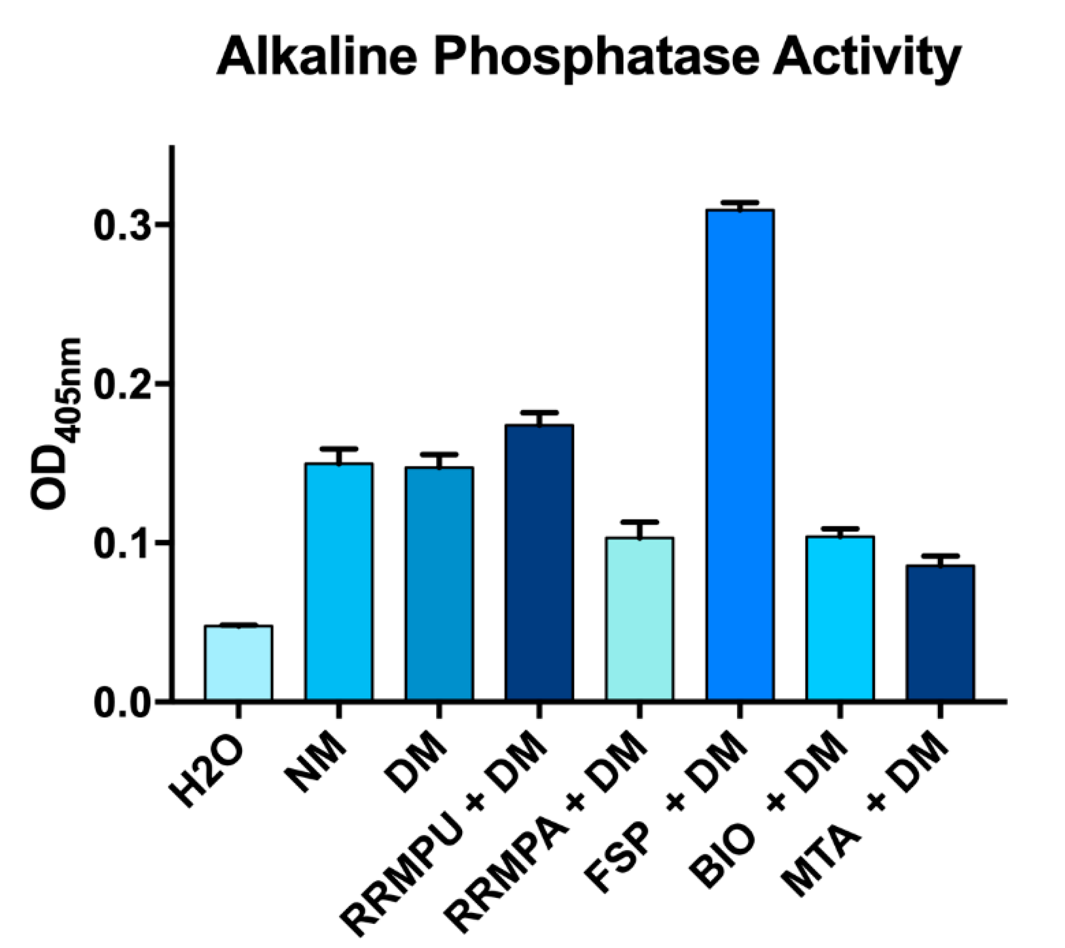
2.5. Alkaline Phosphatase Activity

Alkaline phosphatase (ALP) activity of cells cultured with or without a bioceramic disc was evaluated using the QuantiChromTM Alkaline Phosphatase Assay Kit (DALP-250; BioAssay Systems Hayward, CA, USA) after 15 days of culture in DM. Reagent application and sample preparation were performed according to the manufacturer’s instructions. The specimens were read at 405 nm and data were expressed as optical density (OD) at 405 nm. The experiment was performed in triplicate.

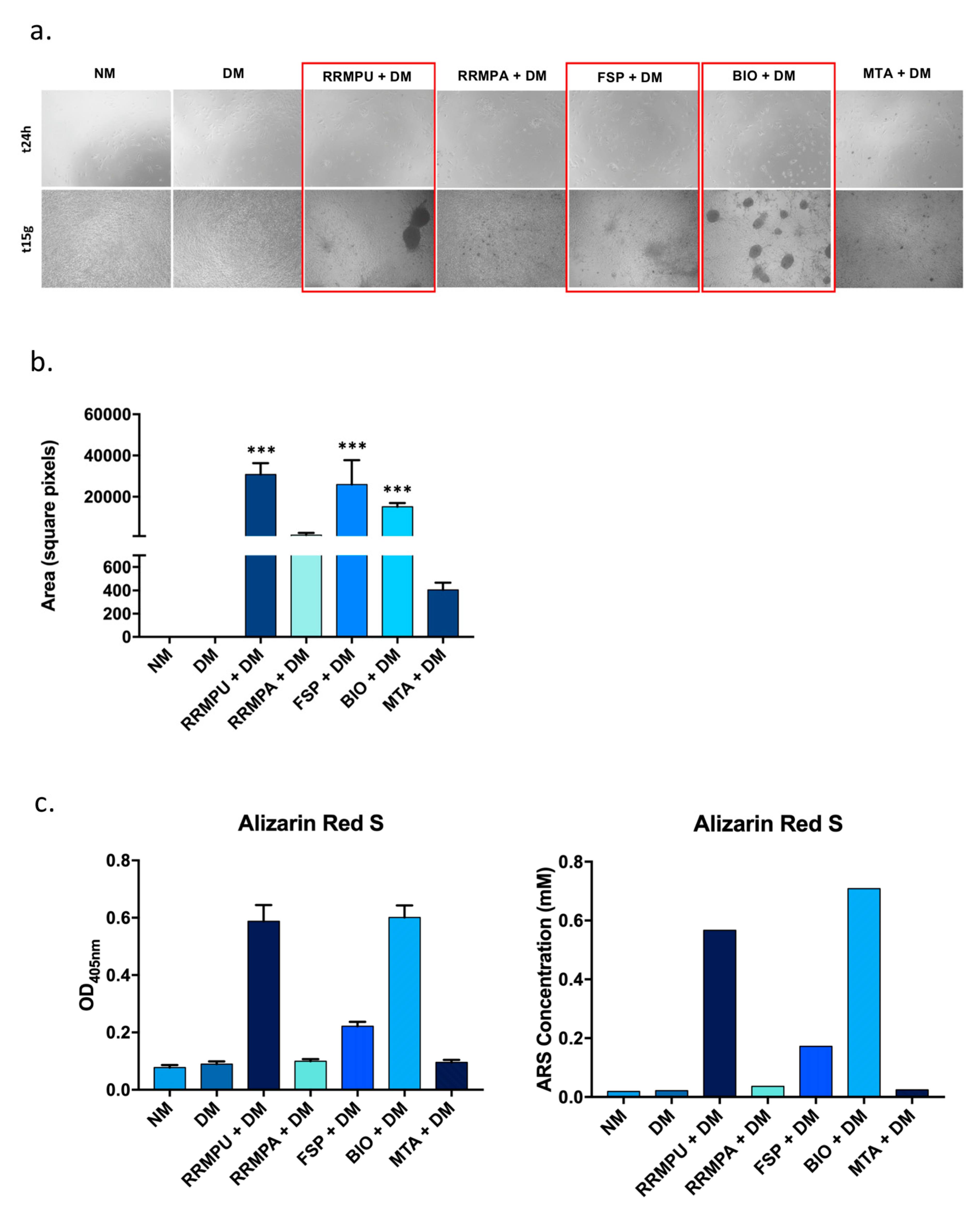
2.6. Mineralization and Alizarin Red S Staining

Alizarin Red S is a red dye used to identify calcium deposits in cell culture. Mineralization is evaluated by the extraction of calcified material at low pH, neutralization with ammonium hydroxide and colorimetric detection at 405 nm [25]. The MG-63 cells were cultured in DM for 15 days. Analysis was performed using the Alizarin Red S Staining Quantification Assay (ARed-Q Kit; ScienCell Research Laboratories, Carlsbad, CA, USA), following the manufacturer’s instructions. The dye (1 mL of 40 mm) was added to each well, incubated for 30 min at ambient temperature and removed using 10% acetic acid. The acid was neutralized with 10% ammonium hydroxide. Three aliquots from each well were transferred to a 96-well plates and their absorbance was read at 405 nm with a plate reader. Data were expressed as OD at 405 nm and as dye concentration (mm). Dye concentration was calculated using an Alizarin Red S standard curve, according to the manufacturer’s instructions.

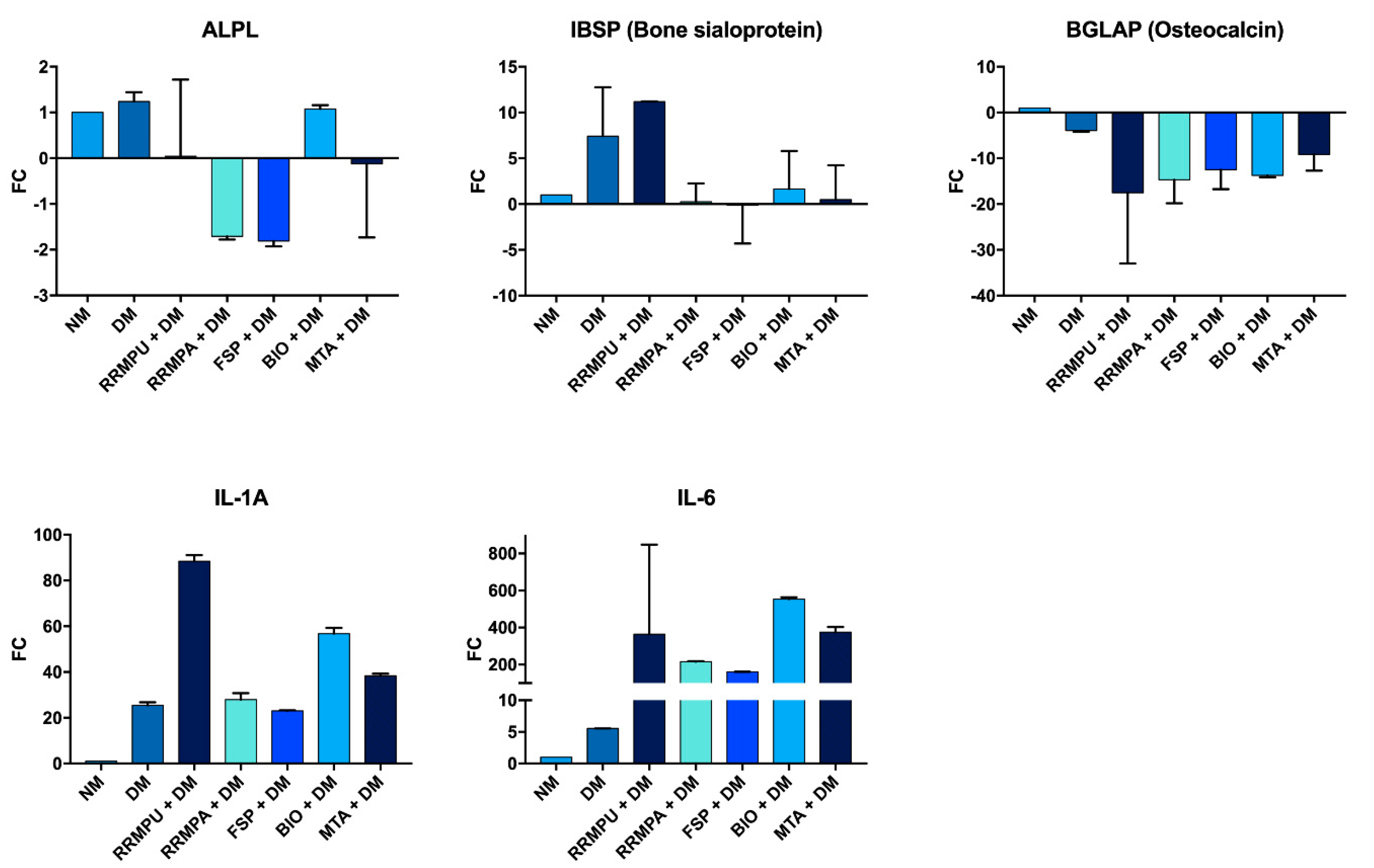
2.7. Quantitative Real-Time PCR (RT-qPCR)

Ribonucleic acid (RNA) was extracted from the MG-63 cells after osteogenic differentiation with TRIzol reagent (Thermo Fisher Scientific, Waltham, MA, USA), according to manufacturer’s instructions. The RNA pellets were resuspended in diethylpyrocarbonate-treated water, and RNA concentrations were measured with a spectrophotometer (NanoDrop™ 2000, Thermo Fisher Scientific). The RNA was reverse-transcribed with a High-Capacity cDNA Reverse Transcription Kit (Applied Biosystems, Foster City, CA, USA) after a DNase treatment with a TURBO DNA-freeTM Kit (Thermo Fisher Scientific). The RNA samples without reverse transcriptase were reverse transcribed as negative controls for DNA contamination in the PCR analyses. Complementary DNA (cDNA) was amplified by real-time PCR using the TaqMan® Gene Expression Master Mix (Applied Biosystems, Foster City, CA, USA) and the StepOne machine (Applied Biosystem, Foster City, CA, USA), according to the manufacturer’s protocols. The following TaqMan®-qPCR assays (Applied Biosystem) were employed: Glyceraldehyde-3-phosphate dehydrogenase, GAPDH (Hs99999905_m1); actin beta, ACTB (Hs99999903_m1); pumilio RNA binding family member 1, PUM1 (Hs00472881_m1); alkaline phosphatase, ALPL (Hs01029144_m1); bone gamma-carboxyglutamate protein, BGLAP (Hs01587814_g1); integrin binding sialoprotein, IBSP (Hs00173720_m1); interleukin 1 alpha, IL-1a (Hs00174092_m1); interleukin 6, IL-6 (Hs00985639_m1). A no-template control was included for each assay. The RT-qPCR reaction was conducted with a 10 min incubation period at 95 °C, 40 cycles of 95 °C amplification for 15 s and 60 °C for 1 min for annealing. GAPDH, ACTB and PUM1 were used as housekeeping genes to normalize the gene expression data of the target genes. The 2−ΔΔCt method was used to analyze relative changes in gene expression between samples in the form of fold changes.

2.8. Statistical Analysis

The GraphPad Prism software version 7.0a was used to analyze the data (GraphPad Software, Inc., San Diego, CA, USA). Parametric statistical methods were adopted after validating the homogeneity (Shapiro–Wilk test) and homoscedasticity (modified Levene test) of the corresponding data sets. Analyses were performed using one-way analysis of variance and the Tukey multiple comparison procedures. Significance was defined following the New England Journal of Medicine style: 0.12 not significant (ns), 0.033 *, 0.002 ** and p < 0.001 *** [26]. A probability value of p < 0.05 was considered to be statistically significant.

3. Results

3.1. Cellular Attachment and Biocompatibility

All the bioceramic materials tested were biocompatible (Figure 1). Adhesion and the morphology of MG-63 cells were not affected by the presence of EM or by direct contact with any of the bioceramics. No morphological differences in attachment or in the growing of cells were identified between the control and treated wells.

3.2. Osteogenic Differentiation

The process of osteogenic differentiation was evaluated via examination of the ALP activity. The ALP levels were significantly higher in MG-63 cells cultured in direct contact with FSP and RRMPU, compared with the cells grown in the presence of MTA (p < 0.001 for both comparisons, Figure 2, Table 2). After 15 days in DM, mineralized nodule formation was enhanced in cells cultured with bioceramic discs, compared to those cultured without, as shown in Figure 3. Furthermore, the area occupied by the mineralized nodules was significantly larger in MG-63 cells grown in direct contact with FSP, RRMPU and BIO (p < 0.001 for all; Figure 3a,b), compared to that obtained from culturing with MTA. The formation of mineralized nodules in the MG-63 cells was also evaluated with Alizarin Red S staining (Figure 3c). Staining was significantly increased when cells were cultured in the presence of RRMPU (p < 0.0001), BIO (p < 0.0001) and FSP (p = 0.0012) (Figure 3c). Osteogenic differentiation was also investigated in MG-63 cells cultured through the expression of ALPL, IBSP, BGLAP, IL-1a and IL-6, using RT-qPCR (Figure 4). ALPL expression was significantly reduced in MG-63 cells cultured with FSP and RRMPA. IBSP expression was significantly increased in cells cultured with RRMPU. BGLAP was downregulated in all the tested conditions and there was no statistical difference in the fold changes among the different biomaterials. No differences in ALPL, IBSP and BGLAP expressions between FSP and the other bioceramic materials were evident. The upregulation of IL-1A and IL-6 was detected when culture was performed in the presence of some of the biomaterials. No difference in IL-6 expression was identified between FSP and the other bioceramic materials. IL-1A expression was significantly increased in MG-63 cells cultured with RRMPU or BIO (p < 0.0001 for both) and MTA (p = 0.0009), versus MG-63 cells cultured in DM alone. Significant difference in IL-1A expression was also observed in cells cultured in BIO, versus those cultured in the presence of MTA (p < 0.0001).

4. Discussion

Biocompatibility is one of the most important characteristics for dental products [27,28]. In vitro biocompatibility tests are regulated by ISO 7405 and ISO 10993-5 standards [29]. Although many studies have evaluated the biocompatibility of dental materials [19,30,31], the use of different in vitro models (different cell cultures, medium or exposure time) precludes the meaningful comparison of the results reported in those studies [29]. The osteosarcoma-derived MG-63 cells have been used occasionally for evaluating the biocompatibility of calcium silicate cements, due to their similar physiologic and adhesive properties as human osteoblasts [32],as well as their reliability in biocompatibility testing [19,30,31,33]. All the cements tested in the present study demonstrated high biocompatibility with MG-63 cells. The osteogenic response of MG-63 cells was evaluated by ALP activity, an important marker of bone matrix mineralization. The two bioceramic materials RRMPU and FSP, both composed mainly of calcium silicates, zirconium oxide, tantalum oxide and calcium phosphate monobasic, exhibited higher ALP activity compared to MTA, suggesting a better capability of these two materials to induce mineralization. Indeed, MTA is composed by similar elements (calcium, silicate, phosphate, carbon and oxygen), but shows a different overall composition (tricalcium silicate, dicalcium silicate, bismuth oxide, and small proportions of tricalcium aluminate and calcium sulfate). The capability of mineralization potential of bioceramic materials was confirmed by the observation of mineralized nodules in cell cultures and by Alizarin Red S staining. The FSP has been claimed to have optimal mineralizing properties [34]. The previously reported result is in agreement with findings from the present study. The osteogenic differentiation of mesenchymal stem cells is regulated by osteocytes and osteoblasts in a simplified bone niche. To monitor the potential effects of the biomaterials on osteogenic differentiation, expressions of ALPL, IBSP and BGLAP and ALPL genes were used as markers of different phases of osteogenesis. After an initial peak, ALPL levels started to decline as other genes (e.g., BGLAP) were upregulated [35]. In the present study, the expression of these markers was determined after 15 days of culture in an osteogenic differentiation medium. ALPL expression was downregulated in all the conditions tested. The results are in line with the notion that the early osteogenesis phase declines after 15 days of culture. Accordingly, even if the ALP enzyme is still active, gene expression is diminished.
As expected, MG-63 cells cultured in DM expressed high levels of IBSP, as this gene is involved in the middle phase of osteogenesis. However, high levels of BGLAP expression were not detected. The results suggest that the MG-63 cells in DM had not reached the last phase of osteogenesis after culturing for 15 days. The absence of statistically significant differences in ALPL, IBSP and BGLAP expressions between FSP and the other bioceramic materials suggests that the effect of FSP on osteogenic induction is comparable to the other bioceramic materials tested. Up-regulations of IL-1A and IL-6 gene expressions were evident in the MG-63 cells cultured with FSP, RRMPU or Biodentine. Interleukin-6 plays an important role in tissue regeneration by enhancing ALP activity and promoting osteogenic differentiation, while IL-1A is positively involved in the regulation, production and secretion of IL-6 [36]. Up-regulation of IL-6 in the presence of FSP confirms that the activity of ALP is stimulated in the cells and that osteogenic differentiation is ongoing.

5. Conclusions

Within the limits of the present study, it may be concluded that FSP exhibits better biocompatibility and bioactivity in comparison with MTA. There is no difference in these activities between FSP and RRMPU. The FSP also promotes cellular differentiation and demonstrates the potential to contribute to the re-mineralization process of osteolytic lesions of endodontic origin.

Author Contributions

The submitting author affirms that all individuals listed as authors agree that they have met the criteria of authorship, agree to the conclusions of the study and that no individual meeting the criteria of authorship has been omitted. Conceptualization: D.P., P.C. and E.B.; Methodology: L.A., V.M., A.M. and A.C.; Formal analysis: L.B., L.A., A.M. and N.S.; Investigation: L.A., V.M., D.P., P.C. and M.A.; Data curation: A.C., P.C., L.A. and N.S.; Writing-original draft preparation: A.C., D.P., M.A. and V.M.; Writing—review and editing: A.C., M.A., N.S., A.M., L.B., D.P., P.C. and E.B.; Supervision: E.B., L.B., D.P. and P.C. All authors have read and agreed to the published version of the manuscript.

Funding

This research received no external funding.

Acknowledgments

This research received no specific grant from any funding agency in the public, commercial, departmental or not-for-profit sectors.

Conflicts of Interest

L. Annaratone is funded by Fondazione Umberto Veronesi (Post-Doctoral Fellowship 2018 and 2019). D. Pasqualini declares lecturing personal fees for Dentsply Maillefer in the last three years. The other Authors declare no competing conflicts of interest with the materials discussed in this manuscript.

References

  1. Peters, L.B.; Wesselink, P.R.; Buijs, J.F.; Van Winkelhoff, A.J. Viable Bacteria in Root Dentinal Tubules of Teeth with Apical Periodontitis. J. Endod. 2001, 27, 76–81. [Google Scholar] [CrossRef] [PubMed]
  2. Ørstavik, D.; Pitt Ford, T.R. Essential Endodontology: Prevention and Treatment of Apical Periodontitis, 2nd ed.; Blackwell Munksgaard: Oxford, UK, 2008. [Google Scholar]
  3. Parirokh, M.; Torabinejad, M.; Dummer, P.M.H. Mineral trioxide aggregate and other bioactive endodontic cements: An updated overview—Part I: Vital pulp therapy. Int. Endod. J. 2017, 51, 177–205. [Google Scholar] [CrossRef] [PubMed]
  4. Torabinejad, M.; Parirokh, M.; Dummer, P.M.H. Mineral trioxide aggregate and other bioactive endodontic cements: An updated overview—Part II: Other clinical applications and complications. Int. Endod. J. 2017, 51, 284–317. [Google Scholar] [CrossRef] [PubMed]
  5. Torabinejad, M.; Watson, T.; Ford, T.P. Sealing ability of a mineral trioxide aggregate when used as a root end filling material. J. Endod. 1993, 19, 591–595. [Google Scholar] [CrossRef]
  6. Yoshino, P.; Nishiyama, C.K.; Modena, K.C.D.S.; Santos, C.F.; Sipert, C.R. In vitro cytotoxicity of white MTA, MTA Fillapex® and portland cement on human periodontal ligament fibroblasts. Braz. Dent. J. 2013, 24, 111–116. [Google Scholar] [CrossRef] [Green Version]
  7. Nowicka, A.; Lipski, M.; Parafiniuk, M.; Sporniak-Tutak, K.; Lichota, D.; Kosierkiewicz, A.; Kaczmarek, W.; Buczkowska-Radlińska, J. Response of human dental pulp capped with biodentine and mineral trioxide aggregate. J. Endod. 2013, 39, 743–747. [Google Scholar] [CrossRef]
  8. Camilleri, J.; Sorrentino, F.; Damidot, D. Investigation of the hydration and bioactivity of radiopacified tricalcium silicate cement, Biodentine and MTA Angelus. Dent. Mater. 2013, 29, 580–593. [Google Scholar] [CrossRef]
  9. Gandolfi, M.G.; Taddei, P.; Siboni, F.; Modena, E.; Ginebra, M.-P.; Prati, C. Fluoride-containing nanoporous calcium-silicate MTA cements for endodontics and oral surgery: Early fluorapatite formation in a phosphate-containing solution. Int. Endod. J. 2011, 44, 938–949. [Google Scholar] [CrossRef]
  10. Trope, M.; Bunes, A.; Debelian, G. Root filling materials and techniques: Bioceramics a new hope? Endod. Top. 2015, 32, 86–96. [Google Scholar] [CrossRef]
  11. Haapasalo, M.; Parhar, M.; Huang, X.; Wei, X.; Lin, J.; Shen, Y. Clinical use of bioceramic materials. Endod. Top. 2015, 32, 97–117. [Google Scholar] [CrossRef]
  12. Wang, Z. Bioceramic materials in endodontics. Endod. Top. 2015, 32, 3–30. [Google Scholar] [CrossRef]
  13. Zhou, H.M.; Shen, Y.; Wang, Z.; Li, L.; Zheng, Y.; Häkkinen, L.; Haapasalo, M. In vitro cytotoxicity evaluation of a novel root repair material. J. Endod. 2013, 39, 478–483. [Google Scholar] [CrossRef] [PubMed] [Green Version]
  14. Taha, N.A.; Safadi, R.A.; Alwedaie, M.S. Biocompatibility evaluation of Endosequence root repair paste in the connective tissue of rats. J. Endod. 2016, 42, 1523–1528. [Google Scholar] [CrossRef] [PubMed]
  15. Willershausen, I.; Wolf, T.; Kasaj, A.; Weyer, V.; Willershausen, B.; Briseño-Marroquín, B. Influence of a bioceramic root end material and mineral trioxide aggregates on fibroblasts and osteoblasts. Arch. Oral Biol. 2013, 58, 1232–1237. [Google Scholar] [CrossRef] [PubMed]
  16. Guo, Y.J.; Du, T.F.; Li, H.B.; Shen, Y.; Mobuchon, C.; Hieawy, A.; Wang, Z.J.; Yang, Y.; Ma, J.; Haapasalo, M. Physical properties and hydration behavior of a fast-setting bioceramic endodontic material. BMC Oral Health 2016, 16, 23. [Google Scholar] [CrossRef] [Green Version]
  17. Primus, C.M.; Tay, F.R.; Niu, L.-N. Bioactive tri/dicalcium silicate cements for treatment of pulpal and periapical tissues. Acta Biomater. 2019, 96, 35–54. [Google Scholar] [CrossRef]
  18. Jiang, Y.; Zheng, Q.; Zhou, X.; Gao, Y.; Huang, D. A comparative study on root canal repair materials: A cytocompatibility assessment in L929 and MG63 cells. Sci. World J. 2014, 2014, 1–8. [Google Scholar] [CrossRef] [Green Version]
  19. Chen, C.L.; Huang, T.H.; Ding, S.J.; Shie, M.Y.; Kao, C.T. Comparison of calcium and silicate cement and mineral trioxide aggregate biologic effects and bone markers expression in MG63 cells. J. Endod. 2009, 35, 682–685. [Google Scholar] [CrossRef]
  20. Pizzoferrato, A.; Ciapetti, G.; Stea, S.; Cenni, E.; Arciola, C.R.; Granchi, D. Cell culture methods for testing biocompatibility. Clin. Mater. 1994, 15, 173–190. [Google Scholar] [CrossRef]
  21. Zhu, Q.; Haglund, R.; Safavi, K.E.; Spangberg, L.S. Adhesion of human osteoblasts on root-end filling materials. J. Endod. 2000, 26, 404–406. [Google Scholar] [CrossRef]
  22. Bidar, M.; Afshari, J.T.; Shahrami, F. Evaluation of adhesion and morphology of human osteoblasts to white MTA and Portland cement. Iran. Endod. J. 2007, 2, 87–90. [Google Scholar] [PubMed]
  23. Gomes-Cornélio, A.L.; Rodrigues, E.M.; Salles, L.; Mestieri, L.B.; Faria, G.; Guerreiro-Tanomaru, J.M.; Tanomaru-Filho, M. Bioactivity of MTA Plus, Biodentine and an experimental calcium silicate-based cement on human osteoblast-like cells. Int. Endod. J. 2016, 50, 39–47. [Google Scholar] [CrossRef] [PubMed]
  24. Margunato, S.; Taşlı, P.N.; Aydın, S.; Kazandag, M.K.; Şahin, F. In vitro evaluation of ProRoot MTA, Biodentine, and MM-MTA on human alveolar bone marrow stem cells in terms of biocompatibility and mineralization. J. Endod. 2015, 41, 1646–1652. [Google Scholar] [CrossRef] [PubMed]
  25. Gregory, C.; Gunn, W.; Peister, A.; Prockop, D.J. An Alizarin red-based assay of mineralization by adherent cells in culture: Comparison with cetylpyridinium chloride extraction. Anal. Biochem. 2004, 329, 77–84. [Google Scholar] [CrossRef] [PubMed]
  26. Harrington, D.; D’Agostino, R.B.; Gatsonis, C.; Hogan, J.W.; Hunter, D.J.; Normand, S.L.T.; Drazen, J.M.; Hamel, M.B. New guidelines for statistical reporting in the journal. N. Engl. J. Med. 2019, 381, 285–286. [Google Scholar] [CrossRef]
  27. Cheng, L.; Ye, F.; Yang, R.; Lu, X.; Shi, Y.; Li, L.; Fan, H.; Bu, H. Osteoinduction of hydroxyapatite/beta-tricalcium phosphate bioceramics in mice with a fractured fibula. Acta Biomater. 2010, 6, 1569–1574. [Google Scholar] [CrossRef] [PubMed]
  28. Wang, Z.; Shen, Y.; Haapasalo, M. Dental materials with antibiofilm properties. Dent. Mater. 2014, 30, e1–e16. [Google Scholar] [CrossRef] [PubMed]
  29. Attik, G.N.; Villat, C.; Hallay, F.; Pradelle-Plasse, N.; Bonnet, H.; Moreau, K.; Colon, P.; Grosgogeat, B. In vitro biocompatibility of a dentine substitute cement on human MG63 osteoblasts cells: Biodentine™ versus MTA. Int. Endod. J. 2014, 47, 1133–1141. [Google Scholar] [CrossRef] [PubMed]
  30. Mitchell, P.; Ford, T.P.; Torabinejad, M.; McDonald, F. Osteoblast biocompatibility of mineral trioxide aggregate. Biomaterials 1999, 20, 167–173. [Google Scholar] [CrossRef]
  31. Chiang, T.Y.; Ding, S.J. Comparative physicochemical and biocompatible properties of radiopaque dicalcium silicate cement and mineral trioxide aggregate. J. Endod. 2010, 36, 1683–1687. [Google Scholar] [CrossRef]
  32. Clover, J.; Gowen, M. Are MG-63 and HOS TE85 human osteosarcoma cell lines representative models of the osteoblastic phenotype? Bone 1994, 15, 585–591. [Google Scholar] [CrossRef]
  33. Ding, S.J.; Kao, C.T.; Chen, C.L.; Shie, M.Y.; Huang, T.H. Evaluation of human osteosarcoma cell line genotoxicity effects of mineral trixoide aggregate and calcium silicate cements. J. Endod. 2010, 36, 1158–1162. [Google Scholar] [CrossRef] [PubMed]
  34. Laurent, P.; Camps, J.; About, I. BiodentineTM induces TGF-β1 release from human pulp cells and early dental pulp mineralization. Int. Endod. J. 2012, 45, 439–448. [Google Scholar] [CrossRef] [PubMed]
  35. Golub, E.E.; Boesze-Battaglia, K. The role of alkaline phosphatase in mineralization. Curr. Opin. Orthop. 2007, 18, 444–448. [Google Scholar] [CrossRef]
  36. Xie, Z.; Tang, S.; Ye, G.; Wang, P.; Li, J.; Liu, W.; Li, M.; Wang, S.; Wu, X.; Cen, S.; et al. Interleukin-6/interleukin-6 receptor complex promotes osteogenic differentiation of bone marrow-derived mesenchymal stem cells. Stem Cell Res. Ther. 2018, 9, 13. [Google Scholar] [CrossRef] [Green Version]
Figure 1. Cellular attachment and biocompatibility. To evaluate cell attachment and biocompatibility, MG-63 cells were observed with an EVOS inverted microscope for morphological alteration and adhesion evaluation. If cells appeared flat or partially-round and attached to the surface, the material was considered biocompatible. If cells were totally round and detached from the surface, the material was considered toxic and not biocompatible. (a) MG-63 cells cultured without any bioceramic material were used as positive control. (b) MG-63 cells were not affected by direct contact with bioceramics. In the fast set putty (FSP) sample, cells acquired a round shape (white arrows) and detached from the disc surface. The cells remained in suspension after 24 h of treatment. After 48 h, the cells had re-attached to the surface. (c) MG-63 cells cultured in 1 mL of eluate medium (EM) were attached to the disc surface and no morphological differences in the attachment or the growing of cells were identified. In (b,c), adherent cells are highlighted by black arrows.
Applsci 10 06713 g001
Figure 2. Alkaline phosphatase activity. Osteogenic differentiation was monitored by evaluation of alkaline phosphatase (ALP) activity: ALP levels were higher in MG-63 cells cultured in direct contact with FSP or RRMPU, compared with the cells grown in the presence of MTA. The ALP activity is expressed as absorbance at 405 nm.
Applsci 10 06713 g002
Figure 3. Formation of mineralized nodules. The presence of biomaterials enhanced the formation of mineralized nodules. (a) Mineralized nodules were detected in MG-63 cells grown in direct contact with RRMPU, BIO or FSP, as highlighted in red. (b) The area of nodules was increased in RRMPU, FSP and BIO cultures (p < 0.001 ***). (c) Alizarin Red S staining demonstrated that biomineralization increased when cells were cultured in direct contact with RRMPU (p < 0.0001 ***), BIO (p < 0.0001 ***) or FSP (p = 0.0012 ***). Alizarin Red S is expressed as absorbance at 405 nm (left) and as mM concentration (right).
Applsci 10 06713 g003
Figure 4. Evaluation of gene expression by RT-qPCR. The osteogenic differentiation process was monitored by RT-qPCR: gene expression of alkaline phosphatase (ALPL), bone sialoprotein (IBSP), osteocalcin (BGLAP), interleukin 1 alpha (IL-1a) and interleukin 6 (IL-6) was investigated. ALPL expression was significantly downregulated in all the conditions tested (top, left). IBSP expression was increased in culture with RRMPU, but the differences were not statistically significant (top, middle). BGLAP expression was downregulated in all the conditions tested, without any statistically significant difference (top, right). Upregulation of IL-1A (bottom, left) and IL-6 was detected in all the conditions tested (bottom, right).
Applsci 10 06713 g004
Table 1. Chemical formulation of the tested materials: Root Repair Material Putty (RRMPU, Brasseler), Root Repair Material Paste (RRMPA, Brasseler), Fast-set Putty (FSP, Brasseler), Biodentine (BIO) and ProRoot® MTA.
MaterialCompositionpH
Root Repair Material Putty (RRMPU, Brasseler)Calcium silicates, zirconium oxide, tantalum oxide, calcium phosphate monobasic>12
Root Repair Material Paste (RRMPA, Brasseler)Calcium silicates, zirconium oxide, tantalum oxide, calcium phosphate monobasic>12
Fast-set Putty (FSP, Brasseler)Calcium silicates, zirconium oxide, tantalum oxide, calcium phosphate monobasic>12
Biodentine (BIO, Septodont, Saint-Maur-des-Fosses, France)Powder: tricalcium silicate, dicalcium silicate, calcium carbonate, zirconium oxide, calcium oxide, iron oxide Liquid: calcium chloride, a hydrosoluble polymer, water9
ProRoot® Mineral Trioxide Aggregate (MTA, Dentsply Maillefer, Ballaigues, Switzerland)Powder: tricalcium silicate, dicalcium silicate, bismuth oxide, tricalcium aluminate, calcium sulfate dihydrate or gypsum Liquid: water9
Table 2. Comparative analysis of the tested bioceramic materials. ARS alizarin red staining; ALP alkaline phosphatase; NM normal medium, DM differentiation medium, RRMPU root repairing material putty, RRMPA root repairing material paste, FSP root repairing material putty fast set, BIO Biodentine™, MTA mineral trioxide aggregate (ns = not significant, * p < 0.033, ** p < 0.002, *** p < 0.001, **** p < 0.0001).
ARS p-ValueALP p-Value
NM vs. DMns0.998NM vs. DMns>0.99
NM vs. RRMPU + DM****<0.0001NM vs. RRMPU + DM*0.02
NM vs. RRMPA + DMns0.9549NM vs. RRMPA + DM***<0.001
NM vs. FSP + DM***0.0003NM vs. FSP + DM***<0.001
NM vs. BIO + DM****<0.0001NM vs. BIO + DM***<0.001
NM vs. MTA + DMns0.9843NM vs. MTA + DM***<0.001
DM vs. RRMPU + DM****<0.0001DM vs. RRMPU + DM**0.01
DM vs. RRMPA + DMns0.9993DM vs. RRMPA + DM***<0.001
DM vs. FSP + DM***0.0008DM vs. FSP + DM***<0.001
DM vs. BIO + DM****<0.0001DM vs. BIO + DM***<0.001
DM vs. MTA + DMns>0.9999DM vs. MTA + DM***<0.001
RRMPU + DM vs. RRMPA + DM****<0.0001RRMPU + DM vs. RRMPA + DM***<0.001
RRMPU + DM vs. FSP + DM****<0.0001RRMPU + DM vs. FSP + DM***<0.001
RRMPU + DM vs. BIO + DMns0.9959RRMPU + DM vs. BIO + DM***<0.001
RRMPU + DM vs. MTA + DM****<0.0001RRMPU + DM vs. MTA + DM***<0.001
RRMPA + DM vs. FSP + DM**0.0016RRMPA + DM vs. FSP + DM***<0.001
RRMPA + DM vs. BIO + DM****<0.0001RRMPA + DM vs. BIO + DMns>0.99
RRMPA + DM vs. MTA + DMns>0.9999RRMPA + DM vs. MTA + DMns0.13
FSP + DM vs. BIO + DM****<0.0001FSP + DM vs. BIO + DM***<0.001
FSP + DM vs. MTA + DM**0.0012FSP + DM vs. MTA + DM***<0.001
BIO + DM vs. MTA + DM****<0.0001BIO + DM vs. MTA + DMns0.1

