Article

Classification Methods for Airborne Disease Spores from Greenhouse Crops Based on Multifeature Fusion

1
School of Agricultural Engineering, Jiangsu University, Zhenjiang 212013, China
2
Key Laboratory of Modern Agricultural Equipment and Technology, Ministry of Education, Jiangsu University, Zhenjiang 212013, China
*
Author to whom correspondence should be addressed.
Appl. Sci. 2020, 10(21), 7850; https://doi.org/10.3390/app10217850
Submission received: 6 October 2020 / Revised: 27 October 2020 / Accepted: 3 November 2020 / Published: 5 November 2020

Abstract

:
Airborne fungal spores have always played an important role in the spread of fungal crop diseases, causing great concern. The traditional microscopic spore classification method mainly relies on naked eye observations and classification by professional and technical personnel in a laboratory. Due to the large number of spores captured, this method is labor-intensive, time-consuming, and inefficient, and sometimes leads to huge errors. Thus, an alternative method is required. In this study, a method was proposed to identify airborne disease spores from greenhouse crops using digital image processing. First, in an indoor simulation, images of airborne disease spores from three greenhouse crops were collected using portable volumetric spore traps. Then, a series of image preprocessing methods were used to identify the spores, including mean filtering, Gaussian filtering, OTSU (maximum between-class variance) method binarization, morphological operations, and mask operations. After image preprocessing, 90 features of the spores were extracted, including color, shape, and texture features. Based on these features, logistics regression (LR), K nearest neighbor (KNN), random forest (RF), and support vector machine (SVM) classification models were built. The test results showed that the average accuracy rates for the 3 classes of disease spores using the SVM model, LR model, KNN model, and RF model were 94.36%, 90.13%, 89.37%, and 89.23%, respectively. The harmonic average of the accuracy and the recall rate value (F value) were higher for the SVM model and its overall average value reached 91.68%, which was 2.03, 3.59, and 3.96 percentage points higher than the LR model, KNN model, and RF model, respectively. Therefore, this method can effectively identify 3 classes of diseases spores and this study can provide a reference for the identification of greenhouse disease spores.

1. Introduction

In recent years, with the continuous development of the social economy, the demand for so-called “vegetable basket” projects has increased. The vegetable industry is developing rapidly in China, which currently has a greenhouse cultivation area measuring more than 4 million hectares—the largest in the world in terms of total area [1,2]. The temperature and humidity in the greenhouse environment are conducive to the proliferation and spread of airborne diseases (tomato gray mold (Botrytis cinerea), cucumber downy mildew (Pseudoperonospora cubensis), cucumber powdery mildew (Podosphaera xanthii), etc.), which will increase with further expansion of the cultivation area and increases to the continuous planting period. In severe cases, production can be reduced, leading to a yield loss of 20–50% or even no harvest at all [3,4,5]. Therefore, an increasing need for accurate and real-time monitoring of airborne disease spores from greenhouse crops has emerged.
With the improvement and popularization of spore trap devices, airborne fungal spores are commonly being collected by these devices. Previous studies have used them for sampling spores on microscope slides or plastic tape, which were coated with a thin film of petroleum jelly and then examined microscopically at one-hundred times magnification [6,7]. The traditional microscopic spore counting method mainly relies on naked eye observation and classification by professional and technical personnel in a laboratory. Because of the great number of trapped spores, this method is labor-intensive, time-consuming, and inefficient, and sometimes leads to huge errors. Thus, the timely identification and counting of trapped spores is hindered [8,9]. In addition, polymerase chain reaction (PCR) testing has been extensively used in the detection of crop pathogenic fungi. However, it is difficult to translate these techniques to practical applications because of the high technical requirements and great operational complexity involved [10,11].
Machine vision techniques have been widely used for the automatic diagnosis and grading of plant diseases in recent years, providing a basis for the intelligent and automated detection of crop diseases [12,13]. The image recognition method, which is based on morphological features and machine learning, can complete the identification while capturing a large number of microparticle images. This method usually uses image acquisition devices to collect samples and then to analyze the morphological features to establish a library, then finally classifies them to finish the detection [14,15,16]. Recently, some studies on fungal spores using micro-image processing have been reported [17,18]. Lei et al. [19], in order to achieve quantitative monitoring of airborne spores, proposed an algorithm for the automatic detection and counting of spores based on digital image processing using the K-means clustering algorithm, image preprocessing, the identification of touching spores based on their shape factor and area, and touching spore contour segmentation based on concavity and contour segment merging. Wang et al. [20] proposed a method based on image processing and an artificial neural network to automatically detect P. xanthii spores. The correct identification rate of 63.6% when testing the pictures showed that it could automatically detect and count P. xanthii spores. Yang et al. [21] proposed a rapid rice blast detection and identification method based on the diffraction fingerprint textures of crop disease spores. This method can enrich and detect spores and allow early monitoring of crop diseases.
Although the above research methods have achieved certain results, these results have only been for one type of spore. The diseases in greenhouses are complex and the morphologies are similar for some spores. For example, B. cinerea spores are almost oval, P. cubensis spores are lemon shaped, and P. xanthii spores are cylindrical. These factors make it more difficult to identify the different spores. Therefore, on the basis of simulating the greenhouse environment, in this study a multifeature fusion algorithm is developed for classification of airborne disease spores from greenhouse crops. Firstly, the resolution of the images is set to 80 × 80 pixels and the average filter is used to reduce the noise and to convert the color images to grayscale. Gaussian filtering is performed on the grayscale images and noise points are removed, then the maximum between-class variance method is used for automatic threshold segmentation. Secondly, the internal holes in the spore are filled through morphological expansion and corrosion operations and other interference targets are removed to obtain binary images. Then, the target images are obtained by masking the binary and RGB images. Finally, in order to improve the accuracy and effectiveness of spore identification, the color, texture, and shape features of the spore images are extracted and airborne disease spore classification methods are established based on multifeature fusion.

2. Materials and Methods

2.1. Spore Extraction and Identification

The process of extraction and identification for the airborne disease spores is shown in Figure 1. In May 2020, B. cinerea spores, P. cubensis spores, and P. xanthii spores were collected in a Venlo-type greenhouse at the Key Laboratory of Modern Agricultural Equipment and Technology of the Ministry of Education of Jiangsu University, China. First, the aged spores were rinsed and bacteria on the plants’ leaves were cleaned with sterile water. The leaves were placed on potato dextrose agar (PDA; Sinopharm Chemical Reagent Co., Ltd., Shanghai, China) medium. When the PDA medium was covered with spore colonies, the sterile inoculation needles were used to select different colonies and inoculate them on the new PDA medium until only one colony was growing. Then, the colonies on the PDA medium were rinsed with sterile water to prepare a sporangia or conidia suspension. Finally, the morphology of spores was observed under an ultra-deep, three-dimensional microscope (VHX-900F, made by KEYENCE Co., Osaka, Japan) with a cell counting plate and the pathogens were screened out according to relevant data. The measurement results for airborne disease spores are shown in Table 1.

2.2. Spore Image Collection

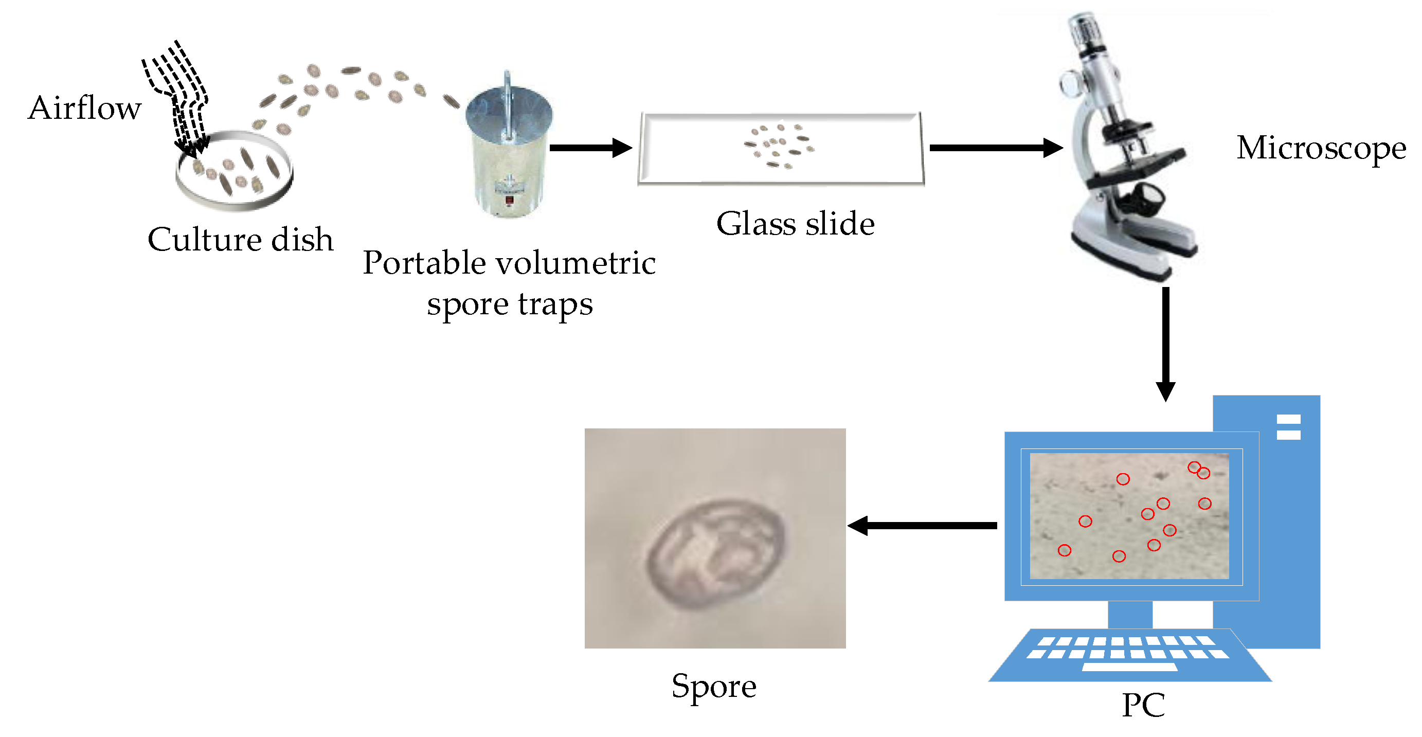
The experiment was conducted in a Venlo-type greenhouse at the Key Laboratory of Modern Agricultural Equipment and Technology of the Ministry of Education of Jiangsu University, China. The collection of airborne disease spores was achieved by simulating a greenhouse environment. A portable volumetric spore trap (OK-BZ1, Zhengzhou Oukeqi Instrument Manufacturing Co., Ltd., Zhengzhou, China) was used to collect the airborne disease spores. First, an aurilave was used to slowly and continuously blow the spores above the trap to spread them into the air. Then, airborne spores were deposited onto microscope slides that were uniformly coated with a thin film of petroleum jelly. The spores on the slides were observed and photographed at the genus level with the aid of a light microscope under × 400 magnification (Nikon TS100-F, made by Nikon Co, Tokyo, Japan). All 600 experimental images of airborne disease spores (200 images of each of spore) were acquired for validation of the proposed algorithm using a digital imaging system with the RGB model. The spore image collection process is shown in Figure 2.

2.3. Preprocessing for Spore Images

In order to avoid the interference of other factors besides the crop disease spores, it was necessary to preprocess the original images of the disease spores. The preprocessing flow is shown in Figure 3. First, the resolution of the images was set to 80 × 80 pixels and the average filter was used to reduce the noise and convert the color images to grayscale. Gaussian filtering was performed on the grayscale images and noise points were removed, then the maximum between-class variance method was used for automatic threshold segmentation. Then, the internal holes of the spore were filled through the expansion and corrosion morphological operations, and other interference targets were removed to obtain the binary image. Finally, the target image was obtained by masking the binary image and the RGB image. Some spore images and their preprocessed images are shown in Figure 4.

2.4. Feature Extraction of Spore Images

The spores of P. cubensis are ovoid or lemon-shaped, with milky protrusions on the top and a light brown color. The spores of P. xanthii are oblong and colorless. The spores of B. cinerea are round or oval in shape and nearly colorless. Combined with the size parameters of the spores of the three diseases shown in Table 1 and the contour shapes of the three spores shown in Figure 4, the color, texture, and shape features of the spore images were extracted in this study to improve the accuracy and effectiveness of the spore identification.

2.4.1. Color Features

The spores of P. cubensis have milky protrusions on the top and are light brown in color. The spores of P. xanthii are colorless. The spores of B. cinerea are nearly colorless. Therefore, in this study, the color features were extracted based on image pixels, which have the advantage of having stable features after rotation, scale, and translation changes. The low-order color moments contained abundant color distribution information. The first moment was used to describe the light and dark information in the image by calculating the mean value of the colors. The second moment was used to describe the standard deviation of the color to reflect the color distribution range of the image. The third moment highlighted the color deviation and was used to describe the symmetry of the color distribution. Therefore, low-order moments were used to extract color features in this study. The HSV (Hue, Saturation, Value) color space model was converted from the RGB (Red, Green, Blue) color space model, which can be used for color expression in the machine vision process. Therefore, the HSV color space model was calculated. However, the V component of the HSV color model is independent of color. In this study, only the color moment features of the H and S components (mean, variance, and skewness) from the HSV color space models of spore images were extracted, giving a total of 6 color features. The calculation is shown in Equations (1)–(3):
μ i = 1 N j = 1 N P i j
σ i = [ 1 N j = 1 N ( P i j μ i ) 2 ] 1 2
ξ i = [ 1 N j = 1 N ( P i j μ i ) 3 ] 1 3
In the equations, μi, σi, and ξi are the mean, variance, and skewness of the color, respectively. Pij is the i-th color channel component of the j-th pixel. N represents the number of pixels in the image.

2.4.2. Shape Features

Shape features were used to describe the shape parameters of objects in order to provide a worthy correlation with human visual perception systems. From Table 1 and Figure 4, it can be seen that the spores of P. cubensis are ovoid or lemon-shaped, with a size range of 21.1–39.8 µm. The spores of P. xanthii are oblong, with a size range of 30.2.1–39.5 µm. The spores of B. cinerea are round or oval in shape, with a size range of 11.4–26.7 µm. The spores of the three diseases have different shapes. In this study, for the seven Hu invariant moments, the roundness and slenderness ratios of the spores were selected as their shape features. The calculation methods for roundness and slenderness are shown in Equations (4) and (5), respectively:
(1)
Roundness ratio
F o r m   f a c t o r = 4 π a r e a p e r i m e t e r 2
In the equation, the area represents the area of the target (total number of pixels). The perimeter represents the perimeter of the target, which is the outermost contour length of the target.
(2)
Slenderness ratio
E i o n g a t e d n e s s = a r e a t h i c k c n e s s 2
In the equation, thickness represents the width of the smallest circumscribed rectangle of the target.

2.4.3. Texture Features

The surface properties of the scene correspond to the image or image area described by texture feature, which is a value calculated from the image. This value quantifies the characteristics of the gray-level change within the image area. Texture includes regular texture and random texture. It can be seen from Figure 4 that the three disease spores have unique texture features, and that the texture features of the spore images are a combination of regular and random textures. Therefore, spores can be identified by extracting their texture features.
At present, the image texture feature extraction methods included grayscale difference statistics, auto-correlation function, gray-level co-occurrence matrix (GLCM), local binary patterns (LBP), and spectrum-based feature analysis methods. However, the most common and most studied texture extraction methods are the statistics-based texture analysis methods. Therefore, in this study, the gray-level co-occurrence matrix and the local binary mode were selected and the two texture features were fused as the texture features of spore images.
(1)
GLCM
Bhunia et al. [22] studied various statistical features in the GLCM and obtained four critical features through experiments—namely the contrast, asm (Angular Second Moment), entropy, and correlation features. The GLCM should be normalized before calculation. The calculation equations are shown in Equations (6)–(10):
P φ , d ( i , j ) = G G L C M ( i , j ) i = 0 N 1 j = 0 N 1 G G L C M ( i , j )
C o n t r a s t = i = 0 N 1 j = 0 N 1 | i j | 2 P φ , d ( i , j )
A s m = i = 0 N 1 j = 0 N 1 P φ , d 2 ( i , j )
E n t r o p y = i = 0 N 1 j = 0 N 1 P φ , d ( i , j ) log 2 P φ , d ( i , j )
C o r r e l a t i o n = i = 0 N 1 j = 0 N 1 [ ( i , j ) P φ , d ( i , j ) μ x μ y ] σ x σ y
In the equation:
μ x = i = 0 N 1 i j = 0 N 1 P φ , d ( i , j )
μ y = j = 0 N 1 j i = 0 N 1 P φ , d ( i , j )
σ x 2 = i = 0 N 1 ( i μ x ) 2 j = 0 N 1 P φ , d ( i , j )
σ y 2 = j = 0 N 1 ( i μ y ) 2 i = 0 N 1 P φ , d ( i , j )
In the experiment, the preprocessed images were converted into 16 gray levels and then four key features of GLCM in 4 directions (0°, 45°, 90°, and 135°) were calculated to obtain a total of 16 feature values.
(2)
LBP
The LBP texture feature description method has the advantages of having simple principles, a small calculation amount, grayscale invariance, and rotation invariance [23]. It uses the difference between the gray value of the center pixel and the adjacent pixel to generate the LBP code. The basic symbols related to LBP were defined as follows: gc represents the gray value of the center point of the local area, gp (p = 0, 1,…, 7) corresponds to the points distributed equally around the center point, and (xc, yc) represents the center point coordinate. The LBP local area texture calculation equation centered on (xc, yc) is shown in Equations (15) and (16):
s ( g p g c ) = { 1 g p g c 0 0 g p g c < 0
L B P ( x c , y c ) = p = 0 7 s ( g p g c ) 2 p
An LBP operator with 8 points in the neighborhood will generate 28 kinds of LBP values. The number of sampling points in the neighborhood determines the dimension of texture features. To solve the problem of having too many binary modes and to improve the statistics, Kou et al. [24] proposed the “equivalent mode” to reduce the dimensions of the mode types of LBP operators. The adjacent element changes from 0 to 1 or 1 to 0 were regarded as transitions. Transition values that do not exceed two during the calculation process are called LBP equivalent modes, of which there are a total of 58 different type, otherwise they are called mixed modes. In this study, this method was used to change the binary mode from the original 256 dimensions to 59 dimensions.

3. Results

3.1. Test Platform and Parameter Settings

In this study, the experimental data included 600 pictures of airborne disease spores, of which 70% were used for training and 30% were used for testing. The low-order color moments contained abundant color distribution information. The HSV color space model was converted from the RGB color space model, which can be used for color expression in machine vision. The V component of the HSV color model is independent of color [25]. In this study, we used the color moment algorithm to extract the mean, variance, and skewness of the H and S color components, giving a total of 6 color features. The surface properties of the scene correspond to the image or image area described by the texture feature [26]. The gray level co-occurrence matrix was used to extract 16 texture features. The LBP algorithm was used to extract 59 texture features. The size range of P. cubensis spores was 21.1–39.8 µm. The size range of P. xanthii spores was 30.2.1–39.5 µm. The size range of B. cinerea spores was 11.4–26.7 µm. Therefore, in this study, the circularity, slenderness ratio, and 7 Hu invariant moment features of the spores were extracted as their shape features. The classification experiment was performed in the MATLAB R2016b environment. The computer system configuration used was as follows: Win10 (64-bit) (Microsoft Corporation, Washington, DC USA), running 16 GB memory(SAMSUNG, Gyeonggi Province, South Korea), with an Intel(R) Core (TM)i5-9400F CPU processor @ 2.90 GHz(Intel Corporation, Santa Clara, CA, USA). When the LR model was used to classify the spores of the three diseases, the instability caused by the possible bias of the sample number was considered and the adopted classification method was one-vs.-one. Some characteristic parameters of the spores of the three diseases intersected (such as the shape parameters). A logistics classifier was adopted and the Sigmoid function was used for the quantization function. In order to avoid overfitting, the LR algorithm chooses L2 as the regular term. When the three disease spores were classified by the KNN model, 70% of the training sets were reclassified considering the number of samples, and the K value of the KNN algorithm was estimated by 10-fold cross-validation. The number of nearest neighbors of the KNN algorithm was set to 4. Euclidean distance was adopted for the distance function. When the RF model was used to classify the spores of the three diseases, in order to achieve the aim of drawing an equal number of samples, the sampling method used was “put back” sampling. The number of trees was set to 100. The minimum sample required to split the internal nodes was set to 2. The minimum number of samples on the leaf node of the RF algorithm was set to 1. The SVM algorithm uses a Gaussian kernel function and the classification method used was one-vs.-one.

3.2. Analysis of Classification Results

3.2.1. Evaluation Index

In the field of machine learning, the confusion matrix is also called the possibility table or error matrix. It is a visualization tool used by a specific matrix to show the performance of the algorithm [27]. In this study, a confusion matrix was used to evaluate the performance of the classifier. As shown in Figure 5, each row of the matrix represents the actual category for the sample, and each column represents the predicted category for the sample. The numbers on the diagonal of the matrix represent the number of samples correctly identified for each category. It can be seen from Figure 5 that 55, 58, and 52 samples were correctly identified from classes 1, 2, and 3. Among them, 5 samples from class 1 were predicted to be class 3. For class 3, 7 samples were predicted to be class 1 and 1 sample was predicted to be class 2. For class 2, 1 sample was predicted to be class 1 and 1 sample was predicted to be class 3. This shows that some spores of the three types of airborne disease spores of greenhouse crops may be similar in color, texture, and shape characteristics. In particular, the three types of airborne disease spores have similar shape characteristics. The results were consistent with the size parameters for the three types of airborne disease spores measured in this study.
In addition, common classification performance metrics are used to evaluate classification models in this study. These classification performance metrics include accuracy, precision, recall, and a comprehensive evaluation index F value (F1-score). The calculation equations are shown in Equations (17)–(20):
A = T P + T N T P + T N + F P + F N
P = T P T P + F P
R = T P T P + F N
F = 2 P R P + R
In the equation, TP (True positive) is used to represent the actual number of samples predicted to be positive. TN (True negative) is used to indicate the number of samples that are actually in the negative class and predicted to be negative. FP (Fasle positive) is used to indicate the number of samples that are actually negative and predicted to be positive. FN (Fasle negative) is used to indicate the number of samples that are actually in the positive class and predicted to be negative. When the classifier performs the classification task, the actual number of disease spores to be predicted is regarded as the number of positive samples, while the sum of the other two types of disease spores is the number of negative samples. The classification results for the support vector machine are shown in Table 2.

3.2.2. Classification Results for Different Models

In this study, color, shape, and texture features were extracted for spore images. The classification models for LR, KNN, RF, and SVM were established. The accuracy rate, recall rate, and comprehensive evaluation index values for the three kinds of spore classifications using different classification models were compared and analyzed and the overall recognition ability of the classifier was summarized. The results of the comparative analysis are shown in Table 3 and Table 4.
From Table 3 and Table 4, it can be seen that the SVM model has better overall classification performance. The average accuracy rate for the spore recognition using the SVM model was 94.36%, while the accuracy rates of the LR, KNN, and RF models were 90.13%, 89.37%, and 89.23%, respectively.
Regarding the recognition of the three types of spores, the SVM model had higher accuracy, recall, and comprehensive evaluation indicators than the three other classification models. Regarding the comparison of the accuracy rates, the accuracy rate of the SVM model for class 1 spores was 87.30%, which was lower than the 89.26% achieved by the LR model and the 88.57% achieved by the RF model. The accuracy rates of SVM model for class 2 was 98.31%, which was lower than that of the LR model (98.21%). However, regarding the comparison of the accuracy rates, the SVM model as a whole reached 91.76%, which was better than the other three classification models.
As a comprehensive evaluation index, the F1-score represents the overall performance of the classifier. Regarding the recognition of the three categories, the F value of the SVM model was higher and the overall average value reached 91.68%, which was 2.03, 3.59, and 3.96 percentage points higher than LR model, KNN model, and RF model, respectively, showing that the SVM model performed well.
As we can see from Table 4, the recognition times for the LR model, KNN model, RF model, and SVM model were 1.1241 s, 1.6473 s, 1.8597 s, and 1.5346 s, respectively. The recognition time for the SVM model was higher than that of the LR model, however it was lower than the KNN model and RF model. The recognition time for spores for the classification model was much lower than the artificial recognition time [21]. If the dataset of the spore images is large, in order to improve the recognition speed, the work station can be used in real-world applications [28].

3.2.3. Recognition Rate of Different Feature Combinations

From Table 1 and Figure 4, it can be seen that each spore has its own color, shape, and texture features. In order to analyze the effects of the color, shape, and texture features on the recognition rate of spores, the classification results for the different feature combinations under the SVM model are shown in Figure 6.
We can see from Figure 6 that the recognition rates for spores based on color; shape; texture; color and shape; color and texture; shape and texture; and fusion features were 45.86%, 79.62%, 71.45%, 82.84%, 75.36%, 86.18%, and 94.36%, respectively. The shape feature had the greatest influence on the recognition of spores. The color feature had the least influence on the recognition of spores. However, the recognition rates for fusion features were higher than those for other feature groups at 48.5, 14.74, 22.91, 11.52, 19, and 8.18 percentage points higher than for color; shape; texture; color and shape; color and texture; and shape and texture feature groups, respectively.

4. Discussion

Cucumbers and tomatoes are the most common vegetables in people’s daily diets and are widely consumed around the world. Tomato (B. cinereal) and cucumber (P. cubensis and P. xanthii) diseases are the most common diseases in greenhouse cultivation. When these diseases are severe, they can cause extensive losses. Crop yield losses can be reduced through disease spore identification and control. In this study, a combination of spore trapping and image processing was used to identify the disease spores.
Each spore has its own shape characteristics based on its perimeter, surface area, number of spines, spine size, maximum and minimum ray radii, aspect ratio, and roundness [29]. The spores of P. cubensis are ovoid or lemon-shaped, with a size range of 21.1–39.8 µm. The spores of P. xanthii are oblong, with a size range of 30.2.1–39.5 µm. The spores of B. cinerea spores are round or oval in shape, with a size range of 11.4–26.7 µm. Therefore, in this study, seven Hu invariant moments and roundness and slenderness ratios were selected as the spore shape features. The spores of P. cubensis are milky on the top and light brown in color. The spores of P. xanthii are colorless. The spores of B. cinerea are nearly colorless. The low-order color moments contained abundant color distribution information. The HSV color space model was converted from the RGB color space model, which can be used for color expression in machine vision [25]. Different color characteristics can be used to distinguish spores [21]. In addition, pollen and spores in the air have unique texture features that can be identified [30]. The characteristics of these spores can provide a basis for the classification of spores. Hence, in this study, to improve the accuracy and effectiveness of spore identification, the color, texture, and shape features of the spore images were extracted.
Analysis was conducted for misclassified spores. Taking the SVM model as an example, this had a classification accuracy rate, precision rate, recall rate, and F1-score of 94.36%, 91.76%, 91.67%, and 91.68%, respectively. Although the classification results meet the requirements, there were still some spores that were not correctly classified. For example, when using the SVM model to classify the spores of the three diseases, 5 B. cinerea spores were mistakenly classified as cucumber downy mold spores, 7 cucumber down mold spores were mistakenly classified as B. cinerea spores, 1 P. cubensis spore was mistakenly classified as a P. xanthii spore, 1 P. xanthii spore was mistakenly classified as a P. cubensis spore, and 1 P. xanthii spore was mistakenly classified as a B. cinerea spore. Analyzing Table 1 and Figure 4, it can be seen that the reason for the misclassification of B. cinerea and cucumber downy mold spores may be that the spores of these two diseases are similar in size and the morphologies of the two spores are similar. The reason for the misclassification of P. xanthii spores may be that some of the spores were in the growth stage when the images were collected, and the morphological characteristics of the growth stage are inconsistent those of the mature stage [31]. The relevant research shows that when the number of spores is small, the accuracy of the manual identification may be high. However, when the number is large, the advantages of machine recognition become apparent (the accuracy of machine recognition is always stable and the recognition time is very short), saving a lot of time and labor [32]. In this study, the average accuracy rate for the recognition of 3 classes of disease spores using the SVM model was 94.36% and the overall average F1-score reached 91.68%. Therefore, this method can effectively identify 3 classes of diseases spores and this study can provide a reference for the identification of greenhouse disease spores.

5. Conclusions

The traditional microscopic spore classification method mainly relies on naked eye observations and classification by professional and technical personnel in a laboratory. This method is labor-intensive and time-consuming and has low efficiency, sometimes leading to huge errors. In order to solve this problem, in this study a multifeature fusion classification method was proposed greenhouse airborne disease spores in order to classify B. cinerea, P. xanthii, and P. cubensis spores. First, disease spores were extracted from greenhouse crops and purified for identification. Second, images of 3 classes of airborne disease spores were collected using portable volumetric spore traps in an indoor simulation environment. Third, the spores were identified using a series of image processing approaches, including mean filtering, gaussian filtering, OTSU method binarization, morphological operations, and mask operation. Then, the color moment algorithm was used to extract weed color features and the gray level co-occurrence matrix and the LBP algorithm were used to extract weed shape features. Additionally, two geometric features, namely the circularity and slenderness ratios, and 7 Hu invariant moment features were extracted. Based on these features, LR, KNN, RF and SVM classification models were built respectively. The test results showed that the average accuracy rates for recognition of the three spores using the SVM model, LR model, KNN model, and RF model were 94.36%, 90.13%, 89.37%, and 89.23%, respectively. The F value of the SVM model was higher and the overall average value reached 91.68%, which was 2.03, 3.59, and 3.96 percentage points higher than the LR model, KNN model, and RF model, respectively.

Author Contributions

Conceptualization, Y.W. and H.M.; methodology, Y.W., X.D., G.M., and Y.L.; data analysis and writing—original draft preparation, Y.W. and B.W.; writing—review and editing, Y.W., X.D., and G.M.; project administration, H.M.; funding acquisition, H.M. All authors have read and agreed to the published version of the manuscript.

Funding

This work was partially supported by the National Natural Science Foundation of China (32071905), the Jiangsu Demonstration Project of Modern Agricultural Machinery Equipment and Technology (NJ2019-19), and the China Agriculture Research System (CARS-23-C03).

Acknowledgments

The author would like to thank the Key Laboratory of Agricultural Engineering in Jiangsu University for supporting the experimental conditions of the research.

Conflicts of Interest

The authors declare no conflict of interest.

References

  1. Wang, T.Y.; Wu, G.X.; Chen, J.W.; Cui, P.; Chen, Z.X.; Yan, Y.Y.; Zhang, Y.; Li, M.C.; Niu, D.X.; Li, B.G. Integration of solar technology to modern greenhouse in China: Current status, challenges and prospect. Renew. Sustain. Energy Rev. 2017, 70, 1178–1188. [Google Scholar] [CrossRef]
  2. Jin, C.; Mao, H.P.; Chen, Y.; Shi, Q.; Wang, Q.R.; Ma, G.X.; Liu, Y. Engineering-Oriented dynamic optimal control of a greenhouse environment using an improved genetic algorithm with engineering constraint rules. Comput. Electron. Agric. 2020, 177, 105698. [Google Scholar] [CrossRef]
  3. Hafez, Y.M.; Attia, K.A.; Kamel, S.; Alamery, S.F.; El-Gendy, S.; Al-Doss, A.A.; Mehiar, F.; Ghazy, A.I.; Ibrahim, E.I.; Abdelaal, K.A.A. Bacillus subtilis as a bio-agent combined with nano molecules can control powdery mildew disease through histochemical and physiobiochemical changes in cucumber plants. Physiol. Mol. Plant Pathol. 2020, 111, 101489. [Google Scholar]
  4. Tanaka, K.; Fukuda, M.; Amaki, Y.; Sakaguchi, T.; Inai, K.; Ishihara, A.; Nakajima, H. Importance of prumycin produced by Bacillus amyloliquefaciens SD-32 in biocontrol against cucumber powdery mildew disease. Pest Manag. Sci. 2017, 73, 2419–2428. [Google Scholar] [CrossRef]
  5. Wallace, E.C.; D’Arcangelo, K.N.; Quesada-Ocampo, L.M. Population analyses reveal two host-adapted clades of Pseudoperonospora cubensis, the causal agent of cucurbit downy mildew, on commercial and wild cucurbits. Phytopathology 2020, 110, 1578–1587. [Google Scholar] [CrossRef] [PubMed]
  6. Choudhury, R.A.; Koike, S.T.; Fox, A.D.; Anchieta, A.; Subbarao, K.V.; Klosterman, S.J.; McRoberts, N. Season-Long dynamics of spinach downy mildew determined by spore trapping and disease incidence. Phytopathology 2016, 106, 1311–1318. [Google Scholar] [CrossRef] [PubMed] [Green Version]
  7. Dhar, N.; Mamo, B.E.; Subbarao, K.V.; Koike, S.T.; Fox, A.; Anchieta, A.; Klosterman, S.J. Measurements of aerial spore load by qPCR facilitates lettuce downy mildew risk advisement. Plant Dis. 2020, 104, 82–93. [Google Scholar] [CrossRef]
  8. Torfs, S.; Van Poucke, K.; Van Campenhout, J.; Ceustermans, A.; Croes, S.; Bylemans, D.; Van Hemelrijck, W.; Keulemans, W.; Heungens, K. Venturia inaequalis trapped: Molecular quantification of airborne inoculum using volumetric and rotating arm samplers. Eur. J. Plant Pathol. 2019, 155, 1319–1332. [Google Scholar] [CrossRef]
  9. Chan, B.D.; Icoz, K.; Huang, W.F.; Chang, C.L.; Savran, C.A. On-Demand weighing of single dry biological particles over a 5-order-of-magnitude dynamic range. Lab Chip 2014, 14, 4188–4196. [Google Scholar] [CrossRef]
  10. Sireesha, Y.; Velazhahan, R. Rapid and specific detection of Peronosclerospora sorghi in maize seeds by conventional and real-time PCR. Eur. J. Plant Pathol. 2017, 150, 521–526. [Google Scholar] [CrossRef]
  11. Bandamaravuri, K.B.; Nayak, A.K.; Bandamaravuri, A.S.; Samad, A. Simultaneous detection of downy mildew and powdery mildew pathogens on Cucumis sativus and other cucurbits using duplex-qPCR and HRM analysis. AMB Express 2020, 10, 135. [Google Scholar] [CrossRef]
  12. Akhmadeev, A.A.; Salakhov, M.K. A new approach of recognition of ellipsoidal micro- and nanoparticles on AFM images and determination of their sizes. Meas. Sci. Technol. 2016, 27, 105402. [Google Scholar] [CrossRef]
  13. Xu, P.F.; Zhang, R.B.; Yang, N.; Oppong, P.K.; Sun, J.; Wang, P. High-Precision extraction and concentration detection of airborne disease microorganisms based on microfluidic chip. Biomicrofluidics 2019, 13, 024110. [Google Scholar] [CrossRef] [PubMed]
  14. Yang, N.; Hu, J.Q.; Zhou, X.; Wang, A.Y.; Yu, J.J.; Tao, X.Y.; Tang, J. A rapid detection method of early spore viability based on AC impedance measurement. J. Food Process Eng. 2020, e13520. [Google Scholar] [CrossRef]
  15. Wang, P.; Yuan, S.Q.; Yang, N.; Wang, A.Y.; Fordjour, A.; Chen, S.B. The collection method for crop fungal spores based on an efficient microfluidic device. Aerosol Air Qual. Res. 2020, 20, 72–79. [Google Scholar] [CrossRef] [Green Version]
  16. Quesada, T.; Hughes, J.; Smith, K.; Shin, K.; James, P.; Smith, J. A Low-Cost spore trap allows collection and real-time PCR quantification of airborne Fusarium circinatum spores. Forests 2018, 9, 586. [Google Scholar] [CrossRef] [Green Version]
  17. Zhu, Y.D.; Zhang, J.Y.; Li, M.Y.; Zhao, L.J.; Ren, H.R.; Yan, L.G.; Zhao, G.M.; Zhu, C.Z. Rapid determination of spore germinability of Clostridium perfringens based on microscopic hyperspectral imaging technology and chemometrics. J. Food Eng. 2020, 280, 109896. [Google Scholar] [CrossRef]
  18. Setlow, P.; Wang, S.W.; Li, Y.Q. Germination of spores of the orders Bacillales and Clostridiales. Annu. Rev. Microbiol. 2017, 71, 459–477. [Google Scholar] [CrossRef]
  19. Lei, Y.; Yao, Z.F.; He, D.J. Automatic detection and counting of urediniospores of Puccinia striiformis f. sp. tritici using spore traps and image processing. Sci. Rep. 2018, 8, 13647. [Google Scholar] [CrossRef] [Green Version]
  20. Wang, D.P.; Wang, B.T.; Yan, Y. The identification of powdery mildew spores image based on the integration of intelligent spore image sequence capture device. In Proceedings of the 2013 Ninth International Conference on Intelligent Information Hiding and Multimedia Signal Processing (Iih-Msp 2013), Beijing, China, 16–18 October 2013; pp. 177–180. [Google Scholar]
  21. Yang, N.; Yu, J.J.; Wang, A.Y.; Tang, J.; Zhang, R.B.; Xie, L.L.; Shu, F.Y.; Kwabena, O.P. A rapid rice blast detection and identification method based on crop disease spores’ diffraction fingerprint texture. J. Sci. Food Agric. 2020, 100, 3608–3621. [Google Scholar] [CrossRef]
  22. Bhunia, A.K.; Bhattacharyya, A.; Banerjee, P. A noval feature descriptor for image retrieval by combining modified color histogram and diagonally symmetric co-occurrence texture pattern. Pattern Anal. Appl. 2020, 2, 703–723. [Google Scholar] [CrossRef] [Green Version]
  23. Liu, L.; Fieguth, P.; Guo, Y.; Wang, X.; Pietikainen, M. Local binary features for texture classification: Taxonomy and experimental study. Pattern Recognit. 2017, 62, 135–160. [Google Scholar] [CrossRef] [Green Version]
  24. Kou, Q.Q.; Cheng, D.Q.; Chen, L.L.; Zhao, K. A Multiresolution gray-scale and rotation invariant descriptor for texture classification. IEEE Access 2018, 6, 30691–30701. [Google Scholar] [CrossRef]
  25. Das, S.; Rudrapal, D. Analysis of color moment as a low level feature in improvement of content based image retrieval. In Proceedings of the Fourth International Conference on Signal and Image Processing 2012 (ICSIP); Springer: New Delhi, India, 2013; pp. 387–397. [Google Scholar]
  26. Yang, H.; Yin, J.; Jiang, M. Perceptual image hashing using latent low-rank representation and uniform LBP. Appl. Sci. 2018, 8, 317. [Google Scholar] [CrossRef] [Green Version]
  27. Vijayaragavan, P.; Ponnusaamy, R.; Aramudhan, M. An optimal support vector machine based classification model for sentimental analysis of online product reviews. Future Gener. Comput. Syst. Int. J. eSci. 2020, 111, 234–240. [Google Scholar] [CrossRef]
  28. Zeng, S.N.; Zhang, B.B.; Du, Y. Joint distances by sparse representation and locality-constrained dictionary learning for robust leaf recognition. Comput. Electron. Agric. 2017, 142, 563–571. [Google Scholar] [CrossRef]
  29. Chesmore, D.; Bernard, T.; Inman, A.J.; Bowyer, R.J. Image analysis for the identification of the quarantine pest Tilletia indica. EPPO Bull. 2003, 33, 495–499. [Google Scholar] [CrossRef]
  30. Marcos, J.V.; Nava, R.; Cristobal, G.; Redondo, R.; Escalante-Ramirez, B.; Bueno, G.; Deniz, O.; Gonzalez-Porto, A.; Pardo, C.; Chung, F.; et al. Automated pollen identification using microscopic imaging and texture analysis. Micron 2015, 68, 36–46. [Google Scholar] [CrossRef] [Green Version]
  31. Lu, Y.; Wang, W.; Huang, M.G.; Ni, X.Z.; Chu, X.; Li, C.Y. Evaluation and classification of five cereal fungi on culture medium using Visible/Near-Infrared (Vis/NIR) hyperspectral imaging. Infrared Phys. Technol. 2020, 105, 103206. [Google Scholar] [CrossRef]
  32. Yang, N.; Qian, Y.; EL-Mesery, H.S.; Zhang, R.B.; Wang, A.Y.; Tang, J. Rapid detection of rice disease using microscopy image identification based on the synergistic judgment of texture and shape features and decision tree-confusion matrix method. J. Sci. Food Agric. 2019, 99, 6589–6600. [Google Scholar] [CrossRef]
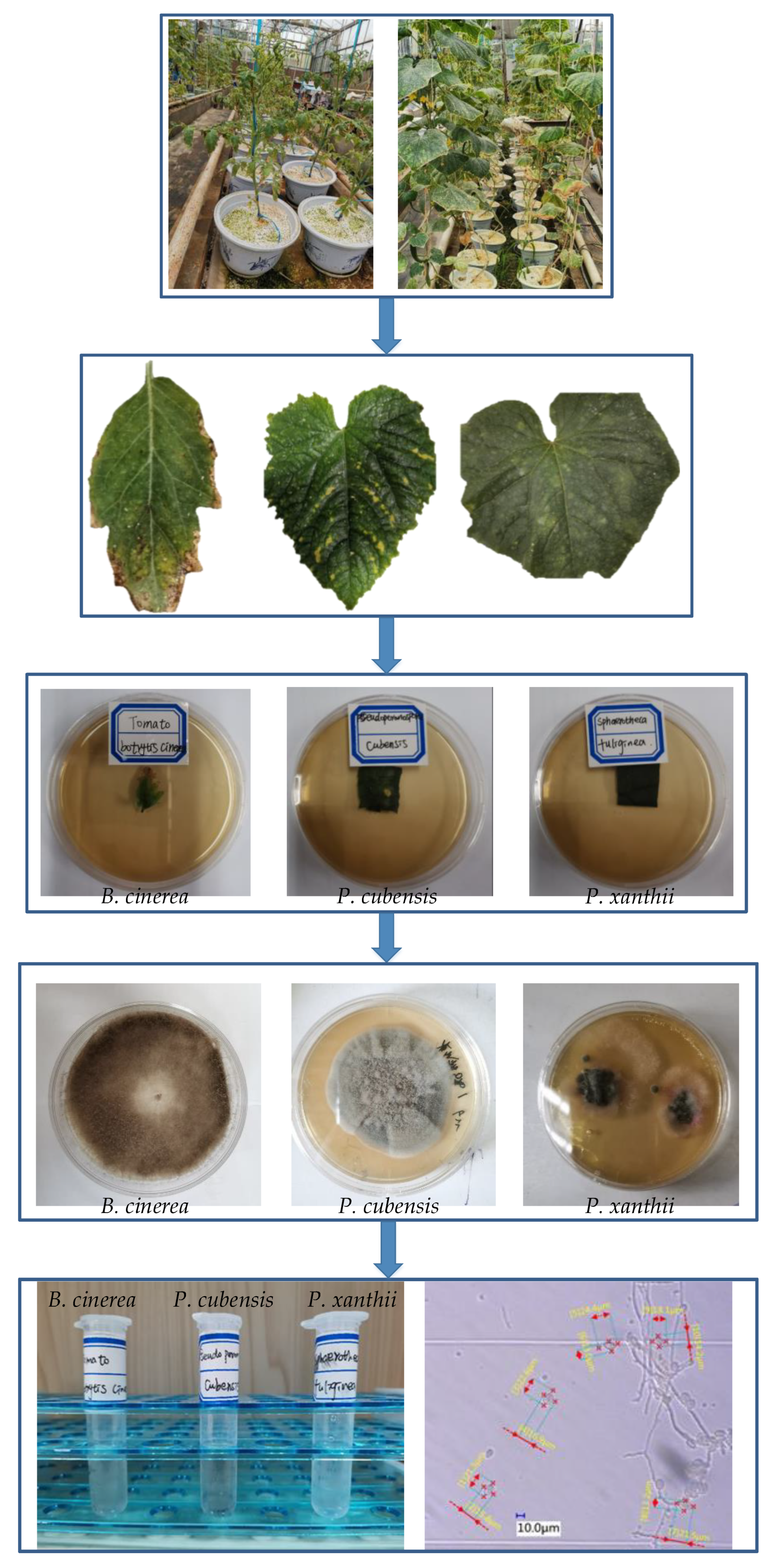
Figure 1. Extraction and identification process for the airborne disease spores.
Applsci 10 07850 g001
Figure 2. Spore image collection.
Applsci 10 07850 g002
Figure 3. Spore image pre-processing.
Applsci 10 07850 g003
Figure 4. Some spore images and their preprocessed images: (a) B. cinerea spores; (b) P. xanthii spores; (c) P. cubensis spores.
Applsci 10 07850 g004
Figure 5. Confusion matrix for the SVM model. Note: Axis number representations: (1) B. cinereal spores; (2) P. xanthii spores; (3) P. cubensis spores.
Applsci 10 07850 g005
Figure 6. Classification results for different feature combinations under the SVM model.
Applsci 10 07850 g006
Table 1. Spore sizes of the airborne diseases from the three greenhouse crops.
SpeciesSpore Size/um
P. xanthii35.4(30.2~39.5) × 14.2(7.3~22.2)
P. cubensis30.6(21.1~39.8) × 20.5(13.8~23.6)
B. cinerea19.3(11.4~26.7) × 11.7(8.3~14.5)
Table 2. Classification results for the SVM.
Sample ClassBasic Indicators
TPTNFPFN
15511085
25810712
35211368
Table 3. Comparative analysis of different classification models (%).
ClassClassification Results
PrecisionRecallF1-Score
LRKNNRFSVMLRKNNRFSVMLRKNNRFSVM
189.2682.4088.5787.3091.3589.1388.1691.6786.5785.6388.3689.43
298.3295.1697.0898.3192.7592.8389.3496.6795.4593.9893.0597.48
387.9485.3583.2889.6685.9783.9880.2686.6786.9486.9481.7488.14
Table 4. Overall average recognition ability comparisons of different classification models.
IndexesClassification Model
LRKNNRFSVM
Accuracy (%)90.1389.3789.2394.36
Precision (%)89.5187.6489.6491.76
Recall (%)90.0288.6585.9291.67
F1-Score (%)89.6588.0987.7291.68
Recognition time (s)1.12411.64731.85971.5346
Publisher’s Note: MDPI stays neutral with regard to jurisdictional claims in published maps and institutional affiliations.