Article

Preparation and Evaluation of Microcapsules Encapsulating Royal Jelly Sieve Residue: Flavor and Release Profile

Department of Food Science and Technology, Zhejiang University of Technology, Hangzhou 310032, China
*
Author to whom correspondence should be addressed.
Appl. Sci. 2020, 10(22), 8126; https://doi.org/10.3390/app10228126
Submission received: 10 October 2020 / Revised: 6 November 2020 / Accepted: 10 November 2020 / Published: 17 November 2020
(This article belongs to the Section Food Science and Technology)

Abstract

:
This study aimed to improve the flavor of royal jelly residue via microencapsulation technology using Arabic gum and gelatin as wall materials. This microencapsulation technology showed a good encapsulation yield of 85.71 ± 2.84% and encapsulation efficiency of 92.34 ± 3.17%. The intact structures of the microcapsules were observed using optical and scanning electron microscopes. The results of the simulated gastrointestinal digestion proved that the microcapsules were well-tolerated in the gastric environment (a release rate of 32.95 ± 2.34%). Both electronic nose and electronic tongue evaluations showed that microencapsulation improved the sensory index of the royal jelly sieve residue. After microencapsulation, the astringency, bitterness, and irritant odors of the royal jelly residue were reduced. Simultaneously, the release rate in the intestine was 98.77 ± 1.91%, which demonstrated that microencapsulation would not prevent the human body from absorbing the royal jelly. The results from this study are expected to facilitate the development of mild flavor products made from royal jelly.

Graphical Abstract

1. Introduction

Royal jelly (RJ) is a thick fluid produced and secreted by nurse bees [1]. Fatty acids constitute 80–85% of RJ’s lipid composition [2] and form the main part of royal jelly sieve residue (RJSR). Trans-10-hydroxy-2-decenoic acid (10-HDA) is found only in RJ in nature [3]; thus, it is often used to determine the product quality index of RJ. 10-HDA is an insoluble medium-chain unsaturated fatty acid that is found as a solid crystal at room temperature; it exerts antibacterial [4] and anti-inflammatory activity [5] and regulates immune cell activity [6]. China is unanimously acknowledged to be the leading world producer and exporter of RJ. Chinese-produced RJ represents over 60% of the RJ produced worldwide and is exported to Japan, the United States, and Europe [2].
Fatty acids in RJ have a spicy and astringent taste and pungent odor [7]. These fatty acids will crystallize at room temperature during storage or processing (10-HDA’s melting point is 52 °C [8]), producing a gritty taste. These unpleasant feelings will remain in the throat for a long time, sometimes leading to vomiting. At the beginning of royal jelly product processing, royal jelly sieve residue (RJSR) is separated from RJ and passed through 125-μm mesh screens, aiming to balance the differences of 10-HDA content in various batches of RJ raw materials.
Methods for the preparation of microcapsules have been widely reported. Complex coacervation and spray drying are the most common methods used in microencapsulation and provide many advantages, such as enhancing the core material stability, isolating the external environment to reduce degradation, controlling the release, and alleviating unpleasant flavors [9,10]. Microencapsulation can also change the gastrointestinal solubility of the embedded material, making it resistant to gastric acid digestion, despite being released in the small intestine [11,12]. In this work, it was necessary to find a type of microcapsule wall material suitable to be used in food. Gelatin (GE) and Arabic gum (AG) were selected because they meet the following conditions: they are non-toxic, environmentally friendly [13], low-priced, and have no obvious unpleasant taste. These two materials have unique emulsification qualities, high water solubility, low viscosity, and low cost, which make them suitable as commercial options [14].
Because of the production standards and biological activity of RJSR, the jelly cannot be directly discarded during production. Thus, developing a technology that can modify the taste of RJ is highly desirable. The objective of the current study was to prepare RJSR microcapsules. The sensory index and in vitro digestibility under the simulated gastrointestinal environment were also investigated. The results will be of great significance to improve the flavor of RJ products and the process of RJ.

2. Materials and Methods

2.1. Materials and Reagents

RJSR with more than 70% 10-HDA was supplied by Zhejiang Jiangshan Health Bee Enterprise Co. Ltd. (Jiangshan, China). GE, AG, and the standard chemicals (10-HDA and methyl 4-hydroxybenzoate) for High performance liquid chromatography (HPLC) were purchased from Shanghai Yuanye Biotechnology Co., Ltd. (Shanghai, China). Enzymes (pepsin, pancreatin, and transglutaminase) were purchased from Sinopharm Chemical Reagent Co. Ltd. (Shanghai, China). HPLC-grade methanol, ethanol, and ethyl acetate and analytically pure hydrochloric acid, NaOH, acetic acid, NaCl, KH2PO4, and anhydrous sodium sulfate were purchased from China National Pharmaceutical Group Co., Ltd. (Beijing, China).

2.2. Pretreatment of RJSR

According to our feasibility test, the particle size of the RJSR was too large and would produce failure of embedding. The RJSR and sodium hydroxide were then mixed according to a mass ratio of 1:0.3, and deionized water was added and dissolved by ultrasound (KQ-400E, Ultrasonic Instrument Co., Ltd., Kunshan, China). Trace insoluble substance was removed via centrifugation (CR21N, Hitachi, Tokyo, Japan) at 4000× g. Then, the pH of the mixture was adjusted to 5.0 using a pH meter (206, Testo, Shanghai, China) with 10% (v/v) acetic acid under stirring at 100 rpm. The pretreated royal jelly sieve residue (PRJSR) was centrifuged at 4000× g, and the precipitation was retained for microencapsulation.

2.3. Preparation of Microcapsules

The process parameters of gelatin Arabic gum microencapsulation were described in [15] and [16]. GE solution (2%, w/w) and AG solution (2%, w/w) were prepared at 40 °C in advance of the preparation of PRJSR. According to the core–wall ratio (0.5:1) and wall material ratio (1:1), the mass ratio of the materials was set as RJSR:GE:AG = 1:1:1. PRJSR was mixed with the GE solution via an Ultra-Turrax (T18 Digital IKA, Staufen, Germany) at 12,000 rpm for 3 min. The mixture was stirred for 15 min in a water bath at 40 °C at 800 rpm using a constant temperature magnetic stirrer (HWCL-5, Greatwall scientific industrial and trade Co.,Ltd., Zhenzhou, China). Next, the AG solution was added using an Ultra-Turrax under the same conditions. The pH was then adjusted to 4.1 with 10% (v/v) acetic acid and stirred for 20 min. The temperature of the mixture was then reduced from 40 to 10 °C by stirring at 200 rpm in another constant temperature magnetic stirrer. Transglutaminase was added at the same time. Finally, the microcapsule powder was obtained by a spray dryer (B-260 mini, BUCHI, Shanghai, China) with the following parameters: The temperature of the inlet was 190 °C, the temperature of the outlet was 70 °C, and the flow rate was 3 mL/min.
The content of 10-HDA was selected as the index to evaluate the microencapsulation efficiency of the RJSR.

2.4. Efficiency of Microencapsulation

The content of 10-HDA was determined by an HPLC system (Waters1525, Waters, Milford, MA, USA), and an XBridge C18 column (5 μm, 4.6 × 250 mm, Waters, Milford, MA, USA) was used for separation. The isocratic mobile phase consisted of 55% methanol and 35% water with 0.03 M hydrochloric acid. The detective wavelength was 210 nm according to a UV detector (Waters 2487, Waters, Milford, MA, USA), the flow rate was 1 mL/min, and the column temperature was 35 °C. Methyl 4-hydroxybenzoate was used as an internal standard. The standard curve was made by plotting the relationship between the peak area ratio and the 10-HDA concentration and internal standard and was used to calculate the content in the microcapsules [17].
To evaluate the core material’s recovery rate and embedding effect, the encapsulation yield (EY) and encapsulation efficiency (EE) were calculated according to the following equations [18,19]:
EE   ( % ) = 1 Surface   10 HDA   amount   in   the   microcapsules Total   10 HDA   amount   in   the   microcapsules × 100 %
EY   ( % ) = Total   10 HDA   amount   in   the   microcapsules 10 HDA   amount   used   in   the   preparation   of   microcapsules × 100 %
Surface 10-HDA was extracted by ethyl acetate, and the total 10-HDA was extracted using sodium hydroxide and ethanol. All extracts were dehydrated with anhydrous sodium sulfate before introducing HPLC and were then filtered through a 0.45-μm membrane.

2.5. Characterization of Microcapsules

2.5.1. Optical Microscope

An optical microscope (IX2-SLP, OLYMPUS, Tokyo, Japan) was used to observe the internal morphology of the wet microcapsules. The wet microcapsules were obtained by a dropper from the liquid before spray drying.

2.5.2. Scanning Electron Microscope (SEM)

To observe the surface conditions of the dry microcapsules, powder was collected for SEM after spray drying. A scanning electron microscope (S-4700Ⅱ, Hitachi, Tokyo, Japan) was used to observe the morphology of the microcapsules, with a voltage acceleration of 15 kV, and the same spray-dried microcapsules were used for particle size determination.

2.5.3. Particle Size

A particle size analyzer (Mastersizer 3000, Malvern, Malvern, UK) was used to determine the sizes of the microcapsules under the dry process, thereby avoiding the influence of wall material swelling on the size.

2.5.4. FTIR Analysis

To demonstrate that PRJSR was packaged in GE and AG, the infrared spectra of GE, AG, PRJSR, Mix., and the microcapsules were analyzed separately by an FTIR spectrometer (Nicolet 6700, Thermo Scientific, Waltham, MA, USA) in the range of 400–4000 cm−1. The microcapsules were then washed with ethyl acetate to remove the PRJSR on the surface and dried again in advance.

2.6. Sensory Evaluation

The electronic nose was composed of different selective gas sensor arrays, signal acquisition units, and suitable pattern recognition software. Traditional food analysis methods based on fingerprints are not based on traditional chemical analysis methods but instead use fingerprints to provide product volatility information. Electronic nose technology remains a research field with great development potential. It provides a fast-analytical method that can be easily combined with chemometrics [20]. To obtain quantitative taste data, the electronic tongue can be used to measure the differences of the taste signals between raw materials and microcapsules based on potentiometric measurements. The sensors interacted with active ingredients possessing specific tastes in an aqueous medium, and then the chemical interaction was converted into an electrical signal by measuring the voltage difference between the reference electrode and the probe [21].
The differences between the microcapsules and Mix. were assessed by sensory evaluation. To avoid a situation where the wal -materials diluted the core material and led to flavor changes, the mixture of GE and AG (marked as the wall materials) was also evaluated.

2.6.1. Electronic Nose

An electronic nose (E-nose) system (PEN 3, Air sense Analytics, Schwerin, Germany) was applied to evaluate the aroma information. The E-nose included an array of 10 different metal oxide sensors, as listed in Table 1, which also shows the main application purposes of the sensors [22]. The samples were accurately weighed (1.0 g), placed in a 15-mL headspace bottle with a sealed cap, and then maintained under static balance at 30 °C for 30 min before testing. The results were analyzed by principal component analysis (PCA) and loading analysis (LA).

2.6.2. Electronic Tongue

An electronic tongue system (SA402B, Insent, Atsugi-Shi, Japan) was used to evaluate the taste traits [23]. All sensors were placed in the reference solution for one day before measurements, and self-inspection, activation, calibration, diagnosis, and other steps were completed as required. Then, a 5 g sample was diluted in a 50-mL volumetric flask with deionized water, and 25 mL of the solution was accurately transferred to the sample cup dedicated to the electronic tongue. The results were analyzed by PCA and LA.

2.7. Kinetics of Release during In Vitro Digestion

The in vitro release behavior of the microcapsules was investigated by employing both simulated gastric (SGF) and intestinal fluid (SIF) models [24,25]. In total, 0.5g NaCl and 4000U pepsin were added to the proper amount of HCl and distilled water to obtain 1 L SGF (pH1.2). Next, 6.8g KH2PO4 and 10 g pancreatin were added to distilled water, adjusting the pH to 6.8, and 1 L SIF was obtained. SGF and SIF were stored at 4 °C before use.
The spray-dried microcapsules were subjected to the simulated gastric phase. The 0.5 g microcapsules were weighed and mixed with 50 mL SGF, and then the mixture was put in a shaker (SKY-111B, Sukun, Shanghai, China) at 120 rpm under 37 °C for 2 h and kept in darkness. Next, the mixture was transferred to 50 mL of SIF and put in a shaker under the same conditions.
F r a c t i o n   r e l e a s e   ( % ) = c t c a × 100 %
where ct is the amount of 10-HDA determined at time t, and ca is the amount of 10-HDA initially encapsulated.

2.8. Statistical Analysis

All the samples were measured in triplicate. The data were analyzed by a one-way analysis of variance, and the results are expressed as the mean value ± standard deviation. PCA and LA were conducted using the SPSS 25 software. Differences were considered significant at p < 0.05.

3. Results and Discussion

3.1. Efficiency of Microencapsulation

The results for the microencapsulation efficiency were as follows: EY = 85.71 ± 2.84% and EE = 92.34 ± 3.17%. This means that most of the RJSR was recycled and this was mostly inside the microcapsules [26]. However, some free RJSR remained outside the microcapsules. This free RJSR was removed before the FTIR analysis.

3.2. Characterization of Microcapsules

3.2.1. Optical Microscope

As shown in Figure 1, GE and AG were used as a shell of globules, and some irregular crystals were wrapped in the spheres. Thanks to the characteristics of the core material, our microcapsules could have their internal structures observed more clearly compared to embedded lipids or probiotics [15,16]. Due to the different RJSR crystals having different growth rates, the size of the RJSR in each microcapsule differed, resulting in a broad size distribution of the microcapsules (Figure 1a). Figure 1b shows that although the shape of the wet capsule was round, a solid rather than a liquid droplet was located inside, thereby affecting the surface morphology of the dried microcapsule. In addition, as shown in Figure 1b, the crystals of RJSR grew and became sharper, preventing the colloid from evenly adhering onto the surface. Therefore, RJSR should be recrystallized to avoid these issues.

3.2.2. SEM

The morphology of the microcapsules was observed by SEM (Figure 2). The microcapsules had complete surfaces with approximately spherical shapes. Some wrinkles were observed on the surfaces of the microcapsules, and a small number of microcapsules had dents on the surface. The spray-dried microcapsules did not show a perfect spherical shape. Although they were spherical in an aqueous solution, embedded oil microcapsules [16,27] were more likely to remain spherical after drying, while microcapsules with irregular core materials, such as probiotics [15] or PRJSR, tended to shrink. The reason for this phenomenon is that PRJSR is an irregular solid crystal, but this feature also ensured that the microcapsules would not be excessively wrinkled and shrunken. However, there were hardly any holes on the microcapsule surfaces. This was due to the proper ratio of PRJSR:GE:AG. If the concentration of the outer shell material was too high, the microcapsules might have become irregular or adhesive, resulting in damage after spray drying [27]. The unbroken surfaces indicate that these wall materials could offer protection against leakage [28].
These observations confirmed that the microcapsules were successfully prepared through the complex coacervation and spray drying process.

3.2.3. Particle Size

Based on Figure 3, the particle sizes of spray-dried microcapsules were between 1 and 100 μm, and most of them were located at 10–30 μm. The polymer disperse index (PDI) was 0.272, indicating that the particle size distribution was relatively uniform. Colloidal particles in this size range tended to show softness [29] and smoothness [30]. The difference in particle size could be due to the number and volume of the cores in the multicore microcapsules.

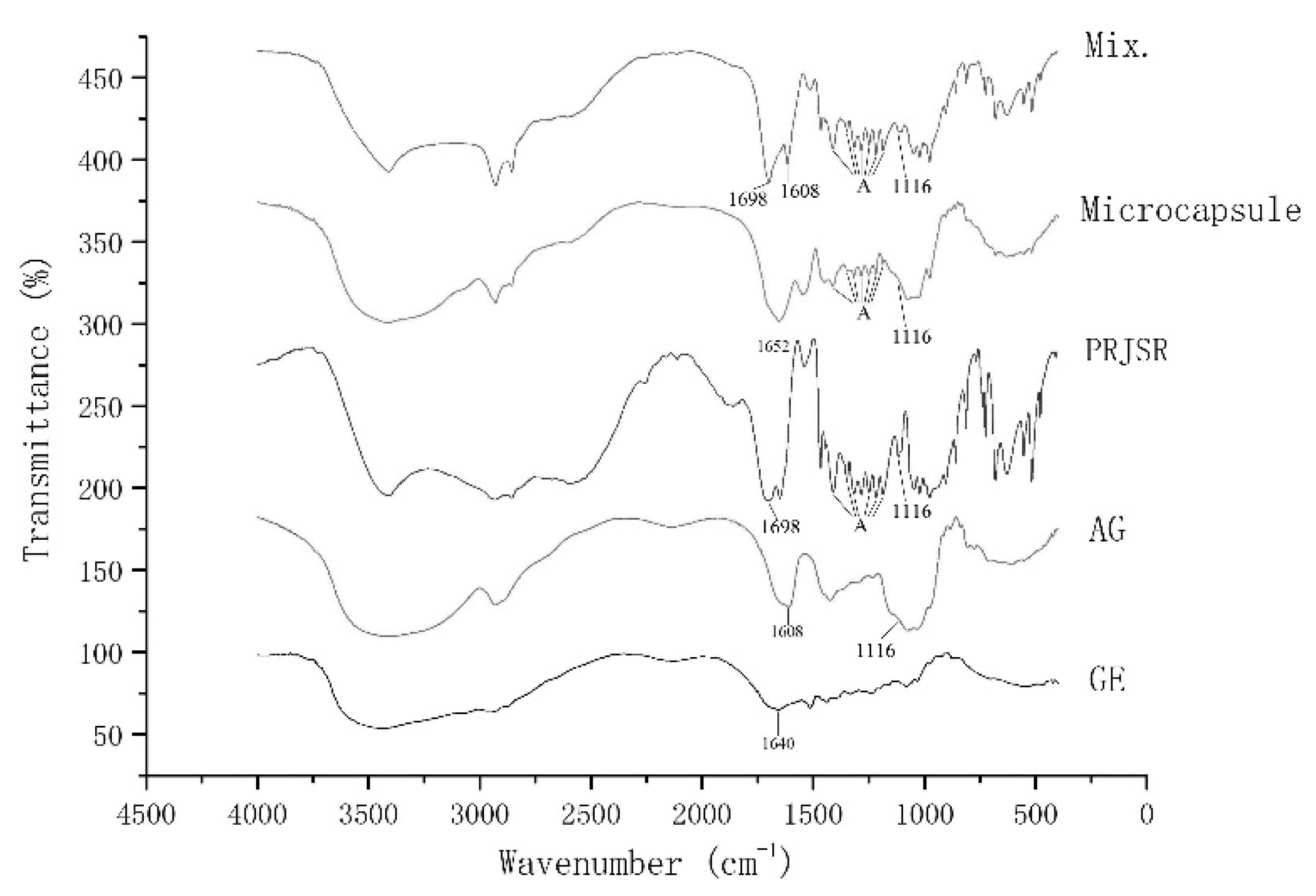
3.2.4. FTIR Analysis

The FTIR spectra were shown in Figure 4. In the region below 1116 cm−1, the microcapsules revealed the same characteristic peaks as AG, while the Mix. was similar to PRJSR. A peak at 1698 cm−1 due to COOH stretching [31,32] only appeared in PRJSR and Mix. but not in the microcapsules. The group peaks (marked as A in Figure 4) only appeared in PRJSR, Mix., and microcapsules, indicating the characteristic peaks of PRJSR. The peak at 1640 cm−1 in GE corresponded to NH3+, and the peak at 1608 cm−1 in AG corresponded to COO. However, in the microcapsules, there was a peak at 1652 cm−1, which confirmed that these two groups stimulated interactions [26,33,34]. The results confirmed that PRJSR was successfully embedded by GE and AG. In PRJSR, a peak at 1608 cm−1 also appeared, which might indicate that some fatty acids still existed in the form of sodium salt.

3.3. Sensory Evaluation

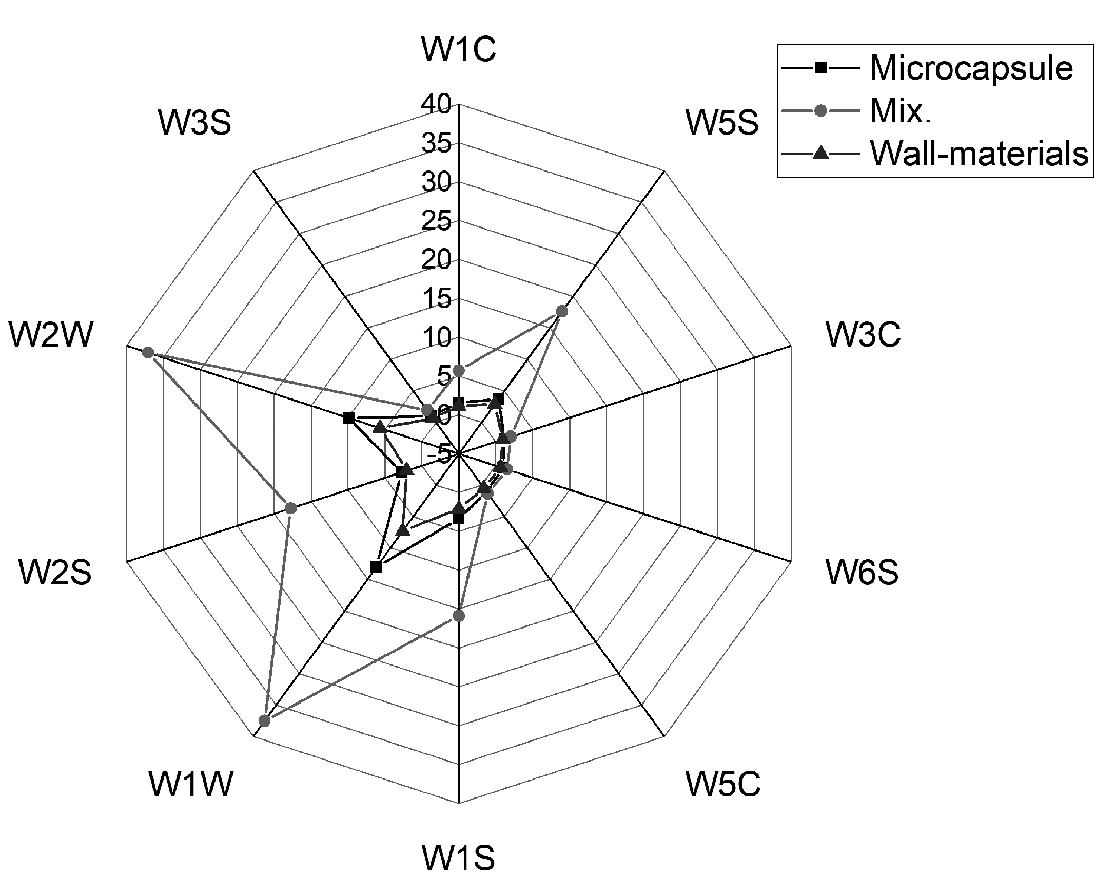
3.3.1. Electronic Nose

According to Figure 5, the response values of the two samples in W1C, W3S, W3C, W6S, and W5C were very small (less than 5), indicating that the corresponding odors of the four sensors were very light. Sensors W1W, W2W, W2S, and W5S showed a relatively strong response value, indicating that the similar odors of nitrogen oxides, sulfides, and alcohols corresponding to those sensors were relatively strong in RJSR [35]. However, the response value of microcapsules (W1W 12.88 ± 0.43, W2W 9.72 ± 0.31, W2S 2.71 ± 0.02, and W5S 3.54 ± 0.22) was significantly smaller than that of the mixture (W1W 36.76 ± 0.75, W2W 36.35 ± 0.64, W2S 17.43 ± 0.92, and W5S 17.43 ± 0.21); thus, microencapsulation was able to mask these odors well. This was also demonstrated in LA (Figure 6a). The distribution of sensors W1C, W3S, W3C, W6S, and W5C was close to the coordinate zero point, indicating that their contribution to PC1 and PC2 was very small. On the contrary, W1W and W2W greatly contributed to LA1 (contribution > 0.5), while W1S and W2S greatly contributed to LA2 (contribution > 0.4). The contribution variances of PC1 and PC2 for direct electronic nose measurement (Figure 6b) were 97.2 and 2.6%, adding up to 99.8%. This means that the first two PCs were sufficient to explain the total variance of the datasets. In addition, as shown in Figure 6b, there was no overlap between the Mix. and the microcapsules. This selective microencapsulation phenomenon was also observed by Ren et al. [10] and Zhu et al. [36], demonstrating that these two samples had differences in odor. The above results demonstrate that the odor of RJSR was successfully lightened.

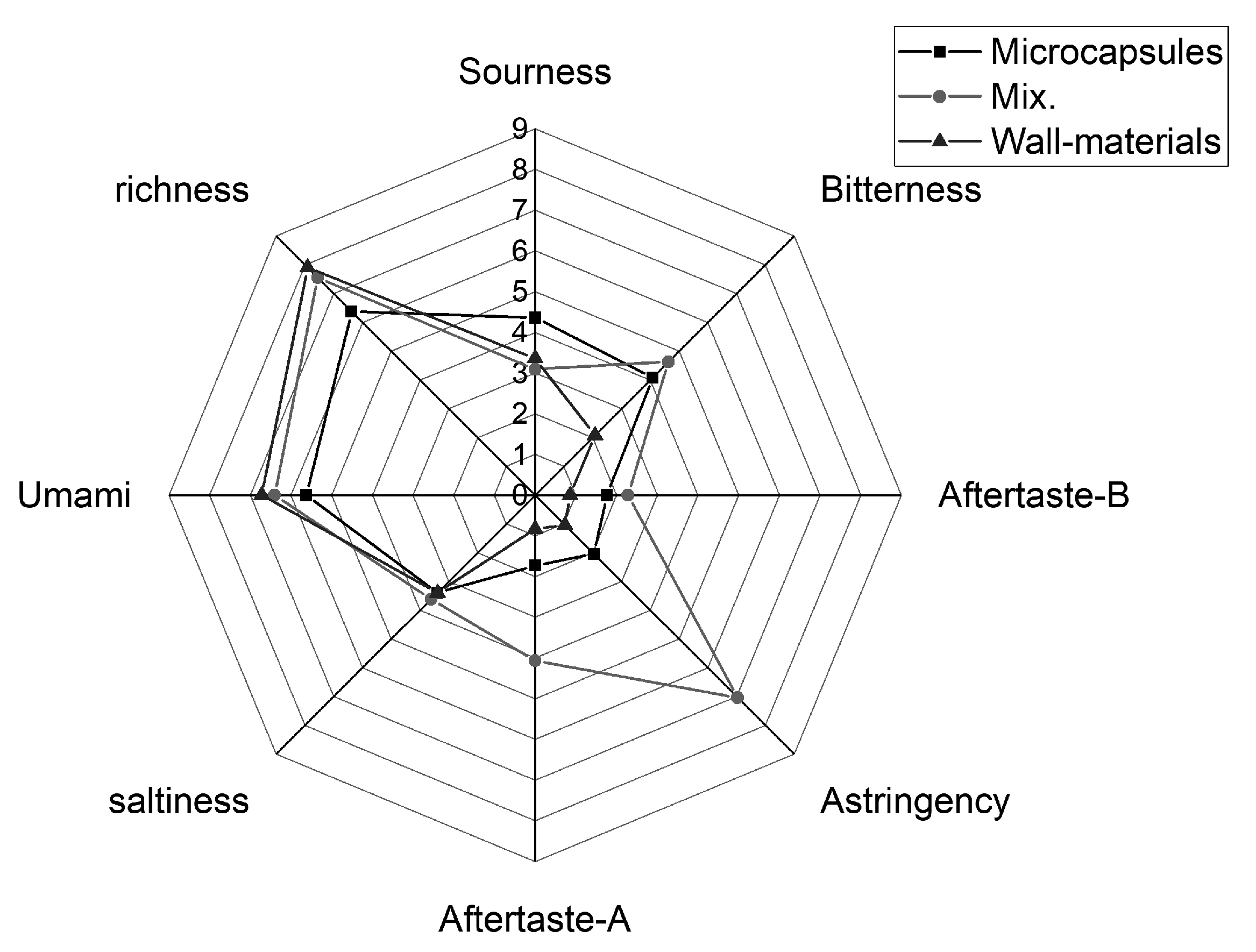
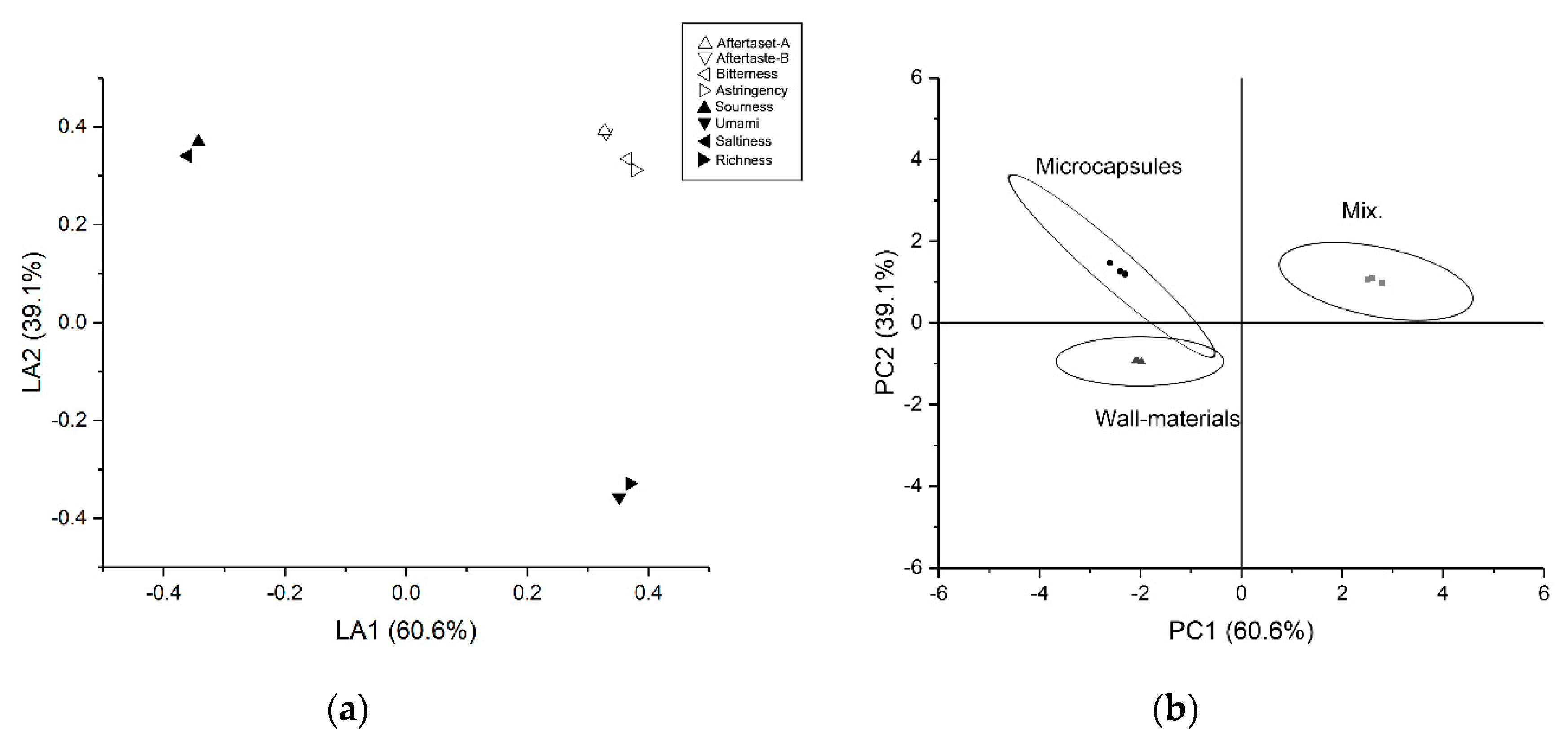
3.3.2. Electronic Tongue

The electronic tongue was used to evaluate the basic tastes, including umami, saltiness, acidity, bitterness, and astringency, while further observing the richness, astringent aftertaste (aftertaste-A), and bitter aftertaste (aftertaste-B) [23].
As shown in Figure 7, the response values of the microcapsule (astringency 1.88 ± 0.16 and aftertaste-A 1.77 ± 0.04) and mixture (astringency 7.13 ± 0.10 and aftertaste-A 4.07 ± 0.05) showed a difference in astringency and aftertaste-A. This indicates that the astringency of RJSR was well-masked. However, the difference between the response values of bitter and aftertaste-B was not as significant (microcapsule bitterness 3.58 ± 0.03 and aftertaste-B 1.77 ± 0.02; mixture bitterness 5.70 ± 0.09 and aftertaste-B 3.16 ± 0.12), indicating that the bitter substances in RJSR might have relatively good water solubility, leading to their location on the surfaces of the microcapsules. The difference between sour and salty might have been caused by the acid and salt introduced during microencapsulation. As shown in Figure 8a, astringency, aftertaste-A, bitterness, and aftertaste-B contributed greatly to both LA1 and LA2 (both >0.3), while the contribution rates of the other indicators were relatively small (one item or both <0). The total contribution variances of PC1 and PC2 for direct electronic nose measurement (Figure 8b) were 60.6 and 39.1%, adding up to 99.7%. This also means that the first two PCs were sufficient to explain the total variance of the datasets. The greater the degree of dispersion was between the data points, the greater the differences among the samples [37]. In addition, as shown in Figure 8b, there was no overlap between the Mix. and the microcapsules. This selective microencapsulation phenomenon was also observed by Yi et al. [21], demonstrating that these two samples had differences in taste. The above results confirm that the unpleasant flavor of RJSR was successfully altered.

3.4. Kinetics of Release during In Vitro Digestion

To investigate the potential application of microcapsules containing 10-HDA in practical delivery [38], in vitro release was carried out, and the results are shown in Figure 9. During SGF, a rapid release was observed during the first 30 min, possibly due to unembedded 10-HDA. However, over the 2-h exposure period, the release rate eventually stabilized to 32.95 ± 2.34%. In the stomach environment, GE and AG had opposite charges, and electrostatic attraction prevented the microcapsules from disintegrating. However, this force disappeared in the intestinal environment because both GE and AG were negatively charged [12]. Due to the hydrolysis of pepsin, the release rate of pepsin began to rise after 90 min. Under exposure to SIF, the release rate increased rapidly. The electrostatic interaction between GE and AG changed due to the intestinal pH, leading to disintegration of the microcapsules. Trypsin specifically hydrolyzed hydrophilic amino acid residues, which accelerated the disintegration of the microcapsules [39]. The microencapsulated 10-HDA was released almost completely (98.77 ± 1.91%) in 240 min, which would be conducive to absorption in the intestine.
The in vitro digestion experiments showed that the final release rate in the gastric phase was only about 30%, with a more rapid release observed in the intestinal phase, suggesting that the microencapsulation of RJSR provided better enteric-coated properties [40].

4. Conclusions

Royal jelly sieve residue was successfully microencapsulated through complex coacervation and spray drying using gelatin and Arabic gum as the wall materials. This microcapsule technology offered a good encapsulation yield (85.71 ± 2.84%) and encapsulation efficiency (92.34 ± 3.17%). Additionally, the RJSR microcapsule had a small mean particle size and a more uniform particle size distribution (10–30 μm). The microcapsules and RJSR had significant differences in their sensory values. In terms of taste, astringency and bitterness were reduced. In terms of smell, pungent odor was reduced. In the in vitro digestion experiment, the stomach and small intestine environment showed different release rates, indicating that the microcapsules have gastric environmental tolerance and enteric solubility. Royal jelly is a functional food limited by its unpleasant flavor. The results of this study are expected to help develop more royal jelly products in the field of food processing. In addition, this study proved that a GE–AG system could be used to embed not only liquid oil or bacteria but also lipophilic solids.

Author Contributions

Conceptualization, R.H.; Methodology, R.H.; Validation, J.Y.; Formal analysis, J.Y.; Investigation, R.H.; Resources, R.H.; Data curation, J.Y.; Writing—original draft preparation, J.Y.; Writing—review and editing, R.H. and L.W.; Visualization, J.Y.; Supervision, P.S.; Project administration, R.H.; Funding acquisition, R.H. and P.S. All authors have read and agreed to the published version of the manuscript.

Funding

This study was supported by Key Research and Development Project of Zhejiang Province, China (No. 2018C02045).

Conflicts of Interest

The authors declare no conflict of interest.

Abbreviations

RJRoyal jelly
RJSRroyal jelly sieve residue
PRJSRpretreated royal jelly sieve residue
GEgelatin
AGArabic gum
Mix.the mixture of GE, AG and PRJSR
PCAprincipal component analysis
LAloading analysis

References

  1. Ramanathan, A.N.K.G.; Nair, A.J.; Sugunan, V.S. A review on Royal Jelly proteins and peptides. J. Funct. Foods 2018, 44, 255–264. [Google Scholar] [CrossRef]
  2. Ramadan, M.F.; Al-Ghamdi, A. Bioactive compounds and health-promoting properties of royal jelly: A review. J. Funct. Foods 2012, 4, 39–52. [Google Scholar] [CrossRef]
  3. Isidorov, V.A.; Bakier, S.; Grzech, I. Gas chromatographic–mass spectrometric investigation of volatile and extractable compounds of crude royal jelly. J. Chromatogr. B 2012, 885–886, 109–116. [Google Scholar] [CrossRef] [PubMed]
  4. Fratini, F.; Cilia, G.; Mancini, S.; Felicioli, A. Royal Jelly: An ancient remedy with remarkable antibacterial properties. Microbiol. Res. 2016, 192, 130–141. [Google Scholar] [CrossRef] [PubMed]
  5. Chen, Y.F.; You, M.M.; Liu, Y.C.; Shi, Y.Z.; Wang, K.; Lu, Y.Y.; Hu, F.L. Potential protective effect of Trans-10-hydroxy-2-decenoic acid on the inflammation induced by Lipoteichoic acid. J. Funct. Foods 2018, 45, 491–498. [Google Scholar] [CrossRef]
  6. Mihajlovic, D.; Rajkovic, I.; Chinou, I.; Colic, M. Dose-dependent immunomodulatory effects of 10-hydroxy-2-decenoic acid on human monocyte-derived dendritic cells. J. Funct. Foods 2013, 5, 838–846. [Google Scholar] [CrossRef]
  7. Watanabe, T.; Terada, Y. Food Compounds Activating Thermosensitive TRP Channels in Asian Herbal and Medicinal Foods. J. Nutr. Sci. Vitam. 2015, 61, S86–S88. [Google Scholar] [CrossRef]
  8. Barker, S.A.; Foster, A.B.; Lamb, D.C.; Hodgson, N. Identification of 10-hydroxy-delta 2-decenoic acid in royal jelly. Nature 1959, 183, 996–997. [Google Scholar] [CrossRef]
  9. Costa, A.M.M.; Moretti, L.K.; Simões, G.; Silva, K.A.; Calado, V.; Tonon, R.V.; Torres, A.G. Microencapsulation of pomegranate (Punica granatum L.) seed oil by complex coacervation: Development of a potential functional ingredient for food application. LWT 2020, 131, 109519. [Google Scholar] [CrossRef]
  10. Ren, W.; Tian, G.; Zhao, S.; Yang, Y.; Gao, W.; Zhao, C.; Zhang, H.; Lian, Y.; Wang, F.; Du, H.; et al. Effects of spray-drying temperature on the physicochemical properties and polymethoxyflavone loading efficiency of citrus oil microcapsules. LWT 2020, 133, 109954. [Google Scholar] [CrossRef]
  11. Quan, J.; Kim, S.M.; Pan, C.H.; Chung, D. Characterization of fucoxanthin-loaded microspheres composed of cetyl palmitate-based solid lipid core and fish gelatin–gum arabic coacervate shell. Food Res. Int. 2013, 50, 31–37. [Google Scholar] [CrossRef]
  12. De Almeida Paula, D.; Martins, E.M.F.; de Almeida Costa, N.; de Oliveira, P.M.; de Oliveira, E.B.; Ramos, A.M. Use of gelatin and gum arabic for microencapsulation of probiotic cells from Lactobacillus plantarum by a dual process combining double emulsification followed by complex coacervation. Int. J. Biol. Macromol. 2019, 133, 722–731. [Google Scholar] [CrossRef] [PubMed]
  13. Yang, X.; Gao, N.; Hu, L.; Li, J.; Sun, Y. Development and evaluation of novel microcapsules containing poppy-seed oil using complex coacervation. J. Food Eng. 2015, 161, 87–93. [Google Scholar] [CrossRef]
  14. Shaddel, R.; Hesari, J.; Azadmard-Damirchi, S.; Hamishehkar, H.; Fathi-Achachlouei, B.; Huang, Q. Use of gelatin and gum Arabic for encapsulation of black raspberry anthocyanins by complex coacervation. Int. J. Biol. Macromol. 2018, 107, 1800–1810. [Google Scholar] [CrossRef]
  15. Da Silva, T.M.; Jacob Lopes, E.; Codevilla, C.F.; Cichoski, A.J.; de Moraes Flores, É.M.; Motta, M.H.; de Bona da Silva, C.; Grosso, C.R.F.; de Menezes, C.R. Development and characterization of microcapsules containing Bifidobacterium Bb-12 produced by complex coacervation followed by freeze drying. LWT 2018, 90, 412–417. [Google Scholar] [CrossRef]
  16. Li, Y.; Dou, X.; Pang, J.; Liang, M.; Feng, C.; Kong, M.; Liu, Y.; Cheng, X.; Wang, Y.; Chen, X. Improvement of fucoxanthin oral efficacy via vehicles based on gum Arabic, gelatin and alginate hydrogel: Delivery system for oral efficacy enhancement of functional food ingredients. J. Funct. Foods 2019, 63, 103573. [Google Scholar] [CrossRef]
  17. Tu, X.; Sun, F.; Wu, S.; Liu, W.; Gao, Z.; Huang, S.; Chen, W. Comparison of salting-out and sugaring-out liquid–liquid extraction methods for the partition of 10-hydroxy-2-decenoic acid in royal jelly and their co-extracted protein content. J. Chromatogr. B 2018, 1073, 90–95. [Google Scholar] [CrossRef]
  18. Pitigraisorn, P.; Srichaisupakit, K.; Wongpadungkiat, N.; Wongsasulak, S. Encapsulation of Lactobacillus acidophilus in moist-heat-resistant multilayered microcapsules. J. Food Eng. 2017, 192, 11–18. [Google Scholar] [CrossRef]
  19. Du, Y.L.; Huang, G.Q.; Wang, H.O.; Xiao, J.X. Effect of high coacervation temperature on the physicochemical properties of resultant microcapsules through induction of Maillard reaction between soybean protein isolate and chitosan. J. Food Eng. 2018, 234, 91–97. [Google Scholar] [CrossRef]
  20. Benedetti, S.; Drusch, S.; Mannino, S. Monitoring of autoxidation in LCPUFA-enriched lipid microparticles by electronic nose and SPME-GCMS. Talanta 2009, 78, 1266–1271. [Google Scholar] [CrossRef]
  21. Yi, E.-J.; Kim, J.-Y.; Rhee, Y.-S.; Kim, S.-H.; Lee, H.-J.; Park, C.-W.; Park, E.-S. Preparation of sildenafil citrate microcapsules and in vitro/in vivo evaluation of taste masking efficiency. Int. J. Pharm. 2014, 466, 286–295. [Google Scholar] [CrossRef] [PubMed]
  22. Xu, M.; Wang, J.; Gu, S. Rapid identification of tea quality by E-nose and computer vision combining with a synergetic data fusion strategy. J. Food Eng. 2019, 241, 10–17. [Google Scholar] [CrossRef]
  23. Ismail, I.; Hwang, Y.H.; Joo, S.T. Low-temperature and long-time heating regimes on non-volatile compound and taste traits of beef assessed by the electronic tongue system. Food Chem. 2020, 320, 126656. [Google Scholar] [CrossRef] [PubMed]
  24. Chew, S.C.; Tan, C.P.; Nyam, K.L. In-vitro digestion of refined kenaf seed oil microencapsulated in β-cyclodextrin/gum arabic/sodium caseinate by spray drying. J. Food Eng. 2018, 225, 34–41. [Google Scholar] [CrossRef]
  25. Ryu, D.; Koh, E. Stability of anthocyanins in bokbunja (Rubus occidentalis L.) under in vitro gastrointestinal digestion. Food Chem. 2018, 267, 157–162. [Google Scholar] [CrossRef] [PubMed]
  26. Shaddel, R.; Hesari, J.; Azadmard-Damirchi, S.; Hamishehkar, H.; Fathi-Achachlouei, B.; Huang, Q. Double emulsion followed by complex coacervation as a promising method for protection of black raspberry anthocyanins. Food Hydrocoll. 2018, 77, 803–816. [Google Scholar] [CrossRef]
  27. Samakradhamrongthai, R.S.; Thakeow Angeli, P.; Kopermsub, P.; Utama-ang, N. Optimization of gelatin and gum arabic capsule infused with pandan flavor for multi-core flavor powder encapsulation. Carbohydr. Polym. 2019, 226, 115262. [Google Scholar] [CrossRef]
  28. Pham, L.B.; Wang, B.; Zisu, B.; Truong, T.; Adhikari, B. Microencapsulation of flaxseed oil using polyphenol-adducted flaxseed protein isolate-flaxseed gum complex coacervates. Food Hydrocoll. 2020, 107, 105944. [Google Scholar] [CrossRef]
  29. Shewan, H.M.; Stokes, J.R.; Smyth, H.E. Influence of particle modulus (softness) and matrix rheology on the sensory experience of ‘grittiness’ and ‘smoothness’. Food Hydrocoll. 2020, 103, 105662. [Google Scholar] [CrossRef]
  30. Hossain, M.K.; Keidel, J.; Hensel, O.; Diakité, M. The impact of extruded microparticulated whey proteins in reduced-fat, plain-type stirred yogurt: Characterization of physicochemical and sensory properties. LWT 2020, 134, 109976. [Google Scholar] [CrossRef]
  31. Gao, X.; Zhang, Y.; Liu, Y. Temperature-dependent hygroscopic behaviors of atmospherically relevant water-soluble carboxylic acid salts studied by ATR-FTIR spectroscopy. Atmos. Environ. 2018, 191, 312–319. [Google Scholar] [CrossRef]
  32. Lu, R.; Mori, S.; Tani, H.; Tagawa, N.; Koganezawa, S. Low friction properties of associated carboxylic acids induced by molecular orientation. Tribol. Int. 2017, 113, 36–42. [Google Scholar] [CrossRef]
  33. Li, Y.; Zhang, X.; Zhao, Y.; Ding, J.; Lin, S. Investigation on complex coacervation between fish skin gelatin from cold-water fish and gum arabic: Phase behavior, thermodynamic, and structural properties. Food Res. Int. 2018, 107, 596–604. [Google Scholar] [CrossRef] [PubMed]
  34. Rousi, Z.; Malhiac, C.; Fatouros, D.G.; Paraskevopoulou, A. Complex coacervates formation between gelatin and gum Arabic with different arabinogalactan protein fraction content and their characterization. Food Hydrocoll. 2019, 96, 577–588. [Google Scholar] [CrossRef]
  35. Gu, S.Q.; Wang, X.C.; Tao, N.P.; Wu, N. Characterization of volatile compounds in different edible parts of steamed Chinese mitten crab (Eriocheir sinensis). Food Res. Int. 2013, 54, 81–92. [Google Scholar] [CrossRef]
  36. Zhu, H.; Zhang, Y.; Tian, J.; Chu, Z. Effect of a new shell material—Jackfruit seed starch on novel flavor microcapsules containing vanilla oil. Ind. Crop. Prod. 2018, 112, 47–52. [Google Scholar] [CrossRef]
  37. Zhu, D.; Ren, X.; Wei, L.; Cao, X.; Ge, Y.; Liu, H.; Li, J. Collaborative analysis on difference of apple fruits flavour using electronic nose and electronic tongue. Sci. Hortic. 2020, 260, 108879. [Google Scholar] [CrossRef]
  38. Wang, L.; Zhao, H.; Brennan, M.; Guan, W.; Liu, J.; Wang, M.; Wen, X.; He, J.; Brennan, C. In vitro gastric digestion antioxidant and cellular radical scavenging activities of wheat-shiitake noodles. Food Chem. 2020, 330, 127214. [Google Scholar] [CrossRef]
  39. Mao, Y.; Krischke, M.; Hengst, C.; Kulozik, U. Comparison of the influence of pH on the selectivity of free and immobilized trypsin for β-lactoglobulin hydrolysis. Food Chem. 2018, 253, 194–202. [Google Scholar] [CrossRef]
  40. Souza, C.J.F.; Comunian, T.A.; Kasemodel, M.G.C.; Favaro-Trindade, C.S. Microencapsulation of lactase by W/O/W emulsion followed by complex coacervation: Effects of enzyme source, addition of potassium and core to shell ratio on encapsulation efficiency, stability and kinetics of release. Food Res. Int. Int. 2019, 121, 754–764. [Google Scholar] [CrossRef]
Figure 1. Wet microcapsules under an optical microscope: (a) microcapsules contain small crystals, (b) Microcapsules contain large crystals.
Applsci 10 08126 g001
Figure 2. Scanning Electron Microscope (SEM) images of the spray-dried microcapsules.
Applsci 10 08126 g002
Figure 3. Particle size of the spray-dried microcapsules.
Applsci 10 08126 g003
Figure 4. FTIR spectra of the microcapsules and their materials.
Applsci 10 08126 g004
Figure 5. Radar image of the electronic nose results.
Applsci 10 08126 g005
Figure 6. Loading analysis (LA) and principal component analysis (PCA) results for the electronic nose: (a) LA and (b) PCA.
Applsci 10 08126 g006
Figure 7. Radar image of electronic tongue results.
Applsci 10 08126 g007
Figure 8. LA and PCA results for the electronic tongue: (a) LA and (b) PCA.
Applsci 10 08126 g008
Figure 9. Release rate of microcapsules under in vitro digestion.
Applsci 10 08126 g009
Table 1. Sensors used in PEN 3.
NumberNameMain PerformanceReference
R1W1CAromatic compoundsToluene, 10 mg/L
R2W5SVery sensitive, broad range sensitivity, reacts to nitrogen oxides, sensitive with a negative signalNO2, l mg/L
R3W3CAmmonia, used as a sensor for aromatic compoundsBenzene, 19 mg/L
R4W6SMainly hydrogen, selectivelyH2, 100 mg/L
R5W5CAlkenes, aromatic compounds, fewer polar compoundsPropane, 100 mg/L
R6W1SSensitive to methaneCH4, 100 mg/L
R7W1WReacts to sulfur compounds. Otherwise, sensitive to many terpenes and sulfur organic compounds, which are important for smell, limonene, pyrazineH2S, l mg/L
R8W2SDetects alcohols, partially aromatic compounds, broad rangeCO, 100 mg/L
R9W2WAromatic compounds, sulfur organic compoundsH2S, l mg/L
R10W3SLong chain alkanesCH3, 100 mg/L
Publisher’s Note: MDPI stays neutral with regard to jurisdictional claims in published maps and institutional affiliations.

