Article

Evaluation of Sub-Lethal Toxicity of Benzethonium Chloride in Cyprinus carpio Liver

1
National Research and Development Institute for Industrial Ecology—ECOIND, 71-73 Drumul Podu Dambovitei, 060652 Bucharest, Romania
2
Department of Biochemistry and Molecular Biology, University of Bucharest, 91–95 Spl. Independentei, 050095 Bucharest, Romania
3
David Geffen School of Medicine, University of California, Los Angeles, CA 90002, USA
*
Authors to whom correspondence should be addressed.
Appl. Sci. 2020, 10(23), 8485; https://doi.org/10.3390/app10238485
Submission received: 20 October 2020 / Revised: 21 November 2020 / Accepted: 25 November 2020 / Published: 27 November 2020
(This article belongs to the Special Issue Emerging Effects of Pollutants in the Aquatic Environment)

Abstract

:
Benzenthonium chloride (BEC, Hyamine 1622) is a quaternary ammonium surfactant with cationic properties widely used in cleaning, sanitation, and medical products that can become harmful to humans and also to the environment. This study aimed to evaluate its acute effects on Cyprinus carpio fish in terms of oxidative stress and morphological changes on hepatic tissue in order to show the sub-lethal toxicity of BEC. Fish were exposed to 1 mg/L BEC for 24, 48, and 96 h, and the liver samples were collected. The most significant changes were noticed after 96 h of exposure when the entire antioxidant enzyme system was affected. The activities of catalase, glutathione peroxidase, glutathione reductase, and glutathione S-transferase decreased by 44%, 31%, 30%, and 45%, respectively, compared to control. Glucose-6-phosphate dehydrogenase activity decreased by 29% after 96 h of control, inducing a reduction of NADPH formation which decreased by half the level of reduced glutathione, the main non-enzymatic antioxidant. These effects correlated with the raised value of lipid peroxidation after 96 h and the morphology changes on hepatic tissue, such as cytoplasmic vacuolization and nuclear hypertrophy that could affect the normal function of the liver. All of these results showed acute toxicity of BEC on C. carpio after 96 h of exposure, causing oxidative stress response at the hepatic level.

1. Introduction

Nowadays, cationic surfactants are widely used for various household and industrial applications, in the composition of personal care products, but also in pharmacy, chemistry, biocide (antimicrobials), metallurgy, and petrochemistry products [1]. Therefore, large amounts of surfactants, as well as their by-products, are discharged into sewage with the final destination being the surface aquatic systems. Many surfactants, along with their degradation outcomes, have been found all over the world in fresh and sea waters [2]. These compounds are able to block the normal biogeochemical cycles and thus, significantly affect the ecosystems. Previous studies performed on surfactants for the last decades [3,4,5,6] have focused mainly on anionic and nonionic surfactants; therefore, there is only a little information about the toxicity of cationic surfactants. In natural waters, the concentration of surfactants ranges from 0.001 to 10 mg/L and rarely is higher than 0.5 mg/L [7]. The toxicity of surfactants for aquatic organisms (fish, invertebrates, algae) was reviewed to be between 0.08 and 156 mg/L [8], depending on each organism and various factors [9].
Most surfactants have an ionic or polar head group connected to a hydrophobic tail with a straight or branched hydrocarbon chain. The cationic surfactants contain a minimum of one alkyl chain linked to a positively charged nitrogen atom, and other alkyl groups which are mostly short-chain substituents such as methyl or benzyl groups. This structure gives them the capacity to interact with sewage sludge, soil, and sediment, which are predominantly negatively charged [2,10]. Some studies showed significant concentrations of cationic surfactants in the sewage water 20–5600 μg/L [11,12,13], sludge 0.2–1640 μg/L, and surface waters 0.01–3900 μg/L [11,14]. These surfactants are aerobically biodegradable in activated sludge systems, but their degradation capacity depends on various factors such as chemical structure, the presence of anionic compounds, microbial systems, and their adaption, with their removal rates estimated up to 90% [14]. The toxic actions of these surfactants are different according to their structure, lipophilicity, membrane permeability, and biodegradability/persistence, metabolites occurrence, the interaction with other pollutants, synergism phenomena, etc. [15]. Ecotoxicological studies reported toxic effects of some cationic surfactants on aquatic organisms (fish, Daphnis, rotifers, algae, protozoa, and bacteria) for concentrations ranging from 0.07 to 42 mg/L depending on various abiotic and biotic factors [16].
Benzenthonium chloride (BEC, Hyamine 1622) is one of the quaternary ammonium salts with cationic properties, mainly used in different concentrations depending on the application (0.1–1.6% in detergent products and >50% in sanitation products). Moreover, it is used in the manufacture of pharmaceuticals for human and animal use, and for obtaining personal care products (such as moisturizers, foot odor control sprays, hand sanitizers, antiperspirants, conditioners, scalp treatment masks, etc.) in concentrations under 1% according to the Cosmetic Ingredient Review [17]. Due to its biocidal characteristics and uses, BEC is under the incidence of Biocide Regulation no. 528/2012, Detergent Regulation no. 648/2004, and also Registration, Evaluation, Authorization, and Restriction of Chemicals (REACH) Regulation no. 1907/2006. The aquatic environmental risk for BEC (Hyamine 1622) was estimated to be high due to the inhibitory effects on small aquatic organisms such as algae and planktonic crustacean, at very low concentrations, below 10 μg/L, which were detected in the Danube river in the period 2010–2011 [18]. Sreevidya et al. [13] reported damage effects of BEC (LC50 30.5 mg/L) for nematode C. elegans after 24 h. Additionally, at 5 mg/L, this caused 52% mortality of zebrafish embryos [13].
Generally, a higher concentration of surfactants can cause a suppression of metabolic processes in the cells. The liver is one of the most affected organs because it is the center of the detoxification process of xenobiotics. The toxic effect of cationic surfactants on cells is mainly caused by membrane penetration by the surfactant alkyl chain [19]. The disruption of the cellular membrane through oxidative stress leads to a rise in ion permeability and loss of fluidity, cell death, and tissue necrosis [20,21]. The oxidative stress might be caused by reactive oxygen species (ROS), whose production is intensified by the hydrocarbon metabolism of surfactants [21]. Surfactants, by their chemical properties, can bind or disrupt enzyme structure, as a result, this alters enzyme activity [22], altering the enzyme defense system implicated in detoxification of xenobiotics [23]. According to previous reports, the surfactants can be arranged in the following order of decreasing toxicity: cationic > nonionic > anionic > amphoteric [24,25].
The environmental behavior and toxicity data of BEC are limited, the available studies being focused on the effects on rodents and antimicrobial efficiencies. Therefore, the aim of this study was to evaluate the toxicity of the cationic surfactant BEC on the liver of Cyprinus carpio, a freshwater fish common for European surface waters. The acute effects of this compound on the antioxidant defense system have been highlighted by the activities’ assessment of enzymes involved in different metabolic pathways, and by measuring the levels of other biomarkers of oxidative stress, such as reduced glutathione (GSH) and lipid peroxidation. Moreover, these biochemical determinations were correlated with the histopathological analyses performed on hepatic tissue.

2. Materials and Methods

2.1. Chemicals

The cationic surfactant benzethonium chloride (BEC) (IUPAC name: benzyl-dimethyl-[2-[2-[4-(2,4,4-trimethylpentan-2-yl) phenoxy] ethoxy] ethyl] azanium chloride) under the commercial name Hyamine 1622, purity >96%, was purchased from Fluka Chemie GmbH (Buchs, Switzerland). The chemical reagents used in biochemical and histological analyses were purchase from Sigma-Aldrich (St. Louis, MO, USA) and had purity >98%.

2.2. Test Organisms and Experimental Procedure

One-year-old Cyprinus carpio individuals with health and origin certificate no. 54031 were purchased from a Romanian authorized fish farm. Fish with similar length, weight, and age were acclimatized in laboratory conditions at the Aquatic Biobase of the National Research and Development Institute for Industrial Ecology (ECOIND, Bucharest, Romania) accredited for acute toxicity studies. Fish maintenance and experimental tests were made in accordance with the Guide for the Use and Care of Laboratory Animals [26] and Organization for Economic Co-operation and Development (OECD) recommendation related to the animal suffering and to reduce the number of animals used in tests. This in vivo study was approved and supervised by the Commission of Ethics and Professional Deontology of ECOIND (Intern Regulation no. 18671).
An acute semi-static toxicity test was performed, in accordance with OECD test no. 203. In 100 L aquariums, 40 fish (length 15 ± 2 cm, weight 58 ± 10 g) were exposed to 1 mg/L BEC solution (BEC was firstly prepared in distilled water and then diluted to 1 mg/L with free chlorine tap water in test aquariums) for a period of 24, 48, and 96 h. The test medium was 80% renewed every 24 h. The average analytical concentration of cationic surfactant in the test aquariums was 0.8 mg/L as measured using the Specord 205 spectrometer (Analytik Jena, Germany) according to Annex IIC of Detergent Regulation no. 648/2004.
The BEC concentration used for exposure within the present research was chosen based on our previous studies concerning the assessment of lethal concentration 50 (LC50) for the same fish species [16]. To estimate the maximum acceptable toxicant concentration (MATC) we used the equation of Sprague:
MATC = LC50 value after 96 h (which was 4.57 mg/L) × 0.1
Due to the surfactant hydrophobic properties and to obtain a minim concentration of 0.5 mg/L in the test solution, a value of 1 mg/L BEC was used. Our environmental investigation in the local municipal wastewater and surface water showed the presence of this cationic surfactant in the range of 0.003 to 0.35 mg/L. Only the sum of anionic and non-ionic surfactant concentration is limited to 0.5 mg/L for the wastewater discharged in water bodies [16].
Another lot of 40 fish not exposed to the surfactant was used as control. No feeding during the test period was done.
After each time period of BEC exposure (24, 48, and 96 h), 5 fish were sacrificed and their liver was collected and frozen at −80 °C for biochemical analyses. Some parts of the liver were collected in formalin 10% to be used in histopathological investigations.

2.3. Biochemical Analyses

Protein concentration and enzymes activities (except for superoxide dismutase (SOD)) were spectrometric assessed using a V-530 JASCO spectrophotometer (JASCO, Tokyo, Japan).

2.3.1. Tissue Homogenate Preparation

Liver tissues (1/10 weight/volume) were suspended in ice-cold 0.1 M Tris buffer (pH 7.4) containing 5 mM Ethylenediaminetetraacetic acid (EDTA) and homogenized for two cycles of 2 min at 20 movements/sec using a mixer mill (type MM 301, Retsch GmbH & Co, Haan, Germany). After incubation for 60 min at 4 °C, the homogenates were centrifuged at 10,000 rpm for 30 min at 4 °C to remove the cell debris. The supernatants were collected and divided into aliquots for biochemical determinations.
The protein content of the liver tissues was spectrophotometrically measured at 660 nm according to Lowry’s method using bovine serum albumin as a standard [27]. All of the enzymatic activities were normalized to protein concentration in order to be expressed in terms of units of activity/mg of protein and as a percentage from control.

2.3.2. Enzymatic Activities Measurement

Catalase (CAT) (Enzyme Commission (EC) no. 1.11.1.6) activity was determined by monitoring the decrease of H2O2 concentration at 240 nm [28], one unit of CAT activity being equal to the decomposition of one µmol of H2O2/min/mg protein.
Superoxide dismutase (SOD) (EC 1.15.1.1) was estimated based on NADPH oxidation [29]. The decrease in absorbance at 340 nm was followed using a Tecan Genios microplate reader (Tecan, Salzburg, Austria) for 21 min (30 cycles of 42 s each), to allow NADPH oxidation by the generated superoxide anion. One unit of SOD activity was defined as the amount of enzyme which was necessary to inhibit by 50% the oxidation of NADPH in the reference.
Glutathione peroxidase (GPx) (EC 1.11.1.9) activity was assessed using tert-butyl hydroperoxide and GSH as substrates [30]. The conversion of NADPH to NADP+ due to the reduction of GSSG to GSH by GR was followed by recording the changes in absorption intensity at 340 nm. One unit was expressed as one µmol of NADPH consumed per minute, using a molar extinction coefficient of 6.22 × 10³ M−1 cm−1.
Glutathione reductase (GRed) (EC 1.6.4.2) activity was calculated according to Golberg and Spooner’s method [31], using a mix of 0.1 M phosphate buffer (pH 7.4) with 0.66 mM of oxidized glutathione (GSSG) and 0.1 mM NADPH. One unit of GRed activity was calculated as one μmol of NADPH consumed per minute.
Glutathione S-transferase (GST) (EC 2.5.1.18) activity was spectrometric assayed at 340 nm by measuring the rate of 1- chloro-2, 4-dinitrobenzene (CDNB) conjugation with GSH [32]. One unit of GST activity was defined as the formation of one μmol of conjugated product per minute. The extinction coefficient 9.6 mM−1 cm−1 of CDNB was used for the calculation.
Glucose 6-phosphate dehydrogenase (G6PDH) (EC 1.1.1.49) activity was measured according to Lohr and Waller method [33]. The rate of the NADPH formation was a measure of G6PDH activity, and this can be followed throughout the increase in extinction at 340 nm.
Lactate dehydrogenase (LDH) (EC 1.1.1.27) activity was assayed based on the reversible reaction of NADH oxidation. The reaction speed was determined by a decrease in absorbance at 340 nm due to NADH oxidation. One unit of LDH activity catalyzed the oxidation of one μmol of NADH per minute at 25 °C under specific conditions [34].

2.3.3. GSH Level Measurement

The GSH level was established in liver tissue supernatants using the Glutathione Assay Kit (Sigma-Aldrich, St. Louis, MO, USA) according to the manufacturer’s instructions. The absorbance was measured at 405 nm using a Tecan Genios microplate reader (Tecan, Salzburg, Austria). GSH levels were calculated as nmol/mg protein.

2.3.4. Lipid Peroxidation Measurement

Lipid peroxidation in the fish liver was estimated by the measurement of malondialdehyde (MDA) concentration, which is one of the final products of lipid peroxidation. The MDA content was assayed using a fluorimetric method adapted in our laboratory [35]. A volume of 200 µL of the sample was incubated with 700 µL HCl 0.1 M for 20 min at room temperature. Then, the mixture was incubated with 0.025 M thiobarbituric acid at 37 °C for 65 min. The fluorescence of MDA was recorded using a 520/549 nm (excitation/emission) wavelength on a FP-6300 Spectrofluorometer (JASCO, Tokyo, Japan). A calibration curve with 1,1,3,3-tetramethoxypropane in the range of 0.05–0.5 µM was used to calculate the MDA concentration in the samples. The results were expressed as nmoles of MDA/mg protein.

2.4. Histopathology Staining

Histological changes induced by acute intoxication of Cyprinus carpio with 1 mg/L BEC were evaluated through hematoxylin and eosin staining. After 24, 48, and 96 h of exposure, the hepatic tissues were harvested and preserved in 10% formalin in order to stop the degradation of tissues. The dehydrating and tissue clarification steps were performed using successive baths with ethanol of increasing concentrations (70°, 90°, and 100°) and toluene. The samples were embedded in paraffin wax at 57 °C and sectioned using a Lipshaw rotary microtome (size sections of 5–6 µm). The paraffin tissue sections were placed on slides overnight at a constant temperature of 37 °C. After that, the slides were deparaffinized in toluene, washed with ethanol of decreasing concentrations (100°, 90°, and 70°). The sections were stained with hematoxylin and eosin, dehydrated with ethanol, and clarified using toluene. The samples were mounted in Canada balsam and microscopically examined using a light microscope Olympus BX43 (Olympus, Tokyo, Japan).

2.5. Statistical Analysis

All analyses were performed in three technical replicates for each fish (n = 5). The average values were used to calculate the specific activities of enzymes and standard deviations values (SD) for each experimental group. The statistical analysis was performed using GraphPad Prism software (Version 9.0.0; GraphPad Software, Inc., La Jolla, CA, USA). Comparisons between groups (control and exposed fish) were evaluated by one-way ANOVA followed by a post-hoc Bonferroni test. A value of p < 0.05 was considered to be statistically significant according to this software.

3. Results

3.1. Characterization of Water Parameters

The water quality parameters were constantly monitored in the experimental tanks, controls, and BEC samples (Table 1). The monitored abiotic parameters assured the survival conditions of fish according to test criteria. After 96 h of fish exposure to BEC, no mortality was recorded for the test substance, and the control groups complied with the validity test requirements, with mortality less than 10%. There were no visible behavior alterations.

3.2. Hepatosomatic and Gonadosomatic Index after Exposure to BEC

Each sacrificed fish was analyzed and the average length was 15.95 cm/fish, and the average body mass was about 61.3 g/fish. The mean values of liver and gonad weights were also checked and the results showed that the fish groups were homogenous based on their gonadosomatic (GSI) [36] and hepatosomatic index (HIS) [37] (Table 2). No significant differences in body mass and length between control and exposed groups (p = 0.8004) were observed.
The HSI and GSI were unchanged (p = 0.2033 for liver and p = 0.3648 for gonads) indicating that BEC did not influence vitellogenesis in the liver and vitellogenin endocytosis in ovarian follicles during the 96 h of exposure. Additionally, no mimetic estrogen activity caused by surfactant exposure was evidenced.

3.3. Effect of BEC Exposure on Liver Metabolism

Within the present study, the levels of lipid peroxidation were measured after 24, 48, and 96 h of fish exposure to BEC. Although the activity of CAT in the hepatic tissue decreased after 24 and 48 h by 20% of the control level, this activity decreased after 96 h by 44% compared to the control (Figure 1a), suggesting the accumulation of hydrogen peroxide and enzyme inhibition. In the case of SOD activity, this slightly decreased after 24 and 48 h and diminished only by 10% of control after 96 h of exposure to BEC (Figure 1b). CAT activity significantly decreased (p < 0.05) after 96 h, indicating a toxic effect of BEC on this antioxidant enzyme.
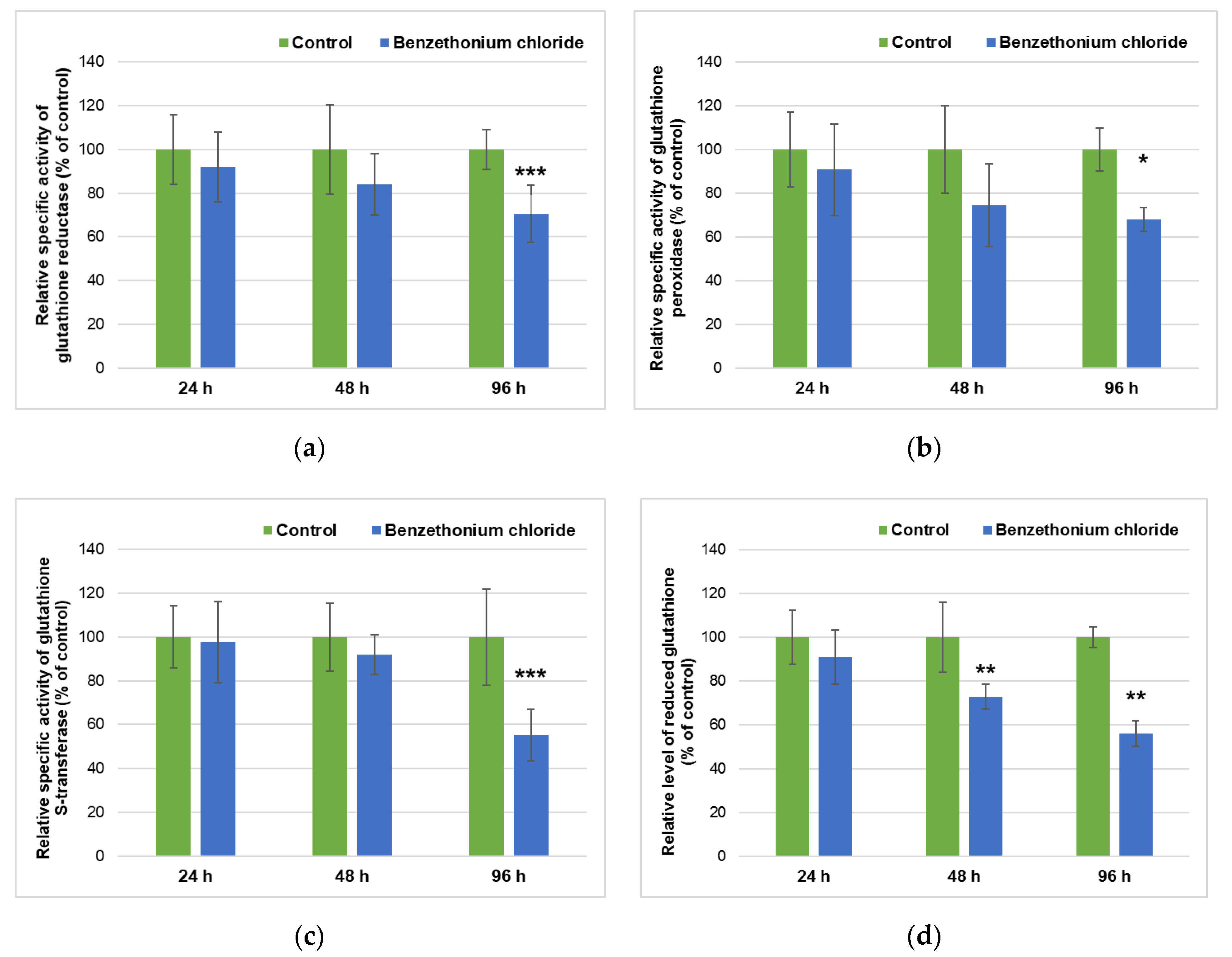
Regarding the metabolism of GSH, the most abundant cellular thiol antioxidant, we investigated the variance of GPx, GRed, and GST activities in correlation with GSH level during the fish exposure to BEC (Figure 2). In the hepatic tissue, all of these three enzymes exhibited lower activities compared to control groups. In the presence of BEC, the specific activities of GRed and GPx diminished in a time-dependent manner, with almost 30% (compared to control) after 96 h. Although the GST activity was not significantly decreased after the first intervals of exposure, its activity was reduced by half compared to its control after the 96 h (Figure 2c). A time-dependent decrease was also observed in the case of GSH level, a significant reduction by 44% of control was measured after 96 h, which suggested a depletion of this antioxidant level in hepatic tissues from fish exposed the longest period to BEC. This correlated very well with the decreased activities of GSH-related enzymes (Figure 2).
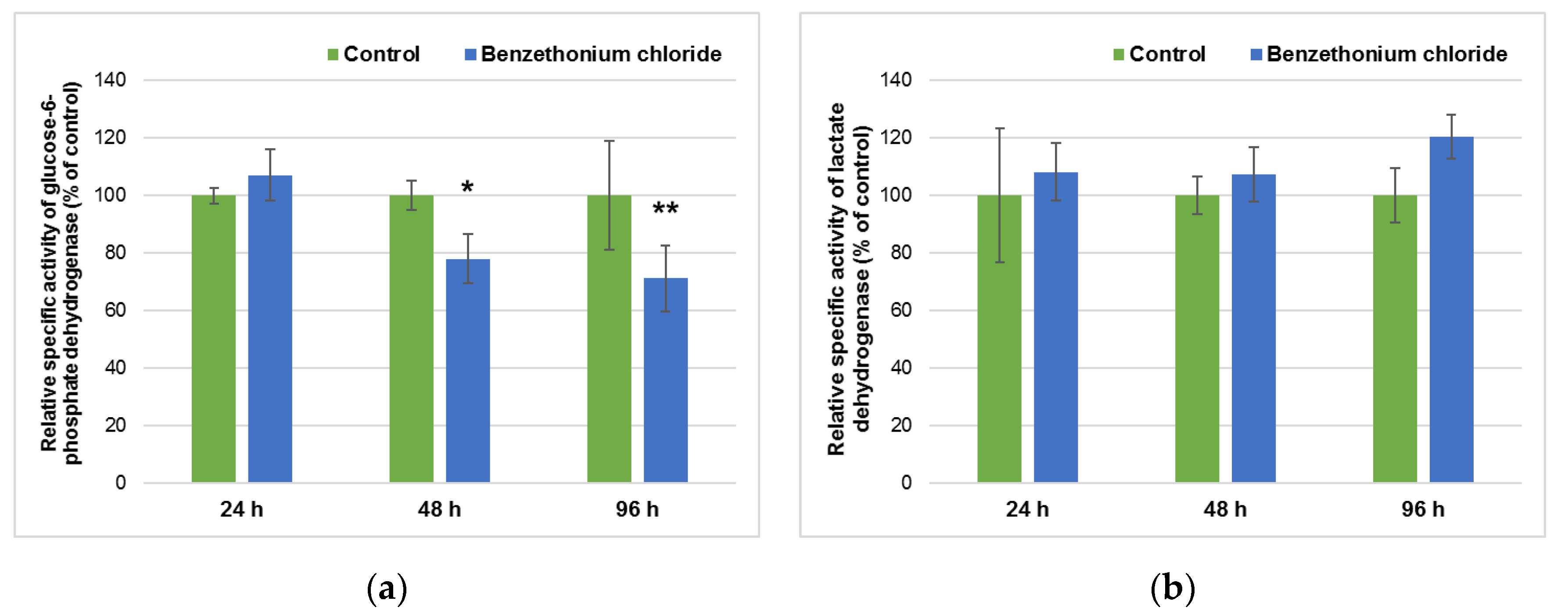
G6PDH activity decreased only after 48 and 96 h of BEC incubation by 23% and 29% of control, respectively (Figure 3a), revealing the reduction of NADPH formation which affected the GSH level (Figure 2d), as well as GRed activity (Figure 2a). LDH activity slightly increased after 24 and 48 h compared to control, a 20% elevation above the control being assessed after 96 h (Figure 3b).
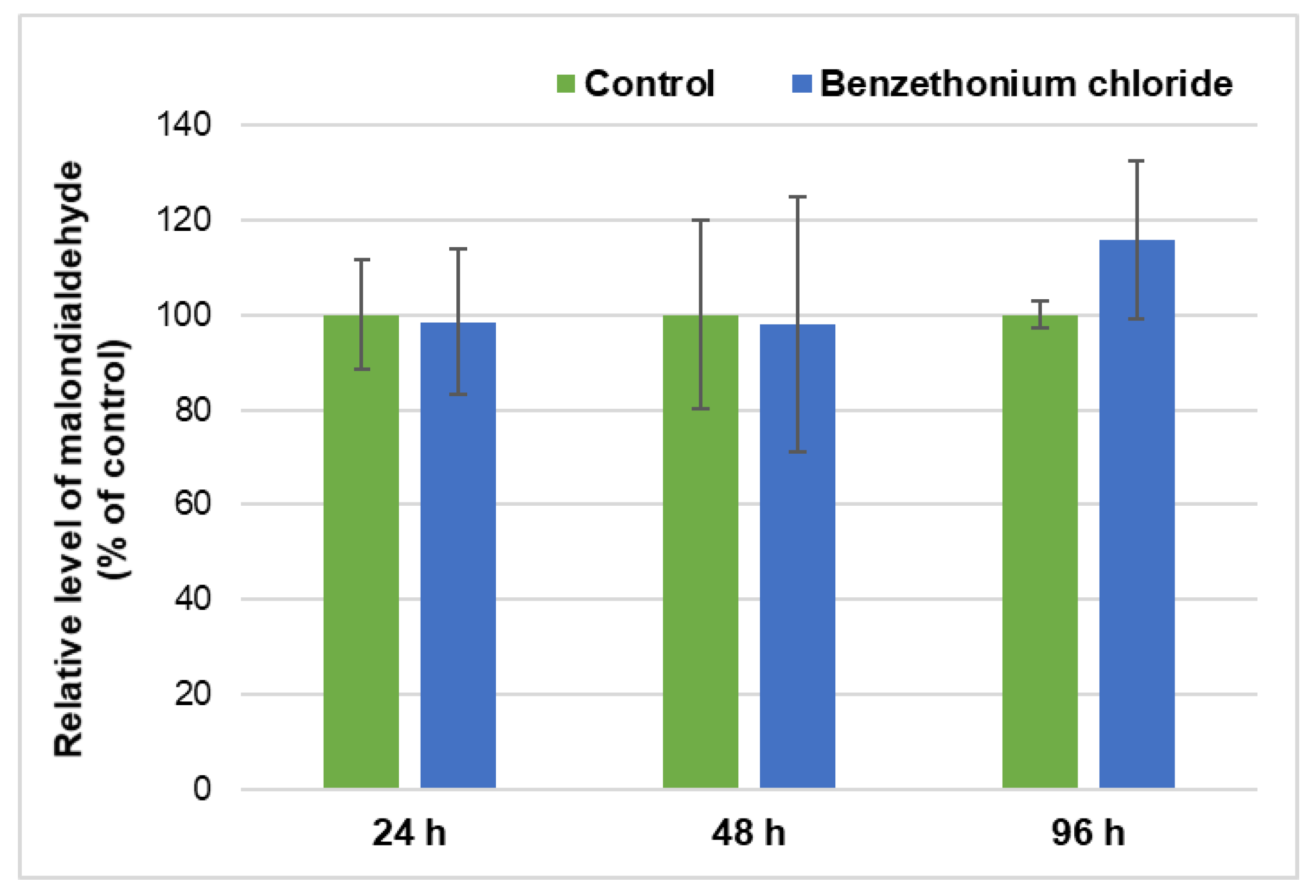
In order to evaluate the lipid peroxidation induced by BEC exposure, the MDA values were measured in the hepatic tissue of fish. No changes were detected compared to control after 24 and 48 h, just a 15% increase after 96 h (Figure 4), indicating a low rate of lipid peroxidation process initiated at the longest period of time tested as exposure interval.

3.4. Histopathological Changes after Exposure to BEC

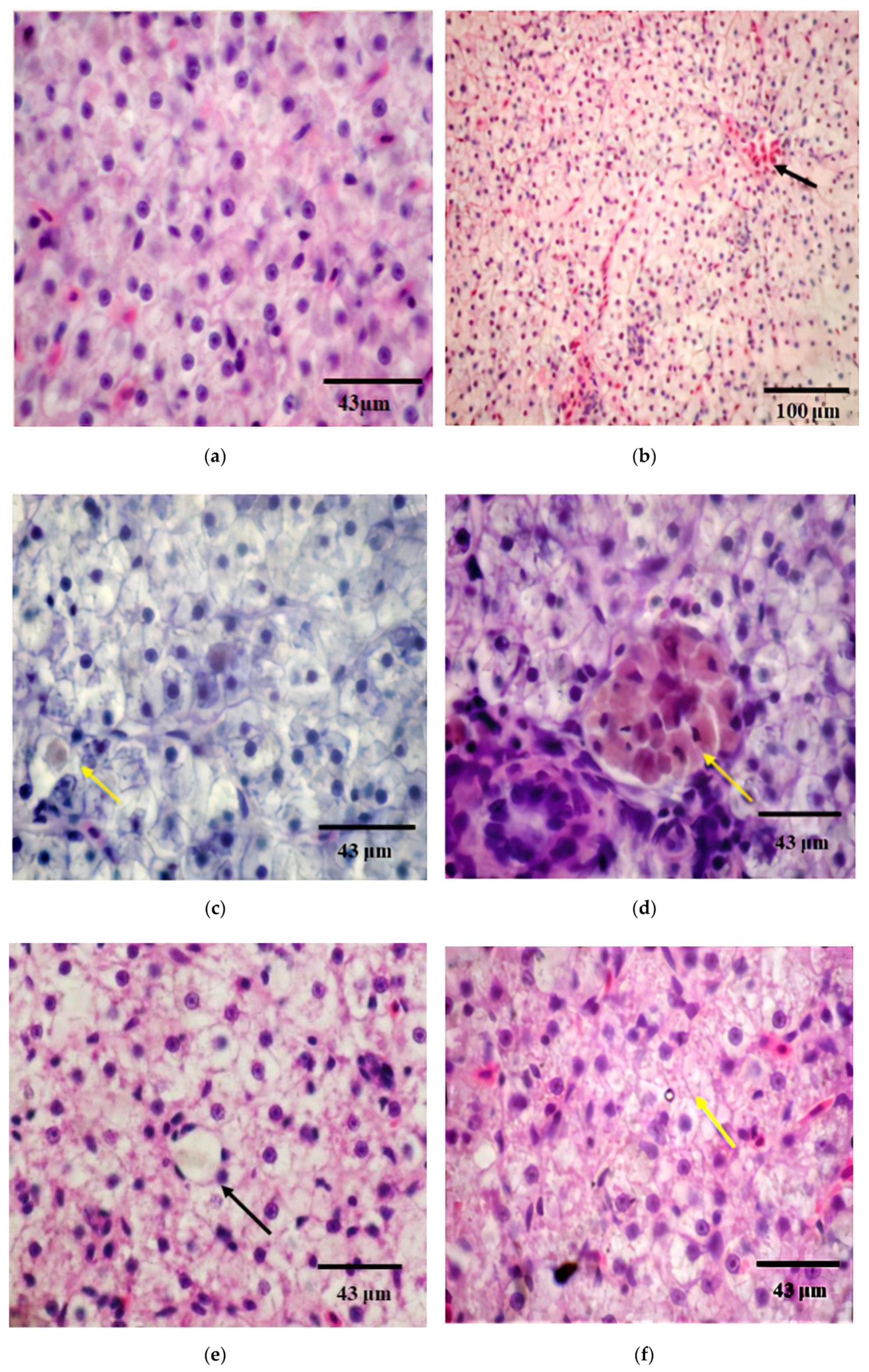
The hepatic tissue of the control fish was evidenced as a normal and homogenous mass of hepatic cells sprinkled with a network of blood vessels and sinusoids (Figure 5a). After 24 h of exposure to BEC, few cells with nuclear hypertrophy, aggregates of macrophages, and cytoplasmic vacuolization were observed (Figure 5b–d). The fish exposed for 96 h to BEC showed an accentuated cytoplasmic vacuolization and nuclear hypertrophy (Figure 5e,f). These observed modifications can be evaluated as changes of grade I and II [38] considering that these slight damages in hepatic tissue could affect the normal function of the liver. The assessment of liver histopathological alterations was qualitatively described taking into consideration the severity of lesions using the methods specified previously [38,39].

4. Discussion

European Commission has imposed strict criteria for the use of chemicals in products to protect human health and environmental safety. One of these criteria includes the ecotoxicological assessments of acute, sub-acute, and chronic effects of chemicals on various aquatic and terrestrial organisms. Fish are the most sensitive species to pollutants, representing a bioindicator of the water quality and of the status of aquatic ecosystems, being suitable as a biological model in ecotoxicological studies [40]. Aerobic organisms, including the fish, have developed an antioxidant defense system against oxygen radicals derived from the exposed xenobiotics, which can prevent excess oxidation and the damage of cell structures. Among the indicators of oxidative stress, changes in antioxidant enzymes activities, and oxidative damage products, such as protein carbonyls and lipid peroxidation are well-known.
Our previous studies on Cyprinus carpio revealed the acute effects of BEC, the LC50 value after 96 h of exposure being calculated at 4.57 mg/L, which indicated acute toxicity on fish according to REACH classification criteria [41]. In this context, our study aimed to reveal the acute toxicity of BEC at a concentration of 1 mg/L in water (simulating the maximum of cationic surfactants detected in surface water) on Cyprinus carpio by assessing the enzymatic and non-enzymatic antioxidative system, the level of lipid peroxidation and histological changes in hepatic tissue.
The liver represents the principal system involved in the xenobiotics’ metabolism of animals and its antioxidant enzymes are taken into consideration as biomarkers of hepatotoxicity induced by various chemicals. During the xenobiotics’ transformation in reactions catalyzed by monooxygenases belonging to CYP 450 families, ROS are generated. The activities of antioxidant enzymes often increase when oxidative stress is generated in fish, in order to contribute to the detoxification of oxygen free radicals and prevent or limit macromolecule damage [42,43]. However, it was shown that some enzymes can also be partially inactivated by the free radicals; hydrogen peroxide can inactivate SOD [44], superoxide anion and hydrogen peroxide inactivate CAT, and GST is sensitive to products of the Haber-Weiss reaction [45].
Our data showed that CAT and SOD activities decreased following BEC exposure for 96 h (Figure 1). Previously, it was shown that CAT enzymatic activity in Oncorhynchus mykiss liver was significantly reduced under benzalkonium chloride (a cationic surfactant from the same class as BEC) starting from a concentration of 0.18 mg/L [15]. Similar results have been obtained in Carassius auratus gibelio liver in case of exposure to deltamethrin, a decrease in the activities of antioxidant enzymes SOD and CAT as a result of superoxide anions accumulation [46]. However, a sub-lethal exposure for 30 days of freshwater fish Oreochromis mossambicus at concentrations of 1 mg/L of anionic, cationic, or nonionic surfactants has led to severe oxidative stress in the liver, the activities of CAT, SOD, and GPx increasing significantly in the case of all tested surfactants [24]. The difference in fish responses could be explained by the different fish species (subtropical versus tropical fish) and the exposure time, which was longer compared to our experiment. Acute exposure of fish to surfactant induced an antioxidant defense response suggested by the changes of enzyme activities, which was in contrast to a chronic exposure that could induce the expression of an adaptive response.
The previous studies showed that the organism’s responses could be different and influenced by various factors, such as: fish species and their characteristics, exposure time (acute or chronic), surfactant type and tested concentration, laboratory test conditions (static/semi-static/dynamic test solutions, dilution water characteristics, aeration, and test vessels). Therefore, the ecotoxicological data and the magnitude of monitoring effects are very varied [1,3,5,8,9,14,16].
GPx reduces the lipid hydroperoxides to hydroxylated lipid derivatives and GST is an enzyme capable of catalyzing multiple reactions in order to detoxify oxidized lipids. The depletion of these two enzymes activities in the liver showed their incapacity to decrease the level of lipid peroxides, explaining the increase of MDA level observed after 96 h of exposure to BEC (Figure 4). The decrease of GPx and GST activity (Figure 2) can be explained by the low level of GSH, the principal non-enzymatic antioxidant of cells that are used substrate by these enzymes. A balance between GSH synthesis and its utilization has to be maintained for proper cell functions. GSH is involved in the diminution of ROS level by acting as an electron-carrier substrate for several enzymes or by reacting directly with radicals in non-enzymatic reactions [47]. The significant decrease of GSH level observed within our study showed the impact of ROS accumulation on cell defense mechanisms. The same response was reported in the carp liver exposed to other pollutants [35].
GRed is involved in the recycling of GSH as this enzyme catalyzes the reaction in which oxidized glutathione returns to its reduced form in the presence of NADPH generated by G6PDH within the pentose phosphate pathway [48]. A decrease of G6PDH activity (Figure 3a) determined a decrease in NADPH, which was necessary for GRed to reduce oxidized glutathione. Therefore, the exposure to BEC induced a reduction of all these three antioxidants, GRed, G6PDH, and GSH. In addition, the increased level of ROS affected the activity of LDH too (Figure 3b). This could be due to an increase of glycolysis rate versus Krebs cycle, consequent to a mitochondrial dysfunction induced by the exposure of fish to this cationic surfactant as previously was noticed [49,50].
Lipid peroxidation induces oxidation of polyunsaturated phospholipids and changes of peroxidation products, such as MDA, can indirectly reflect cellular membrane injuries in hepatic tissue [51]. In our study, the unchanged level of MDA after 48 h compared to control could be explained by the conjugation of lipid peroxides with GSH catalyzed by GST. The increased level of MDA after 96 h highlighted the idea of ROS generation by BEC presence that interacted with polyunsaturated fatty acids in the context of antioxidant enzyme deficiency, such as GST activity decreased, as it was described also in other previous reports [52].
The present study showed that BEC can induce some unspecific structural changes in liver tissue (Figure 5), identified also for other types of xenobiotics [53,54]. Generally, the observed changes in the liver tissues are considered compatible with functioning and survival but reported also some moderate effects, such as hepatocytes with cytoplasm vacuolization and an increased macrophage number related also with the decreases of antioxidant enzyme activities, lipid peroxidation being obtained only after 96 h of exposure to BEC. Previous studies on quaternary ammonium compounds, such as benzalkonium chloride and cetyltrimethylammonium bromide, reported oxidative stress, cell apoptosis, necrosis, and cell death, as these pollutants induced overproduction of hydroperoxide and superoxide anion [55].
Similar studies on surfactants’ toxicity reported histological changes in the liver of Cirrhinus mrigala associated with SOD and CAT changes [40]. There were reported accumulation of melanomacrophage centers, inflammation, and congestion of hepatocytes [40]. The increase in the density of melanomacrophage aggregates is generally correlated with important liver lesions [53]. Macrophage aggregates are considered areas of inflammation, oxygen-free radical formation, and lipid peroxidation, increasing in fish collected from heavily contaminated waters [56]. The function of melanomacrophages in fish liver remains unclear, but some studies have suggested that their presence was associated with the destruction, detoxification, or recycling of endogenous and exogenous compounds [57]. Aspects, such as irregular hepatocyte shapes and cytoplasmic vacuolation, have also been described for Corydoras paleatus intoxicated with pesticides [54] and Leuciscus cephalus exposed to heavy metals [58]. Hepatocytes vacuolization was described by some researchers as an indicator of a degenerative process that suggests metabolic damage, consisting of organelles distension or fluid accumulation, possibly correlated with exposure to contaminated water [38,53]. Changes in nuclear shape or chromatin structure appeared during exposure to cytotoxic, mutagenic, or mitogenic compounds [59].
The negative effects of BEC revealed after the 96 h of exposure could be related to its accumulation in the liver. Generally, the surfactants have a low concentration level in fish due to their biotransformation. However, they have been predominantly detected in the cephalothorax circulatory system (gill, heart, and hepatopancreas) [8]. Some surfactants, such as perfluorooctanesulfonate (PFOS), are founded in high concentration in the hepatic tissue of fish [60]. Anionic surfactants (such as C12-2-LAS) are rapidly transported in the circulatory system firstly to the liver and then to the other organs [61]. Similar cationic compounds (quaternary alkylammonium surfactants) showed the following distribution within the fish body: muscle < blood < skin < gills < liver, after one week of exposure to 1.3 and 59 μg/L, no mortality registered [62].

5. Conclusions

The cationic surfactant BEC induced oxidative stress responses in the liver of Cyprinus carpio after an exposure of 96 h in water tanks. The effects on antioxidant defense systems were related to the decrease of enzyme activities and reduced level of the main non-enzymatic antioxidant GSH. The slightly increased levels of MDA and also, the outlined moderate structural changes support the low toxicity induced by BEC on hepatic tissue after four days of exposure. Our research revealed that the biochemical and histological assays of the liver samples of Cyprinus carpio could be used as biomarkers in the water quality monitoring and assessment of the ecotoxicological risk of chemicals.

Author Contributions

Conceptualization, S.G., M.S.S., I.E.L., M.N.-L. and A.D.; Formal analysis, S.G., D.N.M., M.S.S., M.C. and A.D.; Funding acquisition, A.D.; Investigation, S.G., D.N.M., M.S.S., C.A.S. and M.C.; Methodology, S.G., D.N.M., M.S.S., C.A.S., M.C., I.E.L. and A.D.; Project administration, M.N.-L.; Writing—original draft, S.G., D.N.M., M.S.S. and I.E.L.; Writing—review & editing, C.A.S., M.N.-L. and A.D. All authors have read and agreed to the published version of the manuscript.

Funding

This research was funded by the Romanian National Authority for Scientific Research and Innovation (UEFISCDI) through the project no. 10PCCF/2018 RADAR (PN-III-P4-ID-PCCF-2016-0114—“Selection and dissemination of antibiotic resistance genes from wastewater treatment plants into the aquatic environment and clinical reservoirs”).

Conflicts of Interest

The authors declare no conflict of interest. The funders had no role in the design of the study; in the collection, analyses, or interpretation of data; in the writing of the manuscript, or in the decision to publish the results.

References

  1. Kaczerewska, O.; Martins, R.; Figueiredo, J.; Loureiro, S.; Tedim, J. Environmental behavior and ecotoxicity of cationic surfactants towards marine organisms. J. Hazard. Mater. 2020, 392, 122299. [Google Scholar] [CrossRef]
  2. Ying, G.G. Fate, behavior and effects of surfactants and their degradation products in the environment. Environ. Int. 2006, 32, 417–431. [Google Scholar] [CrossRef]
  3. Abel, P.D. Toxicity of synthetic detergents to fish and aquatic invertebrates. J. Fish Biol. 1974, 6, 279–298. [Google Scholar] [CrossRef]
  4. Lewis, M.A.; Suprenant, D. Comparative acute toxicities of surfactants to aquatic invertebrates. Ecotoxicol. Environ. Saf. 1983, 7, 313–322. [Google Scholar] [CrossRef]
  5. Lewis, M.A. Chronic and sub lethal toxicities of surfactants to aquatic animals: A review and risk assessment. Water Res. 1991, 25, 101–113. [Google Scholar] [CrossRef]
  6. Mandal, R.; Mandal, D.; Mishra, N.; Bahadur, A. Effect of surfactants on phosphatase level of fresh water fish Labeo rohita. J. Environ. Biol. 2010, 31, 395–398. [Google Scholar]
  7. Zhu, X.; Wang, Z.; Sun, Y.; Gu, L.; Zhang, L.; Wang, J.; Huang, Y.; Yang, Z. Surfactants at environmentally relevant concentrations interfere the inducible defense of Scenedesmus obliquus and the implications for ecological risk assessment. Environ. Pollut. 2020, 261, 114131. [Google Scholar] [CrossRef]
  8. Jackson, M.; Eadsforth, C.; Schowanek, D.; Delfosse, T.; Riddle, A.; Budgen, N. Comprehensive review of several surfactants in marine environments: Fate and ecotoxicity. Environ. Toxicol. Chem. 2016, 35, 1077–1086. [Google Scholar] [CrossRef] [Green Version]
  9. Pretti, C.; Chiappe, C.; Baldetti, I.; Brunini, S.; Monni, G.; Intorre, L. Acute toxicity of ionic liquids for three fresh water organisms: Pseudokirchneriella subcapitata, Daphnia magna and Danio rerio. Ecotoxicol. Environ. Saf. 2009, 72, 1170–1176. [Google Scholar] [CrossRef]
  10. Clara, M.; Scharf, S.; Scheffknecht, C.; Gans, O. Occurrence of selected surfactants in untreated and treated sewage. Water Res. 2007, 41, 4339–4348. [Google Scholar] [CrossRef]
  11. Agrawal, K.; Agnihotri, G.; Shrivas, K.; Mundhara, G.L.; Patel, K.S.; Hoffmann, P. Determination of cationic surfactants in environmental samples by flow injection analysis. Microchim. Acta 2004, 147, 273–278. [Google Scholar] [CrossRef]
  12. Martínez-Carballo, E.; Sitka, A.; González-Barreiro, C.; Kreuzinger, N.; Fürhacker, M.; Scharf, S.; Gans, O. Determination of selected quaternary ammonium compounds by liquid chromatography with mass spectrometry. Part I. Application to surface, waste and indirect discharge water samples in Austria. Environ. Pollut. 2007, 145, 489–496. [Google Scholar] [CrossRef] [PubMed]
  13. Sreevidya, V.S.; Lenz, K.A.; Svoboda, K.R.; Ma, H. Benzalkonium chloride, benzethonium chloride, and chloroxylenol—Three replacement antimicrobials are more toxic than triclosan and triclocarban in two model organisms. Environ. Pollut. 2018, 235, 814–824. [Google Scholar] [CrossRef] [PubMed]
  14. Zhang, C.; Cui, F.; Zeng, G.M.; Jiang, M.; Yang, Z.Z.; Yu, Z.G.; Zhu, M.Y.; Shen, L.Q. Quaternary ammonium compounds (QACs): A review on occurrence, fate and toxicity in the environment. Sci. Total Environ. 2015, 15, 518–519. [Google Scholar] [CrossRef] [PubMed]
  15. Antunes, S.C.; Nunes, B.; Rodrigues, S.; Nunes, R.; Fernandes, J.; Correia, A.T. Effects of chronic exposure to benzalkonium chloride in Oncorhynchusmykiss: Cholinergic neurotoxicity, oxidative stress, peroxidativedamage and genotoxicity. Environ. Toxicol. Pharmacol. 2016, 45, 115–122. [Google Scholar] [CrossRef]
  16. Gheorghe, S.; Lucaciu, I.; Paun, I.; Stoica, C.; Stanescu, E. Ecotoxicological behavior of some cationic and amphoteric surfactants (biodegradation, toxicity and risk assessment). In Biodegradation—Life Science; Chamy, R., Rosenkranz, F., Eds.; IntechOpen: London, UK, 2013; pp. 83–114. [Google Scholar]
  17. CIR. Compendium, Containing Abstracts, Discussions, and Conclusions of CIR Cosmetic Ingredient Safety Assessments; Cosmetic Ingredient Review: Washington, DC, USA, 2006. [Google Scholar]
  18. Gheorghe, S.; Lucaciu, I.; Paun, I.; Stoica, C.; Stanescu, E. Environmental exposure and effects of some micropollutants found in the Romanian surface waters. J. Environ. Prot. Ecol. 2014, 15, 878–888. [Google Scholar]
  19. Pérez, P.; Fernández, E.; Beiras, R. Toxicity of benzalkonium chloride on monoalgal cultures and natural assemblages of marine phytoplankton. Water Air Soil Pollut. 2009, 201, 319–330. [Google Scholar] [CrossRef]
  20. Christen, V.; Faltermann, S.; Brun, N.R.; Kunz, P.Y.; Fent, K. Cytotoxicity and molecular effects of biocidal disinfectants (quaternary ammonia, glutaraldehyde, poly (hexamethylene biguanide) hydrochloride PHMB) and their mixtures in vitro and in zebrafish eleuthero-embryos. Sci. Total Environ. 2017, 586, 1204–1218. [Google Scholar] [CrossRef]
  21. Li, M.H. Effects of nonionic and ionic surfactants on survival, oxidative stress, and cholinesterase activity of planarian. Chemosphere 2008, 70, 1796–1803. [Google Scholar] [CrossRef]
  22. Cserhati, T.; Forgacs, E.; Oros, G. Biological activity and environmental impact of anionic surfactants. Environ. Int. 2002, 28, 337–348. [Google Scholar] [CrossRef]
  23. Jee, J.H.; Kang, J.C. Biochemical changes oif enzymatic defence system after phenanthrene exposure in olive flounder, Paralichthyus olivaceus. Physiol. Res. 2005, 54, 585–591. [Google Scholar]
  24. Bindu, P.C.; Babu, P. Surfactant-induced lipid peroxidation in a tropical euryhaline teleost Oreochromis mossambicus (Tilapia) adapted to fresh water. Indian J. Exp. Biol. 2001, 39, 1118–1122. [Google Scholar] [PubMed]
  25. Park, C.J.; Song, S.H.; Kim, D.H.; Gye, M.C. Developmental and acute toxicity of cetylpyridinium chloride in Bombina orientalis (Amphibia: Anura). Aquat. Toxicol. 2016, 177, 446–453. [Google Scholar] [CrossRef] [PubMed]
  26. Sharples, F.; Anestidou, L.; Beil, K.; Fletcher, C.H.; Haycraft, R.; Zurlo, J. Guide for the Use and Care of Laboratory Animals, 8th ed. In National Research Council (US) Committee or the Update of the Guide for the Care and Use of Laboratory Animals; National Academies Press: Washington, DC, USA, 2011. [Google Scholar]
  27. Lowry, O.H.; Rosenbrough, N.J.; Farr, A.L.; Randall, B.J. Protein measurement with the Folin phenol reagent. J. Biol. Chem. 1951, 193, 265–275. [Google Scholar]
  28. Aebi, H. Catalase. In Methods of Enzymatic Analysis; Bergmeyer, H.V., Ed.; Academic Press: New York, NY, USA, 1984; pp. 673–677. [Google Scholar]
  29. Paoletti, F.; Mocali, A. Determination of superoxide dismutase activity by purely chemical system based on NADP(H) oxidation. Methods Enzymol. 1990, 186, 209–221. [Google Scholar]
  30. Beutler, E. Red Cell Metabolism: A Manual of Biochemical Method, 3rd ed.; Grune and Stratton: Orlando, FL, USA, 1984; pp. 68–73. [Google Scholar]
  31. Goldberg, D.M.; Spooner, R.J. Glutathione reductase. In Methods of Enzymatic Analysis, 3rd ed.; Bergmeyer, H.V., Ed.; Verlag Chemie: Weinheim, Germany, 1983; pp. 258–265. [Google Scholar]
  32. Habig, W.H.; Pabst, M.J.; Jakoby, W.B. Glutathione S-transferases. The first enzymatic step in mercapturic acid formation. J. Biol. Chem. 1974, 249, 7130–7139. [Google Scholar]
  33. Lohr, G.W.; Waller, H.D. Glucose-6-phosphate dehydrogenase. In Methods of Enzymatic Analysis; Bergmeyer, H.V., Ed.; Academic Press: London, UK, 1974; pp. 744–751. [Google Scholar]
  34. Korzeniewski, C.; Callewaert, D.M. An enzyme-release assay for natural cytotoxicity. J. Immunol. Methods 1983, 64, 313–320. [Google Scholar] [CrossRef]
  35. Stanca, L.; Petrache, S.N.; Serban, A.I.; Staicu, A.C.; Sima, C.; Munteanu, M.C.; Zărnescu, O.; Dinu, D.; Dinischiotu, A. Interaction of silicon-based quantum dots with gibel carp liver: Oxidative and structural modifications. Nanoscale Res. Lett. 2013, 8, 254. [Google Scholar] [CrossRef] [Green Version]
  36. Htun-han, M. The reproductive biology of the dab Limanda limanda (L) in the North Sea: Gonadosomatic index, hepatosomatic index and condition factor. J. Fish Biol. 1978, 13, 369–378. [Google Scholar] [CrossRef]
  37. King, M. Fisheries Biology, Assessment and Management; Blackwell Publishing: Oxford, UK, 1995; p. 341. [Google Scholar]
  38. Camargo, M.M.P.; Martinez, C.B.R. Histopathology of gills, kidney and liver of a Neotropical fish caged in an urban stream. Neotrop. Ichthyol. 2007, 5, 327–336. [Google Scholar] [CrossRef] [Green Version]
  39. Abdel-Moneim, A.M.; Al-Kahtani, M.A.; Elmenshawy, O.M. Histopathological biomarkers in gills and liver of Oreochromisniloticus from polluted wetland environments, Saudi Arabia. Chemosphere 2012, 88, 1028–1035. [Google Scholar] [CrossRef] [PubMed]
  40. Dani, U.; Bahadur, A.; Kuperkar, K. Biotoxicity and tissue-specific oxidative stress induced by Gemini surfactant as a protocol on fingerlings of Cirrhinus mrigala (Ham.): An integrated experimental and theoretical methodology. Ecotoxicol. Environ. Saf. 2019, 183, 109478. [Google Scholar] [CrossRef] [PubMed]
  41. Gheorghe, S.; Lucaciu, I.; Grumaz, R.; Stoica, C. Acute toxicity assessment of several cationic and amphoteric surfactants on aquatic organisms. J. Environ. Prot. Ecol. 2012, 13, 541–553. [Google Scholar]
  42. Valavanidis, A.; Vlahogianni, T.; Dassenakis, M.; Scoullos, M. Molecular biomarkers of oxidative stress in aquatic organisms in relation to toxic environmental pollutants. Ecotox. Environ. Saf. 2006, 64, 178–189. [Google Scholar] [CrossRef] [PubMed]
  43. Craig, P.M.; Wood, C.M.; McClelland, G.B. Oxidative stress response and gene expression with acute copper exposure in zebrafish (Danio rerio). Am. J. Physiol. 2007, 293, R1882–R1892. [Google Scholar] [CrossRef] [PubMed] [Green Version]
  44. Ivan, Ş.; Lucaciu, I.; Rusu, G.; Iancu, V. Toxicity of some dangerous chemicals on Superoxide dismutase enzymatic activity. J. Environ. Prot. Ecol. 2010, 11, 247–252. [Google Scholar]
  45. Atli, G.; Canli, M. Response of antioxidant system of freshwater fish Oreochromis niloticus to acute and chronic metal (Cd, Cu, Cr, Zn, Fe) exposures. Ecotoxicol. Environ. Saf. 2010, 73, 1884–1889. [Google Scholar] [CrossRef]
  46. Dinu, D.; Marinescu, D.; Munteanu, M.C.; Staicu, A.C.; Costache, M.; Dinischiotu, A. Modulatory effects of deltamethrin on antioxidant defense mechanisms and lipid peroxidation in Carassius auratus gibelio liver and intestine. Arch. Environ. Contam. Toxicol. 2010, 58, 757–764. [Google Scholar] [CrossRef]
  47. Winterbourn, C.C.; Metodieva, D. The reactions superoxide with reduced glutathione. Arch. Biochem. Biophys. 1994, 314, 284–290. [Google Scholar] [CrossRef]
  48. Kletzien, R.F.; Harris, P.K.; Foellmi, L.A. Glucose-6-phosphate dehydrogenase: A ‘‘housekeeping’’ enzyme subject to tissue-specific regulation by hormones, nutrients and oxidant stress. FASEB J. 1994, 8, 174–181. [Google Scholar] [CrossRef]
  49. Inácio, A.S.; Costa, G.N.; Domingues, N.S.; Santos, M.S.; Moreno, A.J.M.; Vaz, W.L.C.; Vieira, O.V. Mitochondrial dysfunction is the focus of quaternary ammonium surfactant toxicity of mammalian epithelial cells. Antimicrob. Agents Chemother. 2013, 57, 2631–2639. [Google Scholar] [CrossRef] [PubMed] [Green Version]
  50. Rogov, A.G.; Goleva, T.N.; Sukhanova, E.I.; Epremyan, K.K.; Trendeleva, T.A.; Ovchenkova, A.P.; Aliverdieva, D.A.; Zvyagilskaya, R.A. Mitochondrial dysfunctions may be one of the major causative factors underlaying detrimental effects of benzalkonium chloride. Oxid. Med. Cell Longev. 2020, 2020, 8956504. [Google Scholar] [CrossRef] [PubMed] [Green Version]
  51. Eaton, D.L.; Klaassen, C.D. Principles of toxicology. In Casarett & Doull’s Toxicology: The Basic Science of Poisons; Klaassen, C.D., Ed.; McGraw Hill Press: New York, NY, USA, 2001; pp. 11–32. [Google Scholar]
  52. Falfushynska, H.I.; Stolyar, O.B. Responses of biochemical markers in carp Cyprinus carpio from two field sites in Western Ukraine. Ecotoxicol. Environ. Saf. 2009, 72, 729–736. [Google Scholar] [CrossRef] [PubMed]
  53. Pacheco, M.; Santos, M.A. Biotransformation, genotoxic and histopathological effects of environmental contaminants in European eel (Anguilla anguilla L.). Ecotoxicol. Environ. Saf. 2002, 53, 331–347. [Google Scholar] [CrossRef] [Green Version]
  54. Fanta, E.; Rios, F.S.; Romão, S.; Vianna, A.C.; Freiberger, S. Histopathology of the fish Corydoras paleatus contaminated with sublethal levels of organophosphorus in water and food. Ecotoxicol Environ Saf. 2003, 54, 119–130. [Google Scholar] [CrossRef]
  55. Pauloin, T.; Dutot, M.; Warnet, J.M.; Rat, P. In vitro modulation of preservative toxicity: High molecular weight hyaluronan decreases apoptosis and oxidative stress induced by benzalkonium chloride. Eur. J. Pharm. Sci. 2008, 34, 263–273. [Google Scholar] [CrossRef]
  56. Hinton, D.; Segner, H.; Au, D.; Kullman, S.; Harman, R. Liver toxicity. In Toxicology of Fishes; Di Giulio, R.T., Hinton, D.E., Eds.; CRC Press Inc.: Florida, CA, USA, 2008; pp. 327–400. [Google Scholar]
  57. Haaparanta, A.; Valtonen, E.T.; Hoffaman, R.; Colmes, J. Do macrophages centers in freshwater fishes reflect the differences in water quality? Aquat. Toxicol. 1996, 34, 253–272. [Google Scholar] [CrossRef]
  58. Hermenean, A.; Damache, G.; Albu, P.; Ardelean, A.; Ardelean, G.; Ardelean, P.D.; Horge, M.; Nagy, T.; Braun, M.; Zsuga, M.; et al. Histopatological alterations and oxidative stress in liver and kidney of Leuciscus cephalus following exposure to heavy metals in the Tur River, North Western Romania. Ecotoxicol. Environ. Saf. 2015, 119, 198–205. [Google Scholar] [CrossRef]
  59. Wolf, J.C.; Wheeler, J.R. A critical review of histopathological findings associated with endocrine and non-endocrine hepatic toxicity in fish models. Aquat. Toxicol. 2018, 197, 60–78. [Google Scholar] [CrossRef]
  60. Moody, C.A.; Martin, J.W.; Kwan, W.C.; Muir, D.C.G.; Mabury, S.A. Monitoring perfluorinated surfactants in biota and surface water samples following an accidental release of fire-fighting foam into etobicoke creek. Environ. Sci. Technol. 2002, 36, 545–551. [Google Scholar] [CrossRef]
  61. Tolls, J.; Haller, M.; Seinen, W.; Sijm, D.T.H.M. LAS bioconcentration: Tissue distribution and effect of hardness-implications for processes. Environ. Sci. Technol. 2000, 34, 304–310. [Google Scholar] [CrossRef]
  62. Amelie, K.; Chang’er, C.; James, M.A.; Jon, A.A.; Steven, D.; Michael, S.M. Tissue distribution of several series of cationic surfactants in rainbow trout (Oncorhynchus mykiss) following exposure via water. Environ. Sci. Technol. 2020, 54, 4190–4199. [Google Scholar]
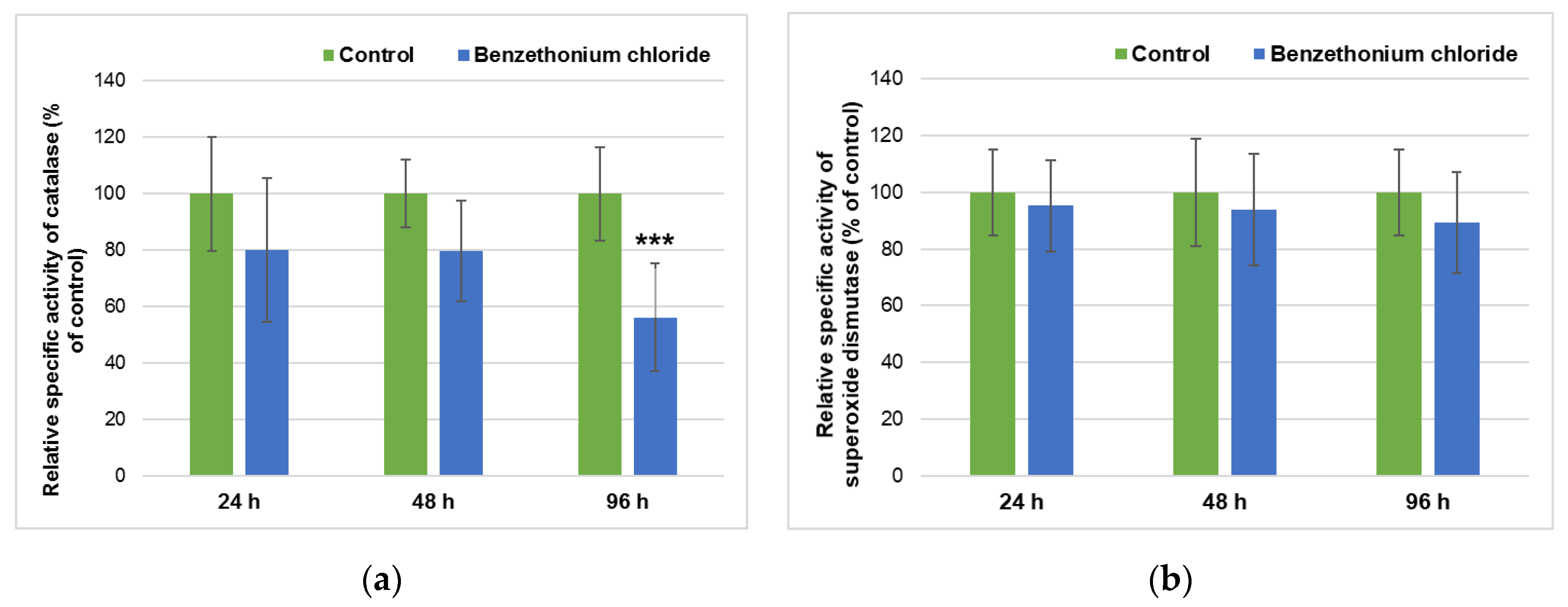
Figure 1. Hepatic activities of catalase (a) and superoxide dismutase (b) in Cyprinus carpio exposed to 1 mg/L of BEC for 24, 48, and 96 h. Results were expressed relative to control as mean levels of relative specific activities ± standard deviation (n = 5 fish for each group). *** p ≤ 0.001 compared to control.
Applsci 10 08485 g001
Figure 2. Hepatic activities of glutathione reductase (a), glutathione peroxidase (b), glutathione S-transferase (c) and superoxide dismutase, and levels of reduced glutathione (d) in Cyprinus carpio exposed to 1 mg/L of BEC for 24, 48, and 96 h. Results were expressed relative to control as mean levels ± standard deviation (n = 5 fish for each group). * p ≤ 0.05, ** p ≤ 0.01, *** p ≤ 0.001 compared to control.
Applsci 10 08485 g002
Figure 3. Hepatic activities of glucose-6-phosphate dehydrogenase (a) and lactate dehydrogenase (b) in Cyprinus carpio exposed to 1 mg/L of BEC for 24, 48, and 96 h. Results were expressed relative to control as mean levels of relative specific activities ± standard deviation (n = 5 fish for each group). * p ≤ 0.05 and ** p ≤ 0.01 compared to control.
Applsci 10 08485 g003
Figure 4. Hepatic levels of malondialdehyde in Cyprinus carpio exposed to 1 mg/L of BEC for 24, 48, and 96 h. Results were expressed relative to control as mean levels ± standard deviation (n = 5 fish for each group).
Applsci 10 08485 g004
Figure 5. Liver morphology after hematoxylin and eosin staining for (a) control group: normal hepatic cells with sinusoidal capillaries (arrow), 100×; (bd) fish exposed to BEC for 24 h: dilated sinusoidal capillaries (arrow in Figure 5b), hepatocytes with nuclear hypertrophy, cytoplasmic vacuolization (arrow in Figure 5c) and macrophage aggregates (arrow in Figure 5d); (e and f) fish exposed to BEC for 96 h: fat vacuoles in the cytoplasm that dislocate and compress the nucleus (arrow in Figure 5e), nuclear pleomorphism (arrow in Figure 5f), 40×.
Applsci 10 08485 g005
Table 1. Mean values of water quality parameters in the experimental tanks.
ParameterMean ValuesInternational Standards
pH6.94–7.86SR EN ISO 10523:2012
Temperature20 ± 0.5 °C-
Dissolved oxygen4–6 mg O2/LSR EN ISO 5814:2013
Total hardness as CaCO3184 mg/LSR ISO 6059:2008
Suspended matter5.2–12.4 mg/LSR EN 872:2005
Chemical oxygen demand14.4–25.92 mg/LSR ISO 6060:1996
Table 2. Hepatosomatic index (HSI) and gonadosomatic index (GSI) of fish groups (n = 5 fish/group) based on the average weight of body mass, liver, and gonads weight measured at 24 h and 96 h of exposure. GSI = (gonad weight/body mass) × 100; HSI = (liver weight/body mass) × 100.
ParameterControl FishExposed Fish
24 h96 h24 h96 h
Body mass (g/fish)58.5 ± 6.9559 ± 11.4064.1 ±1 4.4365.4 ± 10.25
Total length (cm/fish)15.87 ± 0.713.38 ± 1.9816.04 ± 1.1816.2 ± 1.63
Liver weight (g/fish)1.57 ± 0.051.20 ± 0.331.49 ± 0.241.39 ± 0.30
Gonad weight (g/fish)0.64 ± 0.030.20 ± 0.150.40 ± 0.230.73 ± 0.31
HSI2.68 ± 0.212.03 ± 0.252.32 ± 0.212.12 ± 0.21
GSI1.86 ± 0.030.55 ± 0.390.62 ± 0.221.11 ± 0.63
Publisher’s Note: MDPI stays neutral with regard to jurisdictional claims in published maps and institutional affiliations.