Article

Biomechanical Comparison of Different Volar Screw Placements for Horizontal Oblique Scaphoid Fractures

1
Department of Biomedical Engineering, I-Shou University, Kaohsiung 824, Taiwan
2
Department of Orthopedics, E-Da Hospital, Kaohsiung 824, Taiwan
3
School of Medicine, College of Medicine, I-Shou University, Kaohsiung 824, Taiwan
4
Department of Orthopedics, E-Da Cancer Hospital, Kaohsiung 824, Taiwan
*
Author to whom correspondence should be addressed.
Appl. Sci. 2020, 10(23), 8592; https://doi.org/10.3390/app10238592
Submission received: 7 November 2020 / Revised: 26 November 2020 / Accepted: 27 November 2020 / Published: 30 November 2020

Abstract

:
Recently, some surgeons reported that most scaphoid waist fractures were horizontal oblique and not transverse in orientation. Therefore, this cadaveric study aimed to biomechanically compare fixation strength between central and eccentric screw placements for the volar fixation of this most common scaphoid waist fracture. Eight matched pairs of fresh-frozen forearm cadaver specimens were prepared for testing and randomly assigned to two groups: group I specimens were fixed by screws in a central placement, and group II specimens were fixed by screws in an eccentric placement. Horizontal oblique osteotomy was performed along the scaphoid waist. Then, each specimen was placed under the increasing load of a pneumatically driven plunger. We recorded stiffness, load to failure, and failure mechanisms between the central and eccentric screw placement groups. Stiffness was higher in central screw placement (74.1 N/mm) than in eccentric screw placement (29.39 N/mm). The median loads to failure in groups I and II were 54.14 and 26.22 N, respectively. In this biomechanical model, we demonstrated that central screw placement is superior to eccentric placement in terms of fixation strength. However, further clinical investigation is warranted to evaluate whether the different screw placements for volar approach of horizontal oblique scaphoid fractures affect the clinical outcomes.

1. Introduction

In the last few decades, screw fixation of acute scaphoid fractures has become a favorable alternative after the development of headless cannulated screws [1,2,3]. Some surgeons have used this technique for displaced scaphoid fractures and nonunions [4]. In general, screw fixation of scaphoid fractures may be performed through a dorsal or volar approach [1,4,5]. Nowadays, both approaches are used for waist scaphoid fractures on the basis of the surgeon’s preference. Theoretically, the dorsal percutaneous approach to screw fixation of the scaphoid waist fractures allows for a more central placement [6], but some studies did not support that this might translate into a stronger fixation. Meermans and Verstreken [7] conducted a study using three-dimensional (3D) computer modelling with computed tomography (CT) scans of the scaphoid bone to compare two approaches and showed that a volar approach could allow the surgeon to use longer screws. Another finite element analysis demonstrated that centrally placed volar compression screw fixation might be biomechanically advantageous over dorsal screw fixation [8]. Therefore, some surgeons preferred volar screw fixation [9] and conducted biomechanical studies to analyze the stability of different scaphoid screw placement [10,11].
According to the literature, two main techniques can be used for volar scaphoid fixation [10]. One is the eccentric screw placement [1,11]. The other is the central screw placement technique [10,12]. The optimal screw placement for volar fixation of scaphoid waist fractures, herein, continues to be a subject of debate. Recently, some surgeons performed computerized 3D analyses and reported that most waist fractures were horizontal oblique and not transverse in orientation [13]. To date, only a few studies have directly compared different screw placements for horizontal oblique scaphoid fractures in similar groups of patients managed with the same advanced anesthetic and rehabilitation program. There is a need to know whether different screw placements for the most common scaphoid waist fractures affect fixation strength.
We designed this cadaveric study to biomechanically compare fixation strength between central and eccentric screw placements for the volar fixation of horizontal oblique scaphoid fractures. We hypothesized that central screw placement would achieve superior fixation strength for scaphoid fractures. This study was conducted in accordance with the Declaration of Helsinki and was approved by the institutional review board of our hospital on 30 March 2018 (EMRP-107-011).

2. Materials and Methods

Eight matched pairs of fresh-frozen forearm cadaver specimens were prepared for testing. We excluded specimens with visual or radiographic evidence of previous injury, such as scaphoid nonunions and/or scapholunate instability, before the experiment. The specimens were then randomly assigned to either a central or eccentric screw placement group (Table 1). The contralateral one received the opposite screw placement. This arrangement attempted to avoid bias due to the left- or right-handedness of the specimens. The cadaver specimens of both groups were prepared by the same person.

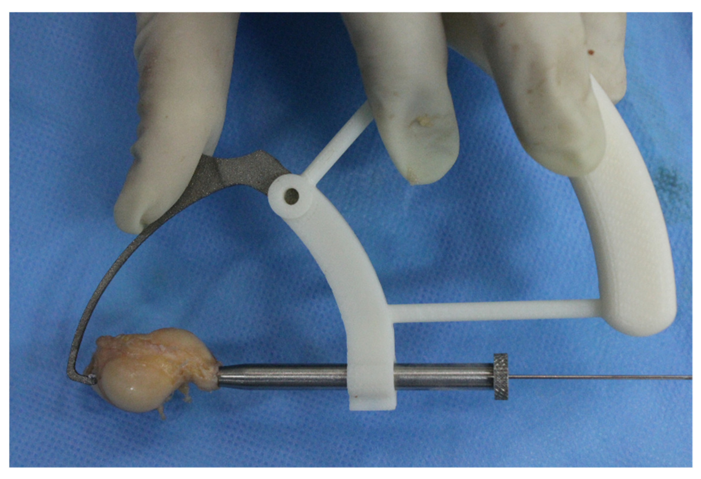
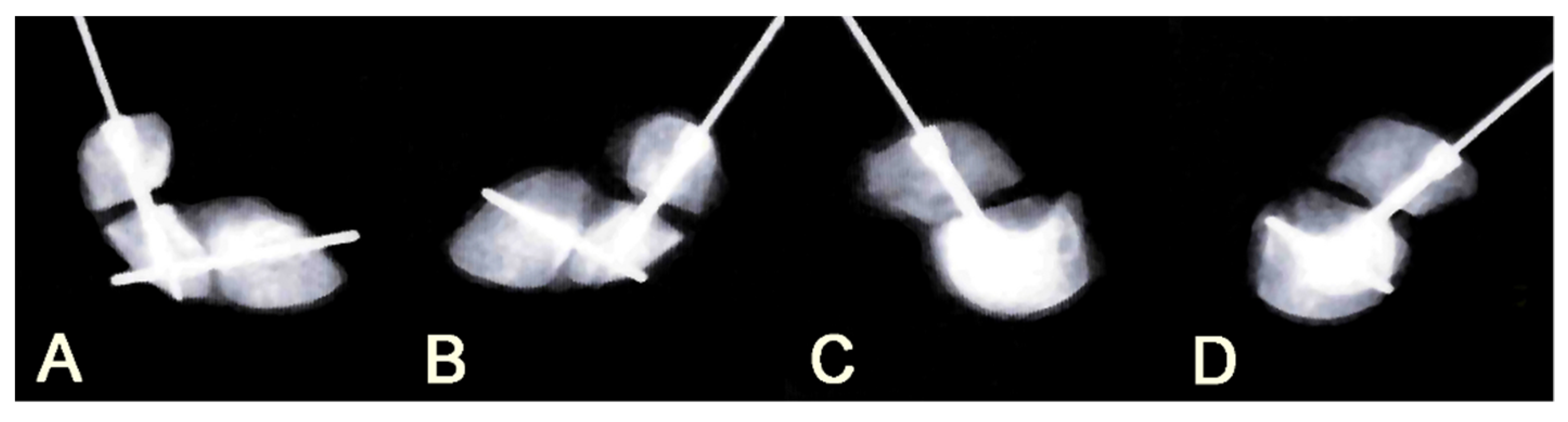
2.1. Surgical Procedures

The specimens were thawed using a standard protocol, 0 ℃ for 24 h following room temperature for another 24 h. We carefully dissected 16 scaphoids with lunates from 8 pairs of cadaveric arms. Subsequently, the scaphoids were cleaned of all soft tissue except scapholunate ligament and were symmetrically marked circumferentially around the scaphoid waist. The angle between the longitudinal axis and marked plane was 60 degrees according to a previous 3D analysis report [13]. Under the guidance of a custom-made aiming device, we drilled a 1.1-mm wire into the scaphoid either with a central or with an eccentric position according to their assignment (Figure 1). The correct position of wire was confirmed using orthogonal radiographs, and a drill-bit was applied along the wire axis. The length of the guidewire was measured, and the screw length was computed by subtracting 4 mm from that length. Using this technique, we could ensure a buried depth of 2 mm below the articular cartilage and avoid screw prominence at the articular surface. This method is frequently used in our practice and recommended in the literature [14]. Next, a smooth horizontal oblique osteotomy was made along the previously marked scaphoid waist using an electrically driven circular bone saw. Then, we enlarged the osteotomy gap to 2 mm. After a 2-mm osteotomy gap was created to simulate the horizontal oblique fracture of the scaphoid waist, we applied the cannulated screws (Wright, Arlington, TN, USA) into the scaphoid either with a central or with an eccentric position. A 2 mm thickness block was inserted into the osteotomy gap to prevent it from being close. We then took additional orthogonal radiographs to confirm the screw trajectory (Figure 2).

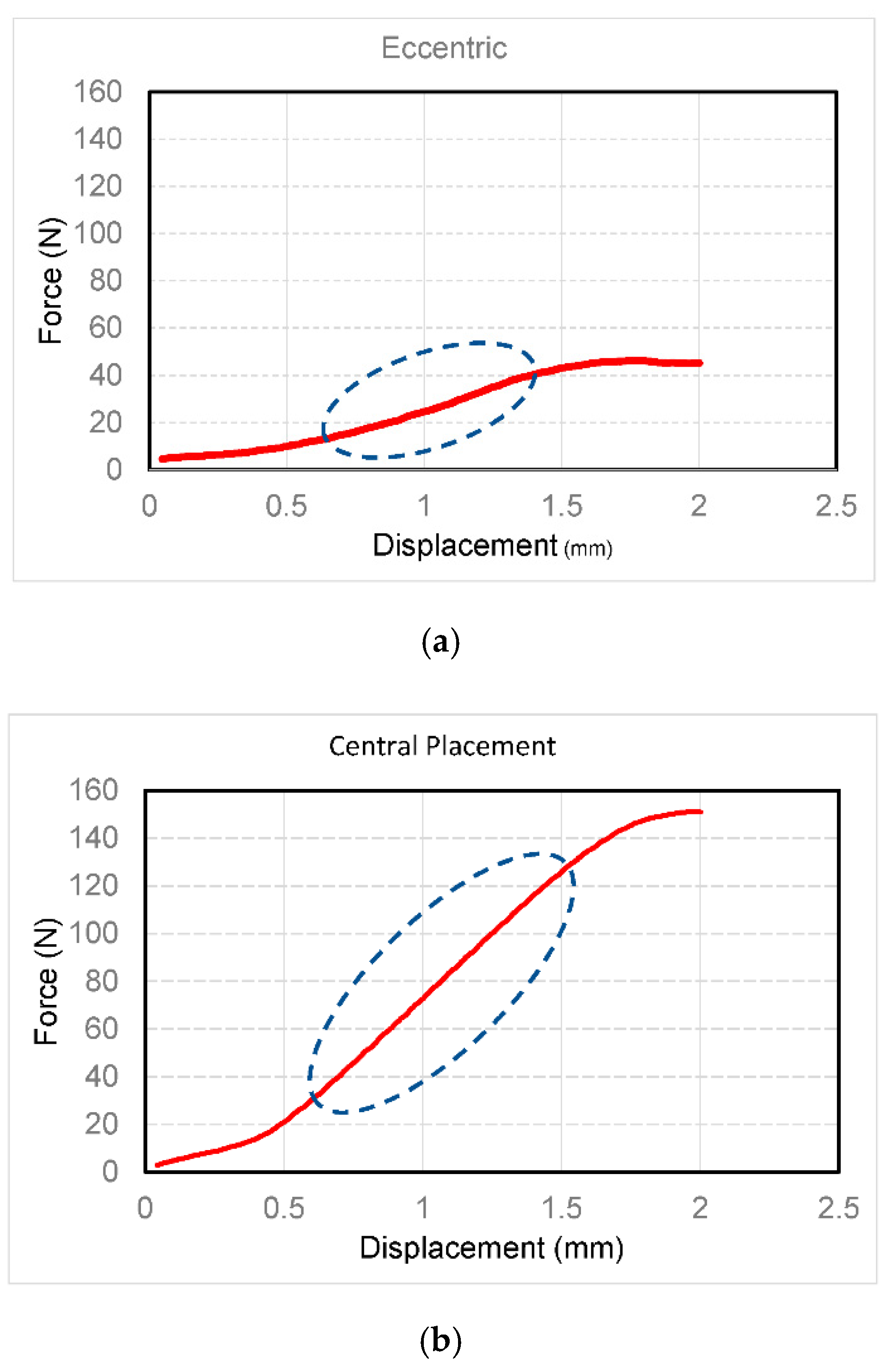
2.2. Biomechanical Testing of Fixation Stability

For biomechanical testing, a 1.2-mm Kirschner wire was horizontally passed through the proximal end of the scaphoid and lunate to provide additional stability, and the proximal fragment of each specimen was potted in a holder using polymethylmethacrylate. The scaphoid was oriented at a 45° angle to the horizontal plane to mimic its normal position in a wrist in the neutral position (Figure 3), and this orientation was consistent with previously published protocols [10,15,16]. This enabled the delivery of a dorsal-to-volar cantilever load, which represents the primary physiologic load encountered by the scaphoid [17]. Each specimen was then placed into a fixture with a pneumatically driven plunger resting on the surface of the distal pole.
A load was applied using a displacement-controlled test protocol in a material testing machine (MTS QTest-10 Testing System, MTS Systems Corporation, MN, USA) at a rate of 0.1 mm/min (MTS 4,501,030 Load Cell, the maximum capacity: 10 kN). The displacement rate was slow enough to mimic the quasi-static status of the biomechanical testing environment. Displacement of the apparatus was increased until it reached 2 mm, which has been known to result in higher nonunion rates of scaphoid waist fractures [18,19]. After applying 5 N preload to the specimen, we then used the load at 2 mm of displacement as endpoint and recorded it for further comparison. The stiffness was defined as the slope of the force–displacement curve during the linear region (Figure 4). It is a biomechanical index to evaluate the ability to withstand the deformation of a structure. This index could be compared objectively without considering the geometry and material of the structure. Therefore, the stiffness of both central and eccentric groups was also evaluated in our study. Finally, we recorded the failure mode of the screw-bone construct.

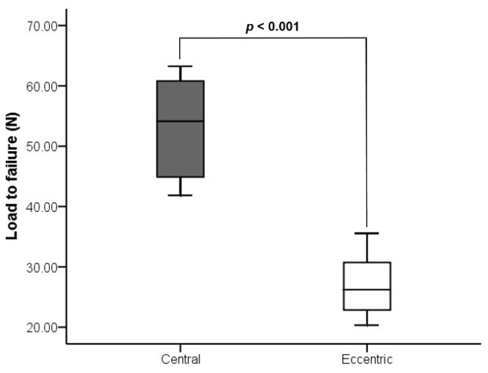
2.3. Statistical Analysis

We calculated the sample size using previous biomechanical testing data and by assuming equal numbers of specimens in each group. The sample size analysis showed that 8 matched pairs of scaphoids would be sufficient to provide significant data for 80% power with 95% confidence [10,15]. Stiffness and load to failure were compared between the groups using Mann–Whitney U tests. The tests were two-tailed, and p < 0.05 was considered statistically significant.

3. Results

The average specimen age was 57.5 years (±9.65 years) (Table 1). After we inspected all cadaveric specimens and found no macroscopic evidence of pathology, we identified one cadaveric specimen with scaphotrapeziotrapezoid arthritis during dissection of a scaphoid with a lunate from a cadaveric arm. However, biomechanical testing was undergone because the scaphoid and scapholunate ligament were intact. Next, we calculated measurements for all specimens. The mean length of scaphoids was 27.8 (standard deviation (SD) 1.90) mm. Furthermore, the use of a screw 4 mm shorter than what was measured worked well [14]. There was no screw prominence at the articular surface. The bone-screw construct failed on distal part and there was no difference between central or eccentric screw placement. Stiffness was higher in central (74.1 N/mm) than in eccentric screw placement (29.39 N/mm) (Figure 5). The median loads to failure in groups I and II were 54.14 and 26.22 N, respectively (Figure 6). Regarding the failure mode of the screw-bone construct, all were fractured at the screw–bone interface.

4. Discussion

The purpose of this cadaveric study was to biomechanically compare fixation strength between central and eccentric screw placements for the volar fixation of horizontal oblique scaphoid fractures. Our hypothesis was that central screw placement would achieve superior fixation strength for this issue. The results showed that central screw placement for volar fixation of horizontal oblique scaphoid fractures provided relatively better strength than eccentric placement in terms of biomechanical stability.
The complexity of the scaphoid structure results in different fracture patterns, making comparison of clinical outcomes difficult. Hence, we adopted a cadaveric study to evaluate the biomechanical stability of different screw placement techniques in the same fracture pattern. However, several limitations restrict the scope of this biomechanical comparison investigation. First is its post-mortem nature. Other factors such as postoperative rehabilitation and bone healing could not be taken into consideration in this cadaveric study. Such factors may underlie differences in the clinical results reported by other surgeons. Furthermore, the average specimen age was 57.5 years, whereas patients with scaphoid fractures are younger in our daily practice. Second, we focused our attention on the biomechanical stability of different screw placements, which is only one factor in choosing a fixation of scaphoid fractures. Additional research is needed to address other issues such as different screw designs, dorsal vs. volar approaches to the scaphoid, and proximal vs. waist fractures of the scaphoid. Since most orthopedic devices were made by metallic alloy, the mechanical performance of the orthopedic devices could be similar when the devices were applied under room temperature and body temperature. Therefore, the experiment did not include a normal saline bath under controlled temperature. Here, we only evaluated different screw placements for volar fixation of horizontal oblique scaphoid fractures because we wanted to simplify the conditions of the experiment. Another limitation of this study was that the strength of the scaphoid fixation in both central and eccentric placement groups seemed to be lower than that observed in previous studies [10,15,16]. These biomechanical cadaveric studies reported that load to failure (2 mm displacement) ranged from 59.1 to 324.4 N. The cadaveric scaphoids in this study were harvested from an older population (range: 42–71 years), and therefore the bone quality would be weaker than that of younger populations. This is a common shortcoming of all cadaveric studies. We attempted to determine the possible reasons to explain this result. First, other studies chose the longest possible screw because screw protrusion could be visually checked [10,15,16]. In contrast, our screw length was computed by subtracting 4 mm from the length of the guidewire. This method is frequently used in our clinical practice [14]. Using this technique, we could ensure a buried depth of 2 mm below the articular cartilage and avoid screw prominence at the articular surface. This resulted in a shorter screw in this study that could affect the fixation strength. However, Patel et al. demonstrated maximizing screw length may not provide superior fixation for proximal scaphoid fractures [20]. Therefore, a 2 mm gap at the fracture site that simulated a scaphoid waist fracture may have been the reason for inferior fixation strength. This setup did not represent the clinical practice that the fracture gap should be closed in order to achieve proper reduction. Nevertheless, this gap ensured that the applied force was carried solely by the implanted screw. Since the objective of this study was to evaluate the stability of the scaphoid fixation, the mechanical effect of bone contact should be eliminated like the test models of scaphoid (3 mm gap) and long bone fracture (10 mm gap) [21,22]. Other studies designed a simple osteotomy to simulate non-displaced or minimally displaced scaphoid waist fractures [10,15,16]. Therefore, the value for load to failure evaluated from the experiment could be lower than that of other studies. A 3D analysis of scaphoid waist fracture revealed that comminution and displacement rates were 11% and 28%, respectively [13]. This meant the fracture surface apposition may not be as good as we previously perceived. Further studies are warranted to analyze the effect of osteotomy gap and to prove which osteotomy is close to the reality.
Not surprisingly, the screw placement for volar fixation of scaphoid fractures may play an important role and affect clinical outcomes. Over the past few decades, several biomechanical studies have demonstrated that central screw placement in the proximal fragment, perpendicular screw trajectory, and longer screw length could provide better biomechanical and clinical results [15,22,23,24]. The common assumption that screw fixation of scaphoid fractures should be undergone in a uniform procedure and that a single screw direction would give optimal fracture fixation may simplify operations; however, this is usually not true for other fractures and should not be true for scaphoid ones [25]. Clearly, a large number of variables must be taken into account when assessing the healing potential of a scaphoid by screw fixation. The relationship between the fracture plane and scaphoid long axis may influence the healing potential. Scaphoid fractures are generally divided into three types: horizontal oblique, transverse, and vertical oblique, on the basis of the fracture plane [26].
With regards to vertical oblique fractures, Luria et al. conducted several studies and concluded that eccentric screw placement achieved perpendicular screw trajectory and resulted in a similar stability of fixation as central screw placement [16,25]. Next, the transverse waist fracture, which was considered the most common, accounted for 60% of scaphoid fractures [26]. Meermans et al. [10] designed a cadaveric osteotomy-simulated transverse fracture model to analyze the influence of different approaches and showed that the transtrapezial approach achieved central screw placement and perpendicular screw trajectory, and these offered a biomechanical advantage.
The current study emphasized the horizontal oblique fracture, which accounted for 35% of all scaphoid fractures [26]. In the past, it was considered the second most common. Recently, some surgeons have reported that most waist fractures were horizontal oblique and not transverse in orientation [13]. Therefore, it is necessary to discern the screw placement that would provide a biomechanical advantage for the most common scaphoid waist fractures. This study showed that central screw placement provided better strength than eccentric placement in terms of biomechanical stability. In contrast, several biomechanical studies demonstrated that more stable fixation of horizontal oblique fractures may be achieved by a screw placed perpendicular to the fracture plane in comparison with that along the long axis of the scaphoid [25,27], with perpendicular screw placement potentially maximizing the surface area available for healing [28]. Perpendicular screw fixation provides a biomechanical advantage in any fracture, not only in scaphoid fractures [24,25]. However, the entry point of perpendicular screw for horizontal oblique fractures is different from the current approaches, either central or eccentric. Faucher et al. proposed directing a perpendicular screw using a volar and proximal approach in a fully extended wrist and reported that the load to failure averaged 258 N for the perpendicular screw group and 294 N for the central screw group [24]. Due to the proximity of the major neurovascular structures and flexor tendons around the volar wrist, care must be taken during this approach, and further investigation is needed to evaluate the feasibility of this technique. Regarding the fracture angle, Faucher et al. created a horizontal oblique fracture, the so-called dorsal sulcus pattern of scaphoid fracture [24]. This study used a different fracture plane identified by Luria et al. The orientation of current fracture plane is between transverse and traditional horizontal oblique types [13].
In summary, while performing volar fixation of horizontal oblique scaphoid fractures, central screw placement demonstrated superior stability to eccentric placement in terms of biomechanical strength. However, further clinical investigations are warranted to evaluate whether the different screw placements for the volar approach of horizontal oblique scaphoid fractures impact the clinical outcomes.

Author Contributions

Conceptualization, C.-H.W. and T.-S.L.; methodology, C.-H.W., T.-S.L., C.-H.M., and C.-Y.Y.; experimental data analysis, C.-H.W. and T.-S.L.; writing—original draft preparation, C.-H.W.; writing—review and editing, C.-H.W. and T.-S.L.; supervision, I.-M.J.; project administration, C.-H.W. and Y.-K.T.; funding acquisition, C.-H.W. All authors have read and agreed to the published version of the manuscript.

Funding

The research was funded by E-DA Hospital, Taiwan (grant number EDAHP104018).

Conflicts of Interest

All authors declare no conflict of interest.

References

  1. Haddad, F.S.; Goddard, N.J. Acute percutaneous scaphoid fixation. J. Bone Joint Surg. Br. 1998, 80-B, 95–99. [Google Scholar] [CrossRef]
  2. Bond, C.D.; Shin, A.Y.; McBride, M.T.; Dao, K.D. Percutaneous screw fixation or cast immobilization for nondisplaced scaphoid fractures. J. Bone Joint Surg. Am. 2001, 83, 483–488. [Google Scholar] [CrossRef] [PubMed]
  3. Geissler, W.B.; Adams, J.E.; Bindra, R.R.; Lanzinger, W.D.; Slutsky, D.J. Scaphoid fractures: What’s hot, what’s not. J. Bone Joint Surg. Am. 2012, 94, 169–181. [Google Scholar] [CrossRef] [PubMed]
  4. Merrell, G.; Slade, J. Technique for percutaneous fixation of displaced and nondisplaced acute scaphoid fractures and select nonunions. J. Hand Surg. Am. 2008, 33, 966–973. [Google Scholar] [CrossRef] [PubMed]
  5. Slade, J.F.; Grauer, J.N.; Mahoney, J.D. Arthroscopic Reduction and Percutaneous Fixation of Scaphoid Fractures With a Novel Dorsal Technique. Orthop. Clin. N. Am. 2001, 32, 247–261. [Google Scholar] [CrossRef]
  6. Chan, K.W.; McAdams, T.R. Central screw placement in percutaneous screw scaphoid fixation: A cadaveric comparison of proximal and distal techniques. J. Hand Surg. Am. 2004, 29, 74–79. [Google Scholar] [CrossRef]
  7. Meermans, G.; Verstreken, F. Influence of screw design, sex, and approach in scaphoid fracture fixation. Clin. Orthop. Relat Res. 2012, 470, 1673–1681. [Google Scholar] [CrossRef] [Green Version]
  8. Acar, B.; Kose, O.; Kati, Y.A.; Egerci, O.F.; Turan, A.; Yuksel, H.Y. Comparison of volar versus dorsal screw fixation for scaphoid waist fractures: A finite element analysis. Orthop. Traumatol. Surg. Res. 2018, 104, 1107–1113. [Google Scholar] [CrossRef]
  9. Bushnell, B.D.; McWilliams, A.D.; Messer, T.M. Complications in dorsal percutaneous cannulated screw fixation of nondisplaced scaphoid waist fractures. J. Hand Surg. Am. 2007, 32, 827–833. [Google Scholar] [CrossRef]
  10. Meermans, G.; Van Glabbeek, F.; Braem, M.J.; van Riet, R.P.; Hubens, G.; Verstreken, F. Comparison of two percutaneous volar approaches for screw fixation of scaphoid waist fractures: Radiographic and biomechanical study of an osteotomy-simulated model. J. Bone Joint Surg. Am. 2014, 96, 1369–1376. [Google Scholar] [CrossRef]
  11. Zlotolow, D.A.; Knutsen, E.; Yao, J. Optimization of volar percutaneous screw fixation for scaphoid waist fractures using traction, positioning, imaging, and an angiocatheter guide. J. Hand Surg. Am. 2011, 36, 916–921. [Google Scholar] [CrossRef] [PubMed]
  12. Meermans, G.; Verstreken, F. Percutaneous transtrapezial fixation of acute scaphoid fractures. J. Hand Surg. Eur. Vol. 2008, 33, 791–796. [Google Scholar] [CrossRef] [PubMed]
  13. Luria, S.; Schwarcz, Y.; Wollstein, R.; Emelife, P.; Zinger, G.; Peleg, E. 3-dimensional analysis of scaphoid fracture angle morphology. J. Hand Surg. Am. 2015, 40, 508–514. [Google Scholar] [CrossRef] [PubMed]
  14. Fowler, J.R.; Ilyas, A.M. Headless compression screw fixation of scaphoid fractures. Hand Clin. 2010, 26, 351–361. [Google Scholar] [CrossRef]
  15. McCallister, W.V.; Knight, J.; Kaliappan, R.; Trumble, T.E. Central placement of the screw in simulated fractures of the scaphoid waist: A biomechanical study. J. Bone Joint Surg. Am. 2003, 85, 72–77. [Google Scholar] [CrossRef]
  16. Luria, S.; Lenart, L.; Lenart, B.; Peleg, E.; Kastelec, M. Optimal fixation of oblique scaphoid fractures: A cadaver model. J. Hand Surg. Am. 2012, 37, 1400–1404. [Google Scholar] [CrossRef]
  17. Smith, D.K.; Cooney, W.P., 3rd; An, K.N.; Linscheid, R.L.; Chao, E.Y. The effects of simulated unstable scaphoid fractures on carpal motion. J. Hand Surg. Am. 1989, 14, 283–291. [Google Scholar] [CrossRef]
  18. Kulkarni, R.W.; Wollstein, R.; Tayar, R.; Citron, N. Patterns of healing of scaphoid fractures. The importance of vascularity. J. Bone Joint Surg. Br. 1999, 81, 85–90. [Google Scholar] [CrossRef]
  19. Amirfeyz, R.; Bebbington, A.; Downing, N.D.; Oni, J.A.; Davis, T.R. Displaced scaphoid waist fractures: The use of a week 4 CT scan to predict the likelihood of union with nonoperative treatment. J. Hand Surg. Eur. Vol. 2011, 36, 498–502. [Google Scholar] [CrossRef]
  20. Patel, S.; Giugale, J.; Tiedeken, N.; Debski, R.E.; Fowler, J.R. Impact of Screw Length on Proximal Scaphoid Fracture Biomechanics. J. Wrist Surg. 2019, 8, 360–365. [Google Scholar] [CrossRef] [Green Version]
  21. Ma, C.H.; Wu, C.H.; Tu, Y.K.; Lin, T.S. Metaphyseal locking plate as a definitive external fixator for treating open tibial fractures--clinical outcome and a finite element study. Injury 2013, 44, 1097–1101. [Google Scholar] [CrossRef] [PubMed]
  22. Dodds, S.D.; Panjabi, M.M.; Slade, J.F., 3rd. Screw fixation of scaphoid fractures: A biomechanical assessment of screw length and screw augmentation. J. Hand Surg. Am. 2006, 31, 405–413. [Google Scholar] [CrossRef] [PubMed]
  23. Trumble, T.E.; Gilbert, M.; Murray, L.W.; Smith, J.; Rafijah, G.; McCallister, W.V. Displaced scaphoid fractures treated with open reduction and internal fixation with a cannulated screw. J. Bone Joint Surg. Am. 2000, 82, 633–641. [Google Scholar] [CrossRef] [PubMed]
  24. Faucher, G.K.; Golden, M.L., 3rd; Sweeney, K.R.; Hutton, W.C.; Jarrett, C.D. Comparison of screw trajectory on stability of oblique scaphoid fractures: A mechanical study. J. Hand Surg. Am. 2014, 39, 430–435. [Google Scholar] [CrossRef] [PubMed]
  25. Luria, S.; Hoch, S.; Liebergall, M.; Mosheiff, R.; Peleg, E. Optimal fixation of acute scaphoid fractures: Finite element analysis. J. Hand Surg. Am. 2010, 35, 1246–1250. [Google Scholar] [CrossRef] [PubMed]
  26. Russe, O. Fracture of the carpal navicular. Diagnosis, non-operative treatment, and operative treatment. J. Bone Joint Surg. Am. 1960, 42-a, 759–768. [Google Scholar] [CrossRef] [PubMed] [Green Version]
  27. Volk, I.; Gal, J.; Peleg, E.; Almog, G.; Luria, S. Three-dimensional comparison of alternative screw positions versus actual fixation of scaphoid fractures. Injury 2017, 48, 1183–1189. [Google Scholar] [CrossRef]
  28. Hart, A.; Mansuri, A.; Harvey, E.J.; Martineau, P.A. Central versus eccentric internal fixation of acute scaphoid fractures. J. Hand Surg. Am. 2013, 38, 66–71. [Google Scholar] [CrossRef]
Figure 1. An eccentric placement of guidewire under the guidance of an aiming device.
Applsci 10 08592 g001
Figure 2. Posteroanterior (A) and lateral (C) radiographs of the scaphoid specimen showing central screw position. Posteroanterior (B) and lateral (D) radiographs of the scaphoid specimen showing eccentric screw position.
Applsci 10 08592 g002
Figure 3. The potted scaphoid specimen and testing apparatus for delivery of a dorsal-to-volar cantilever bending load.
Applsci 10 08592 g003
Figure 4. The force–displacement curve of (a) eccentric and (b) central placement of the bone screw. The stiffness was defined as the slope of the force–displacement curve during the dotted circle (linear) region.
Applsci 10 08592 g004
Figure 5. Boxplot of interquartile range (IQR) showed the median values for stiffness in the central placement and eccentric placement groups. Whiskers indicate the maximum or minimum value.
Applsci 10 08592 g005
Figure 6. IQR of load to failure in the central placement and eccentric placement groups. Whiskers indicate the maximum or minimum value.
Applsci 10 08592 g006
Table 1. Specimen characteristics.
NumberAge/GenderGroupSideLength (mm)NumberAge/GenderGroupSideLength (mm)
154/FCentralL25.0971/MCentralR31.0
254/FEccentricR25.51071/MEccentricL31.5
366/FCentralL26.01151/MCentralL30.0
466/FEccentricR26.51251/MEccentricR29.5
559/MCentralR27.51342/MCentralR28.5
659/MEccentricL27.51442/MEccentricL28.5
750/FCentralL27.01567/FCentralR27.0
850/FEccentricR26.51667/FEccentricL27.5
Publisher’s Note: MDPI stays neutral with regard to jurisdictional claims in published maps and institutional affiliations.

