Article

Ageing-Related Gait Adaptations to Knee Joint Kinetics: Implications for the Development of Knee Osteoarthritis

Institute for Health and Sport (IHES), Victoria University, Melbourne, VIC 3011, Australia
*
Author to whom correspondence should be addressed.
Appl. Sci. 2020, 10(24), 8881; https://doi.org/10.3390/app10248881
Submission received: 26 October 2020 / Revised: 8 December 2020 / Accepted: 10 December 2020 / Published: 12 December 2020
(This article belongs to the Special Issue Biomechanical and Biomedical Factors of Knee Osteoarthritis)

Abstract

:
The prevalence of knee osteoarthritis (OA) increases with ageing and this research aimed to identify gait adaptations that could reduce OA by investigating ageing effects on knee joint biomechanics. Participants were 24 healthy young males (18–35 yrs) and 14 healthy older males (60–75 yrs). Three-dimensional motion capture (Optotrak) and walkway-embedded force plates (AMTI) recorded their natural preferred-speed walking and the following parameters were computed: knee adduction moment, knee joint vertical force, foot contact angle, toe-out angle, foot centre of pressure displacement, time to foot flat, step length, step width and double support time. A 2 × 2 (age × limb) repeated measures mixed model analysis of variance design determined main effects and interactions. Pearson’s correlations between knee kinetic parameters and stride phase variables were also calculated. Both knee adduction moment and vertical joint force were larger in the older group. Relative to the young controls, older individuals showed a longer time to foot flat, less toe-out angle and wider steps. Correlation analysis suggested that reduced toe-out angle and increased step width were associated with lower knee adduction moment; furthermore, knee joint vertical force reduced with greater step length. Future research could focus on intervention strategies for managing excessive knee joint stresses to prevent the ageing-related development of knee OA.

1. Introduction

Knee osteoarthritis (OA) is a chronic and progressive condition that affects gait function and associated qualify of life. The condition is prevalent in senior individuals [1] and although the causes of OA are multifactorial, some risk factors are considered to be manageable. For example, people take 10,000 steps or more every day and modifications to walking patterns may, therefore, reduce the effects of suboptimal gait features that exacerbate OA. Bodyweight management can also effectively control knee joint kinetics in association with OA risk. Biomechanically, knee joint kinetics, particularly in early stance following foot contact, are expected to be indicative of OA risk [2]. The aim of the present investigation was to determine whether the management of foot contact impact and associated knee joint forces could be effective in controlling the progression of OA.
For OA prevention, more energy-efficient loading is key to reducing damage to the knee. This can be achieved by oscillating “impact” of foot contact through the stance phase as mechanical energy to activate toe-off without relying excessively on voluntary push-off [3]. The energy efficiency of loading in walking has been reported to be 60–70% due to mechanical energy dissipated in early stance; later required to be compensated by an injection of push-off forces [4,5]. Inefficient limb loading at foot contact may, therefore, lead to excessive mechanical energy transfer to the knee joint, stressing the protective articular cartilage and ligaments [6,7]. While a physical stimulus is important in promoting articular cartilage synthesis, excessive loading in everyday walking for a prolonged period of time can contribute to OA progression [6,8]. It is, therefore, important to investigate how ageing affects gait biomechanics influence foot–ground impact and associated knee joint kinetics. Our focus in this study was ageing effects on knee joint adduction moments and vertical forces (Figure 1).
Knee adduction moment is recognised as the key kinetic marker for Varus deformity and associated OA development, principally in the medial compartment [9]. Reducing adduction moment during early stance has, therefore, been proposed as an OA intervention and gait adaptations; less toe-out angle and increasing step width have been reported to reduce initial peak adduction moment and associated OA risk [10]. In addition, peak vertical knee joint force during early stance reflects the primary load and has been associated with damage to joint structures [11,12]. Due, in part, to difficulties in modelling knee joint forces their role in OA has been less thoroughly investigated but of the total knee joint force, the vertical component is largest due to gravitational effects on the whole-body centre of mass [13]. Chen et al. [14], for example, reported that increased body mass due to obesity imposed the larger loads on the knee joint and increased OA risks, indicating that vertical force is a risk factor.
Most previous experimental protocols designed to modify walking patterns in OA patients instructed participants to walk in certain ways to examine whether the gait adaptations would change knee joint kinetics [15,16]. In this project, however, as OA is more prevalent in older people, we compared the “natural” gait patterns of healthy, physically active older adults and young controls. It was hypothesised that older adults’ gait patterns would reflect increased knee loads, indicated by greater knee adduction moments and peak vertical force. We also correlated the knee kinetics and stride phase variables to determine the qualitative aspects of gait adaptations that may be effective in reducing the adverse effects of knee joint kinetics. A major advantage of correlation analysis is the capacity to probe the links between gait parameters without manipulating walking patterns experimentally.
To investigate potential associations with the loading response on the knee [17,18] in the current study we expanded the focus from fundamental spatio-temporal parameters, such as step width and toeing out [15], to other foot control variables; including foot contact angle, foot centre of pressure (COP) movement and time to foot flat. The research question addressed here was whether ageing-related gait adaptations may contribute to changes in knee joint kinetics that are specifically associated with the development of OA. As described above, if less toeing-out and wider steps reduce knee adduction moments, it was expected that correlation analysis would reveal other gait parameters that contribute to alleviating stresses on the knee joint. Such findings would be important in suggesting gait adaptations in older adults that could reduce both knee joint forces and adduction moments, leading to improved interventions for preventing OA, such as targeted, practical exercise programs specialised remedial footwear designs.

2. Materials and Methods

2.1. Participants

Participants were 24 healthy young (Mean 23.8 ± 3.2 yrs, Range 18–35 yrs) and 14 healthy older males (Mean 73.4 ± 8.0 yrs, Range 60–75 yrs); all participants were free of injuries or health conditions that may have adversely affected their gait. Physical characteristics were; height (young: 1.75 ± 0.05 m, older: 1.76 ± 0.06 m) and mass (young: 73.1 ± 12.7 kg, older: 77.8 ± 4.4 kg). Age groups were differentiated by age (p < 0.01) but not height (p > 0.05) or body mass (p > 0.05). To exclude gender effects and gait pathologies, including those due to OA, participants were limited to healthy males. Three participants in each group were left foot dominant, as determined using a procedure [19]. Older participants all maintained independent lifestyles, were capable of walking freely for 30 min or longer and reported no falls within the previous two years. Young participants were university volunteers while the older group was recruited through advertisements in a local newsletter. The Victoria University Human Research Ethics Committee approved the experimental protocol and all participants completed the approved informed consent procedures prior to participation.

2.2. Experimental Setup

Three Optotrak (Optotrak®, NDI, Waterloo, ON, Canada) motion capture cameras and two AMTI force plates sampling at 1000 Hz were set up on an 8-metre laboratory walkway. The three-dimensional (3D) position coordinates of joint anatomical landmarks were sampled from the Optotrack Infrared Light Emitting Diodes (IREDs) at 100 Hz. Prior to data collection, static trials were taken in the anatomical position for 5 s to later estimate joint kinematics and kinetics using biomechanical modelling within Visual 3D suits (C-motion, Inc., Germantown, MD, USA). A lower body skeleton model was developed to compute joint kinetics based on inverse dynamics. The femur was modelled by the greater trochanter, quadrate tubercle, lateral and medial epicondyle. The lateral and medial condyles and lateral and medial malleolus of tibia defined the shank. The foot complex was built using the heel (i.e., the proximal end of the foot) and the 2nd and 5th metatarsal heads, toe (i.e., the most anterior and superior surface of the foot) and lateral and medial malleolus.

2.3. Protocol

All participants walked in a straight line at preferred speed to collect 30 trials and either two or three complete gait cycles from both limbs were sampled on each trial (i.e., a total of 60 to 90 stride cycles per subject). A start position was determined such that at preferred walking speed either foot would strike the first plate and the other foot would land completely on the further (second) force plate. Raw position–time data were first interpolated using a window of up to 10 frames (0.1 s) to compensate any occluded signals. A 4th order zero-lag Butterworth Filter with a cut-off frequency of 15 Hz was then applied to smooth the kinematic data [20]. To define gait cycle phases and subsequent spatio-temporal parameters, toe-off and heel contact events were identified using algorithms based on widely-accepted kinematic parameters; validated using a criterion vertical ground reaction force threshold of 5N for both heel contact and toe-off, e.g., [21,22,23].

2.4. Parameters

There were no significant differences in physical characteristics between age groups as reported and, therefore, normalisation procedures were not applied to any variables, including kinetics, kinematics and spatio-temporal gait parameters. All parameters were described by the mean and standard deviation (SD), where SD reflects intrasubject variability, indicating consistency of gait parameters across multiple gait cycles (i.e., 60–90).

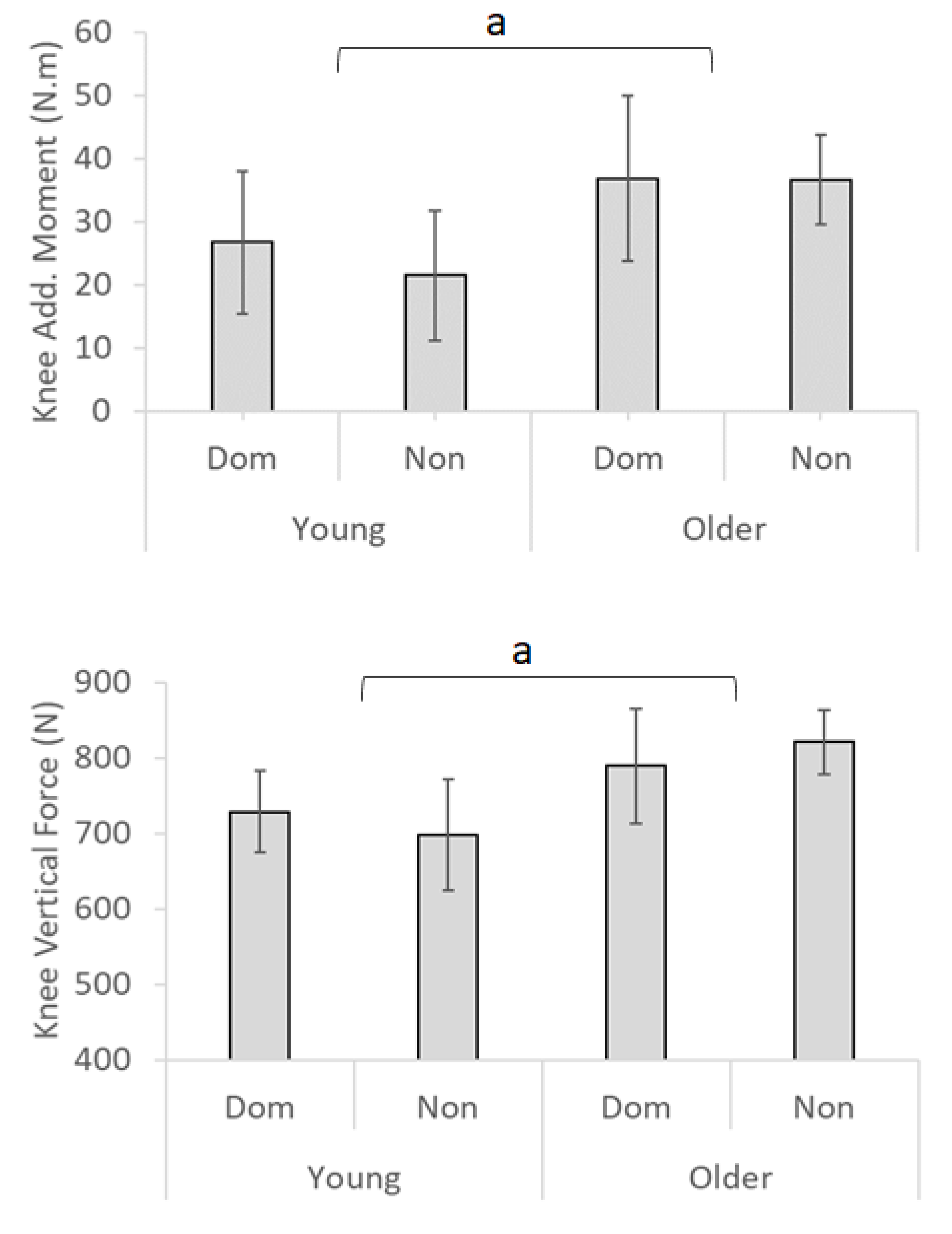
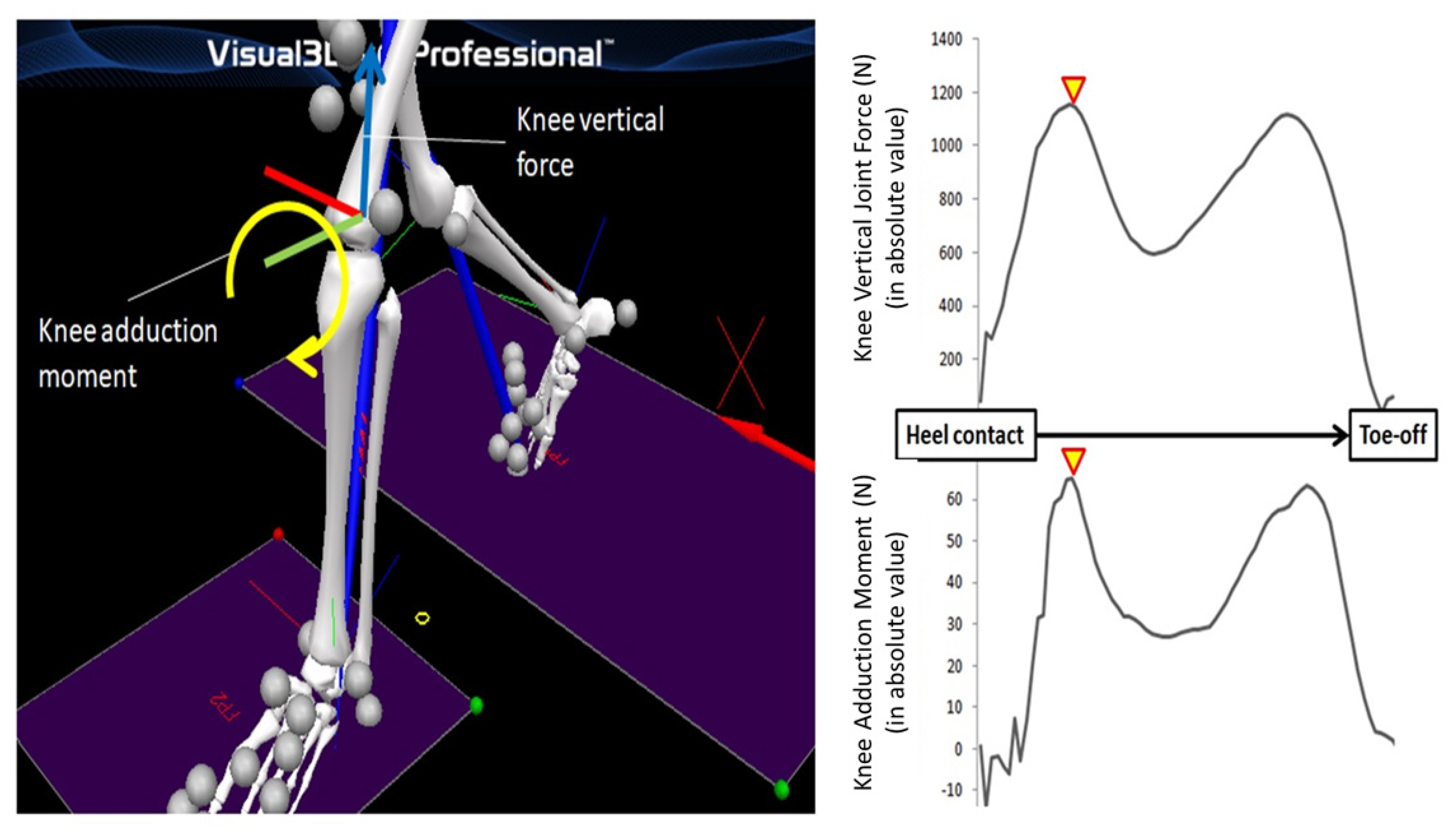
2.4.1. Knee Joint Kinetics
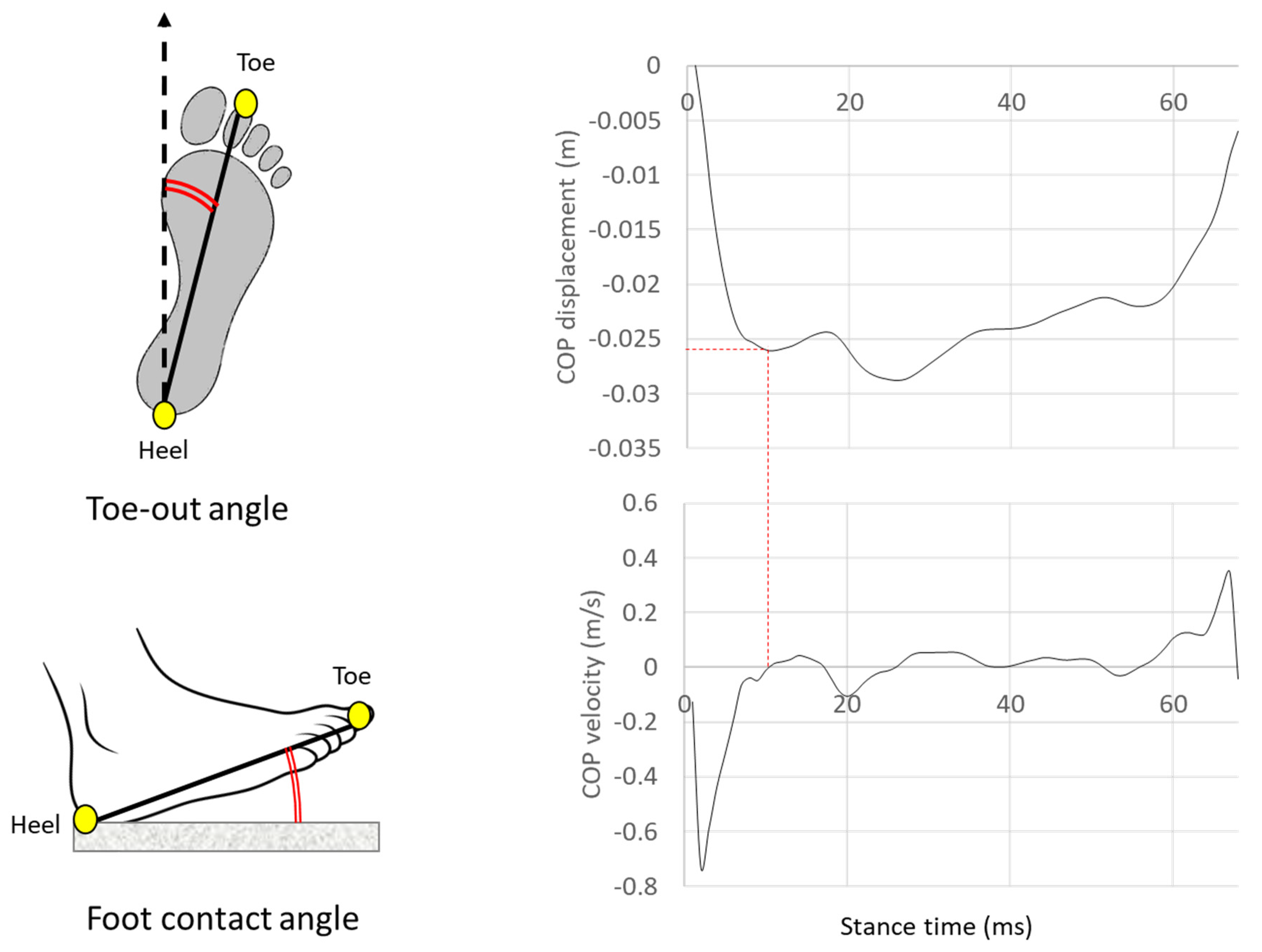
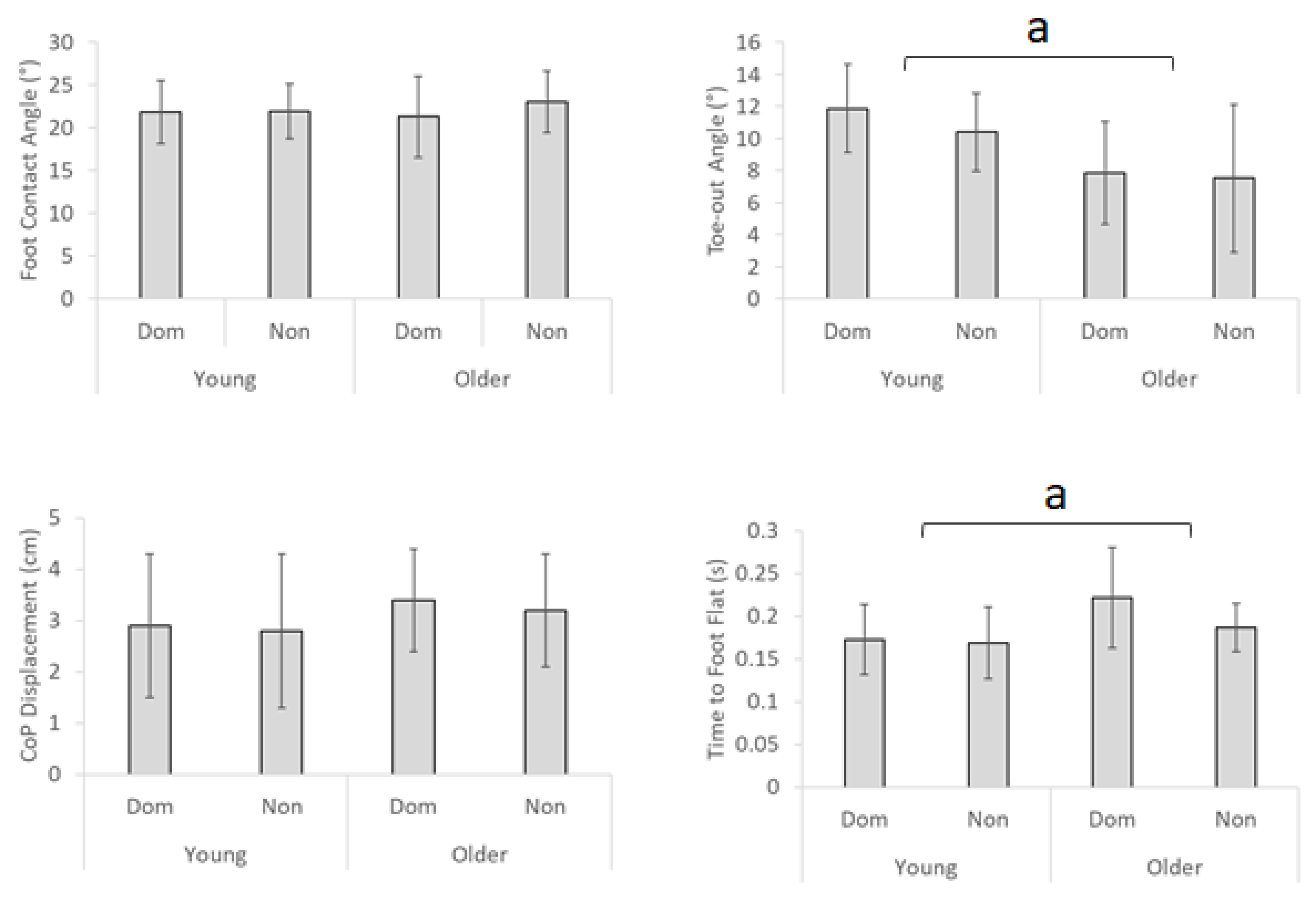
Knee joint kinetics were computed by inverse dynamics based on 3D modelling of lower limb joints including, foot, shank and femur (Figure 1). Rotational torque around the anterior–posterior axis was defined as knee adduction moment and the first peak during the stance phase was obtained. Knee vertical force was also measured at the first peak. Both kinetic parameters were indicated in absolute value as in Figure 1.

2.4.2. Foot Motion

As illustrated in Figure 2 (left) foot contact angle was formed between an axis connecting the toe-heel relative to the ground reference in the sagittal plane at heel contact. Toe-out angle was defined at heel contact in the transverse plane as toe-heel line relative to the walking (anterior) direction.

2.4.3. COP Displacement

As described in Figure 2 (right), COP displacement was defined from heel contact to the initial peak lateral displacement at zero velocity, analysed in absolute values. COP displacement indicates lateral COP motion immediately following foot contact.

2.4.4. Time to Foot Flat

Foot flat was identified at the initial frame where the vertical toe location after heel contact descended to the height obtained from the static calibration data recorded for 3D modelling. Time was recorded from heel contact to foot flat [24].

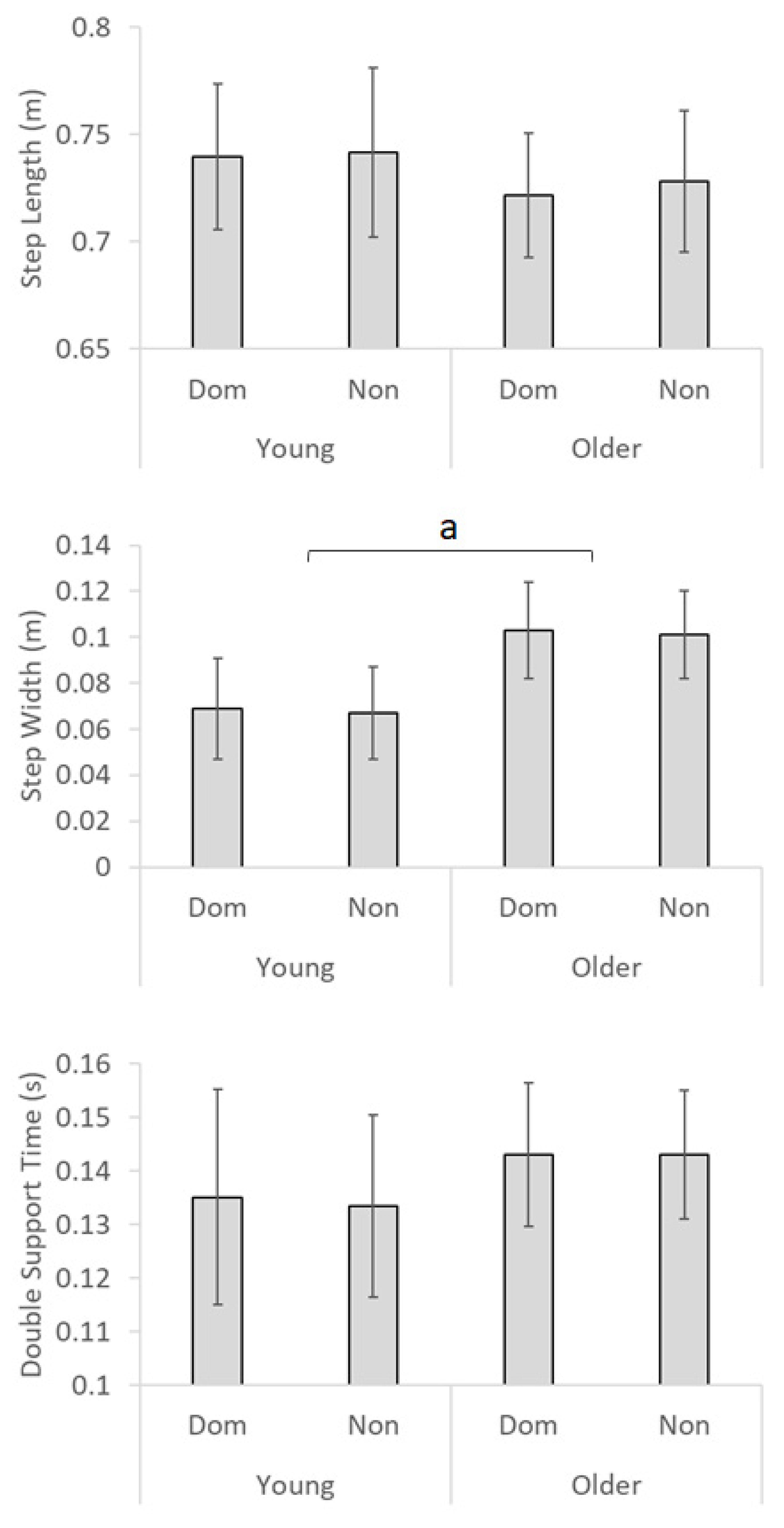
2.4.5. Spatio-Temporal Parameters

Step length and width were displacements between two consecutive heel contacts in anterior–posterior and medio-lateral directions, respectively. Double support time was temporal period from heel contact of one limb to contralateral toe-off, when both feet are on the walking surface. Lead-limb defined the dominance of parameters.

2.5. Statistical Analysis

A 2 × 2 (age × limb) mixed model analysis of variance (ANOVA) design was applied separately to all dependent variables to determine main effects and interactions. Tukey’s posthoc analysis was conducted for any interaction effect. Pearson’s correlations between knee kinetic parameters and other gait variables were calculated. All statistical tests were accepted as significant when computed “p” values ≤ 0.05.

3. Results

As described in Figure 3, the knee adduction moment was higher in the older group by 12.5 Nm as the main effect (F1, 36 = 12.80, p < 0.01). Limb effect indicated that knee adduction moment in the dominant limb was more variable than the non-dominant side by approximately 3 Nm (F1, 36 = 6.13, p < 0.05).
Vertical knee joint force was also 91.5 N greater in the older group (F1, 36 = 6.25, p < 0.05).
In Figure 4, ageing effects were observed in 0.034 s longer time to foot flat (F1, 36 = 7.09, p < 0.01) and 3.45° less toe-out angle (F1, 36 = 7.56, p < 0.01). No other statistical effects were identified.
As described in Figure 5, statistical significance was obtained only in the ageing effect on larger step width by 0.034 m (F1, 36 = 13.32, p < 0.01).
Table 1 presents correlations between knee kinetics and other examined gait parameters. High knee adduction moment was found to accompany greater variability, while the variability of knee joint force was negatively associated with increased adduction moment. Knee force and adduction moment demonstrated inverse correlations but this effect was qualified only in the young group. For foot contact angle, positive correlations were seen with knee adduction moment in the older group. Toe-out angle showed positive correlations with knee adduction moment only in the older group.
Correlation patterns with spatio-temporal parameters demonstrated that increased step length was interlinked with reduced knee force. Step length variability was negatively correlated with knee adduction moment. Increased step width in the older group was linked with lower knee adduction moment. Furthermore, step length was positively associated with foot contact angle (young: r = 0.454, p < 0.01, older: r = 0.419, p < 0.05) and negatively with double support time (young: r = −0.397, p < 0.05, older: r = −0.535, p < 0.01). Toe-out angle was negatively correlated with step width for the young (r = −0.650, p < 0.01) and older group (r = −0.493, p < 0.01). Increased toe-out angle was further correlated with COP movement (young r = 0.425, p < 0.01; older r = 0.707, p < 0.01).

4. Discussion

Older adults demonstrated higher knee joint loading confirmed by greater knee adduction moment and joint vertical force. Positive correlations between mean and SD suggest that increased knee adduction moment may be intermittent rather than on every step for the current healthy populations. In contrast, knee vertical force did not show this tendency, possibly indicating that high joint vertical forces may be more prevalent than adduction moment. As physical characteristics such as height and body mass were similar between the two age groups, the observed differences in knee joint kinetics can be attributed to ageing effects on walking patterns. While ageing has been reported to cause shorter step length, larger step width and prolonged double support [20], only increased step width was identified as the ageing effect on spatio-temporal parameters in the current study. This inconsistency can be explained by the fact that older participants in the current study were limited to healthy individuals, capable of walking for 30 min or longer and testing was conducted on an unobstructed laboratory walkway.
Although gait parameters were comparable between the two age groups, some foot control variables showed ageing effects, including less toe-out angle, greater time to foot flat and increased step width. Consistent with previous studies [15,16], correlations results suggested that both reduced toe-out angle and increased step width seen in older adults were associated with lower knee adduction moment. Toe-in gait has recently received attention for its effects on reducing knee adduction moment, possibly due to foot internal rotation preventing tibial abduction and shortening moment arm [2]. Traditionally, eversion support during early stance was considered to reduce moment arm of knee adduction moment and lateral wedge shoe-insole is widely recognised as the footwear intervention to evert the ankle and reduce adduction moment [25,26]. It is, therefore, possible that both eversion and internal rotation of the ankle during early stance may be effective in reducing knee adduction moment. Charlton et al. [27] also reported the possibility that toe-in gait may stimulate certain lower limb muscles and potentially help relieve knee joint loading. Negative correlations between toe-out angle and step width identified in the current research implied that wider steps would be closely associated with less toe-out angle (i.e., toeing-in), also found by Bennett et al. [15]. Increased step width, the medio-lateral displacement between the two heels, tends to result in pointing the toes inward relative to the heels. Age-related increase in step width and reduced toe-out angle may, therefore, be adaptive in achieving lower knee adduction moment among older individuals.
Contrary to expectation, our investigation of foot control mechanics, including foot contact angle, COP control and time to foot flat showed few direct associations with knee joint kinetics. Preventing flat-foot contact and the associated increase in time to foot flat were hypothesised to dissipate foot contact impact but the obtained results suggest that knee joint kinetics may be independent of these foot control mechanics. It is, however, possible that such foot impact dissipation strategies could reduce stress on the ankle or other lower limb joints. Toe-out gait was, however, found to be associated with larger COP displacement, possibly disadvantageous in dynamic balance control [28]. A further interesting finding, common to both age groups, was negative correlations between step length and knee joint force. Taking longer steps is possibly related to reduced knee joint force. This is a reasonable hypothesis because rather than stressing the knee joint, force may be utilised to increase step length. Efforts to take longer steps may reduce knee joint vertical compression. Interestingly, larger foot contact angle was associated with increased step length. Dorsiflexion at heel contact is responsible for increasing foot contact angle and ageing often impairs dorsiflexor strength [29].
Strength training or footwear interventions to compensate reduced dorsiflexion can possibly contribute to longer strides, which may reduce vertical knee joint forces by directing ground reaction force anterior. Caution should, however, be exercised with older adults because increased foot contact angle was accompanied by larger knee adduction moment. Shorter double support time is also considered to promote longer step length. As prolonged double support time has been identified as a typical gait adaptation to secure balance, particularly among frail populations including seniors [20], gait training to reduce excessively long double support time may help increase step length and potentially contribute to a reduction in knee joint vertical force. Balance support should be carefully considered when walking with longer steps or shorter double support time than usual for training purpose [9].
While the current research aimed to investigate whether ageing was a factor affecting knee joint kinetics and associated OA risks [7], the primary aim was to identify effective gait modifications to reduce OA risks by focusing on knee adduction moment and knee joint vertical force. Based on the current findings, practical intervention strategies to reduce OA risk should be devised with the following considerations. First, some reports have not identified direct links between these parameters and OA risk. From the perspective of gait biomechanics, knee joint kinetics should indicate stress of the knee joint, whereby damage beyond a threshold intensity, frequency and duration could lead to OA. Most well-known is adduction moment that compresses the medial part of the knee but controversy remains as to whether knee adduction moment is related to OA [7].
Second, modelling of knee joint kinetics is complex leading to different normalisation techniques, making direct comparisons between studies difficult. Body mass and stature can affect joint kinetics but in a comparison between groups, if there are no differences in physical characteristics, normalisation may not be necessary. When mean values are normalised, however, coefficient of variation, indicated by standard deviation divided by mean (SD/mean), may be the more suitable measure for variability. Joint moment calculation is relative to the length of the segment (i.e., bone) but normalisation by height may cause inaccuracy. Furthermore, joint kinetics are based on 3D biomechanics and there are multiple components to be considered for comprehensive modelling, including muscle forces and contributions from surrounding connective tissues [11]. In non-invasive software simulation, however, the addition of multiple factors can often require extra assumptions for modelling. In the current study, therefore, a relatively simple approach based on inverse dynamics within Visual 3D (C-motion) has been employed to measure knee adduction moment and vertical joint force.
Third, other biomechanical factors should be investigated to fully understand the role of joint kinetics in knee OA. One essential component is a sagittal plane analysis of knee joint kinetics involving flexion-extension moment and associated eccentric work and power absorption [30]. Quadriceps and other muscle groups (i.e., hamstrings, calf, plantarflexors and dorsiflexors) have been reported to help impact absorption [12,30,31,32]. It is also necessary to acknowledge that knee joint loading usually has two peaks in a gait cycle, as shown in Figure 1: early and late stance for foot contact and toe-off, respectively. While the current research focused on foot contact mechanics and associated OA risk, the push-off phase of the gait cycle may be equally important, with knee flexion moment considered a key kinetic marker [33]. Connective tissues such as ligaments should also be taken into account to fully describe the causes of OA and in terms of recovery, cartilage resynthesis in addition to mechanical factors relating to OA risks should be the essential aspect [8,34].
For the future direction of the study about biomechanical gait adaptations to control knee joint kinetics, the limitations of the current study have been addressed here. For the elimination of gender effects on obtained parameters, only the male population has been included in analysis. In future studies, it is necessary to confirm whether the findings of the current study are applicable to the female population. Comparison of healthy young and older individuals was appropriate to address the research aim of whether ageing alone influences joint kinetics associated with OA. While the current research design has identified the OA implications of knee adduction moment and vertical compression force, frailer seniors or older adults with OA symptoms may have different OA-related joint kinetics.
The current research thus provides the foundation for future studies to further our biomechanical and biomedical understanding of knee OA. In conclusion, both knee adduction moment and knee joint vertical compression force were found to be larger among the older group. Increased step width and reduced toe-out angle seem to be ageing-related gait adaptations to reduce knee adduction moment. Increased step length was, in contrast, possibly a strategy to reduce knee joint vertical force, which can be attained by an increased foot contact angle. The current outcomes should be used to design future studies to propose practical intervention strategies to minimise OA risk, particularly among older adults.

Author Contributions

Conceptualization, H.N. and R.B.; methodology, H.N.; formal analysis, H.N.; writing—original draft preparation, H.N.; writing—review and editing, R.B.; supervision, R.B.; project administration, R.B. All authors have read and agreed to the published version of the manuscript.

Funding

This research received no external funding.

Acknowledgments

The authors wish to thank Tony Sparrow for helpful comments on the manuscript prior to submission.

Conflicts of Interest

The authors declare no conflict of interest.

References

  1. Xie, Y.; Yu, Y.; Wang, J.-X.; Yang, X.; Zhao, F.; Ma, J.-Q.; Chen, Z.-Y.; Liang, F.-R.; Zhao, L.; Cai, D.-J.; et al. Health-related quality of life and its influencing factors in Chinese with knee osteoarthritis. Qual. Life Res. 2020, 29, 2395–2402. [Google Scholar] [CrossRef] [PubMed]
  2. Levinger, P.; Menz, H.B.; Morrow, A.D.; Bartlett, J.R.; Feller, J.; Bergman, N.R. Relationship between foot function and medial knee joint loading in people with medial compartment knee osteoarthritis. J. Foot Ankle Res. 2013, 6, 33. [Google Scholar] [CrossRef] [PubMed] [Green Version]
  3. Huang, T.-W.P.; Shorter, K.A.; Adamczyk, P.G.; Kuo, A.D. Mechanical and energetic consequences of reduced ankle plantar-flexion in human walking. J. Exp. Biol. 2015, 218, 3541–3550. [Google Scholar] [CrossRef] [PubMed] [Green Version]
  4. Sasaki, K.; Neptune, R.R. Muscle mechanical work and elastic energy utilization during walking and running near the preferred gait transition speed. Gait Posture 2006, 23, 383–390. [Google Scholar] [CrossRef] [PubMed]
  5. Cavagna, G.; Thys, H.; Zamboni, A. The sources of external work in level walking and running. J. Physiol. 1976, 262, 639–657. [Google Scholar] [CrossRef] [PubMed]
  6. Gustafson, J.A.; Anderton, W.; Sowa, G.A.; Piva, S.R.; Farrokhi, S. Dynamic knee joint stiffness and contralateral knee joint loading during prolonged walking in patients with unilateral knee osteoarthritis. Gait Posture 2019, 68, 44–49. [Google Scholar] [CrossRef] [PubMed]
  7. Walter, J.P.; D’Lima, D.D.; Colwell, C.W., Jr.; Fregly, B.J. Decreased knee adduction moment does not guarantee decreased medial contact force during gait. J. Orthop. Res. 2010, 28, 1348–1354. [Google Scholar] [CrossRef] [Green Version]
  8. Liphardt, A.; Mündermann, A.; Koo, S.; Bäcker, N.; Andriacchi, T.; Zange, J.; Mester, J.; Heer, M. Vibration training intervention to maintain cartilage thickness and serum concentrations of cartilage oligometric matrix protein (COMP) during immobilization. Osteoarthr. Cartil. 2009, 17, 1598–1603. [Google Scholar] [CrossRef] [Green Version]
  9. Fukaya, T.; Mutsuzaki, T.; Mori, K. Relations between external moment and movement of the knee joint during the stance phase in patients with severe knee osteoarthritis. J. Orthop. 2019, 16, 101–104. [Google Scholar] [CrossRef]
  10. Shull, P.B.; Shultz, R.; Silder, A.; Dragoo, J.L.; Besier, T.F.; Cutkosky, M.R.; Delp, S.L. Toe-in gait reduces the first peak knee adduction moment in patients with medial compartment knee osteoarthritis. J. Biomech. 2013, 46, 122–128. [Google Scholar] [CrossRef]
  11. Messier, S.; Legault, C.; Loeser, R.F.; Van Arsdale, S.J.; Davis, C.; Ettinger, W.H.; DeVita, P. Does high weight loss in older adults with knee osteoarthritis affect bone-on-bone joint loads and muscle forces during walking? Osteoarthr. Cartil. 2011, 19, 272–280. [Google Scholar] [CrossRef] [PubMed] [Green Version]
  12. Tajima, T.; Tateuchi, H.; Koyama, Y.; Ikezoe, T.; Ichihashi, N. Gait strategies to reduce the dynamic joint load in the lower limbs during a loading response in young healthy adults. Hum. Mov. Sci. 2018, 58, 260–267. [Google Scholar] [CrossRef] [PubMed]
  13. Obrębska, P.; Skubich, J.; Piszczatowski, S. Gender differences in the knee joint loadings during gait. Gait Posture 2020, 79, 195–202. [Google Scholar] [CrossRef] [PubMed]
  14. Chen, L.; Zheng, J.J.Y.; Li, G.; Yuan, J.; Ebert, J.; Li, H.; Papadimitriou, J.; Wang, Q.; Wood, D.; Jones, C.W.; et al. Pathogenesis and clinical management of obesity-related knee osteoarthritis: Impact of mechanical loading. J. Orthop. Transl. 2020, 24, 66–75. [Google Scholar] [CrossRef] [PubMed]
  15. Bennett, H.J.; Shen, G.; Cates, H.E.; Zhang, S. Effects of toe-in and toe-in with wider step width on level walking knee biomechanics in varus, valgus, and neutral knee alignments. Knee 2017, 24, 1326–1334. [Google Scholar] [CrossRef] [PubMed]
  16. Simic, M.; Hinman, R.S.; Wrigley, T.V.; Bennell, K.L.; Hunt, M.A. Gait modification strategies for altering medial knee joint load: A systematic review. Arthritis Rheum. 2010, 63, 405–426. [Google Scholar] [CrossRef] [PubMed]
  17. Ota, S.; Ueda, M.; Aimoto, K.; Suzuki, Y.; Sigward, S. Acute influence of restricted ankle dorsiflexion angle on knee joint mechanics during gait. Knee 2014, 21, 669–675. [Google Scholar] [CrossRef]
  18. Goryachev, Y.; Debbi, E.M.; Haim, A.; ROzen, N.; Wolf, A. Foot center of pressure manipulation and gait therapy in fluence lower limb muscle activation in patients with osteoarthritis of the knee. J. Electromyogr. Kinesiol. 2011, 21, 704–711. [Google Scholar] [CrossRef]
  19. Seeley, M.; Umberger, B.R.; Shapiro, R. A test of the functional asymmetry hypothesis in walking. Gait Posture 2008, 28, 24–28. [Google Scholar] [CrossRef]
  20. Nagano, H.; Begg, R.K.; Sparrow, W.A.; Taylor, S. A Comparison of Treadmill and Overground Walking Effects on Step Cycle Asymmetry in Young and Older Individuals. J. Appl. Biomech. 2013, 29, 188–193. [Google Scholar] [CrossRef]
  21. Fusco, N.; Cretual, A. Instantaneous treadmill speed determination using subject’s kinematic data. Gait Posture 2008, 28, 663–667. [Google Scholar] [CrossRef] [PubMed]
  22. O’Connor, C.M.; Thorpe, S.K.; O’Malley, M.J.; Vaughan, C.L. Automatic detection of gait events using kinematic data. Gait Posture 2007, 25, 469–474. [Google Scholar] [CrossRef] [PubMed]
  23. Osaki, Y.; Kunin, M.; Cohen, B.; Raphan, T. Three-dimensional kinematics and dynamics of the foot during walking: A model of central control mechanisms. Exp. Brain Res. 2006, 176, 476–496. [Google Scholar] [CrossRef] [PubMed]
  24. Mariani, B.; Rouhani, H.; Crevoisier, X.; Aminian, K. Quantitative estimation of foot-flat and stance phase of gait using foot-worn inertial sensors. Gait Posture 2013, 37, 229–234. [Google Scholar] [CrossRef]
  25. Nagano, H.; Begg, R.K. Shoe-Insole Technology for Injury Prevention in Walking. Sensors 2018, 18, 1468. [Google Scholar] [CrossRef] [PubMed] [Green Version]
  26. Zafar, A.Q.; Zamani, R.; Akrami, M. The effectiveness of foot orthoses in the treatment of medial knee osteoarthritis: A systematic review. Gait Posture 2020, 76, 238–251. [Google Scholar] [CrossRef] [PubMed]
  27. Charlton, J.M.; Hatfield, G.L.; Guenette, J.A.; Hunt, M.A. Toe-in and toe-out walking require different lower limb neuromuscular patterns in people with knee osteoarthritis. J. Biomech. 2018, 76, 112–118. [Google Scholar] [CrossRef] [PubMed]
  28. Khan, S.J.; Khan, S.S.; Usman, J.; Khana, S.J.; Khana, S.S.; Usmana, J. The effects of toe-out and toe-in postures on static & dynamic balance, risk of fall and TUG score in healthy adults. Foot 2019, 39, 122–128. [Google Scholar] [CrossRef]
  29. Perry, M.C.; Carville, S.F.; Smith, I.C.H.; Rutherford, O.M.; Newham, D.J. Strength, power output and symmetry of leg muscles: Effect of age and history of falling. Graefe’s Arch. Clin. Exp. Ophthalmol. 2006, 100, 553–561. [Google Scholar] [CrossRef]
  30. Nagano, H.; Tatsumi, I.; Sarashina, E.; Sparrow, W.; Begg, R.K. Modelling knee flexion effects on joint power absorption and adduction moment. Knee 2015, 22, 490–493. [Google Scholar] [CrossRef]
  31. Schmitz, R.J.; Harrison, D.; Wang, H.-M.; Shultz, S.J. Sagittal-Plane Knee Moment during Gait and Knee Cartilage Thickness. J. Athl. Train. 2017, 52, 560–566. [Google Scholar] [CrossRef] [PubMed]
  32. Sonoo, M.; Iijima, H.; Kanemura, N. Altered sagittal plane kinematics and kinetics during sit-to-stand in individuals with knee osteoarthritis: A systematic review and meta-analysis. J. Biomech. 2019, 96, 109331. [Google Scholar] [CrossRef] [PubMed]
  33. Teng, H.-L.; MacLeod, T.D.; Link, T.M.; Majumdar, S.; Souza, R.B. Higher Knee Flexion Moment during the Second Half of the Stance Phase of Gait Is Associated With the Progression of Osteoarthritis of the Patellofemoral Joint on Magnetic Resonance Imaging. J. Orthop. Sports Phys. Ther. 2015, 45, 656–664. [Google Scholar] [CrossRef] [PubMed] [Green Version]
  34. Tiku, M.L.; Sabaawy, H.E. Cartilage regeneration for treatment of osteoarthritis: A paradigm for nonsurgical intervention. Ther. Adv. Musculoskelet. Dis. 2015, 7, 76–87. [Google Scholar] [CrossRef] [PubMed] [Green Version]
Figure 1. Knee adduction moment and vertical force, the first peaks as highlighted. Both parameters indicated in absolute value.
Applsci 10 08881 g001
Figure 2. Toe-out angle, foot contact angle, lateral centre of pressure (COP) displacement.
Applsci 10 08881 g002
Figure 3. Knee joint kinetics: adduction moment (up) and vertical force (bottom); Dom/Non = dominant/non-dominant limb’s values; a = significant ageing effect, l = significant limb effect.
Applsci 10 08881 g003
Figure 4. Foot Motion: (top left) foot contact angle; (bottom left) centre of pressure displacement; (top right) toe-out angle; (bottom right) time to foot flat, mean/SD; Dom/Non = dominant/non-dominant limb’s values; a = significant ageing effect, l = significant limb effect.
Applsci 10 08881 g004
Figure 5. Spatio-temporal parameters. (top) step length; (middle) step width; (bottom) double support time, mean/SD; Dom/Non = dominant/non-dominant limb’s values; a = significant ageing effect, l = significant limb effect.
Applsci 10 08881 g005
Table 1. Correlations, knee joint kinetics (adduction moment and vertical force) and gait parameters; SD = standard deviation indicating intrasubject variability over multiple gait cycles. * = p < 0.05, ** = p < 0.01.
Add MomentVertical Force
YoungOlderYoungOlder
Knee joint kineticsAdduction Moment----
Adduction Moment (SD)0.552 **0.795 **−0.389 *−0.288
Knee Vertical Force−0.405 *−0.199--
Knee Vertical Force (SD)0.346 *−0.0200.068−0.180
Foot motionFoot Contact Angle0.2230.511 *−0.271−0.358
Foot Contact Angle (SD)−0.0780.212.084−0.031
Toe-out Angle−0.1820.422 *.0970.007
Toe-out Angle (SD)−0.0640.292.0710.162
COP Displacement−0.0470.250.0640.052
COP Displacement (SD)−0.110−0.003−0.212−0.194
Time to Foot Flat−0.005−0.0370.2760.318
Time to Foot Flat (SD)−0.184−0.0020.2690.295
Spatio-temporal parametersStep Length0.1130.258−0.445 **−0.703 **
Step Length (SD)−0.347 *−0.0820.0450.052
Step Width0.174−0.426 *−0.2820.074
Step Width (SD)−0.095−0.0050.1560.021
Double Support Time0.0560.0520.1390.352
Double Support Time (SD)−0.1550.0160.220−0.027
Publisher’s Note: MDPI stays neutral with regard to jurisdictional claims in published maps and institutional affiliations.

