Article

Development of Dual-Scale Fluorescence Endoscopy for In Vivo Bacteria Imaging in an Orthotopic Mouse Colon Tumor Model

1
Department of Nuclear Medicine, Chonnam National University Hwasun Hospital, Jeollanam-do 58128, Korea
2
Interdisciplinary Program of Molecular Medicine, Chonnam National University, Jeollanam-do 58128, Korea
3
Department of Biomedical Engineering, Chonnam National University, Jeollanam-do 59626, Korea
4
Department of Nuclear Medicine, Chonnam National University Medical School, Jeollanam-do 58128, Korea
*
Authors to whom correspondence should be addressed.
Appl. Sci. 2020, 10(3), 844; https://doi.org/10.3390/app10030844
Submission received: 30 November 2019 / Revised: 11 January 2020 / Accepted: 21 January 2020 / Published: 24 January 2020
(This article belongs to the Special Issue Optical Devices and Systems for Biomedical Applications)

Abstract

:
Colorectal cancer is a representative cancer where early diagnosis and proper treatment monitoring are important. Recently, cancer treatment using bacteria has actively progressed and has been successfully monitored using fluorescence imaging techniques. However, because subcutaneous tumor models are limited in reflecting the actual colorectal cancer situation, new imaging approaches are needed to observe cancers growing in the colon. The fluorescence endoscopic approach is an optimal monitoring modality to evaluate the therapeutic response of bacteria in orthotopic colon cancer. In this study, we developed dual-scaled fluorescence endoscopy (DSFE) by combining wide-field fluorescence endoscopy (WFE) and confocal fluorescence endomicroscopy (CFEM) and demonstrated its usefulness for evaluating bacterial therapy. Firstly, the endoscopic probe of DSFE was developed by integrating the CFEM probe into the guide sheath of WFE. Secondly, colorectal cancer tumor growth and tumors infiltrating the fluorescent bacteria were successfully monitored at the multi-scale using DSFE. Finally, the bacterial distribution of the tumor and organs were imaged and quantitatively analyzed using CFEM. DSFE successfully exhibited fluorescent bacterial signals in an orthotopic mouse colon tumor model. Thus, it can be concluded that the DSFE system is a promising modality to monitor bacterial therapy in vivo.

1. Introduction

Colorectal cancer is the second leading cause of cancer-related death in the United States, and the American Cancer Society (ACS) estimates that 51,020 people will die in 2019 [1]. Various drugs have been evaluated to treat patients with advanced-stage cancer with conventional chemotherapeutic agents, such as monoclonal antibodies [2,3], receptor tyrosine kinase inhibitors [3], and immune checkpoint inhibitors [4,5]. However, despite these efforts, the five-year survival rate of stage IV colorectal cancer remains 14% [1], and resistance to chemotherapy, radiotherapy, and immunotherapy causes cancer treatment to fail [6].
Bacteria is a potential anti-tumor drug that can be used to overcome conventional therapeutic limitations [7]. Many types of bacteria species have the unique ability to invade and colonize solid tumors, resulting in the retardation of tumor growth and even tumor clearance in preclinical studies [8,9,10,11]. In addition, bacterial infection activates host immunity and promotes a therapeutic effect [12,13]. Salmonella is bacteria with multiple anti-tumor functions, including tumor targeting, intratumoral penetration, direct tumor killing, anti-angiogenic, and immune activation [14,15,16,17]. Phase I clinical trials using salmonella have been allowed by the United State Food and Drug Administration (FDA) for advanced cancer patients [18,19,20].
The visualization of the bacteria is important to understand the biodistribution of the bacteria in vivo. Therefore, the fluorescence imaging techniques, which have characteristics including high-sensitivity, real-time imaging, and easy accessibility, are introduced. Fluorescence imaging enables the monitoring of the bacteria without euthanization of the animal tumor model. However, owing to the limited tissue penetration of light, the tumor needs to be located in a superficial area of the body [21,22]. The majority of the preclinical bacterial therapies for colon cancer have used mouse models with subcutaneous xenograft tumors [23,24]. It is convenient to evaluate tumor response by measuring the tumor size. However, the subcutaneous tumor model is less relevant to clinical cancer and has a limitation in representing both primary and metastatic sites [25,26]. The orthotopic colon tumor model, in which the colon tumor locates on the mouse colon, is better in terms of understanding the biological features of cancer. Therefore, the fluorescence endoscopic approach may be an optimal tool to directly evaluate bacterial cancer therapy in the orthotopic colon tumor model.
Fluorescence endoscopic systems have been widely used in colon cancer research. Wide-field fluorescence endoscopy (WFE) enables the early detection of small tumors with tumor-specific fluorescence materials in vivo [27]. WFE specialized for near-infrared fluorescence dye has demonstrated the early detection of dysplastic lesions with high efficiency [28]. Liquid-crystal tunable filter (LCTF)-equipped WFE has demonstrated multispectral fluorescence imaging with various target fluorescent materials in vivo [29]. However, these WFE systems only show gross, tissue-scaled images, rather than cellular-scaled images. The confocal fluorescence endomicroscopy (CFEM) system was introduced to obtain cellular-scaled colon imaging [30]. The CFEM has provided a detailed cellular structure of colon cancer [31,32,33,34]. CFEM has demonstrated colonic mucosa in vivo with high resolution [35]. However, the CFEM system has a small field of view (FOV) and a short working distance. The tip of the CFEM probe should be in contact with the colonic mucosa. It is impossible to image the whole surface of the colon using CFEM. Therefore, it is important to identify suspicious colonic lesions prior to locating the CFEM probe. However, it is extremely difficult to identify suspicious small lesions with a wide-field endoscope under white-light imaging, especially in the early-stage tumors. Therefore, if WFE and CFEM can be used simultaneously, fluorescence signals targeted to cancer can be quickly and simply observed from the tissue to cell scale.
In this study, we developed a new dual-scale fluorescence endoscopy (DSFE) system by combining the WFE and CFEM systems. Additionally, we used the DSFE system to evaluate the distribution of fluorescent bacteria in an orthotopic mouse colon tumor model in vivo.

2. Materials and Method

2.1. Development of Dual-Scale Fluorescence Endoscopy System

Figure 1a,b show a schematic diagram and photograph of the developed DSFE system, respectively. First, a rigid-type endoscope probe (HOPKINS II Telescope 27301AA, Karl Storz, Tuttlingen, Germany) was used to build up the WFE system as the guided sheath consisted of two probing paths with inner diameters of 3.0 mm and 1.0 mm, respectively. The larger path was utilized as the laser excitation and florescence-signal detection path of WFE, and the smaller path was used for the insertion of the CFEM probe. The guiding sheath had three channels: Main rigid endoscope (channel 1), air-pump connection for bowel inflation (channel 2), and working channel for an additional probe (channel 3). A high-power light emitting diode (LED) light source (TouchBright X6, Live Cell Instrument, Seoul, Republic of Korea) with intensity-adjustable multi LEDs of various wavelengths (405 nm, 480 nm, 525 nm, 630 nm, and white light) was connected to the light post of the endoscopic probe. A single color camera (acA1920-40uc, Basler AG, Germany) for white-light imaging and a single monochrome camera (Retiga R1, Qimaging, Surrey, BC, Canada) for fluorescence imaging were used to capture white-light and fluorescence image signals, respectively. An achromatic lens (focal length = 50 mm, LA1131-A-ML, Thorlabs, NJ, USA) and a beam splitter (30:70 (Reflection:Transmission)), BS049, Thorlabs, NJ, USA) were assembled between the guiding sheath and detection cameras to simultaneously obtain images from the white-light and fluorescence cameras. The rotating filter wheel contained a green bandpass filter (FF01-540-50-25, Semrock, NY, USA) that was positioned before the fluorescence camera as an emission filter. An air pump (DK-200, DAE-KWANG electronics, Seoul, Republic of Korea) was applied via channel 2 to optimize the colon-imaging condition inside the mouse colon. Using the developed WFE system, we could observe the internal condition of the colon in real time (i.e., 29 frames per second (fps) for white-light imaging and 10 fps for fluorescence imaging), as depicted in Figure 1d. The FOV and spatial resolution at a working distance of 5 mm using the USAF 1951 resolution target (R3L3S1P, Thorlabs, NJ, USA) were 7.5 mm and 57.0 line-pair/mm, respectively.
After the WFE system was set up, a commercial CFEM system (Cellvizio DualBand®, Mauna Kea Technologies, Paris, France) with two excitation-wavelength lasers (488 nm and 660 nm, respectively) and two emission filters (502—633 nm and 673—800 nm, respectively) with tunable fluorescence imaging speed (9 to 12 fps) were integrated with the WFE system to generate DSFE by inserting the confocal probe into channel 3 (Figure 1c). Because channel 3 is directly connected to the smaller path of the guiding sheath, the probe of CFEM was able to reach the targeted area easily. Flexible CFEM probes (Mini Z, Mauna Kea Technologies, Paris, France) with a 0.94-mm diameter were used for mouse colonic endomicroscopic examination. Moreover, the FOV and spatial resolution of the CFEM probes were 325 μm and 3.5 μm, respectively.

2.2. Phantom Study for DSFE

The relationships between the fluorescence signal with the concentration of fluorescent molecules and the excitation light intensity of both WFE and CFEM were evaluated. A thin silicone tube (outer diameter: 1.0 mm, inner diameter: 0.5 mm) was filled with fluorescein isothiocyanate (FITC)-dextran (average mol. wt. 70,000; Sigma-Aldrich, MO, USA; cat. no. 46945). A fluorescence image of the silicone tube was obtained using the fluorescence camera of WFE (exposure time: 100 ms, no binning, no gain) with the emission filter under the excitation of a 480-nm LED light source (92.1 mW/cm2). A rectangular region-of-interest (ROI) was drawn on the tube area to quantify the fluorescence signal intensity using Image J software (National Institutes of Health, Bethesda, MD, USA). For CFEM evaluation, the probe tip of CFEM was dipped into the FITC containing the tube under the excitation of a 488-nm laser (exposure time: 110 ms). The obtained fluorescence signal was analyzed using IC viewer software (Maunakea Technology, Kea Technologies, Paris, France).

2.3. Cell Culture

MC38 murine-colon adenocarcinoma cells were used to generate the orthotopic colon tumor model. MC38 cells were cultured in Dulbecco’s modified eagle medium (DMEM) with 4500 mg/L D-glucose, L-glutamine, 110 mg/L sodium pyruvate, 10% fetal bovine serum (FBS), and 1% penicillin-streptomycin suspended media. All media and reagents were purchased from Welgene Inc. (Gyeongsangbuk-do, Republic of Korea).

2.4. Development of Attenuated Bacteria Expressing GFP by Eletroporation

The attenuated Salmonella typhimurium defective in guanosine 5′- diphosphate-3′-diphosphate (ppGpp) synthesis (∆ppGpp strain), SHJ2037 (RelA::cat. SpoT::kan), was used as the host bacteria (hereafter indicated as SL); its use has been reported previously [36]. A single colony of the salmonella strain on an Luria-Bertani (LB) plate without antibiotics was removed and cultured overnight in LB media in a shaking incubator (37 °C, 200 rpm). The overnight bacterial culture was diluted 100-fold into a fresh medium and grown to the early exponential phase (A600 ≤ 0.5) for 2 h. Then, the cells were harvested and washed three times by centrifugation with 4000 rpm at 4 °C for 15 min using ice-cold 10% glycerol. Finally, the cells were gently resuspended in ice-cold 10% glycerol to reach a density of 2 × 10⁹ colony-forming-unit (cfu)/mL and then mixed with 20–30 µg of purified green fluorescent protein (GFP) plasmids isolated from bacteria on ice for 20 min. The mixture of cells and plasmids was transferred into a 1-mm cuvette and run on a GenePulser Xcell system (Biorad, CA, USA) following an appropriate program (1800 V, 200 Ω, 25 µF capacitors). GFP plasmid-transformed bacteria were recovered by adding 500 µL of LB medium and shaking in an incubator (37 °C) for 45 min. This was then spread on the LB agar with an ampicillin antibiotic. The next day, a single colony of bacteria expressing GFP was used for overnight culture to create bacterial stocks with 80% glycerol ready for use.

2.5. Development of Orthotopic Colon Tumor Model

This study was conducted in accordance with the Declaration of Helsinki, and the protocol was approved by the Animal Research Committee of the Chonnam National University (CNU IACUC-H-2017-64, 25/September/2017). We produced an orthotopic tumor model in a previous study [37]. Briefly, a C57BL/6 (7-week-old) mouse was anesthetized with a mixture of Ketamin (200 mg/kg) and xylazine (10 mg/kg) and the tip of the WFE was inserted via the mouse anus. An 8-inch-long, 30-gauge needle (Cadence, Inc., VA, USA) was inserted via the instrument channel 3 of the endoscopic guide sheath. Then, 1 × 10⁵ MC38 cells suspended in 30-μL phosphate buffered saline (PBS) were injected into the mouse colonic wall. A serial follow-up endoscopic investigation with WFE was performed to evaluate tumor formation after tumor-cell implantation of the mouse colonic wall. The tumor size was evaluated by endoscopic scoring methods according to a previous report [38].

2.6. Administration of Attenuated, GFP-Expressing Salmonella in Orthotopic Mouse Model

The Salmonella ∆ppGpp GFP strain from stock was streaked on an LB agar plate containing an appropriate antibiotic. A single colony was collected and cultured in an LB/antibiotic medium overnight (12–18 h). The overnight bacterial culture was diluted 100-fold into a fresh medium and reached the early exponential phase (A600 = 0.5). The cells were harvested by centrifugation (4000 rpm, 4 min), and then washed by PBS. The number of bacteria (1 × 10⁷ cfu/mice) used for intravenous injection was calculated by considering 1.0 A600 = 8 × 10⁸ cfu. After the endoscopic confirmation of tumor formation in the orthotopic tumor mouse, the bacteria were injected via the tail vein.

2.7. In Vivo Bacterial Imaging with the DSFE System

The orthotopic colon tumor was monitored using the DSFE system under anesthesia. White-light (exposure time: 35 ms, no binning, gain: 15) and fluorescence (exposure time: 100 ms, no binning, no gain) imaging was obtained with the WFE system. The 480-nm LED light source with 92.1 mW/cm2 energy was used as an excitation light source for fluorescence imaging of the WFE system. Fluorescence imaging with CFEM was also obtained under the excitation of a 488-nm laser with 13.5 mW/cm2 (exposure time: 110 ms).

2.8. Ex Vivo Bacterial Imaging with Confocal Endoscopic System

The fluorescence signal from each organ was measured to evaluate the bacterial distribution of each main organ. The excised tumor, lung, spleen, and liver of each mouse were extracted. The fluorescence signals were measured using CFEM with an S-1500 probe (exposure time: 110 ms). The signal intensity of each organ was quantified using IC viewer, and the results were compared with viable bacterial counts.

2.9. Bacterial Viable Counting

The extracted tumor, lung, spleen, and liver were weighed separately for bacterial enumeration. After appropriate tissue homogenization and dilution with PBS, the specimens were plated in modified LB media and incubated for 24 h at 37 °C. The number of viable bacterial colonies is expressed as cfu/gram for each specimen.

3. Results

3.1. Development of DSFE System and Phantom Study

The fused tip of DSFE successfully entered the mouse colon via the anus (Figure 1d). We successfully observed the inside wall of the mouse colon and the embedded tumor from the view of the white camera of WFE. Additionally, the small probe of CFEM easily reached the target tumoral area under the guidance of the white-light WFE image, as depicted in Figure 1e.
A FITC-contained silicone tube phantom was tested using WFE (Figure 2a). The results revealed that the mean fluorescence signal demonstrated a concentration-dependent linear increment, and then reached a plateau at 3.1 mg/mL of FITC (asterisk in Figure 2b). The fluorescence signal of WFE also demonstrated a linear increment according to the excited LED light intensity at 1.6 mg/mL concentration (Figure 2c). The fluorescence signal from the FITC-contained tube using CFEM (Figure 2d) exhibited a similar increase pattern of concentration-dependent FITC (Figure 2e) and excitation LED light intensity-dependent (Figure 2f) signal intensity. Owing to the difference in the experimental sample probing approaches, the fluorescence saturation concentration of CFEM was 0.8 mg/mL (asterisk in Figure 2e), which is lower than that of WFE.

3.2. Generation of Orthotopic Colon Tumor Model

We generated the elevation of the colonic wall 2–3 weeks after cancer-cell implantation with the white light endoscopic images (Figure 3). The growth of the colon tumor in the mouse colon was successfully observed during serial endoscopic examination. Figure 3a–c indicate the orthotopic colon tumors at 20, 22, and 27 days, respectively, after cancer-cell implantation. The visual scoring of the tumor changed from score 3 (tumor covering up to one-quarter of the colonic circumference) at day 20 to score 4 (tumor covering up to half of the colonic circumference) on day 27.

3.3. In Vivo Bacterial Imaging with the DSFE System

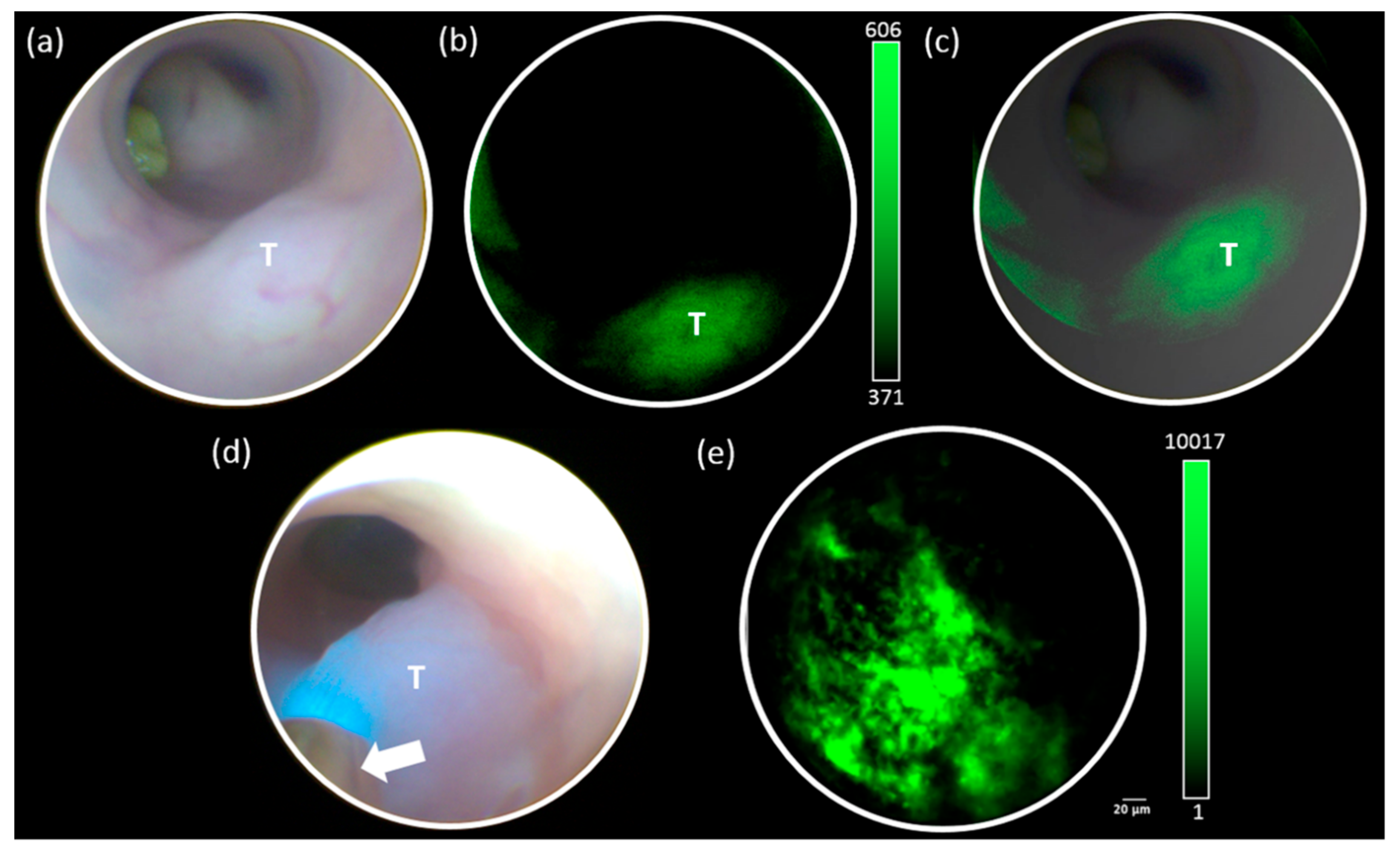
The mouse colon tumor was imaged with the DSFE system one day after the administration of green fluorescent bacteria via the tail vein. The white light image of WFE exhibited mucosal elevated lesions within the mouse colon (Figure 4a). The fluorescence image of WFE exhibited an increased signal intensity around the tumor-bearing colonic wall area (Figure 4b). Figure 4c indicates the fused image of Figure 4a,b. The measured fluorescence signal was 425.5 ± 52.5. The tip of the CFEM probe was approached on the tumor site, which was monitored with the white-light image, as depicted in Figure 4d. Multiple clustered green fluorescence signals of 1866.0 ± 2113.3 were detected in the tumor (Figure 4e).

3.4. Ex Vivo Quantification of Fluorescent Bacteria

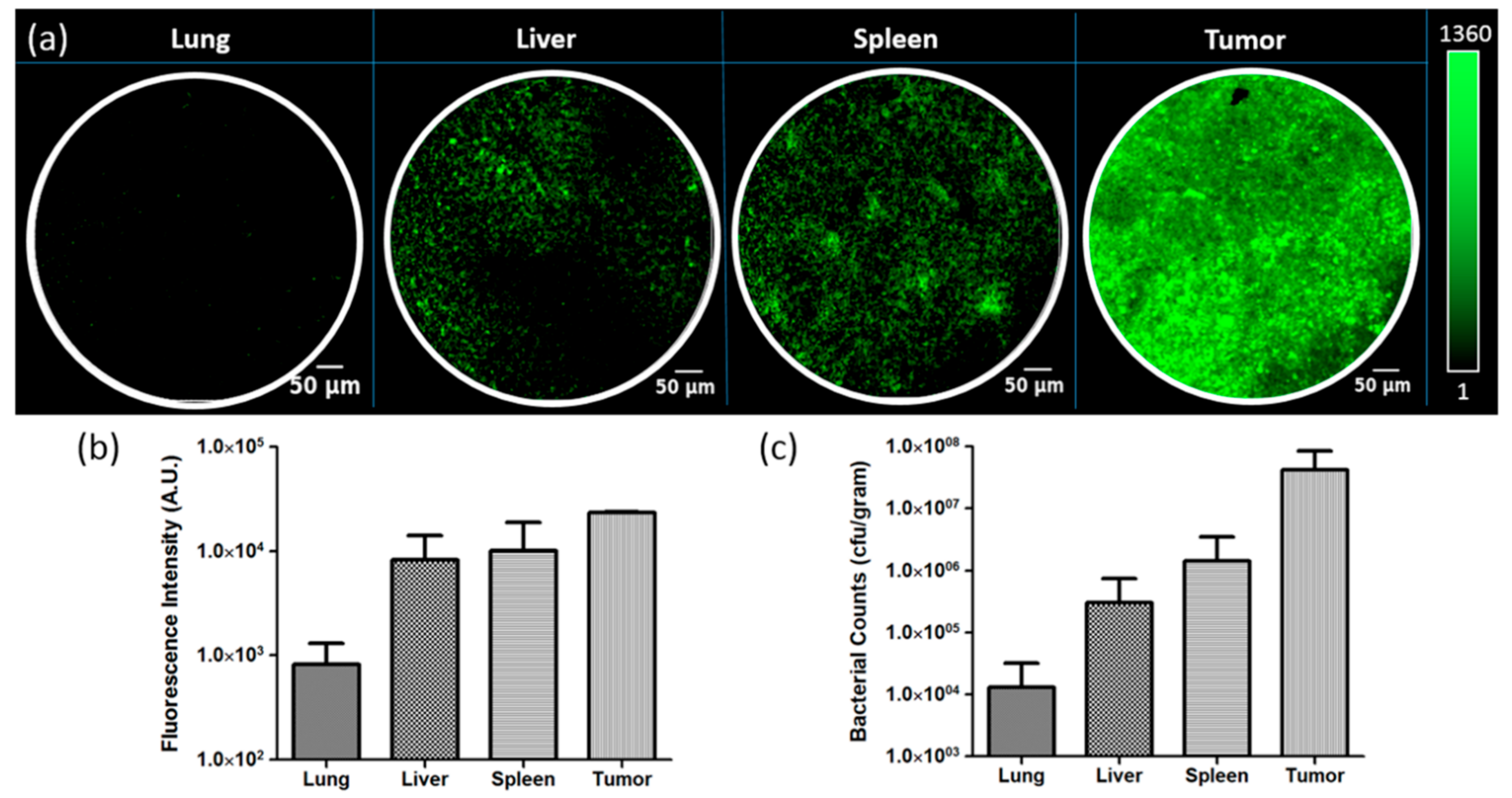
The spleen, liver, lung, and tumor were extracted from the bacteria-injected orthotopic colon tumor mice after euthanization. The CFEM probe exhibited different levels of green fluorescent signals in each examined organ and tumor (Figure 5a). The tumor demonstrated the highest green fluorescent signal (23,439.0 ± 250.3), followed by the spleen (10,133.0 ± 8880.0), liver (8284.0 ± 5931.0), and lung (818.5 ± 497.1), respectively (Figure 5b). The number of viable bacterial colonies demonstrated similar results to that of CFEM. The tumor site exhibited the largest number of viable bacteria (4.2 × 10⁷ cfu/gram), followed by the spleen (1.4 × 10⁶ cfu/gram), liver (3.0 × 10⁵ cfu/gram), and lung (1.3 × 10⁴ cfu/gram), respectively (Figure 5c).

4. Discussion and Conclusions

We achieved in vivo dual-scale fluorescent bacterial imaging using the developed DSFE system in an orthotopic mouse colon tumor model. The DSFE system successfully demonstrated the bacterial accumulation of tumor tissue from the gross level to the cellular level. To the best of our knowledge, this is the first case of imaging bacterial accumulation in WFE and CFEM simultaneously in vivo. The fused DSFE system is a promising imaging modality to evaluate the therapeutic response of colon cancer by bacterial therapy in vivo.
The DSFE system has the advantage of compensating the limitation of the single WFE and CFEM system. WFE screened the colonic wall to identify tumor lesions with a large FOV. Additionally, it localized fluorescence-emitting suspicious lesions to contact the CFEM probe within a short examination time. Meanwhile, the CFEM probe exhibited a bacteria-specific fluorescence image at a cellular-scaled resolution, which WFE could not. As a result, the DSFE system enables fast bacterial fluorescence imaging with high sensitivity.
The distribution of fluorescent bacteria in multiple organs was imaged with the CFEM probe ex vivo. The number of viable bacteria was higher in the tumor than in the vital organs. The trends of the fluorescence signal were correlated to the viable bacterial counting results. This bacterial distribution in the mouse tumor model is similar to those in previous reports [39]. Therefore, the fluorescent signal measured by CFEM can be used to monitor the viable bacterial accumulation in the tumor tissue. Longitudinal in vivo imaging with the DSFE system enables further understanding of bacteria-related cancer therapy.
However, this study has two weaknesses. Firstly, our CFEM exhibited a green-dotted image without any specific structure of the colonic mucosa. To obtain high-sensitive imaging, we did not use other nonspecific mucosal staining dyes, such as fluorescein or acriflavine [40,41]. The vascular imaging dye can improve the morphological characterization of the tumor lesion. Further multi-colored fluorescence imaging study with tissue-specific dye will be needed to maximize the usefulness of the DSFE system. Secondly, our WFE exhibited relatively low fluorescence signals on the tumor in comparison with the adjacent normal-looking tissue due to autofluorescence of the tissue (Figure S1). However, the additional CFEM probe exhibited bacteria-specific fluorescence at the tumor site. The bacterial fluorescence signal was much higher when comparing with autofluorescence of the normal mouse organ (Figure S2). This compensatory imaging is an advantage of the DSFE system. Further optimization of the bacteria-specific fluorescence system will improve the quality of the image.
In conclusion, although there are some areas to be improved, the developed DSFE system has successfully demonstrated the feasibility of visualizing and quantifying the distribution of bacteria in orthotopic mouse colon cancer. Therefore, the DSFE system is expected to be successfully applied to research on the therapy of colon cancer using bacteria.

Supplementary Materials

The following are available online at https://www.mdpi.com/2076-3417/10/3/844/s1. Figure S1: In vivo DSFE imaging of normal mouse colon without bacterial injection; Figure S2: Ex vivo autofluorescence image of normal mouse organ without bacterial injection using CFEM.

Author Contributions

Conceptualization: S.W.Y., C.L., C.L., and J.M.; methodology: S.W.Y.; experiment: S.W.Y., D.-h.N., S.P., and H.L.; data analysis: S.W.Y., D.N., S.P., and C.L.; data curation: S.W.Y. and C.L.; writing of original draft preparation: S.W.Y. and D.N.; writing of review and editing: C.L., C.L., and J.M.; visualization: S.W.Y. and C.L.; supervision: C.L. and J.M.; funding acquisition: S.W.Y. All authors have read and agreed to the published version of the manuscript.

Funding

This research was supported by the Bio & Medical Technology Development Program of the National Research Foundation (NRF) funded by the Korean government (MSIT) (No. NRF-2019M3E5D1A02067958), the National Research Foundation Korea (NRF) grants funded by the Korean government (MSIT) (NRF-2018R1D1A1B07050011, NRF-2017R1A2B3012157), the Korea Health Technology R&D Project (HI18C0858) through the KHIDI (Korea Health Industry Development Institute) funded by the Ministry of Health & Welfare, and a grant (HCRI 19134, HCRI 19006) by the Chonnam National University Hwasun Hospital Institute for Biomedical Science.

Conflicts of Interest

The authors declare no conflict of interest.

References

  1. Siegel, R.L.; Miller, K.D.; Jemal, A. Cancer statistics, 2019. CA Cancer J. Clin. 2019, 69, 7–34. [Google Scholar] [CrossRef] [PubMed] [Green Version]
  2. Guren, T.K.; Thomsen, M.; Kure, E.H.; Sorbye, H.; Glimelius, B.; Pfeiffer, P.; Osterlund, P.; Sigurdsson, F.; Lothe, I.M.B.; Dalsgaard, A.M.; et al. Cetuximab in treatment of metastatic colorectal cancer: Final survival analyses and extended RAS data from the NORDIC-VII study. Br. J. Cancer 2017, 116, 1271–1278. [Google Scholar] [CrossRef] [PubMed] [Green Version]
  3. Grothey, A.; Van Cutsem, E.; Sobrero, A.; Siena, S.; Falcone, A.; Ychou, M.; Humblet, Y.; Bouche, O.; Mineur, L.; Barone, C.; et al. Regorafenib monotherapy for previously treated metastatic colorectal cancer (CORRECT): An international, multicentre, randomised, placebo-controlled, phase 3 trial. Lancet 2013, 381, 303–312. [Google Scholar] [CrossRef]
  4. Overman, M.J.; Lonardi, S.; Wong, K.Y.M.; Lenz, H.J.; Gelsomino, F.; Aglietta, M.; Morse, M.A.; Van Cutsem, E.; McDermott, R.; Hill, A.; et al. Durable Clinical Benefit With Nivolumab Plus Ipilimumab in DNA Mismatch Repair-Deficient/Microsatellite Instability-High Metastatic Colorectal Cancer. J. Clin. Oncol. 2018, 36, 773–779. [Google Scholar] [CrossRef]
  5. Idrees, M.; Tejani, M. Current Treatment Strategies for Elderly Patients with Metastatic Colon Cancer. Cureus 2019, 11, e4713. [Google Scholar] [CrossRef] [Green Version]
  6. Datta, N.R.; Ordonez, S.G.; Gaipl, U.S.; Paulides, M.M.; Crezee, H.; Gellermann, J.; Marder, D.; Puric, E.; Bodis, S. Local hyperthermia combined with radiotherapy and-/or chemotherapy: Recent advances and promises for the future. Cancer Treat Rev. 2015, 41, 742–753. [Google Scholar] [CrossRef]
  7. Forbes, N.S.; Coffin, R.S.; Deng, L.; Evgin, L.; Fiering, S.; Giacalone, M.; Gravekamp, C.; Gulley, J.L.; Gunn, H.; Hoffman, R.M.; et al. White paper on microbial anti-cancer therapy and prevention. J. Immunother Cancer 2018, 6, 78. [Google Scholar] [CrossRef]
  8. Song, S.; Vuai, M.S.; Zhong, M. The role of bacteria in cancer therapy-enemies in the past, but allies at present. Infect. Agent Cancer 2018, 13, 9. [Google Scholar] [CrossRef]
  9. Roberts, N.J.; Zhang, L.; Janku, F.; Collins, A.; Bai, R.Y.; Staedtke, V.; Rusk, A.W.; Tung, D.; Miller, M.; Roix, J.; et al. Intratumoral injection of Clostridium novyi-NT spores induces antitumor responses. Sci. Transl. Med. 2014, 6, 249ra111. [Google Scholar] [CrossRef] [Green Version]
  10. Agrawal, N.; Bettegowda, C.; Cheong, I.; Geschwind, J.F.; Drake, C.G.; Hipkiss, E.L.; Tatsumi, M.; Dang, L.H.; Diaz, L.A., Jr.; Pomper, M.; et al. Bacteriolytic therapy can generate a potent immune response against experimental tumors. Proc. Natl. Acad. Sci. USA 2004, 101, 15172–15177. [Google Scholar] [CrossRef] [Green Version]
  11. Thamm, D.H.; Kurzman, I.D.; King, I.; Li, Z.; Sznol, M.; Dubielzig, R.R.; Vail, D.M.; MacEwen, E.G. Systemic administration of an attenuated, tumor-targeting Salmonella typhimurium to dogs with spontaneous neoplasia: Phase I evaluation. Clin. Cancer Res. 2005, 11, 4827–4834. [Google Scholar] [CrossRef] [PubMed] [Green Version]
  12. Chang, W.W.; Lee, C.H. Salmonella as an innovative therapeutic antitumor agent. Int. J. Mol. Sci. 2014, 15, 14546–14554. [Google Scholar] [CrossRef] [PubMed] [Green Version]
  13. Kim, J.E.; Phan, T.X.; Nguyen, V.H.; Dinh-Vu, H.V.; Zheng, J.H.; Yun, M.; Park, S.G.; Hong, Y.; Choy, H.E.; Szardenings, M.; et al. Salmonella typhimurium Suppresses Tumor Growth via the Pro-Inflammatory Cytokine Interleukin-1beta. Theranostics 2015, 5, 1328–1342. [Google Scholar] [CrossRef] [PubMed] [Green Version]
  14. Liang, K.; Liu, Q.; Li, P.; Luo, H.; Wang, H.; Kong, Q. Genetically engineered Salmonella Typhimurium: Recent advances in cancer therapy. Cancer Lett. 2019, 448, 168–181. [Google Scholar] [CrossRef]
  15. Zheng, J.H.; Nguyen, V.H.; Jiang, S.N.; Park, S.H.; Tan, W.; Hong, S.H.; Shin, M.G.; Chung, I.J.; Hong, Y.; Bom, H.S.; et al. Two-step enhanced cancer immunotherapy with engineered Salmonella typhimurium secreting heterologous flagellin. Sci. Transl. Med. 2017, 9, eaak9537. [Google Scholar] [CrossRef]
  16. Sedighi, M.; Zahedi Bialvaei, A.; Hamblin, M.R.; Ohadi, E.; Asadi, A.; Halajzadeh, M.; Lohrasbi, V.; Mohammadzadeh, N.; Amiriani, T.; Krutova, M.; et al. Therapeutic bacteria to combat cancer; current advances, challenges, and opportunities. Cancer Med. 2019, 8, 3167–3181. [Google Scholar] [CrossRef]
  17. Nguyen, V.H.; Kim, H.S.; Ha, J.M.; Hong, Y.; Choy, H.E.; Min, J.J. Genetically engineered Salmonella typhimurium as an imageable therapeutic probe for cancer. Cancer Res. 2010, 70, 18–23. [Google Scholar] [CrossRef] [Green Version]
  18. Heimann, D.M.; Rosenberg, S.A. Continuous intravenous administration of live genetically modified salmonella typhimurium in patients with metastatic melanoma. J. Immunother. 2003, 26, 179–180. [Google Scholar] [CrossRef] [Green Version]
  19. Toso, J.F.; Gill, V.J.; Hwu, P.; Marincola, F.M.; Restifo, N.P.; Schwartzentruber, D.J.; Sherry, R.M.; Topalian, S.L.; Yang, J.C.; Stock, F.; et al. Phase I study of the intravenous administration of attenuated Salmonella typhimurium to patients with metastatic melanoma. J. Clin. Oncol. 2002, 20, 142–152. [Google Scholar] [CrossRef]
  20. Cunningham, C.; Nemunaitis, J. A phase I trial of genetically modified Salmonella typhimurium expressing cytosine deaminase (TAPET-CD, VNP20029) administered by intratumoral injection in combination with 5-fluorocytosine for patients with advanced or metastatic cancer. Protocol no: CL-017. Version: April 9, 2001. Hum. Gene Ther. 2001, 12, 1594–1596. [Google Scholar]
  21. Min, J.J.; Nguyen, V.H.; Kim, H.J.; Hong, Y.; Choy, H.E. Quantitative bioluminescence imaging of tumor-targeting bacteria in living animals. Nat. Protoc. 2008, 3, 629–636. [Google Scholar] [CrossRef] [PubMed]
  22. Jiang, S.N.; Park, S.H.; Lee, H.J.; Zheng, J.H.; Kim, H.S.; Bom, H.S.; Hong, Y.; Szardenings, M.; Shin, M.G.; Kim, S.C.; et al. Engineering of bacteria for the visualization of targeted delivery of a cytolytic anticancer agent. Mol. Ther. 2013, 21, 1985–1995. [Google Scholar] [CrossRef] [PubMed] [Green Version]
  23. Phan, T.; Nguyen, V.H.; D’Alincourt, M.S.; Manuel, E.R.; Kaltcheva, T.; Tsai, W.; Blazar, B.R.; Diamond, D.J.; Melstrom, L.G. Salmonella-mediated therapy targeting indoleamine 2, 3-dioxygenase 1 (IDO) activates innate immunity and mitigates colorectal cancer growth. Cancer Gene Ther. 2019. [Google Scholar] [CrossRef] [PubMed]
  24. Liu, X.; Jiang, S.; Piao, L.; Yuan, F. Radiotherapy combined with an engineered Salmonella typhimurium inhibits tumor growth in a mouse model of colon cancer. Exp. Anim. 2016, 65, 413–418. [Google Scholar] [CrossRef] [Green Version]
  25. Bibby, M.C. Orthotopic models of cancer for preclinical drug evaluation: Advantages and disadvantages. Eur. J. Cancer 2004, 40, 852–857. [Google Scholar] [CrossRef]
  26. Hoffman, R.M. Orthotopic is orthodox: Why are orthotopic-transplant metastatic models different from all other models? J. Cell Biochem. 1994, 56, 1–3. [Google Scholar] [CrossRef]
  27. Oh, G.; Yoo, S.W.; Jung, Y.; Ryu, Y.M.; Park, Y.; Kim, S.Y.; Kim, K.H.; Kim, S.; Myung, S.J.; Chung, E. Intravital imaging of mouse colonic adenoma using MMP-based molecular probes with multi-channel fluorescence endoscopy. Biomed. Opt. Express 2014, 5, 1677–1689. [Google Scholar] [CrossRef] [Green Version]
  28. Gounaris, E.; Ishihara, Y.; Shrivastrav, M.; Bentrem, D.; Barrett, T.A. Near-Infrared Fluorescence Endoscopy to Detect Dysplastic Lesions in the Mouse Colon. Methods Mol Biol 2016, 1422, 137–147. [Google Scholar]
  29. Bae, S.M.; Bae, D.J.; Do, E.J.; Oh, G.; Yoo, S.W.; Lee, G.J.; Chae, J.S.; Yun, Y.; Kim, S.; Kim, K.H.; et al. Multi-Spectral Fluorescence Imaging of Colon Dysplasia InVivo Using a Multi-Spectral Endoscopy System. Transl. Oncol. 2019, 12, 226–235. [Google Scholar] [CrossRef]
  30. Dunbar, K.; Canto, M. Confocal endomicroscopy. Curr. Opin. Gastroenterol. 2008, 24, 631–637. [Google Scholar] [CrossRef]
  31. Mielke, L.; Preaudet, A.; Belz, G.; Putoczki, T. Confocal laser endomicroscopy to monitor the colonic mucosa of mice. J. Immunol. Methods 2015, 421, 81–88. [Google Scholar] [CrossRef] [PubMed] [Green Version]
  32. Waldner, M.J.; Wirtz, S.; Neufert, C.; Becker, C.; Neurath, M.F. Confocal laser endomicroscopy and narrow-band imaging-aided endoscopy for in vivo imaging of colitis and colon cancer in mice. Nat. Protoc. 2011, 6, 1471–1481. [Google Scholar] [CrossRef] [PubMed]
  33. Salvatori, F.; Siciliano, S.; Maione, F.; Esposito, D.; Masone, S.; Persico, M.; De Palma, G.D. Confocal Laser Endomicroscopy in the Study of Colonic Mucosa in IBD Patients: A Review. Gastroenterol. Res. Pract. 2012, 2012, 525098. [Google Scholar] [CrossRef] [PubMed] [Green Version]
  34. Deng, F.; Fang, Y.; Shen, Z.; Gong, W.; Liu, T.; Wen, J.; Zhang, W.; Zhu, X.; Zhong, H.; Wang, T.; et al. Use of confocal laser endomicroscopy with a fluorescently labeled fatty acid to diagnose colorectal neoplasms. Oncotarget 2017, 8, 58934–58947. [Google Scholar] [CrossRef] [Green Version]
  35. Gheonea, D.I.; Saftoiu, A.; Ciurea, T.; Popescu, C.; Georgescu, C.V.; Malos, A. Confocal laser endomicroscopy of the colon. J. Gastrointestin. Liver Dis. 2010, 19, 207–211. [Google Scholar]
  36. Song, M.; Kim, H.J.; Kim, E.Y.; Shin, M.; Lee, H.C.; Hong, Y.; Rhee, J.H.; Yoon, H.; Ryu, S.; Lim, S.; et al. ppGpp-dependent stationary phase induction of genes on Salmonella pathogenicity island 1. J. Biol. Chem. 2004, 279, 34183–34190. [Google Scholar] [CrossRef] [Green Version]
  37. Yoo, S.W.; Oh, G.; Safi, A.M.; Hwang, S.; Seo, Y.S.; Lee, K.H.; Kim, Y.L.; Chung, E. Endoscopic non-ablative fractional laser therapy in an orthotopic colon tumour model. Sci. Rep. 2018, 8, 1673. [Google Scholar] [CrossRef]
  38. Becker, C.; Fantini, M.C.; Neurath, M.F. High resolution colonoscopy in live mice. Nat. Protoc. 2006, 1, 2900–2904. [Google Scholar] [CrossRef]
  39. Low, K.B.; Ittensohn, M.; Le, T.; Platt, J.; Sodi, S.; Amoss, M.; Ash, O.; Carmichael, E.; Chakraborty, A.; Fischer, J.; et al. Lipid A mutant Salmonella with suppressed virulence and TNFalpha induction retain tumor-targeting in vivo. Nat. Biotechnol. 1999, 17, 37–41. [Google Scholar] [CrossRef]
  40. Mascolo, M.; Staibano, S.; Ilardi, G.; Siano, M.; Vecchione, M.L.; Esposito, D.; De Rosa, G.; De Palma, G.D. Probe-based confocal laser endomicroscopy evaluation of colon preneoplastic lesions, with particular attention to the aberrant crypt foci, and comparative assessment with histological features obtained by conventional endoscopy. Gastroenterol. Res. Pract. 2012, 2012, 645173. [Google Scholar] [CrossRef]
  41. Li, C.Q.; Yu, T.; Zuo, X.L.; Xie, X.J.; Li, W.B.; Chu, C.L.; Zuo, F.; Li, Y.Q. Effects on confocal laser endomicroscopy image quality by different acriflavine concentrations. J. Interv. Gastroenterol. 2011, 1, 59–63. [Google Scholar] [CrossRef] [PubMed]
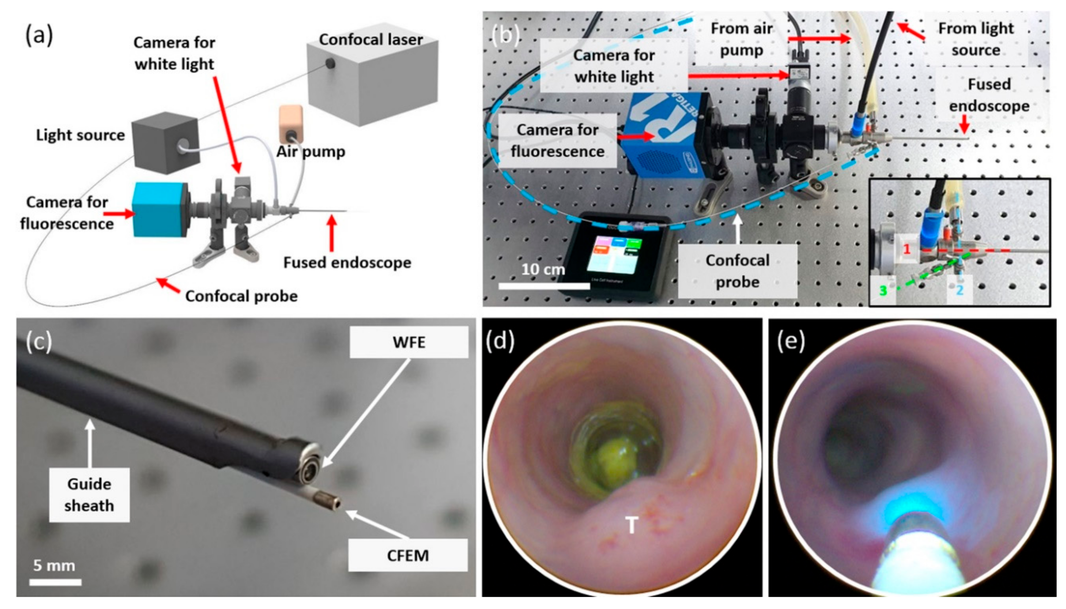
Figure 1. Dual-scale fluorescence endoscopy (DSFE) system. (a) Scheme of major component of the DSFE system. (b) Corresponding photography of the DSFE system. Inset photography shows three channels of the guiding sheath. (c) Magnified endoscopy tip of the fused DSFE probe. The tip diameter of the wide-field fluorescence endoscope (WFE) and confocal fluorescence endomicroscopy (CFEM) probes are 1.9 mm and 0.94 mm, respectively. (d,e) In vivo white-light WFE endoscopic images of inside the mouse colon with orthotropic mouse colon tumor (T) and the laser emitting from the CFEM probe into the tumoral area.
Applsci 10 00844 g001
Figure 2. Results of the phantom test of DSFE. (a) Photography of phosphate buffered saline and 15 different concentrations of fluorescein isothiocyanate (FITC)-contained silicone tubes. (b) The concentration-dependent fluorescence signal of FITC measured via WFE. (c) The fluorescence signal of FITC according to excitation light intensity via WFE. (d) FITC-contained tubes with and without excitation light emitting from the CFEM probe (red arrow) (e) The concentration-dependent fluorescence signal of FITC measured via CFEM. (f) The fluorescence signal of FITC according to excitation light intensity via CFEM.
Applsci 10 00844 g002
Figure 3. Endoscopic follow-up of orthotopic colon tumor. Tumor growth after (a) 20 days, (b) 22 days, and (c) 27 days from MC38 colon cancer cell implantation. A gradually increased tumor volume (white arrows) is shown with serial endoscopic imaging.
Applsci 10 00844 g003
Figure 4. In vivo DSFE imaging of fluorescent bacteria in the orthotopic mouse colon tumor model. (a) White light, (b) fluorescence, and (c) merged images of the bacteria injected orthotopic tumor (T) using the WFE system. (d) CFEM probe (white arrow) contacted the tumor (T) lesion. (e) Bacteria-emitting fluorescence signal imaged using the CFEM probe.
Applsci 10 00844 g004
Figure 5. Ex vivo quantification of green fluorescent protein (GFP) bacteria in a mouse model with CFEM (n = 2). (a) Confocal fluorescence imaging of GFP bacteria in various extracted tissues from the orthotopic tumor mouse. (b) The quantified fluorescence signal of bacteria from each tissue. (c) Viable bacterial counting analysis of each extracted tissue.
Applsci 10 00844 g005