Communication

Behavior of Astrocytes Derived from Human Neural Stem Cells Flown onto Space and Their Progenies

1
Department of Psychiatry, UCLA, Los Angeles, CA 90095, USA
2
Department of Psychiatry, Semel Institute for Neuroscience and Human Behavior, University of California, Los Angeles, CA 90095, USA
*
Author to whom correspondence should be addressed.
Appl. Sci. 2021, 11(1), 41; https://doi.org/10.3390/app11010041
Submission received: 15 October 2020 / Revised: 14 December 2020 / Accepted: 17 December 2020 / Published: 23 December 2020
(This article belongs to the Special Issue Advances in Space Biology: Cell Behavior in Microgravity)

Abstract

:
Long-term travel and prolonged stays for astronauts in outer space are imminent. To date more than 500 astronauts have experienced the extreme conditions of space flight including microgravity and radiation. Here we report that human neural stem cells (NSCs) flown onto space were successfully induced to the astrocyte phenotype when grown in fetal calf serum (FCS) supplemented medium. We want to emphasize that these astrocytes were generated after the space flight through a slow process lasting several weeks. Interestingly, we also found that these cells newly formed astrocytes, proliferated slowly but significantly and they showed a tendency to continue proliferating at the same pace. Astrocytes, a major type of glial cells, are key for the normal function of the central nervous system (CNS). They are also emerging as a critical component in most neurodegenerative diseases. Knowledge on the effects of space microgravity on them is of utmost importance for long duration space travel.

Graphical Abstract

1. Introduction

Long-term travel and lengthy stays for astronauts in outer space are imminent. To date, more than 500 astronauts have experienced the extreme conditions of space flight, including microgravity and radiation [1]. There is emerging evidence that both these factors impact the physiology and, possibly, the behavior of crew members. Therefore, studies on how the space environment affects the central nervous system (CNS) are essential.
Neural stem cells (NSCs) are a key population in the CNS because they maintain tissue integrity and function. They also possess the potential of generating the various cell types during development and adulthood in health and disease. We have previously shown that simulated microgravity increases neural cell proliferation [2]. Astrocytes, a major type of glial cells are key components of neuronal proper function. They have a variety of essential roles including glutamate and potassium buffering, axonal guidance, trophic support, astrocyte-mediated inflammatory response, wound healing, formation of the blood-brain barrier, neuronal synapse formation, and plasticity [3,4]. Astrocytes are emerging as a critical component in most neurodegenerative diseases [5]. Although the full contribution of astrocytes to neurological disease remains unresolved, astrocyte cell-autonomous deficits have been implicated in a variety of neurological disorders [6,7].
In the present study, in order to ascertain if space (SPC)-flown NSCs would respond to cues known to induce them to the astrocyte lineage, we cultured them in medium containing 10% fetal calf serum (FCS) and gradually, post-flight NSCs transitioned into the astroglial phenotype. We confirmed them as astrocytes because they expressed glial fibrillary acidic protein (GFAP), which is one of the most known astrocyte markers because it does not only identify the cell type, but also its expression is upregulated when astrocytes suffer an insult [8]. GFAP is a major intermediate filament composing their cytoskeleton [9,10]. Another commonly used astrocytic marker is S-100β, a Ca2+ binding peptide abundant in the cytoplasm and nucleus of astrocytes. This peptide is involved in cell cycle regulation and cytoskeleton modification of astrocytes in health and disease [11]. These cells expressed both markers in our cultures indicating their successful specification into the astroglial phenotype.

2. Materials and Methods
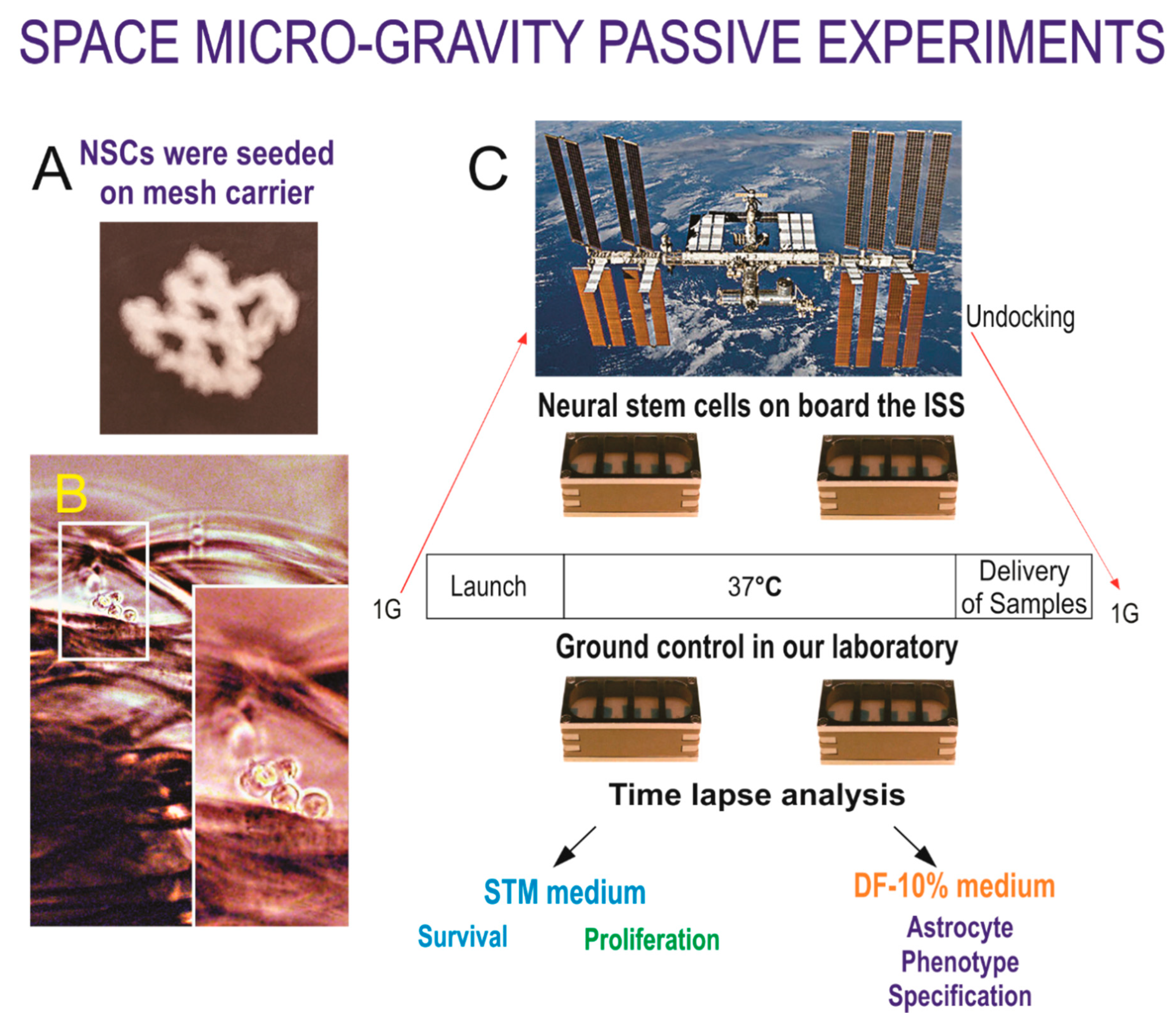
2.1. Cells

A homogeneous population of NSCs was obtained from human induced pluripotent stem cells (hiPS). The original cells, known as “CS83iCTR-33nxx” (such as skin cells), were “reprogrammed” and provided to us by Cedars-Sinai Medical Center via a material transfer agreement. For space flight, NSCs were flown onboard the ISS for 39.3 days and then returned to Earth.
To induce the astrocyte phenotype, we used DULBECO and F-12 medium (DF) and 10% fetal calf serum, we used DF-10% when referring to this culture medium.

2.2. Space Flight

The BioScience-4 mission, launched onboard the Space-X 16 Dragon capsule on 5th November 2018, and it is the first study to investigate the proliferation of central nervous system stem cells in microgravity. This experiment has been very instrumental as it has allowed us to better understand features of neural cells while in space, as well as after returning to Earth [12,13]. The hardware used for this study (Figure 1) was called passive unit(s) because they travelled round trip with the same culture medium. We worked several years in order to choose the adequate hardware, cell density, culture medium and cell origin that would allow for a successful space flight. Thus, from our “Experimental Verification Tests” prior to the space flight we learned and optimized the conditions for human NSCs for our study and determined how should the study be conducted. Ground control cells were grown in the same conditions and hardware as space-flown NSCs in the investigator’s laboratory.

2.3. Recovery of the Hardware and Harvesting of Samples

After splash-down, samples were transported in controlled environment from Long Beach airport to UCLA. The secretome was recovered from each well separately. Subsequently, NSCs were detached from the floor and walls of each well and recovered separately. Secretome samples were frozen for further use. Next, NSCs that were attached to the mesh-carrier, were retrieved from the hardware, plated onto poly-d-lysine coated flasks in stem cell medium (STM) [14] and allowed to recover from space flight. After 20 h in the incubator 5% CO2 and 36.8 °C, flasks were placed in a Zeiss Axio Observer 7 fully motorized inverted research microscope with the Zeiss Axiocam 506 monochrome camera with Zeiss ZEN software. The system was equipped with the Zeiss Full Incubation XL chamber for temperature and CO2 control with motorized scanning stage.

2.4. Time-Lapse Microscopy

We used the Zeiss Axio Observer 7 fully motorized inverted research microscope with the Zeiss Axiocam 506 monochrome camera, Zeiss ZEN software, and definite focus equipped with the full Incubation XL chamber (Zeiss, Oberkochen, Germany) for temperature and CO2 control with motorized scanning stage. Twenty-one days after space flight, the NSCs were placed in DF-10% and time-lapse was started.

2.5. Immunofluorescence

Cell lineage specification was confirmed using double immunofluorescence with established markers for astrocytes. The following antibodies were used at a 1:100 dilution: nestin (BD Pharmingen, Franklin Lakes, NJ, USA); glial fibrillary acidic protein (GFAP); S100β and vimentin (all Sigma, St. Louis, MO, USA). The immunofluorescence procedures were carried out as described previously [15], plating cells onto coated plastic 8-well chambers (Nunc 177445), or on poly-d-lysine coated glass coverslips. Three secondary antibodies were used to visualize the markers mentioned above: goat anti-mouse IgM AMCA (Jackson Immunology Research Laboratories, West Grove, PA, USA); goat anti-rabbit IgG Texas Red (JIR); and goat anti-mouse IgG FITC (Sigma).

3. Results

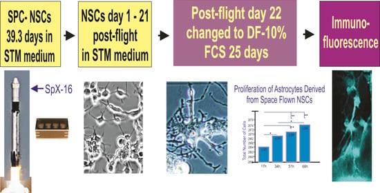
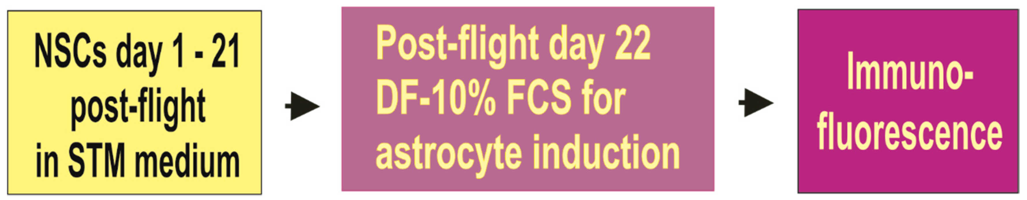
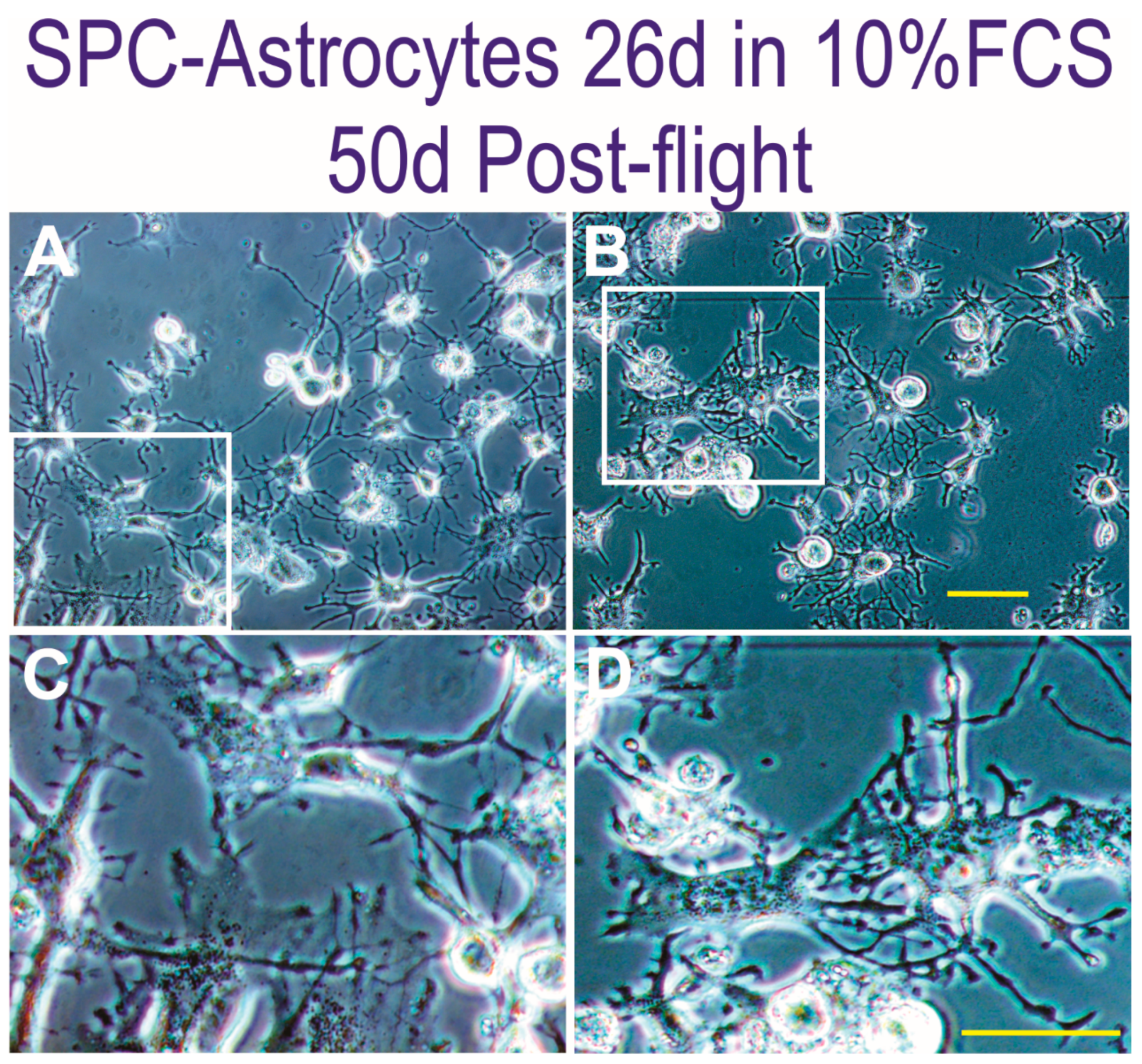
3.1. Space Flown NSCs Give Rise to Astrocytes

After 39.3 days in space and three weeks post-flight in NSCs culture medium, we cultured NSCs and their progenies derived from those flown to space in DF-10% FCS to direct their specification to the astrocyte phenotype (Figure 2).
In time cells grew healthy and slowly started to acquire the astroglial typical morphology. They were becoming flat and with numerous cell processes (Figure 3). We waited 26 days because we monitored the composition of our cultures at earlier time points by immunocytochemistry for NSCs and astrocytes and cells were still transitioning given rise to subpopulations at earlier time points, where nestin and vimentin positive cells were still present in these cultures. Therefore, we had to allow enough time for astroglial specification in order to work with a homogeneous population of astrocytes. For comparison of Naïve or SPC-NSCs derived astrocytes see Supplemental Figure S1.

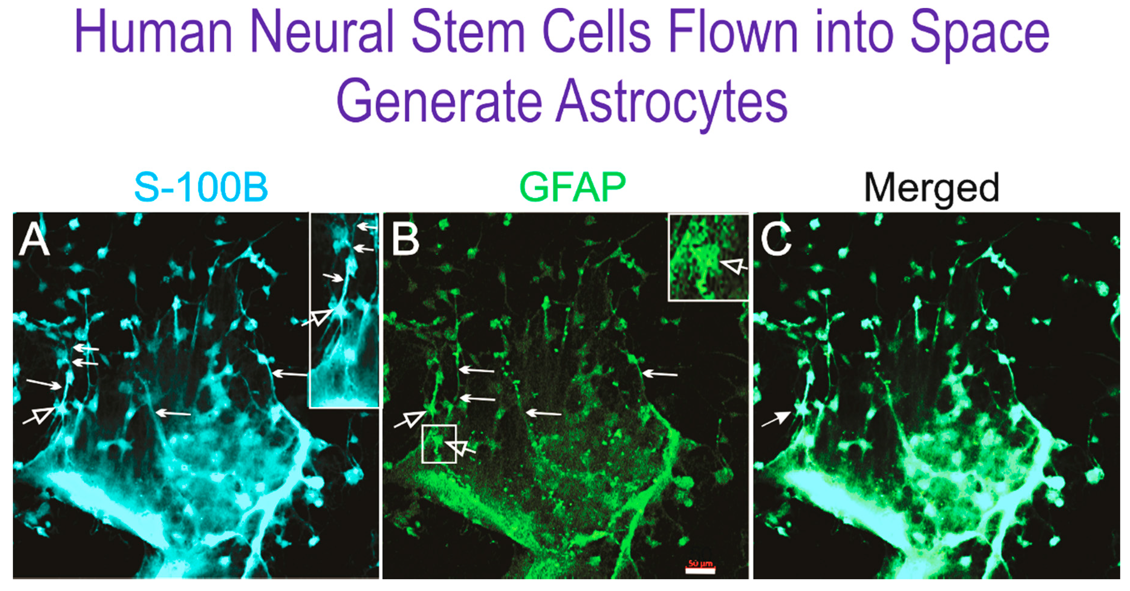
3.2. Immunofluorescent Detection of Astrocyte Markers

Initially, the morphology of most cells being generated by culturing NSCs in DF-10% FCS appeared to be mainly round or bipolar. Their organization was reminiscent of radial glia elongating processes, and some appeared to be migrating along some of those cell processes. Moreover, they appeared to orient all in the same direction, as it occurs during brain development. Despite the fact that their morphology had not yet completely transformed into the typical flat cell, these cells expressed both GFAP and S-100β (Figure 4) with the concomitant loss of two neural progenitor markers, nestin and vimentin that were negative (not shown).

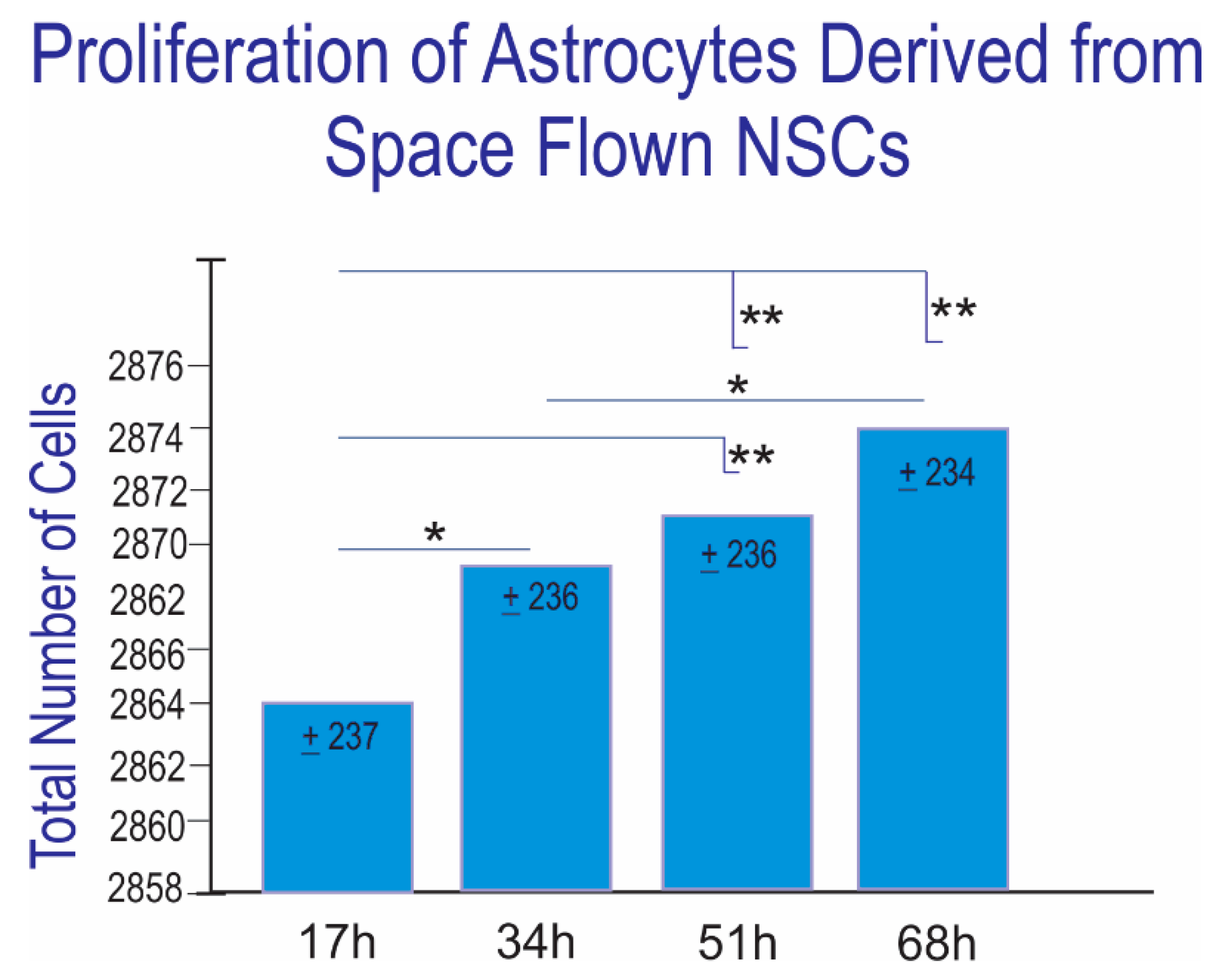
3.3. Proliferation of Newly Formed Astrocytes Derived from NSCs Flown to Space

Three weeks post-flight, NSCs progenies derived from SPC-NSCs were cultured in 10% FCS to direct their specification to the astrocyte phenotype. The vast majority of astrocytes derived from SPC-flown NSCs did not proliferate. Nonetheless, a few cells divided in an interval of 17 h per microscope field (Figure 5).
The proliferation of astrocytes is essential for normal brain development, while in the adult brain astrocytes exhibit little or no proliferation [11]. Nonetheless, upon injury or disease, astrocytes respond with a phenomenon known as astrogliosis, and in this stage they regain their proliferative state [16]. However, even in this state of prominent reactive astrogliosis, astrocytic proliferation is modest at best, with a proliferation rate of 1.1% being labelled as quite high for acute demyelinating lesions [17]. The extent of proliferation of astrocytes in nonneoplastic circumstances is therefore expected to be quite low, with proliferation rates lower than 1% of the total astrocyte population being quite normal.

4. Discussion

Glia, also called glial cells or neuroglia, are non-neuronal cells (astrocytes and oligodendrocytes) in the brain and spinal cord that maintain CNS homeostasis, form and maintain myelin, and provide support and protection for neurons. In the present study, we obtained astrocytes derived from NSCs flown into space. These astrocytes were generated in 1G upon return of the SPC-flown NSCs. This study is important because in the CNS, there are five times more astroglial cells (astrocytes) than neurons. In the past, the concept of neuroglia was introduced by Rudolf Virchow neuro-glia (nerve-cement) [18,19], when astrocytes were considered solely a physical support material for neurons. Currently, it is well known that astrocytes have a multitude of essential functions in the healthy CNS, including primary roles in synaptic transmission and information processing by neural circuit function [20]. Moreover, their secreted molecules such as gliotransmitters, neuromodulators, and trophic factors help maintain adult neurogenesis in the normal brain, while an imbalance of such molecules may be deleterious and contribute to CNS pathologies [21]. Astrocytes respond to all forms of CNS insults through reactive astrogliosis, which is a pathological hallmark of CNS structural lesions [11]. This means that instead of dying after injury or disease, astrocytes survive and undergo fundamental changes, as they display different degrees of astrogliosis proportional to the extent of injury [22] by proliferating and forming a “gliotic scar” to fill spaces left when neurons and glia die. Unfortunately, often the gliotic scar is the main obstacle for regeneration of the CNS tissue lost to injury or disease.
In order to ascertain if NSCs flown to space would acquire the astroglial phenotype post-flight in 1G, NSCs were cultured in DF-10% FCS. As expected, the NSCs transitioned into the astrocyte phenotype as ascertained by the expression of the astrocyte markers S-100 and GFAP, indicating that after space flight NSCs had preserved their pluripotency, and with the right environmental cues they are able to generate astroglial cells. Literature on the proliferation of non-injured astrocytes is not as extensive as it is for models of CNS injury [11]. Colodner and collaborators have shown that astrocytes proliferate in response to a variety of insults, nonetheless, they described this proliferative potential as modest in both pediatric and adult populations, suggesting that there may be a limit to the potential of these cells to generate numerous new astrocytes in the injured CNS [23]. According to Dr. Ina Wanner, cell division rate for normal human astrocytes has been determined to be approximately 2.8 days for astrocyte progenitors and 4 days for astrocytes (personal communication). It has been shown that when normal human astrocytes were re-programmed to become tumor initiating cells, thus expected to be more proliferative, their doubling time was longer than 43.6 h [24]. Another paper using human embryonic stem cell-derived astrocytes in the presence of growth factors (bFGF and EGF) and human serum exhibited a doubling time of 21 ±  2.6 h [25]. We further found that SPC-flown NSCs-derived astrocytes proliferated modestly and steadily. Their numbers increased in a statistically significant manner with time in culture. Between one and two cells divided in an interval of 17 h, thus, in a shorter time than previously reported for astrocytes grown in 1G. While with the data analyzed so far, we cannot assert that there is an increased division of these cells in comparison to1G NSCs derived astrocytes, we found preliminary evidence that SPC-flown NSCs generate younger-like astrocytes and radial-glial cells evocative of neural development where radial glial cells are an abundant progenitor population in particular for cortical expansion. [26,27]. Our analysis will continue when the pandemic allows us to go back to the laboratory to complete this ongoing study. Nonetheless, one could hypothesize that these glial cells may play a role in the intracranial hypertension, or pressure inside the skull, that has been observed during human spaceflight. On one hand, our data confirmed the moderate contribution that one could expect from astrocytes generating new astrocytes in non-injury conditions. On the other hand, four questions come to mind: (1) Are astrocytes in the brain of astronauts increasing in number, even if slowly, and therefore contributing to intracranial hypertension? (2) Are space flown NSCs giving rise to radial-glia in addition to astrocytes? (3) Are space-flown-NSCs transmitting their proliferative potential to cells that are specified to the astrocyte phenotype in 1G? And (4) are these newly specified astrocytes in a state of “inherited stress” resulting from the space flight, or the return of their mother NSCs re-entry and splash down? Currently, we do not have answers to these important questions. A subsequent space flight would be necessary to generate enough material to pursue studies aimed at answering them. Moreover, studies in samples containing other cell types, such as organotypic cultures or animal work where the entire CNS experiences space microgravity are of the essence to understand if the regulation of proliferation in space differs when cells are in homogeneous cultures as opposed to the entire brain.

Supplementary Materials

The following are available online at https://www.mdpi.com/2076-3417/11/1/41/s1, Figure S1: Comparison of the morphological features and general aspect of astrocytes derive from naïve-1G NSCs vs. astrocytes derive from SPC-flown NSCs 50 days after being in DF-10% FCS. (A) Astrocytes derived from hiPS-NSCs grown solely in 1G display their typical flat morphology. (B) Astrocytes derived from hiPS-derived NSCs flown into space displayed a less flat morphology, presented elongated cell processes, and some preserved the bipolar elongated radial glia-like morphology frequently observed during CNS development (inset). Bar = 50 µm).

Author Contributions

S.S., N.C. and V.T., performed extensive image analysis of time-lapse files, consolidated the data for graphs and statistics, contributed to manuscript preparation; A.E.-J. designed the entire study, chose the hardware, performed experimental verification tests, as well as the work at Kennedy Space Center for flight integration. She harvested all cells from the space hardware upon their return to Earth and performed image acquisition of live cells and thereafter performed immunocytochemistry, analysis of the data and prepared the manuscript. All authors have read and agreed to the published version of the manuscript.

Funding

We thank NASA Space Biology for Grant: NNX15AB43G; The IDDRC Cell Culture Core is supported by NIH/NICHD grant number U54HD087101-05.

Data Availability Statement

The authors confirm that the data supporting the findings of this study are available within the article and its supplementary materials.

Acknowledgments

We are grateful to Mark Mobilia and the entire Zeiss team for their support with the microscopy needs for this study, without their support this study would not have been possible. We also thank Amy Rowat for support with her microscope system as well. Amy Gresser. Elizabeth Pane, and Medaya Torres for their help with the implementation of the study. The NASA Space Biology Project team at Ames Research Center D. Tomko, K. Sato, E. Taylor, ARC Space Biology Project Manager and the support personnel at the Space Station Processing Facility at Kennedy Space Center. Thanks to Karin Perkins, Diana Ly, NASA. We also thank Carlos Cepeda for insightful discussions. We are deeply grateful for Fathi Karouia who was the mission scientist from Space Biology, NASA and was in charge of mission preparedness. Special thanks to Maria Birlem and Chriss Bruderrek from Yuri, and STaARS team members Tom Kyler, Craig Walton and BreAnne MacKenzie for supporting the flight implementation and flight de-integration for the automatic hardware. Thanks to Uli Kuebler from Airbus Defense and Space, who introduced me to the flying hardware used for this study. Thanks to Ina Wanner for sharing information on the human astrocytes. We also thank Elida Escalante, for preparation of the mesh micro-carriers. Dorwin Birt for help with computer-related matters and Aurora Espinosa de los Monteros B. for help with the manuscript.

Conflicts of Interest

The authors declare no conflict of interest.

References

  1. Kononikhin, A.S.; Starodubtseva, N.L.; Pastushkova, L.K.; Kashirina, D.N.; Fedorchenko, K.Y.; Brhozovsky, A.G.; Popov, I.A.; Larina, I.M.; Nikolaev, E.N. Spaceflight induced changes in the human proteome. Expert Rev. Proteom. 2017, 14, 15–29. [Google Scholar] [CrossRef]
  2. Espinosa-Jeffrey, A.; Paez, P.M.; Cheli, V.T.; Spreuer, V.; Wanner, I.; de Vellis, J. Impact of simulated microgravity on oligodendrocyte development: Implications for central nervous system repair. PLoS ONE 2013, 8, e76963. [Google Scholar] [CrossRef] [Green Version]
  3. Cepeda, C.; Vergnes, L.; Carpo, N.; Schibler, M.J.; Bentolila, L.A.; Karouia, F.; Espinosa-Jeffrey, A. Human Neural Stem Cells Flown into Space Proliferate and Generate Young Neurons. Appl. Sci. 2019, 9, 4042. [Google Scholar] [CrossRef] [Green Version]
  4. Eroglu, C.; Barres, B.A. Regulation of synaptic connectivity by glia. Nature 2010, 468, 223–231. [Google Scholar] [CrossRef]
  5. Freeman, M.R.; Rowitch, D.H. Evolving concepts of gliogenesis: A look way back and ahead to the next 25 years. Neuron 2013, 80, 613–623. [Google Scholar] [CrossRef] [Green Version]
  6. Sung, K.; Jimenez-Sanchez, M. Autophagy in Astrocytes and its Implications in Neurodegeneration. J. Mol. Biol. 2020, 432, 2605–2621. [Google Scholar] [CrossRef]
  7. Seifert, G.; Schilling, K.; Steinhäuser, C. Astrocyte dysfunction in neurological disorders: A molecular perspective. Nat. Rev. Neurosci. 2006, 7, 194–206. [Google Scholar] [CrossRef]
  8. Tong, X.; Ao, Y.; Faas, G.C.; Nwaobi, S.E.; Xu, J.; Haustein, M.D.; Anderson, M.A.; Mody, I.; Olsen, M.L.; Sofroniew, M.V.; et al. Astrocyte Kir4.1 ion channel deficits contribute to neuronal dysfunction in Huntington’s disease model mice. Nat. Neurosci. 2014, 17, 694–703. [Google Scholar] [CrossRef] [Green Version]
  9. Liddelow, S.A.; Barres, B.A. Reactive Astrocytes: Production, Function, and Therapeutic Potential. Immunity 2017, 46, 957–967. [Google Scholar] [CrossRef] [Green Version]
  10. Bushong, E.A.; Martone, M.E.; Jones, Y.Z.; Ellisman, M.H. Protoplasmic astrocytes in CA1 stratum radiatum occupy separate anatomical domains. J. Neurosci. 2002, 22, 183–192. [Google Scholar] [CrossRef]
  11. Sofroniew, M.V.; Vinters, H.V. Astrocytes: Biology and pathology. Acta Neuropathol. 2010, 119, 7–35. [Google Scholar] [CrossRef] [PubMed] [Green Version]
  12. Donato, R.; Sorci, G.; Riuzzi, F.; Arcuri, C.; Bianchi, R.; Brozzi, F.; Tubaro, C.; Giambanco, I. S100B’s double life: Intracellular regulator and extracellular signal. Biochim. Biophys. Acta 2009, 1793, 1008–1022. [Google Scholar] [CrossRef] [Green Version]
  13. Shaka, S.; Carpo, N.; Tran, V.; Karouia, F.; Espinosa-Jeffrey, A. Human Neural Stem Cells Proliferate More While in Space, Have a Shorter Cell Cycle and are Larger than Ground Control Cells After Space Flight: Implications for Long-term Space Travel. Special Issue “Microgravity and Space Medicine. Int. J. Mol. Sci. 2020. submitted. [Google Scholar]
  14. Espinosa-Jeffrey, A.; Becker-Catania, S.; Zhao, P.M.; Cole, R.; de Vellis, J. Phenotype Specification and Development of Oligodendrocytes and Neurons from Rat Stem Cell Cultures Using two Chemically Defined Media. Special Issue on Stem cells. J. Neurosci. Res. 2002, 69, 810–825. [Google Scholar] [CrossRef]
  15. Espinosa de los Monteros, A.; Zhao, A.P.; de Vellis, J. Transplantation of oligodendrocyte progenitors and CG4 cells into the developing rat brain: Differences and similarities. In Cellular and Molecular Mechanisms of Regeneration and Functional Repair in the CNS; Jeserich, G., Althaus, H., Richter-Landsberg, C., Eds.; Springer: Berlin/Heidelberg, Germany, 1997. [Google Scholar]
  16. Guizzetti, M.; Kavanagh, T.J.; Costa, L.G. Measurements of Astrocyte Proliferation. Methods Mol Biol. 2011, 758, 349–359. [Google Scholar] [CrossRef]
  17. Boghani, Z.; Steele, W.J.; Cykowski, M.D.; Ballester, L.Y.; Britz, G. Creutzfeldt Cell Rich Glioblastoma: A Diagnostic Dilemma. Cureus 2017, 9, e1749. [Google Scholar] [CrossRef] [Green Version]
  18. Kettenmann, H.; Ransom, B.R. Neuroglia. xix; Oxford University Press: New York, NY, USA, 2005; p. 601. [Google Scholar]
  19. Ndubaku, U.; de Bellard, M.E. Glial cells: Old cells with new twists. Acta Histochem. 2008, 110, 182–195. [Google Scholar] [CrossRef] [PubMed] [Green Version]
  20. Araque, A.; Carmignoto, G.; Haydon, P.G.; Oliet, S.H.R.; Robitaille, R.; Volterra, A. Gliotransmitters travel in time and space. Neuron 2014, 81, 728–739. [Google Scholar] [CrossRef] [Green Version]
  21. Cassé, F.; Richetin, K.; Toni, N. Astrocytes’ Contribution to Adult Neurogenesis in Physiology and Alzheimer’s Disease. Front. Cell Neurosci. 2018, 12, 432. [Google Scholar] [CrossRef] [Green Version]
  22. Filous, A.R.; Silver, J. Targeting astrocytes in CNS injury and disease: A translational research approach. Prog. Neurobiol. 2016, 144, 173–187. [Google Scholar] [CrossRef] [Green Version]
  23. Colodner, K.J.; Montana, R.A.; Anthony, D.C.; Folkerth, R.D.; De Girolami, U.; Feany, M.B. Proliferative potential of human astrocytes. J. Neuropathol. Exp. Neurol. 2005, 64, 163–169. [Google Scholar] [CrossRef] [PubMed] [Green Version]
  24. Li, F.; Liu, X.; Sampson, J.H.; Bigner, D.D.; Li, C.Y. Rapid Reprogramming of Primary Human Astrocytes into Potent Tumor-Initiating Cells with Defined Genetic Factors. Cancer Res. 2016, 76, 5143–5150. [Google Scholar] [CrossRef] [PubMed] [Green Version]
  25. Izrael, M.; Slutsky, S.G.; Admoni, T.; Cohen, L.; Granit, A.; Hasson, A.; Itskovitz-Eldor, J.; Krush Paker, L.; Kuperstein, G.; Lavon, N.; et al. Safety and efficacy of human embryonic stem cell-derived astrocytes following intrathecal transplantation in SOD1G93A and NSG animal models. Stem Cell Res. Ther. 2018, 9, 152. [Google Scholar] [CrossRef] [Green Version]
  26. Hansen, D.V.; Lui, J.H.; Parker, P.R.; Kriegstein, A.R. Neurogenic radial glia in the outer subventricular zone of human neocortex. Nature 2010, 464, 554–561. [Google Scholar] [CrossRef]
  27. Matsumoto, N.; Sato, K.; Hishikawa, N.; Kawahara, Y.; Takemoto, M.; Ohta, Y.; Yamashita, T.; Fujii, K.; Kurozumi, K.; Date, I.; et al. A unique case with positive anti-myelin oligodendrocyte glycoprotein antibody presenting multiple brain lesions. Neurol. Clin. Neurosci. 2020, 8, 92–95. [Google Scholar] [CrossRef]
Figure 1. (A) A total of 0.7 × 10⁶ neural stem cells (NSCs) were seeded onto the mesh carrier (proprietary composition). (B) Example of NSCs growing on the mesh carriers, they grow across the mesh fibers. (C). Passive units containing 4 mesh carriers (2 mm × 2 mm) with the cells, were flown onto space onboard SpaceX-16. This experiment was designed to mimic the trajectory astronauts’ brains undergo during space flight (i.e., launch, stay in space and splash down when returning to Earth). NSCs were seeded on mesh-carrier to which cells adhere strongly, this feature was necessary to ensure that cells would not detach and die during launching or while returning to earth. Pre-launch and while in the ISS, cells were maintained at 37 °C and they were at this temperature with slow cool down during splash-down. Upon splash-down they were transferred to the Long Beach airport and brought to the laboratory at 37 °C. Upon arrival cells were seeded onto flasks or flaskettes and allowed to recover during six hours in the incubator prior to setting them in the imaging system.
Applsci 11 00041 g001
Figure 2. NSCs-derived astrocytes timeline. SPC-flown NSCs were harvested and cultured on poly-d-lysine for 21 days. On post-flight day 22, NSCs were switched to a culture medium containing 10% fetal calf serum (FCS).
Applsci 11 00041 g002
Figure 3. Induction of NSCs to the astroglial phenotype. NSCs were cultured in 10% FCS supplemented culture medium to instruct them towards the astrocyte phenotype (A,C). Five days after being in DF-10% medium, most cells were round and developed thin processes. At higher magnification (C), it was easier to see cells that had started to adopt the typical flat morphology. (B) and (D) are another example where most cells did not yet display the astrocyte morphology. View of cells at higher magnification that were becoming flat and connected with neighboring cells via their cell processes (D). Bars = 50 µm.
Applsci 11 00041 g003
Figure 4. View of NSCs cultured in DF-10% FCS. Virtually all cells in these cultures were positive for (A) S-100ß and (B) glial fibrillary acidic protein (GFAP). Cells appeared to emanate from a flat layer positive for both markers, and as they were farther from that layer, they extended radial glia-like cell processes (arrows). Scale bar = 50 µm.
Applsci 11 00041 g004
Figure 5. After 39.3 days in space, astrocytes derived from SPC-flown NSCs, increased their numbers slowly, steadily and in a significant manner with time in culture. Between one and 2 cells divided in an interval of 17 h. Values are expressed as mean + SD (shown inside the corresponding bar per time point) of three separate experiments. * p < 0.05, ** p < 0.01.
Applsci 11 00041 g005
Publisher’s Note: MDPI stays neutral with regard to jurisdictional claims in published maps and institutional affiliations.

