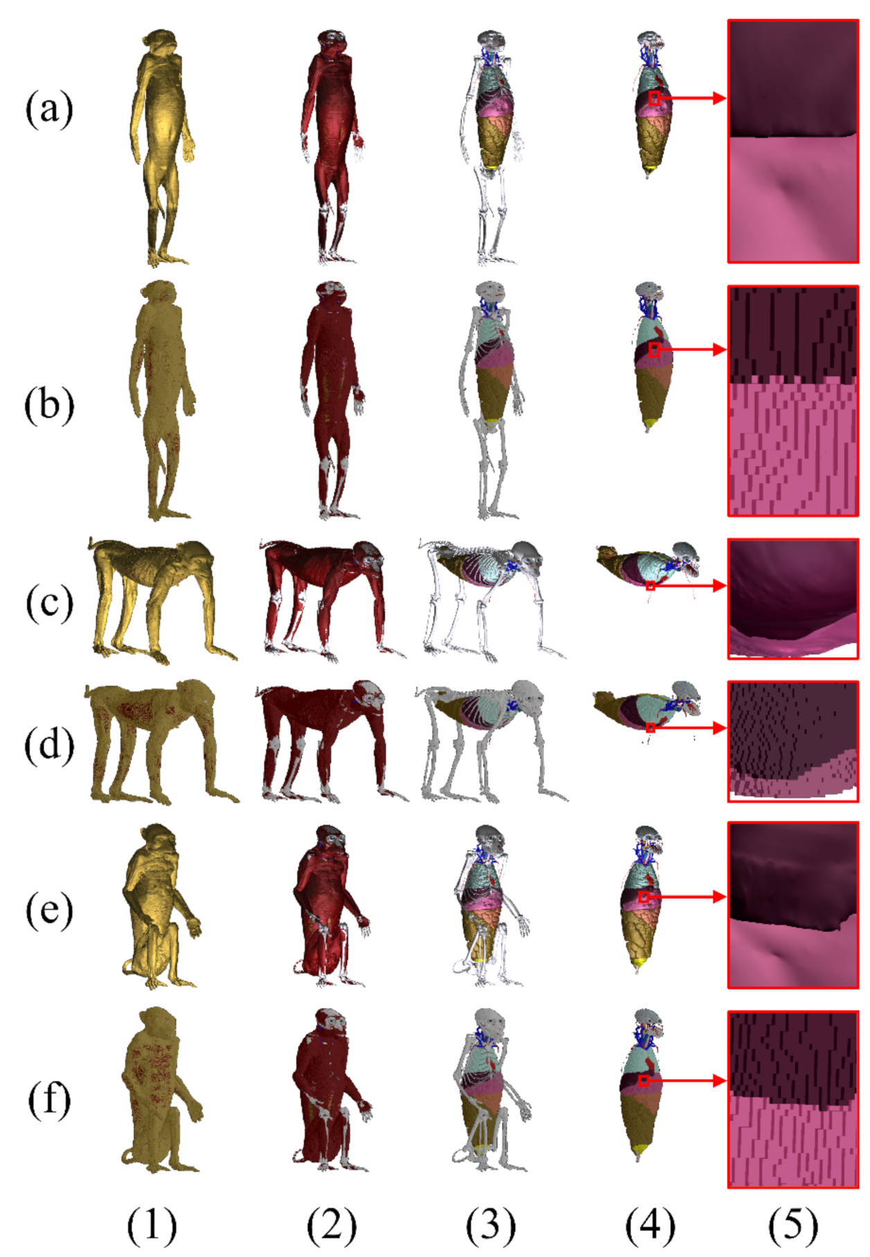
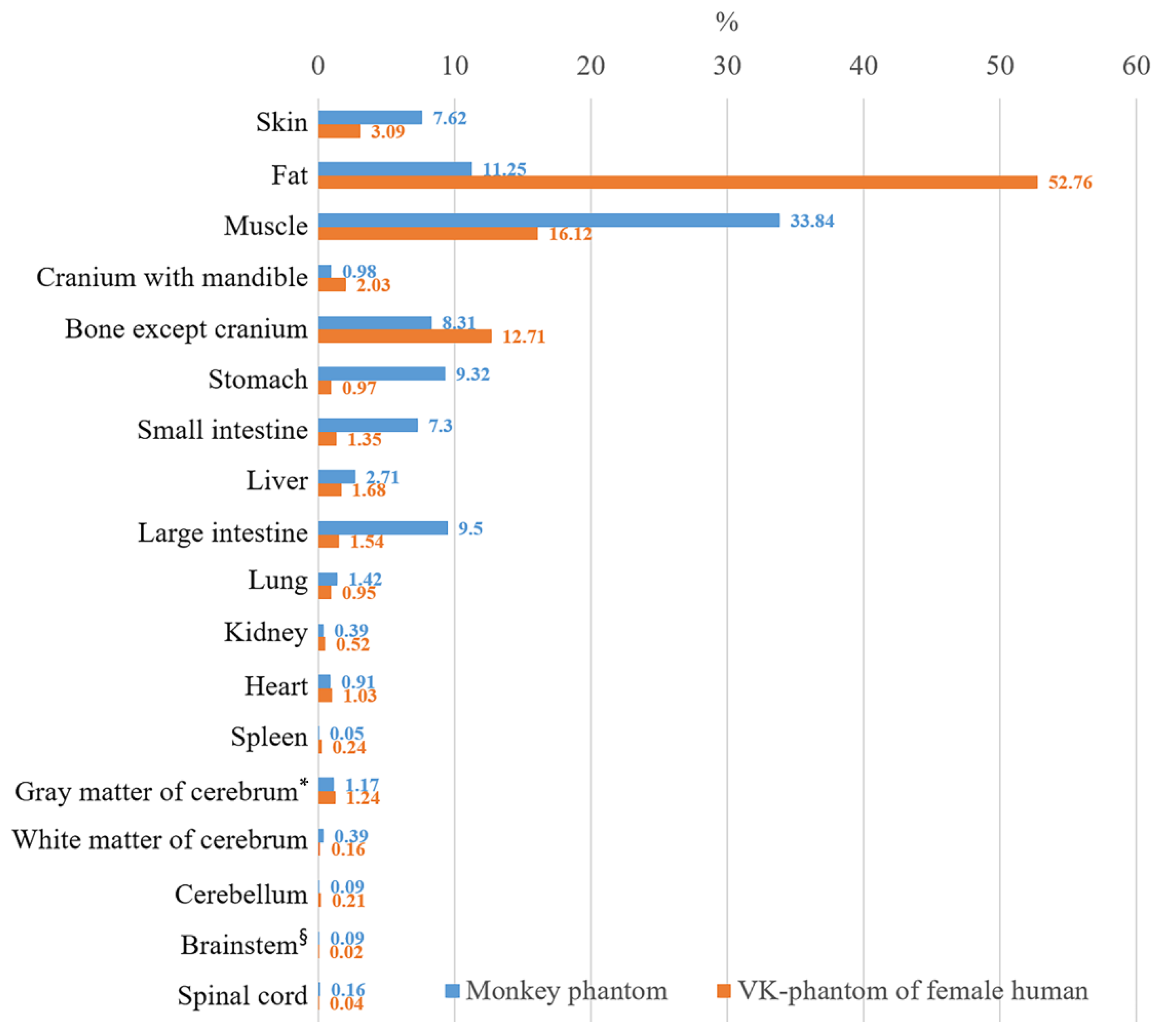
Posture-Transformed Monkey Phantoms Developed from a Visible Monkey
Abstract
:1. Introduction
2. Materials and Methods
3. Results
4. Discussion
5. Conclusions
Author Contributions
Funding
Institutional Review Board Statement
Informed Consent Statement
Data Availability Statement
Conflicts of Interest
Appendix A
| System | Structure | Density (kg/m3) (A) | Number of Voxels of Each Position | Mass of Each Position (g) | ||||
|---|---|---|---|---|---|---|---|---|
| Anatomical (B) | Quadrupedal (C) | Sitting (D) | Anatomical (A × B) | Quadrupedal (A × C) | Sitting (A × D) | |||
| Skeletal | Cranium | 1908 | 22,051 | 2,761 | 21,814 | 42.07 | 41.52 | 41.62 |
| Mandible | 1908 | 6177 | 6164 | 6096 | 11.79 | 11.76 | 11.63 | |
| 1st CV | 1908 | 622 | 614 | 618 | 1.19 | 1.17 | 1.18 | |
| 2nd CV | 1908 | 933 | 913 | 927 | 1.78 | 1.74 | 1.77 | |
| 3rd CV | 1908 | 724 | 696 | 699 | 1.38 | 1.33 | 1.33 | |
| 4th CV | 1908 | 628 | 632 | 631 | 1.20 | 1.21 | 1.20 | |
| 5th CV | 1908 | 641 | 639 | 642 | 1.22 | 1.22 | 1.22 | |
| 6th CV | 1908 | 739 | 755 | 746 | 1.41 | 1.44 | 1.42 | |
| 7th CV | 1908 | 756 | 777 | 769 | 1.44 | 1.48 | 1.47 | |
| 1st CV | 1908 | 1071 | 1076 | 1083 | 2.04 | 2.05 | 2.07 | |
| 2nd CV | 1908 | 1152 | 1155 | 1137 | 2.20 | 2.20 | 2.17 | |
| 3rd CV | 1908 | 1156 | 1184 | 1177 | 2.21 | 2.26 | 2.25 | |
| 4th CV | 1908 | 1218 | 1229 | 1249 | 2.32 | 2.34 | 2.38 | |
| 5th CV | 1908 | 1255 | 1268 | 1266 | 2.39 | 2.42 | 2.42 | |
| 6th CV | 1908 | 1333 | 1348 | 1366 | 2.54 | 2.57 | 2.61 | |
| 7th CV | 1908 | 1451 | 1439 | 1433 | 2.77 | 2.75 | 2.73 | |
| 8th CV | 1908 | 1653 | 1636 | 1635 | 3.15 | 3.12 | 3.12 | |
| 9th CV | 1908 | 1878 | 1845 | 1818 | 3.58 | 3.52 | 3.47 | |
| 10th TV | 1908 | 2237 | 2259 | 2253 | 4.27 | 4.31 | 4.30 | |
| 11th TV | 1908 | 3208 | 3176 | 3151 | 6.12 | 6.06 | 6.01 | |
| 12th TV | 1908 | 3726 | 3722 | 3731 | 7.11 | 7.10 | 7.12 | |
| 1st LV | 1908 | 4271 | 4236 | 4261 | 8.15 | 8.08 | 8.13 | |
| 2nd LV | 1908 | 4754 | 4784 | 4736 | 9.07 | 9.13 | 9.04 | |
| 3rd LV | 1908 | 4784 | 4774 | 4701 | 9.13 | 9.11 | 8.97 | |
| 4th LV | 1908 | 5140 | 5122 | 5151 | 9.81 | 9.77 | 9.83 | |
| 5th LV | 1908 | 5112 | 5106 | 5085 | 9.75 | 9.74 | 9.70 | |
| 6th LV | 1908 | 5625 | 5589 | 5631 | 10.73 | 10.66 | 10.74 | |
| 7th LV | 1908 | 5816 | 5786 | 5815 | 11.10 | 11.04 | 11.10 | |
| Sacrum | 1908 | 3509 | 3500 | 3472 | 6.70 | 6.68 | 6.62 | |
| Coccyx | 1908 | 4852 | 4860 | 4716 | 9.26 | 9.27 | 9.00 | |
| 1st rib | 1908 | 462 | 475 | 464 | 0.88 | 0.91 | 0.89 | |
| 2nd rib | 1908 | 707 | 699 | 696 | 1.35 | 1.33 | 1.33 | |
| 3rd rib | 1908 | 706 | 706 | 713 | 1.35 | 1.35 | 1.36 | |
| 4th rib | 1908 | 931 | 926 | 920 | 1.78 | 1.77 | 1.76 | |
| 5th rib | 1908 | 1108 | 1107 | 1055 | 2.11 | 2.11 | 2.01 | |
| 6th rib | 1908 | 1138 | 1102 | 1066 | 2.17 | 2.10 | 2.03 | |
| 7th rib | 1908 | 1208 | 1256 | 1190 | 2.30 | 2.40 | 2.27 | |
| 8th rib | 1908 | 1317 | 1334 | 1300 | 2.51 | 2.55 | 2.48 | |
| 9th rib | 1908 | 1286 | 1312 | 1271 | 2.45 | 2.50 | 2.43 | |
| 10th rib | 1908 | 966 | 971 | 965 | 1.84 | 1.85 | 1.84 | |
| 11st rib | 1908 | 690 | 687 | 689 | 1.32 | 1.31 | 1.31 | |
| 12nd rib | 1908 | 496 | 469 | 472 | 0.95 | 0.89 | 0.90 | |
| Costal cartilage | 1100 | 6153 | 6279 | 5806 | 6.77 | 6.91 | 6.39 | |
| Sternum | 1908 | 1636 | 1705 | 1486 | 3.12 | 3.25 | 2.84 | |
| Scapula | 1908 | 6514 | 6529 | 6528 | 12.43 | 12.46 | 12.46 | |
| Clavicle | 1908 | 1176 | 1216 | 1176 | 2.24 | 2.32 | 2.24 | |
| Humerus | 1908 | 24,285 | 24,313 | 24,101 | 46.34 | 46.39 | 45.98 | |
| Radius | 1908 | 8861 | 8951 | 8818 | 16.91 | 17.08 | 16.82 | |
| Ulna | 1908 | 9416 | 9451 | 9404 | 17.97 | 18.03 | 17.94 | |
| Scaphoid | 1908 | 401 | 398 | 405 | 0.77 | 0.76 | 0.77 | |
| Lunate | 1908 | 442 | 449 | 450 | 0.84 | 0.86 | 0.86 | |
| Triquetrum | 1908 | 427 | 424 | 415 | 0.81 | 0.81 | 0.79 | |
| Pisiform | 1908 | 289 | 301 | 297 | 0.55 | 0.57 | 0.57 | |
| Trapezium | 1908 | 235 | 235 | 232 | 0.45 | 0.45 | 0.44 | |
| Trapezoid | 1908 | 364 | 359 | 360 | 0.69 | 0.68 | 0.69 | |
| Capitate | 1908 | 396 | 389 | 391 | 0.76 | 0.74 | 0.75 | |
| Hamate | 1908 | 504 | 503 | 495 | 0.96 | 0.96 | 0.94 | |
| 1st MCB | 1908 | 562 | 557 | 556 | 1.07 | 1.06 | 1.06 | |
| 2nd MCB | 1908 | 1062 | 1055 | 1046 | 2.03 | 2.01 | 2.00 | |
| 3rd MCB | 1908 | 1261 | 1254 | 1239 | 2.41 | 2.39 | 2.36 | |
| 4th MCB | 1908 | 983 | 990 | 974 | 1.88 | 1.89 | 1.86 | |
| 5th MCB | 1908 | 725 | 732 | 713 | 1.38 | 1.40 | 1.36 | |
| 1st PP (hand) | 1908 | 215 | 217 | 211 | 0.41 | 0.41 | 0.40 | |
| 2nd PP (hand) | 1908 | 427 | 430 | 435 | 0.81 | 0.82 | 0.83 | |
| 3rd PP (hand) | 1908 | 593 | 588 | 585 | 1.13 | 1.12 | 1.12 | |
| 4th PP (hand) | 1908 | 555 | 551 | 565 | 1.06 | 1.05 | 1.08 | |
| 5th PP (hand) | 1908 | 333 | 338 | 338 | 0.64 | 0.64 | 0.64 | |
| 2nd MP (hand) | 1908 | 136 | 137 | 133 | 0.26 | 0.26 | 0.25 | |
| 3rd MP (hand) | 1908 | 39 | 40 | 33 | 0.07 | 0.08 | 0.06 | |
| 4th MP (hand) | 1908 | 593 | 588 | 585 | 1.13 | 1.12 | 1.12 | |
| 5th MP (hand) | 1908 | 269 | 280 | 276 | 0.51 | 0.53 | 0.53 | |
| 1st DP (hand) | 1908 | 43 | 44 | 42 | 0.08 | 0.08 | 0.08 | |
| 2nd DP (hand) | 1908 | 427 | 430 | 435 | 0.81 | 0.82 | 0.83 | |
| 3rd DP (hand) | 1908 | 136 | 137 | 133 | 0.26 | 0.26 | 0.25 | |
| 4th DP (hand) | 1908 | 39 | 40 | 33 | 0.07 | 0.08 | 0.06 | |
| 5th DP (hand) | 1908 | 593 | 588 | 585 | 1.13 | 1.12 | 1.12 | |
| Sesamoid bone (hand) | 1908 | 46 | 50 | 48 | 0.09 | 0.10 | 0.09 | |
| Pelvis | 1908 | 11,840 | 11,902 | 11,860 | 22.59 | 22.71 | 22.63 | |
| Femur | 1908 | 33,037 | 33,113 | 33,197 | 63.03 | 63.18 | 63.34 | |
| Patella | 1908 | 1586 | 1596 | 1586 | 3.03 | 3.05 | 3.03 | |
| Tibia | 1908 | 19,512 | 19,530 | 19,488 | 37.23 | 37.26 | 37.18 | |
| Fibula | 1908 | 4128 | 4076 | 4086 | 7.88 | 7.78 | 7.80 | |
| Talus | 1908 | 3336 | 3341 | 3300 | 6.37 | 6.37 | 6.30 | |
| Calcaneus | 1908 | 5676 | 5681 | 5667 | 10.83 | 10.84 | 10.81 | |
| Navicular | 1908 | 1228 | 1239 | 1232 | 2.34 | 2.36 | 2.35 | |
| Medial cuneiform | 1908 | 887 | 892 | 884 | 1.69 | 1.70 | 1.69 | |
| Intermediate cuneiform | 1908 | 377 | 382 | 376 | 0.72 | 0.73 | 0.72 | |
| Lateral cuneiform | 1908 | 658 | 671 | 652 | 1.26 | 1.28 | 1.24 | |
| Cuboid | 1908 | 1464 | 1465 | 1463 | 2.79 | 2.80 | 2.79 | |
| 1st MTB | 1908 | 1432 | 1431 | 1455 | 2.73 | 2.73 | 2.78 | |
| 2nd MTB | 1908 | 1397 | 1428 | 1404 | 2.67 | 2.72 | 2.68 | |
| 3rd MTB | 1908 | 1822 | 1811 | 1788 | 3.48 | 3.46 | 3.41 | |
| 4th MTB | 1908 | 1716 | 1733 | 1736 | 3.27 | 3.31 | 3.31 | |
| 5th MTB | 1908 | 1267 | 1264 | 1217 | 2.42 | 2.41 | 2.32 | |
| 1st PP (foot) | 1908 | 420 | 425 | 430 | 0.80 | 0.81 | 0.82 | |
| 2nd PP (foot) | 1908 | 50 | 62 | 74 | 0.10 | 0.12 | 0.14 | |
| 3rd PP (foot) | 1908 | 380 | 368 | 387 | 0.73 | 0.70 | 0.74 | |
| 4th PP (foot) | 1908 | 123 | 138 | 133 | 0.23 | 0.26 | 0.25 | |
| 5th PP (foot) | 1908 | 11 | 17 | 17 | 0.02 | 0.03 | 0.03 | |
| 2nd MP (foot) | 1908 | 123 | 138 | 133 | 0.23 | 0.26 | 0.25 | |
| 3rd MP (foot) | 1908 | 11 | 17 | 17 | 0.02 | 0.03 | 0.03 | |
| 4th MP (foot) | 1908 | 674 | 656 | 699 | 1.29 | 1.25 | 1.33 | |
| 5th MP (foot) | 1908 | 234 | 249 | 243 | 0.45 | 0.48 | 0.46 | |
| 1st DP (foot) | 1908 | 50 | 62 | 74 | 0.10 | 0.12 | 0.14 | |
| 2nd DP (foot) | 1908 | 380 | 368 | 387 | 0.73 | 0.70 | 0.74 | |
| 3rd DP (foot) | 1908 | 123 | 138 | 133 | 0.23 | 0.26 | 0.25 | |
| 4th DP (foot) | 1908 | 11 | 17 | 17 | 0.02 | 0.03 | 0.03 | |
| 5th DP (foot) | 1908 | 674 | 656 | 699 | 1.29 | 1.25 | 1.33 | |
| Intervertebral disc | 1100 | 12,492 | 11,620 | 11,267 | 13.74 | 12.78 | 12.39 | |
| Cartilage | 1100 | 2265 | 2191 | 2169 | 2.49 | 2.41 | 2.39 | |
| Muscular | Muscle | 1090 | 1,699,435 | 1,767,197 | 1,645,241 | 1852.38 | 1926.24 | 1793.31 |
| Alimen-tary | Teeth | 2180 | 5041 | 5164 | 5121 | 10.99 | 11.26 | 11.16 |
| Tongue | 1090 | 6988 | 6986 | 6996 | 7.62 | 7.61 | 7.63 | |
| Esophagus | 1040 | 10,490 | 10,848 | 10,194 | 10.91 | 11.28 | 10.60 | |
| Stomach | 1088 | 468,655 | 467,573 | 390,467 | 509.90 | 508.72 | 424.83 | |
| Small intestine | 1100 | 349,654 | 348,316 | 310,591 | 384.62 | 383.15 | 341.65 | |
| Duodenum | 1102 | 13,417 | 13,297 | 12,039 | 14.79 | 14.65 | 13.27 | |
| Large intestine | 1100 | 472,874 | 472,962 | 449,017 | 520.16 | 520.26 | 493.92 | |
| Liver | 1079 | 137,547 | 137,070 | 126,927 | 148.41 | 147.90 | 136.95 | |
| Gall bladder | 928 | 1550 | 1527 | 1440 | 1.44 | 1.42 | 1.34 | |
| Common bile duct | 1090 | 12 | 23 | 27 | 0.01 | 0.03 | 0.03 | |
| Respira-tory | Trachea | 1080 | 4869 | 5395 | 4741 | 5.26 | 5.83 | 5.12 |
| Bronchus | 1102 | 3411 | 3394 | 3247 | 3.76 | 3.74 | 3.58 | |
| Lungs | 394 | 197,595 | 197,170 | 189,181 | 77.85 | 77.68 | 74.54 | |
| Urinary | Kidney | 1066 | 20,103 | 20,122 | 19,476 | 21.43 | 21.45 | 20.76 |
| Urinary bladder | 928 | 8650 | 8688 | 8644 | 8.03 | 8.06 | 8.02 | |
| Urethra | 1102 | 112 | 106 | 113 | 0.12 | 0.12 | 0.12 | |
| Genital | Ovary | 1048 | 378 | 387 | 391 | 0.40 | 0.41 | 0.41 |
| Uterus | 1105 | 3279 | 3288 | 3293 | 3.62 | 3.63 | 3.64 | |
| Vagina | 1088 | 2936 | 2949 | 2986 | 3.19 | 3.21 | 3.25 | |
| Endo-crine | Thyroid gland | 1071 | 530 | 644 | 562 | 0.57 | 0.69 | 0.60 |
| Adrenal gland | 1071 | 618 | 615 | 587 | 0.66 | 0.66 | 0.63 | |
| Cardio-vascular | Heart | 1071 | 46,668 | 46,607 | 43,353 | 49.98 | 49.92 | 46.43 |
| Common carotid a. | 1050 | 121 | 150 | 130 | 0.13 | 0.16 | 0.14 | |
| External carotid a. | 1050 | 39 | 37 | 48 | 0.04 | 0.04 | 0.05 | |
| Facial a. | 1050 | 22 | 21 | 25 | 0.02 | 0.02 | 0.03 | |
| Superficial temporal a. | 1050 | 46 | 40 | 36 | 0.05 | 0.04 | 0.04 | |
| Alveolar a. | 1050 | 8 | 8 | 10 | 0.01 | 0.01 | 0.01 | |
| Internal carotid a. | 1050 | 47 | 60 | 57 | 0.05 | 0.06 | 0.06 | |
| Middle cerebral a. | 1050 | 31 | 15 | 17 | 0.03 | 0.02 | 0.02 | |
| Basilar a. | 1050 | 7 | 7 | 5 | 0.01 | 0.01 | 0.01 | |
| Vertebral a. | 1050 | 208 | 237 | 212 | 0.22 | 0.25 | 0.22 | |
| the rest arteries | 1050 | 6395 | 6364 | 6130 | 6.71 | 6.68 | 6.44 | |
| Internal jugular v. | 1050 | 570 | 622 | 613 | 0.60 | 0.65 | 0.64 | |
| Retromandibular v. | 1050 | 370 | 369 | 355 | 0.39 | 0.39 | 0.37 | |
| Superficial temporal v. | 1050 | 5 | 3 | 2 | 0.01 | 0.003 | 0.002 | |
| External jugular v. | 1050 | 1048 | 1163 | 1035 | 1.10 | 1.22 | 1.09 | |
| Anterior jugular v. | 1050 | 365 | 440 | 379 | 0.38 | 0.46 | 0.40 | |
| Transverse sinus | 1050 | 50 | 57 | 78 | 0.05 | 0.06 | 0.08 | |
| Confluence of sinuses | 1050 | 30 | 31 | 31 | 0.03 | 0.03 | 0.03 | |
| Sigmoid sinus | 1050 | 82 | 80 | 67 | 0.09 | 0.08 | 0.07 | |
| Superior sagittal sinus | 1050 | 130 | 129 | 142 | 0.14 | 0.14 | 0.15 | |
| Straight sinus | 1050 | 35 | 30 | 28 | 0.04 | 0.03 | 0.03 | |
| the rest veins | 1050 | 13,501 | 13,816 | 13,327 | 14.18 | 14.51 | 13.99 | |
| Lymph-oid | Bone marrow | 1029 | 121,055 | 121,287 | 121,546 | 124.57 | 124.80 | 125.07 |
| Spleen | 1089 | 2655 | 2672 | 2561 | 2.89 | 2.91 | 2.79 | |
| Central nervous | Gray matter of CB | 1046 | 59,217 | 59,180 | 59,271 | 61.94 | 61.90 | 62.00 |
| White matter of CB | 1041 | 20,257 | 20,368 | 20,315 | 21.09 | 21.20 | 21.15 | |
| Dura mater | 1908 | 3873 | 3746 | 3722 | 7.39 | 7.15 | 7.10 | |
| Cerebrospinal fluid | 1007 | 2102 | 2250 | 2164 | 2.12 | 2.27 | 2.18 | |
| Spinal cord | 1075 | 8328 | 8439 | 8496 | 8.95 | 9.07 | 9.13 | |
| Medulla oblongata | 1075 | 1396 | 1392 | 1380 | 1.50 | 1.50 | 1.48 | |
| Pons | 1046 | 1759 | 1731 | 1778 | 1.84 | 1.81 | 1.86 | |
| Midbrain | 1046 | 1530 | 1507 | 1438 | 1.60 | 1.58 | 1.50 | |
| Cerebellum | 1045 | 4929 | 4952 | 4961 | 5.15 | 5.17 | 5.18 | |
| Thalamus | 1045 | 1736 | 1712 | 1693 | 1.81 | 1.79 | 1.77 | |
| Hypothalamus | 1045 | 46 | 41 | 35 | 0.05 | 0.04 | 0.04 | |
| Cingulate gyrus | 1046 | 892 | 917 | 965 | 0.93 | 0.96 | 1.01 | |
| Hippocampus | 1045 | 281 | 262 | 258 | 0.29 | 0.27 | 0.27 | |
| Sensory | Sclera | 911 | 419 | 439 | 463 | 0.38 | 0.40 | 0.42 |
| Cornea | 1051 | 18 | 22 | 19 | 0.02 | 0.02 | 0.02 | |
| Lens cortex | 1076 | 74 | 79 | 85 | 0.08 | 0.09 | 0.09 | |
| Lens nucleus | 1076 | 78 | 84 | 75 | 0.08 | 0.09 | 0.08 | |
| Vitreous humor | 1005 | 5507 | 5512 | 5493 | 5.53 | 5.54 | 5.52 | |
| Integum-entary | Skin | 1109 | 376,101 | 374,396 | 379,802 | 417.10 | 415.21 | 421.20 |
| Breast fat | 911 | 230 | 234 | 236 | 0.21 | 0.21 | 0.21 | |
| Fat | 911 | 675,922 | 732,245 | 644,889 | 615.76 | 667.08 | 587.49 | |
| Total | 5,054,022 | 5,174,453 | 4,803,886 | 5473.69 | 5595.16 | 5211.24 | ||
References
- Nagaoka, T.; Watanabe, S.; Sakurai, K.; Kunieda, E.; Watanabe, S.; Taki, M.; Yamanaka, Y. Development of realistic high-resolution whole-body voxel models of Japanese adult males and females of average height and weight, and application of models to radio-frequency electromagnetic-field dosimetry. Phys. Med. Biol. 2004, 49, 1–15. [Google Scholar] [CrossRef] [PubMed]
- Dimbylow, P. Development of the female voxel phantom, NAOMI, and its application to calculations of induced current densities and electric fields from applied low frequency magnetic and electric fields. Phys. Med. Biol. 2005, 50, 1047–1070. [Google Scholar] [CrossRef] [PubMed]
- Christ, A.; Kainz, W.; Hahn, E.G.; Honegger, K.; Zefferer, M.; Neufeld, E.; Rascher, W.; Janka, R.; Bautz, W.; Chen, J.; et al. The Virtual Family-development of surface-based anatomical models of two adults and two children for dosimetric simulations. Phys. Med. Biol. 2009, 55, N23–N38. [Google Scholar] [CrossRef] [PubMed]
- Segars, W.P.; Sturgeon, G.; Mendonca, S.; Grimes, J.; Tsui, B.M.W. 4D XCAT phantom for multimodality imaging research. Med. Phys. 2010, 37, 4902–4915. [Google Scholar] [CrossRef]
- Gosselin, M.-C.; Neufeld, E.; Moser, H.; Huber, E.; Farcito, S.; Gerber, L.; Jedensjo, M.; Hilber, I.; Di Gennaro, F.; Lloyd, B.; et al. Development of a new generation of high-resolution anatomical models for medical device evaluation: The Virtual Population 3.0. Phys. Med. Biol. 2014, 59, 5287–5303. [Google Scholar] [CrossRef]
- Yeom, Y.S.; Jeong, J.H.; Kim, C.H.; Han, M.C.; Ham, B.K.; Cho, K.W.; Hwang, S.B. HDRK-Woman: Whole-body voxel model based on high-resolution color slice images of Korean adult female cadaver. Phys. Med. Biol. 2014, 59, 3969–3984. [Google Scholar] [CrossRef]
- Lee, A.-K.; Hong, S.-E.; Kwon, J.-H.; Choi, H.-D.; Cardis, E. Mobile phone types and SAR characteristics of the human brain. Phys. Med. Biol. 2017, 62, 2741–2761. [Google Scholar] [CrossRef]
- Kim, C.H.; Yeom, Y.S.; Nguyen, T.T.; Han, M.C.; Choi, C.; Lee, H.; Han, H.; Shin, B.; Lee, J.K.; Kim, H.S.; et al. New mesh-type phantoms and their dosimetric applications, including emergencies. Ann. ICRP 2018, 47, 45–62. [Google Scholar] [CrossRef]
- Yeom, Y.S.; Han, H.; Choi, C.; Nguyen, T.T.; Shin, B.; Lee, C.; Kim, C.H. Posture-dependent dose coefficients of mesh-type ICRP reference computational phantoms for photon external exposures. Phys. Med. Biol. 2019, 64, 075018. [Google Scholar] [CrossRef]
- Lee, A.K.; Park, J.S.; Hong, S.E.; Taki, M.; Wake, K.; Wiart, J.; Choi, H.-D. Brain SAR of average male Korean child to adult models for mobile phone exposure assessment. Phys. Med. Biol. 2019, 64, 045004. [Google Scholar] [CrossRef]
- Davids, M.; Guerin, B.; Vom Endt, A.; Schad, L.R.; Wald, L.L. Prediction of peripheral nerve stimulation thresholds of MRI gradient coils using coupled electromagnetic and neurodynamic simulations. Magn. Reson. Med. 2019, 81, 686–701. [Google Scholar] [CrossRef] [Green Version]
- Yang, B.; Tam, F.; Davidson, B.; Wei, P.S.; Hamani, C.; Lipsman, N.; Chen, C.H.; Graham, S.J. Technical Note: An anthropomorphic phantom with implanted neurostimulator for investigation of MRI safety. Med. Phys. 2020, 47, 3745–3751. [Google Scholar] [CrossRef]
- Yeni-Komshian, G.H.; Benson, D.A. Anatomical study of cerebral asymmetry in the temporal lobe of humans, chimpanzees, and rhesus monkeys. Science 1976, 192, 387–389. [Google Scholar] [CrossRef]
- Frey, S.; Pandya, D.N.; Chakravarty, M.M.; Bailey, L.; Petrides, M.; Collins, D.L. An MRI based average macaque monkey stereotaxic atlas and space (MNI monkey space). Neuroimage 2011, 55, 1435–1442. [Google Scholar] [CrossRef] [Green Version]
- De Schotten, M.T.; Dell’Acqua, F.; Valabregue, R.; Catani, M. Monkey to human comparative anatomy of the frontal lobe association tracts. Cortex 2012, 48, 82–96. [Google Scholar] [CrossRef]
- Takemura, H.; Pestilli, F.; Weiner, K.S.; Keliris, G.A.; Landi, S.M.; Sliwa, J.; Ye, F.Q.; Barnett, M.A.; Leopold, D.A.; Freiwald, W.A.; et al. Occipital White Matter Tracts in Human and Macaque. Cereb. Cortex 2017, 27, 3346–3359. [Google Scholar] [CrossRef] [Green Version]
- Mars, R.B.; Sotiropoulos, S.N.; Passingham, R.E.; Sallet, J.; Verhagen, L.; Khrapitchev, A.A.; Sibson, N.; Jbabdi, S. Whole brain comparative anatomy using connectivity blueprints. Elife 2018, 7, e35237. [Google Scholar] [CrossRef]
- Capogrosso, M.; Milekovic, T.; Borton, D.; Wagner, F.; Moraud, E.M.; Mignardot, J.B.; Buse, N.; Gandar, J.; Barraud, Q.; Xing, D.; et al. A brain-spine interface alleviating gait deficits after spinal cord injury in primates. Nature 2016, 539, 284–288. [Google Scholar] [CrossRef] [Green Version]
- Findlay, R.P.; Lee, A.K.; Dimbylow, P.J. FDTD calculations of SAR for child voxel models in different postures between 10 MHz and 3 GHz. Radiat. Prot. Dosim. 2009, 135, 226–231. [Google Scholar] [CrossRef]
- Lee, A.-K.; Choi, H.-D. Determining the influence of Korean population variation on whole-body average SAR. Phys. Med. Biol. 2012, 57, 2709–2725. [Google Scholar] [CrossRef]
- Xie, T.; Park, J.S.; Zhuo, W.; Zaidi, H. Development of a nonhuman primate computational phantom for radiation dosimetry. Med. Phys. 2020, 47, 736–744. [Google Scholar] [CrossRef]
- Chung, B.S.; Jeon, C.-Y.; Huh, J.W.; Jeong, K.-J.; Har, D.; Kwack, K.-S.; Park, J.S. Rise of the Visible Monkey: Sectioned images of rhesus monkey. J. Korean Med. Sci. 2019, 34, e66. [Google Scholar] [CrossRef]
- Kawai, M.; Mito, U. Quantitative study of activity patterns and postures of Formosan monkeys by the radio-telemetrical technique. Primates 1973, 14, 179–194. [Google Scholar] [CrossRef]
- Hunt, K.D.; Cant, J.G.H.; Gebo, D.L.; Rose, M.D.; Walker, S.E.; Youlatos, D. Standardized descriptions of primate locomotor and postural modes. Primates 1996, 37, 363–387. [Google Scholar] [CrossRef]
- Rose, M.D. Positional behaviour of olive baboons (Papio anubis) and its relationship to maintenance and social activities. Primates 1977, 18, 59–116. [Google Scholar] [CrossRef]
- Chivers, D.J. Malayan Forest Primates: Ten Years’ Study in Tropical Rain Forest; Springer: New York, NY, USA, 2013; pp. 198–199. [Google Scholar]
- Kim, C.Y.; Lee, A.-K.; Choi, H.-D.; Park, J.S. Dawn of the Visible Monkey: Segmentation of the rhesus monkey for 2D and 3D applications. J. Korean Med. Sci. 2020, 35, e100. [Google Scholar] [CrossRef] [Green Version]
- Han, M.; Lee, A.-K.; Choi, H.-D.; Jung, Y.W.; Park, J.S. Averaged head phantoms from magnetic resonance images of Korean children and young adults. Phys. Med. Biol. 2018, 63, 035003. [Google Scholar] [CrossRef] [PubMed]
- Park, J.S.; Jung, Y.W.; Choi, H.-D.; Lee, A.-K. VK-phantom male with 583 structures and female with 459 structures, based on the sectioned images of a male and a female, for computational dosimetry. J. Radiat. Res. 2018, 59, 338–380. [Google Scholar] [CrossRef]
- Gabriel, C.; Gabriel, S.; Corthout, E. The dielectric properties of biological tissues: I. Literature survey. Phys. Med. Biol. 1996, 41, 2231–2249. [Google Scholar] [CrossRef] [Green Version]
- Lafon, Y.; Smith, F.W.; Beillas, P. Combination of a model-deformation method and a positional MRI to quantify the effects of posture on the anatomical structures of the trunk. J. Biomech. 2010, 43, 1269–1278. [Google Scholar] [CrossRef]
- Montagna, W. The Skin of Nonhuman Primates. Am. Zool. 1972, 12, 109–124. [Google Scholar] [CrossRef]
- Aiello, L.C.; Wheeler, P. The expensive-tissue hypothesis: The brain and the digestive system in human and primate evolution. Curr. Anthr. 1995, 36, 199–221. [Google Scholar] [CrossRef]
- Kim, C.H.; Choi, S.H.; Jeong, J.H.; Lee, C.; Chung, M.S. HDRK-Man: A whole-body voxel model based on high-resolution color slice images of a Korean adult male cadaver. Phys. Med. Biol. 2008, 53, 4093–4106. [Google Scholar] [CrossRef] [PubMed] [Green Version]
- Yeom, Y.S.; Han, M.C.; Kim, C.H.; Jeong, J.H. Conversion of ICRP male reference phantom to polygon-surface phantom. Phys. Med. Biol. 2013, 58, 6985–7007. [Google Scholar] [CrossRef]
- Yeom, Y.S.; Jeong, J.H.; Han, M.C.; Kim, C.H. Tetrahedral-mesh-based computational human phantom for fast Monte Carlo dose calculations. Phys. Med. Biol. 2014, 59, 3173–3185. [Google Scholar] [CrossRef]
- Bottauscio, O.; Cassara, A.M.; Hand, J.W.; Giordano, D.; Zilberti, L.; Borsero, M.; Chiampi, M.; Weidemann, G. Assessment of computational tools for MRI RF dosimetry by comparison with measurements on a laboratory phantom. Phys. Med. Biol. 2015, 60, 5655–5680. [Google Scholar] [CrossRef]
- Kim, H.S.; Yeom, Y.S.; Nguyen, T.T.; Choi, C.; Han, M.C.; Lee, J.K.; Kim, C.H.; Zankl, M.; Petoussi-Henss, N.; Bolch, W.E.; et al. Inclusion of thin target and source regions in alimentary and respiratory tract systems of mesh-type ICRP adult reference phantoms. Phys. Med. Biol. 2017, 62, 2132–2152. [Google Scholar] [CrossRef]
- Spitzer, V.; Ackerman, M.J.; Scherzinger, A.L.; Whitlock, D. The visible human male: A technical report. J. Am. Med. Inform. Assoc. 1996, 3, 118–130. [Google Scholar] [CrossRef] [Green Version]
- Park, J.S.; Chung, M.S.; Hwang, S.B.; Lee, Y.S.; Har, D.H.; Park, H.S. Visible Korean human: Improved serially sectioned images of the entire body. IEEE Trans. Med. Imaging 2005, 24, 352–360. [Google Scholar] [CrossRef]
- Zhang, S.X.; Heng, P.A.; Liu, Z.J. Chinese visible human project. Clin. Anat. 2006, 19, 204–215. [Google Scholar] [CrossRef]
- Park, J.S.; Chung, M.S.; Shin, D.S.; Har, D.H.; Cho, Z.H.; Kim, Y.B.; Han, J.Y.; Chi, J.G. Sectioned images of the cadaver head including the brain and correspondences with ultrahigh field 7.0 T MRIs. Proc. IEEE 2009, 97, 1988–1996. [Google Scholar] [CrossRef]
- Park, H.S.; Choi, D.H.; Park, J.S. Improved sectioned images and surface models of the whole female body. Int. J. Morphol. 2015, 33, 1323–1332. [Google Scholar] [CrossRef] [Green Version]
- Park, J.S. Neuroman: Voxel phantoms from surface models of 300 head structures including 12 pairs of cranial nerves. Health Phys. 2020, 119, 192–205. [Google Scholar] [CrossRef]
- Yanamadala, J.; Noetscher, G.M.; Rathi, V.K.; Maliye, S.; Win, H.A.; Tran, A.L.; Jackson, X.J.; Htet, A.T.; Kozlov, M.; Nazarian, A.; et al. New VHP-Female v. 2.0 full-body computational phantom and its performance metrics using FEM simulator ANSYS HFSS. Conf. Proc. IEEE Eng. Med. Biol. Soc. 2015, 3237–3241. [Google Scholar] [CrossRef]


| File Format of Three Positions | Total Number of Files of Three Positions (Number of Files Per Position) | File Size of Anatomical Position | File Size of Quadrupedal Position | File Size of Sitting Position | |
|---|---|---|---|---|---|
| Surface models | STL | 531 (177) | 324 Mbytes | 325 Mbytes | 326 Mbytes |
| Phantoms | TXT * | 3 (1) | 99 Mbytes | 105 Mbytes | 96 Mbytes |
Publisher’s Note: MDPI stays neutral with regard to jurisdictional claims in published maps and institutional affiliations. |
© 2021 by the authors. Licensee MDPI, Basel, Switzerland. This article is an open access article distributed under the terms and conditions of the Creative Commons Attribution (CC BY) license (https://creativecommons.org/licenses/by/4.0/).
Share and Cite
Kim, C.Y.; Lee, A.-K.; Choi, H.-D.; Park, J.S. Posture-Transformed Monkey Phantoms Developed from a Visible Monkey. Appl. Sci. 2021, 11, 4430. https://doi.org/10.3390/app11104430
Kim CY, Lee A-K, Choi H-D, Park JS. Posture-Transformed Monkey Phantoms Developed from a Visible Monkey. Applied Sciences. 2021; 11(10):4430. https://doi.org/10.3390/app11104430
Chicago/Turabian StyleKim, Chung Yoh, Ae-Kyoung Lee, Hyung-Do Choi, and Jin Seo Park. 2021. "Posture-Transformed Monkey Phantoms Developed from a Visible Monkey" Applied Sciences 11, no. 10: 4430. https://doi.org/10.3390/app11104430
APA StyleKim, C. Y., Lee, A.-K., Choi, H.-D., & Park, J. S. (2021). Posture-Transformed Monkey Phantoms Developed from a Visible Monkey. Applied Sciences, 11(10), 4430. https://doi.org/10.3390/app11104430


