Article

Evaluating the Effect of Tissue Selection on the Characteristics of Extracellular Matrix Hydrogels from Decellularized Porcine Bladders

1
Graduate Institute of Biomedical Engineering, National Taiwan University of Science and Technology, No. 43, Sec. 4, Keelung Rd., Da’an Dist., Taipei 10607, Taiwan
2
Biomedical Engineering Research Center, National Defense Medical Center, Taipei 11490, Taiwan
3
Graduate Institute of Applied Science and Technology, National Taiwan University of Science and Technology, Taipei 10607, Taiwan
4
School of Medicine, National Defense Medical Center, Taipei 11490, Taiwan
5
Department of Biomedical Engineering, National Yang Ming Chiao Tung University, Taipei 11221, Taiwan
*
Author to whom correspondence should be addressed.
Appl. Sci. 2021, 11(13), 5820; https://doi.org/10.3390/app11135820
Submission received: 2 June 2021 / Revised: 15 June 2021 / Accepted: 20 June 2021 / Published: 23 June 2021
(This article belongs to the Section Applied Biosciences and Bioengineering)

Abstract

:

Featured Application

The research demonstrated the potential use of sECM and WB for ECM hydrogel fabrication, which could decrease the tissue processing time and increase the possibility for mass production.

Abstract

Porcine urinary bladder is one of the most used organs to fabricate extracellular matrix (ECM) hydrogel. Although there are two different ECM types inside a bladder, i.e., urinary bladder matrix (UBM) and a subtype ECM (sECM), most studies have only employed UBM for hydrogel fabrication, and overlooked the potential use of sECM. In another aspect, the delamination of UBM from bladders is a time-consuming process; consequently, the use of the whole bladder (WB) will likely increase production yield. Therefore, the objective of this study was to fabricate hydrogels from sECM and WB and compare them to UBM. The results indicated that different layers of the bladder shared almost the same biochemical composition. In terms of gelation kinetics, rheology and morphology, although hydrogels from UBM and sECM exhibited some discrepancies, those from UBM and WB interestingly possessed almost the same characteristics. In in vitro studies, all the hydrogels possessed nearly the same biochemical effects towards L929 viability and C2C12 differentiation. These results could preliminarily indicate that the use of sECM should no longer be ignored, and WB could be a promising substitution for UBM hydrogels, eliminating the need for time-consuming delamination processes, as well as increasing the possibility of mass production.

1. Introduction

Hydrogels are defined as highly hydrated viscoelastic polymer materials with specific three-dimensional (3D) networks, which possess similar physical properties to natural tissue [1]. Among various types, hydrogels derived from decellularized tissues offer numerous advantages compared to synthetic hydrogels, as their extracellular matrix (ECM) components contain numerous biochemical and biomechanical signals, which play a pivotal role in the processes of tissue morphogenesis, differentiation and homeostasis [2]. The ECM is composed of two biochemically and morphologically recognizable 3D structures, the basement membrane (BM) and the interstitial matrix (IM) [3]. Although these entities can be investigated as two separate domains, they do not in fact exist in isolation, and are instead interconnected by multiple anchoring and interconnecting fibrils [4]. The BM is a highly organized thin, dense sheet of the ECM, containing primarily collagen (type IV), laminins and fibronectin, which anchor down the epithelium to its underlying lamina propria. The functions of BM are to provide structural support, give the cells physical and functional compartmentalization, and regulate cell behaviors [4]. In contrast, compared to the dense morphology of BM, the interstitial matrix is a loosely organized porous “3D amorphous lattice” located next to the BM. The dominant constituent constructing this ECM include collagens type I, III, V, VI, VII, XII, fibronectins, proteoglycans, which direct the organization of the IM structure, and is responsible for mediating cell attachment and function [5].
Among many types of porcine tissues, one of the most studied ECM is porcine urinary bladder [6]. According to literature, there are two types of ECM inside a porcine urinary bladder, namely urinary bladder matrix (UBM) and another lower type of extracellular matrix, in this context called sub-ECM (sECM) [7]. The UBM contains basement membrane of urothelial cells and the lamina propria, offering cells with nutritive and informative support, while sECM is characterized by muscularis mucosa (submucosa) and the detrusor, which contain an elastic fiber network, accounting for the distension of the bladder [7]. Additionally, the organized smooth muscle cell bundles of IM are also surrounded by a layer of BM [8].
In our previous study [9], we established a reliable protocol to decellularize UBM using sodium dodecyl sulfate (SDS), and successfully prepared UBM hydrogels. At that point, we also questioned the usage of the rest of the bladder after delamination (i.e., the sECM part), and tried to address this problem. To the best of our knowledge, most studies have only prepared hydrogels from UBM and ignored the potential use of sECM or the whole bladder (WB) to fabricate ECM hydrogels, and there is no study investigating the differences among hydrogels made from those types of tissues [10,11,12,13,14]. On one hand, since there is a difference in the complicated biochemical compositions inside the UBM and sECM [15], it can accordingly be hypothesized that the ECM hydrogels made from distinct parts of the porcine bladder will possess dissimilar physical and biochemical properties. On the other hand, the delamination process of separating the UBM from the bladder is a time-consuming task and requires meticulous implantation, as the efficacy of decellularization protocol can be diminished if too many or too few tissue layers are removed [16]. As a result, it is more convenient for mass production if the whole bladder can be used to fabricate hydrogels as well.
Therefore, the objectives of this research are to fabricate hydrogels from sECM and WB and compare them to the widely employed UBM hydrogels in terms of biochemical composition, gelation kinetics, mechanical properties, and surface morphology, as well as the effect on cell viability and differentiation to have a better insight into the influence of tissue selection on hydrogel characteristics.

2. Materials and Methods

2.1. Tissue Decellularization

The pig whole bladders (WB) were collected from 100 to 110 kg pigs in the Taoyuan slaughterhouse (Taoyuan, Taiwan). The basement membrane and lamina propria were delaminated, as previously described [17], and is referred to as UBM in this context. The rest of the bladder was then referred to as sECM thereafter. UBM was used as a control to sECM and WB in this study. The tissues were decellularized using our previously published method [9]. In brief, three to five native UBM, sECM and WB were lyophilized, ground into pieces. The tissues were treated with 1% (wt/v) SDS solution for 24 h, then incubated in 10% fetal calf serum (FCS, GE Healthcare Life Sciences) for an additional two days, sterilized with 0.1% (v/v) peracetic acid (PAA) (Ginyork, Taipei, Taiwan) for 2 h, and finally washed with sterilized PBS. The decellularized tissues were lyophilized and stored at −20 °C until use.

2.2. Decellularization Confirmation

The efficacy of the decellularization process was confirmed on the basis of dsDNA quantification, DAPI staining, HE staining, DNA fragment size according to the procedures presented in our previously published study [9].

2.3. Biochemical Composition

2.3.1. Collagen and sGAG

The amount of collagen and sGAG was measured by the soluble collagen assay Sircol™ (Biocolor, Carrickfergus, UK) and the glycosaminoglycan assay Blyscan™ (Biocolor, Carrickfergus, UK), respectively. The collagen (n = 4) and sGAG (n = 6) were quantified as being below 555 and 656 nm, respectively, using a microplate reader (Epoch, BioTek, Winooski, VT, USA).

2.3.2. Relative Quantification of Protein Profile by Liquid Chromatography–Mass Spectrometry (LC-MS/MS)

The amount of total protein in UBM, sECM and WB hydrogel was first measured, using BCA Protein Assay Kit (Bioman, New Taipei, Taiwan), and the protein composition was identified by LC-MS/MS, using a Orbitrap Fusion Lumos Tribrid quadrupole-ion trap-Orbitrap mass spectrometer (Thermo Fisher Scientific, San Jose, CA, USA) coupled to an Ultimate system 3000 nanoLC system (Thermo Fisher Scientific, Bremen, Germany) with a C18 Acclaim PepMap NanoLC column (250 × 0.075 mm, 2 μm) (Thermo Scientific, San Jose, CA, USA). The buffer formulas were as follows: Mobile phase A was composed of 0.1% formic acid in water, and mobile phase B was composed of 100% acetonitrile with 0.1% formic acid. Peptides were eluted, using a segmented gradient in 70 min from 2% to 40% of solvent B at a flow rate of 300 nL/min. The MS and MS/MS tolerance was set to 10 ppm and 0.02 Da, respectively. The generated MS/MS spectra were searched against the human protein, using the Mascot 2.3 search engine (Matrix Science, Boston, MA, USA) at a false discovery rate (FDR) <1%. All the mass spectroscopy work was performed at the National Taiwan University Consortia of Key Technologies and NTU Instrumentation Center.

2.4. Turbidity Measurement

Turbidimetric gelation kinetics were studied as previously described [17]. Briefly, pepsin-digested pregel solutions were neutralized to pH 7.4 at 4 °C and then incubated at 37 °C in a microplate reader for gelation to occur (Sigma-Aldrich, St. Louis, MO, USA) in pH 2 for 48 h at room temperature (RT). The pregel samples were neutralized to pH 7.4, using 0.01 N NaOH, 10× and 1× PBS at 4 °C. Turbidimetric gelation kinetics were studied as previously described [17]. Briefly, neutralized pregel solution was incubated in a microplate reader (Synergy H4 Hybrid, BioTek, Winooski, VT, USA). The absorbance at 405 nm (n = 3) was measured at every 3 min for 2 h. The normalized absorbance (NA) was calculated using the equation (A – A0)/(Amax − A0), where A was the absorbance value of specific time point; A0 and Amax was the minimum and maximum reading value, respectively; tlag was the time required for the gelation to start; t1/2 and t95 was the time required to reach 50% and 95% of the complete gelation, respectively. Gelation speed was determined by extrapolating the growth portion of the NA curve.

2.5. Rheological Measurements

The viscosity and mechanical strength of the ECM hydrogels were studied using a modular compact rheometer (MRC 102, Anton Paar, Graz, Austria). Briefly, a shear stress with magnitude ranging from 1 to 1000 1/s was applied at 10 °C to record the viscosity of the pre-gel solutions (n = 3). The viscosity constants, i.e., flow index (n) and consistency factor (k), were calculated as previously described [18]. For mechanical strength, an oscillatory time sweep with 0.5% strain at a frequency of 1 Hz at 37 °C over 30 to 60 min was then performed to measure the storage modulus (G’) of the hydrogels (n = 3).

2.6. Scanning Electron Microscopy

The surface ultrastructure of ECM hydrogels was studied as previously described [19]. In brief, after fixing and dehydrating, hydrogels were desiccated, using a critical point dryer (Samdrifi-PVT-3D, Tousimis, MD, USA). The samples were then imaged under a scanning electron microscope (JSM-6500F, JEOL) at 5 kV with 10,000 magnifications. The fiber diameter of each hydrogel (n = 30) was then calculated, using Image J software (Samdrifi-PVT-3D, Tousimis, MD, USA).

2.7. Effect on L929 Fibroblast In Vitro Cell Viability

L929 cells (National Defense Medical Center, Taipei, Taiwan) were added onto the hydrogel surface at a concentration of 6 mg/mL. Cell viability was measured by Alamar blue assay (Bio-rad, Hercules, CA, USA) after 1, 3 and 5 days. Briefly, 10% Alamar blue was added into each well and incubated for 4 h. The fluorescence of the sample was read at excitation 560 nm, emission 590 nm, using a microplate reader (Synergy H4 Hybrid, BioTek, Winooski, VT, USA). Optical images were taken using a DMi8 microscope (Leica, Wetzlar, Germany).

2.8. Effect on C2C12 Myogenic Differentiation

2.8.1. C2C12 Cell Culture

C2C12 cell (Academia Sinica, Taipei City, Taiwan) was cultured on the coated surface as previously described [20,21]. Briefly, 12 mm glass coverslip was coated with 2 mg/mL pregel solution for 1 h at 37 °C and rinsed twice with sterile PBS prior to use. An amount of 1 mg/mL collagen type I (COL1) was used as the control. Successful coating was confirmed by water contact angel, using a goniometer (DSA 100, Krüss GmbH, Hamburg, Germany), and total protein adsorption by BCA assay. The coverslip was then put inside the 24-well plate and 5 × 10³ C2C12 cells were seeded. Growth medium (GM) was DMEM supplemented with 10% FBS and 1% antibiotics. Cell viability on different coating substrates was measured by Alamar blue assay. After the cells reached 70% confluence, myotube differentiation was triggered by switching GM into myogenic differentiation medium (MDM) (DMEM supplemented with 0.2% FBS, and 1% antibiotics) as previously described [22]. Cells were cultured in MDM for 3 days.

2.8.2. Immunocytochemistry

C2C12 cells were cultured on coated coverslips as mentioned above. After 3 days of differentiation, cells were fixed in 4% paraformaldehyde for 20 min at RT, then permeabilized and blocked in PBT (PBS, 0.3% Triton X-100) containing 3% bovine serum albumin (BSA) for 1 h at RT. After that, cells were incubated with mouse monoclonal myosin heavy chain primary antibody (MyHC, 1:200, MMS-460R, Biolegend, San Diego, CA, USA) at 4 °C overnight. MyHC was detected by incubating the cells with Alexa Fluor® 488 Goat anti-mouse IgG secondary antibody (1:800, Biolegend, San Diego, CA, USA) for 1 h at RT. Cell nuclei were stained using mounting medium with DAPI (ab104139, Abcam, Cambridge, MA, USA). Three random fields at 40× magnification were observed by DMi8 microscope (Leica, Wetzlar, Germany). A cell containing 3 or more nuclei was considered a myotube. Fusion index was calculated as the ratio of nuclei within MyHC-labeled myotubes versus the total number of nuclei per field of view as previously described [23].

2.8.3. Western Blot

C2C12 cells were cultured and differentiated in 100 mm coated dishes. Cell lysate was collected, using RIPA buffer containing protease and phosphatase inhibitor (Bioman, New Taipei, Taiwan), and total protein concentration was determined, using BCA assay (Bioman, New Taipei, Taiwan). An amount of 20 µg of protein was loaded into each well of 10% SDS polyacrylamide gel, and run for 120 min at 90 V. Then, the proteins were transferred onto a polyvinylidene fluoride (PVDF) membrane at 200 mA for 90 min. After that, the membrane was then blocked with 5% BSA for 1 h at RT, and incubated with primary anti-myogenin antibody F5D (1:1000, ab1835, Abcam, Cambridge, MA, USA) at 4 °C overnight. The blots were washed and then incubated with horseradish peroxidase-conjugated secondary antibodies (HRP) (1:4000, 111-035-003, Jackson Laboratory, Bar Harbor, ME, USA) for 1 h at RT. Signal was visualized using enhanced chemiluminescence (ECL, Bioman, New Taipei, Taiwan) under UVP ChemStudio PLUS touch (Upland, CA, USA). Bands were then analyzed using ImageJ software. Briefly, the band intensity of the myogenin and GADPH was obtained by ImageJ, and the area under the peak was then measured. After the intensity measurement was acquired, data from each sample were normalized by equation (Band intensity of samples)/(Band intensity of COL1 control group) to eliminate the difference between duplication. Final data analysis was calculated by using equation (Normalized intensity of myogenin)/(Normalized intensity of GADPH) to obtain the relative ratio.

2.9. Effect on C2C12 Osteoblast Differentiation

2.9.1. Osteoblast Differentiation

Cells were cultured on coated surface as mentioned above. After the cells reached 70% confluence, osteoblast differentiation was triggered by switching GM into osteogenic differentiation medium (ODM, GM supplemented with 50 μg/mL ascorbic acid and 10 mM β-glycerophosphate) as previously described [24]. Cells were cultured in ODM for 28 and 35 days.

2.9.2. Alizarin Red Staining (ARS)

The calcium deposit during osteoblastic differentiation was studied, using Alizarin Red S Staining Quantification Assay (Sciencell, Carlsbad, CA, USA), following the manufacturer’s instructions. Briefly, cells were fixed in 4% formaldehyde, and then stained with 40 mM alizarin red at RT for 30 min. The stained cells were observed, and images were captured using an inverted microscope (IMT-2, Olympus, Tokyo, Japan). The stain was extracted, and the supernatant was measured at 405 nm using a microplate reader (Synergy H4 Hybrid, BioTek, Winooski, VT, USA).

2.9.3. Alkaline Phosphatase (ALP) Activity

ALP activity was used to measure the level of osteogenic differentiation of C2C12 cells, using Alkaline phosphatase activity colorimetric assay kit (Biovision, Milpitas, CA, USA). Briefly, washed cells were homogenized in assay buffer and then centrifuged to collect supernatants. Protein lysates were then allowed to interact with p-nitrophenylphosphate (pNPP) at room temperature for 60 min. Absorbance was then read at 405 nm, using a microplate reader (Synergy H4 Hybrid, BioTek, Winooski, VT, USA).

2.10. Statistical Analysis

All data are presented as mean ± standard deviation. One-way ANOVA with Tukey’s post hoc analysis was performed, using SPSS software (IBM Corp., Armonk, NY, USA), to determine statistical significance between experimental groups. Differences were considered to be statistically significant at p < 0.05.

3. Results

3.1. Decellularization Confirmation

The decellularization method employed from our published protocol showed a good efficiency upon removing cellular materials of different layers of the bladders with less than 50 ng ds DNA per mg dried tissue (Supplementary Materials 1 Figure S1).

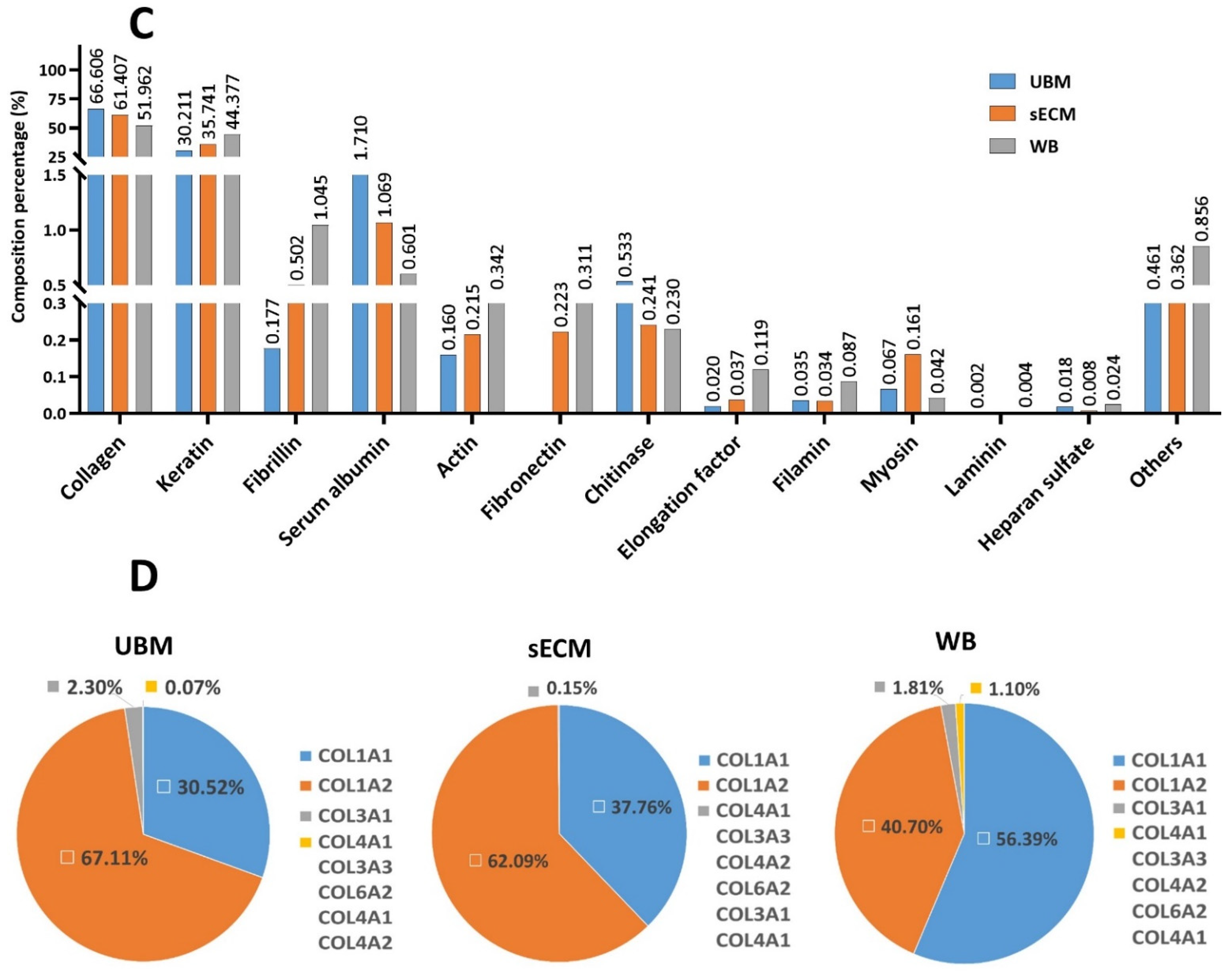
3.2. Biochemical Composition

The two major classes of biomolecules within ECM materials, i.e., sGAG and collagen were accessed quantitatively after decellularization. The results of the UBM were consistent with our previous study [9] with respect to the collagen content (0.48 ± 0.07 compared to 0.49 ± 0.01) and the sGAG content (1.39 ± 0.28 compared to 1.5 ± 0.05, respectively). Interestingly, there was no difference between the soluble collagen and the sGAG amount in UBM, sECM and WB. Additionally, LC-MS/MS was also employed to further investigate the detailed protein profiles from different layers of the bladder. There were more than 100 proteins detected in the materials (Supplementary Materials 2). Although there was a difference in the percentage of each protein component, it was interesting that the UBM, sECM and WB shared relatively the same biochemical compositions. Collagen was found to dominate the ECM composition, representing more than 50% of the total mass of all proteins (Figure 1C). In this collagen domain, collagen type I was the most abundant, accounting for more than 95%, compared to other types of collagens. Collagen type IV, which is ubiquitous in all basement membranes, was present not only in UBM but in sECM as well. Collagen type III and VI were also detected in all experimental groups (Figure 1D). Keratin was the second largest component in the protein composition profile, with more than 30% of the total protein mass. In terms of glycoprotein and proteoglycans/GAG, fibrillin, heparan sulfate, laminin, fibulin were identified in both UBM and sECM. Other ECM proteins, including albumin, myosin and actin, were spotted in the materials as well.

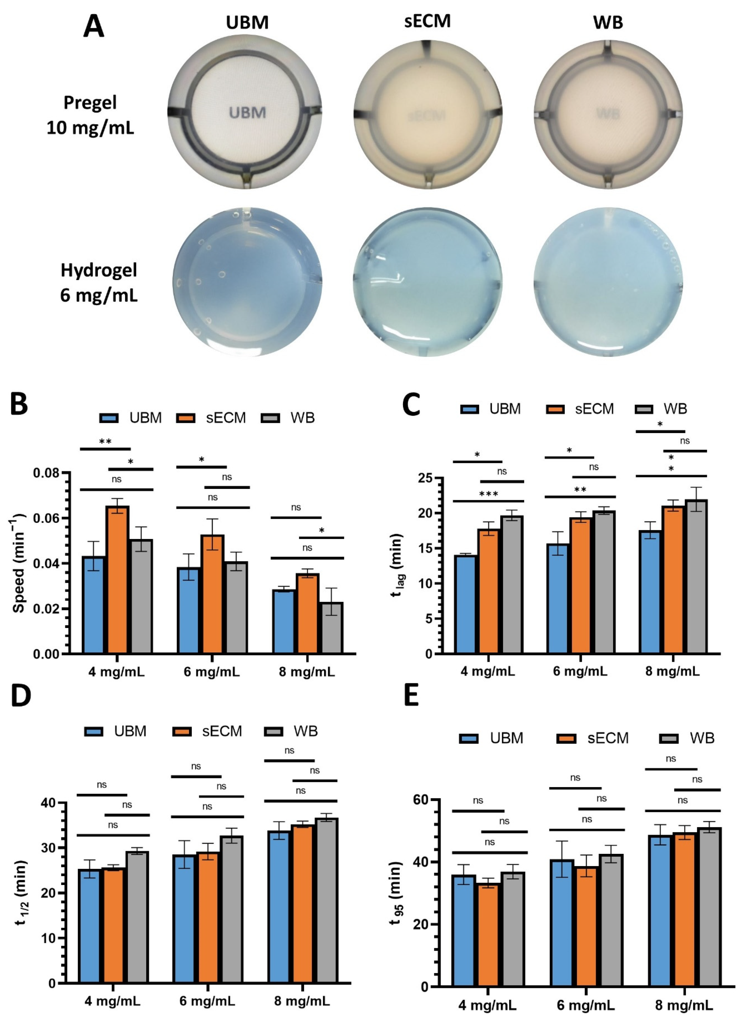
3.3. Turbidity Measurement

In terms of physical appearance, as can be seen from Figure 2A, the pregel solution from different tissue sources in the bladders possessed dissimilar transparency. The UBM pregel was more transparent compared to the other two. The same situation was also observed in the hydrogel form, in which sECM and WB hydrogels were more turbid. The sECM possessed a faster gelation rate than UBM and WB at all concentrations, and sECM and WB had the longer lag phase than UBM (Figure 2B–D). However, an interesting observation was that despite the distinct pattern in gelation speed and lag time, it took almost the same overall time for all types of hydrogels to reach complete gelation (Figure 2E), with no statistical difference, ranging from an average of 35 to 50 min (Supplementary Materials 1 Tables S1–S3).

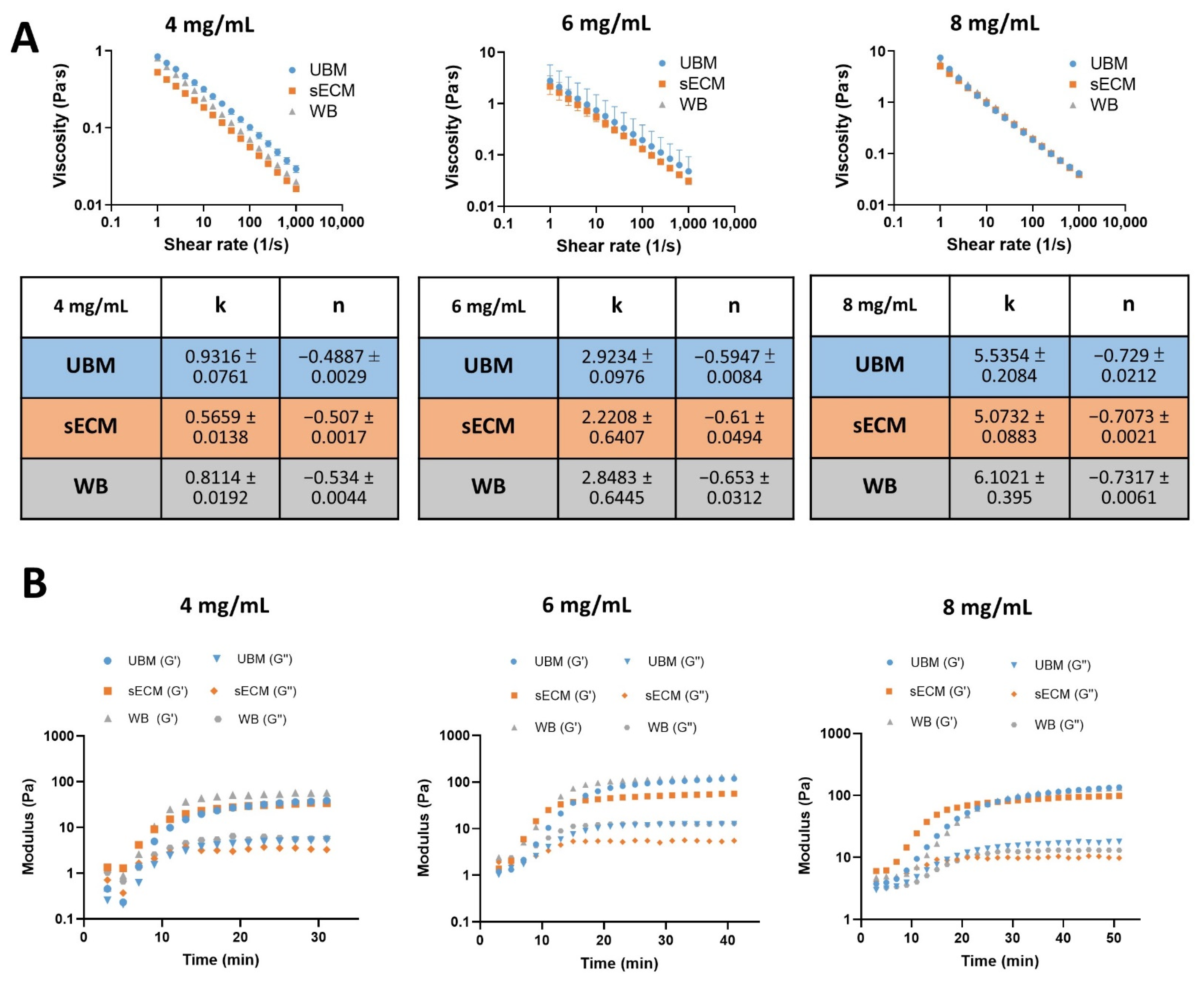
3.4. Rheological Measurements

The rheological properties of the ECM hydrogels were studied to determine the viscosity of the pregel solution and the mechanical strength of the hydrogels. As can be seen from Figure 3A, all of the pregel solutions exhibited shear thinning characteristics at low temperature with almost no difference in the flow index (n) between groups. In terms of viscosity, the sECM pregel was more liquid than the UBM and WB pregels, with lower consistency factors (k) at 8 mg/mL (Figure 3C). In terms of stiffness, it was observed that all the hydrogels had greater G’ (storage modulus) than G″ (loss modulus), indicating a solid-like behavior (Figure 3B). At 4 mg/mL, there were only slight differences among the storage moduli of UBM, sECM and WB, with 35.58 ± 3.37, 30.22 ± 5.07, and 52.94 ± 1.48 Pa, respectively. However, at higher concentrations, hydrogels made from sECM were found to be softer than those from UBM and WB (Figure 3D). It was also evident that hydrogels from UBM and WB possessed almost the same mechanical strength, and there was no dissimilarity in the storage modulus within these groups, ranging from around 110 to 120 Pa at concentrations between 6 and 8 mg/mL, respectively.

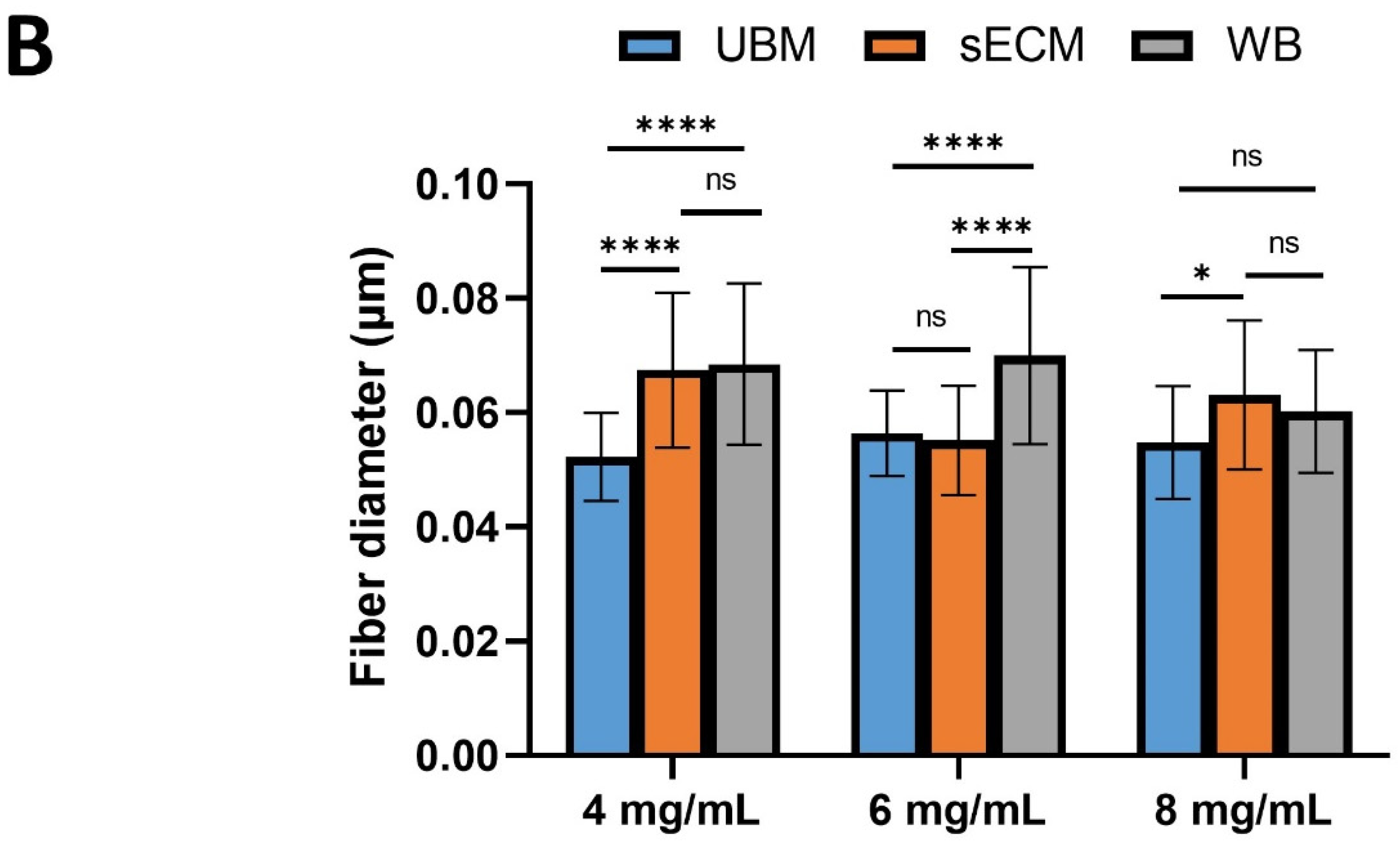
3.5. Scanning Electron Microscopy

As can be seen from the scanning electron micrographs (Figure 4A), all of the hydrogels from different tissue origins possessed a randomly oriented nano-fibrous structure, and the fibers originating from sECM and WB were thicker than those from UBM, with average fiber diameters of 0.0617 and 0.0653, respectively, compared to 0.0528 µm.

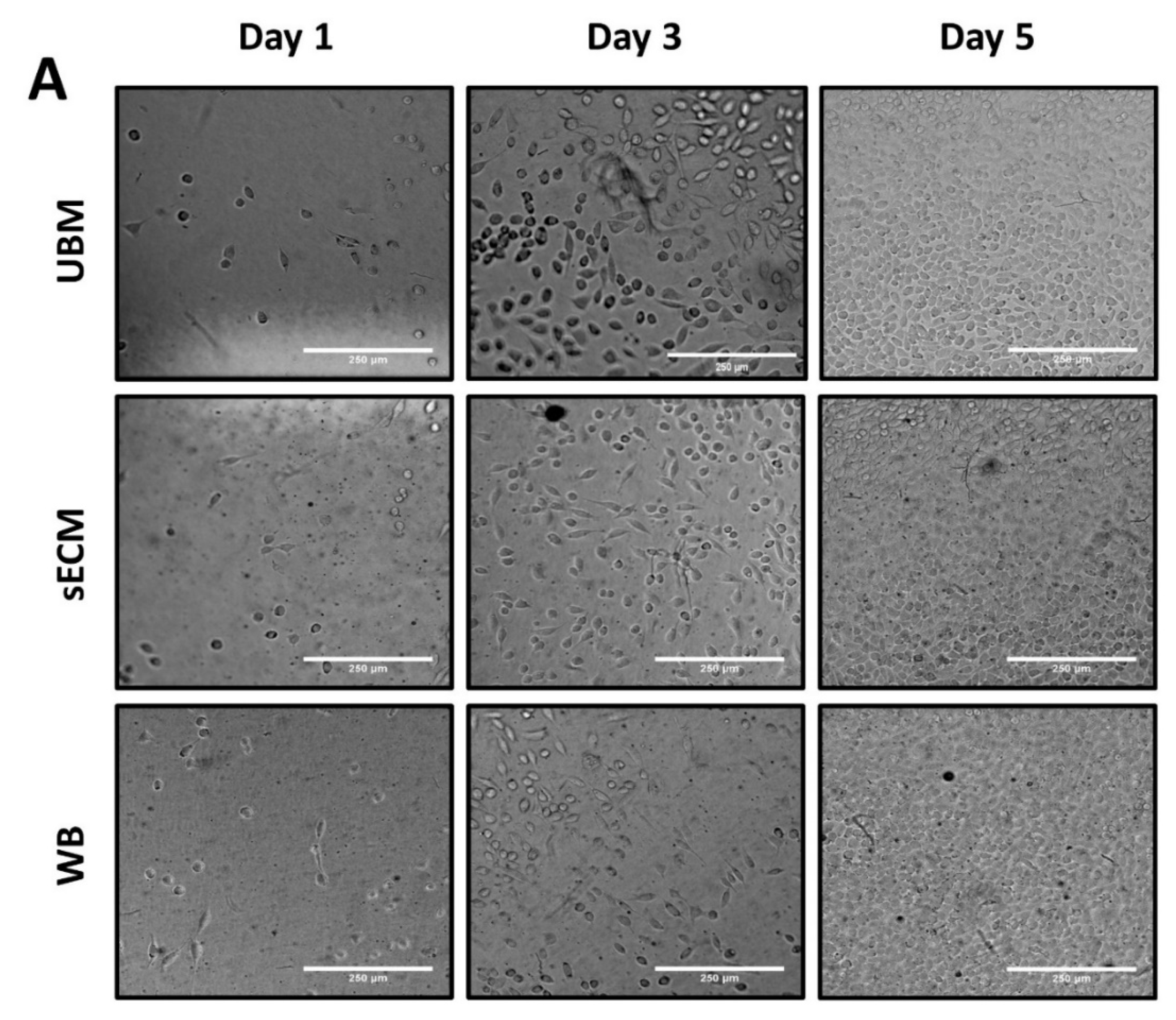
3.6. Effect on L292 Fibroblast In Vitro Cell Viability

On the basis of the Alamar blue assay, it can be observed that L929 grew slowly on day 1 and day 3, but with a more robust speed on day 5, and there was no significant difference among the ECM groups (Figure 5B). This test also confirmed the cytotoxicity of the hydrogels.

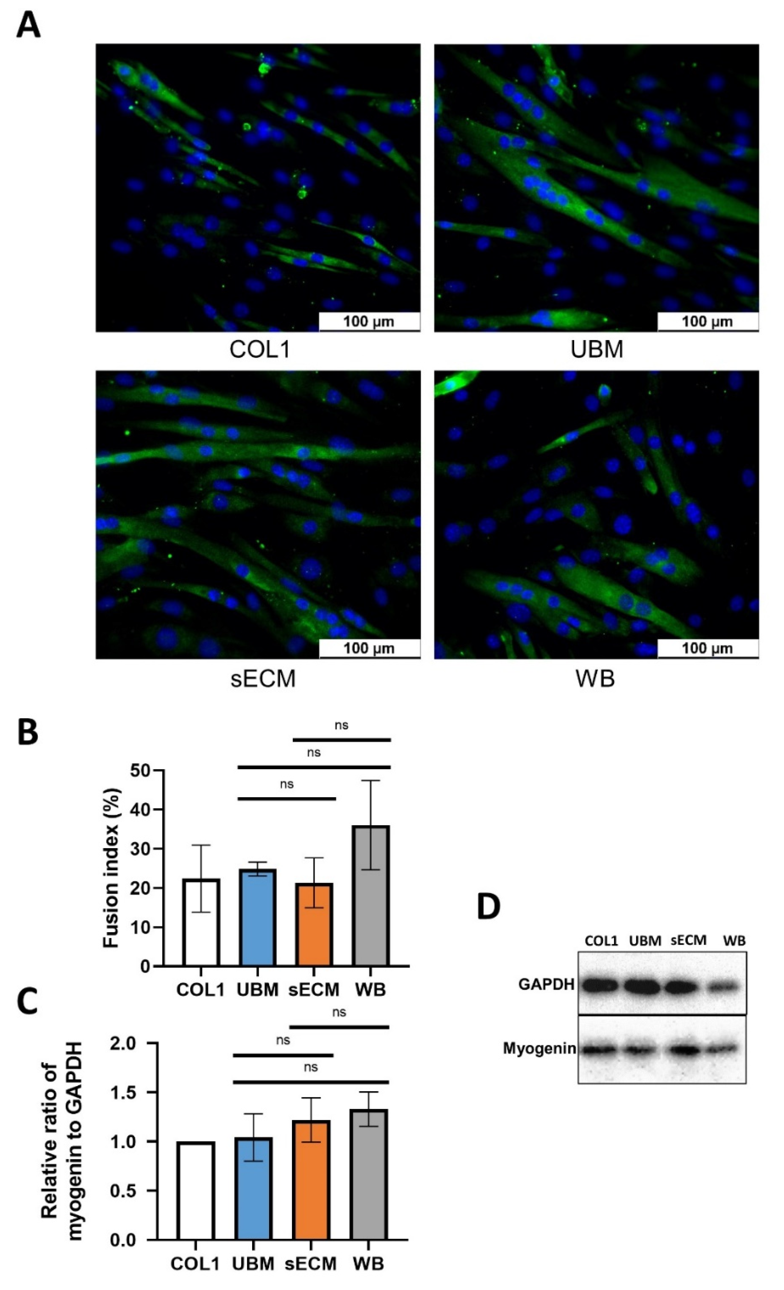
3.7. Effect on C2C12 Myogenic Differentiation

Physical coating was confirmed successfully according to the results of the contact angle measurement and total protein adsorption. The coverslip surface became more hydrophilic with a significantly smaller average contact angle of 32° compared to the uncoated one with approximately 67° (Figure 6A,B). The protein adsorption value of the coverslips coated with UBM, sECM and WB were double that of collagen type I (Figure 6C). This may due to the difference in coating solution concentration (2 mg/mL compared to 1 mg/mL). From the results of the Alamar blue assay (Figure 6D), C2C12 grew rapidly during the first two days in growth medium, and only increased slightly in number on the third day in all experimental groups. There was also no difference in the growth of C2C12 on different coating substrates, suggesting that pregel solutions from different layers of the bladder did not affect the growth of C2C12 cells.
Myogenesis of the C2C12 was confirmed using the following criteria: Upon initiation of differentiation, the cells elongate, adhere, and fuse into multinucleated myotubes that express muscle-specific markers such as myosin heavy chain (MyHC) and myogenic regulatory factors such as myogenin in the terminal differentiation state [25]. As can be seen from the immunocytochemistry study (Figure 7A,B), the C2C12 fused into multinucleated cells on bladder ECM-coated substrates as efficiently as on commercial collagen type I. The effect of UBM, WB and sECM on fusion ability of C2C12 was almost the same. In terms of myogenin expression, there was also no obvious difference among any of the bladder-derived coated groups, and all of these substrates possessed the same function as COL1 as well.

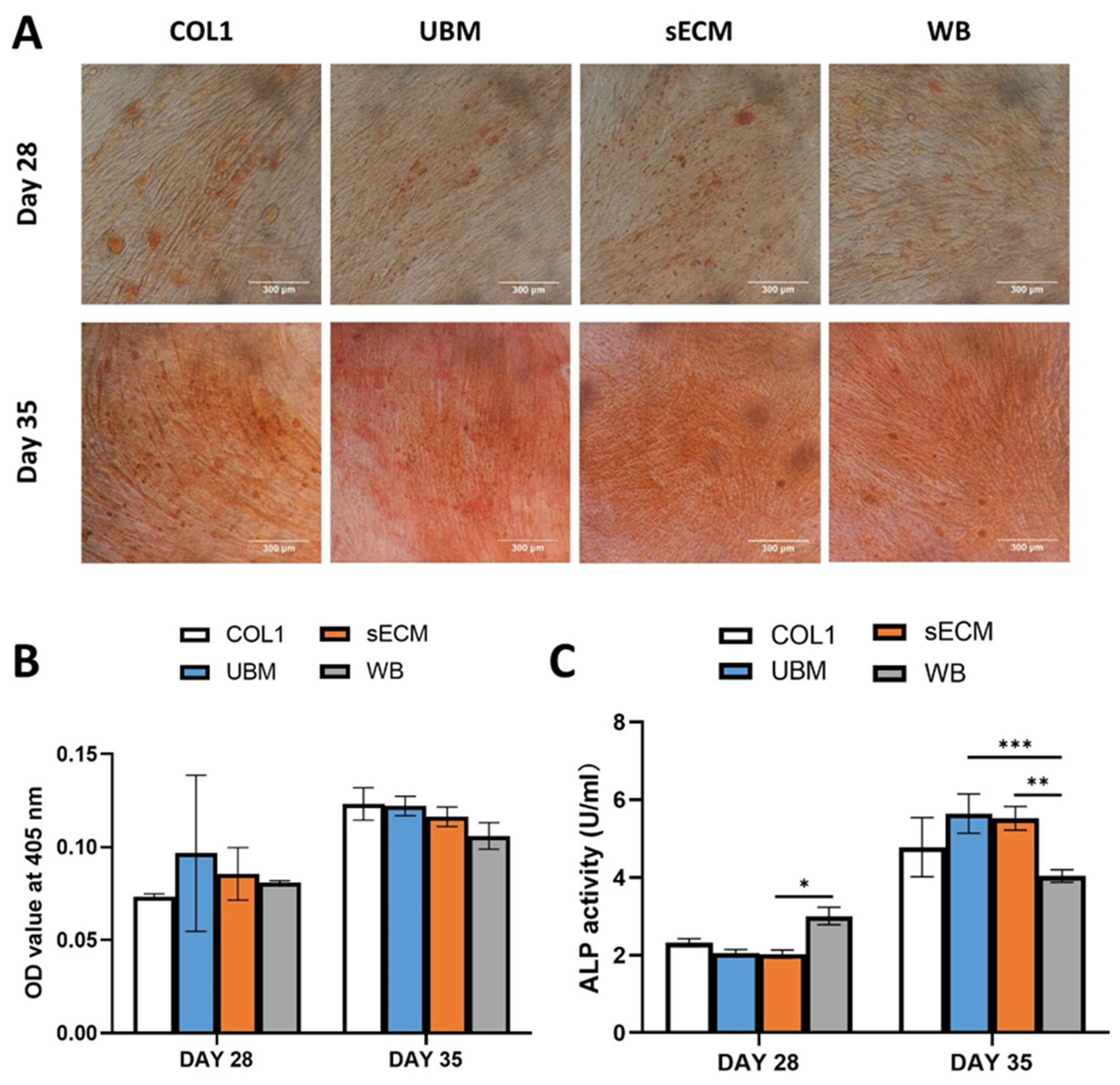
3.8. Effect on C2C12 Osteoblast Differentiation

The calcium deposition was observed after 28 and 35 days in ODM (Figure 8A). C2C12 differentiated into osteoblasts that were almost the same on different substrates, as there was no dissimilarity in the Alizarin red study (Figure 8B). In term of ALP activity, on day 28, C2C12 cultured on WB-coated plates expressed higher amounts of enzyme compared to sECM. On day 35, the ALP content was significantly higher than that of day 28. UBM and sECM groups had higher values than WB, and there was no difference from the COL1 group.

4. Discussion

In the field of tissue engineering, to support cell proliferation, morphogenesis and differentiation, it is prevalent to employ reconstituted matrices with purified biomacromolecules originating from animal tissues or artificial extracellular matrices (ECM). These types of materials can be used for coating in 2D culture systems or fabricated into 3D scaffolds to mimic in vivo environments. One of the most used 3D culture systems consists of hydrogels constructed from soluble collagen type I due to their simplicity as macromolecular components [26]. However, compared to that pure collagenous network, hydrogels with more complex compositions derived from mammalian tissues can aid in supporting cell adhesion, migration, and differentiation by the presence of glycosaminoglycans (sGAG) [26].
To study the properties of ECM hydrogels, the basic criteria required for the characterization are decellularization efficacy, biochemical composition, gelation kinetics, surface morphology and rheology [27]. Therefore, those assays were conducted accordingly. Our decellularization method was efficient at eliminating the cellular contents of three different tissue layers of porcine bladder and all met the established criteria for sufficient decellularization.
According to independent studies, the variations in compositions are probably the primary reason for the different characteristics of hydrogels [19]. Interestingly, there was no difference in the collagen and sGAG content of UBM, sECM and WB after decellularization. The collagen retention was four times as high as that of available commercial ECM materials such as TissueMend (fetal bovine dermis), GraftJacket (dermis), Restore (small intestine submucosa), and AlloPatch (dermis), and the level of sGAG was also higher than those products [28].
The predominant reason for using UBM is that it has a fully intact basement membrane with good integrity, which may offer advantages for cell adhesion in tissue engineering [29]. Despite not possessing a complete sheet of basement membrane, sECM also contained separate basement membrane component around myofibers [8]. UBM is made of basement membrane and loose lamina propria, in which the former contains mainly collagen type IV and VII and the latter is mostly composed of collagen types I, III, and III, IV, VII [15]. On the other hand, sECM is represented by muscularis mucosa matrix and detrusor, in which the first layer comprises collagen types I, IV and the second layer consists collagen types I, III, VI and XII [15]. From the results of LS-MS/MS, it was apparent that UBM and sECM layers shared almost the same types of not only collagens (type I, III, IV, VI), but other compositions as well.
In the gelation kinetics study, there were some different patterns observed in the gelation speed and lag phase of hydrogels from different layers of the bladders. Nevertheless, it took almost the same time for all types of hydrogels to gel completely. Due to the complexity of the compositions of ECM materials, the exact gelation mechanism of hydrogels derived from ECM still remains unknown. It is commonly assumed that the overall interaction of collagen, laminin, elastin and proteoglycan are accountable for the fibrillogenesis self-assembling in multiple complex ways [27]. For pure collagen, gelation occurs through a two-step process with formation of thin filaments taking place in the lag-phase nucleation, and fibril growth and interconnections in the growth phase. In the case of ECM hydrogel, other than collagens, proteoglycans and glycosaminoglycans also affected the length of both the lag and growth phases during gelation process as accelerators and decelerators [30]. The lag phase was hastened with the presence of chondroitin sulfate and keratin sulfate hasten, and impeded by heparin sulfate, but then it was also chondroitin sulfate that detained the growth phase [30]. As there was no statistical difference in the collagen and sGAG concentration of UBM, sECM and WB, it could be preliminarily explainable that all the gels shared similar gelation tendency after the lag phase.
ECM hydrogel formation is a self-assembly process of collagen fibrillogenesis. The polymerization of collagen with long fibrillar structures is regulated by both electrostatic and hydrophobic interactions from glycosaminoglycans, proteoglycans, and other ECM proteins that stimulate the cross-linking of collagen fibers. The ECM digest forms a gel when the liquid is neutralized to physiologic pH (7.4), salt concentration and temperature (37 °C) [27]. In practice, the digested ECM, i.e., pregel solution, is neutralized to physiologic pH and salt concentration and kept at a low temperature until follow-up applications [27]. The results from rheology indicated that pregel solutions obtained from different layers of the bladders in our study possessed the same shear thinning property and viscosity. This may be due to the similarity of protein profiles in those tissue types. In pregel solutions, randomly oriented proteins initially bound with sGAG through hydrogen and Van Der Waals interactions, offering the ability to resist flowing at low shear rate. When higher shear was applied, these bonds were broken, leading to the alignment of proteins and GAGs, causing a shear thinning phenomenon [31]. Hydrogels from UBM and WB possessed similar mechanical strength and were stronger than sECM hydrogels despite the fact that all three hydrogels had the same content of soluble collagen and sGAG. In our studies, only collagen types I, III, IV and VI were detected. However, according to the literature, there are at least 28 different types of collagens inside the bladder. Except for collagen types I and III, which represent the bulk proteins in the bladder, accounting for up to 60% of bladder’s dry weight, the function of the other less abundant collagens and their localization in the bladder is little studied [15]. Collagen types IX, X, XII, XIV and XVI are classified as fibril associated collagens with interrupted triple helices (FACITs), with the ability to cross-link with other collagens [15]. The interactions between complex groups of collagens might be the cause for difference mechanical strengths among hydrogels. Further study must be conducted to confirm this hypothesis.
All the hydrogels obtained from different parts of the bladder exhibited a fibrous surface morphology. The fiber diameter of sECM and WB hydrogels was thicker than that of UBM. This can be theoretically explained by the presence of collagen type VI in the detrusor of sECM, which, according to the literature, can physically associate with collagen I fibrils to increase the thickness of the fibers [15]. These results are also in accordance with studies with liver ECM fibers, in which it was quantitatively found that the skeleton fiber diameter depends on the constituent components [32]. Compared to another study on UBM hydrogel, the UBM hydrogels in this study possessed a thinner fiber diameter with an average thickness of 52 nm compared to 74 nm [19]. The fiber diameters of sECM and WB were also found to be thinner than those from cardiac, SIS, adipose-derived ECM hydrogels, with average fiber diameter of approximately 100 nm [27]. As demonstrated in many studies, ECM hydrogels may share many common characteristics, but the tissue of origin, decellularization process, and protein concentration also affect the structure of the materials [27]. This may be the reason for the variation in fiber dimeter in our research compared to other studies.
Despite having some similarities with respect to the components of ECM, each tissue or organ possesses a distinctive composition, leading to different environments for cells to develop and mature [33]. Typically, when complex compositions of a mixture of single proteins such as collagens, fibronectin, vitronectin or laminin are used as coating substrates, cell behavior can be influenced as a result of the dissimilar ratios of various proteins and polysaccharides compared to the tissue-specific in vivo microenvironment [33]. C2C12 myoblasts have been used as a model to study the effect of ECM materials towards myogenic and osteogenic differentiation for regenerative application [19,34,35]. However, the differentiation of this cell line on sECM, WB and UBM remains unknown. Therefore, C2C12 was selected to examine the cell response when cultured and differentiated on three different ECM substrates. According to our LC-MS results, different layers of the bladder possess dissimilar proportions of various biochemical compositions, and the effect of this compositional discrepancy on the differentiation of the C2C12 muscle progenitor cells was examined. The pregel solutions from UBM, sECM, WB and collagen type I were coated on the coverslip or flasks prior to seeding cells. This physical coating technique has been widely employed in other studies using different ECM sources such as meniscus, liver, brain, pancreata, skeletal muscle and bladder [27]. In order for myogenesis to occur, it is necessary for cell proliferation to be hindered. Commonly, 2% horse serum [23] is used to trigger myogenesis. However, in our study, when the cells reached 70% confluence, FBS content was reduced to 0.2% to kindle myotube formation. This method has also been previously employed in other studies, in which the concentration of growth factors was depleted by tenfold for myoblast differentiation [36]. Myosin heavy chain and myogenin were markers of the end-terminal differentiation of myoblasts [37]; therefore, these proteins were chosen for the immunocytochemistry (ICC) and Western blot experiments.
Fibroblasts are a cell line commonly used for examining cell viability on ECM-based materials [19]. Therefore, L929 fibroblast was chosen to study the cell viability on UBM, sECM and WB hydrogels. It was observed from the Alamar blue assay that different substrates did not influence the viability of L929 after 5 days. In terms of differentiation, the results from ICC and Western blot demonstrated that C2C12 myoblast differentiated on UBM, sECM and WB coated surface was as efficient as on commercial collagen type I. According to the literature, collagens are the main structural protein in the muscle ECM; other various components of the ECM participate in muscle regeneration as well, not only performing structural support, but also influencing the myoblast cell function and myotube formation [38]. Various studies have also been conducted to investigate the influence of single ECM composition on the differentiation of C2C12, including collagen type I, collagen type IV, fibronectin, laminin, decorin, or mixture of complex composition such as Matrigel [39], dermal, UBM and skeletal muscle ECM [19]. The results from these studies suggest that not only single ECM components, but also a complex ECM composition, offer positive effects with respect to myoblast differentiation. Therefore, the effect of three different ECM substrates on the differentiation of C2C12 was then studied. From our LC-MS results, fibrillin, laminin, fibronectin, etc., were detected in all layers of the bladder, and these glycoprotein/proteoglycans have previously been proven to influence cell behavior, signaling and differentiation [35]. Additionally, heparan sulfate, which plays a pivotal role in in muscle regeneration, was also spotted [35]. However, there was no significant difference in differentiation behavior of C2C12 when cultured on coating substrates prepared from different layers of the bladder. Despite having different proportions of each component, these layers shared almost the same biochemical compositions, and the synergistic effects of those single components may lead to the same effect on the myogenesis of myoblasts.
It was previously shown that C2C12 could be transdifferentiated into osteoblastic lineage when cultured in medium supplemented with 10 mM β-glycerophosphate and 50 mg/mL ascorbic acid [40]. In the early stage of osteogenesis, alkaline phosphatase (ALP) is one of the most trustworthy markers, since it is created by osteogenic cells. ALP catalyzes the hydrolysis of phosphate monoesters, and pNPP is commonly used as a phosphatase substrate to detect ALP [41]. For the late stage of osteogenesis, calcium deposition was often used as a reliable indicator [41]. To measure this mineralization process, the cells were stained with Alizarin Red to observe the deposits of calcium phosphate (hydroxyapatite Ca5(PO4)3(OH)), and then the color can be extracted and quantified by a colorimetric assay [41]. According to the results of the ALP assay and Alizarin red staining in our study, C2C12 cultured on ECM-coated substrate was able to undergo osteogenesis as well as on commercial COL1. In terms of bone tissue engineering, collagen type I is often employed as scaffold as it is the main component of bone tissue [34]. Additionally, C2C12 has also been shown to increase osteogenic differentiation capacity when cultured on bone ECM hydrogel [34]. Our results suggest that not only UBM, but also sECM and WB, could be used as a potential substrate for C2C12 osteogenesis. However, this was just a preliminary study. To gain a deeper insight into the effect of different coating compositions on the osteogenic differentiation of C2C12, other assays such as qPCR and Western blot are required.
The objective of this study was to preliminarily compare the physical characteristics and in vitro performance of ECM hydrogel from UBM, sECM and WB. However, the scope of the study was also limited to fibroblast L929 and myoblast C2C12; different cell types may possess different properties toward these types of hydrogels. The interactions between different cell types and hydrogel types might play an essential role in the fabrication of in vitro 3D culture platforms, and might contribute to future applications in developing in vitro disease models and tissue regenerations, which could help to make a step closer to clinical applications.

5. Conclusions

In summary, apart from UBM, sECM and WB were successfully fabricated into hydrogels. These hydrogels shared some similar characteristics, and had almost the same biochemical effects in in vitro studies. Therefore, the use of the sECM part of the bladder should not be ignored, and could further aid in waste management and environmental sustainability. Additionally, hydrogels from WB could be a potential replacement for UBM in tissue engineering applications, avoiding the need for laborious delaminating processes.

Supplementary Materials

The following are available online at https://www.mdpi.com/article/10.3390/app11135820/s1, Figure S1: Decellularization confirmation. Table S1: Turbidimetric analysis of the UBM hydrogel. Table S2: Turbidimetric analysis of the sECM hydrogel. Table S3: Turbidimetric analysis of the WB hydrogel.

Author Contributions

Conceptualization, C.-Y.K. and H.-Q.-D.N.; methodology, C.-Y.K. and H.-Q.-D.N.; validation, C.-Y.K. and H.-Q.-D.N.; formal analysis, H.-Q.-D.N.; investigation, H.-Q.-D.N.; resources, C.-Y.K., C.-M.L. and Y.-H.H.; data curation, H.-Q.-D.N. and Y.-C.W.; writing—original draft preparation, H.-Q.-D.N.; writing—review and editing, C.-Y.K. and Y.-C.W.; visualization, H.-Q.-D.N. and Y.-C.W.; project administration, H.-Q.-D.N.; funding acquisition, C.-Y.K. All authors have read and agreed to the published version of the manuscript.

Funding

This research was funded by Ministry of Science and Technology in Taiwan (MOST-109-2221-E-011-080 -MY2).

Institutional Review Board Statement

Not applicable.

Informed Consent Statement

Not applicable.

Data Availability Statement

The data presented in this study are available in the article.

Acknowledgments

We gratefully thank Shiao-Pieng Lee and Ming-Chien Yang for their enthusiastic support during the whole project; Shu-Kai Yeh and Ming-Hua Ho for sharing the critical point dryer and rheometer chamber selflessly; Jehng-Kang Wang for the assistance in biochemical experiments; Liao, Fang for providing experimental resources, and National Defense Medical Center instrument center for technical support.

Conflicts of Interest

The authors declare no conflict of interest.

References

  1. Catoira, M.C.; Fusaro, L.; Di Francesco, D.; Ramella, M.; Boccafoschi, F. Overview of natural hydrogels for regenerative medicine applications. J. Mater. Sci. Mater. Med. 2019, 30, 159. [Google Scholar] [CrossRef] [PubMed] [Green Version]
  2. Varaprasad, K.; Raghavendra, G.M.; Jayaramudu, T.; Yallapu, M.M.; Sadiku, R. A mini review on hydrogels classification and recent developments in miscellaneous applications. Mater. Sci. Eng. C 2017, 79, 958–971. [Google Scholar] [CrossRef] [PubMed]
  3. Laurila, P.; Leivo, I. Basement membrane and interstitial matrix components form separate matrices in heterokaryons of PYS-2 cells and fibroblasts. J. Cell Sci. 1993, 104, 59. [Google Scholar] [CrossRef] [PubMed]
  4. Matrix, Extracellular and Interstitial. In Reviews in Cell Biology and Molecular Medicine; Available online: https://onlinelibrary.wiley.com/doi/abs/10.1002/3527600906.mcb.200400091 (accessed on 20 June 2021). [CrossRef]
  5. Frantz, C.; Stewart, K.M.; Weaver, V.M. The extracellular matrix at a glance. J. Cell Sci. 2010, 123 Pt 24, 4195. [Google Scholar] [CrossRef] [Green Version]
  6. Pokrywczyńska, M.; Gubanska, I.; Drewa, G.; Drewa, T. Application of Bladder Acellular Matrix in Urinary Bladder Regen-Eration: The State of the Art and Future Directions. BioMed Res. Int. 2014. [Google Scholar] [CrossRef]
  7. Bouhout, S.; Rousseau, A.; Chabaud, S.; Morissette, A.; Bolduc, S. Potential of Different Tissue Engineering Strategies in the Bladder Reconstruction. Regen. Med. Tissue Eng. 2013. [Google Scholar] [CrossRef] [Green Version]
  8. Grounds, M.D. Complexity of Extracellular Matrix and Skeletal Muscle Regeneration. In Skeletal Muscle Repair and Regeneration; Schiaffino, S., Partridge, T., Eds.; Springer: Dordrecht, The Netherlands, 2008; pp. 269–302. [Google Scholar]
  9. Kao, C.-Y.; Nguyen, H.-Q.-D.; Weng, Y.-C. Characterization of Porcine Urinary Bladder Matrix Hydrogels from Sodium Dodecyl Sulfate Decellularization Method. Polymers 2020, 12, 3007. [Google Scholar] [CrossRef]
  10. Ghuman, H.; Massensini, A.R.; Donnelly, J.; Kim, S.-M.; Medberry, C.J.; Badylak, S.F.; Modo, M. ECM hydrogel for the treatment of stroke: Characterization of the host cell infiltrate. Biomaterials 2016, 91, 166–181. [Google Scholar] [CrossRef] [Green Version]
  11. Loneker, A.E.; Faulk, D.M.; Hussey, G.S.; D’Amore, A.; Badylak, S.F. Solubilized liver extracellular matrix maintains primary rat hepatocyte phenotype in-vitro. J. Biomed. Mater. Res. Part A 2016, 104, 957–965. [Google Scholar] [CrossRef]
  12. Tukmachev, D.; Forostyak, S.; Koci, Z.; Zaviskova, K.; Vackova, I.; Vyborny, K.; Sandvig, I.; Sandvig, A.; Medberry, C.J.; Badylak, S.F.; et al. Injectable Extracellular Matrix Hydrogels as Scaffolds for Spinal Cord Injury Repair. Tissue Eng. Part A 2016, 22, 306–317. [Google Scholar] [CrossRef]
  13. Allbritton-King, J.D.; Kimicata, M.; Fisher, J.P. Incorporating a structural extracellular matrix gradient into a porcine urinary bladder matrix -based hydrogel dermal scaffold. J. Biomed. Mater. Res. Part A 2021. [Google Scholar] [CrossRef]
  14. Faust, A.; Kandakatla, A.; Van Der Merwe, Y.; Ren, T.; Huleihel, L.; Hussey, G.; Naranjo, J.D.; Johnson, S.; Badylak, S.; Steketee, M. Urinary bladder extracellular matrix hydrogels and matrix-bound vesicles differentially regulate central nervous system neuron viability and axon growth and branching. J. Biomater. Appl. 2017, 31, 1277–1295. [Google Scholar] [CrossRef]
  15. Aitken, K.; Bägli, D.J. The bladder extracellular matrix. Part I: Architecture, development and disease. Nat. Rev. Urol. 2009, 6, 596–611. [Google Scholar] [CrossRef] [PubMed]
  16. Brown, B.N.; Buckenmeyer, M.J.; Prest, T.A. Preparation of Decellularized Biological Scaffolds for 3D Cell Culture. Methods Mol. Biol. 2017, 1612, 15–27. [Google Scholar] [CrossRef] [PubMed]
  17. Freytes, D.O.; Martin, J.; Velankar, S.S.; Lee, A.S.; Badylak, S.F. Preparation and rheological characterization of a gel form of the porcine urinary bladder matrix. Biomaterials 2008, 29, 1630–1637. [Google Scholar] [CrossRef] [PubMed]
  18. Fernández-Pérez, J.; Ahearne, M. The impact of decellularization methods on extracellular matrix derived hydrogels. Sci. Rep. 2019, 9, 14933. [Google Scholar] [CrossRef] [PubMed] [Green Version]
  19. Wolf, M.T.; Daly, K.A.; Brennan-Pierce, E.P.; Johnson, S.A.; Carruthers, C.A.; D’Amore, A.; Nagarkar, S.P.; Velankar, S.S.; Badylak, S.F. A hydrogel derived from decellularized dermal extracellular matrix. Biomaterials 2012, 33, 7028–7038. [Google Scholar] [CrossRef] [Green Version]
  20. Lee, J.S.; Shin, J.; Park, H.-M.; Kim, Y.-G.; Kim, B.-G.; Oh, J.-W.; Cho, S.-W. Liver Extracellular Matrix Providing Dual Functions of Two-Dimensional Substrate Coating and Three-Dimensional Injectable Hydrogel Platform for Liver Tissue Engineering. Biomacromolecules 2014, 15, 206–218. [Google Scholar] [CrossRef] [PubMed]
  21. DeQuach, J.A.; Yuan, S.H.; Goldstein, L.S.; Christman, K.L. Decellularized Porcine Brain Matrix for Cell Culture and Tissue Engineering Scaffolds. Tissue Eng. Part A 2011, 17, 2583–2592. [Google Scholar] [CrossRef] [Green Version]
  22. Rauch, C.; Brunet, A.-C.; Deleule, J.; Farge, E. C2C12 myoblast/osteoblast transdifferentiation steps enhanced by epigenetic inhibition of BMP2 endocytosis. Am. J. Physiol. Physiol. 2002, 283, C235–C243. [Google Scholar] [CrossRef] [PubMed] [Green Version]
  23. D’Andrea, P.; Civita, D.; Cok, M.; Severino, L.U.; Vita, F.; Scaini, D.; Casalis, L.; Lorenzon, P.; Donati, I.; Bandiera, A. Myoblast Adhesion, Proliferation and Differentiation on Human Elastin-Like Polypeptide (HELP) Hydrogels. J. Appl. Biomater. Funct. Mater. 2017, 15, 43–53. [Google Scholar] [CrossRef] [Green Version]
  24. Wu, Y.; Zhou, J.; Li, Y.; Zhou, Y.; Cui, Y.; Yang, G.; Hong, Y. Rap1A Regulates Osteoblastic Differentiation via the ERK and p38 Mediated Signaling. PLoS ONE 2015, 10, e0143777. [Google Scholar] [CrossRef]
  25. Liu, X.; Gao, Y.; Long, X.; Hayashi, T.; Mizuno, K.; Hattori, S.; Fujisaki, H.; Ogura, T.; Wang, D.O.; Ikejima, T. Type I collagen promotes the migration and myogenic differentiation of C2C12 myoblasts via the release of interleukin-6 mediated by FAK/NF-κB p65 activation. Food Funct. 2020, 11, 328–338. [Google Scholar] [CrossRef]
  26. Arends, F.; Nowald, C.; Pflieger, K.; Boettcher, K.; Zahler, S.; Lieleg, O. The Biophysical Properties of Basal Lamina Gels Depend on the Biochemical Composition of the Gel. PLoS ONE 2015, 10, e0118090. [Google Scholar] [CrossRef] [PubMed]
  27. Saldin, L.T.; Cramer, M.C.; Velankar, S.S.; White, L.J.; Badylak, S.F. Extracellular matrix hydrogels from decellularized tissues: Structure and function. Acta Biomater. 2017, 49, 1–15. [Google Scholar] [CrossRef] [PubMed] [Green Version]
  28. Aurora, A.; McCarron, J.; Iannotti, J.P.; Derwin, K. Commercially available extracellular matrix materials for rotator cuff repairs: State of the art and future trends. J. Shoulder Elb. Surg. 2007, 16, S171–S178. [Google Scholar] [CrossRef]
  29. Yurchenco, P.D. Basement Membranes: Cell Scaffoldings and Signaling Platforms. Cold Spring Harb. Perspect. Biol. 2010, 3, 20. [Google Scholar] [CrossRef] [Green Version]
  30. Oegema, T.R.; Laidlaw, J.; Hascall, V.C.; Dziewiatkowski, D.D. The effect of proteoglycans on the formation of fibrils from collagen solutions. Arch. Biochem. Biophys. 1975, 170, 698–709. [Google Scholar] [CrossRef] [Green Version]
  31. Yan, C.; Altunbas, A.; Yucel, T.; Nagarkar, R.P.; Schneider, J.P.; Pochan, D.J. Injectable solid hydrogel: Mechanism of shear-thinning and immediate recovery of injectable β-hairpin peptide hydrogels. Soft Matter 2010, 6, 5143–5156. [Google Scholar] [CrossRef] [PubMed] [Green Version]
  32. Bual, R.P.; Ijima, H. Intact extracellular matrix component promotes maintenance of liver-specific functions and larger aggregates formation of primary rat hepatocytes. Regen. Ther. 2019, 11, 258–268. [Google Scholar] [CrossRef]
  33. Lutolf, M.P.; Hubbell, J. Synthetic biomaterials as instructive extracellular microenvironments for morphogenesis in tissue engineering. Nat. Biotechnol. 2005, 23, 47–55. [Google Scholar] [CrossRef]
  34. Alom, N.; Peto, H.; Kirkham, G.R.; Shakesheff, K.M.; White, L.J. Bone extracellular matrix hydrogel enhances osteogenic differentiation of C2C12 myoblasts and mouse primary calvarial cells. J. Biomed. Mater. Res. Part B Appl. Biomater. 2018, 106, 900–908. [Google Scholar] [CrossRef] [PubMed]
  35. DeQuach, J.A.; Mezzano, V.; Miglani, A.; Lange, S.; Keller, G.; Sheikh, F.; Christman, K.L. Simple and High Yielding Method for Preparing Tissue Specific Extracellular Matrix Coatings for Cell Culture. PLoS ONE 2010, 5, e13039. [Google Scholar] [CrossRef] [PubMed] [Green Version]
  36. D’Agostino, M.; Torcinaro, A.; Madaro, L.; Marchetti, L.; Sileno, S.; Beji, S.; Salis, C.; Proietti, D.; Imeneo, G.; Capogrossi, M.C.; et al. Role of miR-200c in Myogenic Differentiation Impairment via p66Shc: Implication in Skeletal Muscle Regeneration of Dystrophic mdx Mice. Oxidative Med. Cell. Longev. 2018, 2018, 4814696. [Google Scholar] [CrossRef] [Green Version]
  37. Willkomm, L.; Schubert, S.; Jung, R.; Elsen, M.; Borde, J.; Gehlert, S.; Suhr, F.; Bloch, W. Lactate regulates myogenesis in C2C12 myoblasts in vitro. Stem Cell Res. 2014, 12, 742–753. [Google Scholar] [CrossRef] [PubMed] [Green Version]
  38. Gillies, A.R.; Lieber, R.L. Structure and function of the skeletal muscle extracellular matrix. Muscle Nerve 2011, 44, 318–331. [Google Scholar] [CrossRef] [PubMed] [Green Version]
  39. Chaturvedi, V.; Dye, D.E.; Kinnear, B.F.; Van Kuppevelt, T.H.; Grounds, M.; Coombe, D.R. Interactions between Skeletal Muscle Myoblasts and their Extracellular Matrix Revealed by a Serum Free Culture System. PLoS ONE 2015, 10, e0127675. [Google Scholar] [CrossRef]
  40. Soundharrajan, I.; Kim, D.H.; Kuppusamy, P.; Choi, K.C. Modulation of osteogenic and myogenic differentiation by a phytoestrogen formononetin via p38MAPK-dependent JAK-STAT and Smad-1/5/8 signaling pathways in mouse myogenic pro-genitor cells. Sci. Rep. 2019, 9, 9307. [Google Scholar] [CrossRef] [Green Version]
  41. Chen, J.; Shi, Z.-D.; Ji, X.; Morales, J.; Zhang, J.; Kaur, N.; Wang, S. Enhanced Osteogenesis of Human Mesenchymal Stem Cells by Periodic Heat Shock in Self-Assembling Peptide Hydrogel. Tissue Eng. Part A 2013, 19, 716–728. [Google Scholar] [CrossRef] [Green Version]
Figure 1. Biochemical composition. (A) Collagen content (mg/mg) (n = 4). (B) sGAG content (µg/mg) (n = 6). (C) Protein profiles of the decellularized tissues by LC-MC/MS. (D) Relative percentage of subdomains for collagen.
Applsci 11 05820 g001aApplsci 11 05820 g001b
Figure 2. Difference in transparency of the pregel solution and hydrogel from different tissue source of the bladder (A). Gelation speed (B), tlag (C) and t1/2 (D) and t95 (E) of the hydrogels. *, **, *** denote statistical difference within the same group with p < 0.05, 0.01, 0.001, respectively (n = 3).
Applsci 11 05820 g002
Figure 3. Shear thinning properties of the pregel solution (A). Representative curve of rheologic gelation of hydrogel (B). Consistency factor of the pregel solution (C). Storage modulus (G’) of the hydrogels (D). *, **, **** denote statistical difference within the same group with p < 0.05, 0.01, 0.0001, respectively (n = 3).
Applsci 11 05820 g003aApplsci 11 05820 g003b
Figure 4. Surface morphology of hydrogels from UBM, sECM and WB (scale bar 2 µm) (A). Fiber diameter was calculated using ImageJ with average of 30 fibers per group (B). *, **** denote statistical difference within the same group with p < 0.05, 0.0001, respectively (n = 30).
Applsci 11 05820 g004aApplsci 11 05820 g004b
Figure 5. (A) Optical images of L929 cells on hydrogel surface after 1, 3 and 5 days. (B) L929 cell viability on hydrogel surface evaluated by Alamar blue assay.
Applsci 11 05820 g005aApplsci 11 05820 g005b
Figure 6. Coating confirmation by contact angel measurement (A,B) and total protein adsorption by BCA assay (n = 3) (C). (D) C2C12 viability on different coated surfaces studied by Alamar blue assay (n = 3).
Applsci 11 05820 g006
Figure 7. C2C12 differentiation after 3 days in MDM medium. (A) Representative immunostaining of C2C12 cells with MyHC antibody (green) and nuclei (blue) (scale bar 100 µm). (B) Fusion index of C2C12 on different coated surface (n = 3). Relative expression level myogenin (n = 4) (C) and representative immunoblotting of myogenin (D) in Western blot study.
Applsci 11 05820 g007
Figure 8. C2C12 differentiation after 28 and 35 days in ODM medium. (A) Representative images of cells stained with Alizarin red. (B) Calcium content quantified by spectrophotometer (n = 3). (C) ALP activity measurement by spectrophotometer (n = 3). *, **, *** denote statistical difference within the same group with p < 0.05, 0.01, 0.001, respectively (n = 3).
Applsci 11 05820 g008
Publisher’s Note: MDPI stays neutral with regard to jurisdictional claims in published maps and institutional affiliations.

