Article

Immediate Insulin Treatment Prevents Diabetes-Induced Gut Region-Specific Increase in the Number of Myenteric Serotonergic Neurons

Department of Physiology, Anatomy and Neuroscience, Faculty of Science and Informatics, University of Szeged, H-6726 Szeged, Hungary
*
Author to whom correspondence should be addressed.
Appl. Sci. 2021, 11(13), 5949; https://doi.org/10.3390/app11135949
Submission received: 27 May 2021 / Revised: 21 June 2021 / Accepted: 23 June 2021 / Published: 26 June 2021
(This article belongs to the Special Issue Enteric Nervous System in Health and Disease)

Abstract

:
To evaluate the effects of hyperglycemia and insulin treatment on the proportion of serotonin-immunoreactive (5-HT-IR) myenteric neurons, samples were taken from the duodenum, ileum, and colon of diabetic, insulin-treated diabetic, and control rats 10 weeks after the onset of streptozotocin-induced hyperglycemia. Myenteric whole-mount preparations were immunostained with anti-5-HT and pan-neuronal anti-HuCD markers. In controls, the 5-HT-IR myenteric neurons represent a small proportion (~2.5%) of the total neuronal number in the investigated gut segments. The proportion of 5-HT-IR myenteric neurons was significantly higher in the duodenum (p < 0.01) and colon (p < 0.0001) of diabetic rats compared to the controls but exhibited a slight increase in the ileum. Immediate insulin treatment resulted in a significantly lower proportion of myenteric 5-HT-IR neurons in each segment (duodenum p < 0.0001; ileum p < 0.01; and colon p < 0.0001) compared to the untreated diabetics. Our study demonstrates that the proportion of 5-HT-IR myenteric neurons was enhanced in type 1 diabetes in a region-specific manner. Immediate insulin treatment prevents a higher hyperglycemia-induced amount of 5-HT-IR neurons and restores it to the control level in each investigated gut segment. Despite the low proportion of 5-HT-IR myenteric neurons, hyperglycemia-related changes of these neurons may play a crucial role in gastrointestinal symptoms in type 1 diabetes.

1. Introduction

Diabetic patients often suffer from gastrointestinal (GI) symptoms such as nausea, vomiting, diarrhea, constipation, dyspepsia, or abdominal pain. These complications are associated with structural and functional alterations of the enteric nervous system (ENS), which is known as diabetic enteropathy [1]. The ENS is structured as two major ganglionated plexuses. The myenteric plexus is located between the circular and the longitudinal muscle layers of the gut wall and regulates intestinal motility [2]. The submucous plexus is situated in the submucosal tissue layer close to the muscularis mucosae and the intrinsic vasculature; in small animals, it is found in one layer, while in larger animals and humans, it is in two layers. The submucous neurons influence mucosal secretion, electrolyte absorption across the mucosa, blood flow, and intestinal motility [3,4,5,6].
The ENS is particularly sensitive to hyperglycemia. The persistence of a high level of blood glucose disturbs the intracellular glucose metabolism and leads to the generation of reactive oxygen species damaging the ENS [7,8,9]. Diabetes-related alterations in the enteric neuronal microenvironment contribute to pathological pathways [10]. These alterations include structural neuronal changes, altered secretion of neurotransmitters, or loss of enteric neurons, which in turn modify GI motility and secretory functions and lead to the abovementioned GI symptoms in diabetic patients [11,12].
Serotonin (5-hydroxytryptamine, 5-HT) in the central nervous system is mainly associated with mood, depression, sleep, and appetite, but the gut can store a vast amount of 5-HT. Although enteric 5-HT received more attention recently, most of these studies focused on the 5-HT produced by mucosal enterochromaffin cells [13,14], which accounts for about 90% of the body’s total 5-HT. However, enteric 5-HT is also synthetized by the ENS. 5-HT-immunoreactive (IR) neurons, although in small proportions, are present along the GI tract in rodents [15] and humans [16,17]. The physiological roles of these neurons in GI motility are controversial [18,19]. On the one hand, this discrepancy arises from the large number of different 5-HT receptors, such as 5-HT1, 5-HT2, 5-HT3, 5-HT4, and 5-HT7, and their subtypes [20]. On the other hand, 5-HT-IR neurons have extensive projections. The immunohistochemical study of Hempfling et al. [16] determined the presence of 5-HT-IR neurons and 5-HT-IR nerve fibers in the myenteric ganglia, motor endplates, lamina muscularis mucosae, and blood vessels of a mice esophagus. In a murine colon, Okamoto et al. [21] observed the divergence of myenteric 5-HT-IR neurons throughout the myenteric and submucous plexus, the pacemaker, intramuscular and submucous interstitial cells of Cajal, and submucous arterioles. Moreover, 5-HT-IR neurons appear to synapse with one another, and most neuronal nitric oxide synthase (nNOS)-IR neurons and glial cells are found in the murine colon. This diverse range of targets suggests that these neurons function not only as interneurons or sensory neurons but also as motor neurons innervating interstitial cells of Cajal and blood vessels. Furthermore, this study offers the possibility that myenteric 5-HT-IR neurons coordinate motility with blood flow and secretion in the colon [22].
Despite unanswered questions, such as whether the endogenous 5-HT originates from mucosa or enteric neurons or whether it is essential in GI motility, there is evidence about its important role in gut pathophysiology. 5-HT signaling has been altered in patients with diverticular disease [23], irritable bowel syndrome [24], Hirschsprung’s-associated enterocolitis [25], and autism spectrum disorders [26,27]. Furthermore, the manipulation of 5-HT3; 5-HT4 receptors; and tryptophan hydroxylase, the rate limiting the enzyme of the 5-HT synthesis, is a therapeutic target in the treatment of GI motility disorders [20]. In addition, selective 5-HT4 receptor agonists being considered new prokinetic agents are under investigation for the treatment of diabetic gastroparesis, a syndrome that involves delayed gastric emptying [28].
Although diabetic patients often suffer from GI disturbances, little is known about the gut segment-specific alterations of 5-HT-IR enteric neurons in hyperglycemia or in insulin-treated diabetes. Lincoln et al. [29] detected that the ileal and colonic 5-HT neurons are affected by hyperglycemia in a region-dependent manner. Moreover, Gorio et al. [30] found that insulin treatment is preventive in the neurochemical changes in an alloxan-induced diabetic gut. Segment-specific alterations in the heme oxygenase-IR and nNOS-IR neurons were demonstrated in the small and large intestines of streptozotocin (STZ)-induced diabetic rats [31,32]. In addition, the diabetic alterations in the neuronal microenvironment, including changes in the morphology and function of the capillary endothelium adjacent to the myenteric plexus, were also intestinal region-dependent [10,33]. The protective effects of insulin treatment on the enteric neurons, mainly on nitrergic neurons and on the capillary endothelium supplying the myenteric plexus in type 1 diabetes, have been confirmed in a few studies [10,34,35,36].
Based on these findings, our primary aim was to quantify the 5-HT-IR neurons in the myenteric plexus of the duodenum, ileum, and colon of control animals using fluorescent immunohistochemistry. Furthermore, it was our goal to evaluate the effects of chronic hyperglycemia on the proportion of 5-HT-IR neurons in different gut segments and to investigate whether the insulin treatment results in segment-specific changes in the 5-HT-IR subpopulation of myenteric neurons in diabetes.

2. Materials and Methods

2.1. Animal Model

Adult male Wistar rats (Crl:WI BR; Toxi-Coop Zrt.) weighing 210–230 g kept in type III plastic cages on a 12/12 h day/night cycle with standard laboratory food (Farmer-Mix Kft., Zsámbék) and free access to drinking water were used throughout the experiments. Twenty-five rats were divided randomly into three groups: STZ-induced diabetics (diabetics; n = 7), insulin-treated STZ-induced diabetics (insulin-treated diabetics; n = 8), and sex and age-matched controls (controls; n = 10). Hyperglycemia was induced as described previously [10,34]. The animals were considered diabetic if the non-fasting blood glucose concentration was higher than 18 mM. Hereupon one group of hyperglycemic rats received a subcutaneous injection of insulin (Humulin M3, Eli Lilly Nederland) each morning (3 IU) and afternoon (3 IU). The blood glucose level and weight of each animal were measured weekly. The protocol was designed to minimize pain or discomfort for the animals. In all procedures involving experimental animals, the principles of the National Institutes of Health (Bethesda, MD, USA) guidelines and the EU directive 2010/63/EU for the protection of animals used for scientific purposes were strictly followed, and all experiments were approved by the National Scientific Ethical Committee on Animal Experimentation (National Competent Authority) with the license number XX./1487/2014.

2.2. Tissue Handling

The animals were killed by cervical dislocation under chloral hydrate anesthesia (375 mg/kg i.p.) 10 weeks after the onset of hyperglycemia. The gut segments were dissected and rinsed in a 0.05 M phosphate buffer (PB; pH 7.4). Samples were taken from the duodenum (1 cm distal to the pylorus), the ileum (1 cm proximal to the ileo-cecal junction), and the proximal colon and were processed for quantitative immunohistochemistry. For double-labelling immunohistochemistry, the intestinal segments were cut along the mesentery, pinched flat, and fixed overnight at 4 °C in 4% a paraformaldehyde solution buffered with 0.1 M PB (pH 7.4). The samples were then washed, and whole-mounts with the myenteric plexus were prepared.

2.3. Fluorescent Immunohistochemistry

For double-labelling immunohistochemistry, myenteric whole-mount preparations from different gut segments were immunostained with both a 5-HT and a HuCD pan-neuronal marker. In brief, after blocking in PB containing 0.1% bovine serum albumin (Sigma-Aldrich, Hungary), 10% normal goat serum (Sigma-Aldrich, Hungary), and 0.3% Triton X-100, the samples were incubated overnight with anti-5-HT (rabbit; Sigma-Aldrich, Budapest, Hungary; final dilution 1:700, Cat. No: S5545) and anti-HuCD (mouse; Invitrogen, Thermo Scientific, United States of America; final dilution 1:50, Cat No: A-21271) primary antibodies. After being washed in PB, whole-mounts were incubated with anti-rabbit Alexa Fluor 488 (Life Technologies Corporation, Molecular Probes, Inc., Eugene; final dilution 1:200) and anti-mouse CyTM3 (Jackson ImmunoResearch Laboratories, Inc., Baltimore Pike, PA; final dilution 1:200) secondary antibodies for 2 h. All incubations were conducted at room temperature. Negative controls were performed by omitting the primary antibody when no immunoreactivity was observed.
Two whole-mounts per animal were mounted on slides in an EverBriteTM Mounting Medium (Biotium, Inc., Hayward, CA, USA), observed, and photographed with a Zeiss Imager Z.2 fluorescent microscope equipped with an Axiocam 506 mono camera. In each animal group, 100 ganglia were studied, during which the percentage of 5-HT-IR neurons (per ganglia) relative to the HuCD-IR neurons (per the same ganglia) was determined.

2.4. Statistical Analysis

Statistical analysis was performed with the Kruskal–Wallis test, Dunn’s multiple comparison tests (Figures), or one-way ANOVA and the Newman–Keuls test (Table 1). All analyses were conducted with GraphPad Prism 7.0 (GraphPad Software, La Jolla, CA, USA). A probability of p < 0.05 was set as the level of significance. All data were expressed as mean ± SEM.

3. Results

3.1. Disease Characteristics in Diabetic and Insulin-Treated Diabetic Rats

The general characteristics of the control, STZ-induced diabetic and insulin-treated diabetic animals, ten weeks after the onset of hyperglycemia are shown in Table 1. The diabetic rats were characterized by a significantly reduced body weight and an increased blood glucose concentration (25.54 ± 1.1 mM) compared to the sex and age-matched controls. The immediate insulin treatment resulted in a significantly higher body weight towards the end of the experiment and significantly lower blood glucose concentration (13.75 ± 1.0 mM) during the ten weeks compared to the untreated diabetics.

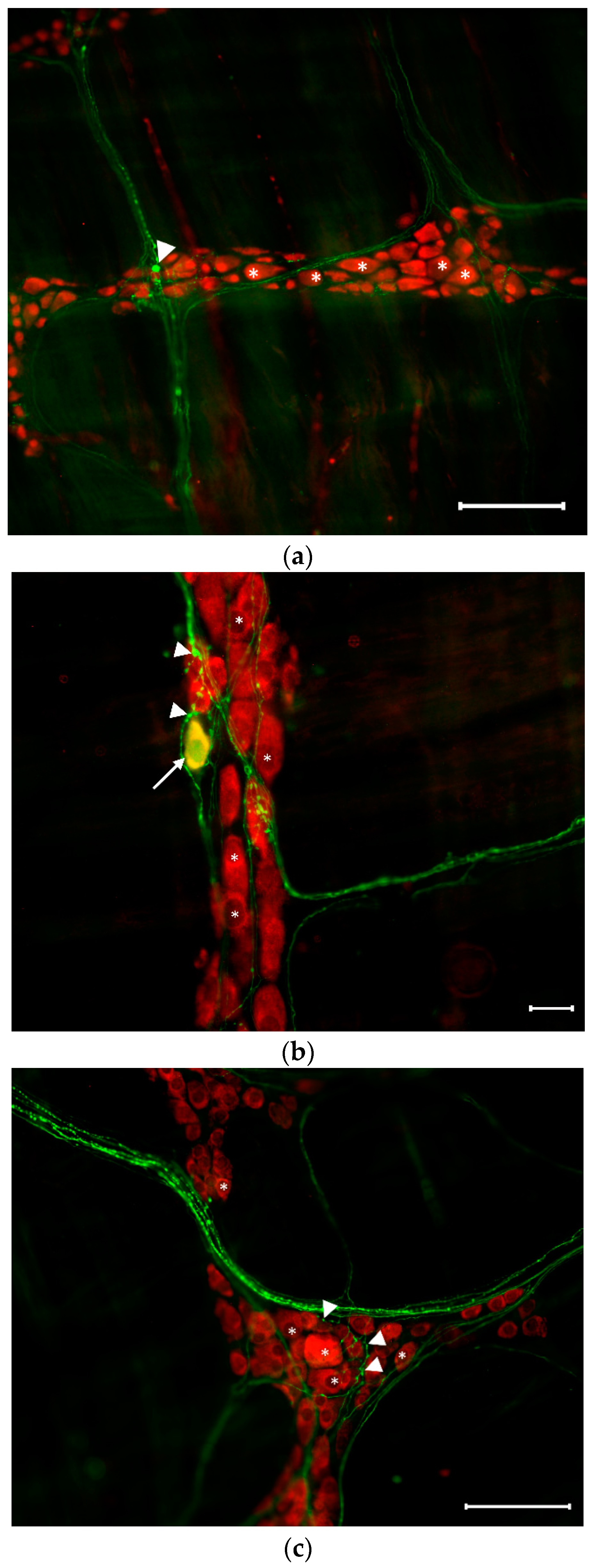
3.2. Distribution of Serotonergic Neurons in the Myenteric Ganglia of Controls

The occurrence of 5-HT-IR myenteric neurons was calculated as the proportion of the total myenteric neuronal number that was HuCD-stained. Despite the extremely low representation of 5-HT-IR neurons in the myenteric ganglia, strong immunoreactivity was observed in the varicosities of projections within the plexus (Figure 1).
In the myenteric ganglia, 5-HT-IR neurons displayed very similar distributions among the different intestinal segments (Figure 2). The proportion of 5-HT-IR neurons varied only by 2–3% in different gut segments: 2.42 ± 0.35% in the duodenum, 2.45 ± 0.33% in the ileum, and 2.35 ± 0.31% in the colon.

3.3. Proportion of Serotonergic Neurons in the Myenteric Ganglia of Diabetics and Insulin-Treated Diabetics

In the myenteric ganglia of diabetic rats, the proportion of 5-HT-IR neurons was increased in each gut segment compared to the controls (Figure 3 and Figure 4).
A 2.5-fold change was observed in the duodenal ganglia of diabetics (5.97 ± 0.7% vs. 2.42 ± 0.35%). A similar proportional increase was observed in the colon where the percentage was 5.91 ± 0.54% relative to 2.35 ± 0.31% of the controls. In the ileum, a slight increasing tendency was observed (3.8 ± 0.41% vs. 2.45 ± 0.33%).
The immediate insulin treatment maintained the proportion of 5-HT-IR myenteric neurons close to the control value in all investigated gut segments (Figure 4 and Figure 5).

3.4. Proportion of Myenteric Ganglia Containing 5-HT-IR Neurons and the Amount of the Serotonergic Neurons in These Ganglia

In controls, the total number of 5-HT-IR neurons per ganglia is extremely low and varied between 0.31 ± 0.06 and 0.46 ± 0.1 in the three investigated gut segments (Figure 6).
However, only 25–30% of the myenteric ganglia contain serotonergic neurons (Figure 7), and the number of 5-HT-IR neurons is 1–2 in these ganglia (Figure 8).
In diabetics, the total number of 5-HT-IR neurons per ganglia increased in each gut segment; however, the increase was significant only in the colon (p < 0.01) where the number of 5-HT-IR neurons was doubled relative to the controls (1.05 ± 0.14 vs. 0.46 ± 0.1; Figure 6). In the diabetic colon, half of the ganglia contained serotonergic neurons (Figure 7) where an average of two cells were found (Figure 8). A significant elevation of the total number of 5-HT-IR neurons per ganglia containing serotonergic neurons was observed in the duodenum compared to the controls (2.13 ± 0.21 vs. 1.25 ± 0.1; Figure 8).
The immediate insulin treatment resulted in low levels of 5-HT-IR myenteric neurons similar to the controls in each intestinal segment in all investigated aspects such as the total number of serotonergic neurons per ganglia (Figure 6), the percentage of myenteric ganglia containing serotonergic neurons (Figure 7), and the total number of 5-HT-IR neurons per serotonergic ganglia (Figure 8).

4. Discussion

The present study was conducted to investigate the gut segment-specific effects of hyperglycemia and insulin treatment on myenteric 5-HT-IR neurons in STZ-induced type 1 diabetes.
A single injection with a high dose of STZ leads to a rapid loss of pancreatic beta cells and concomitant hyperglycemia [37]. In this study, an average 25.5 mM blood glucose level was measured in diabetic rats with a less elevated body weight gain during the 10 weeks compared to controls. Moreover, STZ-induced diabetes is a widely used model of diabetic neuropathy with features including reduced size of nerve fibers, axon and myelin sheath, and decreased nerve conduction velocities [38]. The GI manifestation of neuropathy leads to GI motility disorders that were demonstrated in STZ-diabetic rodents including delayed gastric emptying of both liquids and solids, rapid small intestinal transit, and enhanced spontaneous activity of the colon [34,39].
The proportion of 5-HT-IR myenteric neurons was about 2.5% in controls, which is in good agreement with previous results found in the guinea-pig ileum [17]. Moreover, we found no differences in the proportion of 5-HT-IR neurons between the investigated duodenum, ileum, and colon segments, which is in agreement with the immunohistochemical study of Fuyimiya et al. [40] in which the localization, distribution, and projection of 5-HT-IR neurons and fibers were similar in the stomach, duodenum, jejunum, ileum, and colon of rats.
Although the role of 5-HT in GI motility under physiological conditions is controversial [18,19], a growing amount of evidence demonstrates altered serotonergic signaling as an underlying mechanism in disorders with disturbed GI motility [25,26] including diabetes mellitus [41,42]. The effects of diabetes on the different neurochemical properties of myenteric neuronal populations have been widely investigated. The conclusion of those findings is an altered balance between inhibitory efferent (generally reduced) and excitatory (increased or unchanged) mechanisms [8,39]. We demonstrated a significant 2.5-fold increase in the proportion of 5-HT-IR myenteric neurons in the duodenum and the colon and an increasing tendency in the ileum 10 weeks after STZ treatment application. Based on our earlier findings [33,34] regarding changes in the total neuronal number in diabetes (unchanged in the duodenum and decreased in the ileum and colon), on present results of percentage of myenteric ganglia containing 5-HT-IR neurons, and the total number of serotonergic neurons in these ganglia, we presume that the increase in the percentage of 5-HT-IR neurons relative to the total neuronal number in the duodenum was due to neurochemical alterations of myenteric neurons. We found that more serotonergic neurons in 5-HT-IR ganglia and 5-HT-IR neurons were also detected in those ganglia that previously did not contain 5-HT-IR neurons. The above listed results of studies investigating 5-HT-IR nerves in different segments of the diabetic gut support our findings that the alterations are segment-specific. Increased 5-HT content was found in the duodenum and was unchanged in the rest of the small and large intestines 5 weeks after the alloxan-induced hyperglycemia [30]. Lower brightness of fluorescence in 5-HT-IR nerves in the ileum and increased 5-HT level in the proximal colon were detected 8 weeks after STZ-induced diabetes [29]. In a three-time interval study, alterations in 5-HT levels were demonstrated with a significantly higher 5-HT level in rats’ colons 8 weeks after the induction of STZ-diabetes, followed by a progressive decrease at 16 and 25 weeks, while at later time points, there were no differences in the 5-HT levels between the diabetics and age-matched controls [43]. The inconsistent data from the 1980s and 1990s might be a result of the difference in the duration of diabetes and the difference in the methods used.
Our results are the first to verify the beneficial effects of insulin treatment on the proportion of myenteric 5-HT neurons in different gut segments in 10-week STZ-induced diabetic rats. Immediate insulin treatment prevented the diabetes-induced increased proportion of 5-HT-IR neurons at the control level. Our data are also in good agreement with the findings of Gorio et al. [30] regarding the preventive role of insulin therapy on intestinal 5-HT content in 5-week alloxan-induced diabetes; however, in that stage of the disease, the 5-HT content was increased only in the duodenum and unchanged in the rest of the gut. The segment-specific protective effects of insulin treatment on the neurochemical changes induced by STZ-diabetes have been revealed in some aspects. Izbéki et al. [34] found that insulin treatment has beneficial effect in the ileum and colon but not in the duodenum and jejunum on the density of nitrergic neurons. Jancsó et al. [9] detected that insulin therapy restored the enhanced heme oxygenase 2 protein level to the control level in the duodenum and colon of diabetic rats.
The roles of neuronal 5-HT are generally associated with GI motility, but some of the recently discovered less conventional features of 5-HT-IR neurons should be mentioned [44]. The 5-HT-IR neurons are among the first in the gut to arise, and they coexist later-born neurons. It is possible that 5-HT may be a neuronal growth factor promoting the development/survival of some neurons being among the last to be born including dopaminergic, GABAergic, nitrergic, and calcitonin gene-related peptide-expressing neurons [45]. In support of this, the essential role of 5-HT in the growth and maintenance of enteric neurons [46] and intestinal mucosal epithelium in adult mice were demonstrated [47]. Recent studies focus on the role of 5-HT signaling in enteric neurogenesis in adulthood both in health and diseases [48,49]. The importance of gut microbiota in maintaining the serotonergic network through release of 5-HT and the activation of the 5-HT4 receptor has also been illustrated in a recent study [50]. Significant differences were seen in the composition of luminal microbiota in the fecal samples collected from the ileum and colon of diabetic rats [51]. These findings suggest mechanisms linking microbial dysbiosis to GI disorders. The abovementioned multifaceted role of neuronal 5-HT implies more complex underlying mechanisms in the structural and functional alterations of the ENS in the diabetic state.

5. Conclusions

We demonstrated a gut segment-specific alteration in the proportion of myenteric 5HT-IR neurons, with significantly increased numbers in the duodenum and colon, and illustrated the increasing tendency in the ileum of 10-week STZ-diabetic rats. Moreover, the immediate insulin treatment was preventive of the hyperglycemia-induced enhancement in the amount of 5-HT-IR neurons in all investigated gut segments. The impaired insulin signaling may contribute to these neurochemical changes in 5-HT-IR myenteric neurons found in diabetes.

Author Contributions

Conceptualization, N.B. and M.B.; methodology, D.M., J.B., and Z.M.; investigation, D.M., Z.S., and Z.M.; writing—original draft preparation, Z.S. and M.B.; writing—review and editing, M.B. and D.M.; visualization, Z.S.; supervision, M.B.; funding acquisition, N.B. All authors have read and agreed to the published version of the manuscript.

Funding

This research was funded by the Hungarian National Research, Development, and Innovation Fund project, grant number GINOP-2.3.3-15-2016-00006; Hungarian NKFIH fund project, grant number FK131789 (N.B.); János Bolyai Research Scholarship of the Hungarian Academy of Sciences (N.B.); and the New National Excellence Program of the Ministry for Innovation and Technology for the National Research, Development, and Innovation Fund, No. ÚNKP-20-5 (to Bódi N).

Institutional Review Board Statement

The principles of the National Institutes of Health (Bethesda, MD, USA) guidelines and the EU directive 2010/63/EU for the protection of animals used for scientific purposes were strictly followed, and all experiments were approved by the National Scientific Ethical Committee on Animal Experimentation (National Competent Authority) with the license number XX./1487/2014.

Informed Consent Statement

Not applicable.

Data Availability Statement

Available upon request.

Conflicts of Interest

The authors declare no conflict of interest. The funders had no role in the design of the study; in the collection, analyses, or interpretation of data; in the writing of the manuscript; or in the decision to publish the results.

References

  1. Azpiroz, F.; Malagelada, C. Diabetic neuropathy in the gut: Pathogenesis and diagnosis. Diabetologia 2016, 59, 404–408. [Google Scholar] [CrossRef] [PubMed]
  2. Furness, J.B. The enteric nervous system and neurogastroenterology. Nat. Rev. Gastroenterol. Hepatol. 2012, 9, 286–294. [Google Scholar] [CrossRef] [PubMed]
  3. Furness, J.B. The enteric nervous system: Normal functions and enteric neuropathies. Neurogastroenterol. Motil. 2008, 20, 32–38. [Google Scholar] [CrossRef] [PubMed]
  4. Andres, H.; Rock, R.; Bridges, R.J.; Rummel, W.; Schreiner, J. Submucosal plexus and electrolyte transport across rat colonic mucosa. J. Physiol. 1985, 364, 301–312. [Google Scholar] [CrossRef] [Green Version]
  5. Timmermans, J.P.; Hens, J.; Adriaensen, D. Outer submucous plexus: An intrinsic nerve network involved in both secretory and motility processes in the intestine of large mammals and humans. Anat. Rec. Off. Publ. Am. Assoc. Anat. 2001, 262, 71–78. [Google Scholar] [CrossRef]
  6. Krueger, D.; Michel, K.; Zeller, F.; Demir, I.E.; Ceyhan, G.O.; Slotta-Huspenina, J.; Schemann, M. Neural influences on human intestinal epithelium in vitro. J. Physiol. 2016, 594, 357–372. [Google Scholar] [CrossRef] [PubMed]
  7. Aouacheri, O.; Saka, S.; Krim, M.; Messaadia, A.; Maidi, I. The investigation of the oxidative stress-related parameters in type 2 diabetes mellitus. Can. J. Diabetes 2015, 39, 44–49. [Google Scholar] [CrossRef] [PubMed] [Green Version]
  8. Chandrasekharan, B.; Srinivasan, S. Diabetes and the enteric nervous system. Neurogastroenterol. Motil. 2007, 19, 951–960. [Google Scholar] [CrossRef] [Green Version]
  9. Jancso, Z.; Bodi, N.; Borsos, B.; Fekete, E.; Hermesz, E. Gut region-specific accumulation of reactive oxygen species leads to regionally distinct activation of antioxidant and apoptotic marker molecules in rats with STZ-induced diabetes. Int. J. Biochem. Cell Biol. 2015, 62, 125–131. [Google Scholar] [CrossRef] [Green Version]
  10. Bodi, N.; Talapka, P.; Poles, M.Z.; Hermesz, E.; Jancso, Z.; Katarova, Z.; Izbeki, F.; Wittmann, T.; Fekete, E.; Bagyanszki, M. Gut region-specific diabetic damage to the capillary endothelium adjacent to the myenteric plexus. Microcirculation 2012, 19, 316–326. [Google Scholar] [CrossRef]
  11. Meldgaard, T.; Olesen, S.S.; Farmer, A.D.; Krogh, K.; Wendel, A.A.; Brock, B.; Drewes, A.M.; Brock, C. Diabetic Enteropathy: From Molecule to Mechanism-Based Treatment. J. Diabetes Res. 2018, 2018, 3827301. [Google Scholar] [CrossRef] [PubMed]
  12. Gotfried, J.; Priest, S.; Schey, R. Diabetes and the Small Intestine. Curr. Treat. Options Gastroenterol. 2017, 15, 490–507. [Google Scholar] [CrossRef] [PubMed]
  13. Martin, A.M.; Young, R.L.; Leong, L.; Rogers, G.B.; Spencer, N.J.; Jessup, C.F.; Keating, D.J. The Diverse Metabolic Roles of Peripheral Serotonin. Endocrinology 2017, 158, 1049–1063. [Google Scholar] [CrossRef] [PubMed]
  14. Young, R.L.; Lumsden, A.L.; Martin, A.M.; Schober, G.; Pezos, N.; Thazhath, S.S.; Isaacs, N.J.; Cvijanovic, N.; Sun, E.W.L.; Wu, T.; et al. Augmented capacity for peripheral serotonin release in human obesity. Int. J. Obes. 2018, 42, 1880–1889. [Google Scholar] [CrossRef] [PubMed]
  15. Qu, Z.D.; Thacker, M.; Castelucci, P.; Bagyanszki, M.; Epstein, M.L.; Furness, J.B. Immunohistochemical analysis of neuron types in the mouse small intestine. Cell Tissue Res. 2008, 334, 147–161. [Google Scholar] [CrossRef] [PubMed]
  16. Hempfling, C.; Neuhuber, W.L.; Worl, J. Serotonin-immunoreactive neurons and mast cells in the mouse esophagus suggest involvement of serotonin in both motility control and neuroimmune interactions. Neurogastroenterol. Motil. 2012, 24, e67–e78. [Google Scholar] [CrossRef]
  17. Costa, M.; Brookes, S.J.; Steele, P.A.; Gibbins, I.; Burcher, E.; Kandiah, C.J. Neurochemical classification of myenteric neurons in the guinea-pig ileum. Neuroscience 1996, 75, 949–967. [Google Scholar] [CrossRef]
  18. Spencer, N.J.; Sia, T.C.; Brookes, S.J.; Costa, M.; Keating, D.J. CrossTalk opposing view: 5-HT is not necessary for peristalsis. J. Physiol. 2015, 593, 3229–3231. [Google Scholar] [CrossRef] [PubMed] [Green Version]
  19. Smith, T.K.; Gershon, M.D. CrossTalk proposal: 5-HT is necessary for peristalsis. J. Physiol. 2015, 593, 3225–3227. [Google Scholar] [CrossRef] [PubMed] [Green Version]
  20. Mawe, G.M.; Hoffman, J.M. Serotonin signalling in the gut--functions; dysfunctions and therapeutic targets. Nat. Rev. Gastroenterol. Hepatol. 2013, 10, 473–486. [Google Scholar] [CrossRef] [PubMed] [Green Version]
  21. Okamoto, T.; Barton, M.J.; Hennig, G.W.; Birch, G.C.; Grainger, N.; Corrigan, R.D.; Koh, S.D.; Sanders, K.M.; Smith, T.K. Extensive projections of myenteric serotonergic neurons suggest they comprise the central processing unit in the colon. Neurogastroenterol. Motil. 2014, 26, 556–570. [Google Scholar] [CrossRef] [PubMed]
  22. Furness, J.B.; Costa, M. Neurons with 5-hydroxytryptamine-like immunoreactivity in the enteric nervous system: Their projections in the guinea-pig small intestine. Neuroscience 1982, 7, 341–349. [Google Scholar] [CrossRef]
  23. Banerjee, S.; Akbar, N.; Moorhead, J.; Rennie, J.A.; Leather, A.J.; Cooper, D.; Papagrigoriadis, S. Increased presence of serotonin-producing cells in colons with diverticular disease may indicate involvement in the pathophysiology of the condition. Int. J. Colorectal Dis. 2007, 22, 643–649. [Google Scholar] [CrossRef] [PubMed]
  24. Faure, C.; Patey, N.; Gauthier, C.; Brooks, E.M.; Mawe, G.M. Serotonin signaling is altered in irritable bowel syndrome with diarrhea but not in functional dyspepsia in pediatric age patients. Gastroenterology 2010, 139, 249–258. [Google Scholar] [CrossRef] [Green Version]
  25. Coyle, D.; Murph, J.M.; Doyle, B.; O’Donnell, A.M.; Gillick, J.; Puri, P. Altered tryptophan hydroxylase 2 expression in enteric serotonergic nerves in Hirschsprung’s-associated enterocolitis. World J. Gastroenterol. 2016, 22, 4662–4672. [Google Scholar] [CrossRef]
  26. Israelyan, N.; Margolis, K.G. Serotonin as a link between the gut-brain-microbiome axis in autism spectrum disorders. Pharmacol. Res. 2018, 132, 1–6. [Google Scholar] [CrossRef] [PubMed]
  27. Del Colle, A.; Israelyan, N.; Gross Margolis, K. Novel aspects of enteric serotonergic signaling in health and brain-gut disease. Am. J. Physiol. Gastrointest. Liver Physiol. 2020, 318, G130–G143. [Google Scholar] [CrossRef] [Green Version]
  28. Camilleri, M. Novel Diet; Drugs; and Gastric Interventions for Gastroparesis. Clin. Gastroenterol. Hepatol. 2016, 14, 1072–1080. [Google Scholar] [CrossRef] [PubMed] [Green Version]
  29. Lincoln, J.; Bokor, J.T.; Crowe, R.; Griffith, S.G.; Haven, A.J.; Burnstock, G. Myenteric plexus in streptozotocin-treated rats. Neurochemical and histochemical evidence for diabetic neuropathy in the gut. Gastroenterology 1984, 86, 654–661. [Google Scholar] [CrossRef]
  30. Gorio, A.; Di Giulio, A.M.; Donadoni, L.; Tenconi, B.; Germani, E.; Bertelli, A.; Mantegazza, P. Early neurochemical changes in the autonomic neuropathy of the gut in experimental diabetes. Int. J. Clin. Pharmacol. Res. 1992, 12, 217–224. [Google Scholar]
  31. Bodi, N.; Szalai, Z.; Chandrakumar, L.; Bagyanszki, M. Region-dependent effects of diabetes and insulin-replacement on neuronal nitric oxide synthase- and heme oxygenase-immunoreactive submucous neurons. World J. Gastroenterol. 2017, 23, 7359–7368. [Google Scholar] [CrossRef]
  32. Chandrakumar, L.; Bagyanszki, M.; Szalai, Z.; Mezei, D.; Bodi, N. Diabetes-Related Induction of the Heme Oxygenase System and Enhanced Colocalization of Heme Oxygenase 1 and 2 with Neuronal Nitric Oxide Synthase in Myenteric Neurons of Different Intestinal Segments. Oxidative Med. Cell. Longev. 2017, 2017, 1890512. [Google Scholar] [CrossRef]
  33. Bagyanszki, M.; Bodi, N. Diabetes-related alterations in the enteric nervous system and its microenvironment. World J. Diabetes 2012, 3, 80–93. [Google Scholar] [CrossRef] [PubMed]
  34. Izbeki, F.; Wittman, T.; Rosztoczy, A.; Linke, N.; Bodi, N.; Fekete, E.; Bagyanszki, M. Immediate insulin treatment prevents gut motility alterations and loss of nitrergic neurons in the ileum and colon of rats with streptozotocin-induced diabetes. Diabetes Res. Clin. Pract. 2008, 80, 192–198. [Google Scholar] [CrossRef] [PubMed] [Green Version]
  35. de Mello, S.T.; de Miranda Neto, M.H.; Zanoni, J.N.; Furlan, M.M. Effects of insulin treatment on HuC/HuD; NADH diaphorase; and nNOS-positive myoenteric neurons of the duodenum of adult rats with acute diabetes. Dig. Dis. Sci. 2009, 54, 731–737. [Google Scholar] [CrossRef] [PubMed]
  36. Watkins, C.C.; Sawa, A.; Jaffrey, S.; Blackshaw, S.; Barrow, R.K.; Snyder, S.H.; Ferris, C.D. Insulin restores neuronal nitric oxide synthase expression and function that is lost in diabetic gastropathy. J. Clin. Investig. 2000, 106, 803. [Google Scholar] [CrossRef] [Green Version]
  37. King, A.J. The use of animal models in diabetes research. Br. J. Pharmacol. 2012, 166, 877–894. [Google Scholar] [CrossRef] [Green Version]
  38. Yarandi, S.S.; Srinivasan, S. Diabetic gastrointestinal motility disorders and the role of enteric nervous system: Current status and future directions. Neurogastroenterol. Motil. 2014, 26, 611–624. [Google Scholar] [CrossRef]
  39. Ordog, T.; Hayashi, Y.; Gibbons, S.J. Cellular pathogenesis of diabetic gastroenteropathy. Minerva Gastroenterol. Dietol. 2009, 55, 315–343. [Google Scholar]
  40. Fujimiya, M.; Okumiya, K.; Yamane, T.; Maeda, T. Distribution of serotonin-immunoreactive nerve cells and fibers in the rat gastrointestinal tract. Histochem. Cell Biol. 1997, 107, 105–114. [Google Scholar] [CrossRef]
  41. Crowell, M.D.; Mathis, C.; Schettler, V.A.; Yunus, T.; Lacy, B.E. The effects of tegaserod; a 5-HT receptor agonist; on gastric emptying in a murine model of diabetes mellitus. Neurogastroenterol. Motil. 2005, 17, 738–743. [Google Scholar] [CrossRef]
  42. Farmer, A.D.; Bruckner-Holt, C.; Schwartz, S.; Sadler, E.; Kadirkamanthan, S. Diabetic Gastroparesis: Perspectives from a Patient and Health Care Providers. J. Patient Cent. Res. Rev. 2019, 6, 148–157. [Google Scholar] [CrossRef]
  43. Belai, A.; Lincoln, J.; Milner, P.; Burnstock, G. Progressive changes in adrenergic; serotonergic; and peptidergic nerves in proximal colon of streptozotocin-diabetic rats. Gastroenterology 1988, 95, 1234–1241. [Google Scholar] [CrossRef]
  44. Margolis, K.G.; Cryan, J.F.; Mayer, E.A. The Microbiota-Gut-Brain Axis: From Motility to Mood. Gastroenterology 2021, 160, 1486–1501. [Google Scholar] [CrossRef]
  45. Li, Z.; Chalazonitis, A.; Huang, Y.Y.; Mann, J.J.; Margolis, K.G.; Yang, Q.M.; Kim, D.O.; Cote, F.; Mallet, J.; Gershon, M.D. Essential roles of enteric neuronal serotonin in gastrointestinal motility and the development/survival of enteric dopaminergic neurons. J. Neurosci. 2011, 31, 8998–9009. [Google Scholar] [CrossRef]
  46. Liu, M.T.; Kuan, Y.H.; Wang, J.; Hen, R.; Gershon, M.D. 5-HT4 receptor-mediated neuroprotection and neurogenesis in the enteric nervous system of adult mice. J. Neurosci. 2009, 29, 9683–9699. [Google Scholar] [CrossRef]
  47. Gross, E.R.; Gershon, M.D.; Margolis, K.G.; Gertsberg, Z.V.; Li, Z.; Cowles, R.A. Neuronal serotonin regulates growth of the intestinal mucosa in mice. Gastroenterology 2012, 143, 408–417.e2. [Google Scholar] [CrossRef] [PubMed] [Green Version]
  48. Jonscher, R.; Belkind-Gerson, J. Concise Review: Cellular and Molecular Mechanisms of Postnatal Injury-Induced Enteric Neurogenesis. Stem Cells 2019, 37, 1136–1143. [Google Scholar] [CrossRef]
  49. Grundmann, D.; Loris, E.; Maas-Omlor, S.; Schafer, K.H. Enteric Neurogenesis During Life Span Under Physiological and Pathophysiological Conditions. Anat. Rec. 2019, 302, 1345–1353. [Google Scholar] [CrossRef] [PubMed]
  50. De Vadder, F.; Grasset, E.; Manneras Holm, L.; Karsenty, G.; Macpherson, A.J.; Olofsson, L.E.; Backhed, F. Gut microbiota regulates maturation of the adult enteric nervous system via enteric serotonin networks. Proc. Natl. Acad. Sci. USA 2018, 115, 6458–6463. [Google Scholar] [CrossRef] [Green Version]
  51. Wirth, R.; Bodi, N.; Maroti, G.; Bagyanszki, M.; Talapka, P.; Fekete, E.; Bagi, Z.; Kovacs, K.L. Regionally distinct alterations in the composition of the gut microbiota in rats with streptozotocin-induced diabetes. PLoS ONE 2014, 9, e110440. [Google Scholar] [CrossRef] [PubMed] [Green Version]
Figure 1. Representative fluorescent micrographs from the duodenum (a), ileum (b), and colon (c) of controls after 5-HT-HuCD immunohistochemistry. On the myenteric plexus whole-mount preparations, asterisks indicate neurons that are labelled for HuCD, arrowheads point to myenteric 5-HT-IR varicosities, and the arrow points to a myenteric neuron that is double-labelled for both 5-HT and HuCD. Scale bars are 100 µm (a), 20 µm (b), and 100 µm (c).
Applsci 11 05949 g001
Figure 2. Proportion of 5-HT-IR neurons in the myenteric ganglia of the duodenum, ileum, and colon of controls. The percentage of 5-HT-IR neurons was only 2–3% in the myenteric ganglia in all intestinal segments without any significant differences. Data are expressed as mean ± SEM. CD—control duodenum; CI—control ileum; and CC—control colon.
Applsci 11 05949 g002
Figure 3. Representative fluorescent micrographs from the duodenum (a), ileum (b), and colon (c) of diabetic rats after 5-HT-HuCD immunohistochemistry. On the myenteric plexus whole-mount preparations, asterisks indicate neurons that are labelled for HuCD, arrowheads point to myenteric 5-HT-IR varicosities, and arrows point to myenteric neurons that are double-labelled for both 5-HT and HuCD. Scale bars are 100 µm (a), 20 µm (b), and 50 µm (c).
Applsci 11 05949 g003
Figure 4. Proportion of 5-HT-IR neurons of the duodenum, ileum, and colon of controls, diabetics, and insulin-treated diabetics. The percentage of 5-HT-IR myenteric neurons was increased in each gut segment of diabetics. The insulin treatment reduced the proportion of 5-HT-IR neurons that are similar to the control level. Data are expressed as mean ± SEM. ** p < 0.01 and **** p < 0.0001 (between controls and diabetics); ° p < 0.05 and °°°° p < 0.0001 (between diabetics and insulin-treated diabetics). C—controls; D—diabetics; and ID—insulin-treated diabetics.
Applsci 11 05949 g004
Figure 5. Representative fluorescent micrographs from the duodenum (a), ileum (b), and colon (c) of insulin-treated diabetics after 5-HT-HuCD immunohistochemistry. On the myenteric plexus whole-mount preparations, asterisks indicate neurons that are labelled for HuCD and arrowheads point to myenteric 5-HT-IR varicosities. Scale bars are 20 µm.
Applsci 11 05949 g005
Figure 6. Number of 5-HT-IR neurons of the duodenum, ileum, and colon of controls, diabetics, and insulin-treated diabetics. The total number of 5-HT-IR myenteric neurons was elevated in each gut segment of diabetics. In insulin-treated diabetic rats, the total number of 5-HT-IR neurons remained close to the control level. Data are expressed as mean ± SEM. ** p < 0.01 (between controls and diabetics); °° p < 0.01 (between diabetics and insulin-treated diabetics). C—controls; D—diabetics; and ID—insulin-treated diabetics.
Applsci 11 05949 g006
Figure 7. Percentage of myenteric ganglia containing 5-HT-IR neurons. In controls and insulin-treated diabetics, 20–30% of the myenteric ganglia contain 5-HT-IR neurons. In diabetic rats, a gut segment-specific increase was observed with the most pronounced elevation in the colon where 50% of the myenteric ganglia contain 5-HT-IR neurons. C—controls; D—diabetics; and ID—insulin-treated diabetics.
Applsci 11 05949 g007
Figure 8. Total number of 5-HT-IR neurons in ganglia containing 5-HT-IR neurons. In controls, 1–2 serotonergic neurons were found in 5-HT-IR neurons containing myenteric ganglia. In diabetics, the most pronounced increase was found in the duodenum but the insulin treatment restored the level to close to that of the control. Data are expressed as mean ± SEM. * p < 0.05 (between controls and diabetics); ° p < 0.05 (between diabetics and insulin-treated diabetics). C—controls; D—diabetics; and ID—insulin-treated diabetics.
Applsci 11 05949 g008
Table 1. Weight and glycemic characteristics of the three experimental groups of rats.
Body Weight
(g) ± SEM
Blood Glucose Concentration
(mmol/L) ± SEM
InitialFinalInitialFinal (Average)
Controls (n = 10)226.2 ± 4.3457.1 ± 19.7 *5.46 ± 0.45.56 ± 0.1
Diabetics (n = 7)226.7 ± 6.0344.4 ± 16.0 *°°4.99 ± 0.425.54 ± 1.1 *°°
Insulin-treated diabetics (n = 8)241.3 ± 1.9398 ± 8.9 +4.85 ± 0.113.75 ± 1.0 *°°++
* p < 0.0001 vs. initial; ° p < 0.001 and °° p < 0.0001 vs. final controls; and + p < 0.01 and ++ p < 0.0001 vs. final diabetics.
Publisher’s Note: MDPI stays neutral with regard to jurisdictional claims in published maps and institutional affiliations.

