Article

Development of a Lightweight Tungsten Shielding Fiber That Can Be Used for Improving the Performance of Medical Radiation Shields

Department of Biomedical Engineering, School of Medicine, Keimyung University, 1095 Dalgubeol-daero, Daegu 42601, Korea
Appl. Sci. 2021, 11(14), 6475; https://doi.org/10.3390/app11146475
Submission received: 24 June 2021 / Revised: 8 July 2021 / Accepted: 12 July 2021 / Published: 13 July 2021
(This article belongs to the Section Biomedical Engineering)

Abstract

:
Radiation exposure in medical institutions is mainly due to low doses. Low-dose radiation mainly means scattered radiation, and such scattered radiation can be shielded with a lightweight shielding suit. In this study, the shielding performance of shielding fabrics woven by winding polyethylene (PE) yarn around a 30 μm tungsten wire was evaluated. To improve the shielding performance, an air pressure dispersion process of coating tungsten nanopowder on the fiber was developed. The radiation shielding effectiveness of the shielding fibers with and without dispersed tungsten nanopowder were compared by measuring the spatial dose inside the diagnostic X-ray imaging room of a medical institution. The results of the experiment confirmed that the fabric coated with tungsten nanopowder improved the shielding performance of the general tungsten fiber by approximately 15% and provided relatively effective low-dose radiation shielding at approximately 1.2 m of the X-ray imaging equipment. This study shows that tungsten fiber can be helpful in manufacturing lightweight shielding clothing for protection from scattered radiation in medical institutions.

1. Introduction

The increasing use of artificial radiation in medical institutions has increased radiation exposure in the diagnostic area [1]. Although the dose and quality of the radiation used for the diagnosis of diseases are relatively low compared to the radiation leaked from a nuclear accident, for example, even a very small dose can have a very dangerous effect on the human body [2].
Most of the radiation exposure that is a problem in medical institutions is caused by indirect scattered rays rather than direct rays. Scattered radiation corresponds to a low dose, generally less than 100 mSv [3]. In the case of radiation exposure of less than 100 mSv, the Research Council Biological Effects of Ionizing Radiation report (BEIR VII) supports the theory of linear proportion without a threshold, thus, there is a direct potential for cancer and genetic disorders [4,5]. Therefore, the active protection of medical workers against low-dose radiation exposure is critical.
Current radiation shielding clothing in medical institutions generally uses lead as the shielding material, however, the weight of this substance hinders the physical activity of medical staff and patients. A radiation protection apron having the equivalent to 0.50 mm of lead weighs between 3.01 and 3.15 kg. Because the front part offers protection from the shoulders to the knees, these are widely used in the vicinity of X-rays in medical institutions [6].
A previous study considered that scattered rays exist within 2 m of the radiation generating site, and for this purpose, suggested a shield with a thickness of less than 0.50 mm of lead equivalent [7]. However, in order to reduce the weight of the shielding lining, it is necessary to discover a new shielding material, and this is also related to the manufacturing process technology that is used to replace the shielding material, hence there are many complications in practical use. Recently, radiation shields have been developed using eco-friendly materials to avoid the toxicity of lead. Eco-friendly radiation shielding materials consist of tungsten, bismuth, barium sulfate, and boron, among others, mixed with a polymer material and manufactured in the form of a sheet [8,9]. Any shielding suit used in a medical institution must be lightweight and safe [10]. In order to maintain the same shielding performance for direct X-rays, it is difficult to significantly change the weight of the shield. However, since the scattered radiation generated in a certain distance and direction has a low dose, it can be shielded with lightweight shielding clothing.
Radiation shielding involves absorption using a material made of an element with a large atomic number, such as lead, or reducing the transmission of radiation through interaction with the shielding material. Therefore, lowering the amount of radiation exposure requires selecting a shielding material considering the type and energy of the radiation [11]. In the low energy region of X-rays or γ-rays with high radiation transmittance, sufficient shielding effect can be obtained with relatively thin lead, but in the high energy region, the necessary protective effect cannot be obtained unless the shield is thickened [12]. However, if the thickness of the shield is increased sufficiently, the weight of the shielding suit also increases substantially, which hinders the activity of medical personnel who must wear it for a long time during work.
In this study, PE yarn was wound around a tungsten wire to fabricate a shielding fiber that can be used as a lining for lightweight shielding clothing. The study also developed an air pressure dispersion process to disperse nanotungsten powder to eliminate pinholes in fibers. The shielding performance and weight of the fibers were evaluated to determine the potential efficiency of the fibers in low-dose shielding suits used in medical institutions. Products presented as conventional radiation shielding fibers are mainly manufactured by coating the nonwoven fabric on the shielding sheet with an inorganic mixture [13]. However, the problems of these products concern the weight and flexibility of fibers. To solve these problems, this study proposes a method to improve the shielding performance while maintaining the flexibility of the fiber.

2. Materials and Methods

The shielding material used in this study was tungsten, which has an atomic number of 74, an atomic weight of 183.84 g/mol, and a density of 19.25 g/cm3. As its shielding performance is close to that of lead, it has been widely used recently as an eco-friendly radiation shielding material [14]. In this study, pure tungsten shielding fiber was produced from tungsten yarn made with industrial filament tungsten wire with a diameter of 30 ± 0.05 µm. The fabric was manufactured using a weaving machine (Rapier Loom, Dornier, HTVS 8/S20, Friedrichshafen, Germany). The diameter of the tungsten wire was selected such that it was small enough to maintain the flexibility of the tungsten fabric and provide a high weaving density, but not too small that the yarn could break during the weaving process. Therefore, a diameter of 30 µm was selected in this study [15]. In addition, the fabric was woven by the twill weave method to minimize the spacing between the yarns, which occurs in warp and weft yarns from the plain weave method traditionally used for weaving [16]. Figure 1 shows the technical difference between the twill weave method and the plain weave method. Warp and weft yarns are crossed one by one in the plain weave method, whereas two or more yarns are crossed in the twill weave method [17].
In previous studies, mesh-type connection or knit weaving methods were suggested [18]. However, maintaining a constant shape during processing and achieving reproducibility of the shielding performance requires a more effective weaving method. In addition, the fabric was intended to be stacked for shielding applications involving higher radiation intensities. Therefore, in this study, the fabric was fabricated with high density using the twill weave method. The reproducibility of the shielding performance depends on the thickness of the shield and the consistency of the process technology. Figure 2 presents a fabric woven using tungsten yarn and Figure 3 presents the fabric weaving process with a weaving machine.
Methods for improving the shielding performance of the woven shielding fiber include a laminated structure, that is, using multiple layers of overlapping fabric, and a double coating method [19,20]. In general, a laminated structure of shielding fibers has the disadvantage of increased shielding thickness. Therefore, in this study, a method was developed in which a polyethylene (PE) yarn was wound around a tungsten wire and then woven into a fabric, and tungsten nanopowder was dispersed in the woven shielding fabric.
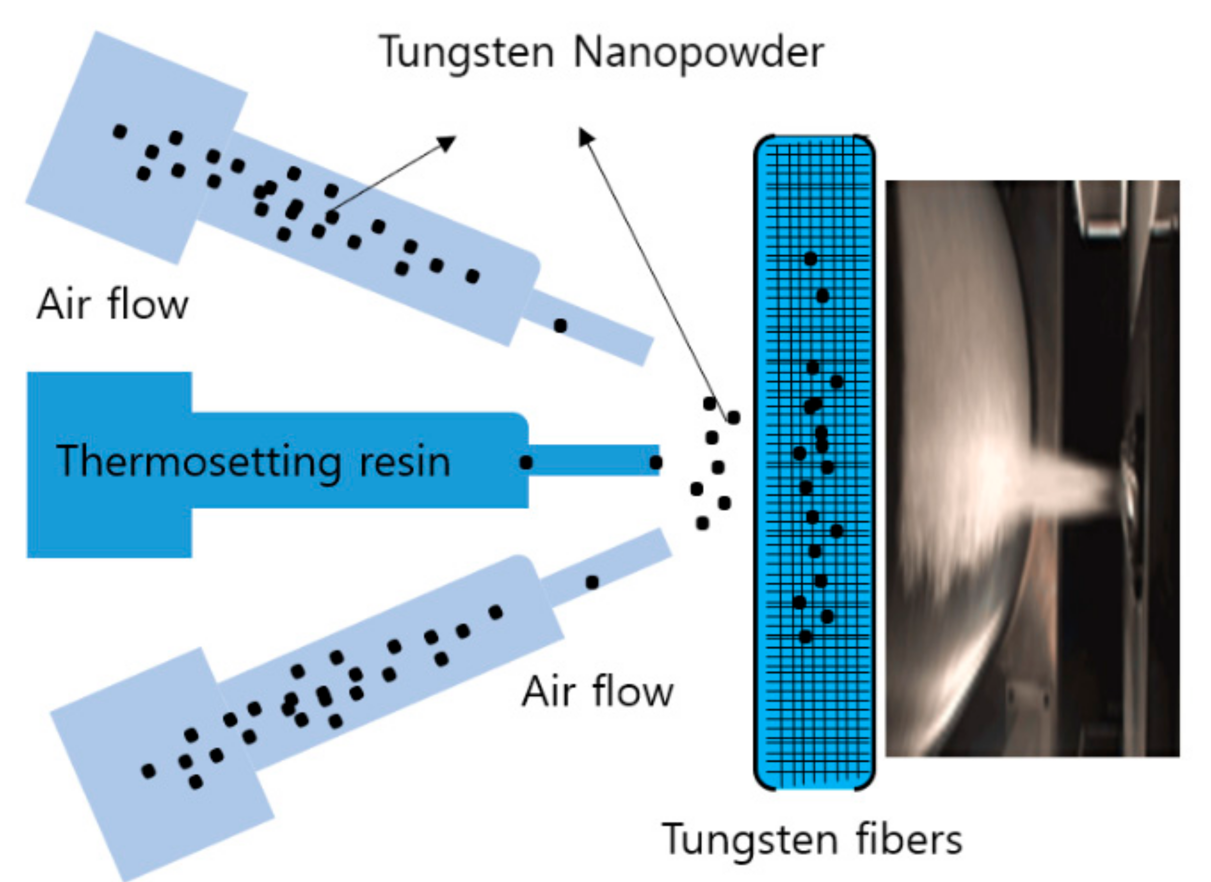
Even with high-density weaving, gaps and voids between the yarns reduce the shielding performance of the tungsten shielding fiber. Therefore, filling the voids with tungsten nanoparticles was expected to improve the shielding performance of the fabric. The air pressure dispersion method of dispersing the tungsten nanoparticles on the shielding fiber fabric was configured as shown in Figure 4 [21].
The air pressure dispersion method disperses tungsten nanoparticles onto the fabric through a thin air nozzle with a diameter of 1.5 mm. In this study, a direct-dispersion form was applied. A trace amount of thermosetting phenolic resin adhesive was used for the complete fixation of tungsten powder particles on the shielding fabric, thus, fixing the particles well, increasing the affinity with the fibers, and filling the voids. The dispersed tungsten powder was applied in a small amount of less than 8 wt% of the total shielding fiber.
In this experiment, to determine the shielding area for medical radiation, the range and energy of the scattered rays inside the imaging room, which is the diagnostic area, were presented, and the shielding performance of direct radiation was evaluated. First, to evaluate the shielding performance of direct rays, the range of X-ray energy used in the diagnosis area was tested based on human body imaging. The following conditions were used in the X-ray energy shielding test—tube voltage: 60–120 kVp, tube current: 200 mA, and irradiation time: 0.1 s.
Because X-ray energy is not a single energy source, the effective energy must be determined. The effective energy of the braking radiation X-rays can be determined using the half-value layer (HVL) measurement method [22]. The HVL is the thickness of a material that attenuates the incident radiation by half. Although there is a difference between materials, the effective energy can be calculated using the following set of equations, after the material for measuring the HVL is determined [23,24].
When the incident radiation intensity I 0 interacts with the shielding material while passing through the shield, the intensity I of the transmitted radiation can be expressed by Equation (1):
I = I 0 e μ t
Here, μ is the linear attenuation coefficient and t is the thickness of the shield. The HVL is the thickness of the material at which the intensity of the incident radiation is halved ( I 0 2 ). Thus, after the expression I = I 0 2 is substituted in Equation (1) with t =   HVL , it becomes I 0 2 = I 0 e μ HVL :
μ = In 2 HVL
Therefore, the effective energy of the incident X-rays can be determined using the linear attenuation coefficient μ calculated from Equation (2) and Hubbell’s mass attenuation coefficient [25].
The medical radiation-shielding performance was evaluated for each calculated effective energy. For the experimental method for testing the shielding performance of the two shielding fibers presented in this study, the geometric conditions were set as shown in Figure 5. The shielding rate calculation of the shielding sheet was 1 W W 0 × 100 [26]. Here, W is the irradiation dose measured when there is a shielding fiber between the X-ray tube (E7239, 150 kV-500 mA, Toshiba, Tokyo, Japan) and the dosimeter (Dosimax plus 1, IBA Dosimetry Corp., Schwarzenbruck, Germany) W 0 is the irradiation dose measured when there is no shielding fiber between the X-ray tube and the dosimeter.
In this study, in order to verify the effectiveness of low-dose radiation shielding in a general imaging room of a medical institution using the fabricated shielding fiber, the irradiation dose was measured from the X-ray generator referenced to the horizontal plane of the patient examination table [27]. The measuring points were set 360° from the radiation generator position and at distances up to 200 cm in 50 cm increments. The spatial dose was measured using a spatial dosimeter (Raysafe 452, Billdal, Sweden).

3. Results

Table 1 shows the characteristics of the yarns and fabrics used to determine the shielding effectiveness of the radiation-shielding fibers. The manufactured yarn was made by winding PE thread on a 30 μm tungsten wire with 98% purity. The reason why the PE thread was wound around the tungsten wire was to ensure the flexibility of the fiber when woven with a shielding fiber and to allow the dispersed tungsten nanopowder to be well settled. The woven fiber was 0.2 mm thick and weighed 954.3 g/m2. Therefore, a shielded suit of less than 1 kg can be produced when used as the apron. Additionally, because the strength of the yarn was 1.45 g/d, weaving with it posed no problem.
The properties of the fibers dispersed with tungsten powder and fibers without dispersion were compared. Figure 6 shows electron micrographs of fabrics with and without tungsten powder dispersed in the fabricated shielding fiber. Figure 7 shows enlarged electron microscope images of the fabrics. Figure 7a shows the voids between the yarns, and Figure 7b shows the tungsten powder particles filling the voids and distributed evenly on the yarn.
Table 2 shows the shielding performance for the two fabricated shielding fibers. Dispersing tungsten powder in the fiber improves the shielding performance by approximately 15%, making it more effective in low-dose areas. Therefore, the dispersion of tungsten powder in the fiber protects against secondary scattering rays.
The low doses encountered in medical institutions are mainly generated in general imaging rooms and the diagnostic areas [28]. In this study, the dose in the space inside the imaging room was measured horizontally within 2 m of the equipment, based on abdominal imaging, which uses the highest dose in the diagnostic area (80 kVp, 30 mAs). The results are shown in Figure 8. Therefore, this shielding fiber fabric is expected to provide shielding at a distance of 1.2 m or more. The experimental results indicated that the spatial dose was 1400 μR within 1 m (Figure 8), and it can be said that ~30% of the spatial dose can be shielded. Therefore, when the distance is greater than 1.2 m, it is expected that the shielding fiber manufactured in this study can be used to protect against scattered radiation, which is indirect radiation.

4. Discussion

The ability to shield X-rays and γ-rays used in the diagnostic area of medical institutions depends on the type and thickness of the shielding material [29,30]. When lead is used as the main material, yhe X-ray protective clothing used for shielding by medical personnel or patients generally uses 0.25 and 0.50 mm lead thickness. The reported shielding performance of these thicknesses are more than 93.8% at 0.25 mm and 98.9% at 0.5 mm [31]. However, for low-dose radiation shielding, economic, environmental, and personnel activity issues arise because of the excessive protection provided by these lead thicknesses [32].
In this study, a tungsten-based shielding fiber for the efficient shielding of low-dose radiation generated in medical institutions was proposed. In addition, it was attempted to improve the shielding performance by dispersing tungsten nanopowder to remove pinholes between yarns, which are a problem when manufacturing with fibers. Using a large amount of powder increases the shielding effect, but reduces the fiber’s flexibility. To retain fiber flexibility, tungsten powder usage was maintained at approximately 8 wt% of the total weight. As a result, the shielding performance was improved by 15% in the low-dose region.
The woven shielding fiber can also be used as a lining for special shielding suits such as partial shielding clothing for flight crews and firefighting suits; furthermore, it can be used in medical institutions for protection against scattered rays, rather than direct rays [33]. In the diagnosis area, as shown by the experimental results, shielding using a shielding fiber fabric is possible at a distance of 1.2 m or greater from the X-ray generator in the imaging room. Therefore, although it is insufficient for direct radiation shielding, it could be possible to manufacture a light-weight shielding suit for shielding against scattered rays. In particular, in places where low-dose radiation is used, such as dentistry or veterinary hospitals, the fabric can reduce the hindering effects of heavy shielding clothing on the users’ activities [34].
A typical conventional method consists of mixing a resin-based polymer substance and a shielding substance and applying a thin coating on a non-woven fabric. This method increases the strength of the shield more than when it is manufactured in the form of a sheet, but the thin coating results in inconsistent shielding reproducibility, which makes mass production difficult. In addition, continuous use incurs problems such as the separation of the coating from the nonwoven fabric and cracks in the coating, resulting in poor durability of the product [35].
Effective shielding of low-dose radiation generated in medical institutions is closely related to distance, and the micro-design of shielding clothing considering distance is possible. Micro-shielding design can be classified according to the radiation sensitivity of major organs of the human body, and an effective radiation defense plan can be realized through customized shielding for each part [36]. In addition, a safe shielding range should be established to minimize the risk to the human body. Because the use of artificial radiation in medical institutions can control the intensity of energy, it can be used to resolve this risk [37,38,39,40].
Therefore, based on the range of scattered radiation and the amount of scattered radiation presented in the results of this study, the extent of low-dose shielding possible with a 1 kg lightweight shielding suit was determined. In particular, a new process technology for filling pinholes in fibers with nanopowders was also presented.
In medical institutions radiation shielding suits are worn by medical staff and patients and the must satisfy the conditions of safety and light weight [41,42,43]. In this study, fibers using tungsten wire were woven to preferentially satisfy the light weight condition, and an effective coating method was proposed through the air pressure dispersion method. In addition, the possibility of improving the shielding performance of the shielding fiber manufactured in this way was verified, and basic data for the development of composite shielding bodies by this verification was provided.

5. Conclusions

To develop a light-weight shielding fiber for use in medical institutions, PE yarn was wound around a 30 µm tungsten wire and woven into a shielding fiber. In addition, to improve the shielding performance, tungsten nanopowder was coated on the shielding fiber by using an air pressure dispersion method. This method ensured the flexibility of the shielding fiber and also improved the shielding performance by ~15%. The developed shielding fiber weighs at 954.3 g/m2, which satisfies the condition of light weight. Furthermore, it could be possible to shield against scattered rays at a distance of 1.2 m or greater with reference to the imaging table with a shielding fiber manufactured by examining the spatial dose distribution inside the imaging room.

Funding

This work was supported by the National Research Foundation of Korea (NRF) grant funded by the Korea government (MEST) (NRF. 2020R1I1A3070451).

Institutional Review Board Statement

Not applicable.

Informed Consent Statement

Not applicable.

Data Availability Statement

Data is contained within the article.

Conflicts of Interest

The author declares no conflict of interest.

References

  1. Kalra, M.K.; Maher, M.M.; Toth, T.L.; Hamberg, L.M.; Blake, M.A.; Shepard, J.; Saini, S. Strategies for CT radiation dose optimization. Radiology 2004, 230, 619–628. [Google Scholar] [CrossRef]
  2. Morishima, Y.; Chida, K.; Katahira, Y. The effectiveness of additional lead-shielding drape and low pulse rate fluoroscopy in protecting staff from scatter radiation during cardiac resynchronization therapy (CRT). Jpn. J. Radiol. 2019, 37, 95–101. [Google Scholar] [CrossRef]
  3. Hunter, D.; Mauldon, E.; Anderson, N. Cost-containment in hypofractionated radiation therapy: A literature review. J. Med. Radiat. Sci. 2018, 65, 148–157. [Google Scholar] [CrossRef] [Green Version]
  4. Henry, D.; Royal, M.D. Effects of low-level radiation—What’s new? Semin. Nucl. Med. 2008, 38, 392–402. [Google Scholar] [CrossRef] [Green Version]
  5. Cooper, J. The 2007 Recommendations of the International Commission on Radiological Protection; ICRP Publication 103; ICPR: Chilton, UK, 2007. [Google Scholar] [CrossRef]
  6. Forward, H.W.; Cardew, P.; Smith, B.; Clack, L.; McWhirter, K.; Johnson, S.; Wessel, K. A comparison of dose savings of lead and lightweight aprons for shielding of 99m-Technetium radiation. Radiat. Prot. Dosim. 2007, 124, 89–96. [Google Scholar] [CrossRef]
  7. McCaffrey, J.P.; Tessier, F.; Shen, H. Radiation shielding materials and radiation scatter effects for interventional radiology (IR) physicians. Radiat. Meas. Phys. 2021, 39, 4537–4546. [Google Scholar] [CrossRef]
  8. Cai, Y.; Hu, H.; Lu, S.; Jia, Q. Optimization of radiation shielding material aiming at compactness, lightweight, and low activation for a vehicle-mounted accelerator-driven DT neutron source. Appl. Radiat. Isot. 2018, 135, 147–154. [Google Scholar] [CrossRef] [PubMed]
  9. Adlienė, D.; Gilys, L.; Griškonis, E. Development and characterization of new tungsten and tantalum containing composites for radiation shielding in medicine. Nucl. Inst. Methods Phys. Res. B 2020, 467, 21–26. [Google Scholar] [CrossRef]
  10. Wang, Y.; Zhong, R.; Li, Q.; Liao, J.; Liu, N.; Joshi, N.S.; Shi, B.; Liao, X.; Guo, J. Lightweight and Wearable X-Ray Shielding Material with Biological Structure for Low Secondary Radiation and Metabolic Saving Performance. Adv. Mater. Technol. 2020, 5, 1–8. [Google Scholar] [CrossRef]
  11. Shi, L.; Tashiro, S. Estimation of the effects of medical diagnostic radiation exposure based on DNA damage. J. Radiat. Res. 2018, 59, 1–9. [Google Scholar] [CrossRef] [PubMed]
  12. Aral, N.; Nergis, F.B.; Candan, C. An alternative X-ray shielding material based on coated textiles. Text. Res. J. 2015, 86, 803–811. [Google Scholar] [CrossRef]
  13. Yang, J.P.; Chen, Z.K.; Yang, G.; Fu, S.Y.; Ye, L. Simultaneous improvements in the cryogenic tensile strength ductility and impact strength of epoxy resins by a hyperbranched polymer. Polymer 2008, 49, 90–96. [Google Scholar] [CrossRef]
  14. Nadin, J.A.; Natasha, A.M.; Noorfatin, B.A.; Rafidah, Z. Tungsten-based material as promising new lead-free gamma radiation shielding material in nuclear medicine. Phys. Med. 2020, 78, 48–57. [Google Scholar] [CrossRef]
  15. Hari, P.K. 1—Types and properties of fibres and yarns used in weaving. In Woven Textiles, 2nd ed.; Woodhead Publishing: Cambridge, UK, 2012; pp. 3–34. [Google Scholar] [CrossRef]
  16. Maghrabi, H.A.; Vijayan, A.; Deb, P.; Wang, L. Bismuth oxide-coated fabrics for X-ray shielding. Text. Res. J. 2015, 86, 649–658. [Google Scholar] [CrossRef]
  17. Jahan, I. Effect of Fabric Structure on the Mechanical Properties of Woven Fabrics. Adv. Res. Text. Eng. 2017, 2, 1–4. [Google Scholar] [CrossRef]
  18. Maghrabi, H.A.; Vijayan, A.; Wang, L.; Deb, P. Design of seamless knitted radiation shielding garments with 3D body scanning technology. Procedia Technol. 2015, 20, 123–125. [Google Scholar] [CrossRef] [Green Version]
  19. Aral, N.; Nerg, F.B.; Candan, C. Investigation of X-ray attenuation and the flex resistance properties of fabrics coated with tungsten and barium sulphate additives. Tekst. Konfeksiyon 2016, 26, 166–171. [Google Scholar]
  20. Maghrabi, H.A.; Vijayan, A.; Mohaddes, F.; Deb, P.; Wang, L. Evaluation of X-ray radiation shielding performance of barium sulphate-coated fabrics. Fibers Polym. 2016, 17, 2047–2054. [Google Scholar] [CrossRef]
  21. Kim, S.C. Development of air pressure mirroring particle dispersion method for producing high-density tungsten medical radiation shielding film. Sci. Rep. 2021, 11, 485. [Google Scholar] [CrossRef] [PubMed]
  22. Neto, A.T.N.; Faria, L.O. Construction and calibration of a multipurpose instrument to simultaneously measure dose, voltage and half-value layer in X-ray emission equipment. Radiat. Meas. 2014, 71, 178–182. [Google Scholar] [CrossRef]
  23. Sakar, E.; Ozpolat, O.F.; Alım, B.; Sayyed, M.I.; Kurudirek, M. Phy-X/PSD: Development of a user-friendly online software for calculation of parameters relevant to radiation shielding and dosimetry. Radiat. Phys. Chem. 2020, 166, 108496. [Google Scholar] [CrossRef]
  24. al-Dhuhaibat, M.J.R. Study of the shielding properties for some composite materials manufactured from polymer epoxy supported by cement, aluminum, iron and lead against gamma rays of the cobalt radioactive source (Co-60). Int. J. Appl. Innov. Eng. Manag. 2015, 4, 90–98. [Google Scholar]
  25. Hubbell, J.H. Photon mass attenuation and energy absorption coefficients from 1 keV to 20 MeV. Int. Appl. Radiat. Isot. 1982, 33, 1269–1290. [Google Scholar] [CrossRef]
  26. Pingale, P.L. Formulation, evaluation or fast dissolving tablet of anti-HIV drugs as fixed dose combination: Use of freeze-dried powder of Annona reticulata and comparison with synthetic super disintegrants. J. Pharm. Sci. Innov. 2019, 8, 38–41. [Google Scholar] [CrossRef]
  27. Chiang, H.W.; Liu, Y.L.; Chen, T.R.; Chen, C.L.; Chiang, H.J.; Chao, S.Y. Scattered radiation doses absorbed by technicians at different distances from X-ray exposure: Experiments on prosthesis. Bio-Med. Mater. Eng. 2015, 26, 1641–1650. [Google Scholar] [CrossRef] [Green Version]
  28. Verdun, F.R.; Bochud, F.; Gundinchet, F.; Aroua, A.; Schnyder, P.; Meuli, R. Quality Initiatives Radiation Risk: What You Should Know to Tell Your Patient. Radiographics 2008, 28, 1807–1816. [Google Scholar] [CrossRef] [Green Version]
  29. Soylu, H.M.; Lambrecht, F.Y.; Ersöz, O.A. Gamma radiation shielding efficiency of a new lead-free composite material. J. Radioanal Nucl. Chem. 2015, 305, 529–534. [Google Scholar] [CrossRef]
  30. Cho, J.H.; Kim, M.S.; Rhim, J.D. Comparison of radiation shielding ratios of nano-sized bismuth trioxide and molybdenum. Radiat. Eff. Defects Solids 2015, 170, 651–658. [Google Scholar] [CrossRef]
  31. Monaco, M.G.L.; Carta, A.; Tamhid, T.; Porru, S. Anti-X apron wearing and musculoskeletal problems among healthcare workers: A systematic scoping review. Int. J. Environ. Res. Public Health 2020, 17, 5877. [Google Scholar] [CrossRef]
  32. Ross, A.M.; Segal, J.; Borenstein, D.; Jenkins, E.; Cho, S. Prevalence of spinal disc disease among interventional cardiologists. Am. J. Card. 1997, 79, 68–70. [Google Scholar] [CrossRef]
  33. Kim, S.C. Double-layered fiber for lightweight flexible clothing providing shielding from low-dose natural radiation. Sci. Rep. 2021, 11, 3676. [Google Scholar] [CrossRef] [PubMed]
  34. Tsapaki, V. Radiation protection in dental radiology—Recent advances and future directions. Phys. Med. 2017, 44, 222–226. [Google Scholar] [CrossRef]
  35. Bychkov, A.N.; Dzhardimalieva, G.I.; Fetisov, G.P.; Valskiy, V.V.; Golubeva, N.D.; Pomogailo, A.D. Synthesis and characterization of metal–polymer nanocomposites with radiation-protective properties. Russ. Metall. 2016, 13, 1207–1213. [Google Scholar] [CrossRef]
  36. Lu, H.; Boyd, C.; Dawson, J. Lightweight lead aprons: The emperor’s new clothes in the angiography suite? Eur. J. Vasc. Endovasc. Surg. 2019, 57, 730–739. [Google Scholar] [CrossRef]
  37. Hobson, J.; Cooper, A. Radiation protection and shielding design—Strengthening the link. Radiat. Prot. Dosim. 2005, 115, 251–253. [Google Scholar] [CrossRef] [PubMed]
  38. Schueler, B.A. Operator shielding: How and why. Tech. Vasc. Interv. Radiol. 2010, 13, 167–171. [Google Scholar] [CrossRef] [PubMed]
  39. Roshani, M.; Phan, G.T.; Ali, P.J.M.; Roshani, G.H.; Hanus, R.D. Evaluation of flow pattern recognition and void fraction measurement in two phase flow independent of oil pipeline’s scale layer thickness. Alex. Eng. J. 2021, 60, 1955–1966. [Google Scholar] [CrossRef]
  40. Roshani, G.H.; Roshani, S. Online measuring density of oil products in annular regime of gas-liquid two phase flows. Measurement 2018, 129, 296–301. [Google Scholar] [CrossRef]
  41. Meisinger, Q.C.; Stahl, C.M.; Andre, M.P.; Kinney, T.B.; Newton, T.G. Radiation protection for the fluoroscopy operator and staff. Am. J. Roentgenol. 2016, 207, 745–754. [Google Scholar] [CrossRef]
  42. Livingstone, R.S.; Varghese, A.; Keshava, S.N. A study on the use of radiation-protective apron among interventionists in radiology. J. Clin. Imaging Sci. 2018, 8, 1–4. [Google Scholar] [CrossRef]
  43. Hubbert, T.E.; Vucich, J.J.; Armstrong, M.R. Lightweight aprons for protection against scattered radiation during fluoroscopy. Am. J. Roentgenol. 1993, 161, 1079–1084. [Google Scholar] [CrossRef] [PubMed] [Green Version]
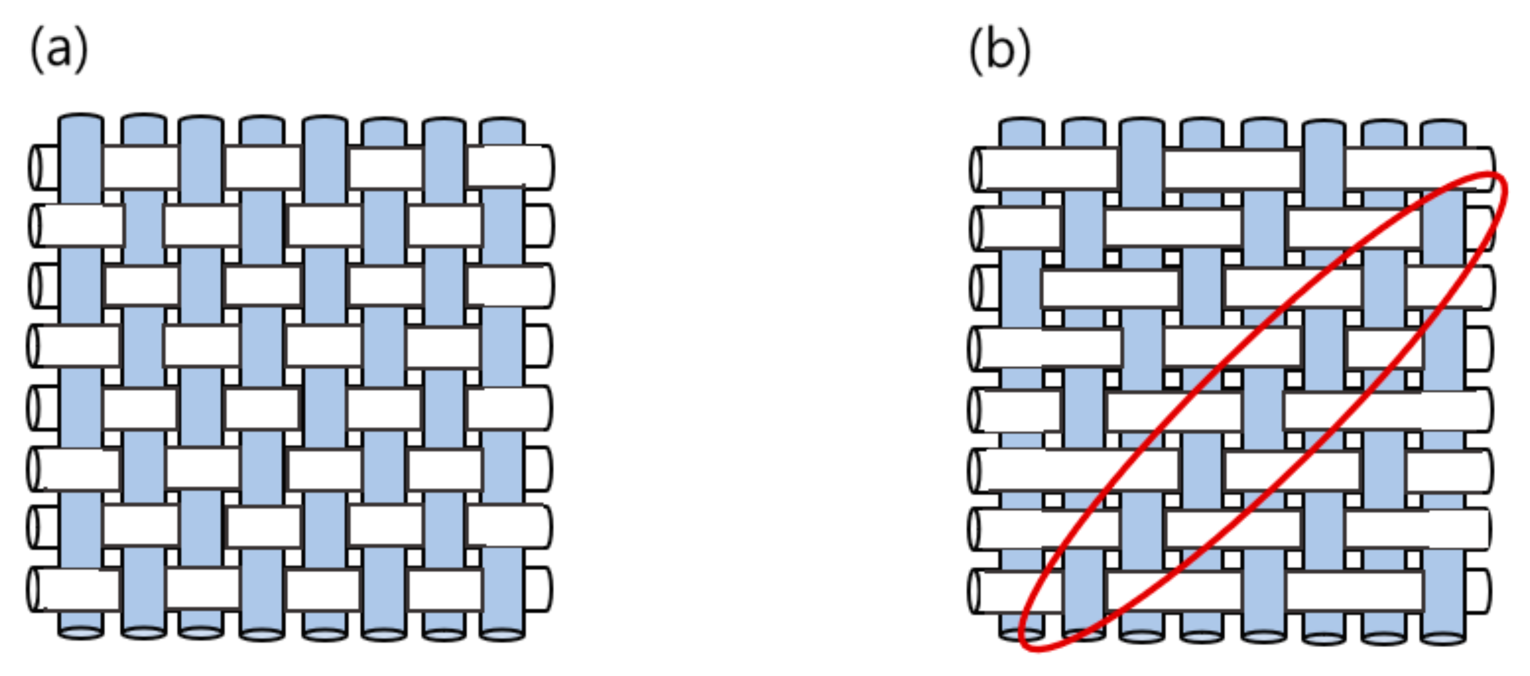
Figure 1. Fabric weaving methods. (a) Plain weave method, (b) Twill weave method.
Applsci 11 06475 g001
Figure 2. Tungsten yarn and fabric.
Applsci 11 06475 g002
Figure 3. High-density weaving process of shielding fiber.
Applsci 11 06475 g003
Figure 4. Tungsten nanopowder dispersion method.
Applsci 11 06475 g004
Figure 5. Method for evaluating the shielding performance of shielding fibers.
Applsci 11 06475 g005
Figure 6. Electron micrographs of the fabricated shielding fabrics. (a) Fabric without dispersed tungsten powder, (b) Fabric with dispersed tungsten powder.
Applsci 11 06475 g006
Figure 7. Enlarged electron microscope images of the fabrics. (a) Tungsten powder is not dispersed in the fabric, (b) Tungsten powder is dispersed.
Applsci 11 06475 g007
Figure 8. Spatial dose distribution inside the imaging room of a medical institution.
Applsci 11 06475 g008
Table 1. Characteristics of medical radiation shielding yarn and fabric.
PE-Tungsten Wire Composite YarnShielding Fiber
FinenessTensile
Strength
(g/d)
Elongation
at Break
(%)
Thread Count
(Thread/Inch)
Weight
(g/m2)
Thickness
(mm)
WarpWeft
501.11.454.28060112–1210.20–0.21
Table 2. Comparison of shielding rate of the two shielding fibers.
Radiation
Type
Effective
X-ray Energy
(keV)
Mean of Exposure (µR)Shielding Rate (%)
NothingTungsten
Fiber
Tungsten
Powder Fiber
Tungsten
Fiber
Tungsten
Powder Fiber
X-ray24.6106.9072.0155.4932.6448.09
28.7381.83284.2231.2725.5339.40
32.5799.70632.82545.8020.8731.75
48.51318.331082.01984.0017.9625.36
54.91648.331407.701312.6714.6020.36
Publisher’s Note: MDPI stays neutral with regard to jurisdictional claims in published maps and institutional affiliations.