
ZFTool: A Software for Automatic Quantification of Cancer Cell Mass Evolution in Zebrafish

 ,
,  ,
,  and
and 
Abstract
:1. Introduction
2. Materials and Methods
2.1. Material
2.2. Methods
3. Results
4. Discussion and Conclusions
Author Contributions
Funding
Institutional Review Board Statement
Informed Consent Statement
Conflicts of Interest
Abbreviations
| 5-FU | 5-Fluorouracil |
| dpf | Days postfertilization |
| GFP | Green-Fluorescent Protein |
| GMV | GFP medium value |
| hpf | Hours postfecundation |
| hpi | Hours postinjection |
| nGFP | Number of GFP pixels |
References
- Ali, S.; Champagne, D.; Spaink, H.; Richardson, M. Zebrafish embryos and larvae: A new generation of disease models and drug screens. Birth Defects Res. Part C Embryo Today 2011, 93, 115–133. [Google Scholar] [CrossRef] [PubMed]
- Drabsch, Y.; He, S.; Zhang, L.; Snaar-Jagalska, B.; Ten Dijke, P. Transforming growth factor-β signaling controls human breast cancer metastasis in a zebrafish xenograft model. Breast Cancer Res. 2013, 15, R106. [Google Scholar] [CrossRef]
- Mikut, R.; Dickmeis, T.; Driever, W.; Geurts, P.; Hamprecht, F.; Kausler, B.; Ledesma-Carbayo, M.; Marée, R.; Mikula, K.; Pantazis, P.; et al. Automated Processing of Zebrafish Imaging Data: A Survey. Zebrafish 2013, 10, 401–421. [Google Scholar] [CrossRef] [Green Version]
- Bakkers, J. Zebrafish as a model to study cardiac development and human cardiac disease. Cardiovasc. Res. 2011, 91, 279–288. [Google Scholar] [CrossRef] [Green Version]
- Mione, M.; Trede, N. The zebrafish as a model for cancer. Dis. Model. Mech. 2010, 3, 517–523. [Google Scholar] [CrossRef] [PubMed] [Green Version]
- Flinn, L.; Bretaud, S.; Lo, C.; Ingham, P.; Bandmann, O. Zebrafish as a new animal model for movement disorders. J. Neurochem. 2008, 106, 1991–1997. [Google Scholar] [CrossRef] [PubMed]
- Bentley, V.; Veinotte, C.; Corkery, D.; Pinder, J.; LeBlanc, M.; Bedard, K.; Weng, A.; Berman, J.; Dellaire, G. Focused chemical genomics using zebrafish xenotransplantation as a preclinical therapeutic platform for T-cell acute lymphoblastic leukemia. Haematologica 2015, 100, 70–76. [Google Scholar] [CrossRef]
- Jung, D.W.; Oh, E.S.; Park, S.H.; Chang, Y.T.; Kim, C.H.; Choi, S.Y.; Williams, D. A novel zebrafish human tumor xenograft model validated for anti-cancer drug screening. Mol. Biosyst. 2012, 8, 1930–1939. [Google Scholar] [CrossRef]
- Konantz, M.; Balci, T.; Hartwig, U.; Dellaire, G.; André, M.; Berman, J.; Lengerke, C. Zebrafish xenografts as a tool for in vivo studies on human cancer. Ann. N. Y. Acad. Sci. 2012, 1266, 124–137. [Google Scholar] [CrossRef]
- Veinotte, C.; Dellaire, G.; Berman, J. Hooking the big one: The potential of zebrafish xenotransplantation to reform cancer drug screening in the genomic area. Dis. Model. Mech. 2014, 7, 745–754. [Google Scholar] [CrossRef] [PubMed] [Green Version]
- Annila, T.; Lihavainen, E.; Marques, I.; Williams, D.; Yli-Harja, O.; Ribeiro, A. ZebIAT, an image analysis tool for registering zebrafish embryos and quantifying cancer metastasis. BMC Bioinform. 2013, 14, S5. [Google Scholar] [CrossRef] [PubMed] [Green Version]
- Teng, Y.; Xie, X.; Walker, S.; White, D.; Mumm, J.; Cowell, J. Evaluating human cancer cell metastasis in zebrafish. BMC Cancer 2013, 13, 453. [Google Scholar] [CrossRef] [PubMed] [Green Version]
- Ghotra, V.; He, S.; de Bont, H.; van der Ent, W.; Spaink, H.; van de Water, B.; Snaar-Jagalska, B.; Danen, E. Automated whole animal bio-imaging assay for human cancer dissemination. PLoS ONE 2012, 7, e31281. [Google Scholar] [CrossRef] [PubMed]
- Cabezas-Sainz, P.; Guerra-Varela, J.; Carreira, M.J.; Mariscal, J.; Roel, M.; Rubiolo, J.; Sciara, A.; Abal, M.; Botana, L.; López, R.; et al. Improving zebrafish embryo xenotransplantation conditions by increasing incubation temperature and establishing a proliferation index with ZFTool. BMC Cancer 2018, 18, 3. [Google Scholar] [CrossRef] [PubMed]
- Guerra-Varela, J.; Terriente, J.; Mariscal, J.; Cabezas-Sainz, P.; Calzolari, S.; Dyballa, S.; Abal, M.; López, R.; Sánchez, L. Assessing proliferation and metastasis of human cancer cells trhough 3D imaging. In Proceedings of the 7th Zebrafish Disease Models Conference, Madison, WI, USA, 28 June–1 July 2014. 100–P1. [Google Scholar]
- Kankaanpää, P.; Paavolainen, L.; Tiitta, S.; Karjalainen, M.; Päivärinne, J.; Nieminen, J.; Marjomäki, V.; Heino, J.; White, D. BioImageXD: An open, general-purpose and high-throughput image-processing platform. Nat. Methods 2012, 9, 683–689. [Google Scholar] [CrossRef] [PubMed]


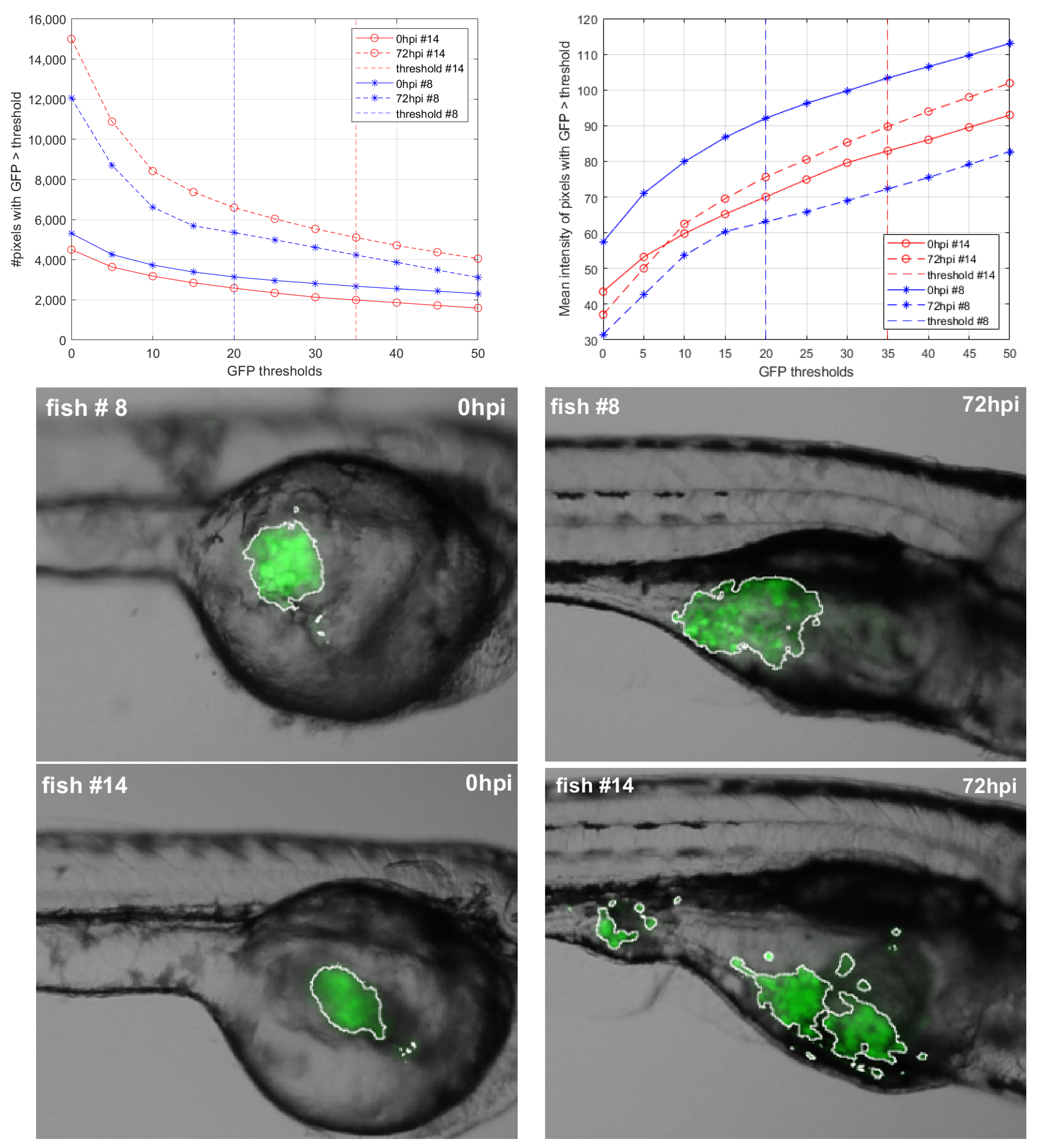
| Zebrafish # | Threshold | PI | ||||
|---|---|---|---|---|---|---|
| #8 | 20 | 3144 | 92.08 | 5349 | 63.07 | 1.16 |
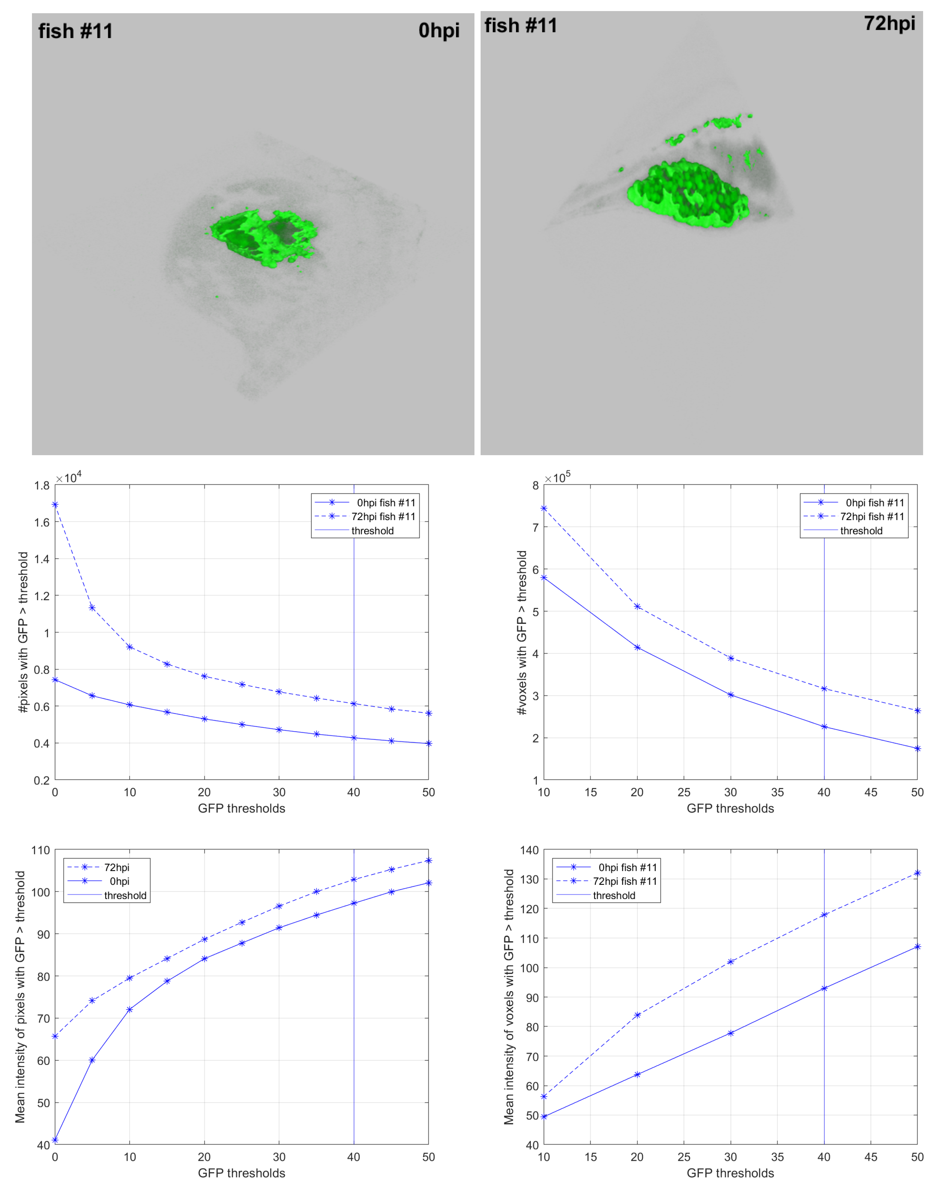
| #11 | 40 | 4279 | 97.25 | 6133 | 102.88 | 1.52 |
| #14 | 35 | 1991 | 82.94 | 5106 | 89.72 | 2.77 |
Publisher’s Note: MDPI stays neutral with regard to jurisdictional claims in published maps and institutional affiliations. |
© 2021 by the authors. Licensee MDPI, Basel, Switzerland. This article is an open access article distributed under the terms and conditions of the Creative Commons Attribution (CC BY) license (https://creativecommons.org/licenses/by/4.0/).
Share and Cite
Carreira, M.J.; Vila-Blanco, N.; Cabezas-Sainz, P.; Sánchez, L. ZFTool: A Software for Automatic Quantification of Cancer Cell Mass Evolution in Zebrafish. Appl. Sci. 2021, 11, 7721. https://doi.org/10.3390/app11167721
Carreira MJ, Vila-Blanco N, Cabezas-Sainz P, Sánchez L. ZFTool: A Software for Automatic Quantification of Cancer Cell Mass Evolution in Zebrafish. Applied Sciences. 2021; 11(16):7721. https://doi.org/10.3390/app11167721
Chicago/Turabian StyleCarreira, María J., Nicolás Vila-Blanco, Pablo Cabezas-Sainz, and Laura Sánchez. 2021. "ZFTool: A Software for Automatic Quantification of Cancer Cell Mass Evolution in Zebrafish" Applied Sciences 11, no. 16: 7721. https://doi.org/10.3390/app11167721
APA StyleCarreira, M. J., Vila-Blanco, N., Cabezas-Sainz, P., & Sánchez, L. (2021). ZFTool: A Software for Automatic Quantification of Cancer Cell Mass Evolution in Zebrafish. Applied Sciences, 11(16), 7721. https://doi.org/10.3390/app11167721