Article

Implications of Permanent Teeth Dimensions and Arch Lengths on Dental Crowding during the Mixed Dentition Period

by
Raisa Daoud
1,
Maria-Angelica Bencze
1,*,
Cristina-Crenguța Albu
2,*,
Elina Teodorescu
1,
Anca-Oana Dragomirescu
1,
Adriana Vasilache
1,
Ioana Suciu
3 and
Ecaterina Ionescu
1
1
Department of Orthodontics and Dentofacial Orthopaedics, Faculty of Dental Medicine, “Carol Davila” University of Medicine and Pharmacy, 020021 Bucharest, Romania
2
Department of Genetics, Faculty of Dental Medicine, “Carol Davila” University of Medicine and Pharmacy, 020021 Bucharest, Romania
3
Department of Endodontics, Faculty of Dental Medicine, “Carol Davila” University of Medicine and Pharmacy, 020021 Bucharest, Romania
*
Authors to whom correspondence should be addressed.
Appl. Sci. 2021, 11(17), 8004; https://doi.org/10.3390/app11178004
Submission received: 19 July 2021 / Revised: 25 August 2021 / Accepted: 27 August 2021 / Published: 29 August 2021
(This article belongs to the Section Applied Dentistry and Oral Sciences)

Abstract

:
Dento-alveolar disharmony with crowding is a common reason for orthodontic treatment with not fully understood or unequivocally demonstrated causes. This study investigated the correlations between teeth dimensions, arch lengths, and crowding during the mixed dentition period. A cross-sectional study on 100 dental casts of patients with class I malocclusions was performed. Dental arches were classified as non-crowded, moderately crowded, severely crowded, and spaced. The mesio-distal widths, bucco-lingual sizes, and crown proportions of permanent teeth were assessed. The results indicated that arch length measurements showed smaller values in crowded arches. The mesio-distal dimensions of upper central incisors and lower lateral incisors were larger in patients with crowding. The bucco-lingual dimensions of upper incisors were decreased, the bucco-lingual dimensions of lower central incisors and permanent first molars were increased in crowded arches. Upper incisors and lower lateral incisors presented larger crown proportions in crowding cases. Low negative correlations were found between mesio-distal diameters of maxillary central incisors, lower lateral incisors, lower permanent first molars, and the values of arch space discrepancies. In conclusion, crowding in the mixed dentition could be associated with reduced arch lengths, increase in mesio-distal sizes of incisors and lower permanent first molars, and variations of bucco-lingual dimensions and crown proportions of incisors and lower permanent first molars.

1. Introduction

Dento-alveolar incongruence with crowding is defined as the discrepancy between dental structures and jaw size, a condition responsible for malpositions and/or teeth rotations [1]. Al Darwish and Farh described dental crowding as the difference between the space needed for proper teeth alignment in the dental arch and the space available in that arch [2]. Crowding is a dynamic process that might be influenced by dental and paradental factors [3]. According to severity, crowding is classified as mild, moderate, or severe. Malocclusions involving dental crowding are much more common in the current population than those consisting of interdental spacing and widening of dental arches.
Dental crowding is not a disease in itself, but it can be considered a catalyst for various diseases such as: periodontal disease, dental caries, and dysfunctions of the temporomandibular joint [4]. Several studies in recent years have confirmed that severe malocclusion is likely to be a “social handicap” [5]. In a sample of Brazilian schoolchildren, dento-alveolar disharmony with crowding was the most common reason for patients to come to an orthodontic consultation, especially due to a high impairment of aesthetics [6].
However, the causes of crowded dento-alveolar incongruence are not fully understood or unequivocally demonstrated. Many authors discussed the involvement of combined etiological factors, recognizing that heredity, environment, ethnicity, and phylogenetic evolution of facial skeleton may play an important role [7,8,9]. Simplifying, dento-alveolar disharmony with crowding, as clinical expression of discrepancies between tooth size and jaw size, is determined by excessively large teeth, excessively small alveolar bones, or a combination of these two conditions.
Regardless of the causes of dento-alveolar disharmony with crowding, several studies have analyzed the extent to which dental size and maxillary size contribute to this pathology. Some authors concluded that groups of individuals with dental crowding have smaller sizes of dental arches compared to those of groups with physiological alignments [1,10].
Other studies indicated that teeth sizes are significantly linked to crowding along dental arches: Rhee and Nahm pointed out that sizes and morphology of incisors are related with crowding, Puri and his co-workers demonstrated that in a group with crowding, the teeth were significantly wider compared to those in the control group [11,12].
Arif and collaborators stated that both mesio-distal tooth diameters and dental arch width are responsible for the onset of crowding [13]. Radnzic concluded that neither mesio-distal crown diameter nor arch dimensions are significantly correlated with dental crowding; his results showed rather a complex interrelationship among cumulative mesio-distal crown dimensions, arch dimensions, and primary dental crowding [14].
Some studies suggested that the trend toward the reduction of the mandibular base size and the maintaining of the size of teeth may also be responsible for the increased number of crowding cases [15]. The majority of the previously mentioned studies were conducted on permanent dentition cases.
For diagnosis and treatment necessities, it is important to quantify the amount of crowding as early as possible using dental casts. Space analysis on dental casts is particularly useful in the mixed dentition period in order to evaluate the potential degree of crowding for children without skeletal problems and with good facial proportions [5]. With the development of technology, the metric analyses of the dental structures are increasingly reliable when performed with three-dimensional modelling software programs, in particular with CBCT, or with other diagnostic tools without continuous ionizing radiation, such as MRI [16]. In addition, the evaluation of the proportions of the face and the soft tissue facial profile measurements benefit from the use of wrapped CBCT images from non-standardized random frontal photographs [17].
Our research was aimed at:
  • analyzing the possible differences related to arch lengths and permanent teeth dimensions between crowded, non-crowded, and spaced arches in a sample of mixed dentition orthodontic patients without skeletal discrepancies;
  • evaluating the possible correlations between teeth morphology and dento-alveolar incongruence with crowding.

2. Materials and Methods

To achieve these goals, a cross-sectional study was conducted at the Orthodontic Department of “Carol Davila” University of Medicine and Pharmacy, Bucharest, Romania. A sample of 100 dental casts of orthodontic patients were selected. As a routine procedure, a signed informed consent for releasing diagnostic records for scientific purposes is always obtained from the patients or legal guardians of the patients prior to the beginning of treatment. The present study did not need ethic approval, as our research just used measurements performed on dental casts, not involving clinical activities on patients.
The selection of dental casts was performed by authors R.D. and M.-A.B. and met the following criteria:
  • Patient’s age between 7 and 10 years;
  • Class I malocclusions;
  • No prior history of orthodontic treatment;
  • Mixed dentitions with all incisors and permanent first molars fully erupted;
  • Dentitions without caries lesions, large coronal restorations, significant attrition, and dental anomalies (congenitally missing teeth, supernumerary teeth, tooth developmental disorders).
The exclusion criteria were related to: patient’s age (less than 7 years or more than 10 years), class II and class III malocclusions, previous history of orthodontic treatment, type of dentition (other than mixed dentition with all incisors and permanent first molars fully erupted), the presence of caries lesions, large coronal restorations, significant attrition, and dental anomalies.
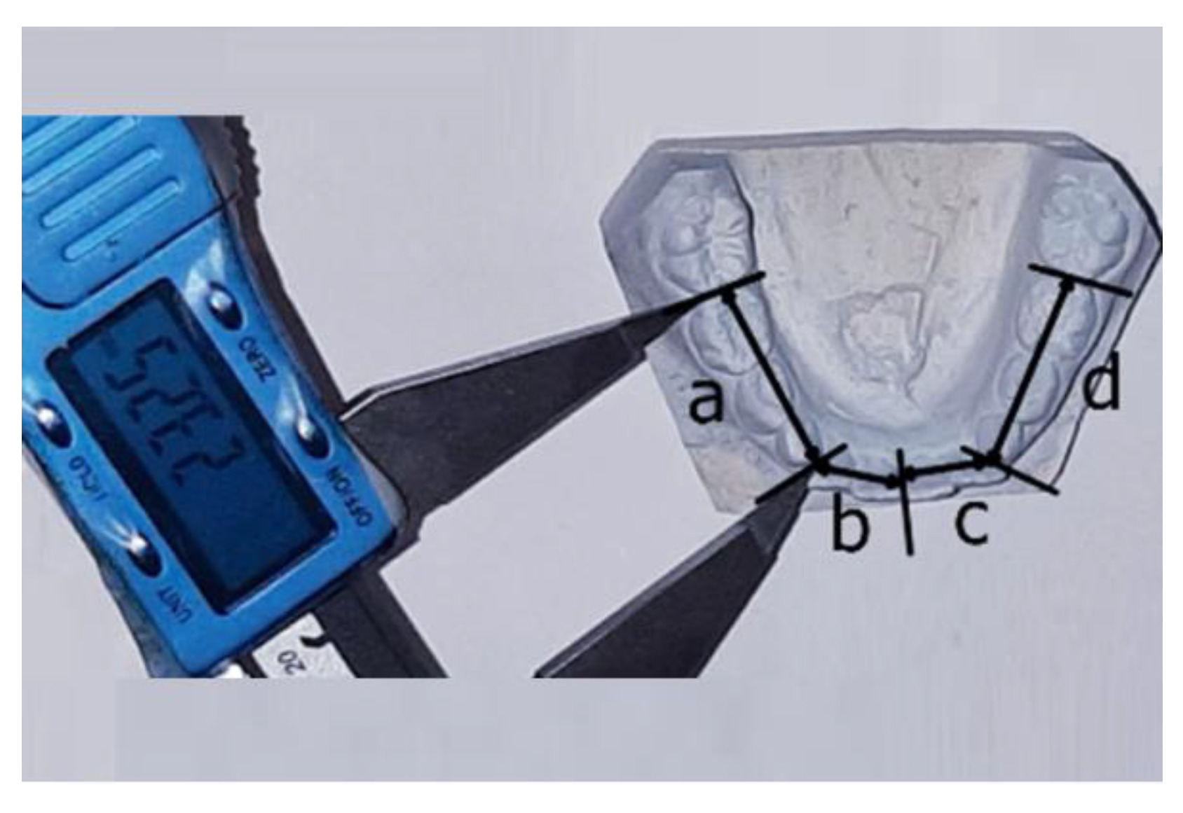
The space analysis was performed for all 200 dental arches (100 maxilla dental arches and 100 mandible dental arches), comparing the amount of space available for the alignment of the teeth and the amount of space required to properly align them [5]. The available space (arch perimeter) was measured from the mesial contact point of one first permanent molar to the other, over the contact points of posterior teeth and incisal edge of anterior teeth. The dental arch was divided into four segments, measured as straight-line approximations of the arch; the measurements were performed with a digital caliper with an accuracy of 0.01 mm (Figure 1 and Figure 2).
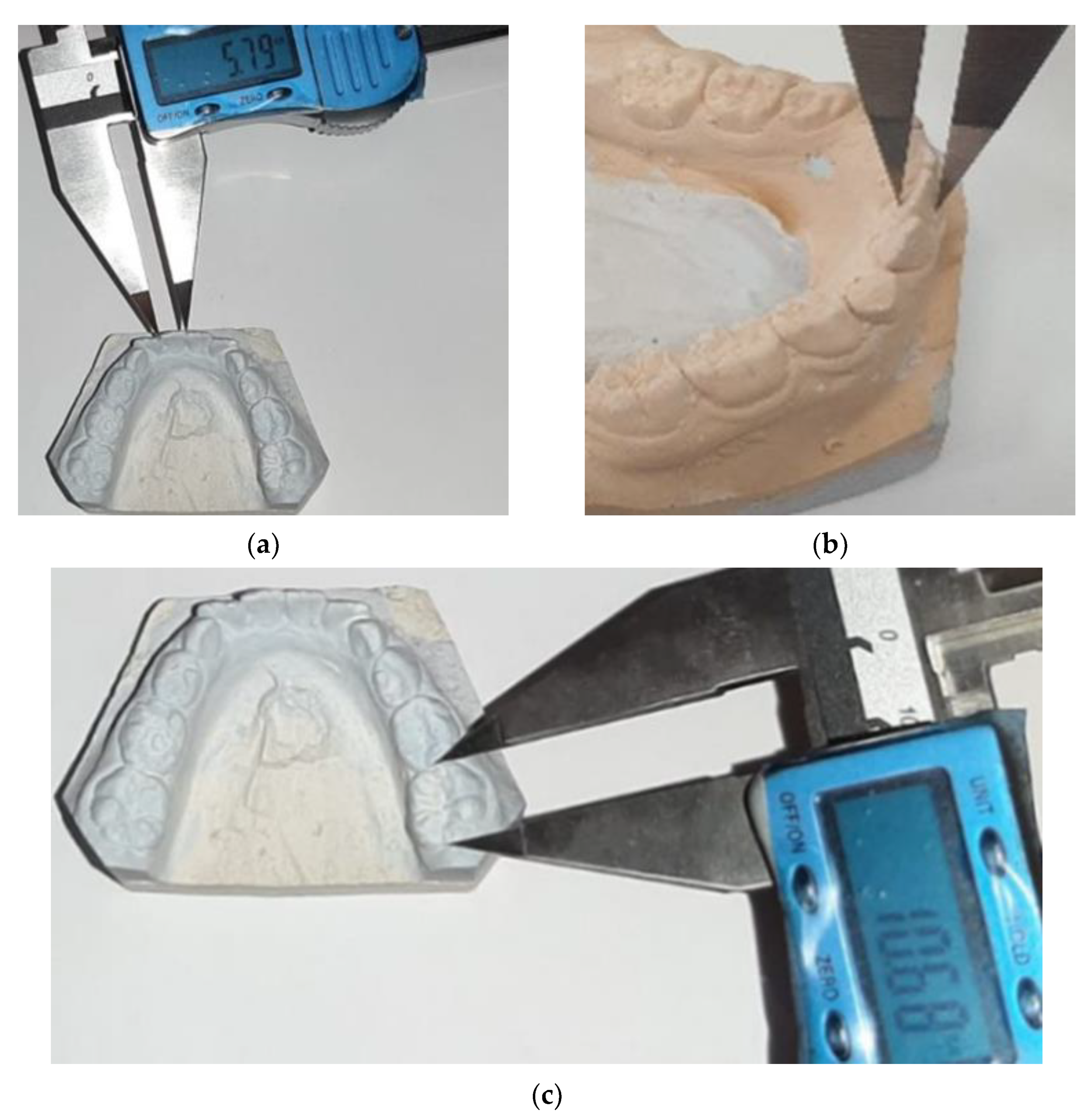
The amount of space required for alignment of the teeth was assessed by measuring the mesio-distal (MD) width of incisors and first permanent molars from contact point to contact point (Figure 3) by estimating the size of permanent canines and premolars according to Tanaka and Johnston prediction formulas and then by summing all these values [18].
The Tanaka and Johnston prediction formulas applied in this study are included in Table 1. The difference between the sum of the mesio-distal teeth dimensions and the arch perimeters value represents the space discrepancy.
For the purpose of this study, similarly with previously published studies, arches with arch length discrepancies of 0 ± 2 mm were considered normal, non-crowded (NCA); arches with a space discrepancy between −4 and −2 mm were defined as moderately crowded (MCA); arches with more than 4 mm space deficiency were labelled as severely crowded (SCA). If the sum of the widths of permanent teeth is more than 2 mm smaller than the available space within the arch, the arch is spaced (SpA) [19]. Maxillary and mandibular arches were classified separately.
In addition to mesio-distal widths of incisors and permanent first molars, bucco-lingual (BL) teeth sizes were also assessed. The mesio-distal crown diameter was measured at the greatest distance between the contact point of the approximal surfaces of the dental crown, and the bucco-lingual crown diameter was the greatest distance between the facial and lingual surfaces of the crown, taken at right angles to the plane of the mesio-distal diameter [20,21]. All measurements were taken in millimeters, by one examiner (R.D.), using the same digital caliper. For each permanent tooth, the MD/BL ratio was calculated using mesio-distal and bucco-lingual widths; this ratio is considered a representation of the crown proportion [22].
One week after the measurements were finished, a randomly selected group of 10 dental casts were measured again for errors depiction and the concordance between the groups of measurements, evaluated with Pearson correlation ratio, was high (r = 0.985, p < 0.01).
Statistical data analysis was performed using the PSPP free software; data were tested for assumption of normality (Kolmogorov–Smirnov test). Descriptive statistical analysis was performed, the mean and standard deviations were estimated for each variable in each group (normal, spaced arches, moderately crowded arches, severely crowded arches). Given the variation and dimorphism between genders, the data were evaluated separately for males and females. Group comparisons were performed with one-way analysis of variance (ANOVA), and post hoc Bonferroni test was applied to evaluate group differences in the crown and arch dimensions. The Pearson correlation parametric test was used to explore statistical correlations between dental parameter values and arch discrepancy values. The minimum probability level for accepting significance of the test results was set at 5% [23].

3. Results

The mean age of patients was 8.6 years; 68% of the selected cases were females and 32% were males. Twenty-four percent of maxillary arches and 40% of mandibular arches were normally aligned; 24% of upper arches and 30% of lower arches were spaced, and the rest were crowded (52% upper arches and 30% lower arches). Evaluating the degree of crowding in our study, the maxillary arches were more frequently affected by crowding than the mandibular ones. Regarding the extent of crowding, 30% of the upper arches were severely crowded, and 22% of the upper arches were moderately crowded; for the mandible, 14% and 16% presented severe and moderate crowding, respectively. There were no significant differences of crowding distribution between genders, but we found statistically significant differences related to the patients’ age (Table 2 and Table 3); the number of crowded arches decreased at nine and ten years of age.
The total available space was statistically significantly smaller in crowded cases at all ages for both upper and lower arches (Table 4).
Comparing teeth parameter values between genders, half of teeth diameters presented statistically significantly different values between male and female patients in all the four groups; therefore, in order to better interpret the study’s result, data concerning teeth morphology were analyzed separately for males and females. The MD dimensions (mean and standard deviation, SD) of permanent incisors and first permanent molars in normal, moderately crowded, severely crowded, and spaced arches are included in Table 5 for female and male patients (group comparisons performed with one-way analysis of variance ANOVA).
The subgroup differences in the MD teeth dimensions were investigated using the post hoc Bonferroni test; the p values, according to patient’s gender, are presented in Table 6. Splitting the data by the gender variable allowed us to discover statistically significant differences in the MD dimensions of upper central incisors between severely crowded and spaced maxillary arches, in the MD dimensions of upper first permanent molars between normal and spaced maxillary arches, and in the MD dimensions of lower lateral incisors between normal and moderately/severely crowded mandibular arches.
Table 7 presents the BL teeth dimensions (mean and standard deviation, SD) of permanent incisors and first permanent molars in normal, moderately crowded, severely crowded, and spaced arches for female and male patients.
The results of the post hoc Bonferroni test are included in Table 8; these results proved that: BL dimensions of upper central incisors were statistically significantly different in maxillary severely crowded arches and in non-crowded arches; BL dimensions of upper lateral incisors were statistically significantly different in maxillary moderately crowded arches and in spaced arches; BL dimensions of lower central incisors were statistically significantly different in mandibular severely crowded arches and in moderately crowded ones; BL dimensions of lower lateral incisors were statistically significantly different in mandibular moderately crowded arches and in spaced arches; BL dimensions of lower first permanent molars were statistically significantly different in mandibular severely crowded arches and in normal and spaced ones.
Crown proportions were different in normal, spaced, and crowded arches (Table 9). Upper incisors and lateral lower incisors presented statistically significantly larger crown proportions in arches with crowding than those in normal and spaced arches.
Table 10 includes a synthesis of the statistically significant results provided by the present study with respect to the involvement of teeth dimensions on the onset of crowding.
Statistical correlations between arch discrepancies and MD diameters of upper and lower incisors and first molars are presented in Table 11 for female and male patients. Statistically significant negative correlations were depicted for mesio-distal diameters of central maxillary incisors, lower lateral incisors, and lower first molars and the values of arch length discrepancies. The highest r values (around 0.4) were recorded for negative correlations between the mesio-distal diameters of maxillary incisors and maxillary arch discrepancy.

4. Discussion

Most authors claim the importance of analyzing the etiology and the most appropriate cause of the crowding. For an appropriate diagnosis and treatment strategy in orthodontic practice, it is necessary to identify, among etiological factors, the tooth size-arch size relationship with crowding [24].
Searching the medical literature database, we found that the majority of studies regarding the etiology of dental crowding were performed on permanent dentition patients. Correlations between teeth dimensions, arch lengths, and crowding have been previously explored with sometimes conflicting results. Little research was conducted on primary or mixed dentition cases. Therefore, the authors of the present study considered the investigation of crowding on mixed dentition cases useful. Our study evaluated a sample of dental casts from orthodontic patients without skeletal discrepancies during the period of mixed dentition, when real or transitional crowding is often encountered. Dental crowding during mixed dentition in most of cases is considered an early sign of treatment need, and early orthodontic treatment has been suggested [25,26].
Arch lengths and crown morphologies were assessed as possible determinant factors for crowding. Research data were collected from dental casts, using the procedure of space analysis for the assessment of crowding [5]. Measurements on dental casts were performed with a digital caliper. This method is still being used in similar studies [27]. A digital caliper with accuracy of 0.01 mm is considered by other specialists as a reliable method for the assessment of dental crowding; its accuracy was even compared to that of measurements on virtual computerized models with dedicated software [28]. Therefore, we appreciate that the analogical methods that are proposed in this study are extremely valid and repeated in all studies with similar objectives. Despite this, lately, the innovation and improvement of reworking software is allowing increasingly reliable and high-level evaluations based on multiplane 3D reconstructions, even in the treatment of severe malocclusions [29].
The 100 patients included in our study group met the inclusion criteria, with female orthodontic patients being more frequent (68% of the selected cases were females and 32% were males). The mean age of patients was 8.6 years. Conducting the space analysis for each dental arch, we found that 52% upper arches and 30% lower arches were crowded; 24% of maxillary arches and 40% of mandibular arches were normally aligned; 24% of upper arches and 30% of lower arches were spaced. There were no significant differences of crowding distribution between genders. Evaluating the degree of crowding in our study, the maxillary arches were more frequently affected by crowding than the mandibular ones. Regarding the extent of crowding, 30% of the upper arches were severely crowded, and 22% of the upper arches were moderately crowded. For the mandible, 14% and 16%, respectively, presented severe and moderate crowding. The number of crowded arches decreased at nine and ten years of age, with statistically significant differences related to patients’ age. This clinical fact can be explained through the natural process of arch length development with age. Our results partially agree with other published data: in a research conducted on 489 children from the general population at the onset of the mixed dentition period, anterior crowding was detected in the maxillary arch in 11.6% of cases and in the mandibular arch in 38.9% of the cases, with girls being more frequently affected by mandibular crowding than boys [25].
In the present analysis, arch length measurements in mixed dentition in the crowded groups constantly showed smaller values than those found among the subjects with normal and spaced arches. The comparison of arch lengths was made for each year of age (subgroups of patients aged 7, 8, 9, and 10 years). Except for the lower arches at seven years, all other results were statistically significant.
Similar results were published by other authors: Howe and co-workers conducted a study on 104 patients with permanent dentition arches (50 patients with gross dental crowding and 54 patients with little or no crowding) and concluded that the crowded group presented significantly smaller arch dimensions than the non-crowded group [12]. Melo et al. published the results of a retrospective study including 23 patients with primary dental arches (12 normal and 11 crowded cases). They concluded that the primary dental arch lengths of the subjects in the normal group tended to be larger than those found among the subjects in the crowded group [30]. The study conducted by Bernabé et al. on a sample of 150 students from Peru tried to determine which intra-arch occlusal variables are the best discriminators for dental arch crowding in the permanent dentition [31]. The results of the cited study showed that arch length is the most important individual factor for crowding in permanent dentition. A study in a Southern Indian population aimed to evaluate dental crowding in relation to the mesio-distal crown widths and arch dimensions and analyzed 132 CT scans of adult patients (genders equally represented, half crowded and half non-crowded), [32]. Its conclusions showed that in males, crowded dentition had statistically significantly larger mesio-distal tooth size and smaller arch dimensions; in females, the results were not statistically significant.
Our results sustain the hypothesis that dental crowding is frequently associated with smaller dental arches, so more attention should be paid to methods of increasing the size of dental arches. This is especially relevant in young patients with mixed dentition, when treatment modalities may include methods to stimulate the development of the jaws in order to properly align the teeth.
Related to teeth dimensions, the statistical analysis was performed separately for each gender; half of teeth diameters presented statistically significantly larger values in male than in female patients, regardless of crowding status. These gender disparities on crown dimensions are in agreement with previous studies [33]. The results of our research showed that the mesio-distal dimensions of upper central incisors were statistically significantly larger in female patients with severely crowded maxillary arches than in those with spaced ones, and the mesio-distal dimensions of lower lateral incisors were statistically significantly larger in male patients with moderately or severely crowded mandibular arches than in male patients with normally aligned lower arches. Analyzing the mesio-distal dimensions of upper first permanent molars, the results of our study were not statistically consistent, probable due to the relatively small sample size.
These results partially confirm some previously published data: a study conducted in India on a group of 90 patients aged 12–18 years and divided into three groups (no dental crowding, mild crowding, moderate crowding) identified that the coronary proportions differed significantly between the three groups only for the central maxillary incisor. Therefore, the authors concluded that the coronal dimensions of the central maxillary incisors are largely responsible for the variations observed in the analyzed dental crowding cases [34]. The study conducted by Bernabé and Flores-Mir also points to a causal relationship between the incisors’ coronary proportions and crowding, in a proportional sense (the larger the size of the incisor crown, the greater the degree of dental crowding) [35].
The research conducted by Radnzic on a study group comprising 60 British boys and 60 Pakistani immigrant boys found no relevant correlations between specific mesio-distal teeth dimensions and dental crowding [14]. However, the author stated that in combination with other parameters, the mesio-distal dimension contributes significantly to the total regression equation. This conclusion suggests the existence of a complex relationship between mesio-distal coronary dimensions, variations in the size of dental arches, and primary dental crowding, therefore, generalizations could not be made, and each arch must be assessed and treated individually.
A cross-sectional study conducted on a sample of 312 dental casts of Sudanese subjects aged 16–26 years (three subgroups of 104 casts with normal, crowded, and spaced dental arches) showed that subjects with dental crowding were more likely to have smaller dental arch measurements and increased tooth material than subjects with a normal arch [36].
In addition to variations of mesio-distal teeth dimensions, in the present study group, the bucco-lingual dimensions of upper incisors were smaller in maxillary crowded arches than in non-crowded and spaced arches (central incisors in female patients, lateral incisors in all patients), the bucco-lingual dimensions of lower central incisors were larger in severely mandibular crowded arches in male patients, the bucco-lingual dimensions of lower first permanent molars were larger in mandibular severely crowded arches than in normal and spaced ones in all patients.
These results partially agree with the assumption of Bora et al., who concluded that the bucco-lingual diameters of the central mandibular incisor, the mandibular and maxillary canine, and the maxillary premolars are the only bucco-lingual teeth diameters that can influence dental crowding [34]. The cited study concluded that the coronary proportions of the maxillary central incisors, the mesio-distal diameter of the maxillary lateral incisors, and the bucco-lingual dimension of mandibular central incisors seem to be largely responsible for variations in dental crowding.
Regarding crown proportions, expressed as the ratio between mesio-distal and bucco-lingual diameters, our study showed that upper incisors and lower lateral incisors presented statistically significantly larger crown proportions (due to increased mesio-distal diameter and/or decreased bucco-lingual diameter) in arches with crowding than in normal and spaced arches. The crown proportions of upper and lower first permanent molars were larger in patients with spaced arches than in those with normal arches. These findings do not support the conclusions of Shah et al., who analyzed a group of 50 untreated white subjects (25 men and 25 women) with varying degrees of crowding and found that mandibular incisor crown shape was no predictor of lower incisor crowding [37]. Our results are similar to the previous assumption of Peck, who included tooth shape among the determining factors in mandibular anterior crowding (persons with ideal incisor alignment were shown to have incisors with smaller mesio-distal and larger bucco-lingual dimensions than persons with incisor crowding) [38].
Analyzing the synthesis of the statistically significant results presented in Table 10, we can appreciate that the study results are consistent regarding crown size variations of upper incisors and lower lateral incisors in crowded dentitions. A statistically significant increase of mesio-distal diameter (for upper central incisors and lower lateral incisors) and a statistically significant decrease of bucco-lingual diameter (for upper incisors) were encountered, and both correlated with a statistically significant increase of crown proportions for each of these teeth. Related to lower central incisors and lower first permanent molars, their bucco-lingual diameter was statistically significantly larger in patients with crowded arches.
Exploring possible data correlations with Pearson test, low negative correlations were depicted for mesio-distal diameters of maxillary central incisors, lower lateral incisors, and lower permanent first molars and the values of arch length discrepancies. The highest r values were recorded for negative correlations between the mesio-distal diameters of maxillary incisors and maxillary arch discrepancy, followed by those for negative correlations between mesio-distal diameters of lower first molars and mandibular arch length discrepancy.
These results are similar to those of other studies that reported a correlation between the degree of lower incisor crowding and mesio-distal dimension of posterior teeth [39]. This association was interpreted as larger posterior teeth occupying more space in the dental arch, which may result in crowding; therefore, the area of posterior teeth may be considered as an important variable related to arch crowding [40].
In order to verify the present findings and to search for other possible statistically significant results, the authors consider it important to conduct future studies on groups consisting of young patients with permanent dentition.

5. Conclusions

According to the results of this investigation, crowding in mixed dentition can be associated with:
  • Maxillary and mandibular reduced dental arch lengths.
  • An increase in the mesio-distal size of upper central incisors, lower lateral incisors, and lower permanent first molars.
  • An increase in the bucco-lingual dimensions of lower central incisors and lower first permanent molars and a decrease in the bucco-lingual dimensions of upper incisors.
  • Larger crown proportions of upper incisors and lower lateral incisors.

Author Contributions

Conceptualization, R.D., M.-A.B., I.S., and E.I.; methodology, E.I., E.T., C.-C.A., and A.-O.D.; software, A.V., R.D., and M.-A.B.; validation, E.I., E.T., and C.-C.A., I.S.; formal analysis, A.V., M.-A.B., and A.-O.D.; investigation, R.D., E.T., and A.-O.D.; resources, M.-A.B., E.T., and E.I.; data curation, R.D., A.V., and I.S.; writing—original draft preparation, R.D., M.-A.B., and E.I.; writing—review and editing, E.T., C.-C.A., and I.S.; visualization, A.V. and A.-O.D.; supervision, E.I.; project administration, M.-A.B. and E.T. All authors contributed equally to this research. All authors have read and agreed to the published version of the manuscript.

Funding

This research received no external funding.

Institutional Review Board Statement

Ethical review and approval were waived for this study, as the research just used measurements performed on dental casts, not involving clinical activities on patients.

Informed Consent Statement

Informed consent was obtained from all subjects involved in the study.

Data Availability Statement

The data presented in this study are available on request from the corresponding author.

Conflicts of Interest

The authors declare no conflict of interests.

References

  1. Yan-Vergnes, W.; Vergnes, J.N.; Dumoncel, J.; Baron, P.; Marchal-Sixou, C.; Braga, J. Asynchronous dentofacial development and dental crowding: A cross-sectional study in a contemporary sample of children in France. J. Physiol. Anthropol. 2013, 32, 22. [Google Scholar] [CrossRef] [PubMed] [Green Version]
  2. Al Darwish, I.; Farh, H. Assessment of maxillary and mandibular dental arches dimensions in down’s syndrome children using digital study models. People 2019, 13, 18. [Google Scholar]
  3. Consolaro, A.; Cardoso, M.D.A. Mandibular anterior crowding: Normal or pathological? Dent. Press J. Orthod. 2018, 23, 30–36. [Google Scholar] [CrossRef] [PubMed]
  4. Hafez, H.S.; Shaarawy, S.M.; Al-Sakiti, A.A.; Mostafa, Y.A. Dental crowding as a caries risk factor: A systematic review. Am. J. Orthod. Dentofac. Orthop. 2012, 142, 443–450. [Google Scholar] [CrossRef]
  5. Proffit, W.R.; Fields, H.W.; Sarver, D.M.; Ackerman, J.L. Contemporary Orthodontics, 5th ed.; Elsevier Mosby: St. Louis, MO, USA, 2013; pp. 391–432. [Google Scholar]
  6. Marques, L.S.; Pordeus, I.A.; Ramos-Jorge, M.L.; Filogônio, C.A.; Filogônio, C.B.; Pereira, L.J.; Paiva, S.M. Factors associated with the desire for orthodontic treatment among Brazilian adolescents and their parents. BMC Oral Health 2009, 9, 34. [Google Scholar] [CrossRef] [PubMed] [Green Version]
  7. Normando, D.; Almeida, M.A.; Quintão, C.C. Dental crowding: The role of genetics and tooth wear. Angle Orthod. 2013, 83, 10–15. [Google Scholar] [CrossRef]
  8. Dávalos, L.M.; Cirranello, A.L.; Geisler, J.H.; Simmons, N.B. Understanding phylogenetic incongruence: Lessons from phyllostomid bats. Biol. Rev. 2012, 87, 991–1024. [Google Scholar] [CrossRef] [PubMed] [Green Version]
  9. Surdu, A.; Feier, R.; Matei, M.; Maris, M.; Ciupilan, C.; Romanec, C. Principles, Objectives and Therapeutic Measures in Dento-alveolar Malalignment in the Period of Growth. Rev. Chim. 2020, 71, 381–385. [Google Scholar] [CrossRef]
  10. Golwalkar, S.A.; Msitry, K.M. An evaluation of dental crowding in relation to the mesiodistal crown widths and arch dimensions. J. Indian Orthod. Soc. 2008, 42, 22–29. [Google Scholar] [CrossRef]
  11. Rhee, S.H.; Nahm, D.S. Triangular-shaped incisor crowns and crowding. Am. J. Orthod. Dentofac. Orthop. 2000, 6, 624–628. [Google Scholar] [CrossRef]
  12. Puri, N.; Pradhan, K.L.; Chandna, A.; Sehgal, V.; Gupta, R. Biometric study of tooth size in normal, crowded, and spaced permanent dentitions. Am. J. Orthod. Dentofac. Orthop. 2007, 132, 279.e7–279.e14. [Google Scholar] [CrossRef]
  13. Arif, A.N.; Rasheed, T.A.; Ali, A.J. Dental crowding and its relationship to tooth size and arch dimensions. J. Nat. Sci. Res. 2014, 4, 133–137. [Google Scholar]
  14. Radnzic, D. Dental crowding and its relationship to mesiodistal crown diameters and arch dimensions. Am. J. Orthod. Dentofac. Orthop. 1988, 94, 50–56. [Google Scholar] [CrossRef]
  15. Hussain, S.S.; Ashraf, B.; Khan, S. Relationship of dental crowding to tooth size and arch dimensions in class I normal & class I malocclusion sample. Pak. Oral Dent. J. 2014, 34, 660–664. [Google Scholar]
  16. Reda, R.; Zanza, A.; Mazzoni, A.; Cicconetti, A.; Testarelli, L.; Di Nardo, D. An Update of the Possible Applications of Magnetic Resonance Imaging (MRI) in Dentistry: A Literature Review. J. Imaging 2021, 7, 75. [Google Scholar] [CrossRef]
  17. Alhammadi, M.S.; Al-Mashraqi, A.A.; Alnami, R.H.; Ashqar, N.M.; Alamir, O.H.; Halboub, E.; Reda, R.; Testarelli, L.; Patil, S. Accuracy and Reproducibility of Facial Measurements of Digital Photographs and Wrapped Cone Beam Computed Tomography (CBCT) Photographs. Diagnostics 2021, 11, 757. [Google Scholar] [CrossRef]
  18. White, L.M.; Kirk, N.E.; Dean, J.A. Accuracy of Model Estimation versus Tanaka and Johnston Arch Length Analysis. J. Clin. Pediatr. Dent. 2021, 45, 54–57. [Google Scholar] [CrossRef]
  19. Faruqui, S.; Fida, M.; Shaikh, A. Comparison of tooth and arch dimensions in dental crowding and spacing. Pak. Orthod. J. 2012, 4, 48–55. [Google Scholar]
  20. Song, J.W.; Leesungbok, R.; Park, S.J.; Chang, S.H.; Ahn, S.J.; Lee, S.W. Analysis of crown size and morphology, and gingival shape in the maxillary anterior dentition in Korean young adults. J. Adv. Prosthodont. 2017, 9, 315–320. [Google Scholar] [CrossRef] [PubMed] [Green Version]
  21. Agenter, M.K.; Harris, E.F.; Blair, R.N. Influence of tooth crown size on malocclusion. Am. J. Orthod. Dentofac. Orthop. 2009, 136, 795–804. [Google Scholar] [CrossRef] [PubMed]
  22. Townsend, G.; Bockmann, M.; Hughes, T.; Brook, A. Genetic, environmental and epigenetic influences on variation in human tooth number, size and shape. Odontology 2012, 100, 1–9. [Google Scholar] [CrossRef]
  23. BeGole, E. Statistics for the Orthodontist. In Current Principles and Techniques, 4th ed.; Vig, K.W.L., Graber, T.M., Vanarsdall, R.L., Eds.; Elsevier, Mosby: St. Louis, MO, USA, 2005; pp. 391–405. [Google Scholar]
  24. Ionescu, E. Anomaliile Dentare si Dento-Maxilare; Editura Universitara “Carol Davila”: Bucharest, Romania, 2021; pp. 176–193. [Google Scholar]
  25. Keski-Nisula, K.; Lehto, R.; Lusa, V.; Keski-Nisula, L.; Varrela, J. Occurrence of malocclusion and need of orthodontic treatment in early mixed dentition. Am. J. Orthod. Dentofac. Orthop. 2003, 124, 631–638. [Google Scholar] [CrossRef] [PubMed]
  26. Gianelly, A.A. Treatment of crowding in the mixed dentition. Am. J. Orthod. Dentofac. Orthop. 2002, 121, 569–571. [Google Scholar] [CrossRef]
  27. Devakrishnan, D.; Gnansambandam, V.; Kandasamy, S.; Sengottuvel, N.; Kumaragurubaran, P.; Rajasekaran, M. Comparative study of tooth size and arch dimensions in class I crowded, proclined malocclusion and class I normal occlusion. J. Pharm. Bioallied Sci. 2021, 13, 783. [Google Scholar] [CrossRef]
  28. Zilberman, O.; Huggare, J.A.; Parikakis, K.A. Evaluation of the validity of tooth size and arch width measurements using conventional and three-dimensional virtual orthodontic models. Angle Orthod. 2003, 73, 301–306. [Google Scholar] [PubMed]
  29. Perrotti, G.; Baccaglione, G.; Clauser, T.; Scaini, R.; Grassi, R.; Testarelli, L.; Reda, R.; Testori, T.; Del Fabbro, M. Total Face Approach (TFA) 3D Cephalometry and Superimposition in Orthognathic Surgery: Evaluation of the Vertical Dimensions in a Consecutive Series. Methods Protoc. 2021, 4, 36. [Google Scholar] [CrossRef] [PubMed]
  30. Melo, L.; Ono, Y.; Takagi, Y. Indicators of mandibular dental crowding in the mixed dentition. Pediatr. Dent. 2001, 23, 118–122. [Google Scholar]
  31. Bernabé, E.; del Castillo, C.E.; Flores-Mir, C. Intra-arch occlusal indicators of crowding in the permanent dentition. Am. J. Orthod. Dentofac. Orthop. 2005, 128, 220–225. [Google Scholar] [CrossRef]
  32. Das, P.J.; Dkhar, W.; Pradhan, A. An evaluation of dental crowding in relation to the mesiodistal crown widths and arch dimensions in southern Indian population. J. Clin. Diagn. Res. 2017, 11, TC10–TC13. [Google Scholar] [CrossRef]
  33. Shahid, F.; Alam, M.K.; Khamis, M.F. Maxillary and mandibular anterior crown width/height ratio and its relation to various arch perimeters, arch length, and arch width groups. Eur. J. Dent. 2015, 9, 490. [Google Scholar] [CrossRef] [PubMed] [Green Version]
  34. Bora, M.; Chokotiya, H.; Banthia, A.; Sharma, M.; Majumder, P. Dental crowding and its relationship to dental morphology in an ethnic population. IJOCR 2014, 3, 63–67. [Google Scholar]
  35. Bernabé, E.; Flores-Mir, C. Dental morphology and crowding: A multivariate approach. Angle Orthod. 2006, 76, 20–25. [Google Scholar]
  36. Mustafa, R.A.; Abuaffan, A.H. Evaluation of Dental Crowding and Spacing in Relation to Tooth Size and Arch Dimensions in a Sample of Sudanese Adults. J. Contemp. Dent. Pract. 2021, 22, 253–258. [Google Scholar] [CrossRef] [PubMed]
  37. Shah, A.A.; Elcock, C.; Brook, A.H. Incisor crown shape and crowding. Am. J. Orthod. Dentofac. Orthop. 2003, 123, 562–567. [Google Scholar] [CrossRef]
  38. Peck, S. Crown dimensions and the alignment or crowding of mandibular incisors. Am. J. Orthod. Dentofac. Orthop. 2003, 124, 20A–21A. [Google Scholar] [CrossRef] [PubMed]
  39. Shah, A.A.; Elcock, C.; Brook, A.H. Posterior tooth morphology and lower incisor crowding. Dent. Anthropol. J. 2005, 18, 37–42. [Google Scholar] [CrossRef] [Green Version]
  40. Hashim, H.A.; Al-Ghamdi, S. Tooth width and arch dimensions in normal and malocclusion samples: An odontometric study. J. Contemp. Dent. Pract. 2005, 6, 36–51. [Google Scholar] [CrossRef]
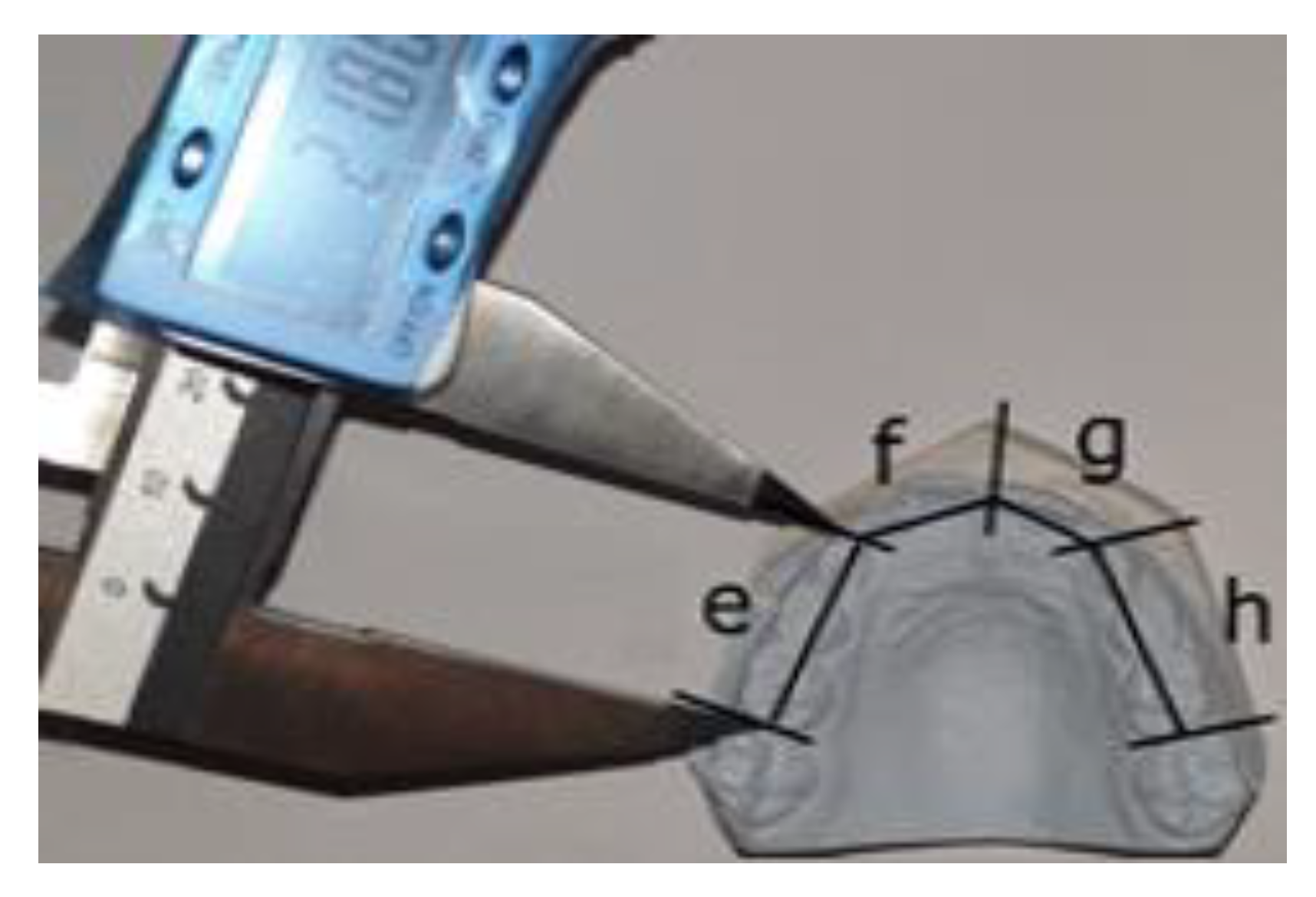
Figure 1. Measurement of the available space on maxillary cast: e = length between the mesial contact point of the upper right first permanent molar and the mesial contact point of the upper right canine, f = length between mesial contact point of the upper right canine and the midline; g = length between the midline and the mesial contact point of the upper left canine; h = length between the mesial contact point of the upper left canine and the mesial contact point of the upper left first permanent molar.
Applsci 11 08004 g001
Figure 2. Measurement of the available space on mandibular cast: a = length between the mesial contact point of the lower right first permanent molar and the mesial contact point of the lower right canine, b = length between mesial contact point of the lower right canine and the midline; c = length between the midline and the mesial contact point of the lower left canine; d = length between the mesial contact point of the lower left canine and the mesial contact point of the lower left first permanent molar.
Applsci 11 08004 g002
Figure 3. Procedures of measuring mesio-distal widths of permanent teeth exemplified through lower left central incisor (a), bucco-lingual dimensions exemplified through lower right central incisor (b), and mesio-distal widths of permanent teeth exemplified through lower right first permanent molar (c).
Applsci 11 08004 g003
Table 1. Tanaka and Johnston prediction formulas used to estimate the size of unerupted canines and premolars based on the width of the lower incisors (modified from [5]).
Data from Cast MeasurementsEstimated Width of Mandibular Canine and Premolars in One QuadrantEstimated Width of Maxillary Canine and Premolars in One Quadrant
Mesio-distal width of the
four lower incisors
½ of the mesio-distal width of the four lower incisors +10.5 mm½ of the mesio-distal width of the four lower incisors +11.0 mm
Table 2. Distribution of crowded, non-crowded, and spaced arches in the study group related to gender; not significant differences according to chi-square test.
ArchesNon-Crowded Arches (NCA)Moderately Crowded Arches (MCA)Severely Crowded Arches (SCA)Spaced Arches (SpA)p
MaxillaFemale (F): 18%F: 16%F: 20%F: 14%0.610 **
Male (M): 6%M: 6%M: 10%M: 10%
MandibleF: 28%F: 14%F: 10%F: 16%0.116 **
M: 12%M: 2%M: 4%M: 14%
** p > 0.05 not statistically significant.
Table 3. Distribution of crowded, non-crowded, and spaced arches in the study group related to age; significant differences among subgroups according to chi-square test.
ArchesNon-Crowded Arches (NCA)Moderately Crowded Arches (MCA)Severely Crowded Arches (SCA)Spaced Arches (SpA)p
Maxilla7 years old (y): 07 y: 4%7 y: 8%7 y: 2%
8 y: 10%8 y: 7%8 y: 9%8 y: 4%0.021 *
9 y: 4%9 y: 8%9 y: 8%9 y: 12%
10 y: 10%10 y: 3%10 y: 5%10 y: 6%
Mandible7 y: 4%7 y: 6%7 y: 2%7 y: 2%
8 y: 18%8 y: 4%8 y: 4%8 y: 4%0.014 *
9 y: 10%9 y: 2%9 y: 6%9 y: 14%
10 y: 8%10 y: 4%10 y: 2% 10 y: 10%
* p < 0.05 statistically significant.
Table 4. Total available space for upper and lower arches related to age; significant differences among subgroups according to ANOVA analysis.
Dental Arch
(N = 200)
Age Group
7 y8 y9 y10 y
Maxilla
(N = 100)
NCA
Mean/SD
73.47/3.4776.28/1.6773.40/4.52
MCA
Mean/SD
71.63/0.7871.40/1.8674.43/2.6975.99/3.63
SCA
Mean/SD
69.63/2.8569.85/1.8773.64/3.3573.88/2.86
SpA
Mean/SD
81.32/079.90/2.2479.02/3.6082.30/1.13
Total
Mean/SD
71.87/4.6272.76/4.0376.18/3.8276.05/5.01
p value<0.001 *<0.001 *0.003 *0.001 *
Mandible
(N = 100)
NCA
Mean/SD
67.92/4.0665.63/2.2268.15/2.9466.57/4.18
MCA
Mean/SD
63.42/2.2365.38/2.2766.82/063.02/3.04
SCA
Mean/SD
65.21/060.97/0.9166.03/1.8859.55/0
SpA
Mean/SD
69.69/070.97/0.8170.31/2.7372.44/1.56
Total
Mean/SD
65.86/3.4865.69/3.2468.61/3.0167.84/5.22
p value0.050<0.001 *0.013 *<0.001 *
* p < 0.05 statistically significant.
Table 5. Mesio-distal dimensions (mm) of permanent incisors and first permanent molars for female and male patients in the four groups (statistical significance according to ANOVA analysis).
Dental Arch
(N = 200)
Tooth
161211212226
Maxilla
(N = 100)
FMFMFMFMFMFM
NCA
Mean/SD
10.41/0.5110.08/1.156.59/0.756.78/0.868.56/0.458.57/1.198.59/0.428.51/0.906.77/0.386.91/0.4510.35/0.3910.10/1.20
MCA
Mean/SD
10.28/0.5610.20/0.626.96/0.507.16/0.438.61/0.359.12/0.488.62/0.399.00/0.696.90/0.466.94/0.4510.17/0.239.95/0.94
SCA
Mean/SD
10.11/0.5910.75/0.416.71/0.397.33/0.608.93/0.689.03/0.178.97/0.729.08/0.246.78/0.557.17/0.6510.09/0.5810.47/0.23
SpA
Mean/SD
10.14/0.3411.07/0.346.94/0.517.13/0.298.44/0.489.17/0.238.43/0.459.21/0.196.85/0.487.23/0.319.81/0.5511.05/0.68
p value0.2990.014 *0.1690.3040.039 *0.2280.025 *0.0790.8270.4770.017 *0.033 *
464241313236
FMFMFMFMFMFM
Mandible
(N = 100)
NCA
Mean/SD
10.43/0.7310.45/0.655.89/0.406.03/0.385.48/0.395.46/0.565.46/0.395.50/0.486.02/0.075.99/0.4310.29/0.5110.86/0.61
MCA
Mean/SD
10.15/0.8010.93/0.005.93/0.416.37/0.005.47/0.285.54/0.005.48/0.295.54/0.005.96/0.446.88/0.0010.14/0.6011.42/0.00
SCA
Mean/SD
10.44/0.4710.87/0.166.20/0.376.57/0.325.72/0.455.90/0.175.60/0.365.71/0.346.09/0.396.71/0.0410.54/0.5210.79/0.18
SpA
Mean/SD
10.33/0.5410.87/0.165.85/0.226.33/0.355.85/0.225.66/0.165.33/0.395.61/0.175.89/0.236.31/0.4110.32/0.8010.53/0.69
p value0.6250.5520.0970.049 *0.3220.2230.3330.7410.5340.004 *0.4740.225
* p < 0.05 statistically significant.
Table 6. Results of the post hoc Bonferroni test (p values of subgroup comparisons) aimed at comparing the MD dimensions of teeth between the four groups for female and male patients.
Dental ArchTooth
161211212226
FMFMFMFMFMFM
MaxillaN-MCA1.0001.0000.3561.0001.0000.6481.0000.6241.0001.0001.0001.000
N-SCA0.4900.2901.0000.3700.1790.8000.1720.2291.0001.0000.5061.000
N-SpA0.9050.029 *0.5151.0001.0000.3021.0000.0761.0001.0000.010 *0.135
MCA-SCA1.0000.6191.0001.0000.4181.0000.2841.0001.0001.0001.0001.000
MCA-SpA1.0000.0731.0001.0001.0001.0001.0001.0001.0001.0000.2290.054
SCA-SpA1.0001.0001.0001.0000.049 *1.0000.027 *1.0001.0001.0000.5480.602
464241313236
FMFMFMFMFMFM
MandibleN-MCA1.0001.0001.0001.0001.0001.0001.0001.0001.0000.035 *1.0001.000
N-SCA1.0001.0000.1440.0840.6820.3101.0001.0001.0000.020 *1.0001.000
N-SpA1.0001.0001.0000.2231.0001.0001.0001.0001.0000.2991.0001.000
MCA-SCA1.0001.0000.4711.0000.7891.0001.0001.0001.0001.0000.7111.000
MCA-SpA1.0001.0001.0001.0001.0001.0001.0001.0001.0000.3701.0000.381
SCA-SpA1.0001.0000.1221.0000.5021.0000.4431.0001.0000.4511.0001.000
* p < 0.05 statistically significant.
Table 7. Bucco-lingual dimensions (mm) of permanent incisors and first permanent molars for female and male patients in the four groups (statistical significance according to ANOVA analysis).
Dental Arch
(N = 200)
Tooth
161211212226
FMFMFMFMFMFM
Maxilla (N = 100)NCA
Mean/SD
11.15/0.5811.49/0.865.85/1.074.34/0.776.64/0.906.10/0.466.87/0.946.28/0.416.09/0.904.42/1.5710.97/0.6111.19/0.67
MCA
Mean/SD
10.90/0.6511.08/1.015.12/0.824.47/1.906.05/1.105.99/0.846.15/1.086.01/0.895.14/0.954.66/2.1110.96/0.4310.82/0.92
SCA
Mean/SD
10.69/0.5811.53/0.204.87/1.695.77/0.585.67/1.516.75/0.635.63/1.456.76/0.365.06/1.525.61/0.8610.79/0.5911.54/0.45
SpA
Mean/SD
10.97/0.4511.49/0.645.80/0.716.84/0.636.43/0.696.53/1.696.44/0.636.71/1.435.77/0.516.78/0.9010.89/0.6511.39/0.38
p value0.1110.5940.0720.002 *0.0560.4990.009 *0.3750.026*0.037 *0.7610.127
464241313236
FMFMFMFMFMFM
Mandible (N = 100)NCA
Mean/SD
10.62/0.4910.46/0.865.17/1.024.70/0.755.45/0.725.40/0.755.51/0.575.46/0.615.28/1.074.44/1.4210.69/0.4610.29/0.66
MCA
Mean/SD
10.33/0.6511.14/0.004.82/1.583.95/0.005.44/0.885.15/0.005.57/0.704.69/0.004.72/1.594.51/0.0010.40/0.7911.19/0.00
SCA
Mean/SD
10.89/0.4511.13/0.125.41/0.725.49/0.805.81/0.586.13/0.116.01/0.466.27/0.015.24/0.665.16/0.4310.84/0.5011.29/0.07
SpA
Mean/SD
10.42/0.4411.07/0.345.75/0.505.39/1.455.56/0.286.03/0.855.70/0.325.90/0.745.82/0.625.47/1.1310.22/0.4610.90/0.38
p value0.043 *0.0550.1040.2010.4830.0980.0860.020 *0.0570.1810.015 *0.003 *
* p < 0.05 statistically significant.
Table 8. Results of the post hoc Bonferroni test (p values of subgroup comparisons) aimed at comparing the BL dimensions of teeth between the four groups for female and male patients.
Dental ArchTooth
161211212226
FMFMFMFMFMFM
MaxillaN-MCA1.0001.0000.5171.0000.7851.0000.3631.0000.0681.0001.0001.000
N-SCA0.0971.0000.1820.1590.0621.0000.006 *1.0000.0930.8961.0001.000
N-SpA1.0001.0001.0000.005 *1.0001.0001.0001.0001.0000.0651.0001.000
MCA-SCA1.0001.0001.0000.2561.0001.0000.9970.7891.0001.0001.0000.142
MCA-SpA1.0001.0000.7330.008 *1.0001.0001.0000.9560.5910.1211.0000.415
SCA-SpA1.0001.0000.2730.3400.3501.0000.2281.0000.5980.6571.0001.000
464241313236
FMFMFMFMFMFM
MandibleN-MCA0.5380.8571.0001.0001.0001.0001.0000.7360.6941.0000.7220.139
N-SCA0.9040.3671.0001.0000.8890.6030.0810.2041.0001.0001.0000.009 *
N-SpA1.0000.0820.4900.7831.0000.2501.0000.5120.6690.2190.0540.023 *
MCA-SCA0.0601.0001.0000.7631.0000.8320.2970.044 *1.0001.0000.3481.000
MCA-SpA1.0001.0000.1050.6071.0000.7801.0000.1020.040 *1.0001.0001.000
SCA-SpA0.1521.0001.0001.0001.0001.0000.8851.0001.0001.0000.040 *1.000
* p < 0.05 statistically significant.
Table 9. Comparison of crown proportions between normal, spaced, and crowded arches for female and male patients (statistical significance according to ANOVA analysis).
Dental Arch
(N = 200)
MD/BL Tooth Ratio
161211212226
Maxilla (N = 100) FMFMFMFMFMFM
NCA
Mean/SD
0.93/0.030.88/0.061.16/0.291.53/0.021.32/0.221.40/0.101.27/0.191.35/0.081.13/0.151.66/0.470.95/0.060.90/0.05
MCA
Mean/SD
0.95/0.080.92/0.031.42/0.241.87/0.711.47/0.271.55/0.211.44/0.271.52/0.201.43/0.381.75/0.690.93/0.040.92/0.06
SCA
Mean/SD
0.95/0.030.93/0.031.59/0.671.30/0.151.68/0.461.35/0.141.68/0.391.35/0.091.43/0.371.30/0.160.94/0.030.91/0.04
SpA
Mean/SD
0.93/0.050.97/0.061.21/0.111.03/0.061.32/0.121.52/0.531.32/0.111.43/0.331.20/0.141.08/0.170.90/0.050.97/0.06
p value0.6170.006 *0.017 *0.003 *0.002 *0.550<0.001 *0.4140.010 *0.031 *0.0620.041 *
464241313236
Mandible (N = 100) FMFMFMFMFMFM
NCA
Mean/SD
0.98/0.061.00/0.041.18/0.251.31/0.201.02/0.151.02/0.081.00/0.091.01/0.051.19/0.281.57/0.750.96/0.041.06/0.08
MCA
Mean/SD
0.98/0.060.98/01.41/0.661.61/01.03/0.181.08/01.00/0.111.18/01.45/0.661.53/00.98/0.061.02/0
SCA
Mean/SD
0.96/0.030.98/01.17/0.181.22/0.241.00/0.170.96/0.010.94/0.120.91/0.061.18/0.181.31/0.120.97/0.020.96/0.02
SpA
Mean/SD
0.99/0.050.95/0.071.03/0.101.26/0.360.98/0.080.96/0.120.94/0.110.97/0.141.02/0.121.19/0.211.01/0.060.97/0.08
p value0.4980.1690.033 *0.4210.7370.2350.1860.023 *0.020 *0.2920.027 *0.024 *
* p < 0.05 statistically significant.
Table 10. Statistically significant differences in crown parameters between patients with crowded arches and patients with normally aligned arches in the study group according to ANOVA and post hoc Bonferroni analysis.
Crown ParametersUpper Central IncisorUpper Lateral IncisorUpper First Permanent MolarLower Central IncisorLower Lateral IncisorLower First Permanent Molar
Mesio-distal (MD) diameterIncrease
(F patients)
n.s.Increase (F patients)
Decrease
(M patients)
n.s.Increase
(M patients)
n.s.
Bucco-lingual (BL) diameterDecrease
(F patients)
Decrease
(F, M patients)
n.s.Increase
(M patients)
n.s.Increase
(F, M patients)
MD/BL ratioIncrease
(F patients)
Increase
(F, M patients)
n.s.n.s.Increase
(F patients)
n.s.
n.s.—not statistically significant differences.
Table 11. Statistical correlations between MD teeth dimensions and arch discrepancies for female and male patients (Pearson correlation test).
Dental Arch
(N = 200)
161211212226
Maxillary
arch discrepancy
FMFMFMFMFMFM
r = 0.029r = 0.213r = 0.045r = −0.113r = −0.383r = 0.074r = −0.383r = 0.100r = −0.010r = 0.082r = −0.172r = 0.247
p = 0.817p = 0.242p = 0.716p = 0.537p = 0.001 *p = 0.688p = 0.001 *p = 0.588p = 0.939p = 0.656p = 0.160p = 0.172
464241313236
Mandibular
arch discrepancy
FMFMFMFMFMFM
r = −0.005r = −0.369r = −0.341r = −0.135r = −0.186r = −0.072r = −0.224r = −0.108r = −0.233r = −0.304r = −0.052r = −0.339
p = 0.966p = 0.038 *p = 0.004 *p = 0.461p = 0.129p = 0.694p = 0.066p = 0.555p = 0.056p = 0.091p = 0.677p = 0.058
* p < 0.05 statistically significant.
Publisher’s Note: MDPI stays neutral with regard to jurisdictional claims in published maps and institutional affiliations.