Review

Analytical Methods for Exploring Nutraceuticals Based on Phenolic Acids and Polyphenols

1
Department of Chemical Engineering and Analytical Chemistry, University of Barcelona, 08028 Barcelona, Spain
2
Research Institute in Food Nutrition and Food Safety, Recinte Torribera, University of Barcelona, 08921 Santa Coloma de Gramenet, Spain
3
Serra Húnter Lecturer, Generalitat de Catalunya, 08007 Barcelona, Spain
*
Author to whom correspondence should be addressed.
Appl. Sci. 2021, 11(18), 8276; https://doi.org/10.3390/app11188276
Submission received: 20 July 2021 / Revised: 29 August 2021 / Accepted: 31 August 2021 / Published: 7 September 2021
(This article belongs to the Special Issue Nutraceuticals: Food and Nutritional Applications)

Abstract

:

Featured Application

The relevance of phenolic acids, flavonoids, and other polyphenols as the biologically active components of nutraceuticals is pointed out here. The principal analytical approaches to deal with their characterization and quantification have been introduced as well, with liquid chromatography being the technique of choice in most cases.

Abstract

Phenolic compounds such as phenolic acids, flavonoids, and stilbenes comprise an enormous family of bioactive molecules with a range of positive properties, including antioxidant, antimicrobial, or anti-inflammatory effects. As a result, plant extracts are often purified to recover phenolic compound-enriched fractions to be used to develop nutraceutical products or dietary supplements. In this article, we review the properties of some remarkable plant-based nutraceuticals in which the active molecules are mainly polyphenols and related compounds. Methods for the characterization of these extracts, the chemical determination of the bioactivities of key molecules, and the principal applications of the resulting products are discussed in detail.

1. Introduction

The consumption of nutraceutical products and functional foods has experienced enormous growth in the last decade due to the crucial role to help prevent some diseases, as well as the positive effects for human health associated with the bioactive compounds that they contain [1,2,3]. Among the different groups of phytochemicals, polyphenols are especially remarkable for several reasons, namely: (i) they are responsible for some organoleptic attributes of natural products, such as color, astringency, or bitterness [4]; (ii) they are important descriptors of features of plants and derived products to be used as biomarkers for quality control, classification, and authentication purposes [5]; (iii) there is solid evidence that the consumption of phenolic compounds found in natural foods may lower the risk of suffering health disorders thanks to their antioxidant capacities [2,3,6]. The last point is the most important regarding this paper as it refers to the beneficial and healing properties of this great family of molecules.
Polyphenols comprise a large family of secondary metabolites of plants that are especially abundant in red berries, chocolate, tea, wine, and fresh fruits, displaying concentrations that may vary from milligrams to grams per kilogram depending on the cases. The regular intake of these foods constitutes the main source of polyphenols for the human diet. However, given the great interest of polyphenols, their natural sources are often exploited to obtain enriched and/or purified extracts with potential applications to cosmetics, nutraceuticals, food supplements, and medicines.
In particular, polyphenols may protect the organism against oxidative damage, thus limiting the risk of several degenerative diseases related to the harmful action of free radicals such as cancer, diabetes, and osteoporosis [2,6]. Beyond this generic antioxidant action, more specific antiviral, antimicrobial, or anti-inflammatory properties attributed to given compounds have been reported as well [7,8,9,10]. Specific examples are given in the following sections.

2. Polyphenols and Related Compounds

They are primarily synthesized as signaling and defensive molecules against external stressors, such as climatic and environmental factors (drought, high temperature, solar radiation, and pollution) and pathogens, thus allowing plants to respond promptly to multiple aggressions of different origins [11,12]. Strictly speaking, polyphenols consist of organic compounds containing several phenolic groups. Other related compounds are sometimes included under this generic designation so that, in a broader context, the family of polyphenols and related compounds is as follows: phenolic acids, flavonoids, stilbenes, lignans, as well as a more heterogeneous group of structurally related compounds such as chalcones, humulones, and alcohols [12,13].
The number of polyphenol molecules that have been reported, identified, or characterized is close to 9000. A brief explanation of the four main classes is given as follows (see Figure 1):
(i)
Phenolic acids comprise hydroxybenzoic and hydroxycinnamic acids. They account for 30% of the total dietary phenolics, and, in general, cinnamic derivatives, such as caffeic, ferulic, and coumaric acids, are more abundant than the benzoic ones. Tartaric esters of these acids (e.g., caftaric and coutaric acids) are especially abundant in grape, vine, and wines. In addition, quinic acid derivatives such as chlorogenic acids are present in a broad range of products (e.g., coffee, tea, and pear). Phenolic acids also include hydrolyzable tannins, which consist of sugar residues esterified with gallic or ellagic acids. This type of tannin can be found at 10 to 100 mg kg−1 levels in coffee, fruits, nuts, tea, and wine.
(ii)
Flavonoids are the largest family of polyphenols both qualitatively and quantitatively as more than 5000 different molecules have been described, accounting for 60% of the total dietary polyphenols in humans [14]. The flavonoid backbone consists of two aromatic rings connected through a linking heterocycle with an oxygen and three carbon atoms (C6-C3-C6 skeleton). Flavonoids can be divided into six subclasses, namely: flavonols, flavones, isoflavones, flavanones, anthocyanidins, and flavanols. Additional details are given below.
Flavonols are based on the hydroxyflavone backbone in which the phenyl residues can be variedly hydroxylated (or methoxylated), thus resulting in various remarkable aglycones such as quercetin, kaempferol, fisetin, and morin [15,16,17]. In nature, they are predominantly linked to sugars as a way to enhance their solubility in intra- and extracellular media. Hence, glucosides, galactosides, rhamnosides, and rutinosides are frequently found in plant matrices. Although flavonols are found at low concentrations, they play fundamental roles in growth regulation mechanisms.
Flavones consist of the 2-phenylchromen-4-one backbone, structurally similar to flavonols but without the hydroxyl group [18,19]. Some representative molecules are apigenin, luteolin, and tangeritin, generally occurring at levels below 1 mg kg−1 in vegetables such a broccoli, celery, carrot, parsley, or spinach. C- and O-glycosides have been reported in cereals such as sorghum and millet at levels from 0.05 to 10 mg kg−1.
Isoflavones are also based on the 2-phenylchromen-4-one backbone, differing from the flavones in the position of the phenyl group. Isoflavones are almost specific to the Fabaceae family, occurring at concentrations of 1 mg kg−1 in soybean, chickpea, peanut, and sunflower seeds [20,21]. Some typical aglycones are daidzin and genistin, often glycosided to a high extent. Despite their low concentrations in natural sources, they have great interest due to their structural resemblance with steroid hormones. Hence, isoflavones act as estrogens to alleviate postmenopausal effects (e.g., hot flashes and bone loss) in women and to lower cholesterol levels in blood.
Flavanones consist of flavan-4-one structures without the double bond in the heterocycle so that they have chiral carbons. As in the other cases, phenyl rings display varied hydroxylation (and methoxylation), with some representative aglycones being hesperetin, naringenin, taxifolin, and eriodictyol. They are predominantly combined with saccharides, resulting in compounds such as hesperidin, naringin, or narirutin. Flavanones are the main polyphenols in species of the Citrus genus, reaching concentrations of 100 to 3000 mg kg−1 [22]. The main flavanone bioactivities deal with the antioxidant and radical scavenging power, thus being used as prophylactics to reduce the risk of cancer and cardiovascular diseases.
Anthocyanidin aglycones and anthocyanin glycosides are charged species containing the flavylium cation (the oxygen of the heterocycle is an oxonium cation). The principal aglycones (e.g., cyanidin, delphinidin, malvidin, and pelargonidin) are often attached to sugars. They are the largest group of pigments found in nature responsible for the cyan and red colors of berries, grapes, tomatoes, and wine [23].
Flavanols, also referred to as flavan-3-ols or catechins, are one of the most important families of polyphenols from both quantitative and qualitative issues [24,25,26]. Flavanols are present in high amounts in cocoa, coffee, fruits (berries, grapes, pears, and apples), legumes (peas, lentils, and beans), nuts (walnut, hazelnut, and peanut), and tea. For example, overall concentrations in fresh fruits are about 50 to 250 mg kg−1, reaching even higher values in green tea (ca. 700 mg kg−1) and cocoa (ca. 2000 mg kg−1). As flavanones, the heterocycle lacks the double bond so that they have chiral carbons. (−)-Epicatechin and (+)-catechin, (−)-epigallocatechin, (−)-epicatechin gallate, and (−)-epigallocatechin gallate are the most remarkable flavanols. They are prone to condense to form the so-called proanthocyanidins (PACs) or condensed tannins, which consist of two or several flavonoid moieties attached in different ways, thus resulting in dimers, trimers, and larger oligomers [9,27]. Monomers are often connected via the C4 of the upper unit with the C6 or C8 of the lower counterpart (B-type bond). However, in specific cases, they have an additional link between the C2 of the upper monomer and the hydroxyl group of C5 or C7 of the lower one (i.e., C2-O-C5 or C2-O-C7), thus resulting in the so-called A-type bond. As detailed below, B-type compounds are more abundant in nature but the A-type ones possess a noticeable antimicrobial activity [28].
(iii)
Stilbenes are characterized by a double-bond connecting two aromatic rings. Despite being found in low quantities in the human diet, the significance of compounds such as trans-resveratrol is outstanding. Apart from the more controversial antiaging effects, chemopreventive, chemotherapeutic, cardioprotective, and neuroprotective benefits have been attributed to resveratrol and its derivatives [29]. Curcuminoids are structurally related stilbenoids exhibiting a great range of nutritional and health beneficial properties [30]. Their structure has been used as the basis to design new drugs for the treatment of several types of cancers and microbial infections [30].
(iv)
Lignans are a minor class of polyphenols consisting of two phenylpropane units. The main food source of lignans is linseed, although they are also found at lower concentrations in cereals, fruits, and vegetables [31]. Recently, lignans have attracted the attention of researchers because of their potential anti-inflammatory, anti-neurodegenerative, antiviral, antimicrobial, and phytoestrogenic activities.

3. Nutraceuticals from Plant Extracts

A type of trendy nutraceutical and dietary supplement consists of mixtures of fruit and medicinal plant extracts such as American cranberry and other red and blackberries, turmeric, grapevine, and green tea, with exceptional detoxifying, antiaging, and antioxidant features [32].
American cranberry (Vaccinium macrocarpon) is rich in a wide variety of phytochemicals such as saccharides, carotenoids, phenolic acids, and flavonoids [33,34]. Fresh berries, raisins, and powdered extracts have been used as natural remedies to treat inflammatory and microbial processes. Other properties have also been recognized, including antineoplastic, cardioprotective, or antidegenerative activities [35,36]. Regarding antimicrobial issues, A-type PACs are the main compounds to be used for the prophylaxis and treatment of urinary tract infections (UTIs). The bioactivity of these species is attributed to their planar structure, which may create a protecting barrier on the urinary tract walls, thus avoiding the adhesion of bacteria [9]. Cranberry extracts are sometimes combined with other medicinal plants such as Salvia officinalis, Urtica urens, or Solidago virgaurea that provide additional anti-inflammatory and diuretic properties to facilitate the excretion of urine.
Another group of nutraceuticals is focused on products to reduce weight and control obesity [37]. For this purpose, raspberry (Rubus idaeus) and green tea (Camellia sinensis) extracts have been widely used, often combined in the same pharmaceutical form in slimming treatments [38,39]. Raspberry contains phenolic ketones (e.g., 4-phenylbutan-2-one) that activate the fat metabolism, while green tea is rich in caffeine and epigallocatechin with fat-burning properties. Adjuvant plant extracts with diuretic activity are sometimes included in the composition of this type of product to reinforce the feeling of weight reduction.
Preparations based on grape (Vitis vinifera) extracts are highly rich in phenolic acids, flavonoids, and stilbenes. Their high antioxidant power is undeniable, thus making grape extracts very healthy [40]. Grapevine is another product derived from Vitis vinifera rich in resveratrol and many other polyphenols, such as rutinosides, with cardioprotective and anti-inflammatory properties to be used in venous insufficiency [41].
Nutraceuticals made with artichoke (Cynara scolymus) possess hypolipidemic, detox, and hepatoprotective indications. Artichoke extracts contain high amounts of phenolic acids, sesquiterpenic lactones, and flavonoids as the principal bioactive compounds [42,43].
Soya (Glycine max), as well as other members of Fabaceae, has been used traditionally in multiple nutraceutical products. The high content in polyphenols provides antioxidant, antimicrobial, and hypolipidemic activity. However, its estrogenic property is probably the most remarkable attribute [20,21]. Hence, soya-based capsules containing purified extracts of isoflavones are widely used to treat menopausal symptoms.
Turmeric (Curcuma longa) belongs to the ginger family, used for centuries as a remedy in traditional Asian pharmacy, as well as a valuable condiment in cuisine. Turmeric extracts or powders of dry rhizome are appreciated due to their antioxidant and anti-inflammatory activities. Turmeric is sometimes combined with other species such as ginger, garlic, and cinnamon for both nutraceutical and culinary purposes. Curcumin and related curcuminoids are the main bioactive compounds of turmeric [7].
Finally, a more diverse group of samples commercialized under detoxifying attributes has also been studied, consisting of mixtures of fruit extracts (Vaccinium corymbosum, Fragaria vesca, Vaccinium macrocarpom, Rubus idaeus, Ribes nigrum, Vaccinium myrtillus, Ruscus aculeatus, and Punica granatum) and some medicinal plants (e.g., Aesculus hippocastanum and Hamamelis virginiana). These preparations contain noticeable amounts of flavanols (catechin, epicatechin, and procyanidin B2), as well as other phenolic acids and flavonoids, providing great antioxidant, antimicrobial, anti-inflammatory effects [3].

4. Analytical Methods for the Determination of Active Compounds in Nutraceutical Products

In this section, an introduction to the principal approaches for the determination of active phenolic compounds in the nutraceutical samples is given.

4.1. Spectroscopic Methods

4.1.1. Antioxidant Indexes

Colorimetric assays are broadly used to determine the antioxidant capacity (AC) in nutraceuticals, which is mainly related to the presence of phenolic compounds. Although there is some controversy about the reliability of AC values obtained by these in vitro chemical assays, they are simple and require cheap equipment, which makes them an attractive approach to estimate AC. Among the most used, we can highlight the assays of Folin–Ciocalteu, ferric reducing antioxidant power (FRAP), 1,1-diphenyl-2-picrylhydrazyl (DPPH), and 2,2′-azino-bis(3-ethylbenzothiazoline-6-sulfonic acid) (ABTS) [6].
The assays are based on a redox reaction (e.g., Folin–Ciocalteu and FRAP), some of them involving a radical reagent (e.g., DPPH and ABTS), which leads to a change in the UV-visible spectrum of the solution. In all of the methods, the calibration is performed with an individual compound (e.g., gallic acid and Trolox) and AC is expressed in terms of the equivalent concentration of the compound used for calibration.
The Folin–Ciocalteu (FC) method is, probably, the most applied to determine the total phenolic content, and it is considered a proper assay to estimate AC in samples of plant origin. The method was initially developed for the analysis of proteins and was later extended to the analysis of polyphenols [44] based on a redox reaction involving the reduction of Mo (VI) to blue Mo (V) under alkaline conditions. The FRAP test is based on the reduction of the Fe (III)-2,2,6-tripyridyl-s-triazine (TPTZ) complex (Fe (TPZT)23+) to the Fe (II)-TPTZ complex (Fe (TPZT)22+), which has an intense blue color [45,46]. The FRAP assay is very simple and rugged, provided that the time elapsed between the start of the reaction and the measurements is kept under control. A large number of FRAP data of plant products can be found in the literature [47]. Other ligands (e.g., Fe (CN)63−) have also been proposed to develop alternative colorimetric reactions.
Regarding radical-based reactions, the DPPH assay is related to the scavenging activity of antioxidants against the 1,1-diphenyl-2-picrylhydrazyl radical (DPPH) [45]. DPPH is a stable radical, commercially available, and its solutions are violet in color, with an absorption maximum at 517 nm. The reduced form (DPPH-H) has a pale yellow color. Thus, when DPPH is reduced to DPPH-H by an antioxidant, the absorbance of the solution decreases. Although the method is widely applied to estimate AC, DPPH is not the best option for samples rich in anthocyanins, such as berries, cherries, and plums, because these compounds absorb in a similar range of wavelengths and interfere. The ABTS method is based on the scavenging activity of antioxidants against the ABTS+ radical cation, whose solutions have a dark blue color. ABTS+ is not commercially available and has to be generated by the oxidation of 2,2′-azino-bis(3-ethylbenzothiazoline-6-sulfonic acid) (ABTS) with peroxodisulfate [45]. A lot of AC data from ABTS assays for a wide variety of plant samples have been reported [47].
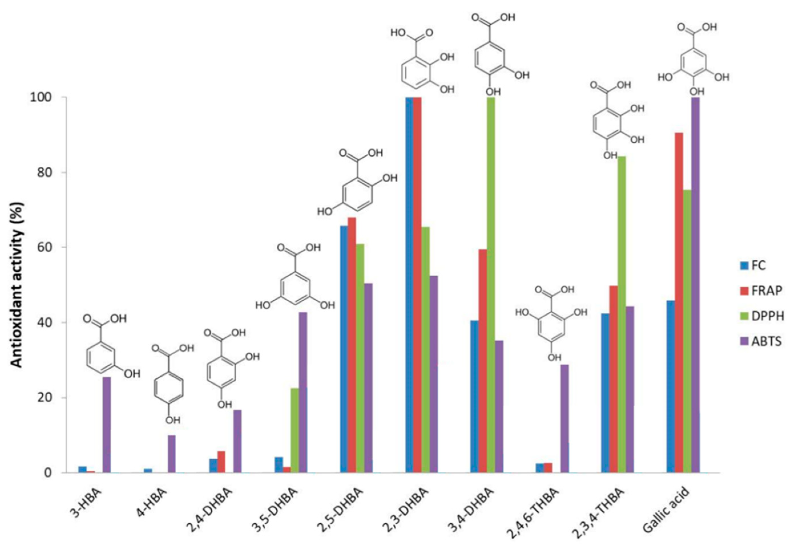
The comparison of results from different assays is not a trivial issue, as the sensitivity of each particular reducing species in each method is characteristic of the compound [48]. Figure 2, adapted from Reference [48] by Alcalde et al., compared various antioxidant indexes for the series of hydroxybenzoic acids. Despite the structural similarity of these molecules, the antioxidant powers differed significantly. The higher activities seemed to be associated with the presence of more than one hydroxyl group conveniently oriented in ortho or para positions, while those in meta or monohydroxylated species were less active. Hence, the strongest reducing agents according to the FC assay were dihydroxy- and trihydroxybenzoic acids with o- and p-configurations, while those not following this pattern were less efficient.
In any case, it is recommended to apply several spectroscopic methods based on different mechanisms to achieve complementary information to evaluate the AC of samples.

4.1.2. Flavonoid Assays

Apart from the indexes that provide information about the total AC, some assays focus on families of flavonoids, such as the aluminum complexation assays, or the 4-dimethylaminocinnamaldehyde (DMAC) spectrophotometric method.
For instance, flavonols and flavones form yellow complexes with Al(III) at neutral pH, and the absorbance of the solution is measured in the 400–430 nm range. Furthermore, Al(III) forms red complexes with some flavonoids (e.g., rutin, luteolin, and catechins) in the presence of sodium nitrite in an alkaline medium, and the absorbance of the solution is measured at 510 nm [49].
Currently, DMAC is the most popular reagent for the quantification of the overall PAC content in nutraceuticals and functional foods. DMAC is an aromatic aldehyde that, under strongly acidic conditions, reacts with flavanols via the formation of a reactive electrophilic carbocation. The DMAC reaction is specific to flavanols and their gallates. DMAC reacts with the C8 position of the terminal unit to form a green chromophore with maximum absorbance at ca. 640 nm. Hence, interferences from the colored anthocyanidins can be avoided [50,51,52,53]. PACs with a high degree of polymerization (DP) have lower responses per unit weight than monomers and dimers [28,50,54].

4.2. Voltammetric Methods

Electroanalytical techniques such as cyclic voltammetry (CV) and differential pulse voltammetry (DPV) have been widely used for the detection of polyphenols. A direct relationship between the concentration of electroactive functional groups and the current intensity generated is often found when they are either oxidized or reduced at the working electrode surface [55,56,57].
The techniques commented above are especially recommended for the analysis of polyphenols as their antioxidant properties are often related to the ability to donate electrons. Voltammetric signals at low oxidation potentials suggest the presence of polyphenolics of high antioxidant activity, whereas those compounds with low antioxidant power show electrochemical activity at more positive potentials [55,57]. Regarding electrodes, recent studies to determine polyphenols in nutraceuticals and related samples have relied on a wide variety of working electrodes, such as the pencil-graphite electrode (PGE), glassy carbon electrode (GCE), platinum (Pt), and different types of biosensors. In general, Pt has been used as an auxiliary electrode and Ag/AgCl (3 M KCl) as the reference electrode [55,57,58,59].
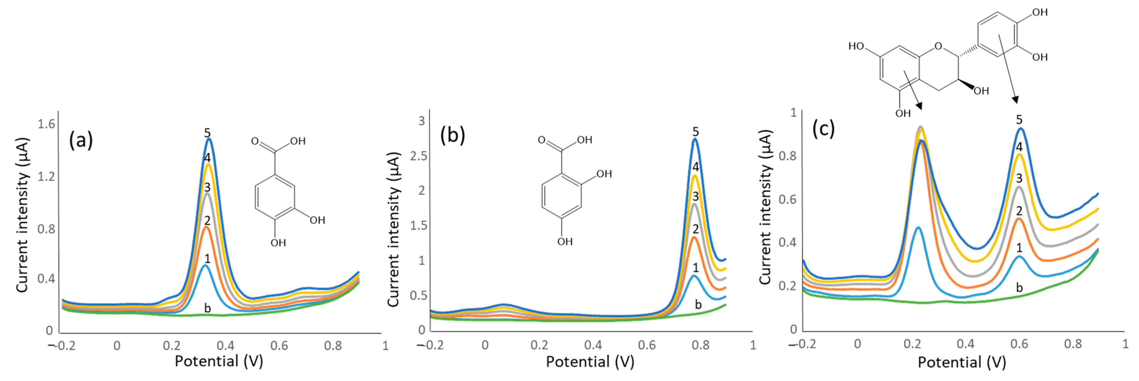
Some representative examples are depicted in Figure 3, which shows the differential pulse voltammograms recorded in the range from −0.2 to + 0.9 V vs. an Ag|AgCl|KCl reference electrode. In the case of catechin, with two independent dihydroxyphenyl moieties, two oxidation peaks corresponding to each side of the molecule have been detected.
These techniques result in powerful alternatives to traditional spectrophotometric measurements due to the high sampling throughput, simplicity, sensitivity, reduced sample, and reagent consumption. However, one of the main issues of voltammetric studies is related to the electrode fouling during the measurements due to the adsorption of several nutraceutical sample components. In such circumstances, a cleaning step may be necessary before each voltammetric measurement to clean or renew the electrode surface [55,57,58,60].
Some representative examples are mentioned as follows. For instance, grape extracts analyzed by CV with GCE showed a first oxidation peak at 420 mV corresponding to the catechol group on the B-ring of flavanols, and a second oxidation peak at 800–860 mV belonging to the resorcinol group on the A-ring [56]. In another study, the determination of TPC in Turkish green, black, and white tea extracts using DPV allowed a sensitive, rapid, and inexpensive evaluation of samples, with recoveries of caffeic acid around 95% [59]. In addition, DPV and CV were demonstrated to be appropriate to express the antioxidant activity of coffee extracts, presenting good correlations with DPPH spectrophotometric results [60].

4.3. Chromatographic Methods

Beyond the spectroscopic and voltammetric methods that provide information on the overall content of polyphenols, separation methods offer excellent opportunities for profiling. Thus, chromatographic techniques result in the best option for the simultaneous quantification of several bioactive components occurring in the samples.
Among the arsenal of techniques available in our current analytical laboratories, liquid chromatography seems to be the system of choice. Capillary electrophoresis (CE) can also be used owing to the ionizable nature of polyphenols. For instance, the phenolic content of herbal, fruit, or vegetable extracts has been determined using different CE modes such as capillary zone electrophoresis (CZE), micellar electrokinetic chromatography (MEKC), capillary isotachophoresis (CITP), and capillary electrochromatography (CEC) [61,62,63,64,65].
The polar and thermolabile nature of polyphenols hinders the use of gas chromatography (GC) for their direct determination. Hence, GC methods for the determination of polyphenols rely on derivatization procedures to reduce the polarity of analytes. In this regard, silylation has been successfully applied to the identification and quantification of phenolic compounds in plant and fruit extracts [66,67,68,69].
As mentioned, liquid chromatography (LC) is the best option for the determination of polyphenols in nutraceutical products and in plant extracts. In general, phenolic acids, stilbenes, and flavonoids can be separated successfully by reversed-phase (RP) mode, typically with C18 or C8 columns. Beyond conventional High Performance LC (HPLC), the newest and most powerful instrumentation based on ultra-high-performance liquid chromatography (UHPLC) and core–shell column technology can be used.
Mobile phases in LC are prepared with MeOH or ACN as organic solvents and acid aqueous solutions (e.g., using formic or acetic acids). The high complexity of the nutraceutical samples often requires customized elution gradients to achieve a good resolution of components. For instance, up to 15 polyphenols were determined in Taurisolo® (grape pomace extract supplement, MBMed, Turin, Italy) using a polar RP-C18 HPLC column and an elution gradient generated with 0.1% formic acid in water (v/v) and acetonitrile [70]. López-Gutierrez et al. determined more than 30 polyphenol-related compounds in nutraceutical samples (tablets and capsules) derived from green tea and grapes. The UHPLC separation was accomplished with sub-2 µm particle-size C18 columns together with mass spectrometry (MS) detection [71,72].
To avoid expensive instrumental platforms, core–shell columns appeared as a good alternative to achieve excellent efficacies. In this regard, Bakhytkyzy et al. used a Kinetex C18 reversed-phase (100 mm × 4.6 mm ID, 2.6-μm particle size) column for the determination of flavanols in dietary products and pharmaceuticals [73]. In another example, sub-2 µm and porous-shell columns were compared for the determination of 29 polyphenols in cranberry-based pharmaceuticals obtaining, in that case, better performances using the UHPLC option [74].
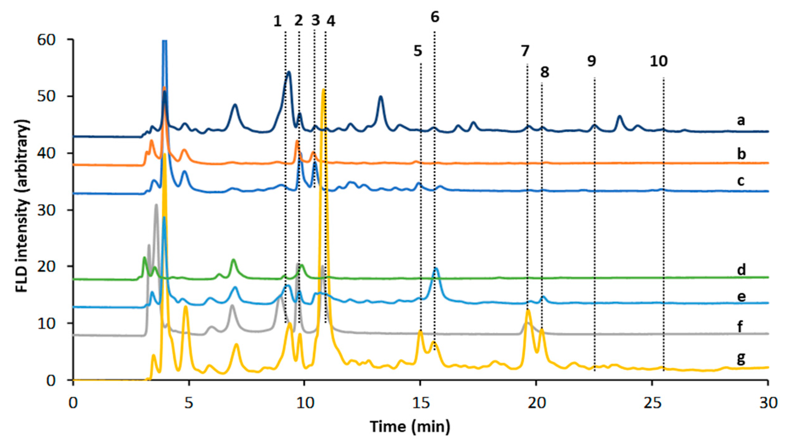
As an alternative to the reverse-phase mode, the hydrophilic interaction chromatography (HILIC) has opened up great possibilities, especially for dealing with the most polar analytes that are weakly retained in RP columns. HILIC was used, for example, for the separation of flavanols and related compounds in dietary supplements and nutraceuticals from berries, grapes, or artichoke extracts [75] (see Figure 4). The mobile phase consisted of acidified acetonitrile and methanol solutions, applying an elution gradient based on increasing the percentage of methanol. In this study, the authors highlighted the improved separation of flavanol oligomers, as well as the great potential to separate small phenolic acid molecules.
The LC × LC strategy, combining different stationary phases, namely RP (or modified RP), HILIC, and, less commonly, size-exclusion chromatography (SEC), has shown to be promising for the determination of polyphenols in food products [76,77]. LC × LC, operating in the off-line or on-line modes, offers improved separations taking advantage of the orthogonality between the first and second dimensions. Consequently, sample clean-up procedures can be simplified by reducing the total analysis time. In this regard, an RP-LC × RP-LC method, combining a micro-bore ES-CN column (1D) with a C18 column (2D), was successfully applied to the determination of 51 polyphenolic compounds in pistachio extracts [78]. Another study performed by Sommella et al. for the determination of polyphenols in apple extracts samples combined HILIC × RP in the two-dimensional LC [79]. In this case, higher peak capacities and sensitivities were obtained with the optimized online HILIC × RP-UHPLC-MS method concerning previously 1D reported methods.
Regarding detection, apart from the widespread UV-vis spectroscopy, fluorescence and mass spectrometry have been utilized to increase the sensitivity and selectivity of the methods. In UV-Vis, current multi-way spectrophotometers such as diode-array detectors allow the simultaneous detection of the different phenolic families. For instance, the range 270–280 nm is used to monitor benzoic acids, flavanols, flavanones, and other species containing phenyl moieties, 310–330 nm is used for hydroxycinnamic acids and stilbenes, 370–380 nm is used for flavones, isoflavones, and flavonols, 420–430 nm is used for curcuminoids, and 450–550 nm is used for anthocyanins.
As most of the polyphenolic compounds have fluorescent moieties in their structures, fluorometric detection is an excellent alternative to the UV counterpart, thus providing enhanced performance from a proper selection of the excitation and emission wavelengths. For instance, flavanols are detected at λex 280 nm and λem 320 nm with negligible interferences from other phenolic families; λex 325 nm and λem 375 nm are good choices for the determination of hydroxycinnamic acids; λex 375 nm and λem 430 nm have been recommended for flavonols, flavones, and isoflavones.
Currently, MS is the most powerful detector for qualitative and quantitative determinations of phenolic compounds. In our opinion, the hyphenation of MS with liquid chromatography deserves a more exhaustive development. Hence, the state of the art of application of HPLC-MS to polyphenol characterization is given below.

Liquid Chromatography Coupled to Mass Spectrometry

HPLC-MS techniques, and more recently, the UHPLC-MS counterparts, have been increasingly used in nutraceutical characterization, especially for bioanalytical applications dealing with pharmacokinetics and metabolism studies. LC-MS has been widely exploited for the identification and structural elucidation of known and unknown bioactive molecules, especially from LC-MS/MS experiments [80,81]. Furthermore, high-resolution mass spectrometers (HRMS) coupled to the LC systems result in exceptional tools for food analysis. Obtaining accurate mass measurements is the major advantage of HRMS to provide almost unambiguous identifications and quantifications. For instance, the presence of curcumin and other curcuminoids in turmeric-based extracts was confirmed using this strategy. As can be seen in Figure 5, the accurate mass spectra obtained by LC-HRMS with an orbitrap analyzer gave the authors reliable information for the identification of curcumin and other major components in the studied samples [82].
By far, ESI is the most generalized ionization source, although atmospheric-pressure chemical ionization (APCI) has also been proposed, especially for those compounds exhibiting low ionization efficiencies under ESI conditions [83,84,85]. This is the case, for instance, of the identification and determination of triterpenes and phenolic acids from ancient apples of Friuli Venezia Giulia as nutraceutical ingredients [85]. While ESI resulted in the best option for the determination of phenolic acids, APCI in negative ion mode was selected as the ion source of terpenes due to the opportunity to obtain the pseudomolecular [M-H] ions with higher intensity than with ESI [86]. Carotenoids are also a typical family of compounds exhibiting better ionization behavior under APCI conditions in comparison to ESI [84,87,88]. For example, Garzón et al. [84] described the determination of carotenoids from Arazá (Eugenia stipitate), an Amazonian fruit with potential value as a nutraceutical source. Recently, UHPLC-APCI-TOF/MS was also proposed for the screening of genistein from selected seeds of Apiaceae [89].
To date, dozens of applications of (U)HPLC-ESI-MS(/MS) have been published. Low-resolution mass spectrometers based on triple quadrupole and ion trap analyzers are typically proposed for the sensitive determination of well-known bioactive substances, as well as for identification purposes when working in tandem mass spectrometry [90,91,92,93,94]. For example, Nzekoue et al. carried out the quantification of 30 bioactive compounds in coffee silverskin extracts by HPLC-MS/MS using a triple quadrupole analyzer [94]. Eighteen phenolic compounds were detected and quantified, with caffeoylquinic acids being the most abundant ones. In contrast, HPLC-DAD-MS using an ion-trap analyzer was employed to address a comprehensive characterization of the phytochemicals present in an Italian ancient apple “Mela Rosa dei Monti Sibillini” [93]. Extracts from lyophilized material were richer in polyphenolic compounds than the dried counterparts. These findings suggested that this ancient apple variety was an excellent source of active components for nutraceuticals.
HRMS based on time-of-flight (TOF) and Orbitrap analyzers is the technique of choice for a comprehensive characterization of plant-based extracts due to the higher-resolution capabilities and the accurate mass measurements (with errors below 2 ppm depending on the instrument). In these cases, hybrid configurations combining quadrupole and ion-trap analyzers coupled with HRMS instruments such as Q-TOF [95,96,97,98,99,100], IT-TOF [101], and Q-Orbitrap [102,103] were preferred to take advantage of the high sensitivity of these platforms under full-scan acquisition mode. Furthermore, fragmentations can be employed for identification purposes, especially when profiling strategies are applied. For instance, Amat-ur-Rasool et al. evaluated the potential nutraceutical properties of leaves from several commonly cultivated plants such as lemon (Citrus limon), red silk-cotton (Bombax ceiba), henna (Lawsonia inermis), eucalyptus (Eucalyptus globulus), basil (Ocimum basilicum), Mandarin orange (Citrus reticulata), and spearmint (Mentha spicata) [95]. RP HPLC coupled to a Q-TOF instrument working in negative ion electrospray mode was proposed as the instrumental technique. Several polyphenols such as 3-caffeoylquinic acid, methyl 4-caffeoylquinate, kaempferol-acetyl-glycoside, quercetin 3-rutinoside, quercetin-acetyl-glycoside, kaempferol 3-O-glucoside, and quercetin 3-O-glucoside were identified. In another work, a Q-Orbitrap HRMS instrument was used to assess a comprehensive investigation of the polyphenolic constituents of fennel waste extract, showing that chlorogenic acids were the most abundant compounds [102]. Some applications based on MS instruments with an even higher resolution than an Orbitrap analyzer, such as Fourier-transform ion cyclotron resonance mass spectrometry (FT-ICR-MS), have also been described in the literature. For example, Maia et al. [104] evaluated Vitis vinifera “Pinot noir” leaves as a source of bioactive nutraceutical compounds using an FT-ICR-MS instrument, identifying the presence of numerous compounds that are known to possess diverse nutritional and pharmacological properties, such as caffeic acid, catechin, kaempferol and quercetin, several phytosterols, and fatty acids.
The use of ambient mass spectrometry (AMS) techniques has also been described for nutraceutical characterization. AMS involves the direct analysis of compounds from sample surfaces, allowing a straightforward study of unconventional bulk samples, such as whole tablets, plant parts, or tissue sections. The analysis takes place at ambient pressure outside the MS instrument, which speeds up the sampling process and without any sample preparation (or minimal sample manipulation). For example, Giffen et al. described the use of direct analysis in real time (DART) for the rapid identification of Salvia species by chemometric processing [105]. The authors analyzed plant leaves in their native form by DART-HRMS without any sample preparation step, obtaining a derived chemical fingerprinting of both fresh and dried salvia material. The presence of essential oil biomarkers such as 3-carene, α-pinene, β-pinene, β-thujone, β-caryophyllene, camphor, and borneol was confirmed. In another work, Riya et al. revealed the important nutraceutical properties of Mauritius fruit (Ananas comosus) against diabetes [106]. In this case, some active compounds identified in ethyl acetate and methanolic extracts of the fruit using DART-HRMS were sinapic acid, daucosterol, 2-methylpropanoate, 2,3-dimethyl-4-hydroxy-4(2H)-furanone, methyl 2-methylbutanoate, and triterpenoid ergosterol.
Desorption electrospray ionization (DESI) is another AMS technique allowing the direct identification and determination of bioactive substances deposited on surfaces and from thin biological tissue sections with minimal (or without) sample treatment. In this case, a charged spray of solvent droplets is directed toward the surface to desorb and ionize the analytes, which are directed to the MS inlet under vacuum conditions. The investigation of phytochemicals from thin-layer chromatography (TLC) imprints or direct plant tissues is well described in the literature. As an example, the chemical profiling and separation of bioactive secondary metabolites in maca (Lepidium peruvianum) by normal- and reversed-phase TLC coupled to DESI-HRMS was proposed by Perez et al. [107]. The authors reported for the first time the identification of six potential plant antibiotics, phytoanticipins, glycosylated ascorbigens, and dihydroascorbigens from Maca seeds.
Matrix-assisted laser desorption ionization (MALDI)−TOF-MS is another excellent technique for the deeper characterization of polyphenols, especially for dealing with polymeric compounds such as PACs [108], especially for the elucidation of intermolecular link positions. The polyphenol profiling of chestnut pericarp, integument, and curing water extracts to qualify these food by-products as a source of bioactive antioxidant compounds for nutraceutical applications was also described by MALDI-TOF-MS [109]. This method allowed the identification of condensed and hydrolyzable tannins such as procyanidins and prodelphinidins.

5. Conclusions

Polyphenols, including phenolic acids, flavonoids, and stilbenes, display great antioxidant activity that may help to combat free radicals resulting from oxidative stress. Other interesting properties such as anti-inflammatory, antineoplastic, antimicrobial, hypolipidemic, estrogenic, or hepatoprotective activities have been described as well. The chemical evaluation of these compounds is essential to ensure the content of the desired active species.
Regarding analytical methods, spectroscopic methods are suitable for the evaluation of the overall antioxidant capacity of nutraceuticals. Electrochemical methods have also demonstrated their great performance to assess the antioxidant properties of products. The simultaneous profiling of multiple analytes relies on separation techniques, among which liquid chromatography is the most recommended one. Polyphenols can be monitored by UV/vis, fluorescence, and mass spectrometry, with the latter being especially suitable for sensitive and selective detection. The hyphenation of liquid chromatography with mass spectrometry is also the technique of choice for the identification and structural elucidation of unknown phytochemicals with bioactive properties.

Author Contributions

J.S. conceived and designed the publication and wrote the core of the document; O.V.-C., O.N., M.G. and S.S. contributed to the bibliographic search, analysis of the information, and writing of different sections of the paper. All authors have read and agreed to the published version of the manuscript.

Funding

This research was funded by the “Agencia Estatal de Investigación”, grant number PID2020-114401RB-C22.

Institutional Review Board Statement

Not applicable.

Informed Consent Statement

Not applicable.

Acknowledgments

Not applicable.

Conflicts of Interest

The authors declare no conflict of interest.

References

  1. Leri, M.; Scuto, M.; Ontario, M.L.; Calabrese, V.; Calabrese, E.J.; Bucciantini, M.; Stefani, M. Healthy effects of plant polyphenols molecular mechanisms. Int. J. Mol. Sci. 2020, 21, 1250. [Google Scholar] [CrossRef] [Green Version]
  2. Tomé-Carneiro, J.; Visioli, F. Polyphenol-based nutraceuticals for the prevention and treatment of cardiovascular disease: Review of human evidence. Phytomedicine 2016, 23, 1145–1174. [Google Scholar] [CrossRef]
  3. Abuajah, C.I.; Ogbonna, A.C.; Osuji, C.M. Functional components and medicinal properties of food: A review. J. Food Sci. Technol. 2015, 52, 2522–2529. [Google Scholar] [CrossRef] [Green Version]
  4. Lucci, P.; Saurina, J.; Núñez, O. Trends in LC-MS and LC-HRMS analysis and characterization of polyphenols in food. Trends Anal. Chem. 2017, 88, 1–24. [Google Scholar] [CrossRef] [Green Version]
  5. Saurina, J. Characterization of wines using compositional profiles and chemometrics. Trends Anal. Chem. 2010, 29, 234–245. [Google Scholar] [CrossRef]
  6. Gülçin, I. Antioxidant activity of food constituents: An overview. Arch. Toxicol. 2012, 86, 345–391. [Google Scholar] [CrossRef] [PubMed]
  7. Farhood, B.; Mortezaee, K.; Goradel, N.H.; Khanlarkhani, N.; Salehi, E.; Nashtaei, M.S.; Najafi, M.; Sahebkar, A. Curcumin as an anti-inflammatory agent: Implications to radiotherapy and chemotherapy. J. Cell. Physiol. 2019, 234, 5728–5740. [Google Scholar] [CrossRef]
  8. Tabrizi, R.; Vakili, S.; Akbari, M.; Mirhosseini, N.; Lankarani, K.B.; Rahimi, M.; Mobini, M.; Jafarnejad, S.; Vahedpoor, Z.; Asemi, Z. The effects of curcumin-containing supplements on biomarkers of inflammation and oxidative stress: A systematic review and meta-analysis of randomized controlled trials. Phyther. Res. 2019, 33, 253–262. [Google Scholar] [CrossRef]
  9. Tao, W.; Zhang, Y.; Shen, X.; Cao, Y.; Shi, J.; Ye, X.; Chen, S. Rethinking the Mechanism of the Health Benefits of Proanthocyanidins: Absorption, Metabolism, and Interaction with Gut Microbiota. Compr. Rev. Food Sci. Food Saf. 2019, 18, 971–985. [Google Scholar] [CrossRef] [Green Version]
  10. Coman, V.; Vodnar, D.C. Hydroxycinnamic acids and human health: Recent advances. J. Sci. Food Agric. 2020, 100, 483–499. [Google Scholar] [CrossRef]
  11. Quideau, S.; Deffieux, D.; Douat-Casassus, C.; Pouységu, L. Plant polyphenols: Chemical properties, biological activities, and synthesis. Angew. Chemie Int. Ed. 2011, 50, 586–621. [Google Scholar] [CrossRef]
  12. Tsao, R. Chemistry and biochemistry of dietary polyphenols. Nutrients 2010, 2, 1231–1246. [Google Scholar] [CrossRef] [PubMed]
  13. Di Lorenzo, C.; Colombo, F.; Biella, S.; Stockley, C.; Restani, P. Polyphenols and human health: The role of bioavailability. Nutrients 2021, 13, 273. [Google Scholar] [CrossRef]
  14. Panche, A.N.; Diwan, A.D.; Chandra, S.R. Flavonoids: An overview. J. Nutr. Sci. 2016, 5, e47. [Google Scholar] [CrossRef] [Green Version]
  15. Flamini, R.; Mattivi, F.; De Rosso, M.; Arapitsas, P.; Bavaresco, L. Advanced knowledge of three important classes of grape phenolics: Anthocyanins, stilbenes and flavonols. Int. J. Mol. Sci. 2013, 14, 19651–19669. [Google Scholar] [CrossRef] [PubMed]
  16. Kubina, R.; Iriti, M.; Kabala-Dzik, A. Anticancer potential of selected flavonols: Fisetin, kaempferol, and quercetin on head and neck cancers. Nutrients 2021, 13, 845. [Google Scholar] [CrossRef] [PubMed]
  17. Ulusoy, H.G.; Sanlier, N. A minireview of quercetin: From its metabolism to possible mechanisms of its biological activities. Crit. Rev. Food Sci. Nutr. 2020, 60, 3290–3303. [Google Scholar] [CrossRef] [PubMed]
  18. Hostetler, G.L.; Ralston, R.A.; Schwartz, S.J. Flavones: Food Sources, Bioavailability, Metabolism and Bioactivity. Adv. Nutr. 2017, 8, 423–435. [Google Scholar] [CrossRef] [Green Version]
  19. Singh, M.; Kaur, M.; Silakari, O. Flavones: An important scaffold for medicinal chemistry. Eur. J. Med. Chem. 2014, 84, 206–239. [Google Scholar] [CrossRef] [PubMed]
  20. Ko, K. Isoflavones: Chemistry, Analysis, Functions and Effects on Health and Cancer. Asian Pac. J. Cancer Prev. 2014, 15, 7001–7010. [Google Scholar] [CrossRef] [Green Version]
  21. Messina, M. Soy foods, isoflavones, and the health of postmenopausal women. Am. J. Clin. Nutr. 2014, 100, 423S–430S. [Google Scholar] [CrossRef] [Green Version]
  22. Testai, L.; Calderone, V. Nutraceutical value of citrus flavanones and their implications in cardiovascular disease. Nutrients 2017, 9, 502. [Google Scholar] [CrossRef] [Green Version]
  23. Tsuda, T. Dietary anthocyanin-rich plants: Biochemical basis and recent progress in health benefits studies. Mol. Nutr. Food Res. 2012, 56, 159–170. [Google Scholar] [CrossRef]
  24. Singh, B.N.; Shankar, S.; Srivastava, R.K. Green tea catechin, epigallocatechin-3-gallate (EGCG): Mechanisms, perspectives and clinical applications. Biochem. Pharmacol. 2011, 82, 1807–1821. [Google Scholar] [CrossRef] [Green Version]
  25. Martin, M.A.; Ramos, S. Impact of dietary flavanols on microbiota, immunity andinflammation in metabolic diseases. Nutrients 2021, 13, 850. [Google Scholar] [CrossRef]
  26. Sabaghi, M.; Hoseyni, S.Z.; Tavasoli, S.; Mozafari, M.R.; Katouzian, I. Strategies of confining green tea catechin compounds in nano-biopolymeric matrices: A review. Colloids Surfaces B Biointerfaces 2021, 204, 111781. [Google Scholar] [CrossRef] [PubMed]
  27. Rauf, A.; Imran, M.; Abu-Izneid, T.; Patel, S.; Pan, X.; Naz, S.; Sanches Silva, A.; Saeed, F.; Rasul Suleria, H.A. Proanthocyanidins: A comprehensive review. Biomed. Pharmacother. 2019, 116, 108999. [Google Scholar] [CrossRef]
  28. Krueger, C.G.; Reed, J.D.; Feliciano, R.P.; Howell, A.B. Quantifying and characterizing proanthocyanidins in cranberries in relation to urinary tract health. Anal. Bioanal. Chem. 2013, 405, 4385–4395. [Google Scholar] [CrossRef] [PubMed]
  29. Mallebrera, B.; Maietti, A.; Tedeschi, P.; Font, G.; Ruiz, M.J.; Brandolini, V. Antioxidant capacity of trans-resveratrol dietary supplements alone or combined with the mycotoxin beauvericin. Food Chem. Toxicol. 2017, 105, 315–318. [Google Scholar] [CrossRef]
  30. Kotha, R.R.; Luthria, D.L. Curcumin: Biological, pharmaceutical, nutraceutical, and analytical aspects. Molecules 2019, 24, 2930. [Google Scholar] [CrossRef] [PubMed] [Green Version]
  31. Zálešák, F.; Bon, D.J.Y.D.; Pospíšil, J. Lignans and Neolignans: Plant secondary metabolites as a reservoir of biologically active substances. Pharmacol. Res. 2019, 146, 104284. [Google Scholar] [CrossRef]
  32. Vidal-Casanella, O.; Nuñez, O.; Saurina, J. Liquid chromatographic fingerprints for the characterization of flavanol-rich nutraceuticals based on 4-dimethylaminocinnamaldehyde precolumn derivatization. Sci. Pharm. 2021, 89, 18. [Google Scholar] [CrossRef]
  33. Gudzinskaite, I.; Stackeviciene, E.; Liaudanskas, M.; Zymone, K.; Zvikas, V.; Viskelis, J.; Urbstaite, R.; Janulis, V. Variability in the qualitative and quantitative composition and content of phenolic compounds in the fruit of introduced American cranberry (Vaccinium macrocarpon Aiton). Plants 2020, 9, 1379. [Google Scholar] [CrossRef] [PubMed]
  34. Zhao, S.; Liu, H.; Gu, L. American cranberries and health benefits—An evolving story of 25 years. J. Sci. Food Agric. 2020, 100, 5111–5116. [Google Scholar] [CrossRef]
  35. Côté, J.; Caillet, S.; Doyon, G.; Sylvain, J.F.; Lacroix, M. Analyzing cranberry bioactive compounds. Crit. Rev. Food Sci. Nutr. 2010, 50, 872–888. [Google Scholar] [CrossRef]
  36. Blumberg, J.B.; Camesano, T.A.; Cassidy, A.; Kris-Etherton, P.; Howell, A.; Manach, C.; Ostertag, L.M.; Sies, H.; Skulas-Ray, A.; Vita, J.A. Cranberries and their bioactive constituents in human health. Adv. Nutr. 2013, 4, 618–632. [Google Scholar] [CrossRef] [Green Version]
  37. Wang, S.; Moustaid-Moussa, N.; Chen, L.; Mo, H.; Shastri, A.; Su, R.; Bapat, P.; Kwun, I.S.; Shen, C.L. Novel insights of dietary polyphenols and obesity. J. Nutr. Biochem. 2014, 25, 1–18. [Google Scholar] [CrossRef] [PubMed] [Green Version]
  38. Ríos-Hoyo, A.; Gutiérrez-Salmeán, G. New Dietary Supplements for Obesity: What We Currently Know. Curr. Obes. Rep. 2016, 5, 262–270. [Google Scholar] [CrossRef] [PubMed]
  39. Tsuda, T. Recent progress in anti-obesity and anti-diabetes effect of berries. Antioxidants 2016, 5, 13. [Google Scholar] [CrossRef] [PubMed] [Green Version]
  40. Georgiev, V.; Ananga, A.; Tsolova, V. Recent advances and uses of grape flavonoids as nutraceuticals. Nutrients 2014, 6, 391–415. [Google Scholar] [CrossRef] [PubMed] [Green Version]
  41. Xia, E.Q.; Deng, G.F.; Guo, Y.J.; Li, H. Bin Biological activities of polyphenols from grapes. Int. J. Mol. Sci. 2010, 11, 622–646. [Google Scholar] [CrossRef]
  42. Pereira, C.; Barros, L.; Ferreira, I.C. Extraction, identification, fractionation and isolation of phenolic compounds in plants with hepatoprotective effects. J. Sci. Food Agric. 2016, 96, 1068–1084. [Google Scholar] [CrossRef] [PubMed]
  43. Gostin, A.I.; Waisundara, V.Y. Edible flowers as functional food: A review on artichoke (Cynara cardunculus L.). Trends Food Sci. Technol. 2019, 86, 381–391. [Google Scholar] [CrossRef]
  44. Singleton, V.L.; Orthofer, R.; Lamuela-Raventós, R.M. Analysis of total phenols and other oxidation substrates and antioxidants by means of folin-ciocalteu reagent. Methods Enzymol. 1999, 299, 152–178. [Google Scholar] [CrossRef]
  45. Gulcin, İ. Antioxidants and antioxidant methods: An updated overview. Arch. Toxicol. 2020, 94, 651–715. [Google Scholar] [CrossRef] [PubMed] [Green Version]
  46. Brainina, K.; Stozhko, N.; Vidrevich, M. Antioxidants: Terminology, methods, and future considerations. Antioxidants 2019, 8, 297. [Google Scholar] [CrossRef] [PubMed] [Green Version]
  47. Shahidi, F.; Zhong, Y. Measurement of antioxidant activity. J. Funct. Foods 2015, 18, 757–781. [Google Scholar] [CrossRef]
  48. Alcalde, B.; Granados, M.; Saurina, J. Exploring the Antioxidant Features of Polyphenols by Spectroscopic and Electrochemical Methods. Antioxidants 2019, 8, 523. [Google Scholar] [CrossRef] [Green Version]
  49. Pękal, A.; Pyrzynska, K. Evaluation of Aluminium Complexation Reaction for Flavonoid Content Assay. Food Anal. Methods 2014, 7, 1776–1782. [Google Scholar] [CrossRef] [Green Version]
  50. Feliciano, R.P.; Shea, M.P.; Shanmuganayagam, D.; Krueger, C.G.; Howell, A.B.; Reed, J.D. Comparison of isolated cranberry (Vaccinium macrocarpon Ait.) proanthocyanidins to catechin and procyanidins A2 and B2 for use as standards in the 4-(dimethylamino)cinnamaldehyde assay. J. Agric. Food Chem. 2012, 60, 4578–4585. [Google Scholar] [CrossRef]
  51. Payne, M.J.; Hurst, W.J.; Stuart, D.A.; Ou, B.; Fan, E.; Ji, H.; Kou, Y. Determination of total procyanidins in selected chocolate and confectionery products using DMAC. J. AOAC Int. 2010, 93, 89–96. [Google Scholar] [CrossRef] [Green Version]
  52. Wang, Y.; Singh, A.P.; Hurst, W.J.; Glinski, J.A.; Koo, H.; Vorsa, N. Influence of Degree-of-Polymerization and Linkage on the Quantification of Proanthocyanidins using 4-Dimethylaminocinnamaldehyde (DMAC) Assay. J. Agric. Food Chem. 2016, 64, 2190–2199. [Google Scholar] [CrossRef] [PubMed]
  53. Vidal-Casanella, O.; Nuñez, O.; Hernández-Cassou, S.; Saurina, J. Assessment of Experimental Factors Affecting the Sensitivity and Selectivity of the Spectrophotometric Estimation of Proanthocyanidins in Foods and Nutraceuticals. Food Anal. Methods 2021, 14, 485–495. [Google Scholar] [CrossRef]
  54. Krueger, C.G.; Chesmore, N.; Chen, X.; Parker, J.; Khoo, C.; Marais, J.P.J.; Shanmuganayagam, D.; Crump, P.; Reed, J.D. Critical reevaluation of the 4-(dimethylamino)cinnamaldehyde assay: Cranberry proanthocyanidin standard is superior to procyanidin A2 dimer for accurate quantification of proanthocyanidins in cranberry products. J. Funct. Foods 2016, 22, 13–19. [Google Scholar] [CrossRef]
  55. Arribas, A.S.; Martinez-Fernandez, M.; Chicharro, M. The role of electroanalytical techniques in analysis of polyphenols in wine. TrAC Trends Anal. Chem. 2012, 34, 78–96. [Google Scholar] [CrossRef]
  56. Petrovic, S.C. Correlation of perceived wine astringency to cyclic voltammetric response. Am. J. Enol. Vitic. 2009, 60, 373–378. [Google Scholar]
  57. Makhotkina, O.; Kilmartin, P.A. The use of cyclic voltammetry for wine analysis: Determination of polyphenols and free sulfur dioxide. Anal. Chim. Acta 2010, 668, 155–165. [Google Scholar] [CrossRef]
  58. David, I.G.; Buleandră, M.; Popa, D.E.; Bîzgan, A.-M.C.; Moldovan, Z.; Badea, I.-A.; Iorgulescu, E.E.; Tekiner, T.A.; Basaga, H. Voltammetric determination of polyphenolic content as rosmarinic acid equivalent in tea samples using pencil graphite electrodes. J. Food Sci. Technol. 2016, 53, 2589–2596. [Google Scholar] [CrossRef] [Green Version]
  59. David, I.G.; Bizgan, A.-M.C.; Popa, D.E.; Buleandra, M.; Moldovan, Z.; Badea, I.A.; Tekiner, T.A.; Basaga, H.; Ciucu, A.A. Rapid determination of total polyphenolic content in tea samples based on caffeic acid voltammetric behaviour on a disposable graphite electrode. Food Chem. 2015, 173, 1059–1065. [Google Scholar] [CrossRef]
  60. Oliveira-Neto, J.R.; Rezende, S.G.; de Fátima Reis, C.; Benjamin, S.R.; Rocha, M.L.; de Souza Gil, E. Electrochemical behavior and determination of major phenolic antioxidants in selected coffee samples. Food Chem. 2016, 190, 506–512. [Google Scholar] [CrossRef]
  61. Memon, A.F.; Solangi, A.R.; Memon, S.Q.; Mallah, A.; Memon, N. Quantitative separation of hesperidin, chrysin, epicatechin, epigallocatechin gallate, and morin using ionic liquid as a buffer additive in capillary electrophoresis. Electrophoresis 2018, 39, 1606–1612. [Google Scholar] [CrossRef]
  62. Arries, W.J.; Tredoux, A.G.J.; de Beer, D.; Joubert, E.; de Villiers, A. Evaluation of capillary electrophoresis for the analysis of rooibos and honeybush tea phenolics. Electrophoresis 2017, 38, 897–905. [Google Scholar] [CrossRef] [PubMed]
  63. Dresler, S.; Bogucka-Kocka, A.; Kováčik, J.; Kubrak, T.; Strzemski, M.; Wójciak-Kosior, M.; Rysiak, A.; Sowa, I. Separation and determination of coumarins including furanocoumarins using micellar electrokinetic capillary chromatography. Talanta 2018, 187, 120–124. [Google Scholar] [CrossRef]
  64. Navarro, M.; Núñez, O.; Saurina, J.; Hernández-Cassou, S.; Puignou, L. Characterization of fruit products by capillary zone electrophoresis and liquid chromatography using the compositional profiles of polyphenols: Application to authentication of natural extracts. J. Agric. Food Chem. 2014, 62, 1038–1046. [Google Scholar] [CrossRef] [PubMed] [Green Version]
  65. Przybylska, A.; Gackowski, M.; Koba, M. Application of capillary electrophoresis to the analysis of bioactive compounds in herbal raw materials. Molecules 2021, 26, 2135. [Google Scholar] [CrossRef]
  66. Ahmad, N.; Zuo, Y.; Lu, X.; Anwar, F.; Hameed, S. Characterization of free and conjugated phenolic compounds in fruits of selected wild plants. Food Chem. 2016, 190, 80–89. [Google Scholar] [CrossRef]
  67. Osman, M.F.; Hassan, N.M.; Khatib, A.; Tolos, S.M. Antioxidant activities of Dialium indum L. Fruit and gas chromatography-mass spectrometry (GC-MS) of the active fractions. Antioxidants 2018, 7, 154. [Google Scholar] [CrossRef] [PubMed] [Green Version]
  68. Park, C.H.; Morgan, A.M.A.; Park, B.B.; Lee, S.Y.; Lee, S.; Kim, J.K.; Park, S.U. Metabolic analysis of four cultivars of liriope platyphylla. Metabolites 2019, 9, 59. [Google Scholar] [CrossRef] [Green Version]
  69. Ifeanacho, M.O.; Ikewuchi, C.C.; Ikewuchi, J.C. Investigation of the profile of phenolic compounds in the leaves and stems of Pandiaka heudelotii using gas chromatography coupled with flame ionization detector. Food Sci. Nutr. 2017, 5, 646–652. [Google Scholar] [CrossRef] [PubMed] [Green Version]
  70. Annunziata, G.; Maisto, M.; Schisano, C.; Ciampaglia, R.; Narciso, V.; Tenore, G.C.; Novellino, E. Effects of grape pomace polyphenolic extract (Taurisolo®) in reducing tmao serum levels in humans: Preliminary results from a randomized, placebo-controlled, cross-over study. Nutrients 2019, 11, 139. [Google Scholar] [CrossRef] [PubMed] [Green Version]
  71. López-Gutiérrez, N.; Romero-González, R.; Plaza-Bolaños, P.; Martínez Vidal, J.L.; Garrido Frenich, A. Identification and quantification of phytochemicals in nutraceutical products from green tea by UHPLC-Orbitrap-MS. Food Chem. 2015, 173, 607–618. [Google Scholar] [CrossRef]
  72. López-Gutiérrez, N.; Romero-González, R.; Martínez Vidal, J.L.; Frenich, A.G. Determination of polyphenols in grape-based nutraceutical products using high resolution mass spectrometry. LWT Food Sci. Technol. 2016, 71, 249–259. [Google Scholar] [CrossRef]
  73. Bakhytkyzy, I.; Nuñez, O.; Saurina, J. Size Exclusion Coupled to Reversed Phase Liquid Chromatography for the Characterization of Cranberry Products. Food Anal. Methods 2019, 12, 604–611. [Google Scholar] [CrossRef] [Green Version]
  74. Parets, L.; Alechaga, É.; Núñez, O.; Saurina, J.; Hernández-Cassou, S.; Puignou, L. Ultrahigh pressure liquid chromatography-atmospheric pressure photoionization-tandem mass spectrometry for the determination of polyphenolic profiles in the characterization and classification of cranberry-based pharmaceutical preparations and natural ext. Anal. Methods 2016, 8, 4363–4378. [Google Scholar] [CrossRef] [Green Version]
  75. Vidal-Casanella, O.; Arias-Alpizar, K.; Nuñez, O.; Saurina, J. Hydrophilic interaction liquid chromatography to characterize nutraceuticals and food supplements based on flavanols and related compounds. Separations 2021, 8, 17. [Google Scholar] [CrossRef]
  76. Cacciola, F.; Rigano, F.; Dugo, P.; Mondello, L. Comprehensive two-dimensional liquid chromatography as a powerful tool for the analysis of food and food products. TrAC Trends Anal. Chem. 2020, 127, 115894. [Google Scholar] [CrossRef]
  77. Cacciola, F.; Arena, K.; Mandolfino, F.; Donnarumma, D.; Dugo, P.; Mondello, L. Reversed phase versus hydrophilic interaction liquid chromatography as first dimension of comprehensive two-dimensional liquid chromatography systems for the elucidation of the polyphenolic content of food and natural products. J. Chromatogr. A 2021, 1645, 462129. [Google Scholar] [CrossRef] [PubMed]
  78. Arena, K.; Cacciola, F.; Mangraviti, D.; Zoccali, M.; Rigano, F.; Marino, N.; Dugo, P.; Mondello, L. Determination of the polyphenolic fraction of Pistacia vera L. kernel extracts by comprehensive two-dimensional liquid chromatography coupled to mass spectrometry detection. Anal. Bioanal. Chem. 2019, 411, 4819–4829. [Google Scholar] [CrossRef]
  79. Sommella, E.; Ismail, O.H.; Pagano, F.; Pepe, G.; Ostacolo, C.; Mazzoccanti, G.; Russo, M.; Novellino, E.; Gasparrini, F.; Campiglia, P. Development of an improved online comprehensive hydrophilic interaction chromatography × reversed-phase ultra-high-pressure liquid chromatography platform for complex multiclass polyphenolic sample analysis. J. Sep. Sci. 2017, 40, 2188–2197. [Google Scholar] [CrossRef]
  80. Tamasi, G.; Pardini, A.; Bonechi, C.; Donati, A.; Pessina, F.; Marcolongo, P.; Gamberucci, A.; Leone, G.; Consumi, M.; Magnani, A.; et al. Characterization of nutraceutical components in tomato pulp, skin and locular gel. Eur. Food Res. Technol. 2019, 245, 907–918. [Google Scholar] [CrossRef]
  81. Simeoni, M.C.; Pellegrini, M.; Sergi, M.; Pittia, P.; Ricci, A.; Compagnone, D. Analysis of Polyphenols in the Lamiaceae Family by Matrix Solid-Phase Dispersion Extraction Followed by Ultra-High-Performance Liquid Chromatography-Tandem Mass Spectrometry Determination. ACS Omega 2018, 3, 17610–17616. [Google Scholar] [CrossRef]
  82. Núñez, N.; Vidal-Casanella, O.; Sentellas, S.; Saurina, J.; Núñez, O. Characterization, classification and authentication of turmeric and curry samples by targeted LC-HRMS polyphenolic and curcuminoid profiling and chemometrics. Molecules 2020, 25, 2942. [Google Scholar] [CrossRef] [PubMed]
  83. Lozano-Mena, G.; Sánchez-González, M.; Parra, A.; Juan, M.E.; Planas, J.M. Identification of gut-derived metabolites of maslinic acid, a bioactive compound from Olea europaea L. Mol. Nutr. Food Res. 2016, 60, 2053–2064. [Google Scholar] [CrossRef]
  84. Garzón, G.A.; Narváez-Cuenca, C.E.; Kopec, R.E.; Barry, A.M.; Riedl, K.M.; Schwartz, S.J. Determination of carotenoids, total phenolic content, and antioxidant activity of Arazá (Eugenia stipitata McVaugh), an amazonian fruit. J. Agric. Food Chem. 2012, 60, 4709–4717. [Google Scholar] [CrossRef] [PubMed]
  85. Sut, S.; Zengin, G.; Maggi, F.; Malagoli, M.; Dall’Acqua, S. Triterpene acid and phenolics from ancient apples of Friuli Venezia Giulia as nutraceutical ingredients: LC-MS study and in vitro activities. Molecules 2019, 24, 1109. [Google Scholar] [CrossRef] [Green Version]
  86. Sut, S.; Poloniato, G.; Malagoli, M.; Dall’acqua, S. Fragmentation of the main triterpene acids of apple by LC- APCI-MSn. J. Mass Spectrom. 2018, 53, 882–892. [Google Scholar] [CrossRef] [PubMed]
  87. Arrizabalaga-Larrañaga, A.; Campmajó, G.; Saurina, J.; Núñez, O.; Santos, F.J.; Moyano, E. Determination of capsaicinoids and carotenoids for the characterization and geographical origin authentication of paprika by UHPLC–APCI–HRMS. LWT- Food Sci. Technol. 2021, 139, 110533. [Google Scholar] [CrossRef]
  88. Giuffrida, D.; Pintea, A.; Dugo, P.; Torre, G.; Pop, R.M.; Mondello, L. Determination of carotenoids and their esters in fruits of sea buckthorn (Hippophae rhamnoides L.) by HPLC-DAD-APCI-MS. Phytochem. Anal. 2012, 23, 267–273. [Google Scholar] [CrossRef]
  89. Bettaiah, A.; Prabhushankar, H.B. Screening of Novel Source for Genistein by Rapid and Sensitive UPLC-APCI-TOF Mass Spectrometry. Int. J. Food Sci. 2021, 2021, 5537917. [Google Scholar] [CrossRef]
  90. Zhou, Y.; Xu, X.Y.; Gan, R.Y.; Zheng, J.; Li, Y.; Zhang, J.J.; Xu, D.P.; Li, H. Bin Optimization of ultrasound-assisted extraction of antioxidant polyphenols from the seed coats of red sword bean (Canavalia gladiate (Jacq.) DC.). Antioxidants 2019, 8, 200. [Google Scholar] [CrossRef] [Green Version]
  91. Sut, S.; Boschiero, I.; Solana, M.; Malagoli, M.; Bertucco, A.; Dall’Acqua, S. Supercritical CO2 extraction of eruca sativa using cosolvents: Phytochemical composition by LC-MS analysis. Molecules 2018, 23, 3240. [Google Scholar] [CrossRef] [PubMed] [Green Version]
  92. El Majdoub, Y.O.; Alibrando, F.; Cacciola, F.; Arena, K.; Pagnotta, E.; Matteo, R.; Micalizzi, G.; Dugo, L.; Dugo, P.; Mondello, L. Chemical Characterization of Three Accessions of Brassica juncea L. Extracts from Different Plant Tissues. Molecules 2020, 25, 5421. [Google Scholar] [CrossRef]
  93. Nkuimi Wandjou, J.G.; Lancioni, L.; Barbalace, M.C.; Hrelia, S.; Papa, F.; Sagratini, G.; Vittori, S.; Dall’Acqua, S.; Caprioli, G.; Beghelli, D.; et al. Comprehensive characterization of phytochemicals and biological activities of the Italian ancient apple ‘Mela Rosa dei Monti Sibillini. Food Res. Int. 2020, 137, 109422. [Google Scholar] [CrossRef] [PubMed]
  94. Nzekoue, F.K.; Angeloni, S.; Navarini, L.; Angeloni, C.; Freschi, M.; Hrelia, S.; Vitali, L.A.; Sagratini, G.; Vittori, S.; Caprioli, G. Coffee silverskin extracts: Quantification of 30 bioactive compounds by a new HPLC-MS/MS method and evaluation of their antioxidant and antibacterial activities. Food Res. Int. 2020, 133, 109128. [Google Scholar] [CrossRef] [PubMed]
  95. Amat-Ur-rasool, H.; Symes, F.; Tooth, D.; Schaffert, L.N.; Elmorsy, E.; Ahmed, M.; Hasnain, S.; Carter, W.G. Potential nutraceutical properties of leaves from several commonly cultivated plants. Biomolecules 2020, 10, 1556. [Google Scholar] [CrossRef]
  96. Reddy, M.N.; Adnan, M.; Alreshidi, M.M.; Saeed, M.; Patel, M. Evaluation of Anticancer, Antibacterial and Antioxidant Properties of a Medicinally Treasured Fern Tectaria coadunata with its Phytoconstituents Analysis by HR-LCMS. Anticancer. Agents Med. Chem. 2020, 20, 1845–1856. [Google Scholar] [CrossRef]
  97. Rocchetti, G.; Senizza, B.; Zengin, G.; Mahomodally, M.F.; Senkardes, I.; Lobine, D.; Lucini, L. Untargeted metabolomic profiling of three Crataegus species (hawthorn) and their in vitro biological activities. J. Sci. Food Agric. 2020, 100, 1998–2006. [Google Scholar] [CrossRef] [PubMed]
  98. Arshad, A.; Ahemad, S.; Saleem, H.; Saleem, M.; Zengin, G.; Abdallah, H.H.; Tousif, M.I.; Ahemad, N.; Mahomoodally, M.F. RP-UHPLC-MS chemical profiling, biological and in silico docking studies to unravel the therapeutic potential of heliotropium crispum desf. As a novel source of neuroprotective bioactive compounds. Biomolecules 2021, 11, 53. [Google Scholar] [CrossRef]
  99. Rouphael, Y.; Bernardi, J.; Cardarelli, M.; Bernardo, L.; Kane, D.; Colla, G.; Lucini, L. Phenolic Compounds and Sesquiterpene Lactones Profile in Leaves of Nineteen Artichoke Cultivars. J. Agric. Food Chem. 2016, 64, 8540–8548. [Google Scholar] [CrossRef]
  100. Lachowicz, S.; Oszmiański, J. Profile of Bioactive Compounds in the Morphological Parts of Wild Fallopia japonica (Houtt) and Fallopia sachalinensis (F. Schmidt) and Their Antioxidative Activity. Molecules 2019, 24, 1436. [Google Scholar] [CrossRef] [Green Version]
  101. Pepe, G.; Salviati, E.; Rapa, S.F.; Ostacolo, C.; Cascioferro, S.; Manfra, M.; Autore, G.; Marzocco, S.; Campiglia, P. Citrus sinensis and vitis vinifera protect cardiomyocytes from doxorubicin-induced oxidative stress: Evaluation of onconutraceutical potential of vegetable smoothies. Antioxidants 2020, 9, 378. [Google Scholar] [CrossRef] [PubMed]
  102. Castaldo, L.; Izzo, L.; De Pascale, S.; Narváez, A.; Rodriguez-Carrasco, Y.; Ritieni, A. Chemical composition, in vitro bioaccessibility and antioxidant activity of polyphenolic compounds from nutraceutical fennel waste extract. Molecules 2021, 26, 1968. [Google Scholar] [CrossRef] [PubMed]
  103. Song, L.; Zheng, J.; Zhang, L.; Yan, S.; Huang, W.; He, J. Phytochemical Profiling and Fingerprint Analysis of Chinese Jujube (Ziziphus jujuba Mill.) Leaves of 66 Cultivars from Xinjiang Province. Molecules 2019, 24, 4528. [Google Scholar] [CrossRef] [PubMed] [Green Version]
  104. Maia, M.; Ferreira, A.E.N.; Laureano, G.; Marques, A.P.; Torres, V.M.; Silva, A.B.; Matos, A.R.; Cordeiro, C.; Figueiredo, A.; Silva, M.S. Vitis vinifera “Pinot noir” leaves as a source of bioactive nutraceutical compounds. Food Funct. 2019, 10, 3822–3827. [Google Scholar] [CrossRef] [PubMed] [Green Version]
  105. Giffen, J.E.; Lesiak, A.D.; Dane, A.J.; Cody, R.B.; Musah, R.A. Rapid Species-level Identification of Salvias by Chemometric Processing of Ambient Ionisation Mass Spectrometry-derived Chemical Profiles. Phytochem. Anal. 2017, 28, 16–26. [Google Scholar] [CrossRef] [PubMed]
  106. Riya, M.P.; Antu, K.A.; Vinu, T.; Chandrakanth, K.C.; Anilkumar, K.S.; Raghu, K.G. An in vitro study reveals nutraceutical properties of Ananas comosus (L.) Merr. var. Mauritius fruit residue beneficial to diabetes. J. Sci. Food Agric. 2014, 94, 943–950. [Google Scholar] [CrossRef]
  107. Perez, C.J.; Conceição, R.S.; Ifa, D.R. Chemical profiling and separation of bioactive secondary metabolites in Maca (Lepidium peruvianum) by normal and reverse phase thin layer chromatography coupled to desorption electrospray ionization-mass spectrometry. J. Mass Spectrom. 2021, 56, 1–14. [Google Scholar] [CrossRef]
  108. Monagas, M.; Quintanilla-López, J.E.; Gómez-Cordovés, C.; Bartolomé, B.; Lebrón-Aguilar, R. MALDI-TOF MS analysis of plant proanthocyanidins. J. Pharm. Biomed. Anal. 2010, 51, 358–372. [Google Scholar] [CrossRef]
  109. Pinto, G.; De Pascale, S.; Aponte, M.; Scaloni, A.; Addeo, F.; Caira, S. Polyphenol profiling of chestnut pericarp, integument and curing water extracts to qualify these food by-products as a source of antioxidants. Molecules 2021, 26, 2335. [Google Scholar] [CrossRef]
Figure 1. Classification of polyphenols according to their structures with some representative molecules of each family.
Applsci 11 08276 g001
Figure 2. Comparison of antioxidant indexes for the series of hydroxybenzoic acids. Data correspond to the normalized slopes of the calibration curves of each analyte and each method. Compound assignation: 3-HBA, 3-hydroxybenzoic acid; 4-HBA, 4-hydroxybenzoic acid; 2,4-DHBA, 2,4-dihydroxybenzoic acid; 3,5-DHBA, 3,5-dihydroxybenzoic acid; 2,5-DHBA, 2,5-dihydroxybenzoic acid; 2,3-DHBA, 2,3-dihydroxybenzoic acid; 3,4-DHBA, 3,4-dihydroxybenzoic acid; 2,4,6-THBA, 2,4,6-trihydroxybenzoic acid; 2,3,4-THBA, 2,3,4-trihydroxybenzoic acid; gallic acid, 3,4,5-trihydroxybenzoic acid. Method assignation: FC, Folin–Ciocalteau; FRAP, ferric reducing antioxidant power; DPPH, 2,-diphenyl-1-picrylhydrazyl; ABTS, 2,2′-azino-bis(3-ethylbenzothiazoline-6-sulfonic acid)(adapted from reference [48]).
Applsci 11 08276 g002
Figure 3. Differential pulse voltammograms recorded in the range from −0.2 to + 0.9 V vs. an Ag|AgCl|KCl reference electrode for three representative examples: (a) 3,4-dihydroxybenzoic acid; (b) 2,4-dihydroxybenzoic acid; (c) catechin (adapted from reference [48]).
Applsci 11 08276 g003
Figure 4. Representative chromatograms of various nutraceutical samples obtained by HILIC-FLD. Sample assignation: a = antioxidant extract; b = cranberry; c = cranberry (combined); d = artichoke; e = red grape (peel and seeds); f = raspberry; g = grapevine. Compound assignation: 1 = catechin; 2 = epicatechin; 3 = procyanidin A2; 4 = procyanidin B2; 5 = trimer 1; 6 = procyanidin C1; 7 = tetramer 1; 8 = tetramer 2; 9 = pentamers; 10 = hexamers (adapted from reference [75]).
Applsci 11 08276 g004
Figure 5. Reversed-phase liquid chromatography coupled to high-resolution mass spectrometry (LC-HRMS) total ion chromatogram (a) and extracted ion chromatogram of curcumin (m/z 367.1187) (b). MS spectrum of peak at retention time of 13.52 min (c). Adapted from reference [82].
Applsci 11 08276 g005
Publisher’s Note: MDPI stays neutral with regard to jurisdictional claims in published maps and institutional affiliations.