Article

First Report and 3D Reconstruction of a Presumptive Microscopic Liver Lipoma in a Black Barbel (Barbus balcanicus) from the River Bregalnica in the Republic of North Macedonia

1
Laboratory of Cytology, Histology and Embryology, Faculty of Natural Sciences and Mathematics-Skopje, Ss. Cyril and Methodius University in Skopje, 1000 Skopje, North Macedonia
2
Laboratory of Histology and Embryology, Department of Microscopy, Institute of Biomedical Sciences Abel Salazar, University of Porto, 4050-313 Porto, Portugal
3
Team of Histomorphology, Physiopathology, and Applied Toxicology, Interdisciplinary Centre of Marine and Environmental Research, University of Porto, 4450-208 Matosinhos, Portugal
*
Author to whom correspondence should be addressed.
Appl. Sci. 2021, 11(18), 8392; https://doi.org/10.3390/app11188392
Submission received: 4 July 2021 / Revised: 3 September 2021 / Accepted: 7 September 2021 / Published: 10 September 2021
(This article belongs to the Special Issue Histopathology of Aquatic Animals)

Abstract

:
A lipoma is a benign tumour of mature adipocytes which may appear in various species, including marine and freshwater fish. It usually occurs in isolated locations, such as a superficial or deep mass, mainly in the skin and seldom in other organs. In non-mammalian vertebrates, there is no agreed minimal size for the mass to be considered a lipoma. This study histologically describes a case proposed to be a microlipoma in the liver of Barbus balcanicus. The structure was an oval-shaped mass of well-differentiated adipocytes, surrounded by hepatic parenchyma. The adipocyte cluster did not contact with major vascular or biliary tracts, the liver capsule, or the hilum. The cell mass reached a maximal linear length and width of ~0.5 mm and ~0.4 mm. A three-dimensional and software-assisted reconstruction of the adipocytic mass showed that it had the shape of a flattened prolate spheroid (~0.01 mm3). Given the histological criteria currently used in the literature, we consider the mass as a lipoma, or, better, a microlipoma because it was tiny. We interpret this structure as an early growing lipoma. This work is the second description of a liver lipoma in a fish to the best of our knowledge.

Graphical Abstract

1. Introduction

Tumours of a neoplastic nature occur across vertebrates, and fish are no exception. The neoplasms in cold-blooded species were early recognised as fundamentally similar in structure and biologic behaviour to the matching tumours in warm-blooded animals [1]. The histological diagnostic criteria defined for benign/malignant neoplasms in mammals stand as guidelines. A lipoma is defined as a benign and slow-growing mesenchymal tumour consisting of well-differentiated mature white adipocytes, typically with minimal connective tissue stroma within [1,2,3]. In humans, this neoplasm is the most common soft tissue tumour, often subcutaneous in location, with an (under)estimated prevalence of 1% [2,3]. This neoplasm is rare and has only been diagnosed in a few fish species, typically appearing as isolated cases. The tumour was either encapsulated [4,5,6,7,8] or nonencapsulated and with the fat cells even merging with the surrounding tissue [9,10].
As it has been described for humans [11], and therefore suggesting phylogenetic conservation, most of the diagnosed lipomas in marine and freshwater fish were localised as dermal or hypodermal masses with visible skin protrusions [4,5,6,8,9,10,12,13,14,15,16,17], sometimes invading the underlying musculature [18,19]. Diagnoses have been grounded on the lesion’s histological features. Contrary to major advances in diagnosing human lipomas, there is not one established histochemical, molecular or cytogenetic biomarker for any fish lipoma. In addition, there is no published data on the prevalence of lipomas in fish populations.
Despite the fact that lipomas rarely appear in other locations in fish, they were detected in the body cavity of yellow-eye mullet (Aldrichetta forsteri) [20], the mesentery of the Senegal seabream (Diplodus bellottii) [21], the musculature around the vertebral column of the southern bluefin tuna (Thunnus maccoyuii) [20] and northern bluefin tuna (Thunnus thynnus) [7], the stomach of the Indian oil sardine (Sardinella longiceps) [22] and the spleen of the largemouth bass (Micropterus salmoides) [23] and perch (Perca fluviatilis) [24]. Regarding liver lipoma, as far as we know, it has been described once, and with very limited imagery, in the marine coly (also known as the coalfish, saithe and Atlantic pollock) (Pollachius virens) almost a century ago [25].
When diagnosing a liver lipoma in fish, it is relevant to have in mind that there are species that naturally have non-hepatic cells inside the organ. Many fish have exocrine pancreatic acini within the liver (around blood vessels spreading into the organ from the hilum) [26]. Still, adipocytes are not seen intrahepatically, from the embryo (adipogenesis) to the adult, even though visceral adipose tissue is often found extrahepatically, at the hilum [26,27,28,29]. Therefore, accumulations of adipocytes inside the liver should be regarded a priori as abnormal features.
This study describes and presumptively diagnoses a microscopic liver lipoma in the black barbel (Barbus balcanicus) while creating the first 3D reconstruction of such nodule from serial histological sections. This finding seems to be the first report in this fish species and is discussed in the light of the literature, drawing attention to a rare and overlooked lesion.

2. Materials and Methods

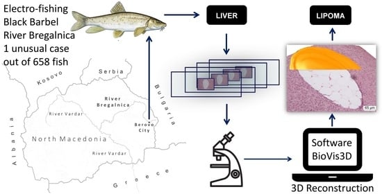
The case black barbel was collected by electro-fishing in the upper part of the River Bregalnica, near the city of Berovo (22°51′27.9″ E, 41°41′59.8″ N), which does not receive effluents from urban, industrial or agricultural origins. This low anthropogenic-impacted area has been used as a reference location in ecotoxicological monitoring studies [30,31]. The animal with the unusual microanatomical feature was collected in summer. It was 1 out of 658 black barbels subjected to liver histopathology—with an intensive systematic sampling strategy—after being randomly caught over one year in the River Bregalnica.
The fish in consideration was measured and weighed, examined for external visual abnormalities and, after the necropsy, examined for internal abnormalities. Immediately after the gross visual inspection, the liver was removed and weighed. The liver was then sliced into a couple of slabs that were fixed in Bouin’s fixative for 48 h, dehydrated in increasing concentrations of ethanol, cleared in xylene and embedded in paraffin wax blocks. All the blocks obtained from this particular fish were exhaustively cut in serial 5 µm-thick sections and routinely stained with haematoxylin and eosin (H&E). One section with the unusual feature was stained with periodic acid–Schiff (PAS) for carbohydrates.
All the serial sections containing the single intrahepatic agglomerate of adipocytes were chosen for the software-supported 3D reconstruction. In every section, the area of interest was photographed with a light microscope (BX50, Olympus, Tokyo, Japan) equipped with a digital camera (Camedia C5050, Olympus, Tokyo, Japan). The photographs for illustration were in TIFF format, and for reconstruction, they were high-resolution images (JPEG, 2560 × 1920 pixels). The latter photos were sequentially imported to a dedicated software (v3.0, BioVis3D, Montevideo, Uruguay), for building the baseline image stack that would support the 3D reconstruction. The software was calibrated for the magnification of the images. The final 3D reconstruction was subjected to digital rendering before being exported as TIFF files.

3. Results

One agglomerate of well-differentiated unilocular adipose cells was noticed when studying the serial histological sections from one black barbel’s liver. Taking into account that 658 captured fish were analysed—in the context of a biomonitoring study—the estimated prevalence of the intrahepatic oddity in the Bregalnica black barbel population was 1.5‰.
The case fish was a two-year-old female with a body weight (BW) of 27.95 g, total length (TL) of 135 mm and liver weight (LW) of 0.68 g. The condition factor of the fish (K = 100 × BW/TL3) was 1.36, and the hepatosomatic index (HSI = 100 × LW/BW) was 2.43.
During the necropsy, neither the external nor the internal examinations revealed gross changes or deformities that could signal pathological conditions (Figure 1). In particular, the liver had a normal appearance, with the typical reddish-brown colour and no gross lesions. However, the microscopic examination of the liver revealed the presence of one “island” made of distinctively empty-looking cells (Figure 2A). The cell mass was encircled by normal liver parenchyma. When sectioned, in the microscopic slides, the cell agglomerate was oval-shaped. It did not protrude the surface of the liver because it was buried deep in the organ, and it was tiny, reaching a maximum diameter of ~0.5 mm.
The nodular lesion was perfectly circumscribed and consisted of well-differentiated and large adipocytes. Their individual size varied little from one another. They contained an unambiguous single roundish empty space that filled most of the unilocular fat cell’s cytoplasm. Indeed, it should be stressed that during the preparation of the tissue for histology, which includes dehydration in ethanol and clearing in xylene, free lipids are expected to dissolve. Therefore, the cytoplasmic location where the major lipid droplet stayed, in vivo, appeared empty in histological sections. The cytoplasm was negative for PAS, excluding the idea that carbohydrates could have caused the empty space. The nucleus was squeezed towards the cell’s periphery. The intercellular limits were prominent. At times, an interstitial connective tissue matrix could be seen. Lipoblasts and mitotic figures were not observed. The internal vasculature was made of a few erratic thin-walled capillaries.
In specific smaller profiles of the adipose cell agglomerate, and at its margins, there were scattered groups of small roundish cells, with a morphology compatible with a leucocytic (mainly lymphocytic) infiltrate (Figure 2B,C). When the agglomerate profile was evidently smaller, we noted a relatively more extended infiltrate in the vicinity of a venule (Figure 2C). We saw no microparasites of any sort at the margin or inside the agglomerate.
The agglomerate of adipocytes was peripherally associated with scant small venules (Figure 2C), but not any major blood vessels or intrahepatic pancreatic tissue, and it was situated far from the liver hilum. Thus, the agglomerate was not a pocket made of an extension of the extrahepatic (peritoneal) fat. It was perfectly well demarked from the normal hepatic parenchyma but not encapsulated by a distinct connective tissue band. The surrounding hepatocytic elements caused no prominent peripheral compression of the agglomerate.
When looking at the 3D reconstructed agglomerate of adipose cells, we concluded that it was not lobulated, and that it approximately formed a flattened oblate spheroid, which looked like a sphere that was markedly squashed from top to bottom (Figure 3). The shorter “a” and longer “b” equatorial (horizontal) axes attained 375 µm and 520 µm, respectively, while the polar (vertical) “c” axis reached 65 µm. Applying the geometric formula for the volume (V) of a perfect oblate spheroid (V = (4/3) × π × a/2 × b/2 × c/2), the fat cell cluster had a V of approximately 6.7 × 106 µm3 (~0.01 mm3).

4. Discussion

This study histologically described and 3D reconstructed an intrahepatic collection of white adipocytes in a black barbel. Besides the structural evidence for such identification, the negativity for PAS staining indirectly supported that the major cytoplasm content in vivo should have been lipids; using paraffin, direct lipid staining was impossible. The oval-shaped and well-demarcated focus of unilocular fat cells was deeply nested into the liver. The histological characteristics of the adipocytic focus, including the absence of lipoblasts, increased mitosis, scarcity of vasculature and association with lymphocytes, nicely matched detailed lipoma descriptions [32]. Objectively, the focus fulfilled the basic structural definition of a lipoma [1] and therefore was presumptively diagnosed as such.
The good circumscription of the cell agglomerate favours a priori this diagnosis over another possibility: ectopic adipocytes. The presence of ectopic fat and ectopic adipocytes in unexpected locations is usually reported in human and experimental animal pathology in strict association with caloric intake (high-fat diets) exceeding expenditure, obesity, insulin resistance and type 2 diabetes mellitus [33,34]. Still, despite the fact the liver may accumulate ectopic fat (within steatotic hepatocytes), it is not among the organs where ectopic adipocytes typically appear under triggering conditions [35,36]. In the case revealed here, it is virtually impossible that just one wild barbel would have been consistently subjected to a high-fat diet, which, even so, would have sparked steatosis (not observed here) well in advance of ectopic adipocyte differentiation and proliferation. Therefore, the structural and circumstantial evidence favours the lipoma diagnosis over a pocket of ectopic adipocytes.
Anyway, it could be questioned if such a small focus of fat cells can be diagnosed as a lipoma in barbels. However, the literature does not specify a minimal size for an abnormal agglomerate of fat cells to be deemed as a lipoma. In humans, the size of (clinically detected) lipomas varies between 2 and 10 cm in diameter [36]. Yet, such benign neoplasms are very slow progressing. Therefore, the macroscopic tumours derive from earlier and much smaller (micro)lipomas, as illustrated well in intramucosal lipomas [37].
According to the predominant histological morphology, the World Health Organization classification of tumours of soft tissue considers the following lipoma types: conventional lipoma (the most common by far), fibrolipoma, angiolipoma, lipomatosis of nerve and lipoblastoma [38]. Since the liver lipoma in the studied black barbel consisted predominantly of mature adipocytes, without an excess of fibrous tissue or of substantial vascular involvement, it should be classified as a conventional lipoma.
Lipomas are rarely reported in fish, and, as far as we know, it seems that a lipoma has only been reported once in the liver [25]. Unlike the present findings in the black barbel, where the lipoma was noted only after histological examination, the liver lipoma found in the marine coly was visually apparent and noticed in the lower lobe of the liver, having a smooth surface and greenish colour [25]. Additionally, this liver lipoma was found in a seven-year-old specimen. Lipomas are generally noticed in older individuals. Their incidence increases with age in domestic animals [39] and in humans [40]. Contrarily, in the current case, the lipoma was noticed in a two-year-old female, an adult but not a particularly old fish. It is common to find specimens of B. balcanicus aged over two years [41,42], and, in general, barbels can live for more than 20 years [43].
The cause(s) of lipoma development in animals is (are) far from clear, but several theories were advanced, mostly from studies in humans. Some authors proposed genetic causes while favouring trauma as the aetiology [44], along with a diversity of risk factors, such as obesity, ethanol abuse, glucose intolerance and liver disease [40]. In farmed fish, dysmetabolic syndrome was suggested as the starting point for lipoma formation [19]. A link with lipid metabolism was reinforced when lipomas (also mentioned as lipoma-like fat tissue accumulations) were induced in a zebrafish model of obesity overexpressing the Akt1 “insulin signalling hub” gene [45]. In channel catfish (Ictalurus punctatus), subcutaneous lipoma was considered to be related to direct exposure to the carcinogenic and mutagenic N-Methyl-N’-Nitro-N-Nitrosoguanidine [14]. In the largemouth bass (Micropterus salmoides), a spleen lipoma was found in one specimen from a zone polluted by a range of contamination sources, but no toxicological aetiology was considered [23].
This microlipoma in a black barbel from a clean location was the only benign lesion found in all studied fish [31]. Facing the condition’s rarity, a definitive causal explanation would be too speculative because the causes could reasonably range from purely genetic to toxicopathic. Although the latter aetiology is unlikely, it cannot be discarded in view of the sampling location, i.e., when knowing that liver lipomatous lesions resembling human hepatic lipomas evolved in rats from carcinogenic assays [46].
As with other lipomas, those in the human liver have no clear aetiology and are very rare [47]. A correlative study supported that, at least in humans, lipomas have a significantly greater association with steatosis when compared to non-lipomatous lesions [48]. In this black barbel single case, it is reiterated that no such association of lesions existed.
The use of 3D technology, including histopathologic reconstruction, to study lesions is a rapidly evolving practice in pathology, recognised as “useful in diagnostic, prognostic, and therapeutic decision-making for the medical and biomedical professions” [49]. Here, a 3D reconstruction of the microlipoma was created for the first time from serial histological sections, exposing a prolate spheroid that can only result from non-homogenous growth. If the fat cells expanded homogeneously in 3D, the focus would be spherical. Therefore, the 3D rebuild not only allowed insights on such kinetics but was also essential in confirming the lack of lobulation and in realistically knowing the mass’s shape and size (volume).
In the end, what is the significance of a liver lipoma for a single fish or a fish population? Given its evident rarity, an impact on populations can be excluded. Regarding the individual, it can be deduced that the health impact will depend on the lipoma volume. In the black barbel, given the small size of the lipoma, it is inferred that it was in an early stage of development and growth. The mass minimally disturbed the nearby parenchyma, and therefore it unlikely altered the liver function. However, it may be theorised that severe morphofunctional impacts could appear if the lipoma expands over the years, finally leading to individual health effects. Notably, one grown liver lipoma in otherwise healthy human beings sparked abdominal pain, nausea and dyspepsia [50,51].
Irrespective of its cause and significance, the microlipoma described here seems to be the second report of a hepatic lipoma in any fish species and the first in a barbel species. The proposed diagnosis and detailed description of the finding in the black barbel can grab the attention of researchers conducting liver histopathology in fish and consequently raise awareness for this very likely underdiagnosed and underreported situation.

Author Contributions

Conceptualisation, K.R., M.J. and E.R.; data curation, K.R.; formal analysis, K.R., M.J., J.A. and E.R.; investigation, K.R., M.J., J.A. and E.R.; methodology, K.R., M.J., J.A. and E.R.; software, J.A.; supervision, M.J. and E.R.; writing—original draft, K.R. and J.A. All authors have read and agreed to the published version of the manuscript.

Funding

This study was partially supported by the Strategic Funding UID/Multi/04423/2019 provided by FCT and ERDF to CIIMAR/CIMAR, in the framework of the programme PT2020.

Institutional Review Board Statement

Not applicable. Ethical review and approval were waived for this study because it does not incur any actions falling within the “procedure” definition established by the Republic of North Macedonian and Portuguese law, in line with EU Directive No. 2010/63 (article 3, number 1). The latter excludes from the “procedure” the killing of animals solely for the use of their organs or tissues. This study used only organs and tissues of fish euthanised solely for the described purpose. In line with the EU Directive No. 2010/63, article 6, number 2, the killing was conducted by competent persons, trained and accredited by law to do so. The killing was at the site of capture.

Informed Consent Statement

Not applicable.

Data Availability Statement

The data are available on request from the corresponding author.

Conflicts of Interest

The authors declare no conflict of interest.

References

  1. Schlumberger, H.H.; Lucké, B. Tumors of fishes, amphibians and reptiles. Cancer Res. 1948, 8, 657–754. [Google Scholar] [PubMed]
  2. Johnson, C.N.; Ha, A.S.; Chen, E.; Davidson, D. Lipomatous soft-tissue tumors. J. Am. Acad. Orthop. Surg. 2018, 26, 779–788. [Google Scholar] [CrossRef] [PubMed]
  3. Charifa, A.; Azmat, C.E.; Badri, T. Lipoma Pathology. In StatPearls; StatPearls Publishing: Treasure Island, FL, USA, 2020. [Google Scholar]
  4. Stolk, A. Tumours of fishes 29. Lipoma in the eel Anguilla anguilla. Beaufortia 1959, 7, 193–198. [Google Scholar]
  5. Tubiash, H.D.; Hendricks, J.D. A possible lipoma in the weakfish, Cynoscion regalis (Bloch and Schneider). Chesap. Sci. 1973, 14, 145–146. [Google Scholar] [CrossRef]
  6. Bruno, D.W.; McVicar, A.H.; Fraser, C.O. Multiple lipoma in the common dab, Limanda limanda L. J. Appl. Ichthyol. 1991, 7, 238–243. [Google Scholar] [CrossRef]
  7. Marino, F.; Monaco, S.; Salvaggio, A.; Macrì, B. Lipoma in a farmed northern bluefin tuna, Thunnus thynnus (L.). J. Fish Dis. 2006, 29, 697–699. [Google Scholar] [CrossRef] [PubMed]
  8. Gómez, S. Multiple dermal lipomas in farmed striped seabream Lithognathus mormyrus on the Spanish Mediterranean coast. Dis. Aquat. Organ. 2009, 85, 77–79. [Google Scholar] [CrossRef] [Green Version]
  9. McCoy, C.P.; Bowser, P.R.; Steeby, J.; Bleau, M.; Schwedler, T.E. Lipoma in channel catfish (Ictalurus punctatus Rafinesque). J. Wild. Dis. 1985, 21, 74–76. [Google Scholar] [CrossRef] [PubMed] [Green Version]
  10. Easa, M.L.S.; Easa, M.; Harshbarger, J.C.; Hetrick, F.M. Hypodermal lipoma in a striped (grey) mullet Mugil cephalus. Dis. Aquat. Organ. 1989, 6, 157–160. [Google Scholar] [CrossRef]
  11. Szewc, M.; Gawlik, P.; Żebrowski, R.; Sitarz, R. Giant lipoma in the fronto-temporo-parietal region in an adult man: Case report and literature review. Clin. Cosmet. Investig. Dermatol. 2020, 13, 1015–1020. [Google Scholar] [CrossRef]
  12. Schwartz, F.J.; Màrquez, R. A lipoma in the striped mojarm, Diapterus plumieri (Pisces: Gerridae) from Mexico. J. Elisha Mitchell Sci. Soc. 1971, 87, 87–90. [Google Scholar]
  13. Hard, G.C.; Williams, R.; Lee, J. Survey of Demersal Fish in Port Phillip Bay for Incidence of Neoplasia. Aust. J. Mar. Freshw. Res. 1979, 30, 73–79. [Google Scholar] [CrossRef]
  14. Chen, H.C.; Pan, I.J.; Tu, W.J.; Lin, W.H.; Hong, C.C.; Brittelli, M.R. Neoplastic Response in Japanese Medaka and Channel Catfish Exposed to N-Methyl-N′-Nitro-N-Nitrosoguanidine. Toxicol. Pathol. 1996, 24, 696–706. [Google Scholar] [CrossRef] [PubMed]
  15. Abowei, J.F.N.; Briyai, O.F.; Bassey, S.E. A Review of Some Viral, Neoplastic, Environmental and Nutritional Diseases of African Fish. Br. J. Pharmacol. Toxicol. 2011, 2, 227–235. [Google Scholar]
  16. De Stefano, C.; Bonfiglio, R.; Montalbano, G.; Giorgianni, P.; Lanteri, G. Multicentric lipoma in a molly (Poecilia velifera). Bull. Eur. Assoc. Fish Pathol. 2012, 32, 220–224. [Google Scholar]
  17. Sood, N.; Swaminathan, T.R.; Yadav, M.K.; Pradhan, P.K.; Kumar, R.; Sood, N.K. First report of cutaneous infiltrative lipoma in goldfish Carassius auratus. Dis. Aquat. Org. 2017, 125, 243–247. [Google Scholar] [CrossRef]
  18. Johnston, C.J.; Deveney, M.R.; Bayly, T.; Nowak, B.F. Gross and histopathological characteristics of two lipomas and a neurofibrosarcoma detected in aquacultured southern bluefin tuna, Thunnus maccoyii (Castelnau), in South Australia. J. Fish Dis. 2008, 31, 241–247. [Google Scholar] [CrossRef]
  19. Marino, F.; Chiofalo, B.; Mazzullo, G.; Panebianco, A. Multicentric infiltrative lipoma in a farmed Mediterranean seabass Dicentrarchus labrax: A pathological and biochemical case study. Dis. Aquat. Organ. 2011, 96, 259–264. [Google Scholar] [CrossRef] [PubMed] [Green Version]
  20. Lester, R.J.G.; Kelly, W.R. Tumour-like growths from southern Australian marine fish. Tasmanian Fish Res. 1983, 25, 27–32. [Google Scholar]
  21. Ramos, P.; Faisca, P.; Carneiro, M.; Rosa, F. Dermal Melanoma and Mesenteric Lipoma in a Senegal Seabream, Diplodus bellottii (Steindachner, 1882) from the Portuguese Coast. Exp. Pathol. Health Sci. 2016, 8, 67–68. [Google Scholar]
  22. Singaravel, V.; Gopalakrishnan, A.; Vijayakumar, R.; Raja, K. Prevalence and pathology of gastric tumours in Indian oil sardine (Sardinella longiceps) from Parangipettai coastal waters, southeast coast of India. J. Coast. Life Med. 2015, 3, 592–595. [Google Scholar] [CrossRef]
  23. Hinck, J.E.; Blazer, V.S.; Denslow, N.D.; Echols, K.R.; Gale, R.W.; Wieser, C.; May, T.W.; Ellersieck, M.; Coyle, J.J.; Tillitt, D.E. Chemical contaminants, health indicators, and reproductive biomarker responses in fish from rivers in the Southeastern United States. Sci. Total Environ. 2008, 390, 538–557. [Google Scholar] [CrossRef] [PubMed] [Green Version]
  24. Stejskal, V.; Kouřil, J.; Policar, T.; Svobodová, Z. Splenic lipidosis in intensively cultured perch, Perca fluviatilis L. J. Fish Dis. 2015, 39, 87–93. [Google Scholar] [CrossRef]
  25. Thomas, L. Sur un lipome abdominal chez un colin. Bull. Ass. Fr. Etude Cancer 1933, 22, 419–435. [Google Scholar]
  26. Figueiredo-Fernandes, A.M.; Fontaínhas-Fernandes, A.A.; Monteiro, R.A.F.; Reis-Henriques, M.A.; Rocha, E. Spatial relationships of the intrahepatic vascular-biliary tracts and associated pancreatic acini of Nile tilapia, Oreochromis niloticus (Teleostei, Cichlidae): A serial section study by light microscopy. Ann. Anat. 2007, 189, 17–30. [Google Scholar] [CrossRef] [PubMed]
  27. Imrie, D.; Sadler, K.C. White adipose tissue development in zebrafish is regulated by both developmental time and fish size. Dev. Dyn. 2010, 239, 3013–3023. [Google Scholar] [CrossRef] [Green Version]
  28. Salmerón, C. Adipogenesis in fish. J. Exp. Biol. 2018, 221, jeb161588. [Google Scholar] [CrossRef] [PubMed] [Green Version]
  29. Sousa, S.; Rocha, M.J.; Rocha, E. Characterization and spatial relationships of the hepatic vascular–biliary tracts, and their associated pancreocytes and macrophages, in the model fish guppy (Poecilia reticulata): A study of serial sections by light microscopy. Tissue Cell 2018, 50, 104–113. [Google Scholar] [CrossRef]
  30. Jordanova, M.; Rebok, K.; Naskovska, M.; Kostov, V.; Rocha, E. Splenic pigmented macrophage aggregates in barbel (Barbus peloponnesius, Valenciennes, 1844) from River Bregalnica—Influences of age, sex and season on a pollution biomarker. Turkish J. Fish Aquat. Sci. 2016, 16, 881–890. [Google Scholar] [CrossRef]
  31. Rebok, K.; Jordanova, M.; Slavevska-Stamenković, V.; Ivanova, L.; Kostov, V.; Stafilov, T.; Rocha, E. Frequencies of erythrocyte nuclear abnormalities and of leucocytes in the fish Barbus peloponnesius correlate with a pollution gradient in the River Bregalnica (Macedonia). Environ. Sci. Pollut. Res. 2017, 24, 10493–10509. [Google Scholar] [CrossRef]
  32. McTighe, S.; Chernev, I. Intramuscular lipoma: A review of the literature. Orthop. Rev. Pavia 2014, 6, 5618. [Google Scholar] [CrossRef] [PubMed] [Green Version]
  33. Church, C.; Horowitz, M.; Rodeheffer, M. WAT is a functional adipocyte? Adipocyte 2012, 1, 38–45. [Google Scholar] [CrossRef] [Green Version]
  34. Snel, M.; Jonker, J.T.; Schoones, J.; Lamb, H.; de Roos, A.; Pijl, H.; Smit, J.W.A.; Meinders, A.E.; Jazet, I.M. Ectopic fat and insulin resistance: Pathophysiology and effect of diet and lifestyle interventions. Int. J. Endocrinol. 2012, 2012, 983814. [Google Scholar] [CrossRef]
  35. De Munck, T.J.I.; Soeters, P.B.; Koek, G.H. The role of ectopic adipose tissue: Benefit or deleterious overflow? Eur. J. Clin. Nutr. 2021, 75, 38–48. [Google Scholar] [CrossRef] [PubMed]
  36. Kosztyuova, T.; Shim, T.N. Rapidly enlarging lipoma. BMJ Case Rep. 2017, 2017, bcr2017221272. [Google Scholar] [CrossRef] [PubMed]
  37. Caliskan, A.; Kohlmann, W.; Affolter, K.; Downs-Kelly, E.; Kanth, P.; Bronner, M.P. Intramucosal lipomas of the colon implicate Cowden syndrome. Mod. Pathol. 2018, 31, 643–651. [Google Scholar] [CrossRef] [PubMed] [Green Version]
  38. WHO Classification of Tumours Editorial Board. WHO Classification of Tumours of Soft Tissue and Bone, 4th ed.; International Agency for Research on Cancer: Lyon, France, 2013; p. 427. [Google Scholar]
  39. Moulton, J.E. (Ed.) Tumors in Domestic Animals; University of California Press: Berkeley, CA, USA, 1990. [Google Scholar]
  40. Kolb, L.; Yarrarapu, S.N.S.; Ameer, M.A.; Rosario-Collazo, J.A. Lipoma. In StatPearls; StatPearls Publishing: Treasure Island, FL, USA, 2020. Available online: https://www.ncbi.nlm.nih.gov/books/NBK507906/ (accessed on 6 October 2020).
  41. Bertoli, M.; Pizzul, E.; Devescovi, V.; Franz, F.; Pastorino, P.; Giulianini, P.G.; Ferrari, C.; Nonnis Marzano, F. Biology and distribution of Danube barbel (Barbus balcanicus) (Osteichthyes: Cyprinidae) at the Northwestern limit of its range. Eur. Zool. J. 2019, 86, 280–293. [Google Scholar] [CrossRef] [Green Version]
  42. Žutinić, P.; Jelić, D.; Jelić, M.; Buj, I. A contribution to understanding the ecology of the large spot barbel-sexual dimorphism, growth and population structure of Barbus balcanicus (Actinopterygii; Cyprinidae) in Central Croatia. N.-West. J. Zool. 2013, 10, 158–166. [Google Scholar]
  43. Amat Trigo, F.; Roberts, C.G.; Britton, J.R. Spatial variability in the growth of invasive European barbel Barbus barbus in the River Severn basin, revealed using anglers as citizen scientists. Knowl. Manag. Aquat. Ecosyst. 2017, 418, 17. [Google Scholar] [CrossRef]
  44. Aust, M.C.; Spies, M.; Kall, S.; Jokuszies, A.; Gohritz, A.; Vogt, P. Posttraumatic lipoma: Fact or fiction? Skinmed 2007, 6, 266–270. [Google Scholar] [CrossRef]
  45. Chu, C.-Y.; Chen, C.-F.; Rajendran, R.S.; Shen, C.-N.; Chen, T.-H.; Yen, C.-C.; Chuang, C.-K.; Lin, D.-S.; Hsiao, C.-D. Overexpression of Akt1 enhances adipogenesis and leads to lipoma formation in zebrafish. PLoS ONE 2012, 7, e36474. [Google Scholar] [CrossRef] [PubMed] [Green Version]
  46. Dixon, D.; Yoshitomi, K.; Boorman, G.A.; Maronpot, R.R. “Lipomatous” lesions of unknown cellular origin in the liver of B6C3F1 mice. Vet. Pathol. 1994, 31, 173–182. [Google Scholar] [CrossRef] [PubMed]
  47. Puljiz, Ž.; Petričević, M.; Bratanić, A.; Barišić, I.; Puljiz, M.; Karin, Ž. An unusually large liver lipoma. Case report. Med. Glas. Zenica 2012, 9, 411–414. [Google Scholar]
  48. Martin-Benitez, G.; Marti-Bonmati, L.; Barber, C.; Vila, R. Hepatic lipomas and steatosis: An association beyond chance. Eur. J. Radiol. 2011, 81, 491–494. [Google Scholar] [CrossRef]
  49. Farahani, N.; Braun, A.; Jutt, D.; Huffman, T.; Reder, N.; Liu, Z.; Yagi, Y.; Pantanowitz, L. Three-dimensional imaging and scanning: Current and future applications for pathology. J. Pathol. Inform. 2017, 8, 36. [Google Scholar] [CrossRef]
  50. Manenti, G.; Picchi, E.; Castrignanò, A.; Muto, M.; Nezzo, M.; Floris, R. Liver lipoma: A case report. BJR Case Rep. 2016, 3, 20150467. [Google Scholar] [CrossRef] [PubMed]
  51. Reddy, O.J.; Gafoor, J.A.; Suresh, B.; Prasad, P.O. Lipoma in liver: A rare presentation. J. Dr. NTR Univ. Health Sci. 2015, 4, 185–187. [Google Scholar] [CrossRef]
Figure 1. View of the necropsy procedure on the black barbel from the River Bregalnica. Gross lesions were not found on the body surface or in internal organs and cavities.
Applsci 11 08392 g001
Figure 2. Representative histological sections (AC) across the intrahepatic adipose cell agglomerate. There is a well-marked frontier between the adipocytes (*) and the liver parenchyma (LP). The flattened nuclei of the adipocytes are perceivable at the cell peripheries (dashed arrows). At specific locations around the adipose cell agglomerate, lymphocytic-like infiltrates are noted (block arrows) in the close vicinity of venules (V).
Applsci 11 08392 g002
Figure 3. Three-dimensional (3D) reconstructions (A,C) of the intrahepatic adipose cell agglomerate, side by side with two “bi-dimensional” aspects (B,D). The last images have digital overlays (in orange) partially superimposed on the histological picture to explain how each histological image contributes to building the serial stack. The microlipoma flattened oval shape became evident when viewing the 3D reconstruction at different angles.
Applsci 11 08392 g003
Publisher’s Note: MDPI stays neutral with regard to jurisdictional claims in published maps and institutional affiliations.