Article

Bilateral Deficits during Maximal Grip Force Production in Late Postmenopausal Women

1
Department of Human Movement Science, Graduate School, Incheon National University, Incheon 22012, Korea
2
Division of Health & Kinesiology, Incheon National University, Incheon 22012, Korea
3
Division of Sport Science, Sport Science Institute & Health Promotion Center, Incheon National University, Incheon 22012, Korea
4
Neuromechanical Rehabilitation Research Laboratory, Incheon National University, Incheon 22012, Korea
*
Author to whom correspondence should be addressed.
Appl. Sci. 2021, 11(18), 8426; https://doi.org/10.3390/app11188426
Submission received: 18 August 2021 / Revised: 8 September 2021 / Accepted: 8 September 2021 / Published: 10 September 2021
(This article belongs to the Special Issue New Trends in Neuromechanics and Motor Rehabilitation)

Abstract

:
The purpose of this study was to investigate bilateral deficit patterns during maximal hand-grip force production in late postmenopausal women. Twenty late postmenopausal and 20 young premenopausal women performed maximal isometric grip force production tasks with dominant and nondominant hands and both hands, respectively. For late postmenopausal women, pulse wave analysis was used for identifying a potential relationship between maximal hand-grip strength and risk factors of cardiovascular disease. The findings showed that late postmenopausal women produced significantly decreased maximal hand-grip strength in dominant and nondominant and bilateral hand conditions compared to those of premenopausal women. Bilateral deficit patterns appeared in late postmenopausal women. For late postmenopausal women, decreased dominant and bilateral hand-grip forces were significantly related to greater bilateral deficit patterns. Further, less maximal hand-grip strength in unilateral and bilateral hand conditions correlated with greater central pulse pressure. These findings suggested that age-related impairments in muscle strength and estrogen deficiency may interfere with conducting successful activities of bilateral movements. Further, assessing maximal dominant hand-grip strength may predict bilateral deficit patterns and risk of cardiovascular disease in late postmenopausal women.

1. Introduction

Menopause typically occurs in women’s 40s [1], and one third of women’s lifespan is spent post-menopause [2]. Progressive reduction of estrogen in postmenopausal women may facilitate more age-related deficits in the central and peripheral nervous system [3,4,5,6]. For example, muscle weakness normally appears in elderly people because of age-induced neurophysiological alterations [7,8,9]. Furthermore, asymmetrical interlimb muscle strength interferes with executing bilateral movements that account for 54% of daily activities in the aging population [10,11]. Importantly, postmenopausal women reveal more significant reduction of muscle strength than premenopausal women and age-matched males [12,13,14,15,16].
Bilateral deficit is a phenomenon when individuals reveal lower force outputs produced simultaneously by both limbs than the sum of unilateral forces generated by each limb. Previous studies indicate that bilateral deficit may appear in either upper or lower extremities during various motor tasks such as maximal voluntary contraction (MVC), reaction time [17,18,19], and different contraction types (e.g., isometric and dynamic contraction) [20]. Moreover, greater levels of bilateral deficit are associated with increased impairment in bilateral performances (e.g., ballistic push-off and vertical squat jumping) [21,22,23], and several aging studies report bilateral deficit patterns in elderly people interfering with various functional movements (e.g., rising from a chair) [24,25].
For postmenopausal and elderly women, previous studies report bilateral deficit patterns in lower limb movements such as leg extension and leg press [24,25,26,27,28]. Specifically, greater bilateral deficit in producing explosive forces increases the time of sit-to-stand performance [25]. In postmenopausal women, increased interhemispheric inhibition and reduced muscle strength potentially induced by deficiency of estrogen and/or progesterone may facilitate bilateral deficit patterns [20,28,29]. However, these previous findings are mostly limited to lower limb movements. Given that successful bilateral upper limb movements are additional critical motor functions for older adults [11], determining whether bilateral deficit patterns in upper limb movements appear in late postmenopausal women is necessary. Thus, the purpose of this study was to examine bilateral deficit patterns in late postmenopausal women using voluntary maximal handgrip force tasks.

2. Materials and Methods

2.1. Particiapants

Twenty healthy late postmenopausal women (mean and standard deviation of age = 65.5 ± 3.1 years) and 20 healthy young premenopausal women (mean and standard deviation of age = 23.4 ± 2.1 years) participated in this study. We recruited participants using flyers in the university and local community centers and confirmed that all participants had no musculoskeletal deficits (e.g., sarcopenia) in their upper extremities, neurological disease, cardiovascular diseases, and significant cognitive impairments. Late postmenopausal women were defined as those with more than four years after menopause [30]. All participants were right-handed as assessed by the Edinburgh handedness inventory [31]. Specific details on demographic information are summarized in Table 1. Before starting the testing, all participants read and signed an informed consent form and experimental protocols approved by the University’s Institutional Review Board.

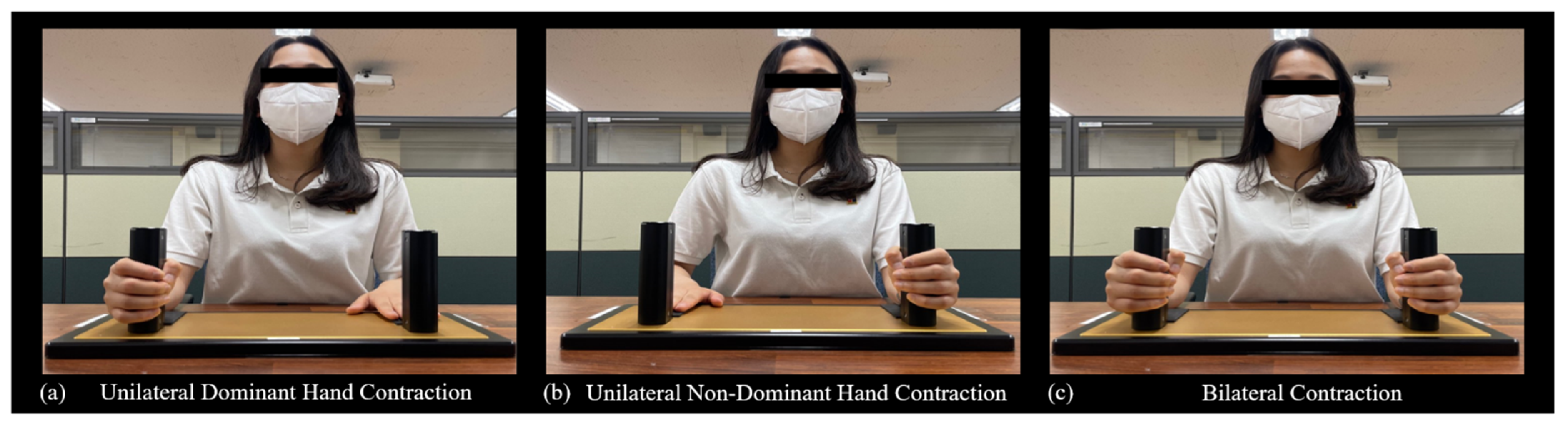
2.2. Experimental Setup

To investigate the bilateral deficit phenomenon in the upper extremities, we used an isometric hand-grip force production paradigm. Before executing isometric force production tasks, participants sat 80 cm away from a 54.6 cm LED monitor (1920 × 1080 pixels; refresh rate = 60 Hz, Dell, Round Rock, TX, USA) and maintained comfortable positions with 15–20° of shoulder flexion and 20–45° of elbow flexion. Using an isometric hand-grip force measurement system (SEED TECH Co., Ltd., Bucheon, Korea), participants grasped the handle (diameter = 30 mm) and produced their maximal isometric force outputs with their unilateral hand and both hands, respectively. Further, we instructed the participants to put their resting hand on the pad during the unilateral tasks and maintain their forearms fixed on the table with same position to avoid inadvertent force output caused by elbow, shoulder, or trunk movements.
We administered two consecutive maximal force production trials for each hand condition: (a) unilateral dominant hand (Figure 1a), (b) unilateral nondominant hand (Figure 1b), and (c) both hands (Figure 1c). For each trial, participants generated as much isometric hand-grip forces as possible for 3 s. They had 60 s of rest between trials and 180 s of rest between hand conditions. The mean of two maximal force production trials for each hand condition was used for further analysis.
Changes in handgrip strength in the aging population may be a risk factor indicating the occurrences of various cardiovascular diseases (e.g., hypertension, coronary artery disease, heart failure, or stroke) [32,33], and reduction of handgrip strength in elderly women is highly related to all-cause mortality [34]. Thus, for late postmenopausal women, we additionally performed non-invasive pulse wave analysis (PWA) using the SphygmoCor Xcel system (AtCor Medical, Sydney, Australia) to investigate the potential relationship between hand-grip force productions and cardiovascular disease risk factors. Before the PWA data collection, participants fasted for at least 10–12 h. All measurements proceeded in a light- and temperature-controlled room after resting for at least 10 min in the supine position. Participants wore a blood pressure cuff on their right upper arm to measure PWA. The blood pressure cuff automatically inflated to measure the brachial blood pressure and after deflating, it re-inflated to capture PWA waveforms. We conducted PWA at least three times, and the mean of two values which ranged within ±5 mmHg in blood pressure, ±5 beats/min in heart rate, and ±3% in augmentation index (AIx) was used for the further analysis.

2.3. Data Analysis

Bilateral index (%) of maximal handgrip force output (MF) was calculated by the following equations [35]. The values of bilateral index below zero indicate that bilateral motor performance was less than the sum of unilateral motor performance from each hand, so more negative values of bilateral index denote greater bilateral deficit patterns.
Bilateral   index   ( % ) = ( 100 × Bilateral   hands   MF ( Dominant   hand   MF + Non dominant   hand   MF ) ) 100
Based on the brachial waveforms obtained from the blood pressure cuff, central aortic pressure waveforms were automatically calculated by the mathematical transfer function [36,37,38]. Central pulse pressure (cPP) is the difference between central systolic blood pressure (cSBP) and central diastolic blood pressure (cDBP). In addition, AIx (%), a measure of arterial stiffness, is calculated as the ratio of augmentation pressure (i.e., cSBP–inflection pressure) and cPP. Increased values of cPP and AIx may be related to a higher appearance rate of cardiovascular disease [39,40,41].
For statistical analyses, we performed an independent t-test to compare the differences of the bilateral index and maximal force production of unilateral hand and both hands between late postmenopausal and young premenopausal women. In addition, one sample t-test was used for determining whether the bilateral index for each group was significantly different from zero. For the late postmenopausal women group, Pearson’s correlation analyses were performed to determine potential relations of maximal hand-grip forces of unilateral hand and both hands to bilateral deficit index as well as cardiovascular disease risk factors. Using the Shapiro–Wilk test, we confirmed that all dependent variables met the assumption of normality. Statistical analyses were performed using IBM SPSS Statistics version 25 (SPSS Inc, Chicago, IL, USA) with alpha set at 0.05.

3. Results

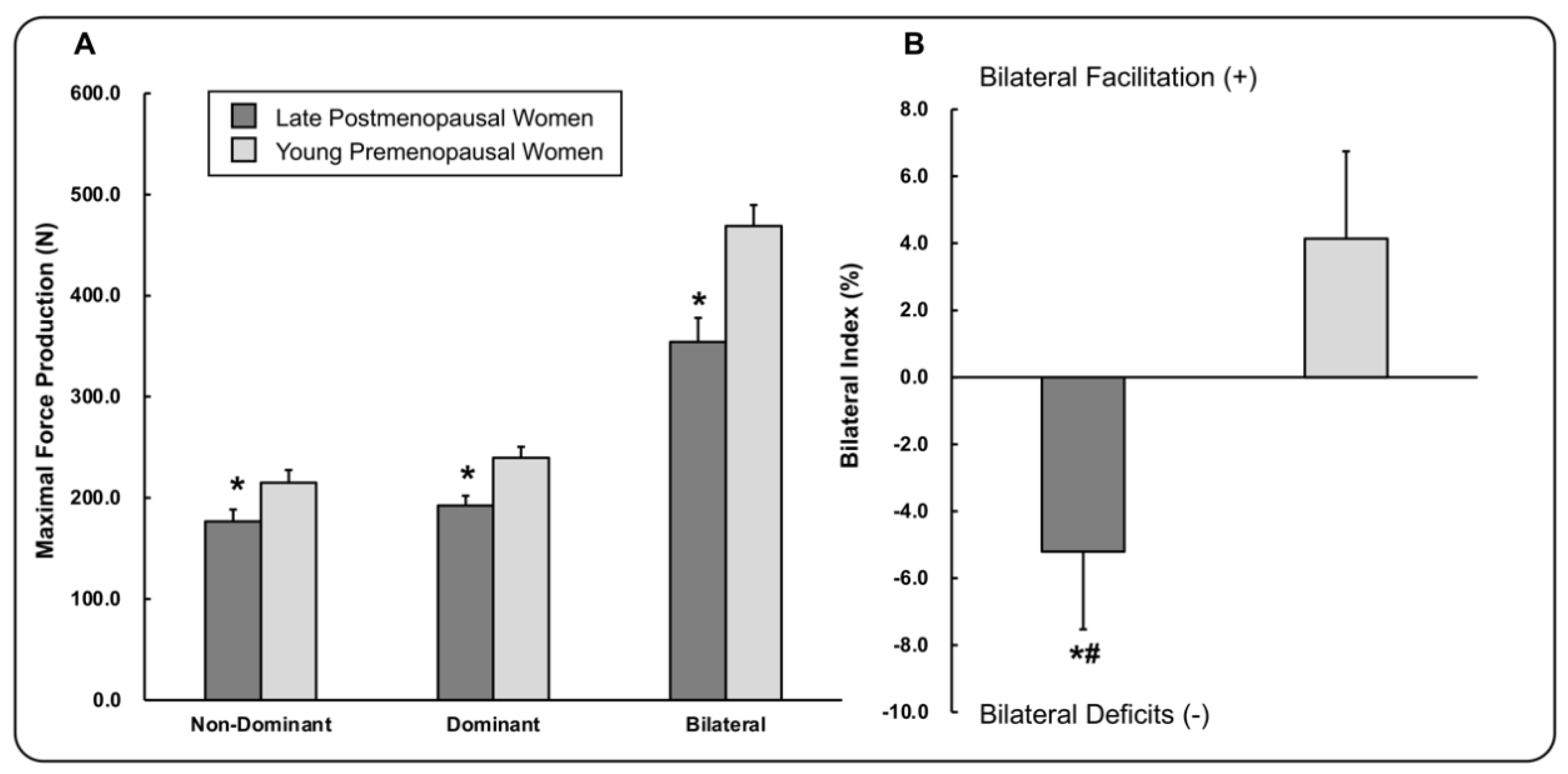
3.1. Maximal Hand-Grip Force Production

Maximal force production in late postmenopausal women was significantly lower than that in young premenopausal women, respectively, in the dominant hand (t38 = −3.26, p = 0.002), nondominant hand (t38 = −2.26, p = 0.03), and both hands (t38 = −3.63, p = 0.001; Figure 2A). Furthermore, the bilateral index values were significantly different between the late postmenopausal and young premenopausal women groups (t38 = −2.68, p = 0.011; Figure 2B). One sample t-test revealed that the bilateral index values in late postmenopausal women were significantly less than zero (t19 = −2.24, p = 0.037), indicating bilateral deficit patterns, whereas the bilateral index values in young premenopausal women were not significantly different from zero (t19 = 1.59, p = 0.13). These findings indicate that late postmenopausal women had reduced maximal hand-grip force in unilateral and bilateral tests and bilateral deficit patterns as compared to those in the young premenopausal women group.

3.2. Correlation Findings for Late Postmenopausal Women

Late postmenopausal women showed significant correlations between greater bilateral deficit patterns and more reduction of maximal hand-grip forces produced by the dominant hand and both hands, respectively (Table 2). Moreover, increased values of cPP were significantly related to less maximal hand-grip forces produced by the nondominant hand, dominant hand, and both hands, respectively. These findings indicate that decreased maximal hand-grip forces in the dominant hand and both hands were related to more bilateral deficit patterns and difference between cSBP and cDBP in late postmenopausal women.

4. Discussion

This study examined bilateral deficit patterns between late postmenopausal and young premenopausal women by estimating the maximal hand-grip force production. Late postmenopausal women showed significantly less hand-grip forces produced in both unilateral (i.e., dominant and nondominant hand) and bilateral tests, and further revealed greater bilateral deficit patterns than young premenopausal women. For late postmenopausal women, decreased maximal hand-grip forces generated by the dominant hand and both hands were significantly related to greater bilateral deficit patterns and increased values in central pulse pressure.
Despite inconsistent findings on the presence of a bilateral deficit pattern in the aging population [28,42], we found that a greater bilateral deficit in the upper extremities appeared in late postmenopausal women. These results expanded previous findings that mainly reported the bilateral deficit phenomenon in the lower extremities [24,25,26,27,28]. Reduced maximal hand-grip forces from each hand during bilateral contraction as compared to those during unilateral contraction may be related to higher interhemispheric inhibition between hemispheres in late menopausal women. Some previous studies asserted that bilateral deficit may be related to suppressive effects of interhemispheric inhibitions between hemispheres during bilateral movement execution [43,44]. In unimanual contraction, increased interhemispheric inhibition from the dominant hemisphere may influence the nondominant hemisphere to suppress the mirror movements of contralateral extremities [45,46]. In a bilateral contraction, both hemispheres may be affected by interhemispheric inhibitions, and these suppressions potentially interfere with motor outputs from each limb [47,48,49]. Interestingly, previous studies reported that greater levels of interhemispheric inhibition in premenopausal women were related to decreased estradiol level during the ovarian cycle, whereas these changes in interhemispheric inhibition level were not observed in males between pre- and post-tests with an interval of 14 days [50,51]. These findings raised a possibility that greater interhemispheric inhibition levels in late postmenopausal women induced by estrogen deficiency may be related to their bilateral deficit patterns during maximal hand-grip force production.
Moreover, our correlation findings indicated that greater reduction of maximal hand-grip strength in the dominant hand was significantly related to increased bilateral deficit patterns in late postmenopausal women. Previous studies reported that maximal hand-grip strength of the dominant hand in postmenopausal women significantly decreased as compared to those in either premenopausal women or age-matched men because of potential interactive effects of aging and estrogen deficiency [16,52]. Impaired muscle strength is frequently observed in older adults because of decreased muscle mass and quality (i.e., muscle strength per muscle mass) as referred to age-related sarcopenia [53]. Moreover, the occurrence rate of sarcopenia highly increases around 50s in women who may experience menopause [54,55]. Several studies posited that estrogen may show an anabolic effect on muscles by stimulating insulin-like growth factor-1 (IGF-1) receptors [56], and decreased levels of estrogen may be related to greater oxidative stress that potentially engenders muscle atrophy [57,58,59]. Moreover, postmenopausal women may have deficits in activation of estrogen receptors highly observed in type II muscle fibers [60,61] influenced by less estrogen and IGF-1 levels, and the deactivation of estrogen receptors presumably impairs muscle strength [58,62]. Consequently, estrogen deficiency in late postmenopausal women may facilitate functional impairments in the dominant hand related to increased bilateral deficits.
In addition, we found that higher cPP in late postmenopausal women was significantly related to less maximal hand-grip force produced by dominant and nondominant hand and both hands. Given the significant relationship between hand-grip force and muscle mass [63], our correlation findings support a proposition that sarcopenic older women showed higher levels of brachial pulse pressure than nonsarcopenic participants [64]. A potential mechanism underlying the relation of muscle mass to altered pulse pressure involves systemic inflammation markers. Increased circulating inflammation markers (e.g., c-reactive protein, interleukin–6, and tumor necrosis factor-alpha) were associated with reduced muscle strength and mass [65], and more inflammation markers may elevate pulse pressure by inducing endothelial dysfunction, increased arterial stiffness, and decreased nitric oxide bioavailability [66,67]. Higher pulse pressure is often associated with an increase of overall cardiovascular events and mortality of cardiovascular diseases [39,68]. Especially in postmenopausal women, managing risk factors of cardiovascular diseases is important because estrogen deficiency caused by menopause rapidly increases the risk of cardiovascular mortality [69]. Potentially, maximal hand-grip force production in either unilateral or bilateral conditions may additionally indicate risk of cardiovascular events in late postmenopausal women.
Although we found bilateral deficit patterns in late postmenopausal women, some limitations that should be cautiously interpreted remain in this study. First, we did not control the ovarian cycle and measure sex hormones in young premenopausal women. Given that estradiol concentrations are presumably related to levels of interhemispheric inhibition [50,51], different ovarian cycles in young premenopausal women might affect the bilateral index during maximal bilateral hand-grip contraction. Thus, future studies need to measure the bilateral index in young premenopausal women throughout the ovarian cycle to assess potential effect of estradiol levels on bilateral deficit patterns. Second, the current bilateral deficit patterns in late postmenopausal women may be influenced by interactive effects of aging and estrogen deficiency. To determine the potential effects of sex hormones on bilateral deficit patterns, future studies need to specify the relationship between altered levels of sex hormones and bilateral deficits in late postmenopausal women, and further test changes in bilateral deficits after hormone therapy interventions. Third, the lower levels of physical activity levels in late postmenopausal women may influence bilateral deficit patterns in their upper extremities, because older women with high levels of physical activity revealed greater muscle strength that potentially decreased bilateral deficit patterns [70]. Although we did not measure and specify different physical activity levels for late postmenopausal women, the potential relationship between physical activity and bilateral deficits in the upper extremities should be investigated in future studies. Lastly, in this study, we did not report the potential effects of greater bilateral deficits and less grip strength in the dominant hand on the execution of activities of daily living in late postmenopausal women. However, previous studies that focused on lower limb function found the relationship between greater bilateral deficits and more impaired daily living performances (e.g., rising from a chair) [24,25]. Despite no functional assessments on upper extremities for this study, further studies should determine whether bilateral deficit patterns in postmenopausal women are associated with activities of daily living requiring successful bimanual upper limb movements.

5. Conclusions

In conclusion, this study revealed bilateral deficit patterns in upper extremity for late postmenopausal women. Furthermore, decreased maximal hand-grip force production in the dominant hand was significantly related with greater bilateral deficit patterns for late postmenopausal women. Increased maximal hand-grip force in the dominant and nondominant hands and both hands correlated with decreased central pulse pressure. These findings suggest that age-related impairments in muscle strength and estrogen deficiency in late postmenopausal women may interfere with conducting successful activities of bilateral movements. Moreover, estimating the dominant hand’s maximal force production may provide beneficial information on progressive bilateral deficit patterns and risk factors of cardiovascular disease in late postmenopausal women.

Author Contributions

Conceptualization, M.-H.H. and N.K.; methodology, J.-S.K., M.-H.H. and N.K.; software, J.-S.K., M.-H.H. and N.K.; data curation, J.-S.K., M.-H.H. and N.K.; writing—original draft preparation, J.-S.K., M.-H.H. and N.K.; writing—review and editing, J.-S.K., M.-H.H. and N.K.; visualization, J.-S.K., M.-H.H. and N.K.; supervision, N.K.; project administration, N.K.; funding acquisition, N.K. All authors have read and agreed to the published version of the manuscript.

Funding

This research was funded by the National Research Foundation of Korea (NRF) grant funded by the Korea government (MSIT), grant number NRF-2018R1C1B5084455.

Institutional Review Board Statement

The study was conducted according to the guidelines of the Declaration of Helsinki and approved by the Institutional Review Board of Incheon National University (approval# 7007971-201810-002A and the study protocol was approved on 16 December 2020).

Informed Consent Statement

Informed consent was obtained from all subjects involved in the study.

Data Availability Statement

Not Available.

Acknowledgments

We thank the study participants for their time and effort.

Conflicts of Interest

The authors declare no conflict of interests.

References

  1. Park, C.Y.; Lim, J.-Y.; Park, H.-Y. Age at natural menopause in Koreans: Secular trends and influences thereon. Menopause 2018, 25, 423–429. [Google Scholar] [CrossRef]
  2. Global Aging into the 21st Century. 1996. Available online: https://www.census.gov/library/publications/1996/demo/96wchart.html (accessed on 18 August 2021).
  3. Davy, K.P.; Desouza, C.A.; Jones, P.P.; Seals, D.R. Elevated heart rate variability in physically active young and older adult women. Clin. Sci. 1998, 94, 579–584. [Google Scholar] [CrossRef]
  4. Huikuri, H.V.; Pikkujämsä, S.M.; Airaksinen, K.E.; Ikäheimo, M.J.; Rantala, A.O.; Kauma, H.; Lilja, M.; Kesäniemi, Y.A. Sex-related differences in autonomic modulation of heart rate in middle-aged subjects. Circulation 1996, 94, 122–125. [Google Scholar] [CrossRef] [PubMed]
  5. Monteleone, P.; Mascagni, G.; Giannini, A.; Genazzani, A.R.; Simoncini, T. Symptoms of menopause—Global prevalence, physiology and implications. Nat. Rev. Endocrinol. 2018, 14, 199–215. [Google Scholar] [CrossRef]
  6. Ribeiro, T.; Azevedo, G.; Crescêncio, J.; Marães, V.; Papa, V.; Catai, A.; Verzola, R.; Oliveira, L.; Silva de Sá, M.; Gallo, L., Jr. Heart rate variability under resting conditions in postmenopausal and young women. Braz. J. Med. Biol. Res. 2001, 34, 871–877. [Google Scholar] [CrossRef] [PubMed] [Green Version]
  7. Ditroilo, M.; Forte, R.; Benelli, P.; Gambarara, D.; de Vito, G. Effects of age and limb dominance on upper and lower limb muscle function in healthy males and females aged 40–80 years. J. Sports Sci. 2010, 28, 667–677. [Google Scholar] [CrossRef] [PubMed]
  8. Macaluso, A.; Nimmo, M.A.; Foster, J.E.; Cockburn, M.; McMillan, N.C.; de Vito, G. Contractile muscle volume and agonist-antagonist coactivation account for differences in torque between young and older women. Muscle Nerve 2002, 25, 858–863. [Google Scholar] [CrossRef]
  9. Vandervoort, A.A. Aging of the human neuromuscular system. Muscle Nerve 2002, 25, 17–25. [Google Scholar] [CrossRef]
  10. Beurskens, R.; Gollhofer, A.; Muehlbauer, T.; Cardinale, M.; Granacher, U. Effects of Heavy-Resistance Strength and Balance Training on Unilateral and Bilateral Leg Strength Performance in Old Adults. PLoS ONE 2015, 10, e0118535. [Google Scholar] [CrossRef]
  11. Kilbreath, S.L.; Heard, R.C. Frequency of hand use in healthy older persons. Aust. J. Physiother. 2005, 51, 119–122. [Google Scholar] [CrossRef] [Green Version]
  12. Calmels, P.; Vico, L.; Alexandre, C.; Minaire, P. Cross-sectional study of muscle strength and bone mineral density in a population of 106 women between the ages of 44 and 87 years: Relationship with age and menopause. Eur. J. Appl. Physiol. Occup. Physiol. 1995, 70, 180–186. [Google Scholar] [CrossRef] [PubMed]
  13. Lindle, R.S.; Metter, E.J.; Lynch, N.A.; Fleg, J.L.; Fozard, J.L.; Tobin, J.; Roy, T.A.; Hurley, B.F. Age and gender comparisons of muscle strength in 654 women and men aged 20–93 yr. J. Appl. Physiol. 1997, 83, 1581–1587. [Google Scholar] [CrossRef] [Green Version]
  14. Phillips, S.K.; Rook, K.M.; Siddle, N.C.; Bruce, S.A.; Woledge, R.C. Muscle weakness in women occurs at an earlier age than in men, but strength is preserved by hormone replacement therapy. Clin. Sci. 1993, 84, 95–98. [Google Scholar] [CrossRef] [Green Version]
  15. Stanley, S.N.; Taylor, N.A.S. Isokinematic muscle mechanics in four groups of women of increasing age. Eur. J. Appl. Physiol. Occup. Physiol. 1993, 66, 178–184. [Google Scholar] [CrossRef]
  16. Cipriani, C.; Romagnoli, E.; Carnevale, V.; Raso, I.; Scarpiello, A.; Angelozzi, M.; Tancredi, A.; Russo, S.; de Lucia, F.; Pepe, J.; et al. Muscle strength and bone in healthy women: Effect of age and gonadal status. Hormones 2012, 11, 325–332. [Google Scholar] [CrossRef] [PubMed]
  17. Henry, F.M.; Smith, L.E. Simultaneous vs. Separate Bilateral Muscular Contractions in Relation to Neural Overflow Theory and Neuromoter Specificity. Res. Q. Am. Assoc. Health Phys. Educ. Recreat. 1961, 32, 42–46. [Google Scholar] [CrossRef]
  18. Taniguchi, Y. Effect of practice in bilateral and unilateral reaction-time tasks. Percept. Mot. Skills 1999, 88, 99–109. [Google Scholar] [CrossRef]
  19. Taniguchi, Y.; Burle, B.; Vidal, F.; Bonnet, M. Deficit in motor cortical activity for simultaneous bimanual responses. Exp. Brain Res. 2001, 137, 259–268. [Google Scholar] [CrossRef] [PubMed]
  20. Škarabot, J.; Cronin, N.; Strojnik, V.; Avela, J. Bilateral deficit in maximal force production. Eur. J. Appl. Physiol. Occup. Physiol. 2016, 116, 2057–2084. [Google Scholar] [CrossRef] [PubMed]
  21. Bobbert, M.F.; de Graaf, W.W.; Jonk, J.N.; Casius, L.J.R. Explanation of the bilateral deficit in human vertical squat jumping. J. Appl. Physiol. 2006, 100, 493–499. [Google Scholar] [CrossRef]
  22. Samozino, P.; Rejc, E.; di Prampero, P.E.; Belli, A.; Morin, J.-B. Force–Velocity Properties’ Contribution to Bilateral Deficit during Ballistic Push-off. Med. Sci. Sports Exerc. 2014, 46, 107–114. [Google Scholar] [CrossRef]
  23. Van Dieen, J.H.; Ogita, F.; de Haan, A. Reduced Neural Drive in Bilateral Exertions: A Performance-Limiting Factor? Med. Sci. Sports Exerc. 2003, 35, 111–118. [Google Scholar] [CrossRef] [Green Version]
  24. Pääsuke, M.; Ereline, J.; Gapeyeva, H.; Joost, K.; Mõttus, K.; Taba, P. Leg-Extension Strength and Chair-Rise Performance in Elderly Women with Parkinson’s Disease. J. Aging Phys. Act. 2004, 12, 511–524. [Google Scholar] [CrossRef]
  25. Ruiz-Cárdenas, J.; Rodríguez-Juan, J.; Jakobi, J.; Ríos-Díaz, J.; Marín-Cascales, E.; Rubio-Arias, J. Bilateral deficit in explosive force related to sit-to-stand performance in older postmenopausal women. Arch. Gerontol. Geriatr. 2018, 74, 145–149. [Google Scholar] [CrossRef]
  26. Janzen, C.L.; Chilibeck, P.D.; Davison, K.S. The effect of unilateral and bilateral strength training on the bilateral deficit and lean tissue mass in post-menopausal women. Eur. J. Appl. Physiol. Occup. Physiol. 2006, 97, 253–260. [Google Scholar] [CrossRef] [PubMed]
  27. Kuruganti, U.; Seaman, K. The bilateral leg strength deficit is present in old, young and adolescent females during isokinetic knee extension and flexion. Eur. J. Appl. Physiol. Occup. Physiol. 2006, 97, 322–326. [Google Scholar] [CrossRef] [PubMed]
  28. Yamauchi, J.; Mishima, C.; Nakayama, S.; Ishii, N. Force–velocity, force–power relationships of bilateral and unilateral leg multi-joint movements in young and elderly women. J. Biomech. 2009, 42, 2151–2157. [Google Scholar] [CrossRef] [PubMed]
  29. Bayer, U.; Hausmann, M. Estrogen treatment affects brain functioning after menopause. Menopause Int. 2011, 17, 148–152. [Google Scholar] [CrossRef]
  30. Sherman, S. Defining the menopausal transition. Am. J. Med. 2005, 118, 3–7. [Google Scholar] [CrossRef] [PubMed]
  31. Oldfield, R.C. The assessment and analysis of handedness: The Edinburgh inventory. Neuropsychologia 1971, 9, 97–113. [Google Scholar] [CrossRef]
  32. Carbone, S.; Kirkman, D.L.; Garten, R.S.; Rodriguez-Miguelez, P.; Artero, E.G.; Lee, D.-C.; Lavie, C.J. Muscular strength and cardiovascular disease: An updated state-of-the-art narrative review. J. Cardiopulm. Rehabil. Prev. 2020, 40, 302–309. [Google Scholar] [CrossRef]
  33. Leong, D.P.; Teo, K.K.; Rangarajan, S.; Lopez-Jaramillo, P.; Avezum, A., Jr.; Orlandini, A.; Seron, P.; Ahmed, S.H.; Rosengren, A.; Kelishadi, R. Prognostic value of grip strength: Findings from the Prospective Urban Rural Epidemiology (PURE) study. Lancet 2015, 386, 266–273. [Google Scholar] [CrossRef]
  34. García-Hermoso, A.; Cavero-Redondo, I.; Ramírez-Vélez, R.; Ruiz, J.R.; Ortega, F.B.; Lee, D.-C.; Martínez-Vizcaíno, V. Mus-cular strength as a predictor of all-cause mortality in an apparently healthy population: A systematic review and meta-analysis of data from approximately 2 million men and women. Arch. Phys. Med. Rehabil. 2018, 99, 2100–2113. [Google Scholar] [CrossRef] [PubMed]
  35. Howard, J.D.; Enoka, R.M. Maximum bilateral contractions are modified by neurally mediated interlimb effects. J. Appl. Physiol. 1991, 70, 306–316. [Google Scholar] [CrossRef] [PubMed]
  36. Butlin, M.; Qasem, A. Large artery stiffness assessment using SphygmoCor technology. Pulse 2016, 4, 180–192. [Google Scholar] [CrossRef] [Green Version]
  37. Butlin, M.; Qasem, A.; Avolio, A.P. Estimation of central aortic pressure waveform features derived from the brachial cuff volume displacement waveform. In Proceedings of the 2012 Annual International Conference of the IEEE Engineering in Medicine and Biology Society, San Diego, CA, USA, 28 August–1 September 2012; pp. 2591–2594. [Google Scholar]
  38. Karamanoglu, M.; O’Rourke, M.F.; Avolio, A.P.; Kelly, R.P. An analysis of the relationship between central aortic and peripheral upper limb pressure waves in man. Eur. Heart J. 1993, 14, 160–167. [Google Scholar] [CrossRef]
  39. Roman, M.J.; Devereux, R.B.; Kizer, J.R.; Okin, P.M.; Lee, E.T.; Wang, W.; Umans, J.G.; Calhoun, D.; Howard, B.V. High Central Pulse Pressure Is Independently Associated with Adverse Cardiovascular Outcome: The Strong Heart Study. J. Am. Coll. Cardiol. 2009, 54, 1730–1734. [Google Scholar] [CrossRef] [PubMed] [Green Version]
  40. Vlachopoulos, C.; Aznaouridis, K.; O’Rourke, M.F.; Safar, M.E.; Baou, K.; Stefanadis, C. Prediction of cardiovascular events and all-cause mortality with central haemodynamics: A systematic review and meta-analysis. Eur. Heart J. 2010, 31, 1865–1871. [Google Scholar] [CrossRef] [Green Version]
  41. Weber, T.; Auer, J.; O’Rourke, M.F.; Kvas, E.; Laßnig, E.; Berent, R.; Eber, B. Arterial Stiffness, Wave Reflections, and the Risk of Coronary Artery Disease. Circulation 2004, 109, 184–189. [Google Scholar] [CrossRef] [PubMed] [Green Version]
  42. Hernandez, J.P.; Nelson-Whalen, N.L.; Franke, W.D.; McLean, S.P. Bilateral index expressions and iEMG activity in older versus young adults. J. Gerontol. Ser. A Boil. Sci. Med. Sci. 2003, 58, M536–M541. [Google Scholar] [CrossRef] [Green Version]
  43. Perez, M.A.; Butler, J.E.; Taylor, J.L. Modulation of transcallosal inhibition by bilateral activation of agonist and antagonist proximal arm muscles. J. Neurophysiol. 2014, 111, 405–414. [Google Scholar] [CrossRef] [Green Version]
  44. Soteropoulos, D.S.; Perez, M.A. Physiological changes underlying bilateral isometric arm voluntary contractions in healthy humans. J. Neurophysiol. 2011, 105, 1594–1602. [Google Scholar] [CrossRef] [Green Version]
  45. Cincotta, M.; Ziemann, U. Neurophysiology of unimanual motor control and mirror movements. Clin. Neurophysiol. 2008, 119, 744–762. [Google Scholar] [CrossRef]
  46. Vercauteren, K.; Pleysier, T.; van Belle, L.; Swinnen, S.P.; Wenderoth, N. Unimanual muscle activation increases interhe-mispheric inhibition from the active to the resting hemisphere. Neurosci. Lett. 2008, 445, 209–213. [Google Scholar] [CrossRef]
  47. DeJong, S.L.; Lang, C.E. The bilateral movement condition facilitates maximal but not submaximal paretic-limb grip force in people with post-stroke hemiparesis. Clin. Neurophysiol. 2012, 123, 1616–1623. [Google Scholar] [CrossRef] [PubMed] [Green Version]
  48. Oda, S.; Moritani, T. Movement-related cortical potentials during handgrip contractions with special reference to force and electromyogram bilateral deficit. Eur. J. Appl. Physiol. Occup. Physiol. 1995, 72, 1–5. [Google Scholar] [CrossRef] [PubMed]
  49. Oda, S.; Moritani, T. Cross-correlation studies of movement-related cortical potentials during unilateral and bilateral muscle contractions in humans. Eur. J. Appl. Physiol. Occup. Physiol. 1996, 74, 29–35. [Google Scholar] [CrossRef]
  50. Weis, S.; Hausmann, M. Sex Hormones: Modulators of Interhemispheric Inhibition in the Human Brain. Neuroscience 2009, 16, 132–138. [Google Scholar] [CrossRef] [PubMed]
  51. Weis, S.; Hausmann, M.; Stoffers, B.; Vohn, R.; Kellermann, T.; Sturm, W. Estradiol Modulates Functional Brain Organization during the Menstrual Cycle: An Analysis of Interhemispheric Inhibition. J. Neurosci. 2008, 28, 13401–13410. [Google Scholar] [CrossRef] [Green Version]
  52. Lee, J.E.; Kim, K.W.; Paik, N.-J.; Jang, H.C.; Chang, C.B.; Baek, G.H.; Lee, Y.H.; Gong, H.S. Evaluation of Factors Influencing Grip Strength in Elderly Koreans. J. Bone Metab. 2012, 19, 103–110. [Google Scholar] [CrossRef] [PubMed] [Green Version]
  53. Cruz-Jentoft, A.J.; Baeyens, J.P.; Bauer, J.M.; Boirie, Y.; Cederholm, T.; Landi, F.; Martin, F.C.; Michel, J.-P.; Rolland, Y.; Schneider, S.M.; et al. Sarcopenia: European consensus on definition and diagnosis: Report of the European Working Group on Sarcopenia in Older People. Age Ageing 2010, 39, 412–423. [Google Scholar] [CrossRef] [Green Version]
  54. Aloia, J.F.; McGowan, D.M.; Vaswani, A.N.; Ross, P.; Cohn, S.H. Relationship of menopause to skeletal and muscle mass. Am. J. Clin. Nutr. 1991, 53, 1378–1383. [Google Scholar] [CrossRef] [PubMed] [Green Version]
  55. Janssen, I.; Heymsfield, S.B.; Ross, R. Low relative skeletal muscle mass (sarcopenia) in older persons is associated with func-tional impairment and physical disability. J. Am. Geriatr. Soc. 2002, 50, 889–896. [Google Scholar] [CrossRef] [PubMed] [Green Version]
  56. Sitnick, M.; Foley, A.M.; Brown, M.; Spangenburg, E.E. Ovariectomy prevents the recovery of atrophied gastrocnemius skeletal muscle mass. J. Appl. Physiol. 2006, 100, 286–293. [Google Scholar] [CrossRef] [PubMed]
  57. Jackson, M.J. Interactions Between Reactive Oxygen Species Generated by Contractile Activity and Aging in Skeletal Muscle? Antioxid. Redox Signal. 2013, 19, 804–812. [Google Scholar] [CrossRef] [Green Version]
  58. Maltais, M.L.; Desroches, J.; Dionne, I.J. Changes in muscle mass and strength after menopause. J. Musculoskelet. Neuronal Interact. 2009, 9, 186–197. [Google Scholar]
  59. Powers, S.K.; Jackson, M.J. Exercise-Induced Oxidative Stress: Cellular Mechanisms and Impact on Muscle Force Production. Physiol. Rev. 2008, 88, 1243–1276. [Google Scholar] [CrossRef] [Green Version]
  60. Brown, M. Skeletal muscle and bone: Effect of sex steroids and aging. Adv. Physiol. Educ. 2008, 32, 120–126. [Google Scholar] [CrossRef]
  61. Wiik, A.; Ekman, M.; Johansson, O.; Jansson, E.; Esbjörnsson, M. Expression of both oestrogen receptor alpha and beta in human skeletal muscle tissue. Histochem. Cell Biol. 2008, 131, 181–189. [Google Scholar] [CrossRef]
  62. Pfeilschifter, J.; Scheidt-Nave, C.; Leidig-Bruckner, G.; Woitge, H.W.; Blum, W.F.; Wüster, C.; Haack, D.; Ziegler, R. Relationship between circulating insulin-like growth factor components and sex hormones in a population-based sample of 50-to 80-year-old men and women. J. Clin. Endocrinol. Metab. 1996, 81, 2534–2540. [Google Scholar]
  63. Kallman, D.A.; Plato, C.C.; Tobin, J.D. The Role of Muscle Loss in the Age-Related Decline of Grip Strength: Cross-Sectional and Longitudinal Perspectives. J. Gerontol. 1990, 45, M82–M88. [Google Scholar] [CrossRef]
  64. Coelho, H.J., Jr.; Aguiar, S.; Gonçalves, I.D.O.; Sampaio, R.A.C.; Uchida, M.C.; Moraes, M.R.; Asano, R.Y. Sarcopenia Is Associated with High Pulse Pressure in Older Women. J. Aging Res. 2015, 2015, 1–6. [Google Scholar] [CrossRef] [PubMed] [Green Version]
  65. Tuttle, C.S.; Thang, L.A.; Maier, A.B. Markers of inflammation and their association with muscle strength and mass: A sys-tematic review and meta-analysis. Ageing Res. Rev. 2020, 64, 101185. [Google Scholar] [CrossRef] [PubMed]
  66. Jain, S.; Khera, R.; Corrales-Medina, V.F.; Townsend, R.R.; Chirinos, J.A. “Inflammation and arterial stiffness in humans”. Atherosclerosis 2014, 237, 381–390. [Google Scholar] [CrossRef]
  67. Zhang, C. The role of inflammatory cytokines in endothelial dysfunction. Basic Res. Cardiol. 2008, 103, 398–406. [Google Scholar] [CrossRef] [PubMed] [Green Version]
  68. Said, M.A.; Eppinga, R.N.; Lipsic, E.; Verweij, N.; van der Harst, P. Relationship of Arterial Stiffness Index and Pulse Pressure with Cardiovascular Disease and Mortality. J. Am. Heart Assoc. 2018, 7, e007621. [Google Scholar] [CrossRef] [Green Version]
  69. Lewis, S.J. Cardiovascular disease in postmenopausal women: Myths and reality. Am. J. Cardiol. 2002, 89, 5–10. [Google Scholar] [CrossRef]
  70. Martin, H.J.; Syddall, H.E.; Dennison, E.M.; Cooper, C.; Sayer, A.A. Relationship between customary physical activity, muscle strength and physical performance in older men and women: Findings from the Hertfordshire Cohort Study. Age Ageing 2008, 37, 589–593. [Google Scholar] [CrossRef] [Green Version]
Figure 1. Hand-grip force production task. (a) Unilateral dominant hand contraction, (b) Unilateral nondominant hand contraction, and (c) Bilateral hands contraction.
Applsci 11 08426 g001
Figure 2. Maximal force production and bilateral index during isometric hand-grip force production tasks (M ± SE). (A) Maximal force production and (B) bilateral index. Asterisk (*) indicates significant difference (p < 0.05) between late postmenopausal and young premenopausal women. Number sign (#) indicates significant difference (p < 0.05) from zero.
Applsci 11 08426 g002
Table 1. Demographic information.
GroupLate Postmenopausal WomenYoung Premenopausal Women
Sample Size (n)2020
Age (years)66 (63–73)23 (22–25)
Handedness20 right20 right
Skeletal Muscle Mass (kg)19.9 ± 1.623.6 ± 2.1
Body Fat Mass (kg)20.6 ± 5.115.9 ± 4.0
Body Mass Index (kg/m2)23.9 ± 2.722.0 ± 2.3
Time Since Menopause (years)14.4 ± 6.2-
Central Pulse Pressure (mmHg)35.6 ± 5.9-
Augmentation index (%)32.0 ± 7.5-
Note. Data are mean ± standard deviation. Age data are median (interquartile range).
Table 2. Correlation findings in late postmenopausal women.
Bilateral IndexCentral Pulse Pressure
Nondominant Hand MFr = 0.388; p = 0.091r = −0.483; p = 0.031 *
Dominant Hand MFr = 0.524; p = 0.018 *r = −0.500; p = 0.025 *
Bilateral Hand MFr = 0.705; p = 0.001 *r = −0.510; p = 0.022 *
Bilateral Index-r = −0.280; p = 0.232
Note: MF, maximal hand-grip force; asterisk (*) indicates p < 0.05.
Publisher’s Note: MDPI stays neutral with regard to jurisdictional claims in published maps and institutional affiliations.

