Abstract
Essential oils from different plant species were found to contain different compounds exhibiting anti-inflammatory effects with the potential to be a valid alternative to conventional chemotherapy that is limited in long-term use due to its serious side effects. Generally, the first mechanism by which an organism counteracts injurious stimuli is inflammation, which is considered a part of the innate immune system. Periodontitis is an infectious and inflammatory disease caused by a dysbiosis in the subgingival microbiome that triggers an exacerbated immune response of the host. The immune–inflammatory component leads to the destruction of gingival and alveolar bone tissue. The main anti-inflammation strategies negatively modulate the inflammatory pathways and the involvement of inflammatory mediators by interfering with the gene’s expression or on the activity of some enzymes and so affecting the release of proinflammatory cytokines. These effects are a possible target from an effective and safe approach, suing plant-derived anti-inflammatory agents. The aim of the present review is to summarize the current evidence about the effects of essentials oils from derived from plants of the Lamiaceae family as complementary agents for the treatment of subjects with periodontitis and their possible effect on the cardiovascular risk of these patients.
1. Periodontitis
Periodontal disease is classically defined as a chronic inflammatory lesion, and gingivitis and periodontitis are the most common diseases derived from periodontium involvement [1]. Periodontitis is a complex chronic inflammatory disease caused by Gram-negative anaerobic bacteria located in the subgingival biofilm [2], which can induce the production of inflammatory mediators, causing the destruction and loss of dental bone support [3]. Periodontitis is also described as an infectious disease which affects the tooth-supporting tissues and leads a numerous clinical, microbiological and immunological symptoms, associated with and, probably, induced by progressive interaction among infectious agents, host immune responses, hazardous environmental exposure and genetic predisposition [4].
Anaerobic bacteria are considered as periodontal pathogens, and the following have been highlighted: Porphyromonas gingivalis, Aggregatibacter actinomycetemcomitans, Prevotella intermedia, Tannerella forsythia, Eikenella spp. and Capnocytophaga spp. However, it is important to highlight that those bacteria are mandatory for the disease development but are not enough and do not account for all cases of periodontitis [5]. The results of one survey in the USA indicate that chronic periodontitis affects about 46% of the adult population, with a higher prevalence among the elderly population [6]. This prevalence refers to the cohort of young adults according to the World Health Organization (WHO), aged 35 to 44 years. Interestingly, forms of periodontitis that occur at younger ages (before the age of 30 years), have other characteristics in addition to age and are known as aggressive periodontitis, with the prevalence ranging from 0.2% in Caucasians to 2.6% in Afro–Americans [7]. It is known that oral microbiome is in equilibrium between these microorganisms and the host response, which has a crucial role in both health and disease development [8]. Unfavourable modifications in the composition of the microbiota are known as dysbiosis [9], which is seen in both cases, periodontitis and CVD. In case of periodontitis, antiseptics and antibiotics such as chlorhexidine or metronidazole are delivered locally in addition to scaling and root planning procedures in order to eradicate the subgingival microbes, therefore creating a healthy subgingival environment. However, the evidence in the literature is still inconclusive [10], and future clinical trials with strict methodological criteria that will allow a more precise evaluation of the efficacy of local antimicrobials in the treatment of chronic periodontitis are required.
In the initial phase, there are no clinical signs; thus, the presence of inflammation cannot be observed. However, when the lesion progresses, vasodilation occurs locally due to the action of bacterial metabolic products, including cytokines [11]. Such initial lesion continues to progress, and a leukocyte infiltrate (mostly lymphocytes and neutrophils) is produced towards the site of inflammation. Crevicular fluid increase occurs, and clinical signs of inflammation appear [12]. In the next phase, or established injury, an inflammatory infiltrate, consisting of T and B lymphocytes, plasma cells and neutrophils, appears, followed by an increase in collagenolytic activity and more collagen-producing fibroblasts. This stage corresponds to moderate to severe gingivitis [12]. The final phase or advanced lesion is distinguished by an unresolved process, fibrosis and an irreversible loss of bone structure, characterized by clinical and histological patterns [2]. In addition, a dense inflammatory infiltrate in connective tissues and predominantly neutrophils in the epithelium are noticed, while, on the other hand, an apical migration of plasma cells to the junctional epithelium occurs to try to defend or keep the epithelial barrier intact, and, consequently, there is a continuous loss of collagen and connective tissue. Finally, if the lesion extends deep, the osteoclasts cause a resorption that affects the alveolar bone [1].
It should be highlighted that periodontitis is a multifactorial disease that requires interdisciplinary treatment concepts and the selection of a therapy that affects the microbiological nature of the disease [13]. In this regard, the recently introduced classification of periodontal diseases [14] aims to identify well-defined clinical entities using clear criteria that are able to link diagnosis with prevention and treatment, thus moving towards precision and individualized dentistry [15].
The interest in the application of natural products has been increased in the last years [16]. Several natural products and herbs have suggested that they have better properties and less side effects compared to chemical agents for irrigation. Furthermore, the use of natural extracts and essential oils (EOs) as an irrigation agent for ultrasonic instrumentation has shown to benefit slight adjunctive effect compared to chlorhexidine or water [17]. Yet, the use of natural extract in subjects with a more severe degree of periodontitis was associated with a greater improvement compared with controls [18]. Natural products in forms of oral spray have shown to be efficient against common oral pathogens, but also safe, without significant cytotoxicity in an in vitro study [19]. Thus, nutraceuticals might have the potential to prevent the infections and may be used as an adjunctive treatment to conventional therapy, as they seem to have the same or even more anti-inflammatory and antimicrobial effect without adding any chemicals. However, still there is not enough scientific evidence on this topic [20,21].
2. Cardiovascular Diseases
Cardiovascular diseases (CVDs) such as coronary heart disease, myocardial infarction, and ischemic stroke are one of the main causes of death worldwide [22]. In 2012, CVDs accounted for around 17.5 million deaths, representing almost a 31% of the worldwide mortality [23]. Atherosclerosis represents one of the main underlying processes for CVDs. It is defined as a condition characterized by formation of an atheroma plaque in the intima layer of the arterial wall; it is composed by an accumulation of lipids, cells and extracellular matrix [24]. High LDL-cholesterol (LDL-C) levels have been traditionally considered as one of the major risk factors for coronary heart disease and, together with triglycerides, are the main risk factors for atherosclerosis [25]. The current approaches for atherosclerosis management focus on the prevention of plaque growth and its destabilization through risk factor control (hypertension, lipid profile, diabetes, smoking, etc.), using lifestyle interventions (diet, physical activity, smoking cessation, etc.) and pharmacological therapies [26]. Recent advances in the understanding of the atherosclerotic process revealed that cholesterol and lipid deposition is not the only causative factor of this disease [27]. Systemic and chronic inflammation plays critical roles in the initial phases, as well as atherosclerotic plaque progression [26,28,29]. During atherosclerotic plaque formation, monocytes are recruited from the blood flow to the arterial wall and differentiate into macrophages of inflammatory phenotype and lipid-containing foam cells. These cells drive the inflammatory process and stimulate plaque maturation and thrombosis [30,31]. Interleukin 10 (IL-10) is an immunoregulatory cytokine with reported anti-inflammatory properties [32]. IL-10 has shown to play a protective role against atherogenesis by inhibiting several inflammatory mediators from activated macrophages and dendritic cells [33,34,35]. Intramuscular injection of IL-10-encoding plasmid DNA in IL10 knockout mice caused an increase in the cytokine level and inhibited plaque formation by 60% [33]. These findings clearly suggest that IL-10 may be a promising therapeutic target for atherosclerosis management [27]. Inflammation plays a key role in atheroma plaque formation and progression [36]. Evidence has suggested that higher levels of circulating C reactive protein (CRP) have a greater risk of suffering an acute myocardial infarction or cerebrovascular event [37]. The main factors of atheroma plaque vulnerability are the composition of the plaque core, the inflammatory process and the formation of a fibrotic layer that covers the nuclei [38]. It has been reported that inflammation of the atheroma plaque interferes with the formation of the fibrous cape, causing apoptosis and degradation of the extracellular matrix by metalloproteinase activation and increasing the risk of plaque rupture and consequent thrombotic cardiovascular events [38,39]. In those situations, alternative therapeutic approaches, such as the use of dietary supplements and nutraceuticals, may be useful [40]. It is known that the main causes of mortality of subjects with non-alcoholic fatty liver disease (NAFLD) are CVDs. Although the available data are not numerous for a final conclusion and relatively few nutraceuticals have been adequately studied for their effects on NAFLD, several nutraceuticals have been shown to contribute to the improvement of lipid infiltration of the liver and of the related anthropometric and/or biochemical parameters [41]. However, such their positive effects are associated with well-chosen dose, supplementation for a medium-long period and lifestyle changes. There are growing data in the literature demonstrating the beneficial effects of nutraceuticals in metabolic diseases and showing significant impact on different cardiometabolic risk factors (including inflammatory markers) and CVD risk [42]. However, more randomized trials as well as observational studies with specific CVD endpoints are needed.
3. Periodontitis and Cardiovascular Risk
Numerous mechanisms have been proposed as links between periodontitis and atherosclerotic CVD, but the most important include systemic inflammation, molecular mimicry and direct plaque colonization by periodontal pathogens [38]. Several systematic reviews and meta-analyses have reported an association between periodontal disease and ischaemic heart disease [43,44,45,46,47]. Some authors have suggested that at clinical exploration level, periodontitis and CVD have a weak association, and that, actually, systemic bacterial exposure from periodontitis could be a more plausible risk factor. In this context, Mustapha et al. [46] reported that periodontitis with increased markers of systemic bacterial exposure (periodontal bacterial burden, periodontitis-related specific serology and CRP) was associated with a greater risk of coronary heart disease compared with subjects without periodontitis [37]. It has also been shown that periodontitis patients present increased levels of inflammatory markers (tumour necrosis factor (TNF), interleukin (IL)-1, 6 and 8) [48]. Short-term adaptive response to inflammation is essential for a correct injury response and cell and tissue repair processes, while long-term consequences of a maintained inflammatory situation are often not beneficial [49]. It has also been reported that low-grade and chronic inflammation are characteristic in cardiometabolic diseases such as obesity, insulin resistance, type 2 diabetes and CVDs [50,51]. An atypical immune response has been referred in these situations, described as metabolically triggered inflammation or “metaflammation”, originated by metabolic surplus, which leads to the activation of different inflammatory molecules and signalling pathways [50]. It is worth mentioning that both metabolic and immune systems are regulated by the same cellular processes, through several hormones, cytokines and bioactive lipids that have a role in the metabolic and immune response. The activation of these “metaflammatory” pathways has been related to extracellular mediators such as cytokines and lipids, especially saturated fatty acids, and also by intracellular mediators such as endoplasmic reticulum stress and elevated production of mitochondria-derived reactive oxygen species. Fatty acid-binding proteins (FABPs), a family of lipid chaperones, have shown molecular and cellular links with “metaflammation”, particularly in cases of cardiometabolic diseases such as obesity, diabetes and atherosclerosis [49].
The most recent consensus document regarding association between periodontitis and CVDs was the results from the joint workshop of the European Federation of Periodontology (EFP) and the World Heart Federation (WHF) in February 2019 [52]. According to its recommendations, periodontitis was considered as an established, novel CV risk factor that influences the management of subjects suffering from CVD or at increased CVD. The management of traditional CV risk factors, such as hypertension, is also required in the presence of periodontitis, and a good periodontal health is of great relevance for achieving CV health [53]. However, it remains to be determined if periodontal treatment in subjects with hypertension would translate into a reduced CV risk. Again, inflammation still appears to be one of the main links between CVD and periodontitis [52]. It should be mentioned that the available evidence mainly comes from observational studies, assessing major CV outcomes such as myocardial infarction, stroke, heart failure or CVD death, but few studies have investigated preclinical markers of CVD in subjects with periodontitis. Very interestingly, some studies suggested that healthy subjects with periodontitis may present signs of early atherosclerosis, and thus, periodontitis may be considered as a risk factor in case of CV events that cannot be fully explained by the presence of other commonly used CV risk factors [37,52]. Future studies are needed to better understand the relationship between both diseases and to detect early stages of CVD or alterations in CV structure and function linked to periodontitis. The last published randomized controlled trials confirm a positive effect of periodontal treatment on surrogate CV measures, while its effect on the incidence of CVD events (myocardial infarction and stroke) have not been investigated in powered randomized controlled studies with adequate control of traditional CV risk factors [54,55].
Interestingly, it has been proposed that botanical products may provide a new perspective in stem cell-based periodontal regeneration thanks to their angiogenic properties that may be beneficial for bone formation and periodontal regeneration [56].
4. Plant-Derived Essential Oils
An essential oil (EO) is generally defined as a product obtained from a natural raw material of plant origin, usually by steam distillation or by mechanical processes [57]. These plant products are very complex natural mixtures of secondary volatile metabolites produced by aromatic plants, where they represent chemical defences against herbivores and pathogens such as bacteria, viruses and fungi [58]. They also exert a dual role in attracting pollinating insects and in repelling the harmful ones [59]. In plants, essential oils are synthesized by all tissues and are stored in secretory cells, epidermic cells or glandular trichomes; consequently, they can be extracted from any plant organs such as buds, stems, twigs, leaves, roots, wood, bark, flowers, fruits and seeds [59]. The particular composition of each EO depends not only on the plant species or plant tissue from which it is extracted but also on the climate, on the soil composition, on the vegetative cycle stage or age and even on the time of the harvest [60]. Moreover, the chemical profile is also affected by the extraction method carried out, thus highlighting the importance of specific extraction techniques over the steam distillation, such as solvent extraction, Soxhlet extraction, microwave-assisted hydro distillation, solvent flavour evaporation, etc., in compliance with the plant material characteristics [61]. Their peculiar chemical composition is mainly represented by various terpenoids and their oxygenated derivatives, along with aldehydes and ketones, esters and alcohols [60]. Generally, they are colourless volatile liquids soluble in organic solvents with a density lower than that of water. The most commercial EOs are extracted from various aromatic plants growing in temperate and warm regions of the Mediterranean and tropical areas, where they are historically used in traditional medicine against a wide variety of pathological conditions due to their numerous pharmacological activities including antimicrobial, antiviral, antioxidant and anti-inflammatory effects [59].
Chronic inflammation and oxidative stress are associated with most of the common chronic disorders and diseases [62,63]. It is known that the normal functions of biological molecules (such as proteins, lipids and DNA) are destabilized by oxidative stress sustained by free radicals (ROS, NRS), which also affects many inflammation-related signalling pathways, thus influencing the cellular and tissues homeostasis. On the other hand, chronic inflammation is characterized by the production of proinflammatory cytokines and chemokines, which leads to pain, redness and swelling of the involved tissue [64]. In traditional medicine, EOs have been used for the treatment of inflammatory processes [65], as they possess many beneficial properties due to the presence of several antioxidant and anti-inflammation compounds such as terpenes, the main class of compounds, and especially monoterpenes [61]. They are also present in numerous pharmaceutical products [66]. In recent studies, Citrus bergamia Risso and Poiteau juice (known as Bergamot) on cardiometabolic risk in dyslipidemic subjects was shown to significantly reduce plasma lipids and improve the atherogenic lipoproteins and subclinical atherosclerosis [67]. Other recent studies with chlorogenic acid and luteolin-based supplement from artichoke extract showed an improvement of two early atherosclerotic markers, carotid intima-media thickness and flow-mediated dilation, evidencing a clinical relevance, considering their beneficial nutraceutical properties, on vascular function and remodelling, including a beneficial cardiovascular and hepatoprotective effects [68].
It is widely known that EOs are recognized for their antimicrobial, antiviral and antifungal activity, but recent studies have also demonstrated potent antioxidant, anti-inflammatory and antidiabetic properties as well as cancer suppressor activity [69]. Thus, the potential of EOs as effective and safe phytotherapeutic agents should not be underestimated, although their efficacy in oral health is well documented [70]. The antibacterial activity of EOs as well as their isolated constituents and their potential applicability in novel dental formulations have been summarized in a systematic review [71], emphasizing the need for further nonclinical and clinical studies. Interestingly, in the last two decades, EOs have been extensively tested for their beneficial properties against a broad spectrum of bacterial species [72] that indicate their use in the files of dentistry and periodontal diseases. All these beneficial actions increase an interest in future investigation of these plants, including the field of periodontitis. Recent research suggests that oxygenated terpenoids found in the EOs diffuse within the bacterial cell membrane, irreversibly damaging it and causing cell death. In this regard, recently Anusha D. et al. assessed the efficacy of mouthwash containing EOs and curcumin (MEC) as an adjunct to nonsurgical periodontal therapy on the disease activity of rheumatoid arthritis (RA) among RA subjects with chronic periodontitis. They also investigated epigenetic modifications including chemical alterations of DNA and associated proteins influencing the remodelling of the chromatin and gene malfunctions, which may be related to both periodontitis and RA. The results revealed that MEC as an adjunct to SRP as an effective approach in reducing the disease activity of both RA and chronic periodontitis [73].
5. Essential Oils from Lamiaceae Family
Based on recent literature data, the Lamiaceae family seems to be one of the richest sources of a wide variety of plants with biological and medical applications [74]. It is a large group with a cosmopolitan distribution that includes 236 genera and about 7000 species that occupy different natural ecosystems [75]. The most interesting members of this family are a variety of aromatic spices traditionally harvested from spontaneous or cultivated population for their culinary use and for their characteristic and unique EOs obtained from them [76]. Numerous Lamiaceae species are native of the Mediterranean area and are widely used in natural medicine, pharmacology, cosmetology and aromatherapy due to the multiple therapeutic effects exerted by the components of their EOs [74]. The volatile compounds most generally found as the main ingredients in EOs among plants of this family are caryophyllene, linalool, limonene, β-pinene, 1,8-cineole, carvacrol, α-pinene, p-cymene, γ-terpinene, thymol and terpineols [77]. Several EOs from the Lamiaceae species are often present in toothpastes and mouthwashes as flavourings and fragrances, and recently also as antibacterial, antifungal and anti-inflammatory agents [77].
It has been proposed that high doses of EOs are needed in order to exert pharmacological effects, but recent studies indicate that this matter can be avoided using the oil formulated as nanoemulsions to improve its bioavailability [78]; thus, more investigations are needed in the future to confirm this finding. Interestingly, in addition to antioxidant, anti-inflammatory, antimicrobial, spasmolytic, antinociceptive, antitumor activity, some EOs, such as those from Thymus vulgaris L. may enhances cognitive function, as shown in animal models [79]. In a recent study, Carbone et al. developed nanostructured lipid carriers (NLC) using EOs from Mediterranean species Rosmarinus officinalis L., Lavandula x intermedia “Sumian”, Origanum vulgare L. subsp. Hirtum (Link) Ietswaart and Thymus capitatus (L.) Hoffmanns. & Link (syn. Coridothymus capitatus (L.) Reichenb. fil., Thymbra capitata (L.) Cav.) with the purpose of examining the antioxidant and anti-inflammatory effects. Their results demonstrated that EOs in these NLC induced a significant and dose-dependent anti-inflammatory effect in the following order from greater to less potency: Lavandula L. > Rosmarinus L. ≥ Origanum L. It is worth noting that encapsulation of both Lavandula and Rosmarinus EOs in the NLC did not modify the anti-inflammatory activity of the EOs compared to the use of them as free compounds [80].
5.1. Lavandula x intermedia (Lavender)
The lavender EOs are generally composed mainly by camphor, terpinen-4-ol, linalool, linalyl acetate, beta-ocimene and 1,8-cineole [81]. Immunomodulatory and anti-inflammatory properties of compounds found in the lavender EOs have also been reported [82]. Many inflammatory processes are associated with leukotriene production catalysed by lipoxygenase (LOX), which can use molecular oxygen or hydrogen peroxide as oxidants. L. x intermedia EOs showed a moderate antioxidant activity, mainly attributed to the effect of linalool and linalyl acetate. These findings supported the use of EOs of L. X intermedia as natural ingredients useful for gastrointestinal disorders and for oxidative stress-related diseases [83].
5.2. Rosmarinus officinalis (Rosemary)
Characteristic EOs of Rosmarinus officinalis include 1,8-cineole, α-pinene, camphor, bornyl acetate, borneol, camphene, α-terpineol, limonene, β-pinene, β-caryophyllene and myrcene. In traditional medicine, it is used for the treatment of inflammation-related disorders [84]. Yet, this plant exerts antioxidant activity and prevents inflammatory ROS-related injury, stimulates smooth muscle relaxation and has low toxicity. Bustanji et al. performed a study to identify the effects of rosemary in blood glucose and lipid profile. They reported through an in vitro assay that rosemary extract (with the component gallic acid: IC50 14.5 μg/mL) inhibited in a dose-dependent manner the activity of cyclic adenosine monophosphate of gluconeogenic genes, cytosolic phosphoenol-pyruvate carboxykinase and glucose-6-phosphatase. Rosemary extract showed hypoglycaemic and hypolipidemic effects by activation of signalling pathways including AMP-activated protein kinase (which induces glycolysis) and proliferator-activated receptor gamma (PPAR-γ). It upregulated the expression of LDL-C receptor (responsible of the endocytosis of LDL-C from blood plasma to liver hepatocytes), sirtuin-1 (increasing the oxidation of fatty acids) and PGC1α (activates PPAR-γ) [85]. Neutrophils are rapidly mobilized, and they are one of the first and main cells to arrive at the inflammation site. Oral treatment with R. officinalis aqueous extract reduced the neutrophil influx, the release of cytokines and the oxidative stress on inflamed exudates [86]. The study of Borges et al. [87] showed that all nanoemulsions showed no toxicity and also showed the ability to potentiate the anti-inflammatory action of essential oils by exerting immunomodulatory activity by inhibiting the production of the proinflammatory mediator nitric oxide.
Plant extracts and their compounds have proven to be an alternative for treating periodontal diseases, since rosemary has shown antibacterial and anti-inflammatory activities in toothpaste presentation, which reduced biofilm formation and improved gingival bleeding [88]. Rasooli et al. also reported an in vivo reduction of biofilm by rosemary EO and suggested its potential use as an anticaries agent [89]. Bernardes et al. also confirmed this antimicrobial activity against oral bacteria in their study. These authors used common bacterial species from the oral cavity (Streptococcus mutans, Streptococcus mitis, Streptococcus sanguinis, Streptococcus salivarius, Streptococcus sobrinus and Enterococcus faecalis) in planktonic form, and the greatest antimicrobial activity of rosemary EO was shown against Streptococcus mitis [90]. The results of Smullen et al. have shown that rosemary and other plant-derived extracts inhibited growth and adhesion of oral bacteria to glass, inhibited both glucosyltransferase activity and glucan production by S. mutans and prevented plaque formation in vitro and on bovine teeth [91].
5.3. Thymus capitatus (Thyme)
The main single constituents of EOs from Thymus genus are thymol, carvacrol, linalool, a-terpineol, 1,8-cineole and borneol [92]. Iauk et al. [93] have investigated the hypoglycaemic activity of T. capitatus (L.) Hoffsgg. & Link via the inhibition of α-amylase and α-glucosidase, inhibitors that offer an attractive strategy to control postprandial hyperglycaemia for type 2 diabetes management. Manconi et al. [94] had suggested that formulations on Thymus capitatus EO in phospholipid vesicles might be used as an antibacterial–antioxidant mouthwash for the treatment of oral cavity diseases. Finally, Valerio et al. [95] indicated the potential use of thyme as biopreservative for bakery products due to its antimicrobial properties. The antioxidant activity of the formulations was evaluated as a protector of keratinocytes against the damage induced by hydrogen peroxide. They were capable of favouring wound repair in keratinocytes. The antibacterial activity of the EO was demonstrated against cariogenic Streptococcus mutans, Lactobacillus acidophilus and commensal Streptococcus sanguinis [94]. Alvarez-Echazú et al. used thymol–chitosan hydrogels to protect the dental biofilm from breakdown and treat inflammation [96]. Thymus zygis has been studied more extensively from an immunological point of view. In a cellular model with human macrophages the gene expression for IL-1β, TNFα and Il-6 were significantly reduced, and anti-inflammatory cytokines such as IL-10 dose-dependently highly increased [97]. Thymus zygis has also been tested in vitro as an antibacterial agent against E. coli, S. enteritidis, S. essen and other bacterial species [98]. Interestingly, in addition to antioxidant, anti-inflammatory, antimicrobial, spasmolytic, antinociceptive and antitumor activity, some EOs, such as those from Thymus vulgaris L. may enhances cognitive function, as shown in animal models [79].
5.4. Selected Compounds as Essential Oils from Lamiaceae Family
The following table presents a summary of the main compounds of EOs from the Lamiaceae family and their effects on inflammatory processes (Table 1).

Table 1.
Anti-inflammatory effects of selected compounds of Essential Oils from Lamiaceae family.
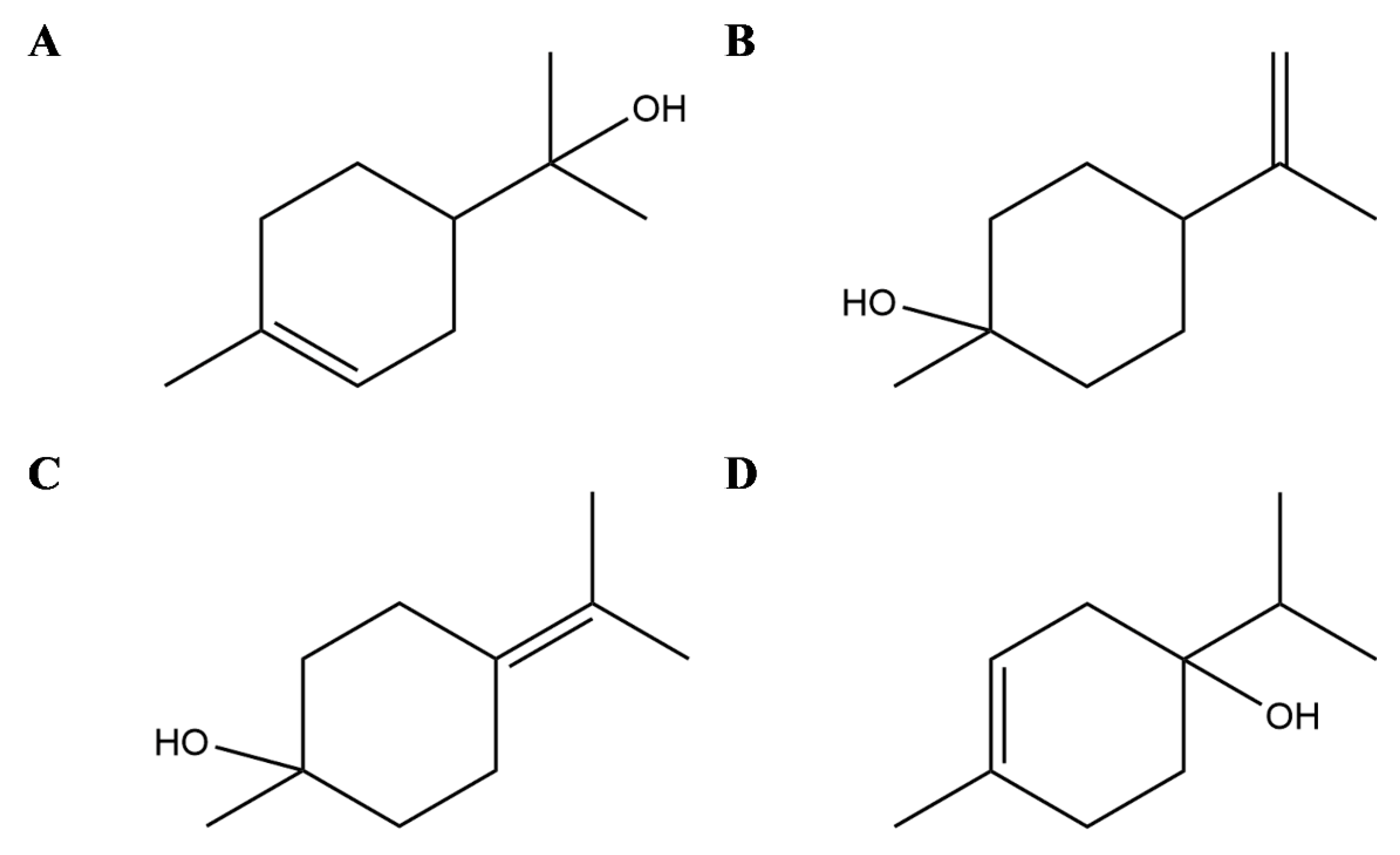
Terpineols are isomers of monocyclic monoterpene alcohol naturally present in different plants, among which the most common are α-terpineol and terpinen-4-ol [120] (Figure 1). In particular, the latter has been shown in vitro to suppress inflammatory mediator production by activation of monocytes [99] and to inhibit inflammatory cytokine generation in LPS-stimulated human macrophages [100] but also in animal models to attenuate inflammation in dextran sulphate sodium-induced colitis [121], prevent LPS-induced acute lung injury by decreasing LPS-induced NF-κB activation and trigger peroxisome PPAR-γ [122] (Figure 2).
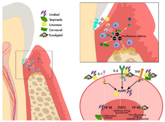
Figure 1.
General Scheme of the effect of the main EOs derived from Lamiaceae family on the periodontium.

Figure 2.
Selected compounds from Essential Oils from Lamiaceae family. Terpineols: α- terpineol (A), β- terpineol (B), γ-terpineol (C), and terpinen-4-ol (D).
Linalool (3,7-dimethyl-1,6-octadien-3-ol) is an acyclic monoterpene found in EOs of hundreds of plants widely spread worldwide and principally in Lamiaceae family [74] (Figure 3A). Several in vitro and in vivo studies demonstrated different anti-inflammatory effects of this monoterpene also interfering with the mediators of the inflammation pathways. In detail, in RAW 264.6 monocyte/macrophage-like cells linalool decreased the generation of lipopolysaccharide (LPS)-induced TNF-α and IL-6 and inhibited the activation of the nuclear factor-κB (NF-κB) and mitogen-activated protein kinase (MAPK) pathways [102]. In addition, in animal models, it has been shown that linalool attenuated acute lung inflammation by reducing TNF-α, IL-6, IL-8, IL-1β and monocyte chemoattractant protein-1 (MCP-1) production [103], further supporting linalool as a promising tool to treat inflammatory related diseases.
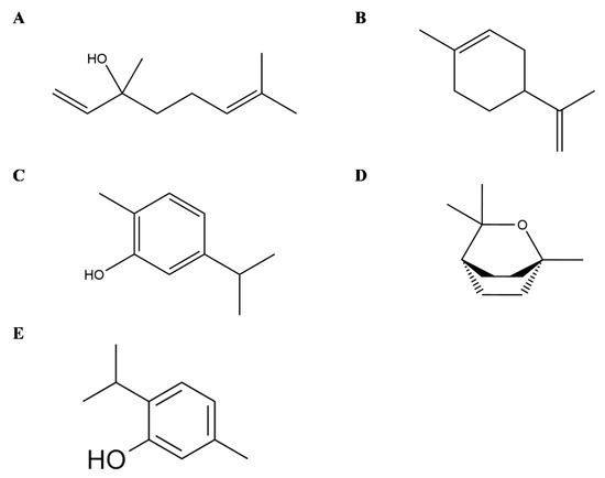
Figure 3.
Selected compounds from Essential Oils from Lamiaceae family. Linalool (A), Limonene (B), Carvacrol (C), Eucalyptol (D), Thymol (E).
Limonene (1-Methyl-4-(prop-1-en-2-yl)cyclohex-1-ene) is a cyclic monoterpene and one of the most common terpenes in nature as well as the main constituent of citrus Eos (Figure 3B). Its anti-inflammatory effects are principally linked to the modulation of cytokines and the interference with the inflammatory-related pathways, as demonstrated by in vitro and in vivo assays. Limonene decreases leukocytes infiltration and neutrophils migration, as well as the levels of TNF-α in cell derived from the peritoneal cavity and in the peritoneal exudate of zymosan-induced peritonitis BALB/C mice [104]. In LPS inflammation-induced RAW 264.7 macrophages, limonene reduced in a dose-dependent manner the levels of proinflammatory cytokines TNF-α, IL-6 and IL-1β, together with the expression of inducible nitric oxide synthase (iNOS), cyclooxygenase (COX) and prostaglandin E2 (PGE2) [105]. Similarly, in vitro model of osteoarthritis with IL-1β-stimulated human chondrocytes, limonene negatively modulated nitric oxide (NO) production by decreasing iNOS, matrix metalloproteinase (MMP)-1 and MMP-13expression, besides NF-κB and p38 activation [106].
Carvacrol (5-isopropyl-2-methylphenol) is a cyclic monoterpene mainly present in the EO of plants from Lamiaceae family (Figure 3C). In an experimental rat model of periodontal disease, carvacrol maintained alveolar bone resorption and decreased tissue lesion at histopathology, with preservation of the gingival tissue also demonstrating anti-inflammatory and antibacterial activities [107]. In addition, in vitro in murine macrophages carvacrol (1, 10, and 100 μg/mL) reduced the LPS-induced nitrite production as well as in vivo in a model of carrageenan-induced pleurisy with a pretreatment 50 or 100 mg/kg, i.e., carvacrol reduced the levels of TNF-α and suppressed leukocytes recruitment in pleural lavage [108]. In human macrophage-like U937 cells, carvacrol suppressed LPS-induced COX-2 expression, activating PPARγ, indicating its anti-inflammatory properties [109] and having no selectivity for both COX-1 and COX-2 enzyme isoforms [110]. Furthermore, it has been shown that carvacrol induces Nav blockade in DRG neurons [123,124], and Gonçalves et al. also reported significant analgesic activity and dose-dependency of T. capitatus EO. It is suggest that it is the main active molecule behind the antinociceptive effects of T. capitatus through peripheral nervous excitability blockade [125]. Other study showed that carvacrol and thymol have the most potent antimicrobial activity against Escherichia coli, Sta. aureus, Str. epidermidis, Enterococcus faecalis, Yersinia enterocolitica, Candida albicans, Bacillus cereus, Listeria monocytogenes, Salmonella typhimurium and Saccharomyces cerevisiae, with the exception of Pseudomonas aeruginosa [126]. Taking into account that T. capitatus fractions are characterized by the presence of carvacrol as dominant constituent, the antibacterial properties may be attributed to this oxygenated monoterpene.
Thymol (2-isopropyl-5-methylphenol), a monoterpene phenol, is a typical compound in EOs from thyme species (Figure 3E). Thymol also ameliorates inflammation in vitro in LPS-induced inflammation in murine macrophage cells [116] as well as in LPS- and interferon (IFN)-γ-induced macrophage inflammation, besides inhibition of the iNO RNA expression in J774A.1 cells [117]. Thymol may also modify prostaglandin catalysed biosynthesis by the inhibition of COX-1 and COX-2 isoforms [118]. Moreover, in mouse mammary epithelial cells, LPS-induced inflammatory response was decreased after thymol treatment (40 μg/mL) by the downregulation of MAPK and NF-κB signalling pathways [119]. Recently, Perrino et al. [127] have reported a high bioactivity also found in endemic wild species as spinulosus Ten., indicating its potential use in organic agriculture, since thymol may also serve as natural agent against phytopathogenic microorganisms.
Eucalyptol or 1,8-cineole (1,3,3-trimethyl-2-oxabicyclo[2.2.2]octane) is a bicyclic monoterpene isolated from EOs from numerous plants [111] (Figure 3D). Its anti-inflammatory properties have also been investigated in human and animal models of respiratory diseases such as asthma, Chronic Obstructive Pulmonary Disease and bronchitis [112,113,128]. In vitro studies on LPS-induced human lymphocytes and monocytes showed a reduced expression of cytokines including TNFα, IL-6 and IL-1β accompanied with a decrease in NFκB activated form [114,115].
6. Conclusions
The available published evidence provides substantial data to consider several species of the Lamiaceae family (Rosmarinus officinalis, Lavandula x intermedia, Thymus capitatus) as potential agents for the treatment of inflammatory diseases. The studies reviewed support the use of EOs of these plants against inflammation-related diseases, and the mechanisms described here provided pathways that explain these anti-inflammatory effects. Furthermore, the wide variety of species of the Lamiaceae family should be considered. Different chemotypes can be found, even within the same species, depending on the region and the environment in which the species have been grown. These factors could affect the percentage of EOs. The phenological phase of the same individual can also change the chemical composition and therefore the EOs obtained from it. Several nutraceutical products based on EOs as an adjuvant therapeutic agent for periodontal treatment might have some additional beneficial effect on periodontitis variables, preventing progression of disease when used as an irrigation solution and/or mouthwash. The utilization of such agents may reduce the intake of the drugs and consequently minimize the risk of a possible appearance of drug resistance as well as other risk factors. The plant extracts may act beneficially on periodontitis possibly through the known anti-inflammatory effects of each compound that remain to be clarified by future studies. Therefore, EOs from the Lamiaceae family can be considered as potential therapeutic agents for the complementary treatment periodontitis, as well as for other immune-inflammatory diseases. The potential applications role of each specific EO needs to be further studied to allow a full understanding of the spectrum of potential applications.
Author Contributions
Conceptualization, G.C., F.M. and A.M.-F.; writing—original draft preparation, G.C., F.C., C.B.-R., G.A.M., I.C. and A.M.-F.; writing—review and editing, A.M.-F. and F.M; supervision, F.M., G.C. and G.A.M. All authors have read and agreed to the published version of the manuscript.
Funding
This research received no external funding.
Institutional Review Board Statement
Not applicable.
Informed Consent Statement
Not applicable.
Data Availability Statement
Not applicable.
Conflicts of Interest
The authors declare no conflict of interest.
References
- Cekici, A.; Kantarci, A.; Hasturk, H.; Van Dyke, T.E. Inflammatory and immune pathways in the pathogenesis of periodontal disease. Periodontol. 2000 2014, 64, 57–80. [Google Scholar] [CrossRef] [PubMed] [Green Version]
- Hasturk, H.; Kantarci, A. Activation and resolution of periodontal inflammation and its systemic impact. Periodontol. 2000 2015, 69, 255–273. [Google Scholar] [CrossRef] [PubMed]
- Teng, Y.T. The role of acquired immunity and periodontal disease progression. Crit. Rev. Oral Biol. Med. 2003, 14, 237–252. [Google Scholar] [CrossRef] [PubMed] [Green Version]
- Kononen, E.; Gursoy, M.; Gursoy, U.K. Periodontitis: A Multifaceted Disease of Tooth-Supporting Tissues. J. Clin. Med. 2019, 8, 1135. [Google Scholar] [CrossRef] [PubMed] [Green Version]
- Tatakis, D.N.; Trombelli, L. Modulation of clinical expression of plaque-induced gingivitis. I. Background review and rationale. J. Clin. Periodontol. 2004, 31, 229–238. [Google Scholar] [CrossRef]
- Eke, P.I.; Wei, L.; Borgnakke, W.S.; Thornton-Evans, G.; Zhang, X.; Lu, H.; McGuire, L.C.; Genco, R.J. Periodontitis prevalence in adults ≥ 65 years of age, in the USA. Periodontol. 2000 2016, 72, 76–95. [Google Scholar] [CrossRef]
- Susin, C.; Haas, A.N.; Albandar, J.M. Epidemiology and demographics of aggressive periodontitis. Periodontol. 2000 2014, 65, 27–45. [Google Scholar] [CrossRef]
- Kilian, M.; Chapple, I.L.; Hannig, M.; Marsh, P.D.; Meuric, V.; Pedersen, A.M.; Tonetti, M.S.; Wade, W.G.; Zaura, E. The oral microbiome—An update for oral healthcare professionals. Br. Dent. J. 2016, 221, 657–666. [Google Scholar] [CrossRef]
- Hajishengallis, G.; Lamont, R.J. Beyond the red complex and into more complexity: The polymicrobial synergy and dysbiosis (PSD) model of periodontal disease etiology. Mol. Oral Microbiol. 2012, 27, 409–419. [Google Scholar] [CrossRef] [Green Version]
- Ramanauskaite, E.; Machiulskiene, V. Antiseptics as adjuncts to scaling and root planing in the treatment of periodontitis: A systematic literature review. BMC Oral Health 2020, 20, 143. [Google Scholar] [CrossRef]
- Kinane, D.F.; Lappin, D.F. Clinical, pathological and immunological aspects of periodontal disease. Acta Odontol. Scand. 2001, 59, 154–160. [Google Scholar] [CrossRef]
- Barros, S.P.; Williams, R.; Offenbacher, S.; Morelli, T. Gingival crevicular fluid as a source of biomarkers for periodontitis. Periodontol. 2000 2016, 70, 53–64. [Google Scholar] [CrossRef]
- Haffajee, A.D.; Socransky, S.S. Microbial etiological agents of destructive periodontal diseases. Periodontol. 2000 1994, 5, 78–111. [Google Scholar] [CrossRef] [PubMed]
- Caton, J.G.; Armitage, G.; Berglundh, T.; Chapple, I.L.C.; Jepsen, S.; Kornman, K.S.; Mealey, B.L.; Papapanou, P.N.; Sanz, M.; Tonetti, M.S. A new classification scheme for periodontal and peri-implant diseases and conditions—Introduction and key changes from the 1999 classification. J. Periodontol. 2018, 89 (Suppl. 1), S1–S8. [Google Scholar] [CrossRef] [PubMed]
- Tonetti, M.S.; Sanz, M. Implementation of the new classification of periodontal diseases: Decision-making algorithms for clinical practice and education. J. Clin. Periodontol. 2019, 46, 398–405. [Google Scholar] [CrossRef]
- Harvey, A.L. Natural products in drug discovery. Drug Discov. Today 2008, 13, 894–901. [Google Scholar] [CrossRef]
- Yilmaz, H.G.; Bayindir, H. Clinical evaluation of chlorhexidine and essential oils for adjunctive effects in ultrasonic instrumentation of furcation involvements: A randomized controlled clinical trial. Int. J. Dent. Hyg. 2012, 10, 113–117. [Google Scholar] [CrossRef] [PubMed]
- Varela-Lopez, A.; Navarro-Hortal, M.D.; Giampieri, F.; Bullon, P.; Battino, M.; Quiles, J.L. Nutraceuticals in Periodontal Health: A Systematic Review on the Role of Vitamins in Periodontal Health Maintenance. Molecules 2018, 23, 1226. [Google Scholar] [CrossRef] [Green Version]
- Nittayananta, W.; Limsuwan, S.; Srichana, T.; Sae-Wong, C.; Amnuaikit, T. Oral spray containing plant-derived compounds is effective against common oral pathogens. Arch. Oral Biol. 2018, 90, 80–85. [Google Scholar] [CrossRef]
- Basu, A.; Masek, E.; Ebersole, J.L. Dietary Polyphenols and Periodontitis—A Mini-Review of Literature. Molecules 2018, 23, 1786. [Google Scholar] [CrossRef] [Green Version]
- Zhu, F.; Du, B.; Xu, B. Anti-inflammatory effects of phytochemicals from fruits, vegetables, and food legumes: A review. Crit. Rev. Food Sci. Nutr. 2018, 58, 1260–1270. [Google Scholar] [CrossRef]
- Weber, C.; Noels, H. Atherosclerosis: Current pathogenesis and therapeutic options. Nat. Med. 2011, 17, 1410–1422. [Google Scholar] [CrossRef]
- World Health Organization. Cardiovascular Diseases (CVDs). Available online: https://www.who.int/en/news-room/fact-sheets/detail/cardiovascular-diseases-(cvds) (accessed on 25 January 2021).
- Wong, B.W.; Meredith, A.; Lin, D.; McManus, B.M. The biological role of inflammation in atherosclerosis. Can. J. Cardiol. 2012, 28, 631–641. [Google Scholar] [CrossRef] [PubMed]
- Rizzo, M.; Berneis, K.; Koulouris, S.; Pastromas, S.; Rini, G.B.; Sakellariou, D.; Manolis, A.S. Should we measure routinely oxidised and atherogenic dense low-density lipoproteins in subjects with type 2 diabetes? Int. J. Clin. Pract. 2010, 64, 1632–1642. [Google Scholar] [CrossRef] [Green Version]
- Raggi, P.; Genest, J.; Giles, J.T.; Rayner, K.J.; Dwivedi, G.; Beanlands, R.S.; Gupta, M. Role of inflammation in the pathogenesis of atherosclerosis and therapeutic interventions. Atherosclerosis 2018, 276, 98–108. [Google Scholar] [CrossRef] [PubMed] [Green Version]
- Kim, M.; Sahu, A.; Hwang, Y.; Kim, G.B.; Nam, G.H.; Kim, I.S.; Chan Kwon, I.; Tae, G. Targeted delivery of anti-inflammatory cytokine by nanocarrier reduces atherosclerosis in Apo E(-/-) mice. Biomaterials 2020, 226, 119550. [Google Scholar] [CrossRef]
- Viola, J.; Soehnlein, O. Atherosclerosis—A matter of unresolved inflammation. Semin. Immunol. 2015, 27, 184–193. [Google Scholar] [CrossRef] [PubMed]
- Paoletti, R.; Gotto, A.M., Jr.; Hajjar, D.P. Inflammation in atherosclerosis and implications for therapy. Circulation 2004, 109, III20–III26. [Google Scholar] [CrossRef] [PubMed] [Green Version]
- Tabas, I.; Bornfeldt, K.E. Macrophage Phenotype and Function in Different Stages of Atherosclerosis. Circ. Res. 2016, 118, 653–667. [Google Scholar] [CrossRef] [Green Version]
- Moore, K.J.; Sheedy, F.J.; Fisher, E.A. Macrophages in atherosclerosis: A dynamic balance. Nat. Rev. Immunol. 2013, 13, 709–721. [Google Scholar] [CrossRef]
- De Vries, J.E. Immunosuppressive and anti-inflammatory properties of interleukin 10. Ann. Med. 1995, 27, 537–541. [Google Scholar] [CrossRef]
- Mallat, Z.; Besnard, S.; Duriez, M.; Deleuze, V.; Emmanuel, F.; Bureau, M.F.; Soubrier, F.; Esposito, B.; Duez, H.; Fievet, C.; et al. Protective role of interleukin-10 in atherosclerosis. Circ. Res. 1999, 85, e17–e24. [Google Scholar] [CrossRef]
- Pinderski Oslund, L.J.; Hedrick, C.C.; Olvera, T.; Hagenbaugh, A.; Territo, M.; Berliner, J.A.; Fyfe, A.I. Interleukin-10 blocks atherosclerotic events in vitro and in vivo. Arterioscler. Thromb. Vasc. Biol. 1999, 19, 2847–2853. [Google Scholar] [CrossRef] [Green Version]
- Murray, P.J. The primary mechanism of the IL-10-regulated antiinflammatory response is to selectively inhibit transcription. Proc. Natl. Acad. Sci. USA 2005, 102, 8686–8691. [Google Scholar] [CrossRef] [Green Version]
- Libby, P.; Ridker, P.M.; Hansson, G.K. Inflammation in atherosclerosis: From pathophysiology to practice. J. Am. Coll. Cardiol. 2009, 54, 2129–2138. [Google Scholar] [CrossRef] [PubMed] [Green Version]
- Carrizales-Sepulveda, E.F.; Ordaz-Farias, A.; Vera-Pineda, R.; Flores-Ramirez, R. Periodontal Disease, Systemic Inflammation and the Risk of Cardiovascular Disease. Heart Lung Circ. 2018, 27, 1327–1334. [Google Scholar] [CrossRef] [PubMed]
- Lockhart, P.B.; Bolger, A.F.; Papapanou, P.N.; Osinbowale, O.; Trevisan, M.; Levison, M.E.; Taubert, K.A.; Newburger, J.W.; Gornik, H.L.; Gewitz, M.H.; et al. Periodontal disease and atherosclerotic vascular disease: Does the evidence support an independent association? A scientific statement from the American Heart Association. Circulation 2012, 125, 2520–2544. [Google Scholar] [CrossRef] [PubMed]
- Golia, E.; Limongelli, G.; Natale, F.; Fimiani, F.; Maddaloni, V.; Pariggiano, I.; Bianchi, R.; Crisci, M.; D’Acierno, L.; Giordano, R.; et al. Inflammation and cardiovascular disease: From pathogenesis to therapeutic target. Curr. Atheroscler. Rep. 2014, 16, 435. [Google Scholar] [CrossRef] [PubMed]
- Patti, M.G.; Allaix, M.E.; Fisichella, P.M. Analysis of the Causes of Failed Antireflux Surgery and the Principles of Treatment: A Review. JAMA Surg. 2015, 150, 585–590. [Google Scholar] [CrossRef] [PubMed]
- Cicero, A.F.G.; Colletti, A.; Bellentani, S. Nutraceutical Approach to Non-Alcoholic Fatty Liver Disease (NAFLD): The Available Clinical Evidence. Nutrients 2018, 10, 1153. [Google Scholar] [CrossRef] [Green Version]
- Patti, A.M.; Al-Rasadi, K.; Giglio, R.V.; Nikolic, D.; Mannina, C.; Castellino, G.; Chianetta, R.; Banach, M.; Cicero, A.F.G.; Lippi, G.; et al. Natural approaches in metabolic syndrome management. Arch. Med. Sci. AMS 2018, 14, 422–441. [Google Scholar] [CrossRef]
- Blaizot, A.; Vergnes, J.N.; Nuwwareh, S.; Amar, J.; Sixou, M. Periodontal diseases and cardiovascular events: Meta-analysis of observational studies. Int. Dent. J. 2009, 59, 197–209. [Google Scholar]
- Zeng, X.T.; Leng, W.D.; Lam, Y.Y.; Yan, B.P.; Wei, X.M.; Weng, H.; Kwong, J.S. Periodontal disease and carotid atherosclerosis: A meta-analysis of 17,330 participants. Int. J. Cardiol. 2016, 203, 1044–1051. [Google Scholar] [CrossRef] [PubMed]
- Humphrey, L.L.; Fu, R.; Buckley, D.I.; Freeman, M.; Helfand, M. Periodontal disease and coronary heart disease incidence: A systematic review and meta-analysis. J. Gen. Intern. Med. 2008, 23, 2079–2086. [Google Scholar] [CrossRef] [PubMed] [Green Version]
- Mustapha, I.Z.; Debrey, S.; Oladubu, M.; Ugarte, R. Markers of systemic bacterial exposure in periodontal disease and cardiovascular disease risk: A systematic review and meta-analysis. J. Periodontol. 2007, 78, 2289–2302. [Google Scholar] [CrossRef] [PubMed]
- Bahekar, A.A.; Singh, S.; Saha, S.; Molnar, J.; Arora, R. The prevalence and incidence of coronary heart disease is significantly increased in periodontitis: A meta-analysis. Am. Heart J. 2007, 154, 830–837. [Google Scholar] [CrossRef]
- Loos, B.G. Systemic markers of inflammation in periodontitis. J. Periodontol. 2005, 76, 2106–2115. [Google Scholar] [CrossRef] [PubMed]
- Furuhashi, M.; Ishimura, S.; Ota, H.; Miura, T. Lipid chaperones and metabolic inflammation. Int. J. Inflam. 2011, 2011, 642612. [Google Scholar] [CrossRef] [PubMed] [Green Version]
- Hotamisligil, G.S. Inflammation and metabolic disorders. Nature 2006, 444, 860–867. [Google Scholar] [CrossRef]
- Gregor, M.F.; Hotamisligil, G.S. Inflammatory mechanisms in obesity. Annu. Rev. Immunol. 2011, 29, 415–445. [Google Scholar] [CrossRef] [Green Version]
- Sanz, M.; Marco Del Castillo, A.; Jepsen, S.; Gonzalez-Juanatey, J.R.; D’Aiuto, F.; Bouchard, P.; Chapple, I.; Dietrich, T.; Gotsman, I.; Graziani, F.; et al. Periodontitis and cardiovascular diseases: Consensus report. J. Clin. Periodontol. 2020, 47, 268–288. [Google Scholar] [CrossRef] [PubMed]
- Del Pinto, R.; Pietropaoli, D.; Munoz-Aguilera, E.; D’Aiuto, F.; Czesnikiewicz-Guzik, M.; Monaco, A.; Guzik, T.J.; Ferri, C. Periodontitis and Hypertension: Is the Association Causal? High Blood Press. Cardiovasc. Prev. 2020, 27, 281–289. [Google Scholar] [CrossRef] [PubMed]
- Orlandi, M.; Graziani, F.; D’Aiuto, F. Periodontal therapy and cardiovascular risk. Periodontol. 2000 2020, 83, 107–124. [Google Scholar] [CrossRef]
- Priyamvara, A.; Dey, A.K.; Bandyopadhyay, D.; Katikineni, V.; Zaghlol, R.; Basyal, B.; Barssoum, K.; Amarin, R.; Bhatt, D.L.; Lavie, C.J. Periodontal Inflammation and the Risk of Cardiovascular Disease. Curr. Atheroscler. Rep. 2020, 22, 28. [Google Scholar] [CrossRef] [PubMed]
- Xue, W.; Yu, J.; Chen, W. Plants and Their Bioactive Constituents in Mesenchymal Stem Cell-Based Periodontal Regeneration: A Novel Prospective. Biomed. Res. Int. 2018, 2018, 7571363. [Google Scholar] [CrossRef]
- Ríos, J.-L. Essential Oils. In Essential Oils in Food Preservation, Flavor and Safety; Preedy, V.R., Ed.; Academic Press: San Diego, CA, USA, 2016; pp. 3–10. [Google Scholar]
- Ul Hassan, M.N.; Zainal, Z.; Ismail, I. Green leaf volatiles: Biosynthesis, biological functions and their applications in biotechnology. Plant Biotechnol. J. 2015, 13, 727–739. [Google Scholar] [CrossRef]
- Sharifi-Rad, J.; Sureda, A.; Tenore, G.C.; Daglia, M.; Sharifi-Rad, M.; Valussi, M.; Tundis, R.; Sharifi-Rad, M.; Loizzo, M.R.; Ademiluyi, A.O.; et al. Biological Activities of Essential Oils: From Plant Chemoecology to Traditional Healing Systems. Molecules 2017, 22, 70. [Google Scholar] [CrossRef]
- Sangwan, N.S.; Farooqi, A.H.A.; Shabih, F.; Sangwan, R.S. Regulation of essential oil production in plants. Plant Growth Regul. 2001, 34, 3–21. [Google Scholar] [CrossRef]
- Aziz, Z.A.A.; Ahmad, A.; Setapar, S.H.M.; Karakucuk, A.; Azim, M.M.; Lokhat, D.; Rafatullah, M.; Ganash, M.; Kamal, M.A.; Ashraf, G.M. Essential Oils: Extraction Techniques, Pharmaceutical And Therapeutic Potential—A Review. Curr. Drug Metab. 2018, 19, 1100–1110. [Google Scholar] [CrossRef]
- Khansari, N.; Shakiba, Y.; Mahmoudi, M. Chronic inflammation and oxidative stress as a major cause of age-related diseases and cancer. Recent Pat. Inflamm. Allergy Drug Discov. 2009, 3, 73–80. [Google Scholar] [CrossRef]
- Dandekar, A.; Mendez, R.; Zhang, K. Cross talk between ER stress, oxidative stress, and inflammation in health and disease. Methods Mol. Biol. 2015, 1292, 205–214. [Google Scholar] [CrossRef]
- Arulselvan, P.; Fard, M.T.; Tan, W.S.; Gothai, S.; Fakurazi, S.; Norhaizan, M.E.; Kumar, S.S. Role of Antioxidants and Natural Products in Inflammation. Oxid. Med. Cell Longev. 2016, 2016, 5276130. [Google Scholar] [CrossRef] [Green Version]
- Bonesi, M.; Loizzo, M.R.; Acquaviva, R.; Malfa, G.A.; Aiello, F.; Tundis, R. Anti-inflammatory and Antioxidant Agents from Salvia Genus (Lamiaceae): An Assessment of the Current State of Knowledge. Antiinflamm. Antiallergy Agents Med. Chem. 2017, 16, 70–86. [Google Scholar] [CrossRef] [PubMed]
- Dajic Stevanovic, Z.; Sieniawska, E.; Glowniak, K.; Obradovic, N.; Pajic-Lijakovic, I. Natural Macromolecules as Carriers for Essential Oils: From Extraction to Biomedical Application. Front. Bioeng. Biotechnol. 2020, 8, 563. [Google Scholar] [CrossRef] [PubMed]
- Toth, P.P.; Patti, A.M.; Nikolic, D.; Giglio, R.V.; Castellino, G.; Biancucci, T.; Geraci, F.; David, S.; Montalto, G.; Rizvi, A.; et al. Bergamot Reduces Plasma Lipids, Atherogenic Small Dense LDL, and Subclinical Atherosclerosis in Subjects with Moderate Hypercholesterolemia: A 6 Months Prospective Study. Front. Pharmacol. 2015, 6, 299. [Google Scholar] [CrossRef] [Green Version]
- Castellino, G.; Nikolic, D.; Magan-Fernandez, A.; Malfa, G.A.; Chianetta, R.; Patti, A.M.; Amato, A.; Montalto, G.; Toth, P.P.; Banach, M.; et al. Altilix((R)) Supplement Containing Chlorogenic Acid and Luteolin Improved Hepatic and Cardiometabolic Parameters in Subjects with Metabolic Syndrome: A 6 Month Randomized, Double-Blind, Placebo-Controlled Study. Nutrients 2019, 11, 2580. [Google Scholar] [CrossRef] [PubMed] [Green Version]
- Leyva-Lopez, N.; Gutierrez-Grijalva, E.P.; Vazquez-Olivo, G.; Heredia, J.B. Essential Oils of Oregano: Biological Activity beyond Their Antimicrobial Properties. Molecules 2017, 22, 989. [Google Scholar] [CrossRef] [PubMed] [Green Version]
- Dagli, N.; Dagli, R.; Mahmoud, R.S.; Baroudi, K. Essential oils, their therapeutic properties, and implication in dentistry: A review. J. Int. Soc. Prev. Community Dent. 2015, 5, 335–340. [Google Scholar] [CrossRef] [PubMed] [Green Version]
- Freires, I.A.; Denny, C.; Benso, B.; de Alencar, S.M.; Rosalen, P.L. Antibacterial Activity of Essential Oils and Their Isolated Constituents against Cariogenic Bacteria: A Systematic Review. Molecules 2015, 20, 7329–7358. [Google Scholar] [CrossRef]
- Hammer, K.A.; Carson, C.F.; Riley, T.V. Antifungal effects of Melaleuca alternifolia (tea tree) oil and its components on Candida albicans, Candida glabrata and Saccharomyces cerevisiae. J. Antimicrob. Chemother. 2004, 53, 1081–1085. [Google Scholar] [CrossRef]
- Anusha, D.; Chaly, P.E.; Junaid, M.; Nijesh, J.E.; Shivashankar, K.; Sivasamy, S. Efficacy of a mouthwash containing essential oils and curcumin as an adjunct to nonsurgical periodontal therapy among rheumatoid arthritis patients with chronic periodontitis: A randomized controlled trial. Indian J. Dent. Res. 2019, 30, 506–511. [Google Scholar] [CrossRef]
- Uritu, C.M.; Mihai, C.T.; Stanciu, G.D.; Dodi, G.; Alexa-Stratulat, T.; Luca, A.; Leon-Constantin, M.M.; Stefanescu, R.; Bild, V.; Melnic, S.; et al. Medicinal Plants of the Family Lamiaceae in Pain Therapy: A Review. Pain Res. Manag. 2018, 2018, 7801543. [Google Scholar] [CrossRef] [Green Version]
- Li, B.; Cantino, P.D.; Olmstead, R.G.; Bramley, G.L.; Xiang, C.L.; Ma, Z.H.; Tan, Y.H.; Zhang, D.X. A large-scale chloroplast phylogeny of the Lamiaceae sheds new light on its subfamilial classification. Sci. Rep. 2016, 6, 34343. [Google Scholar] [CrossRef] [Green Version]
- Mamadalieva, N.Z.; Akramov, D.K.; Ovidi, E.; Tiezzi, A.; Nahar, L.; Azimova, S.S.; Sarker, S.D. Aromatic Medicinal Plants of the Lamiaceae Family from Uzbekistan: Ethnopharmacology, Essential Oils Composition, and Biological Activities. Medicines 2017, 4, 8. [Google Scholar] [CrossRef] [Green Version]
- Karpinski, T.M. Essential Oils of Lamiaceae Family Plants as Antifungals. Biomolecules 2020, 10, 103. [Google Scholar] [CrossRef] [PubMed] [Green Version]
- Borges, R.S.; Ortiz, B.L.S.; Pereira, A.C.M.; Keita, H.; Carvalho, J.C.T. Rosmarinus officinalis essential oil: A review of its phytochemistry, anti-inflammatory activity, and mechanisms of action involved. J. Ethnopharmacol. 2019, 229, 29–45. [Google Scholar] [CrossRef]
- Capatina, L.; Todirascu-Ciornea, E.; Napoli, E.M.; Ruberto, G.; Hritcu, L.; Dumitru, G. Thymus vulgaris Essential Oil Protects Zebrafish against Cognitive Dysfunction by Regulating Cholinergic and Antioxidants Systems. Antioxidants 2020, 9, 1083. [Google Scholar] [CrossRef] [PubMed]
- Carbone, C.; Martins-Gomes, C.; Caddeo, C.; Silva, A.M.; Musumeci, T.; Pignatello, R.; Puglisi, G.; Souto, E.B. Mediterranean essential oils as precious matrix components and active ingredients of lipid nanoparticles. Int. J. Pharm. 2018, 548, 217–226. [Google Scholar] [CrossRef] [PubMed]
- Cavanagh, H.M.; Wilkinson, J.M. Biological activities of lavender essential oil. Phytother. Res. 2002, 16, 301–308. [Google Scholar] [CrossRef] [PubMed]
- Silva, G.L.; Luft, C.; Lunardelli, A.; Amaral, R.H.; Melo, D.A.; Donadio, M.V.; Nunes, F.B.; de Azambuja, M.S.; Santana, J.C.; Moraes, C.M.; et al. Antioxidant, analgesic and anti-inflammatory effects of lavender essential oil. An. Acad. Bras. Cienc. 2015, 87, 1397–1408. [Google Scholar] [CrossRef] [Green Version]
- Carrasco, A.; Martinez-Gutierrez, R.; Tomas, V.; Tudela, J. Lavandin (Lavandula x intermedia Emeric ex Loiseleur) essential oil from Spain: Determination of aromatic profile by gas chromatography-mass spectrometry, antioxidant and lipoxygenase inhibitory bioactivities. Nat. Prod. Res. 2016, 30, 1123–1130. [Google Scholar] [CrossRef]
- Andrade, J.M.; Faustino, C.; Garcia, C.; Ladeiras, D.; Reis, C.P.; Rijo, P. Rosmarinus officinalis L.: An update review of its phytochemistry and biological activity. Future Sci. OA 2018, 4, FSO283. [Google Scholar] [CrossRef] [PubMed] [Green Version]
- Bustanji, Y.; Issa, A.; Mohammad, M.; Hudaib, M.; Tawah, K.; Alkhatib, H.; Almasri, I.; Al-Khalidi, B. Inhibition of hormone sensitive lipase and pancreatic lipase by Rosmarinus officinalis extract and selected phenolic constituents. J. Med. Plants Res. 2010, 4, 2235–2242. [Google Scholar]
- Silva, A.M.; Machado, I.D.; Santin, J.R.; de Melo, I.L.; Pedrosa, G.V.; Genovese, M.I.; Farsky, S.H.; Mancini-Filho, J. Aqueous extract of Rosmarinus officinalis L. inhibits neutrophil influx and cytokine secretion. Phytother. Res. 2015, 29, 125–133. [Google Scholar] [CrossRef] [PubMed]
- Borges, R.S.; Keita, H.; Ortiz, B.L.S.; Dos Santos Sampaio, T.I.; Ferreira, I.M.; Lima, E.S.; de Jesus Amazonas da Silva, M.; Fernandes, C.P.; de Faria Mota Oliveira, A.E.M.; da Conceicao, E.C.; et al. Anti-inflammatory activity of nanoemulsions of essential oil from Rosmarinus officinalis L.: In vitro and in zebrafish studies. Inflammopharmacology 2018, 26, 1057–1080. [Google Scholar] [CrossRef] [PubMed]
- Valones, M.A.A.; Silva, I.C.G.; Gueiros, L.A.M.; Leao, J.C.; Caldas, A.F., Jr.; Carvalho, A.A.T. Clinical Assessment of Rosemary-based Toothpaste (Rosmarinus officinalis Linn.): A Randomized Controlled Double-blind Study. Braz. Dent. J. 2019, 30, 146–151. [Google Scholar] [CrossRef] [PubMed] [Green Version]
- Rasooli, I.; Shayegh, S.; Taghizadeh, M.; Astaneh, S.D. Phytotherapeutic prevention of dental biofilm formation. Phytother. Res. 2008, 22, 1162–1167. [Google Scholar] [CrossRef] [PubMed]
- Bernardes, W.A.; Lucarini, R.; Tozatti, M.G.; Flauzino, L.G.; Souza, M.G.; Turatti, I.C.; Andrade e Silva, M.L.; Martins, C.H.; da Silva Filho, A.A.; Cunha, W.R. Antibacterial activity of the essential oil from Rosmarinus officinalis and its major components against oral pathogens. Z. Nat. C 2010, 65, 588–593. [Google Scholar] [CrossRef] [Green Version]
- Smullen, J.; Finney, M.; Storey, D.M.; Foster, H.A. Prevention of artificial dental plaque formation in vitro by plant extracts. J. Appl. Microbiol. 2012, 113, 964–973. [Google Scholar] [CrossRef]
- Salehi, B.; Mishra, A.P.; Shukla, I.; Sharifi-Rad, M.; Contreras, M.D.M.; Segura-Carretero, A.; Fathi, H.; Nasrabadi, N.N.; Kobarfard, F.; Sharifi-Rad, J. Thymol, thyme, and other plant sources: Health and potential uses. Phytother. Res. 2018, 32, 1688–1706. [Google Scholar] [CrossRef]
- Iauk, L.; Acquaviva, R.; Mastrojeni, S.; Amodeo, A.; Pugliese, M.; Ragusa, M.; Loizzo, M.R.; Menichini, F.; Tundis, R. Antibacterial, antioxidant and hypoglycaemic effects of Thymus capitatus (L.) Hoffmanns. et Link leaves’ fractions. J. Enzyme Inhib. Med. Chem. 2015, 30, 360–365. [Google Scholar] [CrossRef] [PubMed] [Green Version]
- Manconi, M.; Petretto, G.; D’Hallewin, G.; Escribano, E.; Milia, E.; Pinna, R.; Palmieri, A.; Firoznezhad, M.; Peris, J.E.; Usach, I.; et al. Thymus essential oil extraction, characterization and incorporation in phospholipid vesicles for the antioxidant/antibacterial treatment of oral cavity diseases. Colloids Surf. B Biointerfaces 2018, 171, 115–122. [Google Scholar] [CrossRef] [PubMed]
- Valerio, F.; Mezzapesa, G.N.; Ghannouchi, A.; Mondelli, D.; Logrieco, A.F.; Perrino, E.V. Characterization and Antimicrobial Properties of Essential Oils from Four Wild Taxa of Lamiaceae Family Growing in Apulia. Agronomy 2021, 11, 1431. [Google Scholar] [CrossRef]
- Alvarez Echazu, M.I.; Olivetti, C.E.; Anesini, C.; Perez, C.J.; Alvarez, G.S.; Desimone, M.F. Development and evaluation of thymol-chitosan hydrogels with antimicrobial-antioxidant activity for oral local delivery. Mater. Sci. Eng. C Mater. Biol. Appl. 2017, 81, 588–596. [Google Scholar] [CrossRef]
- Ocana, A.; Reglero, G. Effects of Thyme Extract Oils (from Thymus vulgaris, Thymus zygis, and Thymus hyemalis) on Cytokine Production and Gene Expression of oxLDL-Stimulated THP-1-Macrophages. J. Obes. 2012, 2012, 104706. [Google Scholar] [CrossRef] [Green Version]
- Penalver, P.; Huerta, B.; Borge, C.; Astorga, R.; Romero, R.; Perea, A. Antimicrobial activity of five essential oils against origin strains of the Enterobacteriaceae family. APMIS 2005, 113, 1–6. [Google Scholar] [CrossRef]
- Hart, P.H.; Brand, C.; Carson, C.F.; Riley, T.V.; Prager, R.H.; Finlay-Jones, J.J. Terpinen-4-ol, the main component of the essential oil of Melaleuca alternifolia (tea tree oil), suppresses inflammatory mediator production by activated human monocytes. Inflamm. Res. 2000, 49, 619–626. [Google Scholar] [CrossRef]
- Nogueira, M.N.; Aquino, S.G.; Rossa Junior, C.; Spolidorio, D.M. Terpinen-4-ol and alpha-terpineol (tea tree oil components) inhibit the production of IL-1beta, IL-6 and IL-10 on human macrophages. Inflamm. Res. 2014, 63, 769–778. [Google Scholar] [CrossRef]
- Zhang, Y.; Li, D.; Wang, Z.; Zang, W.; Rao, P.; Liang, Y.; Mei, Y. Alpha-terpineol affects synthesis and antitumor activity of triterpenoids from Antrodia cinnamomea mycelia in solid-state culture. Food Funct. 2018, 9, 6517–6525. [Google Scholar] [CrossRef] [PubMed]
- Huo, M.; Gao, R.; Jiang, L.; Cui, X.; Duan, L.; Deng, X.; Guan, S.; Wei, J.; Soromou, L.W.; Feng, H.; et al. Suppression of LPS-induced inflammatory responses by gossypol in RAW 264.7 cells and mouse models. Int. Immunopharmacol. 2013, 15, 442–449. [Google Scholar] [CrossRef] [PubMed]
- Ma, J.; Xu, H.; Wu, J.; Qu, C.; Sun, F.; Xu, S. Linalool inhibits cigarette smoke-induced lung inflammation by inhibiting NF-kappaB activation. Int. Immunopharmacol. 2015, 29, 708–713. [Google Scholar] [CrossRef]
- Kummer, R.; Fachini-Queiroz, F.C.; Estevao-Silva, C.F.; Grespan, R.; Silva, E.L.; Bersani-Amado, C.A.; Cuman, R.K. Evaluation of Anti-Inflammatory Activity of Citrus latifolia Tanaka Essential Oil and Limonene in Experimental Mouse Models. Evid. Based Complement. Alternat. Med. 2013, 2013, 859083. [Google Scholar] [CrossRef] [Green Version]
- Yoon, W.J.; Lee, N.H.; Hyun, C.G. Limonene suppresses lipopolysaccharide-induced production of nitric oxide, prostaglandin E2, and pro-inflammatory cytokines in RAW 264.7 macrophages. J. Oleo Sci. 2010, 59, 415–421. [Google Scholar] [CrossRef] [Green Version]
- Rufino, A.T.; Ribeiro, M.; Sousa, C.; Judas, F.; Salgueiro, L.; Cavaleiro, C.; Mendes, A.F. Evaluation of the anti-inflammatory, anti-catabolic and pro-anabolic effects of E-caryophyllene, myrcene and limonene in a cell model of osteoarthritis. Eur. J. Pharmacol. 2015, 750, 141–150. [Google Scholar] [CrossRef] [PubMed]
- Botelho, M.A.; Rao, V.S.; Montenegro, D.; Bandeira, M.A.; Fonseca, S.G.; Nogueira, N.A.; Ribeiro, R.A.; Brito, G.A. Effects of a herbal gel containing carvacrol and chalcones on alveolar bone resorption in rats on experimental periodontitis. Phytother. Res. 2008, 22, 442–449. [Google Scholar] [CrossRef] [PubMed]
- Guimaraes, A.G.; Xavier, M.A.; de Santana, M.T.; Camargo, E.A.; Santos, C.A.; Brito, F.A.; Barreto, E.O.; Cavalcanti, S.C.; Antoniolli, A.R.; Oliveira, R.C.; et al. Carvacrol attenuates mechanical hypernociception and inflammatory response. Naunyn Schmiedebergs Arch. Pharmacol. 2012, 385, 253–263. [Google Scholar] [CrossRef] [PubMed]
- Hotta, M.; Nakata, R.; Katsukawa, M.; Hori, K.; Takahashi, S.; Inoue, H. Carvacrol, a component of thyme oil, activates PPARalpha and gamma and suppresses COX-2 expression. J. Lipid Res. 2010, 51, 132–139. [Google Scholar] [CrossRef] [PubMed] [Green Version]
- Landa, P.; Kokoska, L.; Pribylova, M.; Vanek, T.; Marsik, P. In vitro anti-inflammatory activity of carvacrol: Inhibitory effect on COX-2 catalyzed prostaglandin E(2) biosynthesis. Arch. Pharm. Res. 2009, 32, 75–78. [Google Scholar] [CrossRef]
- Yalcin, H.; Anik, M.; Sanda, M.A.; Cakir, A. Gas chromatography/mass spectrometry analysis of Laurus nobilis essential oil composition of northern Cyprus. J. Med. Food 2007, 10, 715–719. [Google Scholar] [CrossRef]
- Bastos, V.P.; Gomes, A.S.; Lima, F.J.; Brito, T.S.; Soares, P.M.; Pinho, J.P.; Silva, C.S.; Santos, A.A.; Souza, M.H.; Magalhaes, P.J. Inhaled 1,8-cineole reduces inflammatory parameters in airways of ovalbumin-challenged Guinea pigs. Basic Clin. Pharmacol. Toxicol. 2011, 108, 34–39. [Google Scholar] [CrossRef]
- Kennedy-Feitosa, E.; Okuro, R.T.; Pinho Ribeiro, V.; Lanzetti, M.; Barroso, M.V.; Zin, W.A.; Porto, L.C.; Brito-Gitirana, L.; Valenca, S.S. Eucalyptol attenuates cigarette smoke-induced acute lung inflammation and oxidative stress in the mouse. Pulm. Pharmacol. Ther. 2016, 41, 11–18. [Google Scholar] [CrossRef] [PubMed]
- Juergens, U.R.; Engelen, T.; Racke, K.; Stober, M.; Gillissen, A.; Vetter, H. Inhibitory activity of 1,8-cineol (eucalyptol) on cytokine production in cultured human lymphocytes and monocytes. Pulm. Pharmacol. Ther. 2004, 17, 281–287. [Google Scholar] [CrossRef] [PubMed]
- Kim, K.Y.; Lee, H.S.; Seol, G.H. Eucalyptol suppresses matrix metalloproteinase-9 expression through an extracellular signal-regulated kinase-dependent nuclear factor-kappa B pathway to exert anti-inflammatory effects in an acute lung inflammation model. J. Pharm. Pharmacol. 2015, 67, 1066–1074. [Google Scholar] [CrossRef] [PubMed]
- Chauhan, A.K.; Jakhar, R.; Paul, S.; Kang, S.C. Potentiation of macrophage activity by thymol through augmenting phagocytosis. Int. Immunopharmacol. 2014, 18, 340–346. [Google Scholar] [CrossRef]
- Vigo, E.; Cepeda, A.; Gualillo, O.; Perez-Fernandez, R. In-vitro anti-inflammatory effect of Eucalyptus globulus and Thymus vulgaris: Nitric oxide inhibition in J774A.1 murine macrophages. J. Pharm. Pharmacol. 2004, 56, 257–263. [Google Scholar] [CrossRef]
- Marsik, P.; Kokoska, L.; Landa, P.; Nepovim, A.; Soudek, P.; Vanek, T. In vitro inhibitory effects of thymol and quinones of Nigella sativa seeds on cyclooxygenase-1- and -2-catalyzed prostaglandin E2 biosyntheses. Planta Med. 2005, 71, 739–742. [Google Scholar] [CrossRef] [PubMed]
- Liang, D.; Li, F.; Fu, Y.; Cao, Y.; Song, X.; Wang, T.; Wang, W.; Guo, M.; Zhou, E.; Li, D.; et al. Thymol inhibits LPS-stimulated inflammatory response via down-regulation of NF-kappaB and MAPK signaling pathways in mouse mammary epithelial cells. Inflammation 2014, 37, 214–222. [Google Scholar] [CrossRef]
- Khaleel, C.; Tabanca, N.; Buchbauer, G.J.O.C. α-Terpineol, a natural monoterpene: A review of its biological properties. Open Chem. 2018, 16, 349–361. [Google Scholar] [CrossRef]
- Zhang, H.; Hua, R.; Zhang, B.; Zhang, X.; Yang, H.; Zhou, X. Serine Alleviates Dextran Sulfate Sodium-Induced Colitis and Regulates the Gut Microbiota in Mice. Front. Microbiol. 2018, 9, 3062. [Google Scholar] [CrossRef] [Green Version]
- Peng, L.Y.; Shi, H.T.; Yuan, M.; Li, J.H.; Song, K.; Huang, J.N.; Yi, P.F.; Shen, H.Q.; Fu, B.D. Madecassoside Protects Against LPS-Induced Acute Lung Injury via Inhibiting TLR4/NF-kappaB Activation and Blood-Air Barrier Permeability. Front. Pharmacol. 2020, 11, 807. [Google Scholar] [CrossRef]
- Joca, H.C.; Vieira, D.C.; Vasconcelos, A.P.; Araujo, D.A.; Cruz, J.S. Carvacrol modulates voltage-gated sodium channels kinetics in dorsal root ganglia. Eur. J. Pharmacol. 2015, 756, 22–29. [Google Scholar] [CrossRef] [PubMed]
- Joca, H.C.; Cruz-Mendes, Y.; Oliveira-Abreu, K.; Maia-Joca, R.P.; Barbosa, R.; Lemos, T.L.; Lacerda Beirao, P.S.; Leal-Cardoso, J.H. Carvacrol decreases neuronal excitability by inhibition of voltage-gated sodium channels. J. Nat. Prod. 2012, 75, 1511–1517. [Google Scholar] [CrossRef] [PubMed]
- Goncalves, J.C.; de Meneses, D.A.; de Vasconcelos, A.P.; Piauilino, C.A.; Almeida, F.R.; Napoli, E.M.; Ruberto, G.; de Araujo, D.A. Essential oil composition and antinociceptive activity of Thymus capitatus. Pharm. Biol. 2017, 55, 782–786. [Google Scholar] [CrossRef] [PubMed] [Green Version]
- Cosentino, S.; Tuberoso, C.I.; Pisano, B.; Satta, M.; Mascia, V.; Arzedi, E.; Palmas, F. In-vitro antimicrobial activity and chemical composition of Sardinian Thymus essential oils. Lett. Appl. Microbiol. 1999, 29, 130–135. [Google Scholar] [CrossRef]
- Perrino, E.V.; Valerio, F.; Jallali, S.; Trani, A.; Mezzapesa, G.N. Ecological and Biological Properties of Satureja cuneifolia Ten. and Thymus spinulosus Ten.: Two Wild Officinal Species of Conservation Concern in Apulia (Italy). A Preliminary Survey. Plants 2021, 10, 1952. [Google Scholar] [CrossRef] [PubMed]
- Juergens, U.R.; Dethlefsen, U.; Steinkamp, G.; Gillissen, A.; Repges, R.; Vetter, H. Anti-inflammatory activity of 1.8-cineol (eucalyptol) in bronchial asthma: A double-blind placebo-controlled trial. Respir. Med. 2003, 97, 250–256. [Google Scholar] [CrossRef] [Green Version]


Publisher’s Note: MDPI stays neutral with regard to jurisdictional claims in published maps and institutional affiliations. |
© 2021 by the authors. Licensee MDPI, Basel, Switzerland. This article is an open access article distributed under the terms and conditions of the Creative Commons Attribution (CC BY) license (https://creativecommons.org/licenses/by/4.0/).