Featured Application
During food processing, the surface of food and processing equipment is easily contaminated by microorganisms, which will cause food spoilage or the spread of disease, threatening food safety. Plasma-activated solution can be used as a novel food disinfectant, enhancing food quality. Plasma-activated solution will not leave chemical residues or cause environmental pollution. It is a green and environmentally friendly disinfectant and promises to replace traditional chemical disinfectants.
Abstract
Plasma-activated solution has attracted more attention in the food industry due to no chemical residue and good bacteriostatic properties. This study aimed to evaluate the effects of plasma-activated hydrogen peroxide solution (PAH) on the morphophysiology of Staphylococcus aureus biofilms. PAH was prepared using dielectric-barrier-discharge plasma and incubated with S. aureus biofilms for 0–40 min. Changes in biofilm morphophysiology were evaluated with laser scanning confocal microscopy, electron microscopic images, reactive oxygen species (ROS) content, metabolic capacity, and 1% agarose gel. Results indicated that the population of S. aureus in the biofilms was reduced by 4.04-log after incubation with PAH for 30 min. The thickness and metabolic capacity of biofilms were decreased, the ROS content and DNA fragments of bacteria increased after PAH treatments. Data suggested that PAH treatments significantly destroyed the morphophysiology of S. aureus (ATCC 6538) biofilms and could be considered as a valuable anti-biofilm technology to reduce foodborne pathogens on food and/or in food facilities.
1. Introduction
A biofilm is tightly grouped mass of microorganisms encased in extracellular polymeric substances to provide cellular protection against host cellular and chemical responses [1]. Biofilms are formed on the surface of food products, making food spoilage and leading to serious health problems and economic losses [2]. The non-thermal sterilization techniques commonly applied in the food industry, such as sodium hypochloride and peroxyacetic acid, are very efficient at controlling foodborne pathogens existing in the planktonic state [3,4]. However, most pathogens are found in the biofilm matrix [5], which is 10 to 1000 times more resistant to sanitizer than in the planktonic state [6]. Due to the limitations of existing non-thermal methods for the control of bacterial biofilms in food, such as high cost and negative impact on food, human, and the environment, it is of interest in exploring new non-polluting and effective anti-biofilm methods that can be used in the food industry.
The formation of bacterial biofilms is a dynamic process of repeated cycles and can be divided into four stages: initial attachment, early formation, maturation, and shedding [7]. In the initial attachment, planktonic bacteria attach to the substrate surface through fimbria and/or flagella. This process can be considered as the reversible adhesion [8,9], involving the proliferation of bacteria cells, the increase in extracellular polymers, and the interaction between bacteria and matrix turned from weak to permanent. In the early stage of biofilm formation, bacterial cells grow closely and begin to secrete a polymer matrix to the surroundings, further transforming into discrete groups of cells [10]. For the maturation, small colonies aggregate into large colonies, forming a specific biofilm structure with the extracellular polymer matrix as a protective barrier. Finally, mature biofilm cells shed and disperse into planktonic cells or form new biofilms [11].
Cold plasma is a relatively new non-thermal sterilization technology and has been drawing more interest in food sterilization due to its low cost and ‘green’ feature [12]. Cold plasma can directly treat contaminated surfaces or generate a solution with antimicrobial activity to sanitize surfaces of food and/or processing equipment. When the liquid is exposed to plasma, the reactive species generated in the gas phase are transferred to the liquid phase, such as reactive oxygen species (ROS) and nitrogen species [13]. The treated liquid is named a ‘plasma-activated solution’ (PAS). PAS has been proven to have good antibacterial activity against planktonic bacteria. Gram-positive bacteria are more resistant to PAS than Gram-negative bacteria [14]. Therefore, Staphylococcus aureus biofilms were used as the model to investigate the inactivation effect of the plasma-activated hydrogen peroxide solution (PAH) in the present study. The bactericidal mechanism of PAH was illustrated with the changes in biofilm structures, ROS content in biofilms, the metabolic capacity of biofilms, and intracellular DNA integrity. This study provided a theoretical basis for the effective treatment of S. aureus biofilms with PAH.
2. Materials and Methods
2.1. Formation of Biofilms
Staphylococcus aureus (ATCC 6538) was purchased from China General Microbiological Culture Collection Center, Beijing, China. A single colony of S. aureus grown on the mannitol high salt agar (Qingdao hopebio Biotechnology Co., Ltd., Qingdao, China) plate was transferred to 8 mL of tryptic soy broth (TSB, Qingdao hopebio Biotechnology Co., Ltd., Qingdao, China) medium and incubated overnight at 37 °C. The cultured bacteria were diluted with TSBG (Qingdao hopebio Biotechnology Co., Ltd., Qingdao, China, TSB solution with 0.6% glucose and salt) medium to 105 CFU/mL, and then 10 mL bacterial suspension was transferred to the 6-well cell culture plates. Bacteria were cultured at 37 °C for 4 days. The supernatant was replaced with 10 mL of fresh TSBG medium every 24 h of incubation [2,15]. After incubation, the biofilm was washed with 10 mL of 0.85% sterile NaCl solution three times to remove planktonic or loosely attached bacteria and leave the bacteria biofilm for further analysis.
2.2. Preparation of PAH and Sample Treatments
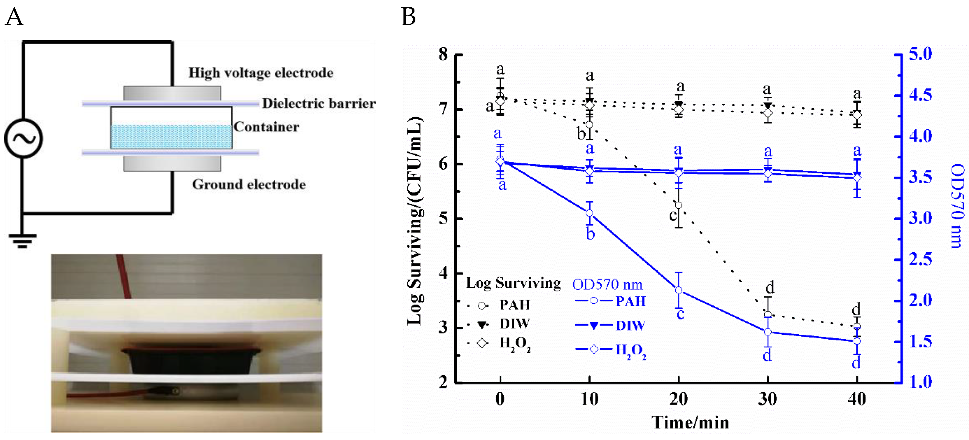
A dielectric barrier discharge (DBD) device as exhibited in Figure 1A was employed for cold plasma treatment. For PAH preparation, 800 µL hydrogen peroxide solution (3%, Sigma-Aldrich (Shanghai) Trading Co., Ltd., Shanghai, China) was added to 100 mL deionized water and then packed in a polypropylene container that was sealed with a polyethylene film (Ningjin Jinyi Packaging Materials Co., Ltd., Xingtai, China). The reason for using 0.8% hydrogen peroxide solution (800 µL H2O2 in 100-mL deionized water) in the present study was to improve the antibacterial efficiency of PAS based on our preliminary experiments, which also showed that the 0.8% H2O2 solution had a little antimicrobial effect on bacterial cells by itself. The container was placed between two electrodes and there was a dielectric barrier layer between the electrode and the sample. Processing parameters were as follows: the voltage = 60 kV; the frequency = 50 Hz; the working gas = air; and treatment time = 4 min. After treatment, the PAH was stored at room temperature for 6 h before further application.
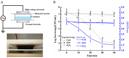
Figure 1.
(A) Dielectric barrier discharge plasma for the preparation of plasma-activated solution. Effects of incubation time on the (B) inactivation and the total amount (OD570 nm) of S. aureus biofilms mixed with the plasma-activated hydrogen peroxide solution. Deionized water (DIW) and hydrogen peroxide solution were chosen as negative controls. Different lowercase letters (a–d) for the same treatment indicate significant differences among different incubation time (p < 0.05).
For an effect on S. aureus biofilms, 10 mL PAH was transferred to the 6-well culture plate with formed biofilms and incubated at room temperature for 0, 10, 20, 30, and 40 min before biofilms were sampled for different analyses. At sampling times, the biofilm was washed with 0.85% sterile NaCl solution to stop the reaction between the biofilm and PAH [16]. To evaluate the effect of PAH on the viability of bacterial cells in biofilms, treated biofilms were transferred to 0.85% sterile NaCl solution, 0.5 mL of the mixed bacteria suspension was taken and diluted serially. Then, 100 µL of the dilution was plated on mannitol high salt agar. Bacterial colonies formed on the agar plate were counted after incubation at 37 °C for 24 h. Results were presented as the surviving bacterial population in log CFU/mL. Deionized water (DIW) and H2O2 solution (prepared by adding 800 µL 3% hydrogen peroxide solution to 100 mL deionized water) treatment were chosen as negative controls.
2.3. Determination of Total Amount of Biofilms
Biofilms after PAH treatment were fixed with methyl alcohol for 1 min, followed by washing with 0.85% sterile NaCl solution. Then, biofilms were incubated with 1% crystal violet for 30 min at room temperature and washed with the NaCl solution again. Ethanol: acetone at a ratio of 4:1 (v/v) was added to the biofilm and the absorbance at 570 nm was measured by the ultraviolet-visible absorption spectrometry (UV-2600/2700, Shimadzu Co., Kyoto, Japan) [17]. Deionized water (DIW) and H2O2 solution (prepared by adding 800 µL 3% hydrogen peroxide solution to 100 mL deionized water) treatment were chosen as negative controls.
2.4. Biofilm Morphology Image
Changes in the morphology of S. aureus biofilms after PAH treatment were observed using scanning electron microscopy (SEM). Biofilms in 6-well culture plates after treatments were washed with PBS five times and resuspended in 2.5% glutaraldehyde at 4 °C overnight for the fixation. After the fixation, samples were washed with PBS and deionized water three times each and then dehydrated in graded ethanol concentrations of 30, 50, 70, 85, 95, and 100%. The samples were freeze-dried and assessed visually using the S4800 SEM (Hitachi Co. Ltd., Tokyo, Japan).
2.5. Laser Scanning Confocal Microscopy (LSCM)
LSCM was employed to determine the damage extent of PAH to the viability of S. aureus biofilms. Biofilms after treatment were stained with SYTO 9 (Thermo Fisher Scientific, Waltham, MA, USA) and propidium iodide (PI, Thermo Fisher Scientific, Waltham, MA, USA) for 15 min at room temperature according to the manufacturer’s protocol. The SYTO 9 labels living bacteria cells and the PI labels the bacteria with a damaged membrane [18]. The stained biofilm was spread on a slide and then covered by a coverslip. The fluorescence was evaluated by the excitation at 480 nm for SYTO 9 and 490 nm for PI using LSCM.
2.6. The Metabolic Capacity of Biofilms
Resazurin, as a dye, can be used to assess the metabolic capacity of live cells. The blue dye is converted to a pink fluorescent substance by the oxidation-reduction reactions in live bacteria, while the dye is not reduced within inactivated bacteria [19]. The bacteria suspension was mixed with 25 µM resazurin at a ratio of 1:1, and incubated in the dark for 3 h at 37 °C. The fluorescence intensity was detected at the excitation wavelengths 560 nm and emission wavelengths 590 nm. The results were expressed as the percentage of metabolic capacity (%): 100 × AFUtreated/AFUcontrol.
2.7. ROS in S. aureus Biofilms
2′,7′-dichlorodihydrofluorescein diacetate (H2DCFDA, Aladdin, Shanghai, China) was employed as a fluorescent probe to assess the ROS level in biofilms. Then, 100 μL of the treated suspension (as described in Section 2.2) was mixed with 100 μL of 40-μM H2DCFDA and incubated in the dark for 30 min at 37 °C. Then, the solution was washed and resuspended in phosphate-buffered saline (PBS, 0.01 M, pH 7.2) at 37 °C for 15 min. The fluorescent analysis was performed with a microplate reader (SpectraMax M2e, Molecular Devices, San Jose, CA, USA) at 495 nm (excitation) and 525 nm (emission) [20]. Intracellular reactive oxygen species (ROS) levels were evaluated by observing changes in the intensity of fluorescent substances. The LSCM images of the biofilm stained with H2DCFDA were also evaluated and the fluorescence intensity was collected using the Zeiss ZEN 2010 software.
2.8. Bacterial DNA Isolation and Analysis
DNA of S. aureus biofilms after PAH treatment was extracted by the DNA Extraction Kit (Qiagen, Duesseldorf, Germany) according to the manufacture’s instruction. For the electrophoresis analysis, DNA samples were loaded on a 1% agarose gel and strained by GoldView.
2.9. Statistical Analysis
The normality of data was checked using the Shapiro-Wilk. One-way variance analysis (ANOVA) was used for statistical analysis of the samples which were repeated independently three times. Duncan’s test in SAS software was used to evaluate the significant difference between the mean values, and p < 0.05 was the significance level.
3. Results and Discussion
3.1. Anti-Biofilm Efficiency of PAH
The antibacterial activity of PAH against the S. aureus biofilms after various incubation time was evaluated (Figure 1B). DIW and H2O2 solution were chosen as negative controls, results indicated that they had no significant (p > 0.05) effect on the inactivation of the biofilm. For the PAH treatment, the number of viable bacteria decreased significantly (p < 0.05) from 7.25 to 3.21 log CFU/mL after the prolonged incubation time from 0 to 30 min, then the reduction showed a flat tendency with increasing incubation time. The total amount of biofilms was determined by the crystal violet staining method (Figure 1B). The higher value of the OD 570 nm, the more amount of biofilm combined with the crystal violet. The decreasing trend in OD values was consistent with the decreasing trend of the bacterial cells. Therefore, the following investigation mainly focused on the influence of PAH on the biofilms after incubation for 0–30 min. Results indicated that the PAH was very effective in the inactivation of S. aureus cells (ATCC 6538) in the biofilms. Reactive species contributing to the antimicrobial efficacy of the PAH have been attributed mainly to hydrogen peroxide (H2O2), ozone (O3), hydrogen radicals (OH•), nitrite (NO2−), and nitric oxide (NO•) [10], which are generated through the following pathways [21,22]:
The ROS, such as OH•, possess high penetrability against extracellular polymeric substances and bacterial cells in the biofilms. Meanwhile, reactive nitrogen species, which are responsible for the decrease in the pH value of PAH [23], affect the stability of biofilm structure [24]. After plasma treatment, the pH of PAH decreases to about 2.28, which might affect the hydrophobicity and surface adhesion of the biofilms. When the pH value of the external environment of bacterial cells decreases, the activity of the membrane proton pump increases, and H+ is expelled from the cytoplasm. Therefore, cell energy is mainly used for avoiding internal acidification, which reduced the adhesion of the biofilm [25].
3.2. Changes in the Appearance of S. aureus Biofilms
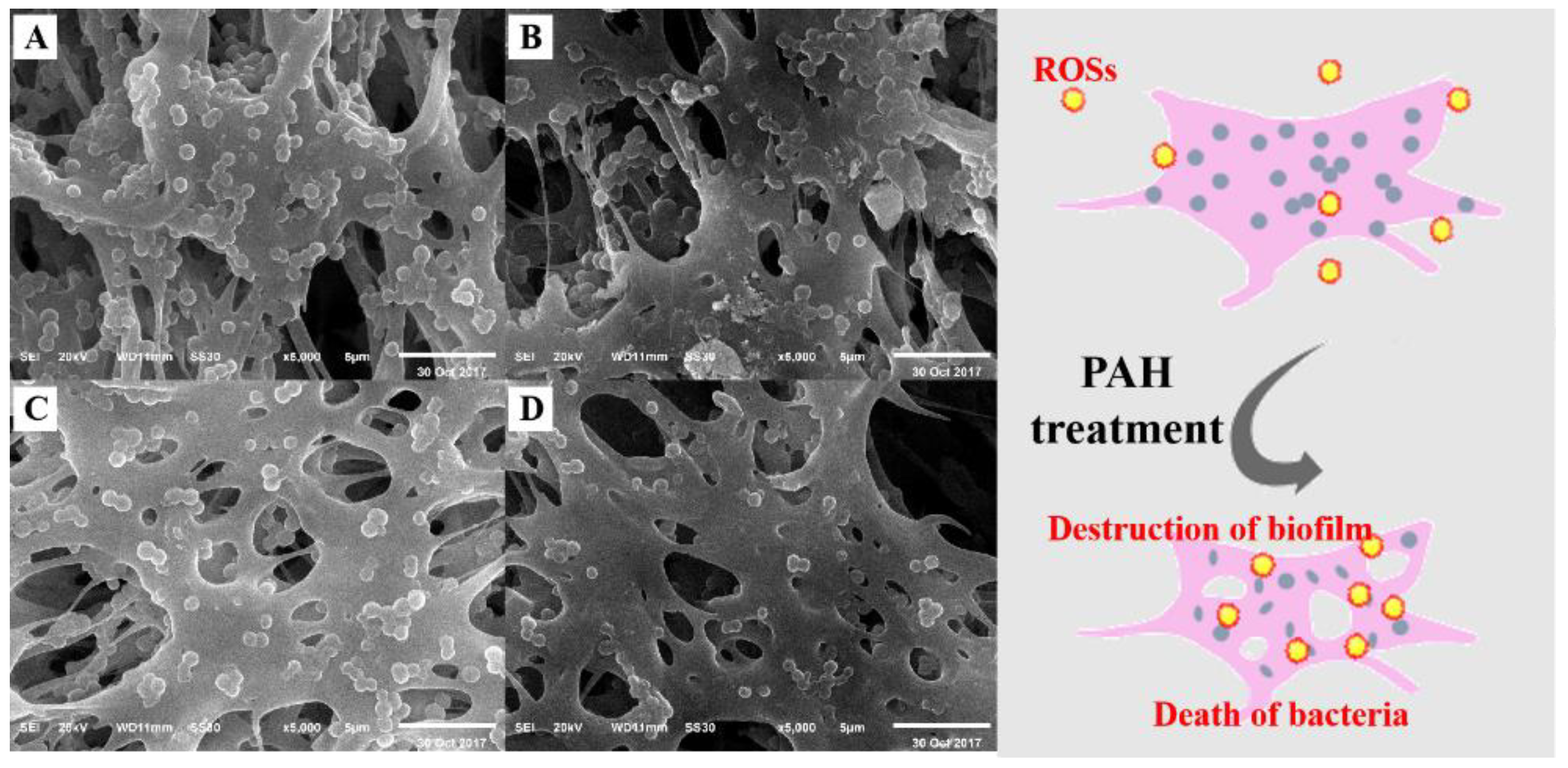
SEM images were used to further investigate the morphology of S. aureus biofilms (Figure 2A–D). For the untreated S. aureus biofilms, the structure of the biofilm network was dense and there were fewer holes. Meanwhile, there appeared a greater number of bacterial cells and most of the cells were single with regular shapes and smooth surfaces. After incubation with PAH for 30 min, the structure of the biofilm became loose with more and bigger holes. More bacterial cells aggregated together and the integrity of the bacteria cells was destroyed. Results suggested that PAH could lead to significant changes in the biofilm matrix, resulting in the direct exposure of the bacterial cells to PAH. The damages in bacterial cells and biofilms could be ascribed to the erosion effects of ROSs in PAH [10], which could induce damage to lipids, proteins, or DNA. Due to the good uniformity and fluidity of PAH, free radicals or reactive species can uniformly cover and fully contact the established biofilm, achieving a remarkable antibacterial effect [26].

Figure 2.
SEM images of S. aureus biofilms after incubation with the plasma-activated hydrogen peroxide solution for (A) 0 min, (B) 10 min, (C) 20 min, and (D) 30 min.
3.3. Changes in the Thickness of S. aureus Biofilms
Two fluorescent dyes were applied to assess the damages caused by PAH to S. aureus biofilms. The green fluorescent SYTO 9 stains only live bacteria within biofilms, while the red fluorescent PI stains only nonviable bacteria [27]. The LSCM images of S. aureus biofilms after PAH treatment exhibited in Figure 3A further confirmed the inactivation of bacterial cells in biofilms. The image of biofilms without PAH treatment was mainly green. The intensity of the red color increased upon increasing incubation time. The merged image of biofilms after the 30 min incubation was almost yellow, suggesting that the majority of live S. aureus cells in biofilms were non-viable after PAH treatments. The initial thickness of S. aureus biofilms (Time 0) was around 10.7 µm. Meanwhile, DIW and H2O2 solution had no significant (p > 0.05) effect on the thickness of the biofilm. After PAH treatment, the thickness of the biofilm decreased significantly (p < 0.05) to 8.1 µm when the incubation time was prolonged to 30 min (Figure 3B). The thickness of dead biofilm cells increased from 0.4 to 6.2 µm and the thickness of live cells reduced from about 10.3 to less than 1.9 µm. Results suggested that PAH treatment might destruct the S. aureus (ATCC 6538) biofilms by killing live cells. With the reduced thickness of biofilms, the protective effect of the biofilm matrix was gradually lost so that more S. aureus cells were directly in contact with PAH.
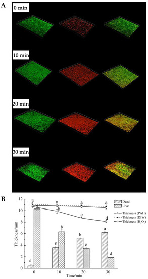
Figure 3.
(A) LSCM images and (B) the thickness of S. aureus biofilms after incubation with plasma-activated hydrogen peroxide solution for 0, 10, 20, and 30 min. The first column of (A) represents the biofilms stained with SYTO 9 (labels living bacteria cells), the second column represents the biofilms stained with propidium iodide (PI, labels the bacteria with a damaged membrane), and the third column is a combination of the first two. Deionized water (DIW) and hydrogen peroxide solution were chosen as negative controls. Dead: the thickness of biofilms in the second column of (A), Live: the thickness of biofilms in the first column. Thickness (PAH), Thickness (DIW), Thickness (H2O2) represents the total thickness of the biofilm after plasma-activated hydrogen peroxide, deionized water, and hydrogen peroxide solution treatment, respectively. Different lowercase letters (a–d) for the same treatment indicate significant differences among different incubation time (p < 0.05).
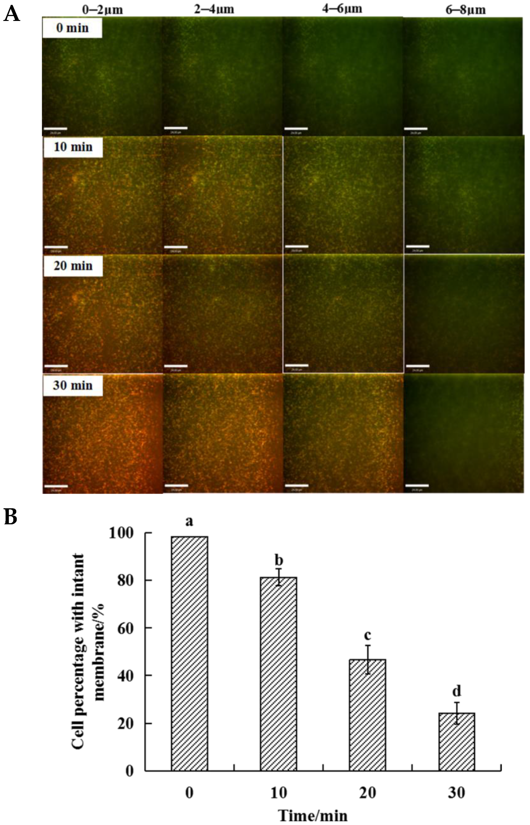
According to the results of the thickness of S. aureus biofilm, the Z-axis LSCM images of 1–4 layers (0–8 μm) biofilms were selected, as shown in Figure 4A. With the increase in the PAH treatment time, the red dots of the first layer (0–2 μm) gradually increased, indicating that the cell membranes in these biofilm layers were gradually destroyed. With the extension of biofilm depth, especially in the fourth layer of biofilm (6–8 μm), the red dots gradually decreased, indicating that the depth of action on S. aureus biofilm was 6–8 μm after 30 min of PAH treatment.
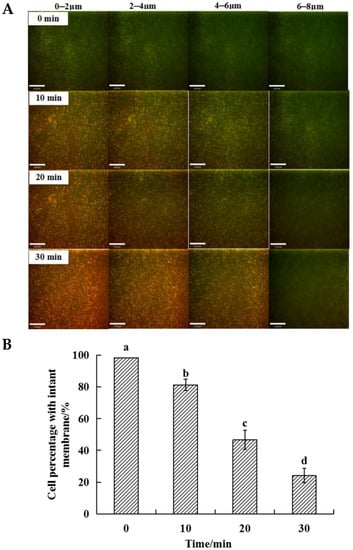
Figure 4.
Membrane integrity of S. aureus biofilm after PAH treatment for 0, 10, 20, 30 min. (A) The Z-axis LSCM images of 1–4 layers (0–8 μm) S. aureus biofilm. 0–2 μm (the first layer): the surface of the biofilm, 6–8 μm (the fourth layer): the interior of the biofilm; (B) Cell percentage with the intact membrane in the S. aureus biofilm. Different letters over the bars indicate a significant difference (p < 0.05) among incubation time.
The percentage of intact cell membrane structure in biofilms after PAH treatment was shown in Figure 4B. Before PAH treatment, about 98% of S. aureus had a complete cell membrane structure. With the extension of treatment time, the percentage of S. aureus with complete cell membrane structure showed a significant (p < 0.05) downward trend, and after 30 min treatment, the percentage decreased to 28.27%. This result was higher than the number of bacteria with reproductive ability in the biofilm as exhibited in Figure 1B. This phenomenon might be attributed to some bacteria cells still had membrane structure after PAH treatment, but intracellular contents had been destroyed and the ability to reproduce was lost.
3.4. Changes in Metabolic Capacity
The metabolic capacity in viable biofilm cells was quantified by the resazurin. As shown in Figure 5, the percentage of metabolic capacity exhibited a continuously decreasing tendency with the prolonged exposure to plasma. For the biofilms without PAH treatment, the percentage of metabolic capacity was 98.23% and decreased to 23.27% when biofilm was immersed in PAH for 30 min. Combined with ROS results, it was found that the balance of the oxidation-reduction system in the biofilms was disturbed by PAH, leading to a significant (p < 0.05) decline in metabolic activity. Meanwhile, compared with the result as shown in Figure 1B, more than 99.9% of the bacteria lost the ability to reproduce in the biofilm after PAH treatment for 30 min, but only about 76% of these bacteria lost the ability to metabolize. This result is similar to the percentage of bacteria with intact cell membrane structure shown in Figure 4B. The metabolic capacity depends on the electron transport chain on the surface of bacteria, which can provide energy. When the bacterial membrane is damaged, the cell will lose the ability to metabolize.
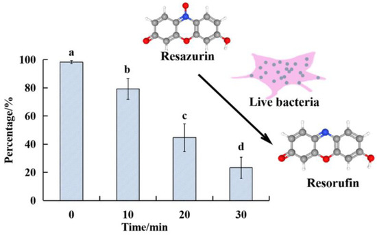
Figure 5.
The metabolic capacity (%) in S. aureus cells in biofilms after incubation with the plasma-activated hydrogen peroxide solution for various time. Different letters over the bars indicate a significant difference (p < 0.05) among incubation time.
3.5. Changes in ROS Levels of Bacteria Cells within the Biofilm
The ROS level in S. aureus biofilms was investigated to clarify the role and mechanism of PAH in sterilization. The relationship between cell death and intracellular ROS level was discussed. The fluorescence intensity of the biofilm after the PAH treatment for 30 min was shown in Figure 6A, compared with the first layer (0–2 μm), the fourth layer of the biofilm had a more obvious fluorescence intensity. It may be because surface bacteria were more exposed to PAH, RONS in PAH led to the destruction of cell membrane structure, which was then washed away by deionized water during sample preparation. Figure 6B showed the total ROS level of bacteria at 6–8 μm in S. aureus biofilms. With the extension of PAH treatment time, the total fluorescence intensity in cells gradually increased, indicating that intracellular ROS gradually accumulated in this layer of biofilm. Combined with the result in Figure 1B, more than 99.9% of the bacteria in the biofilm lost the ability to reproduce after PAH treatment for 30 min. Although the fluorescence intensity of the bacteria in the fourth layer of the biofilm was relatively high after treatment for 30 min, indicating that PAH can not only destroy the surface cell structure of the biofilm through its reactive oxygen species but also induce the production of ROS in the cells at the bottom of the biofilm. As a result, some bacteria have intact cellular structures but lose the ability to reproduce.

Figure 6.
(A) LSCM images of S. aureus biofilms stained with H2DCFDA; (B) The content of ROSs in S. aureus biofilms after incubation with the plasma-activated hydrogen peroxide solution for various time. Different letters over the bars indicate a significant difference (p < 0.05) among incubation time.
3.6. Changes in DNA Integrity
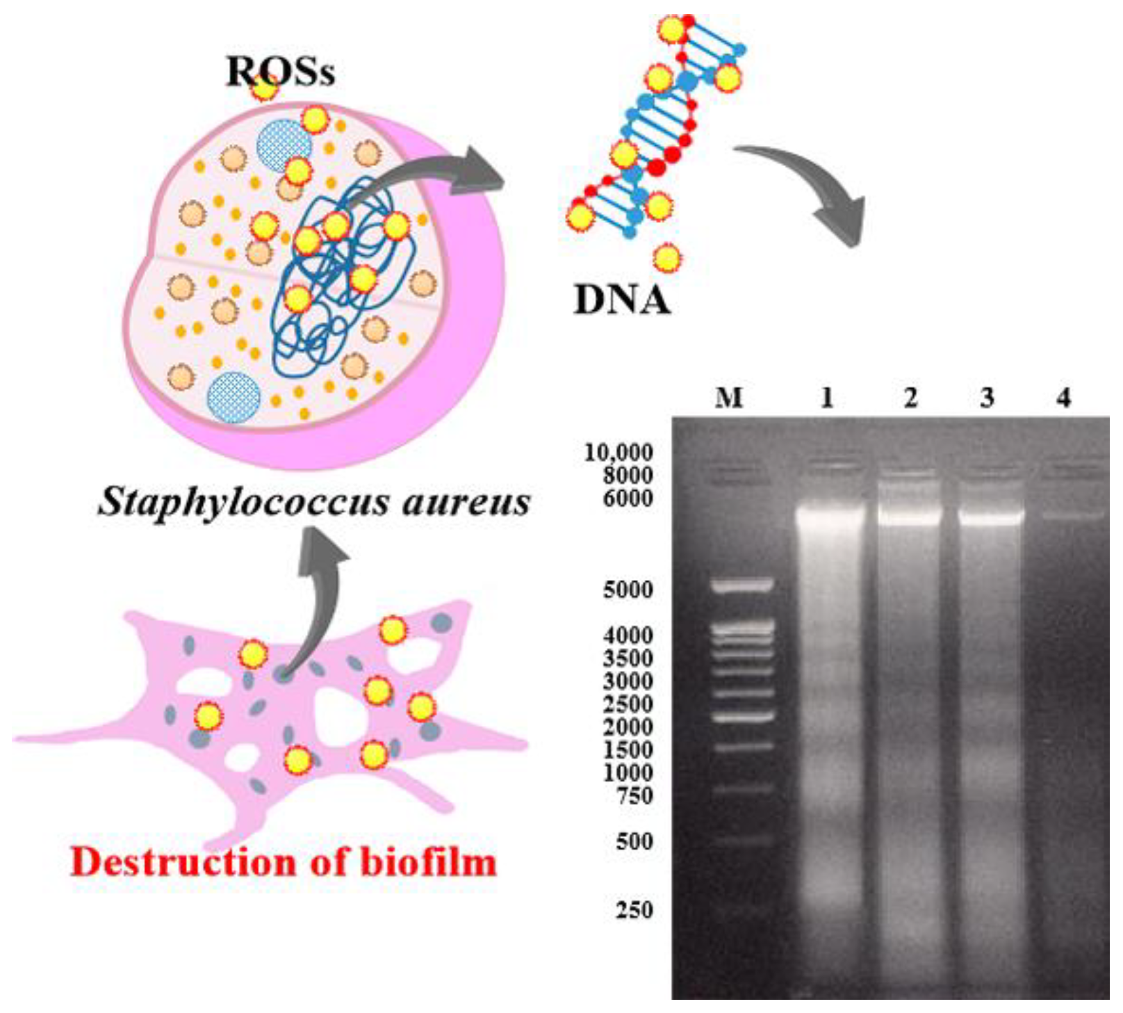
The effect of PAH on S. aureus biofilm can be further elucidated by the DNA analysis of biofilm. DNA electrophoresis showed that the band of DNA fragments with a molecular size of around 6000 appeared thicker and brighter with the extension of PAH exposure to the biofilm (Figure 7), indicating the damage of DNA integrity in S. aureus cells in the biofilms. The reactive species involved in cellular DNA breakdown could be OH•, NO2•, and ONOO− in PAH generated by DBD [28,29]. Results suggested that PAH treatment not only caused damage to the cell integrity but also led to severe damage to the DNA in the biofilms.
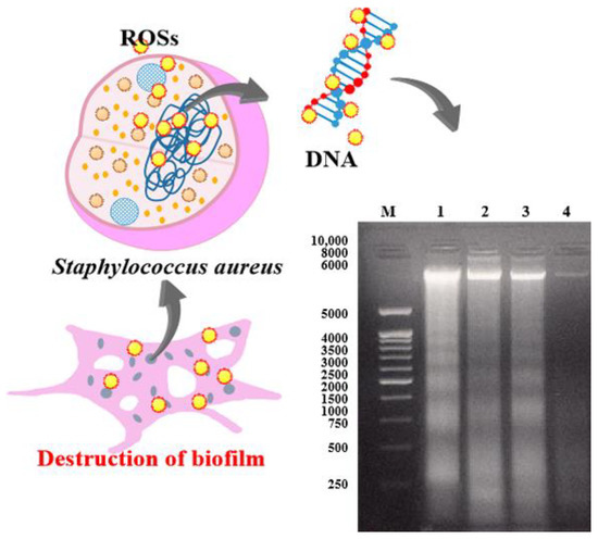
Figure 7.
The agarose gel of S. aureus DNA after plasma-activated solution treatment. M: DL 10,000 DNA Markers, 1–4: S. aureus DNA from samples incubated with the plasma-activated solution for 30, 20, 10, and 0 min, respectively.
In general, when S. aureus biofilms were exposed to PAH for 30 min, 23–28% bacteria were in the sublethal state, which had complete cell membrane structure and metabolic capacity but could not proliferate, mostly in the fourth layer of the biofilm. Due to the existence of apoptosis cells and extracellular matrix on the surface of the biofilm, the penetration of PAH was decreased, and the defense ability of the underlying cells against PAH was improved, the effect of RONS on biofilm cells was reduced. When the underlying cells were subjected to an adverse external environment, the stress response system in the bacteria could produce various proteins to eliminate the oxidative stress induced by PAH. With the extension of PAH treatment time, this self-protection system of cells will reach a critical value, resulting in internal cell damage. The result of the intracellular ROS level further revealed that reactive species in PAH could penetrate cells and destroy the ROS balance in cells. Meanwhile, the oxidative damage of DNA also demonstrated that PAH could induce cell damage by oxidative stress.
4. Conclusions
This work investigated the effects of PAH prepared by DBD on S. aureus (ATCC 6538) biofilms. Results indicated that PAH could significantly affect morphophysiology of the biofilms, including changing biofilm structure and thickness, inactivating bacterial cells in the biofilms, and damaging bacterial metabolic capacity, as well as DNA in the cells. The effectiveness depends upon the incubation time. Reactive species in the biofilms mixed with PAH might be responsible for the effects. In future studies, more S. aureus strains and other bacteria will be studied to further verify whether PAH can be used as an alternative anti-biofilm sanitizer effectively to control foodborne pathogens on the surface of food and/or food processing equipment.
Author Contributions
Conceptualization, J.Z. (Jianying Zhao), J.Q. and M.H.; methodology, J.Z. (Jianying Zhao) and M.H.; software, J.Z. (Jianying Zhao) and J.Q.; writing—review and editing, J.Z. (Jianying Zhao), J.Q. and J.L.; supervision, W.Y. and J.Z. (Jianhao Zhang); funding acquisition, W.Y. and J.Z. (Jianhao Zhang). All authors have read and agreed to the published version of the manuscript.
Funding
This research was funded by the National Key Research and Development Program of China [2018YFD0700802] and the Jiangsu Agricultural Science and Technology Independent Innovation Fund (No. CX (18)3041).
Institutional Review Board Statement
Not applicable.
Informed Consent Statement
Not applicable.
Data Availability Statement
The data that support the findings of this study are available from the corresponding author upon reasonable request.
Conflicts of Interest
The authors declare no conflict of interest.
References
- Xiong, Z.; Du, T.; Lu, X.; Cao, Y.; Pan, Y. How deep can plasma penetrate into a biofilm? Appl. Phys. Lett. 2011, 98, 221503. [Google Scholar] [CrossRef]
- Ziuzina, D.; Boehm, D.; Patil, S.; Cullen, P.J.; Bourke, P. Cold plasma inactivation of bacterial biofilms and reduction of quorum sensing regulated virulence factors. PLoS ONE 2015, 10, 0138209. [Google Scholar] [CrossRef]
- Byun, K.H.; Han, S.H.; Yoon, J.w.; Park, S.H.; Ha, S.D. Efficacy of chlorine-based disinfectants (sodium hypochlorite and chlorine dioxide) on Salmonella Enteritidis planktonic cells, biofilms on food contact surfaces and chicken skin. Food Control 2021, 123, 107838. [Google Scholar] [CrossRef]
- Ellebracht, J.W.; King, D.A.; Castillo, A.; Lucia, L.M.; Acuff, G.R.; Harris, K.B.; Savell, J.W. Evaluation of peroxyacetic acid as a potential pre-grinding treatment for control of Escherichia coli O157:H7 and Salmonella Typhimurium on beef trimmings. Meat Sci. 2005, 70, 197–203. [Google Scholar] [CrossRef] [PubMed]
- Jamal, M.; Ahmad, W.; Andleeb, S.; Jalil, F.; Imran, M.; Nawaz, M.A.; Hussain, T.; Ali, M.; Rafiq, M.; Kamil, M.A. Bacterial biofilm and associated infections. J. Chin. Med. Assoc. 2018, 81, 7–11. [Google Scholar] [CrossRef] [PubMed]
- Haney, E.F.; Trimble, M.J.; Cheng, J.T.; Valle, Q.; Hancock, R.E.W. Critical assessment of methods to quantify biofilm growth and evaluate antibiofilm activity of host defence peptides. Biomolecules 2018, 8, 29. [Google Scholar] [CrossRef] [Green Version]
- Stoodley, P.; Sauer, K.; Davies, D.G.; Costerton, J.W. Biofilms as complex differentiated communities. Annu. Rev. Microbiol. 2002, 56, 187–209. [Google Scholar] [CrossRef] [PubMed] [Green Version]
- Sun, F.J.; Qu, F.; Ling, Y.; Mao, P.Y.; Xia, P.Y.; Chen, H.P.; Zhou, D.S. Biofilm-associated infections: Antibiotic resistance and novel therapeutic strategies. Future Microbiol. 2013, 8, 877–886. [Google Scholar] [CrossRef] [PubMed]
- Homoe, P.; Bjarnsholt, T.; Wessman, M.; Sorensen, H.C.F.; Johansen, H.K. Morphological evidence of biofilm formation in Greenlanders with chronic suppurative otitis media. Eur. Arch. Oto-Rhino-L 2009, 266, 1533–1538. [Google Scholar] [CrossRef]
- Zhu, Y.; Li, C.; Cui, H.; Lin, L. Feasibility of cold plasma for the control of biofilms in food industry. Trends Food Sci. Technol. 2020, 99, 142–151. [Google Scholar] [CrossRef]
- Srey, S.; Jahid, I.K.; Ha, S.D. Biofilm formation in food industries: A food safety concern. Food Control 2013, 31, 572–585. [Google Scholar] [CrossRef]
- Pankaj, S.K.; Keener, K.M. Cold plasma: Background, applications and current trends. Curr. Opin. Food Sci. 2017, 16, 49–52. [Google Scholar] [CrossRef]
- Oehmigen, K.; Winter, J.; Hähnel, M.; Wilke, C.; Brandenburg, R.; Weltmann, K.-D.; von Woedtke, T. Estimation of Possible Mechanisms of Escherichia coli Inactivation by Plasma Treated Sodium Chloride Solution. Plasma Process. Polym. 2011, 8, 904–913. [Google Scholar] [CrossRef]
- Han, L.; Patil, S.; Boehm, D.; Milosavljevic, V.; Cullen, P.J.; Bourke, P. Mechanisms of inactivation by high-voltage atmospheric cold plasma differ for Escherichia coli and Staphylococcus aureus. Appl. Environ. Microbiol. 2015, 82, 450–458. [Google Scholar] [CrossRef] [Green Version]
- Traba, C.; Liang, J.F. Susceptibility of Staphylococcus aureus biofilms to reactive discharge gases. Biofouling 2011, 27, 763–772. [Google Scholar] [CrossRef] [PubMed] [Green Version]
- Ercan, U.K.; Wang, H.; Ji, H.; Fridman, G.; Brooks, A.D.; Joshi, S.G. Nonequilibrium plasma-activated antimicrobial solutions are broad-spectrum and retain their efficacies for extended period of time. Plasma Process. Polym. 2013, 10, 544–555. [Google Scholar] [CrossRef]
- Fernandez, A.; Shearer, N.; Wilson, D.R.; Thompson, A. Effect of microbial loading on the efficiency of cold atmospheric gas plasma inactivation of Salmonella enterica serovar Typhimurium. Int. J. Food Microbiol. 2012, 152, 175–180. [Google Scholar] [CrossRef]
- Tian, Y.; Ma, R.; Zhang, Q.; Feng, H.; Liang, Y.; Zhang, J.; Fang, J. Assessment of the physicochemical properties and biological effects of water activated by non-thermal plasma above and beneath the water surface. Plasma Process. Polym. 2015, 12, 439–449. [Google Scholar] [CrossRef]
- Xiao, J.; Zhang, Y.; Wang, J.; Yu, W.; Wang, W.; Ma, X. Monitoring of cell viability and proliferation in hydrogel-encapsulated system by resazurin assay. Appl. Biochem. Biotechnol. 2010, 162, 1996–2007. [Google Scholar] [CrossRef]
- Pan, Y.Y.; Zhang, Y.; Cheng, J.H.; Sun, D.W. Inactivation of Listeria monocytogenes at various growth temperatures by ultrasound pretreatment and cold plasma. LWT-Food Sci. Technol. 2020, 118, 108635. [Google Scholar] [CrossRef]
- Lukes, P.; Dolezalova, E.; Sisrova, I.; Clupek, M. Aqueous-phase chemistry and bactericidal effects from an air discharge plasma in contact with water: Evidence for the formation of peroxynitrite through a pseudo-second-order post-discharge reaction of H2O2 and HNO2. Plasma Sources Sci. Technol. 2014, 23, 015019. [Google Scholar] [CrossRef]
- Park, J.H.; Kumar, N.; Park, D.H.; Yusupov, M.; Neyts, E.C.; Verlackt, C.C.; Bogaerts, A.; Kang, M.H.; Uhm, H.S.; Choi, E.H.; et al. A comparative study for the inactivation of multidrug resistance bacteria using dielectric barrier discharge and nano-second pulsed plasma. Sci. Rep. 2015, 5, 13849. [Google Scholar] [CrossRef] [PubMed] [Green Version]
- Qian, J.; Zhuang, H.; Nasiru, M.M.; Muhammad, U.; Zhang, J.; Yan, W. Action of plasma-activated lactic acid on the inactivation of inoculated Salmonella Enteritidis and quality of beef. Innov. Food Sci. Emerg. Technol. 2019, 57, 102196. [Google Scholar] [CrossRef]
- Hertwig, C.; Reineke, K.; Ehlbeck, J.; Knorr, D.; Schluter, O. Decontamination of whole black pepper using different cold atmospheric pressure plasma applications. Food Control 2015, 55, 221–229. [Google Scholar] [CrossRef]
- Duffy, G.; Sheridan, J.J. The effect of temperature, pH and medium in a surface adhesion immunofluorescent technique for detection of Listeria monocytogenes. J. Appl. Microbiol. 1997, 83, 95–101. [Google Scholar] [CrossRef] [PubMed] [Green Version]
- Zhou, R.; Zhou, R.; Wang, P.; Xian, Y.; Mai-Prochnow, A.; Lu, X.; Cullen, P.J.; Ostrikov, K.; Bazaka, K. Plasma-activated water: Generation, origin of reactive species and biological applications. J. Phys. D Appl. Phys. 2020, 53, 303001. [Google Scholar] [CrossRef]
- Alkawareek, M.Y.; Algwari, Q.T.; Laverty, G.; Gorman, S.P.; Graham, W.G.; O’Connell, D.; Gilmore, B.F. Eradication of Pseudomonas aeruginosa biofilms by atmospheric pressure non-thermal plasma. PLoS ONE 2012, 7, e44289. [Google Scholar] [CrossRef] [PubMed] [Green Version]
- Devi, Y.; Thirumdas, R.; Sarangapani, C.; Deshmukh, R.R.; Annapure, U.S. Influence of cold plasma on fungal growth and aflatoxins production on groundnuts. Food Control 2017, 77, 187–191. [Google Scholar] [CrossRef]
- Chauvin, J.; Judee, F.; Yousfi, M.; Vicendo, P.; Merbahi, N. Analysis of reactive oxygen and nitrogen species generated in three liquid media by low temperature helium plasma jet. Sci. Rep. 2017, 7, 4562. [Google Scholar] [CrossRef]


Publisher’s Note: MDPI stays neutral with regard to jurisdictional claims in published maps and institutional affiliations. |
© 2021 by the authors. Licensee MDPI, Basel, Switzerland. This article is an open access article distributed under the terms and conditions of the Creative Commons Attribution (CC BY) license (https://creativecommons.org/licenses/by/4.0/).