Article

Effect of Washing Condition on the Fracture Strength, and the Degree of Conversion of 3D Printing Resin

1
Department of Prosthodontics, School of Dentistry, Chonnam National University, Gwangju 61186, Korea
2
School of Dentistry, Chonnam National University, Gwangju 61186, Korea
3
Department of Prosthodontics, Daejeon Dental Hospital, Wonkwang University, Daejeon 35233, Korea
*
Author to whom correspondence should be addressed.
Appl. Sci. 2021, 11(24), 11676; https://doi.org/10.3390/app112411676
Submission received: 30 October 2021 / Revised: 2 December 2021 / Accepted: 7 December 2021 / Published: 9 December 2021
(This article belongs to the Special Issue Restorative and Endodontic Materials for Clinical Dentistry)

Abstract

:
This study compared the surface roughness, contact angle, surface energy, residual monomers, degree of conversion, and flexural strength of 3D-printed dental resin under various washing conditions. The specimens were printed with a digital light processing (DLP) printer and were divided into four groups: the group dipped in IPA for 5 s (IPA-D), the group washed in IPA for 1 min (IPA-1), the group washed in IPA for 10 min (IPA-10), and the group washed with TPM for 10 min (TPM-10). Following, the groups were redivided into two groups: a cured group and an uncured group. All experimental data were statistically analyzed using one-way analysis of variance and Tukey’s test. In all groups, the surface roughness showed a value of 1.2–1.8 μm, with no significant difference (p > 0.05). Contact angle showed a significant difference between the three groups using IPA and the TPM group, whereby the TPM-washed specimen showed a low contact angle (p < 0.05). The degree of conversion (DOC) increased in the following order: IPA-D group, IPA-1 group, IPA-10 group, and TPM-10 group, exhibiting a significant difference between all groups (p < 0.05). Flexural strength was measured at 110–130 MPa in all groups, with no significant difference between groups (p > 0.05). The washing time and washing solution type of the 3D printing material had no significant effect on surface roughness and flexural strength.

1. Introduction

Currently, 3D printing is being widely used in the dental field [1,2]. The development of various materials based on polymers has made it possible to replace existing dental restorations and devices [1,3]. As the use of 3D-printed dental restorations based on the digital method is increasing, the research and products are under development [4].
Accuracy is probably the most important factor in the production of 3D-printed dental restorations because dental care cannot be established if accuracy is not guaranteed. Therefore, when 3D printing is applied to dentistry, it is crucial for dental clinicians to make accurate and precise printouts, and as of now, the various research on this topic is performed and related results have been published [5,6]. Several points should be considered to improve the accuracy of the 3D-printed outcome. First of all, the light source, a method for controlling the amount of light, the Z-axis setting, and a method for overcoming mechanical errors should be determined. After that, the suitable materials for printing need to be selected and developed. Once the printer and material have been selected, the next thing to consider is the method of printing. Accordingly, studies and trial and errors about this topic have been published, such as the thickness of the printing layer, the speed of printing, and the position and number of supporters for the stacked layer [7].
However, the 3D printing process used in actual clinical practice does not only require a printed outcome. The current 3D printing process consists of following three-steps; removing the supporter, washing, and post-curing after printing. Therefore, it is impossible to printout the exact restoration without considering the post-processing conditions. Many printing companies provide guidelines for this, but the guidelines are specific to each company, and the academic basis for this is still poor. Therefore, many clinicians are conducting research on washing and post-curing processes [8,9,10,11]. In particular, research on the post-processing process is very sparse compared with research on improving the accuracy of the printing process [12,13,14].
Therefore, the goal of this study is to comparatively evaluate the characteristics of 3D-printed outcomes under various washing conditions. The temporary resin specimen for this study was printed out. The resin, a photopolymer acrylate type resin, is a material widely used in 3D printing because of its aesthetic color and the ease of process and polishing procedure. After the printouts, the surface characteristics and the degree of conversion (DOC) were measured, and the effect on flexural strength after post-processing was evaluated. The null hypothesis of this study is that there is no difference in surface properties, the DOC, and flexural strength according to the various cleaning conditions.

2. Materials and Methods

2.1. Experimental Materials

The photopolymer resin (Vericom MAZIC D TEMP, Vericom, Gangwon-do, Korea) used for temporary crown was used in this study (Table 1). Disk-shaped specimens were printed out using a digital light processing (DLP) printer, Hunter (Flashforge 3D printer, Jinhua, China). It was designed to be 10 mm in diameter and 2 mm in thickness using the CAD software program (Meshmixer, Autodesk). The groups were divided according to washing methods and materials. Isopropylalochol (IPA, Vaxxen Labs, Cortland, Ohio, USA) and tripropylene glycol monomethyl ether (TPM, Formlabs, Somerville, MA, USA) were used as the washing solutions for cleaning the specimen. According to the regulations recommended by the manufacturer, the TPM group was applied for 10 min.

2.2. Experimental Groups

According to washing conditions, a total of 80 specimens were classified into 4 groups: the IPA-D group was dipped with isopropylalochol for 5 s, the IPA-1 group was washed with isopropyl alcohol for 1 min, the IPA-10 group was washed with isopropyl alcohol for 10 min, and the TPM-10 group was washed with TPM for 10 min. All groups were washed with the resin washing device (FormWash, Formlabs, Somverville, MA, USA) at room temperature. After that each group was divided into uncured and cured groups (Table 2). The surface characteristics were evaluated by using the uncured specimen and the cured specimens were used for measuring the flexural strength.

2.3. Experimental Method

The surface of the specimen was coated with platinum and observed using a field emission scanning electron microscope (SNE-4500M Plus, BRUKER, Pleasanton, CA, USA), and the surface roughness was measured using a nanosurface 3D optical profiler (NV-E1000, Nano System, Daejeon, Korea). To measure the contact angle, a drop of 4 µL of distilled water was dropped onto the specimen and 30 shots were taken at 0.1 s intervals using a contact angle analyzer (video contact angle measuring device: Phoenix 300, SEO Inc., Suwon, Korea). The surface energy value was also measured. The amount of residual monomers remaining on the surface after washing was measured with a Fourier infrared spectrometer (INVENIO-X, BRUKER, USA) in the wavenumber range of 4000–500 cm−1 and with a resolution of 4 cm−1. Ten specimens were tested for each group, and the degree of conversion (DOC) of each group was calculated using the standard baseline method by converting the measured transmittance to absorbance. To determine the degree of conversion, the ratio between unpolymerized aliphatic(C=C) bond at 1640 cm−1 and the aromatic (C=C) at 1610 cm−1 peaks was used. The uncured aromatic (C=C) bond at 1610 cm−1 peak was used as an internal standard.
DC   ( % ) = 1 100   ×   [   Aliphatic   p o l y m e r i z e d   /   Aromatic   p o l y m e r i z e d     Aliphatic   u n p o l y m e r i z e d   /   Aromatic   u n p o l y m e r i z e d     ]
Flexural strength was measured after using a post-curing device (LC-3D Print Box, 3D Systems, Rock Hill, SC, USA) for 30 min to cure all residual monomers remaining on the surface after washing. In the three-point flexural fracture test based on the ISO-178 standard, starting with a preload of 1 N, the load value was measured when the specimen was broken while applying a load rate of 1 mm/min with a universal testing machine (Hydraulic type universal testing machine 1000 kN, ELE, Leighton Buzzard, UK).

2.4. Statistical Analysis
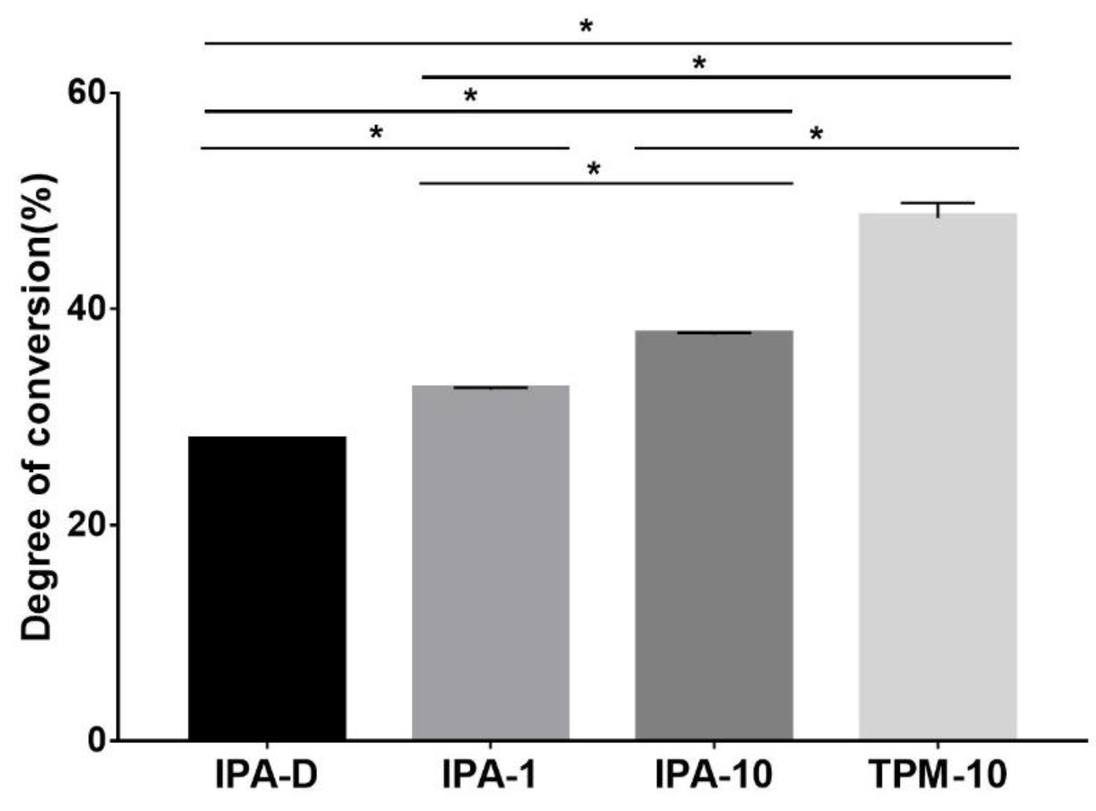
Statistical analysis was performed using a statistical analysis program (GraphPad Prism, Prism 7 for Windows, version 7). One-way analysis of variance and Tukey’s post-hoc tests were performed. They were used to analyze the influence of washing time and solution on the temporary resin surface (roughness, contact angle) and its mechanical characteristics (DOC, flexural strength). All results confirmed the statistical significance at the p < 0.05 level.

3. Results

After the washing process, the surface aspects of the specimens of each group were photographed using FE-SEM. For the IPA-D group, which had a relatively short washing time, it could be observed that suspensions remained on the surface of the specimen, and groups A and B, which had a relatively long washing time of 10 min, contained relatively few foreign substances (Figure 1). Surface roughness was measured with a nano surface 3D optical profiler, and Ra (µm) values were 1.27 ± 0.23 µm for group IPA-D, 1.55 ± 0.60 µm for group IPA-1, 1.47 ± 0.91 µm for group IPA-10, and 1.86 ± 0.83 µm for group TPM-10. Between the four groups, no statistically significant difference was found (Figure 2). As for the contact angle, the results of the TPM-10 group (43.26 ± 0.15°) were statistically more significant than those of the IPA-D group (67.74 ± 1.96°), IPA-1 group (64.58 ± 1.29°), and IPA-10 group (63.92 ± 0.07°). TPM-10 group has a very small contact angle (p < 0.05), indicating that this surface is the most hydrophilic (Figure 3 and Figure 4). In the case of surface energy, the TPM-10 group (125.8 ± 0.13 mN/m) had the highest energy compared with the IPA-D group (100.4 ± 2.31 mN/m), the IPA-1 group (104.0 ± 1.48 mN/m), and the IPA-10 group (104.8 ± 0.07 mN/m), and the difference was determined to be statistically significant (p < 0.05) (Figure 5).
The amount of residual monomers was measured using a Fourier infrared spectrometer (PerkinElmer, Waltham, MA, USA). From this, the amount of residual monomers could be evaluated quantitatively. Due to the substituted alkenes, the residual monomers were calculated from the peak at the 1637 cm−1 [15,16,17]. The FTIR equation was used to evaluate the DOC by referencing the result of uncured state [18]. By comparing the DOC for each resin, the groups washed for a long time such as the IPA-10 and TPM-10 groups were determined to be approximately 37% and 47%, respectively. The short washing time for the IPA-D group and the IPA-1 group resulted in a DOC of 27–32%. In particular, the TPM-10 group showed the highest DOC (46–50%), and the statistical significance was confirmed between the four groups (p < 0.05) (Figure 6). Flexural strength was 131.46 ± 25.97 MPa for group IPA-D, 133.07 ± 19.37 MPa for group IPA-1, 114.56 ± 20.54 MPa for group IPA-10, and 114.64 ± 26.76 MPa for group TPM-10. Overall, the four groups showed a value of about 110–130 MPa. For the groups lacking washing, such as the IPA-D and IPA-1 groups, the flexural strength was slightly higher, but there was no statistically significant difference (Figure 7).

4. Discussion

This study was conducted to examine the characteristics of the surface according to the various 3D printing washing conditions and to determine the clinically optimal washing time and washing solution. The experimental results showed no statistically significant difference in terms of surface roughness and flexural strength, but the evaluation on the contact angle and the DOC showed the difference. Therefore, the null hypothesis of this study was partially rejected.
When the contact angle was compared, the surface of the TPM-10 group had the highest wettability and the highest surface energy compared to the other three groups (IPA-D, IPA-1, IPA-10). There is still much debate about wetting and bacterial adhesion [19,20]. However, several studies have shown that hydrophobic bacteria colonize on hydrophobic surfaces [19,21]. Streptococcus mutans, which is most commonly found in the oral cavity, is a representative hydrophobic bacterium, and the TPM-10 group conducted in the above study is likely to be more resistant to bacterial adhesion [22]. In the case of amount of residual monomers, for long washing times, as for the IPA-10 and TPM-10 groups, the DOC was high because of the small amount of residual monomer. This resulted in a relatively low DOC. In addition, the IPA-D and IPA-1 groups showed relatively high flexural strength, but this difference was determined to be not statistically significant.
The surface roughness of dental prostheses has a significant impact on clinical practice. A rough surface can become easily colonized by bacteria in the oral cavity, and it also increases the wear of the opposing teeth [23]. Moreover, prostheses with rough surfaces are not considered to be aesthetically pleasing [24]. In addition, the fit of a prosthesis may be poor due to its rough surface, which is the most important factor in clinical situations [25]. This is because if the rough surface of the prosthesis is placed in the mouth, it may act as an undercut and the possibility of an inaccurate prosthesis margin increases. Because 3D-printed dental prostheses cannot be polished inside, a smooth inner surface is essential for restoration. However, according to the results of this experiment, there was no significant difference in surface roughness according to the washing solution or time. From the SEM image, residual suspended solids were observed in the IPA-D group, and relatively clear surfaces were observed in the IPA-10 and TPM-10 groups. The clear surface indicates an exposed layer which is unique in 3D printouts. In other words, it was speculated that the well-washed printed surface rather offset the degree of roughness due to residual suspended solids observed in the IPA-D group because the printed layer was exposed. The SEM images showed that the IPA-D and IPA-1 groups, which lacked washing time, showed more residual suspended solids than the IPA-10 and TPM-10 groups. In contrast, if washing is insufficient, such as for the IPA-D group and the IPA-1 group, the residual monomers will be cured naturally, which may lead to over-curing, which may have made the surface lubricated. In the case of the TPM group, the result showed a rougher surface, but visually from SEM image, it showed a smooth state, and there was no statistically significant difference between other three groups (IPA-D, IPA-1, IPA-10). As such, the aspect of surface roughness due to washing should be studied further in the future.
Many studies on residual monomers of dental resins have been reported [26,27]. In particular, in terms of strength and cytotoxicity, many clinicians are concerned about residual monomers, and studies are being conducted on the importance of washing along with efforts to develop substances that are harmless to the human body [28,29,30]. In addition, it has been reported that the subsequent amount of polymerized residual monomers has a negative effect on the marginal adaptation of a temporary crown in clinical practice [31]. Statistical differences between the IPA groups arise only based on the time spent for washing, which shows that proper time spent on washing is essential in dental practice. Usually, the DOC of resins fabricated by dental printing resin companies is reported to be approximately 50–60%, but only the IPA-10 group showed this value in this experiment. Therefore, it can be said that at least 10 min of washing is required to achieve clinical precision. The TPM group showed a high DOC, which may imply the lower amount of residual monomers, compared with that for IPA group. It proves the superb washing ability of the ether functional groups. Of course, the influence of not only the residual monomers, but also of the washing solution in the oral cavity should be considered; however, in light of the results of this experiment, the use of TPM is judged to be more suitable for dental use than IPA.
Flexural strength was evaluated in accordance with ISO standards, and the measurements were performed after post-curing under the same clinical conditions. In flexural strength, since there was no statistically significant difference between the specimens, washing and strength are irrelevant. In all groups, the strength was found to be consistent with the information provided by the resin companies whose products were used in the experiment (approximately 110 MPa). The reason why the IPA-D and IPA-1 groups showed relatively high flexural strength is thought to be that many residual monomers showed a higher resin content as a result of the post-curing process. It is presumed that the amount of resin increased as the residual monomers, which were not washed off the surface, were cured after post-curing and hardened. However, this did not result in a statistically significant difference, and any difficulties in removing the prosthesis due to the presence of residual monomers will eventually be accompanied by relief during the procedure. Therefore, this increase in flexural strength is not expected to be clinically useful.
This experiment was conducted in a short washing time (10 min or less) which is useful for 3D printing in dental clinic. However, there is a possibility that the surface energy may differ depending on the characteristics of the washing material (isopropyl alcohol, TPM). Therefore, further research is needed to associate washing conditions with surface roughness. In addition, the most important part of clinical practice is the adequate adaptation of a 3D-printed prosthesis in the oral cavity. Therefore, additional experiments to confirm the marginal adaptation in various oral environments (prepared teeth or implant abutments) are necessary. However, under these limited conditions, this experiment showed that even a very short washing time (1 min) was clinically acceptable. This result can be used as a guideline for dental 3D printing protocols that have not yet been established.

5. Conclusions

In all four groups, the surface roughness showed a value of 1.2–1.8 μm, and the flexural strength was measured at 110–130 MPa. The contact angle of the TPM-10 group showed better wettability compared to other three groups using IPA. The degree of conversion (DOC) was higher as the washing time increased, and the TPM group showed higher value than in other three IPA groups.
Within the limitations of this experiment, the following conclusions can be drawn when a temporary crown is fabricated with 3D-printed resin:
Even if the washing times and methods of 3D printing were changed, there was no significant difference in the surface roughness of the resin.
However, if the washing time is insufficient, the DOC of resin was low, which is expected to have a negative clinical effect.
In addition, residual monomers in 3D-printed resin do not clinically affect the flexural strength.

Author Contributions

Data curation, H.-P.L.; Formal analysis, Y.K. and S.-K.L.; Funding acquisition, S.-W.P.; Investigation, G.-S.K.; Methodology, K.-D.Y.; Project administration, J.-H.K.; Resources, Y.Y.; Writing—original draft, W.J.; Writing—review & editing, C.P. All authors have read and agreed to the published version of the manuscript.

Funding

This research was funded by the National Research Foundation of Korea (NRF) grant funded by the Korean government (No. 2021R1C1C1008165), a grant (BCRI21030) of Chonnam National University Hospital Biomedical Research Institute, and the Technology development Program (S2850798 and S2975345) funded by the Ministry of SMEs and Startups (MSS, Korea), a grant (2020-1148) of Chonnam National University.

Conflicts of Interest

The authors declare no conflict of interest.

References

  1. Schönhoff, L.M.; Mayinger, F.; Eichberger, M.; Reznikova, E.; Stawarczyk, B. 3D printing of dental restorations: Mechanical properties of thermoplastic polymer materials. J. Mech. Behav. Biomed. Mater. 2021, 119, 104544. [Google Scholar] [CrossRef] [PubMed]
  2. Pillai, S.; Upadhyay, A.; Khayambashi, P.; Farooq, I.; Sabri, H.; Tarar, M.; Lee, K.T.; Harb, I.; Zhou, S.; Wang, Y.; et al. Dental 3D-Printing: Transferring Art from the Laboratories to the Clinics. Polymers 2021, 13, 157. [Google Scholar] [CrossRef] [PubMed]
  3. Kessler, A.; Hickel, R.; Reymus, M. 3D printing in dentistry—State of the art. Operat. Dent. 2020, 45, 30–40. [Google Scholar] [CrossRef] [PubMed]
  4. Bud, E.S.; Bocanet, V.-I.; Muntean, M.H.; Vlasa, A.B.; Sorana, M.; Pacurar, M.D.; Bogdan, R.; Olteanu, C.D.; Bud, A. Accuracy of Three-Dimensional (3D) Printed Dental Digital Models Generated with Three Types of Resin Polymers by Extra-Oral Optical Scanning. J. Clin. Med. 2021, 109, 1908. [Google Scholar] [CrossRef] [PubMed]
  5. Greco, G.B.; Popi, D.; Di Stefano, D.A. Accuracy of 3-dimensional printing of dental casts: A proposal for quality standardization. J. Prosthet. Dent. 2021. [Google Scholar] [CrossRef] [PubMed]
  6. Son, K.; Lee, J.H.; Lee, K.B. Comparison of Intaglio Surface Trueness of Interim Dental Crowns Fabricated with SLA 3D Printing, DLP 3D Printing, and Milling Technologies. Healthcare 2021, 9, 983. [Google Scholar] [CrossRef] [PubMed]
  7. Baek, J.W.; Shin, S.-Y. Accuracy of dies fabricated by various three dimensional printing systems: A comparative study. J. Dent. Rehabil. Appl. Sci. 2020, 36, 242–253. [Google Scholar] [CrossRef]
  8. Song, G.; Son, J.W.; Jang, J.H.; Choi, S.H.; Jang, W.H.; Lee, B.N.; Park, C. Comparing volumetric and biological aspects of 3D-printed interim restorations under various post-curing modes. J. Adv. Prosthodont. 2021, 13, 71–78. [Google Scholar] [CrossRef]
  9. Nowacki, B.; Kowol, P.; Koziol, M.; Olesik, P.; Wieczorek, J.; Waclawiak, K. Effect of Post-Process Curing and Washing Time on Mechanical Properties of mSLA Printouts. Materials 2021, 14, 4856. [Google Scholar] [CrossRef]
  10. Bayarsaikhan, E.; Lim, J.-H.; Shin, S.-H.; Park, K.-H.; Park, Y.-B.; Lee, J.-H.; Kim, J.-E. Effects of Postcuring Temperature on the Mechanical Properties and Biocompatibility of Three-Dimensional Printed Dental Resin Material. Polymers 2021, 13, 1180. [Google Scholar] [CrossRef]
  11. Kim, D.; Shim, J.-S.; Lee, D.; Shin, S.-H.; Nam, N.-E.; Park, K.-H.; Shim, J.-S.; Kim, J.-E. Effects of Post-Curing Time on the Mechanical and Color Properties of Three-Dimensional Printed Crown and Bridge Materials. Polymers 2020, 12, 2762. [Google Scholar] [CrossRef] [PubMed]
  12. Wu, D.; Zhao, Z.; Zhang, Q.; Qi, H.J.; Fang, D. Mechanics of shape distortion of DLP 3D printed structures during UV post-curing. Soft Matter. 2019, 15, 6151–6159. [Google Scholar] [CrossRef] [PubMed]
  13. Reymus, M.; Lumkemann, N.; Stawarczyk, B. 3D-printed material for temporary restorations: Impact of print layer thickness and post-curing method on degree of conversion. Int. J. Comput. Dent. 2019, 22, 231–237. [Google Scholar]
  14. Jindal, P.; Juneja, M.; Bajaj, D.; Siena, F.L.; Breedon, P. Effects of post-curing conditions on mechanical properties of 3D printed clear dental aligners. Rapid Prototyp. J. 2020, 26, 1337–1344. [Google Scholar] [CrossRef]
  15. Herrera-Gonzalez, A.M.; Caldera-Villalobos, M.; Perez-Mondragon, A.A.; Cuevas-Suarez, C.E.; Gonzalez-Lopez, J.A. Analysis of Double Bond Conversion of Photopolymerizable Monomers by FTIR-ATR Spectroscopy. J. Chem. Educ. 2019, 96, 1786–1789. [Google Scholar] [CrossRef]
  16. Moldovan, M.; Balazsi, R.; Soanca, A.; Roman, A.; Sarosi, C.; Prodan, D.; Vlassa, M.; Cojocaru, I.; Saceleanu, V.; Cristescu, I. Evaluation of the Degree of Conversion, Residual Monomers and Mechanical Properties of Some Light-Cured Dental Resin Composites. Materials 2019, 12, 2109. [Google Scholar] [CrossRef] [Green Version]
  17. Suvarna, S.; Chhabra, T.; Raghav, D.; Singh, D.; Kumar, P.; Sahoo, S. Residual monomer content of repair autopolymerizing resin after microwave postpolymerization treatment. Eur. J. Prosthodont. 2014, 2, 28–32. [Google Scholar]
  18. Leloup, G.; Holvoet, P.E.; Bebelman, S.; Devaux, J. Raman scattering determination of the depth of cure of light-activated composites: Influence of different clinically relevant parameters. J. Oral Rehabil. 2002, 29, 510–515. [Google Scholar] [CrossRef]
  19. Grivet, M.; Morrier, J.J.; Benay, G.; Barsotti, O. Effect of hydrophobicity on in vitro streptococcal adhesion to dental alloys. J. Mater. Sci. Mater. Med. 2000, 11, 637–642. [Google Scholar] [CrossRef] [PubMed]
  20. Nassar, U.; Meyer, A.E.; Ogle, R.E.; Baier, R.E. The effect of restorative and prosthetic materials on dental plaque. Periodontology 2000 1995, 8, 114–124. [Google Scholar] [CrossRef] [PubMed]
  21. Mabboux, F.; Ponsonnet, L.; Morrier, J.J.; Jaffrezic, N.; Barsotti, O. Surface free energy and bacterial retention to saliva-coated dental implant materials—An in vitro study. Colloids Surf. B Biointerfaces 2004, 39, 199–205. [Google Scholar] [CrossRef] [PubMed]
  22. Kim, D.H.; Kwon, T.Y. In vitro study of Streptococcus mutans adhesion on composite resin coated with three surface sealants. Restor. Dent. Endod. 2017, 42, 39–47. [Google Scholar] [CrossRef] [PubMed]
  23. Komalsingsakul, A.; Senawongse, P. Effect of the surface roughness of composite resins on the water contact angle and biofilm formation. Mahidol. Dent. J. 2019, 39, 75–84. [Google Scholar]
  24. Gönülol, N.; Yılmaz, F. The effects of finishing and polishing techniques on surface roughness and color stability of nanocomposites. J. Dent. 2012, 40, e64–e70. [Google Scholar] [CrossRef]
  25. Attia, M.A.; Shokry, T.E.; Abdel-Aziz, M. Effect of different surface treatments on the bond strength of milled polyetheretherketone posts. J. Prosthet. Dent. 2021. [Google Scholar] [CrossRef] [PubMed]
  26. Araújo, P.H.H.; Sayer, C.; Giudici, R.; Poço, J.G.R. Techniques for reducing residual monomer content in polymers: A review. Polym. Eng. Sci. 2002, 42, 1442–1468. [Google Scholar] [CrossRef]
  27. Leber, A.P. Human exposures to monomers resulting from consumer contact with polymers. Chem. Biol. Interact. 2001, 135, 215–220. [Google Scholar] [CrossRef]
  28. Ponting, D.J.; Ortega, M.A.; Niklasson, I.B.; Karlsson, I.; Seifert, T.; Steen, J.; Luthman, K.; Karlberg, A.T. Development of New Epoxy Resin Monomers—A Delicate Balance between Skin Allergy and Polymerization Properties. Chem. Res. Toxicol. 2018, 32, 57–66. [Google Scholar] [CrossRef] [PubMed]
  29. Görukmez, E.; Şen Yilmaz, B.; Ramoglu, S.I. Is a Single Rinse Effective on Evacuating the Residual Monomers After Orthodontic Bonding? An In Vivo Study. Bezmiâlem Sci. 2021, 9, 127. [Google Scholar] [CrossRef]
  30. Seo, D.-S. Influence of the washing process on the aligning capability of a nematic liquid crystal on polymer surfaces. Liquid Cryst. 2000, 27, 789–793. [Google Scholar] [CrossRef]
  31. Guerra, R.M.; Duran, I.; Ortiz, P. FTIR monomer conversion analysis of UDMA-based dental resins. J. Oral Rehabil. 1996, 23, 632–637. [Google Scholar] [CrossRef] [PubMed]
Figure 1. FE-SEM images at 1.0 K magnification after washing the surface of the specimen (A) IPA-D group: IPA dipping, (B) IPA-1 group: IPA washing for 1 min, (C) IPA-10 group: IPA washing for 10 min, (D) TPM-10 group: TPM washing for 10 min.
Applsci 11 11676 g001
Figure 2. Surface roughness according to different washing conditions.
Applsci 11 11676 g002
Figure 3. Contact angle of the water droplet on the surface. (A) IAP-D group; (B) IAP-1 group; (C) IAP-10 group; (D) TPM-10 group.
Applsci 11 11676 g003
Figure 4. Contact angle for different washing conditions: * significant difference at p < 0.05.
Applsci 11 11676 g004
Figure 5. Adhesion energy according to different washing conditions: * significant difference at p < 0.05.
Applsci 11 11676 g005
Figure 6. Degree of conversion according to different washing conditions: * significant difference at p < 0.05.
Applsci 11 11676 g006
Figure 7. Flexural strength according to different washing conditions.
Applsci 11 11676 g007
Table 1. The component of photopolymer resin (MAZIC D TEMP).
Photopolymer Resin
MonomersMethacrylic oligomers
Crosslinking agentPhosphine oxide
Table 2. Experimental groups according to washing conditions.
Dipping with IPA (IPA-D)1 min with IPA (IPA-1)10 min with IPA (IPA-10)10 min with TPM (TPM-10)
Uncured10101010
Cured10101010
Publisher’s Note: MDPI stays neutral with regard to jurisdictional claims in published maps and institutional affiliations.

