Conference Report

The 12th International Conference on Instrumental Analysis—Modern Trends and Applications (20–23 September 2021, Virtual Event)

by
Manassis Mitrakas
1,* and
Maria Ochsenkühn-Petropoulou
2,*
1
Analytical Chemistry Laboratory, Department of Chemical Engineering, Aristotle University of Thessaloniki, 54124 Thessaloniki, Greece
2
Laboratory of Inorganic and Analytical Chemistry, Department of Chemical Sciences, School of Chemical Engineering, National Technical University Athens, Iroon Polytechneiou 9, Zographou Campus, 15780 Athens, Greece
*
Authors to whom correspondence should be addressed.
Appl. Sci. 2021, 11(24), 11767; https://doi.org/10.3390/app112411767
Submission received: 28 October 2021 / Accepted: 8 November 2021 / Published: 10 December 2021
The 12th International Conference on “Instrumental Methods of Analysis” www.ima2021.gr (accessed on 8 November 2021)), was organized by the Aristotle University of Thessaloniki and National Technical University of Athens, during 20–23 September 2021 as a virtual event, providing the opportunity for high-level analytical scientists from all around the world to promote their relevant research.
IMA is a biannual series of conferences that started in 1999 and cover all areas of Chemical Analysis, including the development of new techniques, modern trends, and applications in a wide range of scientific disciplines. To date, several leading analytical chemists from Greece and abroad have presented their research work at previous IMA meetings. The 12th IMA conference (in a virtual format for the first time), had the ambition to bring together some of the most talented and innovative analytical chemists from all over the world for an excellent scientific online conference. The program of the 4-day event attended by 260 participants from 23 countries, included 14 invited speakers, 73 oral presentations, and 98 poster contributions.
Covered topics included: spectrometric and electrometric analysis; chromatographic, mass spectrometric, microscopic, and thermal analysis methods; proteomics, metabolomics, metallomics, and elemental speciation analysis; chemical and biosensors; field analysis—mobile analytical instruments; miniaturized analytical systems (lab-on-a-chip), micro-, and nanofluidics; immunoassays and electrophoretic separation techniques; sampling techniques and strategies; robotics and automation; quality control—quality assurance in analysis; metrology; data processing and chemometrics; environmental analysis; biomedical (ecotoxicological and clinical) and pharmaceutical analysis; food analysis; materials analysis (nanomaterials, smart/advanced materials, and surface analysis); archaeometry; and analytical chemistry markets and possibilities for commercialization. Special sessions, focused on aerosol metrology (supported by EU Project AEROMET II), advanced X-ray techniques (supported by the European X-ray Spectrometry Association), and application of chemical analysis in the study of virus spread analytics (airborne and wastewaters), were also organized within the frame of IMA-2021.

Monday 20 September 2021

1. Plenary Lectures: Virus Spread Analytics, Drug Research

1.1. COVID-19: The World’s First Environmental Disease—Integrative Analytical and Computational Perspectives

D. A. Sarigiannis 1,2
1
Environmental Engineering Laboratory, Department of Chemical Engineering, Aristotle University of Thessaloniki, 54124 Thessaloniki, Greece
2
University Institute of Advanced Study IUSS, 27100 Pavia, Italy
COVID-19 challenges the way the global human community addresses public health protection from infectious diseases and the factors that determine their prevalence. The environmental dimension of the disease spread and of the associated health risk is an essential part of the pandemic puzzle. Ambient air temperature, total humidity, and UV-B radiation flux are key parameters determining the potency of the virus to “dock” itself onto the human lung epithelium and thus influence the effective intrinsic transmissibility of different SARS-CoV-2 variants. Prior exposure to immunotoxicants, such as perfluorinated compounds, has been elated to negatively influence the ability of the human immune system to respond effectively to SARS-CoV-2 contamination. Alas, recent human biomonitoring studies have demonstrated that most Europeans are exposed to PFCs at non-negligible levels. Exposure to fine and ultra-fine particles through the air has been shown to increase human susceptibility to SARS-CoV-2, resulting in a 9% increase in mortality rate of vulnerable individuals for 1 μg/m3 increase in ambient air PM2.5. Aerosol-based dispersion of the virus has been deemed the second most important pathway of exposure after direct exposure to biological droplets from sneezing or coughing. Our ability to detect and monitor continuously low levels of environmental pollution may well prove to be an essential element for an effective strategy countering the spread of COVID-19. Wastewater epidemiology has been a very important tool serving predictive models of viral spread dynamics in humans with valuable information regarding the overall viral load independently from the incidence rate of the disease in the community. Enhancement of the current state-of-the-art with metagenomics-based pathochip microarrays may significantly increase the applicability and reliability of waste and wastewater epidemiology accounting explicitly for the total pathogenic load affecting humans. When it comes to humans, there are two main methodological approaches for contamination detection: (i) detection of genetic material (or proteins) from SARS-CoV-2 in respiratory-tract specimens, or (ii) the detection of antibodies against the virus in blood samples. Detection of viral RNA is the only accepted diagnostics to confirm active infection. Analytical science is important to understand what is determined/detected in each type of assay and assess and explain the limitations of each approach (limits of detection and quantification of the existence and practical implications of false positives or false negatives). Integration of the data gathered through these tools requires both a comprehensive framework for instrumental analysis that can cover efficiently all environmental compartments, human matrices, and even waste and wastewater, and an intelligent multi-scalar dynamic computational platform that allows us to capture effectively the environmental and social dynamics that govern the disease spread and severity. The COVID-19 Risk Evaluation (CORE) system incorporates all these elements and, since the beginning of the pandemic in Europe, provides daily analyses of the pandemic dynamics and the time trends of the associated health risk. CORE allows long-term predictions and scenario analysis, taking into account non-pharmacological interventions and pharmacological solutions, while considering the evolutionary pressure on the virus leading to an extended number of mutations that present partial immune escape features.

1.2. Wastewater-Based Epidemiology during COVID-19 Pandemic

N. S. Thomaidis
Laboratory of Analytical Chemistry, Department of Chemistry, National and Kapodistrian University of Athens, Panepistimioupolis Zografou, 15771 Athens, Greece
December 2019 will be remembered as the springboard of Coronavirus Disease 2019 (COVID-19), which causes viral pneumonia and many other health effects. However, the measures taken to restrict the virus spread affected societies, economies, and, indirectly, mental and physical health, habits, and lifestyle. Wastewater-based epidemiology became a promising chemical tool for monitoring SARS-CoV-2 infection dynamics, as well as a population’s chemical exposure, public health, lifestyle, and how the ongoing pandemic affected the aforementioned parameters [1–3]. In the present study, the effects of COVID-19 pandemic on the population of Athens, employing WBE, are presented. During the first wave of the pandemic (March–April 2020), the analysis of wastewater revealed significant increase in surfactants (+196%) and biocides (+152%), while a significant decrease was observed for tobacco and industrial chemicals (−33% and −52%, respectively). For illicit drugs, and especially for stimulants such as amphetamine, methamphetamine, and cocaine, an increasing trend was observed from March 2020 to March 2021. In addition, during March 2020, when there was no effective treatment for SARS-CoV-2, antiviral and antibiotics demonstrated significant increases (+170% and +57%, respectively), while NSAIDs use was decreased (−27%). Oxazepam, one of the most common benzodiazepines, reached peak use levels during December 2020, remaining high during January to March 2021. On 7th of November, when the 2nd total lockdown was announced, SARS-CoV-2 viral load showed a stable trend until the end of January, when extremely high viral loads were observed for the first time in Athens. For the following three months (February to May), the observed increase was the result of the SARS-CoV-2 alpha-variant, as revealed by NGS analysis of RNA extracts. After the end of the restrictions and lockdown (May to June) SARS-CoV-2 viral load demonstrated a decreasing trend during June, while the prevalence of delta-variant and the absence of restriction measures resulted in a fourth wave with high viral load in wastewater in July and August 2021. The time–trend analysis of viral load in the wastewater of Athens correlated with infections dynamics and epidemiological variables, demonstrating the potential of WBE to be used as an early warning system of the pandemic dynamics.

References 

  • Ahmed, W.; Angel, N.; Edson, J.; Bibby, K.; Bivins, A.; O’Brien, J.W.; Choi, P.M.; Kitajima, M.; Simpson, S.L.; Li, J.; et al. First confirmed detection of SARS-CoV-2 in untreated wastewater in Australia: A proof of concept for the wastewater surveillance of COVID-19 in the community. Sci. Total Environ. 2020, 728, 138764.
  • Thomaidis, N.S.; Gago-Ferrero, P.; Ort, C.; Maragou, N.C.; Alygizakis, N.A.; Borova, V.L.; Dasenaki, M.E. Reflection of socioeconomic changes in wastewater: Licit and illicit drug use patterns. Environ. Sci. Technol. 2016, 50, 10065–10072.
  • Diamanti, K.; Aalizadeh, R.; Alygizakis, N.A.; Galani, A.; Mardal, M.; Thomaidis, N.S. Wide-scope target and suspect screening methodologies to investigate the occurrence of new psychoactive substances in influent wastewater from Athens. Sci. Total Environ. 2019, 685, 1058–1065.

    1.3. Biomimetic Chromatography to Predict In Vivo Distribution of Drug Discovery Compounds

    K. Valko 1,2
    1
    School of Pharmacy, UCL, London WC1N 1AX, UK
    2
    Bio-Mimetic Chromatography Ltd., Stevenage Herts SG1 2DX, UK
    Chromatography is a powerful analytical separation technique which can also be used to measure compound interactions with stationary phases. Biomimetic chromatography is designed to mimic the biological partition processes of compounds by using specific protein and phospholipid stationary phases. The carefully standardized gradient chromatographic retention times obtained on biomimetic stationary phases are used in models of the in vivo distribution of compounds [1]. The volume of distribution, tissue binding, and various toxicity parameters can therefore be estimated from the measured biomimetic chromatographic properties of early drug discovery compounds. The non-specific binding of compounds can be predicted by the sum of the albumin and phospholipid binding which also showed an excellent inverse correlation with the drug efficiency [2]. The HPLC based estimation of drug efficiency together with the in vitro potency can be used to estimate the dose [3] that is required to achieve the desired free concentration of the drug at the site of action (Figure 1). In this way, organic chemists are able to use these data at early stages of the lead optimisation process, and the drug design and discovery processes can be accelerated with reduced costs. The models have been successfully used in over 20 drug discovery programs ranging from CNS to respiratory targets. More recently, the applicability of the method has been investigated for potential peptide therapeutics where the traditional methods for lipophilicity determination and protein binding could not be used.
    Figure 1. The chemically bonded Human Serum Albumin (HSA), Apha-1-acid glycoprotein (AGP) and Immobilized Artificial Membrane HPLC stationary phases can model the in vivo tissue distribution, free concentration, and dose of compounds without using animal experiments.
    Figure 1. The chemically bonded Human Serum Albumin (HSA), Apha-1-acid glycoprotein (AGP) and Immobilized Artificial Membrane HPLC stationary phases can model the in vivo tissue distribution, free concentration, and dose of compounds without using animal experiments.
    Applsci 11 11767 g001

    References 

    • Valko, K.L. Application of biomimetic HPLC to estimate in vivo behavior of early drug discovery compounds. Future Drug Discov. 2019, 1, FDD11. https://doi.org/10.4155/fdd-2019-0004.
    • Valko, K.; Chiarparin, E.; Nunhuck, S.; Montanari, D. In Vitro Measurement of Drug Efficiency Index to Aid Early Lead Optimization. J. Pharm. Sci. 2012, 101, 4155–4169. https://doi.org/10.1002/jps.23305.
    • Teague, S.; Valko, K. How to identify and eliminate compounds with a risk of high clinical dose during the early phase of lead optimisation in drug discovery. Eur. J. Pharm. Sci. 2017, 110, 37–50.

      1.4. Magnetoliposomes for Efficient AC-Magnetic Field Remote Controlled Drug Release

      M. E. Fortes Brollo, M. C. Concepción Serrano and M. d. P. Puerto Morales *
      Instituto de Ciencia de Materiales de Madrid, ICMM/CSIC, Sor Juana Inés de la Cruz 3, 28049 Madrid, Spain
      *
      Correspondence:
      Magnetic hyperthermia is an interesting alternative for some kinds of cancer which are difficult to remove, such as brain tumours, or especially resistant to other therapies, such as triple-negative breast cancer [1]. However, there are still a limited number of approved nanoformulations and clinical trials due to some drawbacks of this therapy, i.e., a lack of effective accumulation of nanoparticles in the tumour after intravenous injection, alteration of heating due to agglomeration of nanoparticles in lysosomes inside the cell, and uncertainty and lack of control of the temperature at the nanoscale.
      Here, we will show that by combining liposomes, magnetic nanoparticles, and a drug, we were able to prepare a nanocarrier (<200 nm) for directing drugs to specific tissues or organs, avoiding side effects [2]. It is possible to control the spatial distribution of the nanoparticles in the liposome by controlling the nanoparticle surface charge (Figure 1). Magnetoliposomes accumulate in the cell cytoplasm, reaching 100% labelling efficiency and maintaining cell viability above 80%. Finally, by adjusting the field conditions, drug delivery can be triggered by overcoming the melt transition temperature of the lipid bilayer by remote control using an alternating magnetic field.
      Figure 1. From left to right: TEM images of liposomes and magnetoliposomes having magnetic nanoparticles inside the lipid bilayer (coated with oleic acid), attached to the liposome surface (coated with dimercaptosuccinic acid), and encapsulated inside its aqueous volume (coated with aminopropylsilane).
      Figure 1. From left to right: TEM images of liposomes and magnetoliposomes having magnetic nanoparticles inside the lipid bilayer (coated with oleic acid), attached to the liposome surface (coated with dimercaptosuccinic acid), and encapsulated inside its aqueous volume (coated with aminopropylsilane).
      Applsci 11 11767 g002

      References 

      • Rubia-Rodríguez, I.; Santana-Otero, A.; Spassov, S.; Tombácz, E.; Johansson, C.; de la Presa, P.; Teran, F.J.; Morales, M.d.; Veintemillas-Verdaguer, S.; Thanh, N.T.K.; et al. Whither magnetic hyperthermia? A tentative roadmap. Materials 2021, 14, 706.
      • Brollo, M.E.F.; Domínguez-Bajo, A.; Tabero, A.; Domínguez-Arca, V.; Gisbert, V.; Prieto, G.; Johansson, C.; Garcia, R.; Villanueva, A.; Serrano, M.C.; et al. Combined Magnetoliposome Formation and Drug Loading in One Step for Efficient AC-Magnetic Field Remote Controlled Drug Release. ACS Appl. Mater. Interfaces 2020, 12, 4295.

        2. Aerosol Metrology, Advanced X-ray Techniques I

        2.1. Traceable Analysis of Aerosols and Advanced Materials at the Nano- and Microscales by X-ray Spectrometry

        B. Beckhoff *, K. Bzheumikhova, P. Hönicke, Y. Kayser, C. Stadelhoff, R. Unterumsberger, A. Wählisch, N. Wauschkuhn, J. Weser and C. Zech
        Physikalisch-Technische Bundesanstalt (PTB), Abbestraße 2-12, 10587 Berlin, Germany
        *
        Correspondence:
        The development of new materials and the assessment of aerosols require the correlation of the materials’ functionality or particle toxicity with their chemical and physical properties. To probe these properties, analytical methods that are both sensitive and selective at the nano- and microscales are required. The reliability of most analytical methods is based on the availability of reference materials or calibration samples, the spatial elemental composition of which is as similar as possible to the matrix of the specimens of interest. However, there is a drastic lack of certified reference materials, in particular at the nanoscale. PTB addresses this challenge by means of a bottom-up X-ray analytical method where all instrumental and experimental parameters are determined with known contributions to the uncertainty of the analytical results. This first-principle based approach does not require any reference materials but a complete characterization of the analytical instruments’ characteristics and, in addition, of the X-ray fundamental parameters related to the elements composing the sample. X-ray spectrometric methods allow for the variation of the analytical sensitivity, discrimination capability, and information depth needed to effectively reveal the spatial, elemental, and chemical specimen parameters of interest. Examples of aerosols characterization, interfacial speciation, and elemental depth profiling, as well as layer composition and thickness characterizations in advanced materials, will be given. Recent instrumental achievements provide access to beam profiles in the nanometer range to qualify heterogeneous materials as well as towards the operando speciation of battery materials. X-ray spectrometry (XRS) under grazing incidence or detection is capable to reveal analytical and dimensional information from layered 2D and 3D systems as well as aerosols or particles deposited on flat substrate surfaces. The SI traceable XRS approach of PTB allows for the qualification of calibration samples for surface contamination as well as for nano- and micro-scaled layers to be employed in laboratory XRF instruments for the establishment of chemical traceability related to quality management purposes [1–12].

        References 

        • Beckhoff, B.; Fliegauf, R.; Kolbe, M.; Müller, M.; Weser, J.; Ulm, G. Reference-Free Total Reflection X-ray Fluorescence Analysis of Semiconductor Surfaces with Synchrotron Radiation. Anal. Chem. 2007, 79, 7873.
        • Reinhardt, F.; Beckhoff, B.; Eba, H.; Kanngiesser, B.; Kolbe, M.; Mizusawa, M.; Müller, M.; Pollakowski, B.; Sakurai, K.; Ulm, G. Evaluation of High-Resolution X-ray Absorption and Emission Spectroscopy for the Chemical Speciation of Binary Titanium Compounds. Anal. Chem. 2009, 81, 1770.
        • Unterumsberger, R.; Pollakowski, B.; Müller, M.; Beckhoff, B. Complementary Characterization of Buried Nanolayers by Quantitative X-ray Fluorescence Spectrometry under Conventional and Grazing Incidence Conditions. Anal. Chem. 2011, 83, 8623.
        • Pollakowski, B.; Hoffmann, P.; Kosinova, M.; Baake, O.; Trunova, V.; Unterumsberger, R.; Ensinger, W.; Beckhoff, B. Nondestructive and nonpreparative chemical nanometrology of internal material interfaces at tunable high information depths. Anal. Chem. 2013, 85, 193.
        • Hönicke, P.; Kolbe, M.; Müller, M.; Mantler, M.; Krämer, M.; Beckhoff, B. Experimental Verification of the Individual Energy Dependencies of the Partial L-Shell Photoionization Cross Sections of Pd and Mo. Phys. Rev. Lett. 2014, 113, 163001.
        • Pollakowski, B.; Beckhoff, B. Nondestructive Speciation Depth Profiling of Complex TiOx Nanolayer Structures by Grazing Incidence X-ray Fluorescence Analysis and Near Edge X-ray Absorption Fine Structure Spectroscopy. Anal. Chem. 2015, 87, 7705.
        • Dietrich, P.M.; Streeck, C.; Glamsch, S.; Ehlert, C.; Lippitz, A.; Nutsch, A.; Kulak, N.; Beckhoff, B.; Unger, W.E.S. Quantification of Silane Molecules on Oxidized Silicon: Are there Options for a Traceable and Absolute Determination? Anal. Chem. 2015, 87, 10117.
        • Soltwisch, V.; Hönicke, P.; Kayser, Y.; Eilbracht, J.; Probst, J.; Scholze, F.; Beckhoff, B. Element sensitive reconstruction of nanostructured surfaces with finite elements and grazing incidence soft X-ray fluorescence. Nanoscale 2018, 10, 6177.
        • Hönicke, P.; Detlefs, B.; Nolot, E.; Kayser, Y.; Mühle, U.; Pollakowski, B.; Beckhoff, B. Reference-free grazing incidence X-ray fluorescence and reflectometry as a methodology for independent validation of X-ray reflectometry on ultrathin layer stacks and a depth-dependent characterization. J. Vac. Sci. Technol. A 2019, 37, 41502.
        • Unterumsberger, R.; Hönicke, P.; Kayser, Y.; Pollakowski-Herrmann, B.; Gholhaki, S.; Guo, Q.; Palmer, R.E.; Beckhoff, B. Interaction of nanoparticle properties and X-ray analytical techniques. J. Anal. At. Spectrom. 2020, 35, 1022.
        • Wansleben, M.; Vinson, J.; Wählisch, A.; Bzheumikhova, K.; Hönicke, P.; Beckhoff, B.; Kayser, Y. Speciation of iron sulfide compounds by means of X-ray emission spectroscopy using a compact full-cylinder von Hamos spectrometer. J. Anal. At. Spectrom. 2020, 35, 2679.
        • Zech, C.; Hönicke, P.; Kayser, Y.; Risse, S.; Grätz, O.; Stamm, M.; Beckhoff, B. Polysulfide driven degradation in lithium–sulfur batteries during cycling–quantitative and high time-resolution operando X-ray absorption study for dissolved polysulfides probed at both electrode sides. J. Mater. Chem. A 2021, 9, 10231.

          2.2. Element Mass Concentrations in Ambient Aerosols, a Comparison of Results from Filter Sampling/ICP-MS and Cascade Impactor Sampling/Mobile Total Reflection X-ray Fluorescence Spectroscopy

          S. Seeger 1,*, J. Osan 2, O. Czömpöly 2, A. Gross 3, H. Stosnach 3, L. Stabile 4, M. Ochsenkuehn-Petropoulou 5, L. A. Tsakanika 5, T. Lymperopoulou 5, S. Goddard 6, M. Fiebig 7, F. Gaie-Levrel 8, Y. Kayser 9 and B. Beckhoff 9
          1
          Bundesanstalt für Materialforschung und -Prüfung BAM, D-12203 Berlin, Germany
          2
          Centre for Energy Research (EK), H-1121 Budapest, Hungary
          3
          Bruker Nano GmbH, D-12489 Berlin, Germany
          4
          Department of Civil and Mechanical Engineering, University of Cassino and Southern Lazio UNICAS, 03043 Cassino, Italy
          5
          Laboratory of Inorganic and Analytical Chemistry, School of Chemical Engineering, National Technical University Athens NTUA, Iroon Polytechniou 9, 15773 Athens, Greece
          6
          National Physical Laboratory, NPL, Teddington TW11 0LW, UK
          7
          Norwegian Institute for Air Research NILU, N-2027 Kjeller, Norway
          8
          Laboratoire National de Métrologie et d’Essais, 75724 Paris, France
          9
          Physikalisch-Technische Bundesanstalt, PTB, D-38116 Braunschweig, Germany
          *
          Correspondence:
          Quantitative chemical analysis of airborne particulate matter (PM) is vital for the understanding of health effects in indoor and outdoor environments, and required by EU air quality regulations. Typically, airborne particles are sampled on filters, followed by lab-based analysis, e.g., with inductively coupled plasma mass spectrometry (ICP-MS). Within the EURAMET EMPIR AEROMET project [1,2], cascade impactor aerosol sampling was combined with on-site total reflection X-ray fluorescence (TXRF) spectroscopy. The study aimed at a proof of principles for this new mobile and on-size tool for the quantification of aerosol element compositions and element mass concentrations within short time intervals of less than 12 h. In a field campaign, the method’s technical feasibility could be demonstrated [3]. The TXRF results were traced back to a stationary, reference-free XRS setup in the laboratory of the German national metrology institute PTB at the BESSY II electron storage ring in Berlin, Germany. Simultaneous PM10-filter sampling, followed by standardized lab-based analysis, allowed for a comparison of the field campaign data of both methods. As Figure 1 shows, the correspondence between PM10 filter sampling and ICP-MS and, on the other hand, cascade impactor sampling and TXRF, is quite encouraging. However, for some of the analysed elements, e.g., V and Pb, the observed deviations are higher than expected, and this highlights the fact that spectral deconvolution strategies for TXRF on cascade impactor samples still need some improvement.
          Figure 1. Comparison of element mass concentrations in the PM10 fraction during three runs of a field campaign. Absolute errors determined by respective spectroscopy software.
          Figure 1. Comparison of element mass concentrations in the PM10 fraction during three runs of a field campaign. Absolute errors determined by respective spectroscopy software.
          Applsci 11 11767 g003
          This work was supported by the EMPIR programme, co-financed by the Participating States and from the European Union’s Horizon 2020 research and innovation programme, through grant agreements 16ENV07 AEROMET and 19ENV08 AEROMET II.

          References 

          • AEROMET. Available online: www.aerometproject.com (accessed on 8 November 2021).
          • AEROMET II. Available online: www.aerometprojectii.com (accessed on 8 November 2021).
          • Seeger, S.; Osan, J.; Czömpöly, O.; Gross, A.; Stosnach, H.; Stabile, L.; Ochsenkuehn-Petropoulou, M.; Tsakanika, L.A.; Lymperopoulou, T.; Goddard, S.; et al. Quantification of Element Mass Concentrations in Ambient Aerosols by Combination of Cascade Impactor Sampling and Mobile Total Reflection X-ray Fluorescence Spectroscopy. Atmosphere 2021, 12, 309–326.

            2.3. X-ray Fluorescence and Absorption Methods for Identifying Sources of Urban Aerosol Pollution Episodes

            J. Osán 1,*, O. Czömpöly 1, V. Groma 1, E. Börcsök 1 and S. Pollastri 2
            1
            Environmental Physics Department, Centre for Energy Research, Konkoly-Thege M. út 29-33, H-1121 Budapest, Hungary
            2
            Elettra Sincrotrone Trieste, Strada Statale 14-km 163, 5, Basovizza, I-34149 Trieste, Italy
            *
            Correspondence:
            European countries have made progress in reducing particulate air pollution in recent decades due to concerns about their effect on heath. In particular, submicron particles can be detrimental due to their ability to reach alveolar region of the lung. Ultrafine particles (d < 100 nm) can even enter directly to the blood circulatory system. Although the number concentration and size distribution of particulates can be readily investigated by scanning mobility particle sizers, the chemical composition and speciation of particles are of high importance. The size distribution of elemental concentrations has a time variation dependent on sources and meteorological conditions. Both natural and anthropogenic sources can be strongly variable in time. Identifying unique pollution episodes is a difficult task as the presence of emitted particles could be limited to only a couple of hours.
            Size fractionated urban aerosol samples were collected using a May-type cascade impactor onto seven stages, covering a 70 nm–9 µm diameter range. Sampling onto silicon wafers enables the application of multiple non-destructive analytical methods such as total-reflection X-ray fluorescence (TXRF), X-ray absorption near edge structure (XANES), or even scanning electron microscopy (SEM). Using a low-power, laboratory TXRF spectrometer, a 0.1 ng/m3 detection limit could be reached for transition metals at individual impactor stages from 4 m3 of air sampled [1]. By the combination of cascade impactor sampling and TXRF analysis, the particle size dependence of concentrations was obtained for both major (S, K, Ca, and Fe) and trace elements (Cu, Zn, Br, and Pb). Pollution episodes were identified based on elemental size distribution determined by TXRF. Samples with elevated Cu and Br concentrations were subjected to XANES measurements at the XRF beamline of Elettra (Trieste, Italy). The XANES spectra were analyzed through linear combination fitting of spectra from reference compounds.
            With the combination of TXRF and XANES, Cu present in the samples of most episodes studied could be successfully linked to exhaust and non-exhaust type traffic related sources. For a unique short episode with co-existing elevated Cu and Br in the fine fraction, the ratio of organic/inorganic Br species could be determined. Both Cu and Br XANES results underlined that this pollution episode was caused by a local anthropogenic source.
            This work was supported by the European Structural and Investment Funds jointly financed by the European Commission and the Hungarian Government under grant no. VEKOP-2.3.2-16-2016-00011 and the EMPIR programme co-financed by the Participating States and from the European Union’s Horizon 2020 research and innovation programme, through grant agreement 19ENV08 AEROMET II.

            Reference 

            • Osán, J.; Börcsök, E.; Czömpöly, O.; Dian, C.; Groma, V.; Stabile, L.; Török, S. Spectrochim. Acta Part B 2020, 167, 105852.

              2.4. NPL Studies into Dental Surgery Airborne Particulates for COVID-19 Mitigation

              J. T. Tompkins *, D. M. Butterfield and A. S. Brown
              Air Quality and Aerosol Metrology Group, National Physical Laboratory, Teddington TW11 0LW, UK
              *
              Correspondence:
              The National Physical Laboratory (NPL) has conducted two studies into the measurement of aerosols produced by dental Aerosol Generating Procedures (AGPs) at a MyDentist dental surgery. The first study was a feasibility study in July 2020 using an Optical Particle Counter (OPC) in a surgery where AGPs were performed on real patients. This attempted to recreate the conditions found in a dental surgery (including both high speed and 3-in-1 AGPs), in line with the UK’s COVID-19 guidance at the time. This included the mitigations in use in dental surgeries across the UK: pumped ventilation, an open window, high-bore suction in the mouth, and four-handed dentistry.
              The first study suggested that in this specific mitigated clinical environment:
              • OPC particle number concentrations in the size range 0.2–10 μm were driven by non-AGP background effects.
              • The impact from the range of 41 AGPs on particle number concentrations was limited (Figure 1).
              • The duration of significantly increased particle number concentration events measured by the OPC did not exceed 20 min (Figure 1). The mean event duration time was approximately 10 min.
              Figure 1. Significant aerosol peaks from AGPs in the first study.
              Figure 1. Significant aerosol peaks from AGPs in the first study.
              Applsci 11 11767 g004
              A follow-on study was conducted in February 2021. This study was much larger, with five different OPC instruments in two differently sized rooms with AGPs performed on both dental patients and a surgical manikin. The second study observations supported the first:
              • Manikin tests showed a significant increase in OPC-measured particle mass concentrations when the surgery was unventilated (air filtration off and window closed).
              • The real patient study found little impact on in-surgery particle concentrations from the range of 19 AGPs where listed mitigations were used. This was true for both particle mass concentrations and particle number concentrations.
              • The study highlighted the difference between OPC-measured particle number concentration and particle mass concentration measurements, in relation to aerosol concentrations in the room.
              We present the results from both studies and examine the effectiveness of using OPCs in dental studies. We also highlight the importance of the OPC calibration standardisation work in projects such as the AEROMET II EMPIR project.

              2.5. X-ray Photoelectron Spectroscopy and Scanning Auger Microscopy and Spectroscopy: Theory and Applications

              D. Karfaridis * and G. Vourlias
              AMDe Lab, Department of Physics, Aristotle University of Thessaloniki, 54124 Thessaloniki, Greece
              *
              Correspondence:
              X-ray photoelectron spectroscopy (XPS) is an analytical method for material identification and structural and chemical state analysis. The analysis is performed when the sample’s surface is irradiated with soft X-rays to ionize atoms and release photoelectrons from the nucleus. The kinetic energy of the escaped photoelectrons is limited by the depth that can be obtained, giving the technique a high surface sensitivity by limiting the sampling depth to a few nanometers (~5 nm). Photoelectrons are collected and analyzed by the instrument giving spectra of the emitted intensity as a function of the kinetic or beam energy of the electrons. As each element has a unique set of binding energies, this technique can be used to determine the quality of elements. Furthermore, the peak regions in the nominal bond energy can quantify the concentration of the respective elements. Small changes in the binding energy (chemical shifts) provide vital data on the chemical state of the sample, enabling chemical analysis of the surface (short-range) without destroying the sample (Figure 1). In addition, Auger electron scanning electron microscopy combines scanning electron microscopy (SEM) and Auger electron spectroscopy (AES) techniques. An electron beam scans the surface, and the collected electrons are energetically analyzed to detect Auger peaks. The intensity of the Auger peaks as a function of the position of the incident electron beam allows us to visualize (spatially) the element from the matching peaks. The depth of information of the technique corresponds to the depth of information offered by Auger electron spectroscopy which typically means the first two or three atomic layers of the surface.
              Figure 1. Spot #1 indicates an excellent quality of CNTs due to the nano-scale Ni particles as a substrate for the growth of CNTs (a). Spot #2, on the other hand, shows bad quality CNTs. (b) AES spectra from the two spots.
              Figure 1. Spot #1 indicates an excellent quality of CNTs due to the nano-scale Ni particles as a substrate for the growth of CNTs (a). Spot #2, on the other hand, shows bad quality CNTs. (b) AES spectra from the two spots.
              Applsci 11 11767 g005

              2.6. Technological Advantages in Photoelectron Spectroscopy: Near Ambient Pressure (NAP-XPS) Instrumentation and Applications

              L. Socaciu-Siebert *, P. Dietrich and A. Thissen
              SPECS Surface Nano Analysis GmbH, Voltastr. 5, 13355 Berlin, Germany
              *
              Correspondence:
              Over the last decades, Near Ambient Pressure (NAP)-XPS has demonstrated its promising potential in a wide variety of applications. Starting from operando studies of surface reactions in catalysis, the applications have quickly been enhanced towards studies of processes at liquid surfaces. For more than 15 years, the need for basic studies of the fundamental chemistry at solid–liquid interfaces has attracted growing interest. Dip-and-pull experiments at synchrotrons finally demonstrated that in situ and operando XPS in electrochemical experiments can be realized, mainly using synchrotron beams, significantly contributing to the basic understanding of modern energy converting or storing devices, such as batteries, fuel cells, etc.
              The development of pure laboratory NAP-XPS systems by using small spot and high photon flux density monochromatic X-ray sources, together with the development of optimized sample environments such as Peltier coolers and operando liquid cells, opens the possibility for the preparation and analysis of a multitude of liquid samples or solid–liquid interfaces on a reliable daily basis. Furthermore, the combination of various analysis methods (NAP-SPM, IRRAS, NAP-HAXPES, etc.) allows us to gain insights into the fundamental processes that take place during a chemical reaction. Most sophisticated experiments to date have been operando electrochemistry in a classical three-electrode setup.
              Existing solutions for NAP-XPS will be presented, with an emphasis on latest developments of instruments and material analysis methods. Examples and results will be shown, as well as future perspective of applications and scientific contributions of routine operando NAP-XPS.

              3. Aerosol Metrology. Advanced X-ray Techniques II

              3.1. The Role of Aerosol Science on Understanding and Preventing SARS-CoV-2 Transmission in the Community

              K. Eleftheriadis
              NCSR “Demokritos”, 15310 Ag. Paraskevi, 104 31 Athens, Greece
              The worldwide spread of the SARS-CoV-2 virus is partly due to the fast and unconstrained mechanism or mechanisms of transmission. Infection is usually present in the upper respiratory tract, which is typically the initial site of infection and source of replication for transmission for influenza viruses [1]. Despite the well-known potential for airborne spread of coronavirus disease (COVID-19) the mechanical properties, airborne behaviour, transport, and capture as inhaled micro-nano particles by human subjects present in the same area as the host, has not been adequately recognized. However, there is well documented evidence for the significant potential for inhalation exposure to viruses in microscopic respiratory droplets (aerosol) at short to medium distances (up to several meters, or room scale). Before the current pandemic, airborne infectious disease transmission was found not only to occur by coughing and sneezing, but also during normal speech, which also yields large quantities of particles that are too small to see by eye, but are large enough to carry a variety of communicable respiratory pathogens [2].
              This work will demonstrate examples of the aerosol properties governing residence time and transmission in indoor space and explore the use of preventive measures to mitigate this route of airborne transmission. Studies have demonstrated beyond any reasonable doubt that viruses are released during exhalation, talking, and coughing in aerosol particles small enough to remain aloft in air and pose a risk of exposure at distances beyond 1–2 m from an infected individual.

              References 

              • Lakdawala, S.S.; Jayaraman, A.; Halpin, R.A.; Lamirande, E.W.; Shih, A.; Stockwell, T.; Lin, X.; Simenauer, A.; Hanson, C.T.; Vogel, L.; et al. The soft palate is an important site of adaptation for transmissible influenza viruses. Nat. Cell Biol. 2015, 526, 122–125. https://doi.org/10.1038/nature15379.
              • Asadi, S.; Wexler, A.S.; Cappa, C.; Barreda, S.; Bouvier, N.M.; Ristenpart, W.D. Aerosol emission and superemission during human speech increase with voice loudness. Sci. Rep. 2019, 9, 1–10. https://doi.org/10.1038/s41598-019-38808-z.

                3.2. Total Reflection X-ray Fluorescence Reference Materials for Cascade Impactor Air Quality Monitoring Systems

                (Awarded with the Best Oral Presentation Related to X-ray Spectroscopy by EXSA)

                L. Vigna 1,*, M. Gottschalk 2, E. Cara 3, F. Ferrarese-Lupi 3, A. Verna 1, S. L. Marasso 1,4, S. Seeger 2, L. Boarino 3, N. De Leo 3, F. C. Pirri 1,5 and M. Cocuzza 1,4
                1
                Politecnico di Torino, 10129 Torino, Italy
                2
                Bundesanstalt für Materialforschung und -prüfung (BAM), 12489 Berlin, Germany
                3
                Istituto Nazionale di Ricerca Metrologica (INRiM), 10135 Torino, Italy
                4
                CNR-IMEM, 43124 Parma, Italy
                5
                Istituto Italiano di Tecnologia, Center for Sustainable Future Technologies, 10144 Torino, Italy
                *
                Correspondence:
                The development of traceable methodologies to measure particulate matter concentration and monitor air quality in polluted areas of Europe is an important challenge to be addressed in order to reduce health and environmental backlashes. The use of cascade impactor sampling methods coupled with total reflection X-ray fluorescence (TXRF) spectroscopy for elemental mass concentration measurements is an accurate way with a fast response and low level of uncertainty [1]. Certified reference materials are being developed to mimic the cascade impactors patterns with known quantities and distribution of target elements.
                To this goal, a new method has been developed to obtain flexible, reusable, and low-cost parylene C shadow masks by using photolithographic steps. After the fabrication (Figure 1a), the obtained micro stencils are applied to 30 mm acrylic substrates in order to evaporate different metals and thus replicating the Dekati patterns (Figure 1b).
                Another type of reference samples can be obtained by directing the self-assembly of block copolymers (BCPs) nanotemplates inside pre-lithographed Dekati patterns and by infiltrating the BCPs with metallic oxide (Figure 1c,d). The absolute quantification of infiltrated materials by means of reference-free GIXRF allows tuning the process for the required metallic mass deposition.
                Figure 1. (a) Sequence of steps for micropatterning using reusable parylene C shadow masks and (b) sketch of the fabrication steps used to obtain the parylene C stencils. (c) Scheme of the sequential infiltration synthesis of BCPs cylindrical nanotemplates and SEM image of the resulting metallic oxide replica. (d) Microtemplate on silicon used to direct BCPs’ self-assembly in the Dekati pattern.
                Figure 1. (a) Sequence of steps for micropatterning using reusable parylene C shadow masks and (b) sketch of the fabrication steps used to obtain the parylene C stencils. (c) Scheme of the sequential infiltration synthesis of BCPs cylindrical nanotemplates and SEM image of the resulting metallic oxide replica. (d) Microtemplate on silicon used to direct BCPs’ self-assembly in the Dekati pattern.
                Applsci 11 11767 g006aApplsci 11 11767 g006b
                Reference 
                • Seeger, S.; Osan, J.; Czömpöly, O.; Gross, A.; Stosnach, H.; Stabile, L.; Ochsenkuehn-Petropoulou, M.; Tsakanika, L.; Lymperopoulou, T.; Goddard, S.; et al. Quantification of Element Mass Concentrations in Ambient Aerosols by Combination of Cascade Impactor Sampling and Mobile Total Reflection X-ray Fluorescence Spectroscopy. Atmosphere 2021, 12, 309.

                  3.3. Experimental Measurement Protocol of Aerosol Deposition Relating to a Bend in a Large Ventilation Network with Rectangular Ducts

                  D. Costa 1,2,*, J. Malet 1 and E. Géhin 2
                  1
                  Institut de Radioprotection et de Sûreté Nucléaire (IRSN), PSN-RES, SCA, LEMAC, 91192 Gif-sur-Yvette, France
                  2
                  Univ Paris Est Creteil, CERTES, F-94000 Creteil, France
                  *
                  Correspondence:
                  Aerosol deposition in ventilation plays a key role in particulate pollution transfers inside buildings, but has been mostly studied in ventilation systems by applying models developed on the basis of experimental results obtained mainly on aerosol sampling lines. The size of these lines differs greatly with the size of industrial ventilation ducts whose cross-sections are larger than 50 centimeters. Additionally, in the literature, few experimental studies focused on singularities, such as bends, mostly at a medium scale with circular or square cross-sections. By applying these models, aerosol deposition measurements are affected by a scale effect and possibly by airflow patterns specific to rectangular ducts due to secondary flows.
                  In this study, we focus on aerosol deposition relating to a bend, in rectangular ducts (600 × 400 mm²) of the DIESE research facility, a new ventilation network of industrial size built at IRSN. We present the corresponding experimental protocol and aerosol deposition velocity measurements.
                  Experimentally, we need to generate enough aerosols to compensate the dilution due to high airflows. Furthermore, monodisperse aerosol generators are generally lab-scale devices, leading to detection challenges: we need to come to a compromise and find a measurement technique with a low enough limit of detection. Consequently, we used a fluorescent aerosol and verified (a) the aerosol size distribution and the injected mass flow; (b) the repeatability of the volume concentration in the bulk, sampled on filters, and (c) the repeatability of aerosol deposition measurements, carried out by wiping wall surfaces and completed by fluorescence spectroscopy analyses. Additionally, we carried out aerodynamic measurements, such as velocity measurements with hot wire anemometry and PIV.

                  3.4. Inter-Laboratory Comparison of ED-XRF/PIXE Analytical Techniques in the Elemental Analysis of Filter-Deposited Multi-Elemental Certified Reference Materials Representative of Ambient Particulate Matter

                  M. Gini 1,*, M. I. Manousakas 1,2,3, V. Kantarelou 2, A. G. Karydas 2, M. Chiari 4, A. Migliori 5, N. Civici 6, B. Veleva 7, K. Šega 8, L. Samek 9, C. Samara 10, Z. Kertesz 11, J. Osan 12 and K. Eleftheriadis 1
                  1
                  ERL, Institute of Nuclear & Radiological Sciences & Technology, Energy & Safety, N.C.S.R. “Demokritos”, 15310 Athens, Greece
                  2
                  Institute of Nuclear and Particle Physics, N.C.S.R. “Demokritos”, 15310 Athens, Greece
                  3
                  Laboratory of Atmospheric Chemistry, Paul Scherrer Institute (PSI), 5233 Villigen, Switzerland
                  4
                  National Institute of Nuclear Physics (INFN), 50019 Sesto Fiorentino, Italy
                  5
                  Nuclear Science and Instrumentation Laboratory, Physics Section, International Atomic Energyy Agency, 2444 Seibersdorf, Austria
                  6
                  Institute of Applied Nuclear Physics, University of Tirana, 1010 Tirana, Albania
                  7
                  National Institute of Meteorology and Hydrology, 1784 Sofia, Bulgaria
                  8
                  Institute for Medical Research and Occupational Health, Ksaverska Cesta 2, p.p. 291, 10001 Zagreb, Croatia
                  9
                  Faculty of Physics and Applied Computer Science, AGH University of Science and Technology, 04-218 Krakow, Poland
                  10
                  Department of Chemistry, Environmental Pollution Control Laboratory, Aristotle University of Thessaloniki, University Campus, 54124 Thessaloniki, Greece
                  11
                  Institute for Nuclear Research (Atomki), Bem tér 18/c, H-4026 Debrecen, Hungary
                  12
                  Centre for Energy Research, Konkoly-Thege Miklós út 29-33., H-1121 Budapest, Hungary
                  *
                  Correspondence:
                  The quantification of the elemental concentration of ambient particulate matter is a challenging task as the observed elemental loadings are not well above the detection limit for most analytical techniques. Although non-destructive nuclear techniques are widely used for the chemical characterization of ambient aerosol, only one multi-element standard reference filter material that mimics ambient aerosol composition has become recently available in the market. To ensure the accuracy, reliability, and comparability of instruments’ performance, multiple reference materials with different elemental mass loadings are necessary. In this study, an intercomparison exercise was performed to evaluate the measurement uncertainty and instruments performance using multi-element dust standard reference samples deposited on PTFE filters. The filter samples, produced by means of dust dispersion, were tested in terms of homogeneity, reproducibility, and long-term stability. Eight laboratories participated in the exercise. The evaluation of the results reported by the participants was performed by using two sets of reference values: (a) the concentrations reported by the Expert Laboratory and (b) the robust average concentrations reported by all participants. Most of those reported on the certificate of analysis elements were efficiently detected in the sample loadings prepared as representative for atmospheric samples by the Expert Laboratory. The average absolute relative difference between the reported and the reference values ranged between 0.1% (Ti) and 33.7% (Cr) (CRM-2584). The participants efficiently detected most of the elements, except those elements with atomic number lower than 16 (i.e., P, Al, and Mg). The average absolute percentage difference between the participants results and the assigned value as derived by the expert laboratory was 17.5 ± 18.1 % (CRM-2583; Cr and Pb excluded) and 16.7 ± 16.7 % (CRM-2584; Cr and P excluded).

                  3.5. Evaluation of Bone and Joint Quality in Mice Employing X-ray Microtomography

                  P. Giannoutsou 1, A. Kaspiris 2, P. Kastana 2, C. Kontoyannis 1,3, E. Papadimitriou 2 and M. Orkoula 1,*
                  1
                  Laboratory of Instrumental Analysis, Department of Pharmacy, University of Patras, 26504 Patras, Greece
                  2
                  Laboratory of Molecular Pharmacology, Department of Pharmacy, University of Patras, 26504 Patras, Greece
                  3
                  Institute of Chemical Engineering Sciences, FORTH, 26504 Patras, Greece
                  *
                  Correspondence:
                  The quality of femur bone, lumbar vertebra, and hip joint of aging mice was evaluated in the present study. Male and female mice of a wide range of ages (7 to 80 weeks), strain Black6, were scanned using X-ray microtomography. Parameters such as mineral density were calculated to assess bone quality.
                  The parameter values and the images recorded reveal that bony and cartilage tissues remain in healthy condition until the age of 1 year. From this point, decay takes place in bone (osteoporosis) and joints (osteoarthritis). Mineral density of femur and vertebra trabecular bone decreases, while the free space between bone rods increases. The spongy architecture is maintained with wider gaps. The trabecular bone becomes less (Figure 1).
                  The cartilage covering the femoral head in hip joints mineralizes. It converts to a thick bony layer, losing the elastic properties of the chondral tissue (Figure 2). Osteoarthritis is installed. On the other hand, subchondral bone remains dense until the age of 80 weeks.
                  Figure 1. X-ray microtomographic section of a 7 weeks (left) and 80 weeks (right) lumbar vertebra. Trabecular bone is declining.
                  Figure 1. X-ray microtomographic section of a 7 weeks (left) and 80 weeks (right) lumbar vertebra. Trabecular bone is declining.
                  Applsci 11 11767 g007
                  Figure 2. (Left): 7 weeks (healthy) femoral head section. Outer cartilagenous layer is evident. (Right): 80 weeks (osteoarthritic) femoral head section. Cartilage has completely mineralized.
                  Figure 2. (Left): 7 weeks (healthy) femoral head section. Outer cartilagenous layer is evident. (Right): 80 weeks (osteoarthritic) femoral head section. Cartilage has completely mineralized.
                  Applsci 11 11767 g008

                  3.6. Aethalometer Multiple Scattering Correction in an Arctic Environment

                  S. Vratolis * and K. Eleftheriadis
                  ERL, Institute of Nuclear & Radiological Sciences & Technology, Energy & Safety, National Centre for Scientific Research “Demokritos”, 15310 Athens, Greece
                  *
                  Correspondence:
                  In this work, measurements of aerosol optical properties at Zeppelin station, near Alesund, Svalbard, are reported. The station is located at 78.907° N and 11.889° E (Figure 1a), at 474 m above sea level (a.s.l) in the untouched Arctic environment, far away from substantial contamination sources, and is therefore an ideal place for monitoring global atmospheric gasses and long-transported contaminants. Spectral absorption properties of atmospheric aerosol have been continuously monitored at the station since 1998.
                  Here, we report absorption data from AE-31 and MAAP for the period between July 2015 and December 2017. Observations of the aerosol light absorption at 637 nm from the Multiangle Absorption Photometer (MAAP, Model 5012 Thermo Scientific) were used as reference. The instrument is described in detail in [1].
                  The multiple scattering correction factor C for the Aethalometer is derived from the equation:
                  C = bATN (637 nm)/(babsref (637 nm) * R@637 nm)
                  bATN is the aerosol attenuation coefficient measured by the Aethalometer, babsref is the aerosol absorption coefficient measured by the MAAP, and R is the filter loading effect correction factor [2].
                  In Figure 1b, we present the histogram of C values for the AE31 aethalometer and its lognormal fit.
                  Figure 1. (a): Zeppelin Station, (b): Lognormal frequency distribution of the multiple scattering correction factor for the AE31.
                  Figure 1. (a): Zeppelin Station, (b): Lognormal frequency distribution of the multiple scattering correction factor for the AE31.
                  Applsci 11 11767 g009

                  References 

                  • Petzold, A.; Schonlinner, M. Multi-angle absorption photometry—A new method for the measurement of aero-sol light absorption and atmospheric black carbon. J. Aerosol. Sci. 2004, 35, 421–441.
                  • Weingartner, E.; Saathoff, H.; Schnaiter, M.; Streit, N.; Bitnar, B.; Baltensperger, U. Absorption of light by soot particles: Determination of the absorption coefficient by means of aethalometers. J. Aerosol. Sci. 2003, 34, 1445–1463.

                    4. Spectroscopy and Applications

                    4.1. Timelines in Atomic Spectrometry

                    G. M. Hieftje
                    Department of Chemistry, Indiana University, 980 Cape Marco Drive, #1205, Marco Island, FL 34145, USA
                    Compared to fields such as art, philosophy, and mathematics, atomic spectrometry is a very young discipline. The foundations of atomic spectrophysics and atomic spectrochemistry can be traced back to such scientific giants as Newton, Bunsen, and Kirchhoff. However, it can be argued that many aspects of current practice in analytical atomic spectrometry were introduced in the 19th, 20th, and 21st centuries. Such developments include modern ways of producing and detecting atomic optical and mass spectra, the introduction of alternative sources for generating neutral atoms and atomic ions, advanced understanding of fundamental events that affect analytical figures of merit, an almost continuous improvement of such figures of merit, the melding of atomic and molecular spectrometry, and the application of atomic spectrometric methods to an almost unfathomable range of contemporary problems. However, even the most up-to-date methods, applications, and instrumentation for atomic spectrometry exhibit shortcomings. In this presentation, some of these limitations will be outlined, and possible means to overcome them offered. These considerations will in turn suggest future directions that research in atomic spectrometry might profitably take.

                    4.2. Analytical and Structural Perspectives of NMR in Natural Products Research: From Mixture Analysis to In-Cell Investigations

                    I. P. Gerothanassis
                    Section of Organic Chemistry and Biochemistry, Department of Chemistry, University of Ιoannina, 45110 Ιoannina, Greece
                    A critical overview of recent analytical developments of NMR spectroscopy in natural products will be provided, with emphasis in the following applications:
                    (i)
                    chemical analysis of extracts without isolation or derivatization steps [1], ‘in situ’ direct monitoring of dynamic changes of metabolites [2], investigation of enzymatic reaction products [3], real time biotransformation monitoring [4], and metabolomics analysis [5];
                    (ii)
                    “in-cell” NMR studies of the apoptotic activity of flavonoids [6] and polyunsaturated fatty acids with the Bcl-2 family of proteins;
                    (iii)
                    quantum chemical calculations of high-resolution structures of analytes in solution based on NMR chemical shifts [7–21].
                    Acknowledgments: The research work was supported by the Hellenic Foundation for Research and Innovation (H.F.R.I.) under the “First Call for H.F.R.I. Research Projects to support Faculty members and Researchers and the procurement of high-cost research equipment grant” (Project Number: 2050).

                    References 

                    • Papaemmanouil, C.; Tsiafoulis, C.G.; Alivertis, D.; Tzamaloukas, O.; Miltiadou, D.; Tzakos, A.; Gerothanassis, I.P. Selective 1D TOCSY NMR Experiments for a Rapid Identification of Minor Components in the Lipid Fraction of Milk and Dairy Products: Towards Spin-Chromatography? J. Agric Food Chem. 2015, 63, 5381–5387.
                    • Kontogianni, V.G.; Tsiafoulis, C.G.; Roussis, I.G.; Gerothanassis, I.P. Selective 1D TOCSY NMR Method for the Determination of Glutathione in White Wine. Anal. Methods 2017, 9, 4464–4470.
                    • Charisiadis, P.; Kontogianni, V.G.; Tsiafoulis, C.G.; Tzakos, A.G.; Gerothanassis, I.P. Determination of Polyphenolic Phytochemicals Using Highly Deshielded –OH 1H-NMR Signals. Phytochem. Anal. 2017, 28, 159–170.
                    • Ahmed, R.; Siddiqui, H. Choudhary, M.I.; Gerothanassis, I.P. 1H-13C HMBC NMR Experiments as a Structural and Analytical Tool for the Characterization of Elusive Trans/Cis Hydroperoxide Isomers from Oxidized Unsaturated Fatty Acids in Solution. Magn. Reson. Chem. 2019, 57, S69–S74.
                    • Kontogianni, V.G.; Primikyri, A.; Sakka, M.; Gerothanassis, I.P. Simultaneous Determination of Artemisinin and its Analogues and Flavonoids in Artemisia Annua Crude Extracts with the use of NMR Spectroscopy. Magn. Reson. Chem. 2020, 58, 232–244.
                    • Charisiadis, P.; Tsiafoulis, C.G.; Tzakos, A.G.; Gerothanassis, I.P. Dynamic Changes in Composition of Extracts of Natural Products as Monitored by in Situ NMR. Magn. Reson. Chem. 2014, 52, 764–768.
                    • Ahmed, R.; Varras, P.C.; Siskos, M.G.; Siddiqui, H.; Choudhary, M.I.; Gerothanassis, I.P. NMR and Computational Studies as Analytical and High-Resolution Structural Tool for Complex Hydroperoxides and Endo-Hydroperoxides of Fatty Acids in Solution—Exemplified by Methyl Linolenate. Molecules 2020, 25, 4902.
                    • Kyriakou, E.; Primikyri, A.; Charisiadis, P.; Katsoura, M.; Gerothanassis, I.P.; Stamatis, H.; Tzakos, A.G. Unexpected Enzyme-Catalyzed Regioselective Acylation of Flavonoid Aglycones and Rapid Product Screening. Org. Biomol. Chem. 2012, 10, 1739–1742.
                    • Chatzikonstantinou, A.; Chatziathanasiadou, Μ.; Ravera, Ε.; Fragai, Μ.; Parigi, G.; Gerothanassis, I.P.; Luchinat, C.; Stamatis, H.; Tzakos, A. Enriching the Biological Space of Natural Products, through Real Time Biotransformation Monitoring: The NMR Tube Bioreactor. BBA Gen. Subj. 2018, 1862, 1–8.
                    • Chatzikonstantinou, A.V.; Tsiailanis, A.; Gerothanassis, I.P.; Stamatis, H.; Ravera, E.; Fragai, M.; Luchinat, C.; Parigi, G.; Tzakos, A.G. The NMR Tube Bioreactor. Methods Enzymol. 2020, 633, 71–101.
                    • Tsagogiannis, Ε.; Vandera, E.; Primikyri, A.; Asimakoula, S.; Tzakos, A.; Koukkou, A.I.; Gerothanassis, I.P. Characterization of Protocatechuate 4,5-Dioxygenase from Pseudarthrobacter Phenanthrenivorans Sphe3 and in Situ Reaction Monitoring in the NMR Tube. Int. J. Mol. Sci. 2021, 22, 9647.
                    • Tsiafoulis, C.G.; Papaemmanouil, C.; Alivertis, D.; Tzamaloukas, O.; Miltiadou, D.; Balayssac, S.; Malet-Martino, M.; Gerothanassis, I.P. NMR-Βased Μetabolomics of the Lipid Fraction of Organic and Conventional Bovine Milk. Molecules 2019, 21, 1067.
                    • Primikyri, A.; Sayyad, Ν.; Quilici, G.; Vrettos, E.I.; Lim, K.; Chi, S.-W.; Musco, G.; Gerothanassis, I.P.; Tzakos, A.G. Probing the Interaction of a Quercetin Bioconjugate with Bcl-2 in Living Human Cancer Cells with In-cell NMR Spectroscopy. FEBS Lett. 2018, 592, 3367–3379.
                    • Siskos, M.; Choudhary, M.I.; Tzakos, A.G.; Gerothanassis, I.P. 1H ΝΜR Chemical Shift Assignment, Structure and Conformational Elucidation of Hypericin with the Use of DFT Calculations—The Challenge of Accurate Positions of Labile Hydrogens. Tetrahedron 2016, 72, 8287–8293.
                    • Siskos, M.; Choudhary, M.I.; Gerothanassis, I.P. Refinement of Labile Hydrogen Position of Enol-Enol Tautomers of β-Dicarbonyl Compounds Based on DFT Calculations of 1H NMR Chemical Shifts in Solution: Comparison with X-ray and Neutron Diffraction Methods. Org. Biomol. Chem. 2017, 15, 4655–4666.
                    • Siskos, M.; Choudhary, M.I.; Gerothanassis, I.P. Hydrogen Atomic Positions of O–H···O Hydrogen Bonds in Solution and in the Solid State: The Synergy of Quantum Chemical Calculations with 1H-NMR Chemical Shifts and X-ray Diffraction Methods. Molecules 2017, 22, 415
                    • Siskos, M.; Choudhary, M.I.; Gerothanassis, I.P. DFT-Calculated Structures Based on 1H NMR Chemical Shifts in Solution vs. Structures Solved by Single-Crystal X-ray and Crystalline-Sponge Methods: Assessing Specific Sources of Discrepancies. Tetrahedron 2018, 74, 4728–4737.
                    • Venianakis, T.; Oikonomaki, C.; Siskos, M.G.; Varras, P.C.; Primikyri, A.; Alexandri, E.; Gerothanassis, I.P. DFT Calculations of 1H- and 13C-NMR Chemical Shifts of Geometric Isomers of Conjugated Linoleic Acid (18:2 ω-7) and Model Compounds in Solution. Molecules 2020, 25, 3660.
                    • Siskos, M.G.; Varras, P.C.; Gerothanassis, I.P. DFT Calculations of O-H...O 1H NMR Chemical Shifts in Investigating Enol–Enol Tautomeric Equilibria: Probing the Impacts of Intramolecular Hydrogen Bonding vs Stereoelectronic Interactions Tetrahedron 2020, 76, 130979.
                    • Venianakis, T.; Oikonomaki, C.; Siskos, M.G.; Primikyri, A.; Gerothanassis, I.P. DFT Calculations of 1H NMR Chemical Shifts of Geometric Isomers of Conjugated Linolenic Acids, Hexadecatrienyl Pheromones and Model Triene-Containing Compounds: Structures in Solution and Revision of NMR Assignments. Molecules 2021, 26, 3477.
                    • Venianakis, T.; Primikyri, A.; Alexandri, E.; Papamokos, G.; Gerothanassis, I.P. Molecular Models of Three ω-3 Fatty Acids Based on NMR and DFT Calculations of 1H NMR Chemical Shifts. J. Mol. Liq. 2021, 342, 117460.

                      4.3. Optimising the Workflow for Microplastics Analysis Using FTIR Microscopy

                      I. Robertson
                      Perkin Elmer Limited, Seer Green, Bucks HP9 2FX, UK
                      Analysis of environmental samples containing microplastics is essential to determine their prevalence and their impact. A range of analytical techniques have been applied to the analysis of microplastics. Of the techniques adopted, infrared (IR) spectroscopy, more specifically, IR microscopy, has established itself as a primary analytical technique for the detection and identification of microplastics.
                      The microplastics analysis workflow for IR microscopy consists of several steps involved in progressing from the raw sample to answers, including the initial sampling through to data analysis. The steps involved may be different depending on the type of initial sample and the amount of sample cleanup required to prepare the sample for infrared (IR) analysis. This paper describes the different types of environmental samples, the sample collection methods, and the range of different sample cleanup methods, then deals more specifically with the best ways to optimise sample filtration for measurement by IR microscopy.
                      The principles of IR microscopy and the different sample measurement modes will be described, comparing and contrasting each type. IR microscopy and imaging experiments can generate significant quantities of data that need to be analysed to obtain the required information. The different methods for extracting data and information will be explained and suggestions made for best practice.

                      4.4. Introduction to FTIR Techniques for Materials Characterization

                      L. Malletzidou *, T. Zorba and G. Vourlias
                      Advanced Materials and Devices Laboratory, School of Physics, Aristotle University of Thessaloniki, GR-54124 Thessaloniki, Greece
                      *
                      Correspondence:
                      Fourier transform infrared spectroscopy (FTIR) has been established as one of the fundamental techniques for materials’ molecular characterization [1]. Using infrared irradiation—from Near IR to Far IR—information can be acquired from both organic and inorganic matter, with applications varying from determination and observation during synthesis, up to recording of ageing processes.
                      Among its well-known techniques of examination (transmittance for powders or thin films, and specular reflectance for materials with polished surface), attenuated total reflection (ATR) technique is ideal for examination of liquid, strongly absorbing thick samples, while sample preparation is unnecessary, and the specimens can be retrieved after examination.
                      The combination of an FTIR spectrometer with an infrared microscope (micro-FTIR)—equipped with single element MCT detectors—has the advantage that analysis can be performed on the micrometric scale for both transmittance and reflectance modes, even with the use of a micro-ATR accessory. Over recent years, micro-FTIR spectroscopy has been equipped with focal plane array (FPA) detectors, evolving infrared microanalysis even further [2]. FPA detectors, consisting of thousands of individual detectors, simultaneously provide thousands of individual spectra (each one for every individual detector), which leads to FTIR imaging with spatial and spectral dimensions (Figure 1), in contrast to FTIR mapping using single element detectors, in terms of measurement time and spatial resolution [3]. Thus, FTIR imaging emerges in applications where chemical imaging is needed, such as clinical and biomedical applications, polymers and microplastics, industry, and more [2–5].
                      Figure 1. Microscopic image of a SiC surface including unknown superficial spots (left). FPA chemical images using integrated area under Si–O band (centre). Corresponding FTIR spectra of the SiC surface and the SiO2 spots, the latter as an oxidation product (right).
                      Figure 1. Microscopic image of a SiC surface including unknown superficial spots (left). FPA chemical images using integrated area under Si–O band (centre). Corresponding FTIR spectra of the SiC surface and the SiO2 spots, the latter as an oxidation product (right).
                      Applsci 11 11767 g010

                      References 

                      • Smith, B. Infrared Spectrum Interpretation. In A Systematic Approach; CRC Press: Boca Raton, FL, USA, 1999.
                      • Dorling, K.M.; Baker, M.J. Rapid FTIR chemical imaging: Highlighting FPA detectors. Trends Biotechnol. 2013, 31, 437–438.
                      • Bhargava, R.; Wang, S.Q.; Koenig, J.L. FTIR microspectroscopy of polymeric systems. Adv. Polym. Sci. 2003, 163, 137–191.
                      • Löder, M.G.J.; Kuczera, M.; Mintenig, S.; Lorenz, C.; Gerdts, G. Focal plane array detector-based micro-Fourier-transform infrared imaging for the analysis of microplastics in environmental samples. Environ. Chem. 2015, 12, 563–581.
                      • Levin, I.W.; Bhargava, R. Fourier transform infrared vibrational spectroscopic imaging: Integrating microscopy and molecular recognition. Annu. Rev. Phys. Chem. 2005, 56, 429–474.

                        4.5. Further Developments in Battery-Operated Microplasmas

                        L. Quadeer 1, D. Cebula 1 and V. Karanassios 1,2,*
                        1
                        Department of Chemistry, University of Waterloo, Waterloo, ON 2017, Canada
                        2
                        Waterloo Institute of Nanotechnology, University of Waterloo, Waterloo, ON 2017, Canada
                        *
                        Correspondence:
                        Plasmas, in particular Inductively Coupled Plasmas (ICPs), are widely used in elemental analysis. In an ICP, the plasma is formed on top of a torch. Its torch consumes a typical amount of about 20 L/min of Argon gas and requires about 1.25 kW of electrical power. We have been developing and characterizing miniaturized versions of plasmas called microplasmas. In contrast to laboratory-scale ICPs, our microplasmas can be operated from a battery (requiring about 10 W of electrical power and a flow-rate of about 250 mL/min of Argon gas), thus they be used on-site (i.e., in the field). They can also be fabricated using a variety of inexpensive technologies (e.g., 3D printing) and materials (e.g., flexible polymeric substrates). An example of a microplasma is shown in the Figure 1 below [1]. A microplasma has been arbitrarily defined as one with one critical dimension (e.g., length, or width, or height, or radius), around 1000 μm or less [2,3].
                        Figure 1. The microplasma (above) is formed at the end of a needle (with an ID = 110 μm). The needle is offset from the microchannel for illustration purposes. The channel is covered using a flexible, polymeric substrate (e.g., 0.3 mm poly carbonate plate).
                        Figure 1. The microplasma (above) is formed at the end of a needle (with an ID = 110 μm). The needle is offset from the microchannel for illustration purposes. The channel is covered using a flexible, polymeric substrate (e.g., 0.3 mm poly carbonate plate).
                        Applsci 11 11767 g011
                        In this presentation, recent developments in portable, battery-operated microplasma spectrometry will be described in detail.

                        References 

                        • Tormann, D.A.; Cebula, A.; Karanassios, V. Modified graphene oxide as a chemo-mechanical material for Chromium speciation using a battery operated microplasma on a polymeric substrate. In Proceedings of the IEEE International Conference on Flexible and Printable Sensors and Systems (FLEPS), Manchester, UK, 17 August 2020. https://doi.org/10.1109/FLEPS49123.2020.9239545.
                        • Karanasios, V. Microfluidics and Nanofluidics: Science, Fabrication Technology (from Cleanrooms to 3D Printing) and Their Application to Chemical Analysis by Battery Operated Microplasmas-on-Chips; Open Access Book; InTech Publishing: London, UK, 2018; Chapter 1, pp. 1–34. https://doi.org/10.5772/intechopen.74426.
                        • Karanassios, V. Microplasmas for Portable Optical Emission Spectrometry. In Portable Spectroscopy and Spectrometry 1: Technologies and Instrumentation; Crocombe, R.A., Learye, P.E., Kammrath, B.W., Eds.; John Wiley & Sons: Hoboken, NJ, USA, 2021; Chapter 11, in press.

                          4.6. ATR-FTIR as a Tool for Osteoarthritis Disease Diagnosis

                          N. Pradakis 1,2,*, N. Vordos 1,2, D. A. Gkika 3,4, M. Koffa 5 and K. Tilkeridis 6
                          1
                          Physics Department, International Hellenic University, 65404 Kavala, Greece
                          2
                          Biophysics Laboratory, Physics Department, International Hellenic University, 65404 Kavala, Greece
                          3
                          Chemistry Department, International Hellenic University, 65404 Kavala, Greece
                          4
                          Hephaestus Laboratory, International Hellenic University, 65404 Kavala, Greece
                          5
                          Department of Molecular Biology and Genetics, Democritus University of Thrace, 68100 Alexandroupolis, Greece
                          6
                          Orthopaedic Department, Medical School, University General Hospital of Alexandroupolis, Democritus University of Thrace, St. Niarhos 1, Dragana, 68100 Alexandroupolis, Greece
                          *
                          Correspondence:
                          Osteoarthritis (OA) constitutes one of the major factors contributing to a low-quality life among elders. It is the main cause of disability and limited mobility, especially for people aged 65 and older, and can be described as an abnormal musculoskeletal condition which affects more than 300 million people on a global scale [1]. Pain is the basic symptom, resulting in a low-quality life. Currently, diagnosis is feasible through different means, such as radiographic analysis, ultrasonography, conventional examination, as well as lab analysis [2]. However, a straight-forward, quantitative, global scale categorization based on the degree of damage is missing. ATR-FTIR is an excellent method for analyzing biological samples such as tissues, cells, or fluids [3]. Although this method has been previously applied for a variety of biological materials, there are only a few papers employing this method for the analysis of synovial fluid (SF) to diagnose OA [4,5]. The purpose of this work is to present a sufficient protocol about the analysis of synovial fluids aiming to identify potential biomarkers of OA. To do so, a review was performed to compile sample collection information and present the spectrum analysis protocols. The research period lasted between the 24th of March and the 12th of June 2021. A bibliographic search was performed on the following databases: Scopus, PubMed, and Google Scholar, using the keywords “FTIR”, “ATR”, “synovial”, “fluid”, “osteoarthritis”, and “analysis” (and combinations thereof such as “synovial fluid” AND “analysis” AND “FTIR”). Different databases were used, as they have different search algorithms and hence different results are presented. From the total of 729 papers checked, only a total of 35 were relevant to this work, out of which many were counted multiple times. The analysis of the relevant literature showed significant differences in both the temperature and the storage time of the samples. Ammediate consequence is the large variation in the peaks of the spectra, as well as the conclusions drawn. The best protocol, according to the results of this work, corresponds to a 4 °C storage temperature for a 24 h period before the analysis after joint aspiration. The temperature and storage were selected in terms of SF component quality preservation. In the future, more than 100 samples of synovial fluid will be analyzed to investigate the existence of possible biomarkers.
                          Acknowledgments: We acknowledge support of this work by the project “InTechThrace: Integrated Technologies in biomedical research: multilevel biomarker analysis in Thrace” (MIS: 5047285) funded by the Operational Programme “Competitiveness, Entrepreneurship and Innovation” (NSRF 2014-2020) and co-financed by Greece and the European Union (European Regional Development Fund).

                          References 

                          • Kloppenburg, M.; Berenbaum, F. Osteoarthritis year in review 2019: Epidemiology and therapy. Osteoarthr. Cartil. 2020, 28, 242–248.
                          • Abramoff, B.; Caldera, F.E. Osteoarthritis: Pathology, diagnosis, and treatment options. Med. Clin. N. Am. 2020, 104, 293–311.
                          • Baker, M.J.; Trevisan, J.; Bassan, P.; Bhargava, R.; Butler, H.J.; Dorling, K.M.; Fielden, P.R.; Fogarty, S.W.; Fullwood, N.J.; Heys, K.A.; et al. Using Fourier transform IR spectroscopy to analyze biological materials. Nat. Protoc. 2014, 9, 1771–1791.
                          • Vijarnsorn, M.; Riley, C.B.; Shaw, R.A.; McIlwraith, C.W.; Ryan, D.A.J.; Rose, P.L.; Spangler, E. Use of infrared spectroscopy for diagnosis of traumatic arthritis in horses. Am. J. Veter Res. 2006, 67, 1286–1292.
                          • Ziegler, C.M.; Kircher, P.; Hassfeld, S. Analysis of temporomandibular joint synovial fluid using Fourier transform/infrared spectroscopy. J. Oral. Maxillofac. Surg. 2002, 60, 1302–1306.

                            Tuesday 21 September 2021

                            5. Plenary Lectures—Trends in Analytical Techniques

                            5.1. Instrumental Analysis for Metals in Biology: Ultratraces, Isotopes, Species, and Images

                            R. Lobinski 1,2
                            1
                            Institute of Analytical and Physical Chemistry for the Environment and Materials, (IPREM-UMR 5254), CNRS, E2S UPPA, 64000 Pau, France
                            2
                            Chair of Analytical Chemistry, Warsaw University of Technology, 00-664 Warszawa, Poland
                            The concentrations of the individual metals in biological systems are tightly controlled by the physiology and biochemistry of an organism. Biologically mediated processes lead to the isotopic fractionation and sub-cellular distribution. The metal-induced gene expression results in the synthesis of a myriad of biological ligands on the proteome and metabolome levels implied in the uptake, trafficking, accumulation, and excretion of trace elements. There is increasing evidence that biochemical and physiological processes can contribute to the fractionation of mineral elements on the isotopic level, as it has been well established for oxygen, nitrogen, or sulfur. Consequently, information on the metal concentrations, speciation, isotopic composition, and distribution is essential for the understanding of the role and fate of metals in a biological system.
                            The status of a mineral element in a biological sample can be defined by: (i) concentration (possibly isotopic composition), (ii) chemical speciation, and (iii) localization. The control of all the three is essential for the cellular metal homeostasis. Therefore, there is a need for analytical methods able to provide the necessary information for all the elements, for the maximum of species and with a maximum spatial resolution at an adequate time scale.
                            The lecture discusses the state of the art of the development of such a versatile “toolbox”, its recent advances, and evolution trends. Particular attention will be given to global approaches aimed at a simultaneous comprehensive speciation analysis of one or several elements (metallomics [1]). Recent trends in ultratrace analysis by ICP MS, precise isotope ratio measurements by multicollector ICP MS, high resolution (Fourier Transform) electrospray mass spectrometry, and nanoSIMS imaging will be highlighted.

                            Reference 

                            • Mounicou, S.; Szpunar, J.; Lobinski, R. Metallomics: The concept and methodology. Chem. Soc. Rev. 2009, 38, 1119–1138.

                              5.2. Reviewing Determination Methods for “Free Cu” and Fe(II)/Fe(III) Ratio and Development of New and Fast Speciation Techniques for Reliable Measurements of “Free Cu” and Fe(II)Fe(III) Ratio in Various Samples

                              B. Michalke
                              Helmholtz Center Munich—German Research Center for Environmental Health, Research Unit Analytical BioGeoChemistry, Ingolstädter Landstr. 1, 85764 Neuherberg, Germany
                              Trace elements (or trace metals) are minerals present in living tissues in small amounts. Some of them are known to be essential, others may be essential aside from the remaining elements, which are considered to be nonessential. Trace elements function primarily as catalysts in enzyme systems; some metallic ions, such as iron and copper, participate in oxidation-reduction reactions in energy metabolism. Iron, as a constituent of hemoglobin and myoglobin, also plays a vital role in the transport of oxygen.
                              However, as copper and iron can show different oxidation states—i.e., they can be redox-active—both elements are probably causative in pathologic disease mechanisms of an extended set of severe illnesses. Such diseases include (for iron) forms of cancers, such as prostate cancer and melanoma, as well as ferroptosis, which is a newly discovered peculiar form of non-apoptotic cell death, and (for copper) Menkes and Wilson’s disease. Menkes disease is a congenital metabolic disorder based on a copper metabolic disfunction. Wilson’s disease is a rare inherited disorder that causes copper to accumulate in your liver, brain, and other vital organs. For understanding pathogenesis mechanism and diagnosis purposes, it is of vital importance to know the quantitative level either of free copper or quantitatively the Fe(II) and Fe(III) concentrations to quantify the cellular Fe(II)Fe(III) ratio.
                              Several methods have been published providing different sensitivity, selectivity, and time-spending/saving procedures, for both free copper determination and iron redox state.
                              This contribution summarizes these methods, highlighing the strengths and limitations of each method.
                              For Cu, the “difference“ method is the common clinical method for determining extractable Cu levels in serum, but also an IP-SCX-ICP-sf-MS method is reviewed, which is providing differentiation of Cu(I) and Cu(II), but also (Fe(II) vs. Fe(III). Molecular sieves and measurement of filtrate by ICP-MS or a newly published simple LP-SCX-ICP-MS method will be shortly evaluated. Finally, this lecture introduces a new SCX-ICP-MS or ICP-OES based determination method for free Cu determination, avoiding identified limitations from previous methods. This new approach also allows parallel monitoring of sulfur-compounds (mainly proteins), in this way proving the free Cu signal is not co-eluting with (not bound to) S-ligands (proteins).
                              For iron redox speciation, only semi-quantitative dyes are in use for Fe(II). The Fe(II)/(III) ratio thus cannot be estimated from that approach. To overcome this lack, we developed an innovative CE-ICP-MS technique that allows the simultaneous quantification of redox-active Fe(II) and redox-inactive Fe(III) ions, aside from total iron. Via this method, we found that amyloid precursor protein suppression results in a robust accumulation of Fe(II)-driven lipid peroxidation, sensitizing cancer cells to cell death by ferroptosis. This method was further modified to quantitatively distinguish ferritin-bound iron from other iron species, enabling us to calculate the Fe-load of ferritin in the respective sample. Sequentially, in a second analytical run, the cellular GSH:GSSG ratio in the same cell lysate is assessed.
                              As a proof of principle, the application to several different cell lysate types is shown, as well as the respective changes in Fe(II)/(III) ratio due to cellular knock-downs or iron regulators.

                              5.3. Temperature Gradient Gas Chromatography (TGGC)—Pushing Separation Speed and Separation Efficiency at the Same Time

                              B. Klampfl, R. D. Müller and E. Rosenberg *
                              Institute of Chemical Technologies and Analytics, Vienna University of Technology, Getreidemarkt 9/164, A-1060 Vienna, Austria
                              *
                              Correspondence:
                              The “magic triangle” is well known in chromatography, representing the dilemma that only two of the three parameters separation speed, efficiency, and sample capacity, can be optimized at the same time, however, at the expense of the third parameter [1].
                              Although the development of new stationary phase materials in HPLC (such as monolithic columns and columns with core-shell materials) seems to have alleviated the consequences of this dogma, it still holds true for all forms of chromatography in use nowadays.
                              In gas chromatography, isothermal operation will produce the best separation for a complex mixture; this is, however, at the expense of excessively long separation times and consequently loss of sensitivity. For this reason, temperature programmed separations are typically used, as these allow us to optimize the capacity factor and hence retention, and to thus improve resolution even for a sample of constituents differing widely in their volatility, and thus elution temperature.
                              Normal temperature-programmed separation is characterized by a temperature increase which is changing with time (from low to higher temperatures), but is not changing in space, i.e., along the length of the separation column.
                              In contrast, systems for gas chromatographic separation have been introduced by Boeker et al. [2] and also earlier by Contreras et al. [3] which make use of a negative thermal gradient. This gradient is stationary in time, however non-stationary in space, which means that the temperature decreases along the column from the hot (injector) end to the cold (detector) end. This setup shall be termed temperature gradient gas chromatography (TGGC). While this setup appears counter-intuitive, it leads to a peak focusing as the analytes are moving into a colder part of the column in which their travelling velocity is reduced. This results in very narrow peaks—hence a good chromatographic resolution—and, as very short columns (ca. 2 m) are typically used, also in very fast separations.
                              Since its beginning, there has been intensive discussion as to whether or not TGGC is capable of improving resolution beyond what is achievable in isothermal GC [4,5]. We will present and explain the opposed viewpoints, and arrive at a conclusion which considers the arguments from both sides. Moreover, we will present different options to experimentally realize TGGC from completed and ongoing work in our laboratory [6,7].
                              Acknowledgments: Financial support of this work through by the Austrian Research Promotion Agency (FFG) in the frame of the Project “OPERION” (Project No. 879613) is greatly appreciated.

                              References 

                              • Engelhardt, H. Hochdruck-Flüssigkeits-Chromatographie, 2nd ed.; Springer: Berlin/Heidelberg, Germany, 1977; p. 32.
                              • Boeker, P.; Leppert, Flow Field Thermal Gradient Gas Chromatography. J. Anal. Chem. 2015, 87, 9033–9041.
                              • Contreras, J.A. Axial Temperature Gradients in Gas Chromatography. Ph.D. Thesis, Brigham Young University, Provo, UT, USA, 2010.
                              • Blumberg, L.M. Variance of a zone migrating in a linear medium: II. Time-varying non-uniform medium. Anal. Chem. 1992, 64, 2459–2460.
                              • Jain, V.; Phillips, J.B. High-Speed Gas Chromatography Using Simultaneous Temperature Gradients in Both Time and Distance along Narrow-Bore Capillary Column. J. Chromatogr. Sci. 1995, 33, 601–605.
                              • Müller, R.D. (Vienna University of Technology, Vienna, Austria). Unpublished work, 2021.
                              • Klampfl, B. (Vienna University of Technology, Vienna, Austria). Unpublished work, 2021.

                                5.4. Quo Vadis Electroanalytical Chemistry

                                J. Barek
                                UNESCO Laboratory of Environmental Electrochemistry, Department of Analytical Chemistry, Faculty of Science, Charles University, Albertov 6, CZ 128 43 Prague, Czech Republic
                                In spite of well-known advantages of modern electroanalytical methods (low cost, easy automation, portability resulting in easy on site measurements and compatibility with “green analytical chemistry”), these methods are not used in practical laboratories as they frequently as they deserve to be. In our UNESCIO laboratory of environmental electrochemistry, we are trying to improve the situation by developing new sensors/biosensors applicable for the determination of biologically active compounds (drugs, plant hormones, pesticides, biomarkers, etc.). Our effort will be illustrated by newly developed electroanalytical methods based on (i) new electrode materials, e.g., boron doped diamond [1], silver solid amalgam [2], carbon film [3], (ii) electrodes modified with molecularly imprinted polymers [4], (iii) combinations of electroanalytical and separation methods [5–7], and (iv) measurements in flowing systems [8].
                                Acknowledgments: This research was funded by the Czech Science Foundation (project GACR 20-01589S). We appreciate efficient material, technical and intellectual support of Metrohm.CZ (https://www.metrohm.com/cs-cz/ accessed on 8 November 2021).

                                References 

                                • Nemckova-Makrlikova, A.; Nvratil, T.; Barek, J.; Stenclova, P.; Kromka, A.; Vyskocil, V. Determination of tumour biomarkers homovanillic and vanillylmandelic acid using flow injection analysis with amperometric detection at a boron doped diamond electrode. Anal. Chim. Acta 2019, 1087, 44–50.
                                • Skalova, S.; Langmaier, J.; Barek, J.; Navratil, V.V.T. Doxorubicin determination using two novel voltammetric approaches: A comparative study. Electrochim. Acta 2020, 330, 135180.
                                • Jiranek, I.; Barek, J.; Matysisk, F.M. Voltammetric Determination of 5-Aminoquinoline at Carbon Film Electrode and Carbon and Gold Screen Printed Electrodes—A Comparative Study. Electroanalysis 2020, 32, 2002–2009.
                                • Majidian, M.; Raoof, J.B.; Hosseini, S.R.; Ojani, R.; Barek, J.; Fischer, J. Novel Type of Carbon Nanotube Paste Electrode Modified by Sb2O3 for Square Wave Anodic Stripping Voltammetric Determination of Cd2+ and Pb2+. Electroanalysis 2020, 32, 2260–2265.
                                • Dvorak, P.; Ramos, R.M.; Vyskocil, V.; Rodrigues, J.A. A new electroanalytical methodology for the determination of formaldehyde in wood-based products. Talanta 2020, 217, 121068.
                                • Nemeckova-Makrlikova, A.; Barek, J.; Navratil, T.; Fischer, J.; Vyskocil, V.; Dejmkova, H. Simultaneous determination of tumour biomarkers homovanillic acid, vanillylmandelic acid, and 5-hydroxyindole-3-acetic acid in human urine using single run HPLC with a simple wall-jet glassy carbon electrochemical detector. J. Electroanal. Chem. 2020, 878, 114629.
                                • Hrdlicka, V.; Barek, J.; Navratil, T. Differential pulse oltametric determination of homovanillic acid as a tumor biomarker in human urine after hollow fiber-based liquid-phase microextraction. Talanta 2021, 221, 121594.
                                • Jiranek, I.; Barek, J. The use of non-traditional carbon film electrode based on microcrystalline natural graphite—Polystyrene composite film for amperometric determination of 5-aminoquinoline using flow injection analysis minimising electrode fouling. J. Electroanal. Chem. 2021, 885, 115085.

                                  6. Chromatography, Mass Spectrometry

                                  6.1. Challenges in Cannabis Testing

                                  J. P. Hopkins 1,*, F. Aimone 2, A. Dalmia 3, E. Cudjoe 4, T. Astill 4, J. Jalali 5, J. P. Weisenseel 3, F. Qin 4, M. Murphy 6 and T. Ruthenberg 7
                                  1
                                  Malva S.A. 13 Ilission Street, N. Kifissia, 14564 Athens, Greece
                                  2
                                  Perkin Elmer Viale dell’Innovazione, 3, 20125 Milano, Italy
                                  3
                                  PerkinElmer, Inc., Shelton, CT 06484, USA
                                  4
                                  PerkinElmer, Inc., Woodbridge, ON IP12, Canada
                                  5
                                  PerkinElmer, Inc., San Jose, CA 94088, USA
                                  6
                                  SC Labs, Tigard, OR 97223, USA
                                  7
                                  SC Labs, Santa Cruz, CA 95060, USA
                                  *
                                  Correspondence:
                                  Cannabis (Cannabis Sativa) is a plant of great commercial interest, but its complex resinous parts cause significant technical challenges during analysis. The plant has been known for its medicinal properties since ancient times [1], but owing to its psychotropic effects, it has been banned in most countries around the world to control its potential as a drug of abuse. Subsequently, several States have changed their laws allowing the medical usage of cannabis and/or cannabinoids for conditions such as Lennox–Gastaut syndrome and Dravet syndrome [2], which cause severe and frequent seizures which are often not manageable with traditional anti-epileptic medications, but also for chronic pain [3], terminal cancer [4], and multiple sclerosis [5].
                                  National regulatory drug agencies have not yet agreed the testing required for cannabis, but generally, testing controls gross contamination, pesticides, mycotoxins, microbial contamination, and residual solvents, as well as cannabinoid contents and ratios. Often, recreational cannabis is also tested for its terpene profile which can reflect a user’s subjective enjoyment, but these terpene fractions are of interest to medical researchers as well.
                                  The analysis of cannabis uses chromatographic techniques to separate isobaric components prior to analysis by mass spectrometry. Cannabis matrix is hugely complex—much more so than fruit and vegetables, for example, which also require similar testing for consumer safety. A particular challenge is the rapid contamination and resulting decline in sensitivity of mass spectrometers.
                                  We describe a mass spectrometer which does not suffer from sensitivity declines, as it does not use any voltages in the source and ion-transfer regions, instead using a novel laminar gas flow, combined with heated surfaces in the source and ion transfer regions to prevent contamination and resultant sensitivity loss. We present real-world data of the applicability of this technology in the analysis of very crude cannabis extracts.

                                  References 

                                  • Bridgeman, M.B.; Abazia, D.T. Medicinal Cannabis: History, Pharmacology, and Implications for the Acute Care Setting. Pharm. Ther. 2017, 42, 1–9.
                                  • Epidyolex–Product Information. Available online: https://www.ema.europa.eu/en/medicines/human/EPAR/epidyolex#product-information-section (accessed on 8 November 2021).
                                  • Romero-Sandoval, E.A.; Kolano, A.L.; Alvarado-Vázquez, P.A. Cannabis and Cannabinoids for Chronic Pain. Curr. Rheumatol. Rep. 2017, 19, 67.
                                  • Dariš, B.; Tancer Verboten, M.; Knez, Z.; Ferk, P. Cannabinoids in cancer treatment: Therapeutic potential and legislation. Bosn. J. Basic Med. Sci. 2019, 19, 14–23.
                                  • Gustavsen, S.; Søndergaard, H.B.; Linnet, K.; Thomsen, R.; Rasmussen, B.S.; Sorensen, P.S.; Sellebjerg, F.; Oturai, A.B. Safety and efficacy of low-dose medical cannabis oils in multiple sclerosis. Mult. Scler. Relat. Disord. 2021, 48, 102708.

                                    6.2. Green Microwave-Assisted Cannabis Extraction: From Lab to Industry

                                    V. L. Tsamili *, A. Berberidis and A. Kaloyannis
                                    ALS Analytical Laboratory Systems SA, Thessalias 6 Str., 17456 Alimos, Greece
                                    *
                                    Correspondence:
                                    Cannabaceae plants Cannabis sativa L. (Cannabis), are rich in terpenes. Some terpenes of cannabis are relatively well known for their potential in biomedicine and have been used in traditional medicine for centuries, while others are yet to be studied in detail. Terpenes’ medicinal properties are supported by numerous in vitro, animal, and clinical trials, and show anti-inflammatory, antioxidant, analgesic, anticonvulsive, antidepressant, anxiolytic, anticancer, antitumor, neuroprotective, anti-mutagenic, anti-allergic, antibiotic, and anti-diabetic attributes, among others. Due to the very low toxicity, these terpenes have been proven safe and well-tolerated and are already widely used as food additives and in cosmetic products [1].
                                    Technical advances over the years have allowed the development of green extraction techniques, such as Microwave-Assisted Extraction (MAE). Microwaves (MW), a mature technique for extraction-process intensification, is attracting great amounts of attention in academic research and industrial applications for its technological advantages, while the global market needs suitable technologies for the smart recovery of bioactive Cannabis metabolites [2]. Well-established and innovative extraction protocols in addition to chromatographic separations are reviewed, highlighting their respective advantages, as Gas Chromatography (GC) techniques are also reported for accurate identification and quantification of terpenes in complex cannabis matrices [3].

                                    References 

                                    • Nuutinen, T. Medicinal properties of terpenes found in Cannabis sativa and Humulus lupulus. Eur. J. Med. Chem. 2018, 157, 198–228.
                                    • Gunjević, V.; Grillo, G.; Carnaroglio, D.; Binello, A.; Barge, A.; Cravotto, G. Selective recovery of terpenes, polyphenols and cannabinoids from Cannabis sativa L. inflorescences under microwaves. Ind. Crop. Prod. 2021, 162, 113247.
                                    • Micalizzi, G.; Vento, F.; Alibrando, f.; Donnarumma, D.; Dugo, P.; Mondello, L. Cannabis sativa L.: A comprehensive review on the analytical methodologies for cannabinoids and terpenes characterization. J. Chromatogr. A 2021, 1637, 461864.

                                      6.3. Overcoming Analytical Challenges in Trace Metal Analysis by ICP-MS: A Multi Quadrupole Perspective

                                      H. Ernstberger 1,* and E. Kroukamp 2
                                      1
                                      PerkinElmer, Viale dell’Innovazione 3, 20126 Milano, Italy
                                      2
                                      PerkinElmer, 501 Rowntree Dairy Rd, Woodbridge, ON L4L 8H1, Canada
                                      *
                                      Correspondence:
                                      The need for effective interference removal has been a major driver for innovation in quadrupole ICP-MS over the last 25 years or so. Various implementations of collision and reaction technology were augmented with triple quad instrumentation (ICP-MS/MS) for this purpose. Most recently, a multi-quadrupole instrument was made available [1]. This presentation reviews the distinguishing features of multi-quad analysis vs. triple quad analysis, supported by performance data. It is concluded that the use of multiple quadrupoles in ICP-MS allows the highest level of control of interferences, with ensuing benefits for accuracy, background equivalent concentrations, and detection limits. Supplementary features attributable to the multiple quadrupole technology, such as the possibility to extend the dynamic range in this instrumental implementation, will also be discussed.

                                      Reference 

                                      • Kroukamp, E.; Abou-Shakra, F. Multi-quadrupole ICP-MS: Pushing limits of detection to the next decimal. Spectroscopy 2020, 35, 16–21.

                                        7. Chromatography, Mass Spectrometry II

                                        7.1. Photocatalysis of Diclofenac Using Metal-Organic Framework-Based Oxidant Systems: Kinetics, Transformation Products Elucidation, and In Silico Toxicity

                                        A. Kontogiannis 1, E. Evgenidou 1,2, C. Nannou 1,2, D. Bikiaris 3 and D. Lambropoulou 1,2,*
                                        1
                                        Laboratory of Environmental Pollution Control, Department of Chemistry, Aristotle University of Thessaloniki, GR-54124 Thessaloniki, Greece
                                        2
                                        Center for Interdisciplinary Research and Innovation (CIRI-AUTH), GR-57001 Thessaloniki, Greece
                                        3
                                        Laboratory of Polymer Chemistry and Technology, Department of Chemistry, Aristotle University of Thessaloniki, GR-54124 Thessaloniki, Greece
                                        *
                                        Correspondence:
                                        In the present study, the photocatalytic degradation of Diclofenac under solar simulated light (SSL) using Basolite F300®, a MOF (Metal-organic framework) material, as a catalyst was investigated. The process was enhanced by the addition of oxidants [1,2] (H2O2 or K2S2O8) as Fenton-like reactions appear to take place [3]. The identification of transformation products (TPs) was performed on experiments conducted on the optimum—as derived from the kinetics experiments (Figure 1)—parameters affecting the photocatalytic procedure (i.e., type of oxidant or oxidant concentration) using LC–Orbitrap MS/MS instrumentation. According to the identified TPs, parent compound transformation followed several transformation routes including hydroxylation, decarboxylation, and ring formation, combined with subsequent oxidation and/or ring opening. Lastly, predictions of acute and chronic toxicity on each of the detected TPs on fish, daphnid, and green algae were performed in silico using ECOSAR v2.0 with a view to risk assessment of the photocatalytic process and interpretation of the in vivo toxicity results on daphnia magna. Although many of the identified TPs appear to be more toxic than the parent compound, the overall toxicity of the solution is significantly reduced at the end of the process.
                                        Figure 1. Kinetic profiles of (a) diclofenac under different photocatalytic systems, (b) formed TPs under SSL-MOF-H2O2 system.
                                        Figure 1. Kinetic profiles of (a) diclofenac under different photocatalytic systems, (b) formed TPs under SSL-MOF-H2O2 system.
                                        Applsci 11 11767 g012
                                        Acknowledgments: This research has been co-financed by the European Union and Greek national funds through the Operational Program Competitiveness, Entrepreneurship and Innovation, under the call RESEARCH—CREATE—INNOVATE (2nd Cycle) (Project acronym: UV-LEACH; Project code: T2EDK-00137).

                                        References 

                                        • Yu, D.; Li, L.; Wu, M.; Crittenden, J.C. Enhanced photocatalytic ozonation of organic pollutants using an iron-based metal-organic framework. Appl. Catal. B Environ. 2019, 251, 66–75.
                                        • Wang, C.C.; Li, J.R.; Lv, X.L.; Zhang, Y.Q.; Guo, G. Photocatalytic organic pollutants degradation in metal-organic frameworks. Energy Environ. Sci. 2014, 7, 2831–2867.
                                        • Lv, H.; Zhao, H.; Cao, T.; Qian, L.; Wang, Y.; Zhao, G. Efficient degradation of high concentration azo-dye wastewater by heterogeneous Fenton process with iron-based metal-organic framework. J. Mol. Catal. A Chem. 2015, 400, 81–89.

                                          7.2. Stop Searching—Start Looking. A Breakthrough in Chromatography Technologies

                                          R. Rozenich
                                          Waters Corporation, 1130 Wien, Austria
                                          During the presentation of Waters, you will learn about the latest development in chromatography technology, the evolution of stainless-steel instruments to bioinert systems, and finally our latest addition, the ACQUTIY Premier system, featuring nonmetallic surfaces in the entire flow path of the chromatographic system, including the column used. With this system we can obtain a new level sensitivity and reproducibility for plenty of compound classes, without the burdens of passivation, coatings, use of additives, or use of peek material that all come with consequences on user experience and lab efficiency.

                                          7.3. Development and Validation of a Novel Analytical Approach for the Simultaneous Speciation Analysis of Mercury and Selenium in Fish by HPLC-ICP-MS Following Enzymatic Extraction

                                          M. Ribeiro 1,2,3, A. Leufroy 1, T. Guérin 4, I. Castanheira 2, J. Silva 3 and P. Jitaru 1,*
                                          1
                                          ANSES, Laboratory for Food Safety, F-94700 Maisons-Alfort, France
                                          2
                                          Departamento de Alimentação e Nutrição, Instituto Nacional de Saúde Doutor Ricardo Jorge, INSA IP, Avenida Padre Cruz, 1649-016 Lisboa, Portugal
                                          3
                                          Centro de Química Estrutural, Instituto Superior Técnico, Universidade de Lisboa, Av. Rovisco Pais, 1049-001 Lisboa, Portugal
                                          4
                                          ANSES, Directorate of Strategy and Programs, F-94700 Maisons-Alfort, France
                                          *
                                          Correspondence:
                                          Mercury (Hg), via its organometallic species, methylmercury (MeHg), is one of the most toxic pollutants on a global scale. MeHg bioconcentrates in the marine environment, and hence its presence in human food and particularly in fishery products is a matter of concern; seafood consumption is the major route of human exposure to MeHg. Several authors have hypothesized that selenium (Se) species may reduce the bioaccumulation of MeHg in fish and/or increase its elimination from fish or the human body, but data are very scarce to fully confirm this hypothesis. It also appears that the ability of Se to provide protective effect against Hg toxicity is species dependent. The poor knowledge of MeHg behaviour in fish and fishery products in relation with Se exposure is explained partially by the challenges related to the analytical chemistry, especially the lack of truly accurate methods capable of performing simultaneous speciation analysis of several Hg and Se species in complex biological matrices such as the fish.
                                          This study aims at the development and validation of a new analytical method for the simultaneous speciation analysis of Se and Hg by HPLC–ICP-MS. For this purpose, four Se species, Se(IV), Se(VI), selenomethionine (SeMet), and selenocysteine (SeCys), and two Hg species, namely Hg2+ and MeHg, were separated by an anion–cation exchange HPLC hyphenated to inductively coupled plasma-mass spectrometry (ICP-MS). The species were extracted simultaneously by enzymatic hydrolysis in a closed microwave system.
                                          The method was validated by means of the accuracy profile approach by carrying out three measurement series in duplicate on different days over a timespan of 3 weeks.
                                          These analytical performances allow the application of the method to simultaneous speciation analysis of Se and Hg in fish within the framework of a project aiming to better understand the antagonistic relationship of these elements.

                                          7.4. The Coupling of CE and ICP-MS/MS as a Novel Tool for Investigation of the Gold Nanoparticle–Cisplatin Based Drug Targeted Delivery Systems Formation

                                          A. M. Wróblewska 1,*, A. Milewska 1, M. Drozd 2,3 and M. Matczuk 1
                                          1
                                          Chair of Analytical Chemistry, Faculty of Chemistry, Warsaw University of Technology, Noakowskiego St. 3, 00-664 Warsaw, Poland
                                          2
                                          Chair of Medical Biotechnology, Faculty of Chemistry, Warsaw University of Technology, Noakowskiego St. 3, 00-664 Warsaw, Poland
                                          3
                                          Center for Advanced Materials and Technologies, Warsaw University of Technology, Poleczki Str. 19, 02-822 Warsaw, Poland
                                          *
                                          Correspondence:
                                          Despite the wide application in classical chemotherapy, cisplatin (CDDP) does not exhibit selectivity towards cancerous cells, contributing to severe side effects and intrinsic or acquired resistance of various cancer types. The gold nanoparticle-based (GNPs) drug targeted delivery systems (DTDS) were proposed to overcome the mentioned issues. In trials of such systems (composed from GNPs and CDDP), the optimization of efficient synthetic procedure to obtain the stable in (simulated) human serum conditions conjugates between system constituents is a crucial aspect. In these DTDS, the drug form and the type of nanocarrier surface modification play a significant role. Moreover, it is essential to select analytical methods dedicated to the quantitative analysis of DTDS formation and stability profile. Most often applied are the microscopic and spectrophotometric techniques, which do not guarantee the DTDS determination in the reaction mixture. A promising solution is using the hyphenated techniques composed by the coupling of the separation and detection modules, enabling the simultaneous analysis and accurate identification of sample constituents on the trace level. Herein, we present the research concerning the employment of CE-ICP-MS/MS (capillary electrophoresis-inductively coupled plasma tandem mass spectrometry) to examine the influence of GNPs surface modification type on the CDDP binding effectiveness and stability of formed DTDS (Figure 1). During investigations, the three types of GNPs were tested (GNPs with various functionalization groups: methoxy, carboxy, and biotin), and the differences in drug binding were notified.
                                          Figure 1. The scheme of CE-ICP-MS/MS hyphenation applied in investigations of GNP-CDDP systems.
                                          Figure 1. The scheme of CE-ICP-MS/MS hyphenation applied in investigations of GNP-CDDP systems.
                                          Applsci 11 11767 g013
                                          Acknwoledgment: The study was founded by the Warsaw University of Technology in the frame of the NCHEM2 grant and A.M.W. acknowledges financial support from the IDUB project (Scholarship Plus programme).

                                          7.5. Novel CE-ICP-MS/MS-Based Methodology for the Study of Fe3O4 Nanoparticle’s Interactions with Human Serum Proteins under Physiological Environment

                                          J. Sikorski *, J. Kruszewska, A. Kamińska, M. Jarosz and M. Matczuk
                                          Chair of Analytical Chemistry, Faculty of Chemistry, Warsaw University of Technology, 00-664 Warsaw, Poland
                                          *
                                          Correspondence:
                                          Due to unique magnetic properties, low cytotoxicity, stability, biocompatibility, and biodegradability, superparamagnetic iron oxide nanoparticles (SPIONs) have been extensively researched for medical applications. In particular, their potential use in cancer diagnosis and therapy, e.g., as drug delivery agents, is of great interest. However, this requires the development of appropriate methods to study the behavior of nanoparticles under physiological conditions.
                                          In this work, we present a novel capillary electrophoresis-inductively coupled plasma tandem mass spectrometry (CE-ICP-MS/MS) method to determine SPIONs that provide monitoring of their interactions with human serum proteins in the simulated physiological environment [1]. The separation of differently charged SPIONs was achieved employing capillary electrophoresis, while the use of tandem ICP-MS minimized polyatomic interferences and allowed the quantification of nanoparticles in a wide range of concentrations. The selection of oxygen as collision gas brought better results for simultaneous detection of sulfur (a marker of proteins) and iron (a marker of SPIONs), while the mass-shift approach was chosen to monitor the analytes due to the higher background noted for them using on-mass mode. In the separation method, the temperature of tray and cassette and pH of background electrolyte were adjusted to imitate physiological conditions. The rest of the parameters (type of background electrolyte, applied voltage, and volume of sample) were optimized to obtain the best quality of signals and the recovery of analytes from the capillary.
                                          The optimized method was used to investigate the SPIONs–albumin interactions after 30 min of incubation. The obtained results confirmed the formation of nanoparticle−proteins conjugates. Summarizing the presented methodology could be used in the future for the investigation of the iron oxide nanoparticles changes in biological samples.
                                          Financial support is provided by the National Science Centre, Poland (grant No. 2018/29/B/ST4/00178) and the IDUB project (Scholarship Plus programme).

                                          Reference 

                                          • Kruszewska, J.; Sikorski, J.; Samsonowicz-Górski, J.; Matczuk, M. A CE-ICP-MS/MS method for the determination of superparamagnetic iron oxide nanoparticles under simulated physiological conditions. Anal. Bioanal. Chem. 2020, 412, 8145–8153.

                                            7.6. QSRR Driven Insight into Retention in Multimodal Chromatography

                                            B. Otašević *, B. Svrkota, J. Krmar, A. Protić and M. Zečević
                                            Faculty of Pharmacy, University of Belgrade, Vojvode Stepe 450, 11221 Belgrade, Serbia
                                            *
                                            Correspondence:
                                            A liquid chromatography system in which several separation mechanisms are integrated in the composition of a single column is called multimodal or mixed-mode chromatography (MMC). MMC systems are classified based on combined separation mechanisms as bimodal (RP/HILIC, RP/IEX, and HILIC/IEX) and trimodal (different combinations of RP/HILIC/IEX). The main benefit of MMC lies in widening the spectra of properties of analytes that can be simultaneously chromatographed (nonpolar, polar, organic, inorganic, ionized, and/or non-ionized analytes). In this way, it is possible to reduce the number of required analyses for one complex sample compared to unimodal chromatographic systems. For that reason, the popularity of MMC has been growing fast in recent years. However, in line with this achievement, MMC is characterized by a large number of intermolecular interactions governing separations which are related to the properties of the analyte (charge and polarity) and chromatographic conditions (ionic strength and the pH of the aqueous phase and the content of the organic solvent) [1]. In order to gain insight into the relative contribution of aforementioned factors to retention of selected group of analytes, the preferred chemometric approach is Quantitative Structure Retention Relationship (QSRR) study. The QSRR models relate the physical-chemical properties of analytes reflected by assigned molecular descriptors with their retention behaviour in predefined experimental space described by the range of chromatographic conditions (instrumental and mobile phase composition related factors). Apart from its general purpose to assist in the characterization of observed chromatographic system, the reliable predictions of retention behaviour of so-called system blind analytes (analytes of known chemical structure but not subjected to experimentations) can also be derived from a QSRR model. In such way, the development of an MMC based analytical method can be rationalized by saving time and other resources [2].
                                            This research demonstrates the QSRR study performed on 31 pharmaceuticals covering wide range of polarities, acid-base properties, and divergent retention in RP/WCX system (Thermo Acclaim Mixed Mode WCX-1 3 µm, 2.1 × 150 mm column). This system was subjected to variations of the mobile phase composition (30–50% (v/v) of acetonitrile; 3.8–5.6 pH value and 20–40 mM ionic strength of acetic buffer) and column temperature (30–38 °C) according to the plan of central composite design of experiments. The machine learning algorithm based on Artificial Neural Network was used for relating these independent variables to cube root transformed retention factors of analytes as observed responses. The network comprising of 11-7-1 topology was trained through 1200 cycles with learning rate set at 0.3 and momentum set at 0.5. Cross validation and external validation were used to prove good statistical performances of built model (Root Mean Square Error values 0.131 and 0.147 and Squared Correlation vales 0.963 and 0.944, respectively). According to the weighting scheme used, volume fraction of acetonitrile, pH of aqueous phase, and descriptors related to hydrophobicity and molecule size demonstrated the greatest impact towards retention in MMC.

                                            References 

                                            • Sýkora, D.; Řezanka, P.; Záruba, K.; Král, V. Recent advances in mixed-mode chromatographic stationary phases. J. Sep. Sci. 2019, 42, 89–129.
                                            • Haddad, P.; Taraji, M.; Szücz, R. Prediction of Analyte Retention Time in Liquid Chromatography. Anal. Chem. 2020, 93, 228–256.

                                              7.7. Identification of Transformation Products Derived from Pharmaceutically Active Compounds in Surface Waters by Liquid Chromatography–High-Resolution Mass Spectrometry

                                              K. Anagnostopoulou 1,2, C. Nannou 1,2, E. Evgenidou 1,2 and D. Lambropoulou 1,2,*
                                              1
                                              Laboratory of Environmental Pollution Control, Department of Chemistry, Aristotle University of Thessaloniki, GR-541 24 Thessaloniki, Greece
                                              2
                                              Center for Interdisciplinary Research and Innovation (CIRI-AUTH), Balkan Center, GR-570 01 Thessaloniki, Greece
                                              *
                                              Correspondence:
                                              Surface water quality depends on a variety of parameters, including emerging contaminants (ECs). The interest in ECs has focused on pharmaceutically active compounds (PhACs) among others, as they continuously end up in the environment through human excretion, direct disposal of expired/unused formulations in sewers or household waste etc. Therefore, PhACs result in the discharge of WWTPs, which are the main route for their entry into the water ecosystems. After entering aquatic environment, pharmaceuticals are subjected to biotic (aerobic/anaerobic degradation by microorganisms) and abiotic processes (hydrolysis, photolysis, etc.). Hence, the study of their fate by investigating their transformation products (TPs) is necessary to evaluate any potential toxic effects on aquatic ecosystems. High-resolution mass spectrometry (HRMS) coupled with liquid chromatography (LC) is a promising trend in environmental analysis, as “suspect” compounds such as the expected TPs can be identified, despite not only the limited information but also the unavailability reference standards. HRMS has proved promising for untargeted or suspect profiling of TPs in environmental matrices, as it greatly expands the ability to capture the broad spectrum of potential TPs. The collected data from suspect and non-target screening of TPs can be used either prospectively or retrospectively.
                                              The aim of the present study was to identify TPs of PhACs in surface waters, by means of LC–HRMS, by the cutting-edge Orbitrap analyzer, after applying a generic sample preparation protocol with solid phase extraction (SPE). Initially, a dynamic list of suspect TPs was constructed, based (a) on the occurrence frequency of the parent drugs in surface waters, (b) the prescription/consumption rates of the drugs, and (c) the available bibliographic data on the occurrence of transformation products of PhACs. The list comprised the expected molecular formula, the exact mass of the pseudomolecular ion (calculated automatically), the ESI mode (positive/ negative) and the expected fragment ions, when available. The criteria for the identification of suspect compounds were (a) peak intensity (<104), (b) isotopic profile (70%), (c) S/N > 10, (d) presence of at least one fragment. The processing of the results revealed more than 70 TPs of PhACs. Notably, in some cases, the relative ion abundance corresponding to the TPs was greater than the parent compound, underlining the importance to study the TPs along with the parent compounds. To expand the suspect screening to a wide-scope non-target analysis, the raw data were also processed with the Compound Discoverer 3.2 software, and all the detected TPs were also identified. Finally, an in-silico approach based on the ECOSAR was exploited to assess the ecotoxicity induced by TPs.
                                              Acknowledgments: This research has been funded by 2014–2020 Interreg IPA CrossBorder Cooperation Programme CCI 2014 TC 16 I5 CB 009, Greece –Republic of North Macedonia, Research Project: Aqua-M II: Sustainable management of cross-border water resources, Project number (MIS):5030774, which is gratefully acknowledged. Also, this research is co-financed by Greece and the European Union (European Social Fund—ESF) through the Operational Programme “Human Resources Development, Education and Lifelong Learning” in the context of the project “Reinforcement of Postdoctoral Researchers—2nd Cycle” (MIS-5033021), implemented by the State Scholarships Foundation (ΙKΥ).

                                              8. Chromatography, Speciation, Bioanalytics, Bioseparation

                                              8.1. On-Line Hyphenation of CE with MC-ICP-MS for Species-Specific Isotopic Analysis of Sulfur

                                              S. Faßbender 1, K. Rodiouchkina 2, F. Vanhaecke 2 and B. Meermann 1,*
                                              1
                                              Federal Institute for Materials Research and Testing (BAM), Division 1.1—Inorganic Trace Analysis, Richard-Willstätter-Straße 11, 12489 Berlin, Germany
                                              2
                                              Atomic & Mass Spectrometry—A&MS Research Unit, Department of Chemistry, Ghent University, Campus Sterre, Krijgslaan 281-S12, 9000 Ghent, Belgium
                                              *
                                              Correspondence:
                                              In many scientific fields, isotopic analysis can offer valuable information, e.g., for tracing the origin of food products, environmental contaminants, forensic and archaeological samples (provenance determination), for age determination of minerals (geochronological dating), or for elucidating chemical processes. To date, bulk analysis is typically aimed at measuring the isotopic composition of the entire elemental content of the sample. However, the analyte element is usually present under the form of different species [1]. Thus, separating species of interest from one another and from matrix components prior to isotope ratio measurements can provide species-specific isotopic information [2,3], which could be used to trace the origin of environmental pollutants and elucidate (environmental) speciation. Using on-line hyphenations of separation techniques with multicollector-ICP-MS (MC-ICP-MS) can save time and effort and enables the analysis of different species during a single measurement.
                                              In this work, we developed an on-line hyphenation of CE with multicollector-ICP-MS (CE/MC-ICP-MS) for isotopic analysis of sulfur species using a multiple-injection approach for instrumental mass bias correction by standard-sample bracketing. With this method, the isotopic composition of sulfur in sulfate originating from river water could be analyzed without sample preparation [4]. The results were compared to data from off-line analysis of the same samples to ensure accuracy. The precision of the results of the on-line measurements was promising regarding the differentiation of the river systems by the isotopic signature of river water sulfate. The great potential of this method is based on the versatility of the applied separation technique, not only in the environmental field but also for, e.g., biomolecules, as sulfur is the only covalently bound constituent of proteins that can be analyzed by MC-ICP-MS.

                                              References 

                                              • Rodríguez-González, P.; Epov, V.N.; Pecheyran, C.; Amouroux, D.; Donard, O.F.X. Species-specific stable isotope analysis by the hyphenation of chromatographic techniques with MC-ICPMS. Mass Spectrom. Rev. 2012, 31, 504–521.
                                              • Moldovan, M.; Krupp, E.M.; Holliday, A.E.; Donard, O.F.X. High resolution sector field ICP-MS and multicollector ICP-MS as tools for trace metal speciation in environmental studies: A review. J. Anal. At. Spectrom. 2004, 19, 815–822.
                                              • Epov, V.N.; Rodriguez-Gonzalez, P.; Sonke, J.E.; Tessier, E.; Amouroux, D.; Bourgoin, L.M.; Donard, O.F.X. Simultaneous Determination of Species-Specific Isotopic Composition of Hg by Gas Chromatography Coupled to Multicollector ICPMS. Anal. Chem. 2010, 82, 5652–5662.
                                              • Faßbender, S.; Rodiouchkina, K.; Vanhaecke, F.; Meermann, B. Method development for on-line species-specific sulfur isotopic analysis by means of capillary electrophoresis/multicollector ICP-mass spectrometry. Anal. Bioanal. Chem. 2020, 412, 5637–5646.

                                                8.2. Challenges and Innovations for Trace Level Quantification of Bioactive Compounds in Biological Matrices Using Tandem Mass Spectrometry

                                                L. Barreiros 1,2,*, S. R. Fernandes 1,2, S. Machado 1, E. M. P. Silva 1,3 and M. A. Segundo 1
                                                1
                                                LAQV, REQUIMTE, Department of Chemical Sciences, Faculty of Pharmacy, University of Porto, 4050-313 Porto, Portugal
                                                2
                                                Polytechnic Institute of Porto, School of Health, 4200-072 Porto, Portugal
                                                3
                                                CESPU, Institute of Research and Advanced Training in Health Sciences and Technologies, 4585-116 Gandra, Portugal
                                                *
                                                Correspondence:
                                                The development of analytical methods amenable to deal with real-life samples and to produce accurate results within a short time is a current demand of the modern world. During the last years, tandem mass spectrometry (MS/MS) based techniques have become the method of choice for the targeted analysis of trace levels of chemical compounds in complex matrices due to their inherent high sensitivity and selectivity. Most strategies rely on the coupling to a separative technique prior to MS/MS detection. In this work, the association HPLC-MS/MS for quantification of bioactive compounds in biological matrices will be critically discussed. The steps of sample preparation and analytical determination will be addressed. Moreover, the main analytical features of each developed method, including selectivity, accuracy, precision, limits of detection (LOD) and quantification (LOQ), stability, recovery, and matrix effects will be highlighted.
                                                The implementation of a methodology based on UHPLC-MS/MS for monitoring tranexamic acid (TXA) in human plasma samples at sub-microgram per milliliter levels was pursued [1]. Despite the recognition of TXA as an important antifibrinolytic agent, there is lack of pharmacokinetic and pharmacodynamic data, demanding for the development of accurate and sensitive quantification methods. In a different context, considering the continuous search for new treatment strategies for human immunodeficiency virus (HIV) infection, an HPLC-MS/MS method was developed for the simultaneous quantification of the antiretrovirals tenofovir and efavirenz. The target samples were mice tissue and fluid samples recovered from a pharmacokinetics study with nanoparticles [2]. Finally, BIBP 3226 is a selective neuropeptide Y Y1 receptor antagonist that has proved a positive impact in in vitro bone turnover, thus providing good perspectives towards its application as a pharmacological tool for bone regeneration. Keeping in mind the need to elucidate receptor–antagonist internalization mechanisms, the challenge was to develop a methodology based on HPLC-MS/MS that permitted us to determine the low quantities of antagonist expected to be internalized by cells [3].
                                                Funding: This work received financial support from PT national funds (FCT/MCTES, Fundação para a Ciência e a Tecnologia and Ministério da Ciência, Tecnologia e Ensino Superior) through the project UIDB/50006/2020. L. Barreiros acknowledges funding from FCT through program DL 57/2016—Norma transitória. S.R. Fernandes and S. Machado thank FCT and ESF (European Social Fund) through NORTE2020 (Programa Operacional Regional Norte) for their PhD grants (SFRH/BD/130948/2017 and SFRH/BD/122730/2016).

                                                References 

                                                • Barreiros, L.; Amoreira, J.L.; Machado, S.; Fernandes, S.R.; Silva, E.M.P.; Sá, P.; Kietaibl, S.; Segundo, M.A. Determination of tranexamic acid in human plasma by UHPLC coupled with tandem mass spectrometry targeting sub-microgram per milliliter levels. Microchem. J. 2019, 144, 144–150.
                                                • Barreiros, L.; Cunha-Reis, C.; Silva, E.M.P.; Carvalho, J.R.B.; das Neves, J.; Sarmento, B.; Segundo, M.A. Development and validation of a liquid chromatography-MS/MS method for simultaneous quantification of tenofovir and efavirenz in biological tissues and fluids. J. Pharm. Biomed. Anal. 2017, 136, 120–125.
                                                • Barreiros, L.; Silva, E.M.P.; Alencastre, I.S.; Lamghari, M.; Segundo, M.A. Determination of neuropeptide Y Y1 receptor antagonist BIBP 3226 and evaluation of receptor expression based on liquid chromatography coupled with tandem mass spectrometry. Anal. Bioanal. Chem. 2020, 412, 6625–6632.

                                                  8.3. Prediction of Acute Aquatic Ecotoxicity of Emerging Contaminants Using Biomimetic Chromatographic Measurements

                                                  C. Stergiopoulos 1,*, F. Tsopelas 1, K. Valko 2 and M. Ochsenkühn-Petropoulou 1
                                                  1
                                                  Laboratory of Inorganic and Analytical Chemistry, School of Chemical Engineering, National Technical University of Athens, 10431 Athens, Greece
                                                  2
                                                  Bio-Mimetic Chromatography Ltd., Herts, Stevenage SG1 1AP, UK
                                                  *
                                                  Correspondence:
                                                  The presence of emerging contaminants in aquatic environments can have adverse effects on ecosystems and long-term consequences on human health [1]. Therefore, hazard assessment of compounds seems inevitable in order to evaluate their ecological risk, although no specific regulations seem to exist until nowadays [2]. In this work, the potential of biomimetic chromatography to predict ecotoxicological endpoints of organic compounds of emerging concern was investigated. For this purpose, retention factors of 36 structurally diverse drugs and 39 structurally diverse pesticides were measured on immobilized artificial membrane (IAM), immobilized human serum albumin (HSA) and immobilized alpha-1-acid-glycoprotein (AGP) stationary phases [3]. As ecotoxicological endpoints, half-maximal lethal concentration (LC50) values of fish (rainbow trout, fathead minnow, bluegill sunfish, and sheepshead minnow species) and half-maximal effective concentration (EC50—immobilization) values of water flea (daphnia magna spp.) were considered [4]. Data were compiled from the US Environmental Protection Agency (EPA). For reasons of comparison, corresponding models were derived using calculated physicochemical properties. Models derived with biomimetic properties gave better statistics and superior predictive performance from those based on calculated physicochemical properties. Overall, biomimetic chromatography can be suggested as a promising method in order to screen or rank chemicals in regard to their ecotoxicological risk.

                                                  References 

                                                  • Fawell, J.; Ong, C.N. Emerging Contaminants and the Implications for Drinking Water. Int. J. Water Resour. Dev. 2012, 28, 247–263.
                                                  • Daughton, C.G. Non-regulated water contaminants: Emerging research. Environ. Impact Assess. Rev. 2004, 24, 711–732.
                                                  • Valkó, K.L. Volume 8. Separation Methods in Drug Synthesis and Purification. Handb. Anal. Sep. 2020, 8, 667–758.
                                                  • Stergiopoulos, C.; Makarouni, D.; Tsantili-Kakoulidou, A.; Ochsenkühn-Petropoulou, M.; Tsopelas, F. Immobilized artificial membrane chromatography as a tool for the prediction of ecotoxicity of pesticides. Chemosphere 2019, 224, 128–139.

                                                    8.4. Multiplex Lateral Flow Assay for Liquid Biopsy Applications

                                                    P. Kalligosfyri 1, S. Nikou 2, S. Karteri 3, H. Kalofonos 3, V. Bravou 2 and D. Kalogianni 1,*
                                                    1
                                                    Department of Chemistry, University of Patras, 26504 Patras, Greece
                                                    2
                                                    Department of Medicine, University of Patras, 26504 Patras, Greece
                                                    3
                                                    Division of Oncology, Department of Internal Medicine, University Hospital of Patras, 26504 Patras, Greece
                                                    *
                                                    Correspondence:
                                                    Liquid biopsy is a non-invasive approach alternative to that of conventional tissue biopsy for cancer diagnosis and real-time monitoring of the disease. Liquid biopsies target specific biomarkers, such as blood circulating tumour cells (CTC), circulating tumour DNA (ctDNA), circulating microRNAs, and exosomes that are secreted by the tumours. Body fluid samples, such as blood sample, are used in liquid biopsies [1]. The presence of circulating tumour DNA (ctDNA) or cell-free DNA (cfDNA) in blood circulation has been related to early cancer detection and disease monitoring for proper treatment. The detection and quantification of cfDNA is a great analytical challenge due to its low molecular weight, its extremely low abundance in blood circulation, and the need for the discrimination of ctDNA that carries specific tumour-related gene mutations from normal cfDNA. Therefore, the discrimination of ctDNA from normal cfDNA is based on the detection of specific gene mutations that are located only at the tumour DNA [2–4]. In this project, a new rapid multiplex lateral flow assay was developed for the simultaneous detection of three major single-point mutations of the KRAS gene, related to colorectal cancer, in ctDNA, as well as the normal cfDNA. The multiplex test was initially optimized with synthetic DNA targets and cell lines that express both the normal and the mutated gene. The test was then successfully applied to tissue samples and blood serum samples for cfDNA detection. The proposed lateral flow assay offers a simple, cost-effective, and rapid alternative of liquid biopsy that is completed within 15 min, while the detection is accomplished by bare eye. This lateral flow assay offers multiplex detection of single point mutations, while it is universal and can be applied in the future for the detection and identification of several different kinds of cell-free or circulating tumour DNA molecules.

                                                    References 

                                                    • Palmirotta, R.; Lovero, D.; Cafforio, P.; Felici, C.; Mannavola, F.; Pellè, E.; Quaresmini, D.; Tucci, M.; Silvestris, F. Liquid biopsy of cancer: A multimodal diagnostic tool in clinical oncology. Ther. Adv. Med Oncol. 2018, 10, 1–24.
                                                    • Jia, S.; Zhang, R.; Li, Z.; Li, J. Clinical and biological significance of circulating tumor cells, circulating tumor DNA, and exosomes as biomarkers in colorectal cancer. Oncotarget 2017, 8, 55632–55645.
                                                    • Das, J.; Kelley, S.O. High-performance nucleic acid sensors for liquid biopsy applications. Angew. Chem. Int. Ed. 2019, 58, 2–13.
                                                    • Gorgannezhad, L.; Umer, M.; Islam, N.; Nguyen, N.-T.; Shiddiky, M.J.A. Circulating tumor DNA and liquid biopsy: Opportunities, challenges, and recent advances in detection technologies. Lab Chip 2018, 18, 1174–1196.

                                                      8.5. Study of Scandium Extraction from Greek Bauxite Residue by Acetobacter Tropicalis

                                                      K. Kiskira 1,*, Lymperopoulou 2, L. A. Tsakanika 1, Pavlopoulos 3, K. Papadopoulou 3, K. M. Ochsenkühn 1, G. Lyberatos 3 and M. Ochsenkühn-Petropoulou 1
                                                      1
                                                      Laboratory of Inorganic and Analytical Chemistry, School of Chemical Engineering, National Technical University of Athens, Iroon Polytechniou 9, Zografou Campus, 15773 Athens, Greece
                                                      2
                                                      Products and Operations Quality Control Laboratory, School of Chemical Engineering, National Technical University of Athens, Iroon Polytechniou 9, Zografou Campus, 15773 Athens, Greece
                                                      3
                                                      Laboratory of Organic Chemical Technology, School of Chemical Engineering, National Technical University of Athens, Iroon Polytechniou 9, Zografou Campus, 15773 Athens, Greece
                                                      *
                                                      Correspondence:
                                                      Bauxite residue (BR) is an alkaline by-product generated from the Bayer process for alumina production. It is considered a hazardous material due to its volume (120 million tons of production annually) and alkalinity. The disposal of BR constitutes a complex global environmental problem [1]. BR can be used as a secondary source for valuable metals such as rare earth elements (REEs). Greek BR contains a high scandium (Sc) content of around 0.1 kg/ton that represents 90–92% of the economic value for all REEs [2]. Sc extraction from BR has been studied mainly by hydrometallurgical methods using different mineral acids such as nitric, hydrochloric, and sulfuric acid under different leaching conditions [2–4]. Biotechnologies such as bioleaching can play an important role in metal recovery and they are considered ‘a green technology’ with operational flexibility and low energy requirements [5,6]. The aim of this study was the investigation of the development of a bioleaching procedure using a pure culture of Acetobacter tropicalis, an acetic acid bacterium that produces high amount of organic acids, for Sc recovery from Greek BR and the optimization of the process by testing different parameters such as BR (S/L) ratio, biomass production, final pH, percentage recovery of Sc and the organic acids produced by the microorganism. A chemical analysis for Sc and Fe of the leachate solutions was conducted using ICP-OES; additionally, for Fe, AAS was used. The identification of organic acids was performed with HPLC. The number of bacterial cells was counted by a standard plate count (SPC) method. XRD analysis of BR before and after bioleaching was performed. The micro-morphology of the samples was also examined by SEM equipped with EDAX for semi-quantitative chemical analysis. The maximum extraction of Sc was 42% in a one-step bioleaching process at 1% S/L in 20 d. The main organic acids produced were acetic, oxalic, and citric. The results indicate a synergistic effect of the different organic acids produced by the bacterium. Optimisation of the bioleaching process is in progress, using different microbial cultures such as Aspergillus niger and Penicillium oxalicum aiming to minimise the incubation time and maximise Sc recovery for up-scaling bioleaching for the Sc extraction from BR.

                                                      References 

                                                      • Wang, L.; Sun, N.; Tang, H.; Sun, W. A review on comprehensive utilization of red mud and prospect analysis. Minerals 2019, 9, 362–381.
                                                      • Ochsenkuehn-Petropoulou, M.M.; Tsakanika, L.A.; Lymperopoulou, T.; Ochsenkuehn, K.M.; Hatzilyberis, K.; Georgiou, P.; Stergiopoulos, C.; Serifi, O.; Tsopelas, F. Efficiency of Sulfuric Acid on Selective Scandium Leachability from Bauxite Residue. Metals 2018, 9, 915–931.
                                                      • Ochsenkühn-Petropoulou, M.; Lyberopulu, T.; Parissakis, G. Recovery of lanthanides and yttrium from red mud by selective leaching. Anal. Chim. Acta 1996, 319, 249–254.
                                                      • Borra, C.R.; Blanpain, B.; Pontikes, Y.; Binnemans, K.; van Gerven, T. Recovery of Rare Earths and Other Valuable Metals From Bauxite Residue (Red Mud): A Review. J. Sustain. Metall. 2015, 2, 28–37.
                                                      • Qu, Y.; Li, H.; Wang, X.; Tian, W.; Shi, B.; Yao, M.; Zhang, Y. Bioleaching of Major, Rare Earth, and Radioactive Elements from Red Mud by using Indigenous Chemoheterotrophic Bacterium Acetobacter sp. Minerals 2019, 9, 67.
                                                      • Kiskira, K.; Papirio, S.; van Hullebusch, E.D.; Esposito, G. Fe (II)-mediated autotrophic denitrification: A new bioprocess for iron bioprecipitation/biorecovery and simultaneous treatment of nitrate-containing wastewaters. Int. Biodeterior. Biodegrad. 2017, 119, 631–648.

                                                        8.6. Accumulation and Biological Role of Zolendronate in Rabbit Intervertebral Discs

                                                        A. Kouroumalis, C. Christophoridis and D. Kletsas *
                                                        Laboratory of Cell Proliferation and Ageing, Institute of Biosciences and Applications, NCSR “Demokritos”, 15773 Athens, Greece
                                                        *
                                                        Correspondence:
                                                        Low back pain is considered one of the major chronic disorders, caused mainly due to the degeneration of the intervertebral disc (IVD). IVDs lie between the vertebral bodies constituting the joints of the spine, and they consist of two distinct regions, the outer annulus fibrosus (AF) and an inner gelatinous core (the nucleus pulposus, NP). Biphosphonates, used for a long time in osteoporosis management, have been reported to be beneficial in the homeostasis of tissues resembling IVDs, such as articular cartilage. However, the effect of bisphosphonates, particularly zolendronate (ZOL), on the homeostasis of IVDs at the cellular and tissue level has not been addressed until now, as far as we know. The determination of ZOL in complex biological matrices presents several analytical challenges, thus, an efficient sample treatment procedure, in combination with effective chromatographic separation and highly sensitive detection, are necessary.
                                                        The scope of this work was to develop and validate a SPE-liquid chromatography-tandem mass spectrometry (SPE-LC-MS/MS) analytical method, using on-cartridge derivatization, in order to evaluate the presence of ZOL in various tissues (rabbit blood plasma, skin, and intervertebral disc, including AF and NP), and to study its effect on IVD cells.
                                                        Samples were acquired as follows: Plasma was collected from centrifuged rabbit blood. AF, NP, and skin tissue were dissected from rabbits. The tissues were chopped, enzymatically digested, and then processed for ZOL characterization. For the determination of ZOL in blood plasma and tissues, on-cartridge SPE (Hypersep SAX) derivatization with trimethylsilyl diazomethane (TMS-DAM) was carried out. Ibandronate was used as an internal standard. The final extract was analyzed by LC-MS/MS. Method specificity, linearity, range of measurement, trueness, precision, and limits of detection/quantification were assessed for validation purposes. The cytotoxic effect of ZOL on primary rabbit AF IVD was estimated by MTT and Neutral Red assay and intracellular ROS production with DCFHDA assay. Finally, extracellular matrix genes’ expression was estimated with quantitative RT-PCR.
                                                        The analytical method presented excellent linearity (R2 > 0.990), high recoveries (75–87%), increased repeatability (0.5–5.6%) and low LOD values (15 pg/mg in the case of skin and IVDs) in all matrices studied. Plasma, skin and AF samples, presented a similar temporal variation pattern in the concentration of ZOL, with a peak at 30–60 min and then was quickly reduced approaching baseline levels. ZOL was not detected in NP tissues. Physiological concentrations of ZOL were non-cytotoxic and unable to increase ROS production in rabbit AF cells. Finally, ZOL provoked an anabolic phenotype in AF IVD cells.
                                                        Our data indicate that ZOL accumulates in the AF region of the IVD, and not to the core of the tissue, i.e., NP which is characterized by an absolute absence of vascularization. The presence of ZOL seems to have a positive (anabolic) effect on this tissue.
                                                        Acknowledgments: This research is co-financed by Greece and the European Union (European Social Fund-ESF) through the Operational Programme “Human Resources Development, Education and Lifelong Learning 2014–2020” in the context of the project “Study of antitumor compounds’ accumulation in intervertebral disc tissues and their effects on cellular aging” (MIS 5047829).

                                                        8.7. Machine Leaning Techniques for the Correlation of Retention Factors of Biomimmetic Chromatography with Pharmacokinetic Properties

                                                        P. Danias *, F. Tsopelas and A. Pappa
                                                        Laboratory of Inorganic and Analytical Chemistry, School of Chemical Engineering, National Technical University of Athens, Iroon Polytechniou 9, 15780 Athens, Greece
                                                        *
                                                        Correspondence:
                                                        Biomimetic chromatography, involving Immobilized Artificial Membrane (IAM) and Immobilized Artificial Protein Chromatography as well as micellar chromatography, has gained considerable attention due to its ability to simulate biological processes, in particular for rapid screening of candidate drugs in the early stages of drug discovery. Biomimetic chromatographic indices can be used for the prediction of pharmacokinetic properties, such as volume of distribution, plasma protein binding (%PPB), Human Intestinal Absorption (HIA), and Blood–Brain Barrier (BBB) Penetration [1]. In some cases, however, standard computational techniques such as linear and non-linear regression seem to be insufficient to successfully correlate biomimetic indices with pharmacokinetic properties, such as volume of distribution and BBB penetration. Machine learning and Artificial Intelligence techniques have the potential to provide an insight of the underlying molecular mechanism and construct algorithms with superior predictive performance [2,3].
                                                        In the present work several Machine Learning (ML), Deep Learning (DL), and Artificial Intelligence Techniques were used to construct models based on biomimetic chromatographic indices with certain pharmacokinetic properties. For this purpose, retention factors of over than 100 structurally diverse drugs were considered. Additional physicochemical properties, such as positively and negatively charged molecular fractions, topological polar surface area, and molecular weight were tested. Validation of the constructed models was carried out using appropriate test sets.

                                                        References 

                                                        • Tsopelas, F.; Danias, P.; Pappa, A.; Tsantili-Kakoulidou, A. Biopartitioning micellar chromatography under different conditions: Insight into the retention mechanism and the potential to model biological processes. J. Chromatogr. A 2020, 1621, 461027.
                                                        • Kumar, R.; Sharma, A.; Siddiqui, M.H.; Tiwari, R.K. Prediction of Human Intestinal Absorption of Compounds Using Artificial Intelligence Techniques. Curr. Drug Discov. Technol. 2017, 14, 244–254.
                                                        • Ghahramani, Z. Probabilistic machine learning and artificial intelligence. Nat. Cell Biol. 2015, 521, 452–459.

                                                          Wednesday 22 September 2021

                                                          9. Food Analysis I

                                                          9.1. Verification of Food Authenticity Claims: Consolidated Strategies and New Trends in Data Modelling

                                                          P. Oliveri
                                                          Department of Pharmacy (DIFAR), University of Genova—Viale Cembrano, 4-I-16148 Genova, Italy
                                                          Verification of authenticity claims is a challenging analytical problem of interest to many fields, from cultural heritage studies to the control of trade products, including cloths, drugs, and, above all, food. The question to be addressed—namely compliance of a given product with the declared claims—is qualitative and closely related to the one of quality control. In the scientific literature, most of the published papers answer such a question by means of discriminant classification methods, but it can be easily demonstrated that discriminant strategies are not appropriate, and, in many practical situations, they may lead to incorrect predictions. In fact, all discriminant methods look for a delimiter between two—or more—classes, determined using a contribution from all of the classes considered. This means that all of the classes must be correctly defined, and the samples included must be thoroughly representative of each class as they have a crucial influence on the decision rule to be derived. This is extremely important when the focus is on a single class such as, for example, cases involving verification of an authenticity claim. In fact, in such a case, the discriminant approach would require the collection of two sets of training samples: one representative of the product to be characterized, and a second representative of the entire production of the same product that does not comply with the given claim. Such a condition is rarely realisable in practice, and collected sets of non-compliant samples are often under-representative of the non-compliance possibilities. This inevitably leads to biased decision rules, the outcomes of which are heavily dependent on those samples included in the non-compliant set [1]. The most appropriate family of chemometric methods for addressing this type of problem goes by the name of class modelling [2]. Such methods perform verification of compliance with a specification by defining a multivariate enclosed class space, at a predetermined confidence level, for authentic samples of the class under investigation, enabling what is referred to as one-class classification. The first-class modelling methods introduced into chemometrics were SIMCA (soft independent modelling of class analogy) [3] and UNEQ (unequal dispersed classes) [4]. Recently, strategies based on partial least squares (PLS) regression have been introduced, such as the partial least squares density modelling method (PLS-DM) [5]. Models built in such a way have the advantage of perfectly describing the compliant samples and being free from the distribution of non-compliant samples in the training set. Issues related to development, optimization, and validation of suitable class models for authenticity verification of food products will be critically analysed and discussed.

                                                          References 

                                                          • Oliveri, P. Class-modelling in food analytical chemistry: Development, sampling, optimisation and validation issues—A tutorial. Anal. Chim. Acta 2017, 982, 9–19.
                                                          • Oliveri, P.; Downey, G. Multivariate class modeling for the verification of food-authenticity claims. TrAC Trends Anal. Chem. 2012, 35, 74–86.
                                                          • Wold, M.; Sjöström, S. Chemometrics: Theory and Application; Kowalski, B.R., Ed.; American Chemical Society: Washington, DC, USA, 1977; pp. 243–282.
                                                          • Derde, M.P.; Massart, D.L. UNEQ: A disjoint modelling technique for pattern recognition based on normal distribution. Anal. Chim. Acta 1986, 184, 33–51.
                                                          • Oliveri, P.; López, M.I.; Casolino, M.C.; Ruisánchez, I.; Callao, M.P.; Medini, L.; Lanteri, S. Partial least squares density modeling (PLS-DM)—A new class-modeling strategy applied to the authentication of olives in brine by near-infrared spectroscopy. Anal. Chim. Acta 2014, 851, 30–36.

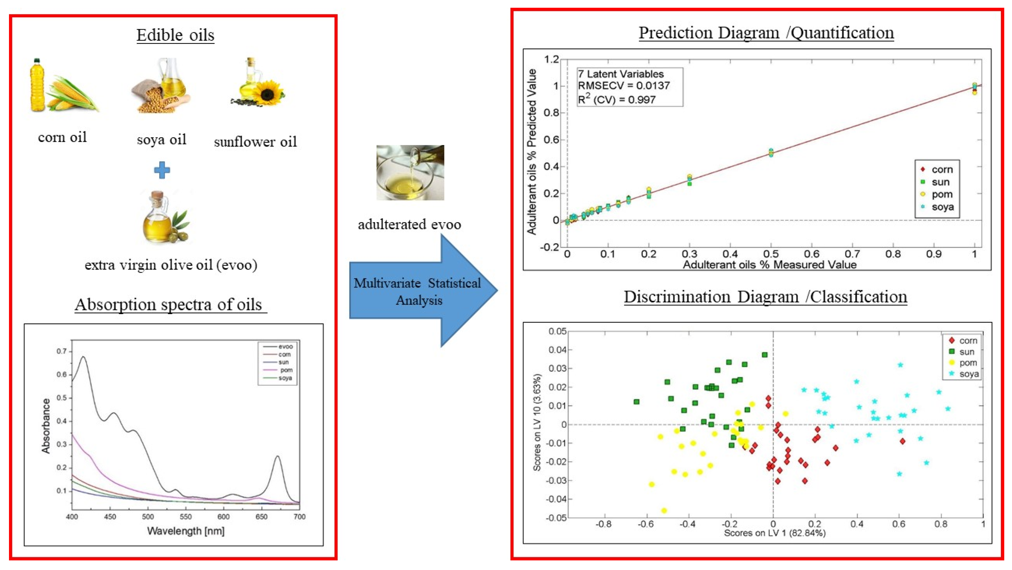
                                                            9.2. High Throughput Analysis for Reliable Detection of EVOOs Adulteration—A Non-Target Metabolomics Approach for Geographical Origin and Varietal Discrimination

                                                            I. F. Tzavellas 1, S. K. Drakopoulou 1, M. E. Dasenaki 1,2 and N. S. Thomaidis 1,2,*
                                                            1
                                                            Laboratory of Analytical Chemistry, Department of Chemistry, National and Kapodistrian University of Athens, Panepistimiopolis Zografou, 15771 Athens, Greece
                                                            2
                                                            Laboratory of Food Chemistry, Department of Chemistry, National and Kapodistrian University of Athens, Panepistimiopolis Zografou, 15771 Athens, Greece
                                                            *
                                                            Correspondence:
                                                            Extra virgin olive oil (EVOO) has been acknowledged as an emblematic food of the Mediterranean diet, partly for its distinctive taste, but foremost for its high nutritional value. The declaration of olive oil superiority, officially substantiated by EU health claim establishment regarding olive oil polyphenols (EU 432/2012), has laid the foundations for in-depth authenticity study in the field to reassure the uniqueness of the product [1].
                                                            Olive oil, due to its high financial impact on the global market, is frequently subjected to some kind of adulteration. Regarding numbers, olive oil tops the food ingredient fraud ranking according to FDA, with one of the most frequent profit-driven fraudulent procedures being its partial substitution with other vegetable oils. However, special attention must also be paid to the assurance of variety and geographical origin in order to highlight the special qualifications of the product [2].
                                                            Serving this purpose, in this study, a non-target screening workflow based on LC-QToF-MS analysis has been developed and optimized for the detection of geographical origin and varietal adulteration in EVOOs samples. Thus, two different adulteration studies have been designed and carried out, including the analysis of samples of different EVOO variety and geographical origin, as well as adulterated samples. The adulterated samples were constructed in the lab, in different adulteration ratios, ranging from 10 to 50%. Noteworthy differentiations in EVOOs profile were recorded, while potential authenticity markers of origin and variety for EVOOs discrimination have also been introduced. Data analysis and evaluation in non-target HRMS screening workflows were performed, significantly assisted using advanced data processing tools combined with supervised and unsupervised chemometric techniques.

                                                            References 

                                                            • EU. Regulation (EC) No. 178/2002 of the European Parliament and of the Council of 28 January 2002, General principles and requirements of food law, establishing the European Food Safety Authority and procedures in matters of food safety. Off. J. Eur. Communities 2002, L31, 1–24.
                                                            • Kalogiouri, N.; Aalizadeh, R.; Thomaidis, N.S. Investigating the organic and conventional production type of olive oil with target and suspect screening by LC-QTOF-MS, a novel semi-quantification method using chemical similarity and advanced chemometrics. Anal. Bioanal. Chem. 2017, 409, 5413–5426.

                                                              9.3. A Multi-Instrumental Approach for the Physicochemical Characterization of a Cotton Honey-Based Spread Produced by Controlling Compositional and Processing Parameters

                                                              V. Sereti 1, A. Lazaridou 1,*, C. Tananaki 2 and C. G. Biliaderis 1
                                                              1
                                                              Laboratory of Food Chemistry and Biochemistry, Department of Food Science and Technology, School of Agriculture, Aristotle University of Thessaloniki, P.O. Box 235, 54124 Thessaloniki, Greece
                                                              2
                                                              Laboratory of Apiculture–Sericulture, Department of Horticulture and Viticulture, School of Agriculture, Aristotle University of Thessaloniki, 54124 Thessaloniki, Greece
                                                              *
                                                              Correspondence:
                                                              Creamed (or set) honey is a type of spread produced by a controlled crystallization process resulting in a product with a large number of small, non-detectable by palate crystals, light color, and a smooth and creamy texture, rendering it into an easily spreadable product similar to peanut butter [1,2]. Honey from cotton blossoms tends to crystallize rapidly resulting in a product with hard texture and low spreadability, even if it is processed via this controlled crystallization treatment. The aim of this study was to develop a cotton honey-based spread (CHS) with acceptable physical and textural properties by adding fructose (1–6%) and water (up to 18% final moisture) into the initial cotton honey, as well as by controlling the temperature during crystallization, Tcryst (5–23 °C × 20 days) and the conditioning (tempering) step as a follow-up process, Tcond (20–30 °C × 10 days). Multi-instrumental analysis (rotational rheometry, differential scanning calorimetry, optical microscopy, colorimetry, and texture analysis) and sensory (spreadability, mouthfeel, and overall acceptability) evaluation were used for characterization of textural properties of the CHS preparations. The addition of fructose and water were found to be the main factors that significantly affected (p < 0.05) the quality parameters of the product. More specifically, with increasing fructose and water contents the crystallinity index (i.e., glucose–water ratio) in CHS formulations decreased leading to significantly (p < 0.05) lower values of apparent and complex viscosities, hardness, spreadability work, and melting enthalpy of sugar microcrystals, as well as improved organoleptic characteristics of this spread product. Moreover, with increasing Tcryst, only the lightness and the melting enthalpy values decreased, while the damping factor increased. Instead, the Tcond did not affect any of the above parameters (p > 0.05). Additionally, the glucose–water ratio was found to correlate positively with all color, thermodynamic, texture analysis parameters, and degree of crystallization of the final product, as well as with all the rheological parameters of the spreads, with exception of loss tangent. Overall, strong correlations were identified between the compositional and physicochemical attributes and the sensory characteristics of honey spread, implying that a multi-instrumental analysis of CHS preparations, along with sensory evaluation, can be a valuable holistic approach for understanding the macrostructure, evaluating the physical properties and quality attributes, as well as predicting the consumer preference of this honey-based product.

                                                              References 

                                                              • Dyce, E.J. Producing Finely Granulated or Creamed Honey; Oxford University Press: Oxford, UK, 1975.
                                                              • Karasu, S.; Toker, O.S.; Yilmaz, M.T.; Karaman, S.; Dertli, E., Thermal loop test to determine structural changes and thermal stability of creamed honey: Rheological characterization. J. Food Eng. 2015, 150, 90–98.

                                                                9.4. Physical Properties and Quality Evaluation of Composite Dough and Bakery Products Using a Multi-Analytical Instrumental Approach

                                                                K. Kotsiou, A. Lazaridou * and C. G. Biliaderis
                                                                Laboratory of Food Chemistry and Biochemistry, Department of Food Science and Technology, School of Agriculture, Aristotle University of Thessaloniki, P.O. Box 235, 54124 Thessaloniki, Greece
                                                                *
                                                                Correspondence:
                                                                Fortification of a staple food rich in rapidly digestible carbohydrates, such as bread, with legumes, which are significant sources of proteins and dietary fibers, might be nutritionally beneficial for improving public health. Nevertheless, inclusion of legume flours into wheat dough formulations is challenging, as it modifies the rheological behavior of the dough [1,2], adversely affecting bread volume and crumb textural characteristics [1–3]. In this study, a multi-analytical instrumental approach was used for the characterization of the physical properties and quality evaluation of composite doughs consisting of wheat and legume flours and the resultant breads. Flours from yellow split pea (YSP) and chickpea (CP) were used, due to their high contents in protein and dietary fiber, to substitute wheat flour at 10–20% levels; moreover, roasting the legume seeds before milling was employed as a quick and cost-effective pretreatment to eliminate the legume off-flavors and therefore to promote acceptability of the final products. Fundamental rotational rheometry revealed, that inclusion of 15 and 20% CP or 20% YSP flours in the dough formulations significantly increased their storage modulus and zero shear viscosity, while reducing maximum creep strain and compliance, attributes that are related to strong and elastic doughs with a great resistance to flow and deformation. Empirical rheological testing with farinograph showed that YSP flour inclusion in the formulations at 15 and 20% levels resulted in doughs with significantly decreased stability, while CP flour inclusion at the same levels improved doughs stability. Extensography showed that incorporation of both CP and YSP flours at high levels in the doughs negatively affected the resistance to extension/extensibility ratio, a factor that is strongly correlated to baking performance. However, the YSP–wheat composite doughs were able to recover their extensograph parameters upon repeated extension, followed by a resting period, thus making them suitable for bread formulations that require multiple cycles of kneading and resting, while the doughs fortified with CP became very resistant to extension after this processing stage. Regarding end-products, higher levels of added legume flours resulted in breads with low specific volumes and harder crumbs as shown by a laser-based scanner and a Texture Analyser, respectively. Monitoring, by Texture Profile Analysis, the changes in crumb hardness during bread storage showed that samples with 20% YSP flour exhibited higher staling rates, which could be partially attributed to the higher rate of amylopectin retrogradation of this formulation as revealed by Differential Scanning Calorimetry. The findings suggest that multi-instrumental analysis of composite doughs and breads is a very useful analytical toolbox for characterization of composite doughs and bakery products with improved nutritional characteristics and for the prediction of their shelf life and consumer acceptability scores.

                                                                References 

                                                                • Mohammed, I.; Ahmed, A.R.; Senge, B. Dough rheology and bread quality of wheat–chickpea flour blends. Ind. Crop. Prod. 2012, 36, 196–202.
                                                                • Portman, D.; Blanchard, C.; Maharjan, P.; McDonald, L.S.; Mawson, J.; Naiker, M.; Panozzo, J.F. Blending studies using wheat and lentil cotyledon flour—Effects on rheology and bread quality. Cereal. Chem. 2018, 95, 849–860.
                                                                • Turfani, V.; Narducci, V.; Durazzo, A.; Galli, V.; Carcea, M. Technological, nutritional and functional properties of wheat bread enriched with lentil or carob flours. LWT Food Sci. Technol. 2017, 78, 361–366.

                                                                  9.5. Discrimination of Feta Cheese from Different Geographical Region, Packaging Type, and Production Date According to Its Chemical Composition and Volatile Profile

                                                                  M. Lygka * and T. Massouras
                                                                  Laboratory of Dairy Research, Agricultural University of Athens, Iera Odos 75, Votanikos, 11855 Athens, Greece
                                                                  *
                                                                  Correspondence:
                                                                  The present work aims to verify whether the chemical composition and the volatile profile could be considered as chemical biomarkers linked to geographical origin, packaging type, and production date of feta cheese. One hundred and ten (110) samples of feta cheeses aged between 3–12 months, packaged in wooden barrels or in tin containers, were collected from nine different geographical regions (throughout mainland Greece and Lesvos). Samples were analyzed for chemical composition (fat, protein, moisture, and salt) using a Foodscan apparatus. The volatile compounds were extracted by headspace Solid phase microextraction (HS-SPME) and analyzed using a gas chromatography-mass spectroscopy (GC/MS). The values of chemical parameters and the volatile compounds were treated for the geographical regions, packaging type and production day variables. A total of 84 volatile compounds were identified, including 18 acids, 16 alcohols, 8 aldehydes, 14 esters, 20 ketones, 8 alkane, and 2 alkene. The most abundant compounds were organic acids followed by alcohols, ketones, and esters. Using Multifactor Anova and discriminant analysis, based on volatile profile of cheese samples achieved a classification rate of 85%, 94.83%, and 84.35% for geographical regions, packaging type, and production day, respectively. Regarding the chemical composition, all samples were within specifications of PDO feta cheese (fat in dry matter > 43% and humidity up to 56%). Statistically significant higher values of fat and protein content and lower moisture content were found in samples of feta cheese packed in wooden barrels compared those packed in tin container. None of the three variables significantly affected the chemical composition of feta cheese.
                                                                  Thus, the volatile profile of feta cheese can be considered as a biomarker to differentiate feta cheese of a certain origin, packaging type and production date.

                                                                  9.6. Application of Spectroscopic Techniques Coupled with Multivariate Statistical Analysis for the Origin Discrimination and Quality Control of Agrofoods

                                                                  R. Kontzedaki and Μ. Velegrakis *
                                                                  Institute of Electronic Structure and Laser, Foundation for Research and Technology-Hellas (IESL-FORTH), 70013 Heraklion, Greece
                                                                  *
                                                                  Correspondence:
                                                                  Food quality control is often based on modern and sophisticated analytical methods and processes that are costly, time-consuming, and require specialized personnel [1–3]. At the Institute of Electronic Structure and Laser of FORTH, we employ an alternative approach in order to investigate the rapid determination of the characteristic substances contained in food samples, with little or no processing. For this study, optical spectroscopic techniques (Absorption (UV-Vis-NIR), Fluorescence, and Raman spectroscopy) are applied. In this way, we can record the optical spectrum that is the characteristic “fingerprint” and reflects the chemical composition of a sample. Different samples have a different fingerprint and the detected differences or similarities, coupled with multivariate statistical analysis methods (Principal Component Analysis (PCA), Partial Least Squares Discriminant Analysis (PLS-DA), and PLS-regression), are used for origin and quality control. In this work, several results of these studies will be presented. Greek white and red wine varieties from different wineries were studied. Additionally, samples of Cretan olive oil were investigated, that were obtained from the Pancretan Olive Oil Competition and had been graded by a trained team of testers using human senses. The samples were discriminated based on their quality and crop type (organic and conventional cultivation).

                                                                  References 

                                                                  • Cassino, C.; Tsolakis, C.; Bonello, F.; Gianotti, V.; Osella, D. Wine evolution during bottle aging, studied by 1H NMR spectroscopy and multivariate statistical analysis. Food Res. Int. 2019, 116, 566–577.
                                                                  • He, Y.; Zhao, Y.; Zhang, C.; Li, Y.; Bao, Y.; Liu, F. Discrimination of Grape Seeds Using Laser-Induced Breakdown Spectroscopy in Combination with Region Selection and Supervised Classification Methods. Foods 2020, 9, 199.
                                                                  • Camin, F.; Larcher, R.; Perini, M.; Bontempo, L.; Bertoldi, D.; Gagliamo, G.; Nicolini, G.; Versinia, G., Characterization of authentic Italian extra-virgin olive oils by stable isotope ratios of C, O and H and mineral composition. Food Chem. 2010, 118, 901–909.

                                                                    9.7. Determination of Volatile Compounds in Spirulina Food Supplements Using HS-SPME–GC/MS

                                                                    A. Paraskevopoulou 1,2, T. Kaloudis 1, A. Hiskia 1 and T. M. Triantis 1,*
                                                                    1
                                                                    Institute of Nanoscience and Nanotechnology, NCSR Demokritos, 15341 Athens, Greece
                                                                    2
                                                                    Chemical Engineering Department, National Technical University, Iroon Politechniou 9, Zografou, 15780 Athens, Greece
                                                                    *
                                                                    Correspondence:
                                                                    Cyanobacteria are photosynthetic microorganisms found in terrestrial and aquatic environments, capable of producing a large number of metabolites with great structural and functional diversity. Arthrospira spp., commercially known as Spirulina, is a cyanobacteria species widely used as a food supplement due to its high content in proteins, vitamins, pigments, and fatty acids. In addition to these bioactive substances, Spirulina produces numerous volatile organic compounds (VOCs) which are important in determining the quality and nutraceutical properties of spirulina products, including their taste and aroma. Most of these secondary metabolites are not toxic, while some exhibit antimicrobial, anticancer, or antibacterial activities [1] and have a wide range of applications in food, cosmetics, and pharmaceutical industries [2].
                                                                    This study focused on the analysis of the VOCs profile in 22 commercial Spirulina food supplements from different geographical origins using headspace solid-phase microextraction (HS-SPME) combined with gas chromatography mass spectrometry (GC-MS). Volatile compounds were identified after mass-spectral deconvolution, matching spectra and retention indices using the NIST MS library and AMDIS software.
                                                                    As many as 229 volatile components were identified and categorized into various chemical classes, while specific compounds were detected in the majority of the analyzed samples, e.g., pyrazines, furans, and furanones (3-acetyl-2,5-dimethylfuran and 2(4H)-benzofuranone-5,6,7,7a-tetrahydro-4,4,7a-trimethyl), esters, aldehydes (hexanal; benzaldehyde; 2-heptenal; 2-octenal; safranal; and β-cyclocitral), alcohols (1-hexanol and 1-octen-3-ol), ketones (2-heptanone,6-methyl; cyclohexanone,2,2,6-trimethyl; α-ionone; β-ionone epoxide; and 5,9-undecadien-2-one), saturated hydrocarbons (pentadecane; hexadecane; and heptadecane), terpenes, alkenes, pyridines, and benzenes. Identification of compounds was based on a high matching score (80–100%) combining a close similarity of their mass spectra and their retention index values with those of reference compounds present in the NIST MS Library. On the basis of total integrated peak areas of the GC-MS elution profile, the most abundant constituents of Spirulina VOCs were heptadecane (relative content 8–20.6%), β-ionone (2–6.56%), pentadecane (1.5–5.95%), trans-β-ionone (1.5–3.36%), hexanal (0.1–2.4%), and 1-octen-3-ol (0.1–2.22%). Results show the potential of Spirulina as an important source of volatile compounds and its possible usage not only as a dietary supplement but also as a raw material for the isolation of these compounds and their further application in the food, pharmaceutical, and cosmetics industries.
                                                                    Acknowledgments: Financial support is acknowledged by the European Union and Greek national funds through the Operational Program Competitiveness, Entrepreneurship and Innovation, under the call RESEARCH—CREATE—INNOVATE (Project acronym: CO2 Bioproducts; project code: T1EDK-02681).

                                                                    References 

                                                                    • Paparella, A.; Shaltier-Harpaza, L.; Ibdah, M. β-Ionone: Its occurrence and biological function and metabolic engineering. Plants 2021, 10, 1–12.
                                                                    • Andrade, L.M.; Andrade, C.J.; Dias, M.; Nascimento, C.; Mendes, M.A. Chlorella and spirulina microalgae as sources of functional foods, nutraceuticals, and food supplements; an overview. MOJ Food Process. Technol. 2018, 6, 45–58.

                                                                      10. Food Analysis II

                                                                      10.1. Physicochemical and Sensorial Properties of Wheat Bread with Sourdough Made from Starter Culture Isolated from a Submerged Chickpea Fermentation Extract

                                                                      C. Nouska, M. Hatzikamari, C. G. Biliaderis and A. Lazaridou *
                                                                      Department of Food Science and Technology, Aristotle University of Thessaloniki, 54124 Thessaloniki, Greece
                                                                      *
                                                                      Correspondence:
                                                                      During bread production, baker’s yeast is used for the development of organoleptic characteristics and the leavening of this food product. Recently, consumers are turning to traditional products, leading the bakery industry to produce bread supplemented with sourdough. Sourdough can be also used as a leavening agent in breadmaking and is responsible for the characteristic flavor and the volume of bread due to the organic acid and gas production. In addition, sourdough may prolong the shelf-life of bread by the production of bacteriocins, organic acids, amylolytic enzymes etc. [1,2]. In this work, a Greek traditional sourdough made from a starter culture isolated from a submerged chickpea fermentation extract was used as a leavening agent in wheat breads. The dominant microflora developed during submerged fermentation, which was responsible for the leavening effect, was found to be clostridia (7.04 ± 0.20 logcfu/g); similar findings have been reported in other studies [3,4]. Provided that clostridia isolated from chickpea sourdough have been found not to form any toxins [3], there was no concern for health hazard. Thus, 10 clostridia strains were isolated from chickpea sourdough and identified with molecular techniques (PCR) as Clostridium perfringens; one of these strains was used as starter culture for the production of sourdough from chickpea flour both in liquid and freeze-dried form before being incorporated into wheat bread. Loaf specific volume increased with the addition of liquid (2.64 ± 0.05 mL/g bread) and freeze-dried (3.19 ± 0.08 mL/g bread) sourdough compared to control bread which was leavened by baker’s yeast (2.20 ± 0.04 mL/g bread). Crumb hardness and melting enthalpy of the retrograded amylopectin in sourdough supplemented breads during storage at 25 °C for 4 days were lower than control formulation as revealed by Texture Profile Analysis (TPA) and Differential Scanning Calorimetry (DSC), respectively, indicating a lower staling extent. Additionally, organic acids analysis conducted by Anion-exchange HPLC-DAD showed higher acetic acid content for sourdough breads compared to control, while butyric and isobutyric acids were produced only by the sourdough starter culture, giving these bread formulations their characteristic flavor; the latter was confirmed by the sensory analysis which was also showed higher crumb softness and overall acceptability rates for sourdough breads, supporting the data from instrumental analysis. Overall, chickpea sourdough from C. perfringens starter culture isolated from a submerged chickpea fermentation extract can improve textural and sensorial properties of wheat breads and extend their shelf-life.

                                                                      References 

                                                                      • Gerez, C.L.; Torino, M.I.; Rollán, G.; de Valdez, G.F. Prevention of bread mould spoilage by using lactic acid bacteria with antifungal properties. Food Control. 2009, 20, 144–148.
                                                                      • Hatzikamari, M.; Kyriakidis, D.A.; Tzanetakis, N.; Biliaderis, C.G.; Litopoulou-Tzanetaki, E. Biochemical changes during a submerged chickpea fermentation used as a leavening agent for bread production. Eur. Food Res. Technol. 2007, 224, 715–723.
                                                                      • Hatzikamari, M.; Yiangou, M.; Tzanetakis, N.; Litopoulou-Tzanetaki, E. Changes in numbers and kinds of bacteria during a chickpea submerged fermentation used as a leavening agent for bread production. Int. J. Food Microbiol. 2007, 116, 37–43.
                                                                      • Hristoskova, S.P.; Yocheva, L.D.; Yankov, D.S.; Danova, S.T. Newly characterized butyrate producing Clostridium sp. Strain 4a1, isolated from chickpea beans (Cicer arietinum L.). Bulg. Chem. Commun. 2018, 50, 459–466.

                                                                        10.2. Polystyrene Biodegradation by Mealworms Larvae: Identification of Degradation Products and Metabolic Alteration Using GC-MS

                                                                        E. D. Tsochatzis 1,*, I. E. Berggreen 1, H. Gika 2,3,4 and G. Theodoridis 2,3,4
                                                                        1
                                                                        Department of Food Science, Aarhus University, Agro Food Park 48, 8200 Aarhus, Denmark
                                                                        2
                                                                        FoodOmicsGR Research Infrastructure, AUTh node, GR 57001 Thessaloniki, Greece
                                                                        3
                                                                        Biomic AUTh, Center for Interdisciplinary Research and Innovation, GR 57001 Thessaloniki, Greece
                                                                        4
                                                                        Aristotle University of Thessaloniki, GR 57001 Thessaloniki, Greece
                                                                        *
                                                                        Correspondence:
                                                                        The present study investigated the biodegradation of polystyrene (PS) plastic by mealworm (Tenebrio molitor) on different diets followed by untargeted screening of larvae gut intestine tissue and frass (manure and feed residuals) to investigate the existence of polymer-generated organic residues. Three different diets, consisting of PS, rolled barley, and water, were tested. PS degradation rates ranged from 16% to 23% within 15 days, with no statistical differences in survival rates. The larvae fed with ad libitum barley:PS (20:1 w/w) and water (diet 2) had the highest growth rate and the highest PS consumption, along with barley:PS of 4:1 w/w (diet 1) (Figure 1).
                                                                        Figure 1. (A) PS consumption throughout the experiment; (B) final survival rates of T. molitor larvae) within the different applied feeding experiments, and (C) survival rate during the experiment. Diet 1 is barley:PS (4:1), Diet 2 is ad libitum barley:PS (20:1) + H2O, and Diet 3 is ad libitum barley:PS (20:1).
                                                                        Figure 1. (A) PS consumption throughout the experiment; (B) final survival rates of T. molitor larvae) within the different applied feeding experiments, and (C) survival rate during the experiment. Diet 1 is barley:PS (4:1), Diet 2 is ad libitum barley:PS (20:1) + H2O, and Diet 3 is ad libitum barley:PS (20:1).
                                                                        Applsci 11 11767 g014
                                                                        A GC-TOF-MS analysis revealed no contaminating substances in the gut intestine tissue, nor styrene or PS oligomers, whilst several bioactive compounds and traces of alkanes, mostly with small carbon chains, were present. Metabolomics analysis on the collected frass, either on the lipophilic (CHCl3) or the polar fraction (MeOH-H2O), was performed. Styrene and PS oligomers (dimers, trimers) were identified, though in a relatively low total amount, up to a total of 346.0 ng/mg. 2,4 di-tert butylphenol was identified in both frass and tissue, coming from the PS polymer (non-intentionally added substances; NIAS) (Figure 2). It was shown that degrading and contaminating organic compounds occur at low levels, in both gut intestine and frass, during biodegradation of PS.
                                                                        Figure 2. Mass fractions (ng/mg) of monomer (styrene) and PS oligomers in the frass from PS. biodegradation by Tenebrio molitor larvae.
                                                                        Figure 2. Mass fractions (ng/mg) of monomer (styrene) and PS oligomers in the frass from PS. biodegradation by Tenebrio molitor larvae.
                                                                        Applsci 11 11767 g015
                                                                        In the polar fraction of frass, bioactive molecules (fatty acids, amides) were identified, together with several hydrocarbons, mostly with longer carbon chains. The formation of these substances indicated enzymatic and biochemical activity in the larvae-gut intestine.

                                                                        10.3. Elemental Metabolomics for Honey Authentication

                                                                        S. A. Drivelos 1, G. Danezis 1, M. Halagarda 2, S. Popek 2 and C. A. Georgiou 1,*
                                                                        1
                                                                        Chemistry Laboratory, Department of Food Science and Human Nutrition, Agricultural University of Athens, 75 Iera Odos, 118 55 Athens, Greece
                                                                        2
                                                                        Department of Food Product Quality, Cracow University of Economics, ul. Sienkiewicza 5, 30-033 Krakow, Poland
                                                                        *
                                                                        Correspondence:
                                                                        Elemental metabolomics concerns food safety and quality. Utilization of elemental profile promotes authenticity and leads to useful markers such as rare earth elements. Elemental profiles have proved reliable indicators for geographical origin determination in many cases and furter they could provide information on genetic origin. Processing methods are also related to elemental fingerprints. Omics technologies harness the impressive power of high-resolution mass spectrometry (HRMS) [1]. Although REEs and precious metals have great potential for determination of geographical origin, they have not been studied extensively. Increased interest in rare earths as authentication markers will grow stronger in the near future.
                                                                        Trace and rare earth elements of 93 honeys of different botanical type, origin, and production method have been studied through ICP-MS. Information obtained using rare earths and trace elements were utilized via chemometric evaluation for the classification of honey samples. Discriminant Analysis (DA) was successful for botanical type and geographical origin classification while Cluster Analysis (CA) was successful only for botanical type. Through Probabilistic Neural Network (PNN) analysis, 85.3% were correctly classified by the network according to their geographical origin and 73.3% according to production method, organic or non-organic. A Partial Least Squares (PLS) model was constructed, giving a prediction accuracy of more than 95%. This study shows that an elemental profile could discriminate honey according geographical and botanic origin. This is an exploratory study pointing out the value of a large-scale sampling study. To explain the incapability of elemental profile to discriminate honey production method, a study on the elemental content of floral nectars and their changes along pesticides use is required [2].

                                                                        References 

                                                                        • Danezis, G.P.; Georgiou, C.A. Food quality, traceability and Foodomics. In Comprehensive Foodomics; Cozzolino, D., Cifuentes, A., Eds.; Elsevier: Amsterdam, The Netherlands, 2021; pp. 244–257.
                                                                        • Drivelos, S.A.; Danezis, G.P.; Halagarda, M.; Popek, S.; Georgiou, C.A. Geographical origin and botanical type honey authentication through elemental metabolomics via chemometrics. Food Chem. 2021, 338, 127936.

                                                                          10.4. Advanced and Comprehensive Screening Methodologies for the Monitoring of Intentionally and Non-Intentionally Added Substances in Food Contact Materials

                                                                          C. Kanakaki *, E. Mermingi and N. Thomaidis
                                                                          Department of Chemistry, National and Kapodistrian University of Athens, Panepistimiopolis Zografou, 157 71 Athens, Greece
                                                                          *
                                                                          Correspondence:
                                                                          Packaging has become an indispensable element in the food manufacturing process, as it protects the food from, e.g., light and microbial contaminations, improving the self-life and stability of products during storage. However, it has been found to represent a source of contamination itself through the migration of substances from the packaging into food. The identification of these substances is an issue that deserves great attention to assure food quality and consumer safety.
                                                                          Aiming the comprehensive screening of intentionally and non-intentionally added substances (IAS and NIAS) in food contact materials (FCMs), GC-MS/MS, and GC-APCI-QTOFMS methods were developed, for the target screening of volatile migrants. Furthermore, corresponding contaminants of low volatility, thermal instability, and high polarity were identified by LC-MS/MS and LC-ESI-QTOFMS methods. In particular, the high-resolution mass spectrometric (HRMS) methods applied were utilized for further suspect and non-target screening and retrospective analysis of the obtained FCMs. For these screening procedures, the entire spectrum of food simulants was evaluated with different migration protocols being employed, depending on the intended use and origin of the analysed samples. A risk assessment of the identified migrating compounds was also performed using the corresponding legislation, online tools, and in-house built software.
                                                                          Application of the developed workflows into a large set of products and comprehensive screening of migrating chemical contaminants will promote the production of inert and consequently safe FCMs. Thus, this work will assess public health risks by facing current and future challenges.
                                                                          Acknowledgments: This work was supported by the State Scholarships Foundation (IKY), which we gratefully acknowledge.

                                                                          11. Chemistry Associations

                                                                          11.1. FoodOmicsGR_RI: A Distributed National Research Infrastructure for the Comprehensive Characterisation of Foods

                                                                          G. Theodoridis
                                                                          Department of Chemistry, Aristotle University of Thessaloniki, 54124 Thessaloniki, Greece
                                                                          Foodomics is the omics field and the technology that combines food and nutrition sciences with advanced analytical techniques and bioinformatics, whereby a hypothesis-free approach is applied to characterize the food composition or food consumers’ biofluids, addressing new challenges such as authentication, traceability, improvement of food produce, food quality, and nutritional value. This holistic approach employs cutting-edge research infrastructure, where big data is produced generating new knowledge, highlighting previously unknown associations of biomolecules with the studied phenotype.
                                                                          FoodOmicsGR_RI is a national research infrastructure aiming to perform and aid omics research in agri-food sector in the Greek research environment. This field is of high importance for the country and its agriculture due to the unique landscape Greece exhibits that result in a diverse and rich portfolio of Greek local products and foods. Therefore, the central scope of FoodOmicsGR_RI is to support the Greek agri-food sector by generating robust data on the composition and nutritional value of the local produce, thereby increasing product’s position and market demand, and provide a higher revenue for the producers.
                                                                          The project comprises eight (8) Greek Universities and Research Centers. Analytical groups and food specialist groups from Universities of Thessaloniki, Athens, Crete, the Aegean, Ioannina, the Agricultural University of Athens, the International Hellenic University, and the Biomedical Research Center of the Academy of Athens comprise a team of 60 staff scientists and 40 newly recruited researchers from 20 scientific disciplines. BIOMIC AUTh, a new interdisciplinary laboratory of the Aristotle University Thessaloniki (http://biomic.web.auth.gr/ (accessed on 8 November 2021), having extensive expertise in bio-analytical chemistry, is coordinating the project and providing the current presentation. More details can be found on our website (http://foodomics.gr/ accessed on 8 November 2021).

                                                                          11.2. EURACHEM Greece: Promoting Quality of Analytical Measurements, QA, and QC in Analytical Laboratories

                                                                          M. Kostakis 1,*, V. Zonaras 2, H. Gika 3, A. Iakovakis 4, C. Virgiliou 5 and G. Theodoridis 5,*
                                                                          1
                                                                          Laboratory of Analytical Chemistry, Department of Chemistry, National and Kapodistrian, University of Athens, Panepistimiopolis Zografou, 15771 Athens, Greece
                                                                          2
                                                                          Laboratory of Pesticide Residues, Scientific Directorate of Pesticides’ Control and Phytopharmacy, Benaki Phytopathological Institute, 14561 Kifissia, Greece
                                                                          3
                                                                          Laboratory of Forensic medicine & Toxicology, School of Medicine, Aristotle University of Thessaloniki, 57022 Thessaloniki, Greece
                                                                          4
                                                                          Veltia Labs, Sindos, 57022 Thessaloniki, Greece
                                                                          5
                                                                          Laboratory of Analytical Chemistry, Department of Chemistry, Aristotle University of Thessaloniki, 57022 Thessaloniki, Greece
                                                                          *
                                                                          Correspondence:
                                                                          Eurachem is a network of national and other organizations in Europe that focuses on measurement quality and quality-related issues [1]. For more than 30 years, Eurachem has organized activities (workshops and training events) and provided documents (guidelines and information leaflets) in order to help laboratories, accreditation bodies, authorities, other stakeholders, practitioners, and researchers to ensure the quality of analytical measurement. Method validation, measurement uncertainty, metrological traceability, internal quality control, and proficiency testing are the main cornerstones of Eurachem [1]. Moreover, upcoming analytical measurement issues, such as sampling and conformity assessment, are also included in Eurachem working activities. In the direction to promote quality assurance in analytical measurement, all materials (guidelines, leaflets, and workshops’ presentations) produced by Eurachem are available free of charge on our website (eurachem.org).
                                                                          The aim of the presentation is to inform the interested parties about Eurachem Greece and about Eurachem’s working groups, to communicate their activities, and to discuss forthcoming issues and problems of interest of the Greek research ecosystem and subsequently bring them to the attention of Eurachem.

                                                                          Reference 

                                                                          • Magnusson, B.; Ellison, S.; Patriarca, M. Impact of Eurachem 25 years of activity. Accred Qual Assur. 2014, 19, 55–57.

                                                                            11.3. EuChemS-WP EiC: EuChemS Working Party on Ethics in Chemistry

                                                                            A. Silvestru and on behalf of the EuChemS-WP EiC Steering Committee
                                                                            Chemistry Department, Babeș-Bolyai University, Arany Janos 11, 400028 Cluj-Napoca, Romania
                                                                            The WP EiC is a network of representatives of the EuChemS member societies with the goal to promote the understanding of the importance of ethical considerations in education and practice of pure and applied chemistry.
                                                                            The WP EiC was founded shortly after 2010, when Hartmut Frank, Professor of environmental chemistry at the University of Bayreuth, together with other remarkable scientists, sustained by the Nobel laureates Richard Ernst and Roald Hoffmann, pointed out that the tremendous development of chemistry at the beginning of the 3rd millennium should be accompanied by an appropriate education and ethical approach. This should be applied to all chemistry-based activities which might raise concerns regarding the use and the impact upon the environment and the development of society. The group was subsequently enlarged by attracting other chemists and scientists, and it was integrated as a Working Party in EuChemS.
                                                                            The vision of WP EiC is based on the responsibilities of chemists on both internal and external level of their decisions and actions. The internal one takes into account a good scientific practice, research, and professional integrity, while the external one includes topics such as the potential dual applications of chemical innovations, sustainability, and risk assessment, as well as the role of specific expertise in science policy, innovation governance, and political decisions.
                                                                            The WP EiC is promoting ethical guidelines in science and education; cooperation and scientific exchanges between specialists from different countries and specific fields related to chemistry encourages interaction and communication between young and experienced scientists and between academia and industry, thus fostering scientific progress and innovation and aiming to better life, a healthy society, peace, and a clean environment.
                                                                            The efforts of the WP EiC to contribute to an ethical education of the young generations of chemists and scientists and our care of the ethical, social, and cultural dimensions of practicing chemistry in the interest of the whole society is also reflected in the materials elaborated by several members of the group: in conferences, in our online course Good Chemistry: Methodological, Ethical, and Social Dimensions (Jan Mehlich), and, offered on the e-learning platform of EuChemS to students in EU, in papers on topics at the intersection of chemistry and applied ethics (i.e., Angew. Chem. Int. Ed. 2011, 50, 8482 and Chem. Eur. J., 2017, 23, 1).
                                                                            The WP EiC today gathers members from 17 countries, representatives of national chemical societies or simply members from academia and industry, interested in the future development of the society in times when chemistry should solve, in an ethical way, a lot of problems: resources economy, environment, health, weapons prohibition, etc. You are invited to join, in a formal or informal way, to share our goals and care by considering the importance of a responsible education, science, and specific activities in chemistry and associated fields.
                                                                            For more information, please visit: https://www.euchems.eu/divisions/ethics-in-chemistry/ (accessed on 8 November 2021).

                                                                            12. Environmental Analysis, Chromatography, Mass Spectrometry

                                                                            12.1. Development of an HRMS-Based Workflow to Quantify PFAs in Leachates and Urban Waters

                                                                            L.-A. Koronaiou 1,2, C. Nannou 1,2 and D. Lambropoulou 1,2,*
                                                                            1
                                                                            Laboratory of Environmental Pollution Control, Department of Chemistry, Aristotle University of Thessaloniki, GR 54124 Thessaloniki, Greece
                                                                            2
                                                                            Centre for Interdisciplinary Research and Innovation (CIRI-AUTH), Balkan Center, 10th km Thessaloniki-Thermi Rd, GR 57001 Thessaloniki, Greece
                                                                            *
                                                                            Correspondence:
                                                                            Per- and polyfluoroalkyl substances (PFASs) are a diverse group of synthetic organofluorine compounds that have been widely used in industrial applications and consumer products such as non-stick cookware, food packaging, fire-fighting foams, carpeting, apparels, and metal plating. PFASs are persistent in the environment, bioaccumulative in wildlife and humans, and known to cause reproductive and developmental toxicity in laboratory animals and wildlife. Landfill leachate is the major pathway for PFAS to exit the municipal solid waste landfills, where their concentrations vary over time and can be much greater than those found in sanitary wastewaters. On the other hand, PFOA and PFOS are particularly poorly removed in the drinking water production chain by conventional purification processes but can be removed with active carbon filtration or by reverse osmosis. The present study comprises a comprehensive development of an analytical methodology aiming to trace and quantify a representative array of PFAs at the sub μg per liter scale, also alleviating the potential sources of cross contamination. In order to achieve sufficiently low LOQs, an optimized sample pre-treatment using solid phase extraction (SPE) was conducted. Extractions from water were performed using weak anionic exchange (WAX) SPE cartridges that were tested over HLB cartridges and proved to be more efficient for a large part of the target compounds. Isotopically-labelled standards were used for internal quantification. A high resolution (HR) Orbitrap Q Exactive Focus mass spectrometer (Thermo Fisher) equipped with a heated ESI source was used, acquiring spectra in negative ionization mode. The overall method was validated in terms of linearity, accuracy, precision, and method limits, while a matrix-effect study demonstrated that the use of matrix-matched calibration is necessary to quantify in such complex matrices with accuracy. The method yielded acceptable recoveries (70%) for the vast majority of compounds, while the LOQs were equal or lower than 10 ng/L. The application of the method in leachates as well as in WWTPs, revealed the occurrence of L-PFBS, PFHxA, and PFOA, while in WWTPs 6:2FtS, PFOSK, and PFOA were detected. The screening capabilities of this system also allows the simultaneous or retrospective monitoring of other PFASs, as well as the suspect screening of other emerging contaminants, using the raw data files obtained by the HRMS instrument.
                                                                            Acknowledgments: This research has been co-financed by the European Union and Greek national funds through the Operational Program Competitiveness, Entrepreneurship and Innovation, under the call RESEARCH—CREATE—INNOVATE (2nd Cycle) (Project acronym: PROMoTE; Project code: T2EDK-04066).

                                                                            References 

                                                                            • Vughs, D.; Baken, K.A.; Dingemans, M.M.L.; de Voogt, P. The determination of two emerging perfluoroalkyl substances and related halogenated sulfonic acids and their significance for the drinking water supply chain. Environ. Sci. Process. Impacts 2019, 21, 1899.
                                                                            • Janda, J.; Nödler, K.; Brauch, H.-J.; Zwiener, C.; Lange, F. Robust trace analysis of polar (C 2-C 8) perfluorinated carboxylic acids by liquid chromatography-tandem mass spectrometry: Method development and application to surface water, groundwater and drinking water. Environ. Sci. Pollut. Res. 2019, 26, 7326–7336.

                                                                              12.2. Combination of Single Algae-ICP-ToF-MS and Multivariate Statistics for (Eco-)Toxicological Assessment

                                                                              M. von der Au 1,*, O. Borovinskaya 2, C. Büchel 3 and B. Meermann 1
                                                                              1
                                                                              Federal Institute for Materials Research and Testing (BAM)—Division 1.1 Inorganic Trace Analysis, 10115 Berlin, Germany
                                                                              2
                                                                              TOFWERK AG, Uttigenstrasse 22, 3600 Thun, Switzerland
                                                                              3
                                                                              Institute of Molecular Biosciences, University of Frankfurt, Max von Laue Str. 9, 60438 Frankfurt, Germany
                                                                              *
                                                                              Correspondence:
                                                                              Diatoms are located at the bottom of the food chain. Thus, toxicologically relevant metals taken up by diatoms can possibly accumulate within the food web and cause harmful effects. Diatoms are a common test system in ecotoxicology. Toxicological effects weaken the growth of algae which is by default investigated by means of fluorescence detection—diminished fluorescence compared to a non-exposed control group indicates an effect. On the basis of the exposed concentration, as well as the obtained fluorescence data, potential threshold exceedance in, e.g., surface waters, is assessed.
                                                                              However, this approach does not allow for the determination of “real” accumulated metal concentration in diatoms. Common approaches are based on microwave assisted digestion and elemental analysis via, e.g., ICP-MS, ICP-OES, or AAS. However, with regard to low absolute metal content in algae, this strategy is only feasible in case of availability of a high biomass.
                                                                              To tackle this problem, alternative, complementary approaches are urgently needed. Within the last years, sp-ICP-MS for nanoparticle, as well as single cell, analysis has stood out as a powerful technique to analyze metal contents as well as size distributions on broad size range (nano- to low micrometer scale) [1]. However, common ICP-MS systems do not allow for multi-element detection within single particle/cell events [2,3]. Thus, simultaneous MS detection devices are needed—just recently, ICP-ToF-MS experienced a revival [4].
                                                                              Within our previous work, we developed an automated sample introduction system based on a HPLC system on line with single particle-ICP-MS, which allowed for ionic background separation and single algae analysis [5]. However, for unambiguous tracing, several fingerprint elements and multielement analysis in single algae (diatoms) are required. Thus, we coupled our previous setup on line to ICP-ToF-MS. Test diatom species were exposed to test substances (Zn) as well as nanoparticles (FeNPs).
                                                                              The developed setup allowed for a fast, automated, and multielement analysis in single diatoms. Furthermore, we combined our approach with multivariate data assessment—multielement detection of characteristic fingerprint elements allowed for an unambiguous diatom tracing. Clustering of diatoms according to metal exposure concentration levels was enabled. Our approach is a new potential tool in ecotoxicological testing.

                                                                              References 

                                                                              • Meermann, B.; Nischwitz, V. ICP-MS for the analysis at the nanoscale—A tutorial review. J. Anal. At. Spectrom. 2018, 33, 1432–1468.
                                                                              • Lau, W.-Y.; Chun, K.-H.; Chan, W.-T. Correlation of single-cell ICP-MS intensity distributions for the study of heterogeneous cellular responses to environmental stresses. J. Anal. At. Spectrom. 2017, 32, 807–815.
                                                                              • Ho, K.-S.; Chan, W.-T. Time-resolved ICP-MS measurement for single-cell analysis and on-line cytometry. J. Anal. At. Spectrom. 2010, 25, 1114–1122.
                                                                              • Borovinskaya, O.; Tanner, M. Multi-Element Detection of Single Nanoparticles with icp-TOF: Application Note. 2016. Available online: https://www.tofwerk.com/application/nanoparticles/ (accessed on 8 November 2021).
                                                                              • Von der Au, M.; Schwinn, M.; Kuhlmeier, K.; Büchel, C.; Meermann, B. Development of an automated on-line purification HPLC single cell-ICP-MS approach for fast diatom analysis. Anal. Chim. Acta 2019, 1077, 87–94.

                                                                                12.3. Relevant Pesticide Transformation Products in Surface Waters Using Target and Suspect Screening with LC-HRMS

                                                                                S. Petromelidou 1,2, C. Nannou 1,2, E. Evgenidou 1,2 and D. Lambropoulou 1,2,*
                                                                                1
                                                                                Laboratory of Environmental Pollution Control, Department of Chemistry, Aristotle University of Thessaloniki, 54124 Thessaloniki, Greece
                                                                                2
                                                                                Center for Interdisciplinary Research and Innovation (CIRI-AUTH), 57001 Thessaloniki, Greece
                                                                                *
                                                                                Correspondence:
                                                                                The intensification of agricultural production in recent decades has forced the rapid increase in the use of pesticides, which end up in the aquatic environment through processes such as surface runoff, rinsing, careless disposal of empty packaging, etc. Upon entering the aquatic bodies, the pesticides undergo biotic and abiotic processes, resulting in their transformation into products that are sometimes even more toxic than the parent compounds. As the determination of the parent compounds in the aqueous samples has been extensively studied, the scientific interest has shifted to their TPs, whose environmental fate is still under investigation. Additionally, their occurrence at relatively low concentrations and the absence of available analytical standards hampers their determination which results in a time consuming and laborious process. The presence of residues of pesticides and/or their transformation products (TPs) in surface water is directly related to water quality. Hence, to determine a worst-case impact of pesticide application in agriculture on surface water quality, the screening of TPs along with the parent compounds is desirable.
                                                                                To this end, the aim of the present study was to utilize liquid chromatography–high-resolution mass spectrometry (LC-HRMS) for the identification of “suspect” TPs (suspect screening). Specifically, the aqueous samples were subjected to solid phase extraction (SPE) prior to analysis, so that a sufficient preconcentration factor be achieved. Afterwards, the analysis was performed by means of a Q Exactive Focus Orbitrap MS, operating in both positive and negative ionization mode. A dynamic list of suspect compounds was created, based on the pesticides that are used systematically and are frequently found in surface waters, according to the recent literature. The a priori information included in the database was the molecular formula, the exact mass of the pseudomolecular ion, the electrospray ionization (positive/negative), and any available fragment ions from literature. The criteria for the identification of the suspect compounds were based on intensity, isotope pattern, retention time, and in silico fragmentation. This way, the suspect hits were prioritized and verified. After processing, 40 transformation products were identified, originating from 25 parent pesticides. The most common detected TPs were those of prometryn, penoxsulam, pyrimethanil, tebuconazole, thiabendazole, and imazalil. Specifically, the TP of pyrimethanil with a molecular weight of 250.1186, was identified in all samples. The frequency of detection of imazalil TP with a molecular weight of 257.0243 was also high, as the TP was detected in 77% of the samples. Most TPs arose either from the process of hydrolysis, as almost all categories of pesticides are subject to hydrolysis, or photolysis, as the samples studied were surface waters and are heavily affected by sunlight. Ultimately, to predict the acute and chronic toxicity of TPs, an in silico approach based on ECOSAR was implemented, revealing increased chronic toxicity for the majority of the identified TPs, while none of them presented serious acute toxicity values.
                                                                                Acknowledgments: This research has been funded by 2014–2020 Interreg IPA CrossBorder Cooperation Programme CCI 2014 TC 16 I5 CB 009, Greece—Republic of North Macedonia, Research Project: Aqua-M II: Sustainable management of cross-border water resources, Project number (MIS):5030774, which is gratefully acknowledged. Also, this research is co-financed by Greece and the European Union (European Social Fund- ESF) through the Operational Programme “Human Resources Development, Education and Lifelong Learning” in the context of the project “Reinforcement of Postdoctoral Researchers—2nd Cycle” (MIS-5033021), implemented by the State Scholarships Foundation (ΙKΥ).

                                                                                12.4. Optimization of the Sample Preparation Step by Central Composite Design for the Analysis of a Wide Array of Emerging Contaminants in Wastewaters and Tap Water Using LC-HRMS

                                                                                A. Ofrydopoulou 1,2, C. Nannou 1,2, E. Evgenidou 1,2 and D. Lambropoulou 1,2,*
                                                                                1
                                                                                Laboratory of Environmental Pollution Control, Department of Chemistry, Aristotle University of Thessaloniki, 57001 Thessaloniki, Greece
                                                                                2
                                                                                Center for Interdisciplinary Research and Innovation (CIRI-AUTH), 57001 Thessaloniki, Greece
                                                                                *
                                                                                Correspondence:
                                                                                Multi-residue analysis is highly desirable for water quality control. To this end, a comprehensive workflow for the quantitative analysis of 172 emerging contaminants (pharmaceuticals and personal care products, illicit drugs, organophosphate flame retardants, and perfluoroalkyl substances) has been developed for application to wastewater and tap water, based on solid phase extraction (SPE) and Orbitrap high resolution mass spectrometry (HRMS). Due to the large number of analytes with various physicochemical characteristics, the response surface methodology (RSM) employing a central composite design (CCD) and desirability function (DF) approach was exploited to optimize the sample preparation process, instead of the conventional single-factor analysis. The factors included in the design of experiments (DoE) were sample pH, eluent solvents composition, and volume. Statistical analysis (ANOVA) proved the adequacy of the proposed model (two-factor interaction) as p-value < 0.05 followed by different diagnostic tests confirmed the good fitting. The best values to acquire DF close to 1 were pH 3.5, methanol/ethyl acetate ratio 87:13 and eluent volume 6 mL. The streamlined method was validated in terms of accuracy, linearity, method limits, reproducibility, and matrix effect. The proposed workflow combines sensitivity and robustness, with recoveries over 70% (Figure 1a,b), method quantification limits <1 ng/L, and relative standard deviations <20% for most of the compounds. Slight matrix effect (ME) was observed except for OPFRs, for which strong ME was calculated. Method applicability was tested over wastewater collected from a municipal wastewater treatment plant in Thessaloniki (Greece), revealing the presence of 69 and 40 compounds in influents and effluents, respectively, at varying concentrations.
                                                                                Figure 1. (a) Percentage of pollutants according to their recoveries at a concentration level of 100 ng L−1 (tap water), (b) 2D mapping of the recovery percentages for all selected compounds over the 15-min LC run.
                                                                                Figure 1. (a) Percentage of pollutants according to their recoveries at a concentration level of 100 ng L−1 (tap water), (b) 2D mapping of the recovery percentages for all selected compounds over the 15-min LC run.
                                                                                Applsci 11 11767 g016aApplsci 11 11767 g016b
                                                                                Funding: LC-Orbitrap MS/MS equipment has been funded by 2014–2020 Interreg IPA Cross Border Cooperation Programme CCI 2014 TC 16 I5CB 009, Research Project: Aqua-M II: Sustainable management of cross-border water resources, Project number (MIS): 5030774, which is gratefully acknowledged.
                                                                                Acknowledgments: A. Ofrydopoulou would like to thank the General Secretariat for Research and Technology (GSRT) and the Hellenic Foundation for Research and Innovation (H.F.R.I.) for providing her scholarship through the action ‘1st Proclamation of Scholarships from ELIDEK for Candidates’—Scholarship Code: 429. This research is co-financed by Greece and the European Union (European Social Fund-ESF) through the Operational Programme “Human Resources Development, Education and Lifelong Learning” in the context of the project “Reinforcement of Postdoctoral Researchers—2nd Cycle” (MIS-5033021), under the Scholarship Code: 17085 (Christina Nannou), implemented by the State Scholarships Foundation (ΙKΥ).This article is based upon work from the Sample Preparation Study Group and Network, supported by the Division of Analytical Chemistry of the European Chemical Society. The authors also acknowledge the staff of Thessaloniki Water and Sewerage Company SA, for the provision of the wastewater samples.

                                                                                12.5. Occurrence of Microplastics in Wastewater Treatment Plant in the City of Thessaloniki

                                                                                D. Kalaronis 1, N. M. Ainali 1,2, E. Evgenidou 1,3, M. Papageorgiou 4, A. Christodoulou 4, I. Lioumbas 4, G. Z. Kyzas 5, D. N. Bikiaris 2 and D. A. Lambropoulou 1,3,*
                                                                                1
                                                                                Laboratory of Environmental Pollution Control, Department of Chemistry, Aristotle University of Thessaloniki, GR 54124 Thessaloniki, Greece
                                                                                2
                                                                                Laboratory of Polymer Chemistry and Technology, Department of Chemistry, Aristotle University of Thessaloniki, GR 54124 Thessaloniki, Greece
                                                                                3
                                                                                Center for Interdisciplinary Research and Innovation (CIRI-AUTH), Balkan Center, GR 54124 Thessaloniki, Greece
                                                                                4
                                                                                Thessaloniki Water Supply and Sewage (EYATH) Co. S.A., GR 54622 Thessaloniki, Greece
                                                                                5
                                                                                Department of Chemistry, International Hellenic University, GR 65404 Kavala, Greece
                                                                                *
                                                                                Correspondence:
                                                                                The large-scale production of plastic products has significant effects on the environment, as the huge amount of polymer waste is difficult to manage properly and thus a significant quantity of them is released into ecosystems each year. Microplastics (MPs) can be defined as those with polymeric fragments with a size of <5 mm. Nowadays, wastewater treatment plants (WWTPs) are considered one of the main pathways for MP release into freshwaters. Although most of the WWTPs have effectively removed the microplastics (removal efficiency can range from 70 to 99%), the municipal WWTP is still an important source of microplastics in the environment. In this light, the present study is focused on MP contamination in a WWTP in the city of Thessaloniki which serves a population of about 1,000,000 people. A systematic sampling was carried out from March 2021 to July 2021 with the aim to identify and quantify MPs particles. The collected samples were concentrated into smaller volume through sieving with metal sieves (4–0.125 mm) and rinsing with ultrapure water. Then, concentrated samples were filtered through fiberglass filters (1.6 μm pore size) and an oxidation digestion was applied with 30 mL of 30% H2O2 for 3 days under continuous stirring to remove the organic matter. Subsequently, filters were dried in room temperature and examined through a stereoscopic microscope equipped with a digital camera for the mapping of filters with possible MPs particles. The next step included the application of a coupled chromatographic technique, using pyrolysis–gas chromatography/mass spectrometry (Py–GC/MS) for the identification as well as quantification of MPs found in the real WWTP samples. Regarding our results, many polymeric fibers were identified along with particles (including fragments and films) in different colors and shapes. According to the identification process, the majority of MP particles were several types of polyethylene, polypropylene, polystyrene, and acrylics, while the polyethylene particles were quantified with a range value between 0.06–0.04 mg/m3.
                                                                                Acknowledgments: This study is financially supported by the Greek Ministry of Development and Investments (General Secretariat for Research and Technology) through the research project “Intergovernmental International Scientific and Technological Innovation-Cooperation. Joint declaration of Science and Technology Cooperation between China and Greece” with the topic “Development of monitoring and removal strategies of emerging micro-pollutants in wastewaters” (Grant No.: T7ΔKI-00220) and it is gratefully acknowledged.

                                                                                12.6. Untargeted Analysis of Cyanobacterial Metabolites in Lake Karaoun, Lebanon, by LC-HRMS with CyanoMetDB

                                                                                S.-K. Zervou 1,*, N. A. Hammoud 1,2, S. Godin 2, T. Kaloudis 1, A. Hiskia 1, J. Szpunar 2 and R. Lobinski 2,*
                                                                                1
                                                                                Laboratory of Photo-Catalytic Processes and Environmental Chemistry, Institute of Nanoscience & Nanotechnology, National Center for Scientific Research “Demokritos”, Agia Paraskevi, 15310 Athens, Greece
                                                                                2
                                                                                Institute of Analytical and Physical Chemistry for the Environment and Materials, (IPREM-UMR 5254), CNRS, E2S UPPA, 64000 Pau, France
                                                                                *
                                                                                Correspondence:
                                                                                Cyanobacteria are photosynthetic prokaryotes that are naturally present in surface waters. Under favorable environmental conditions (temperature, nutrients, and light), they can quickly multiply, forming cyanobacterial blooms. During the last decades, bloom events have been occurring with increasing frequency and severity worldwide due to climate change and an increase in nutrients from anthropogenic activities. Cyanobacteria produce a huge number of secondary metabolites, including potent toxins known as cyanotoxins and other bioactive peptides known as cyanopeptides. Due to the effects of these compounds on public health and on ecosystems, there is an urgent need to detect and identify known and unknown cyano-matabolites in surface waters. This is a great analytical challenge, as the number of analytical standards is extremely limited and fragmentation mass spectra are only available publicly for a few metabolites.
                                                                                In the frame of the present study, a high-resolution mass spectrometry (HRMS) method was developed to investigate the presence of cyano-metabolites in cyanobacterial bloom samples. Samples were collected from Lake Karaoun, Lebanon. Intracellular and extracellular metabolites were extracted with appropriate analytical protocols [1–3]. Analysis was performed by reverse phase LC coupled to an Orbitrap Fusion Lumos Tribrid mass spectrometer equipped with electrospray (ESI) ionization source operated in positive mode. Two fragmentation modes, the collision-induced dissociation (CID) and the higher-energy C-trap dissociation (HCD) were applied in order to obtain fragmentation spectra. Data treatment was carried out using Compound Discoverer software in combination to the new CyanoMetDB mass list [4] and related tools and databases for detection, identification, and structural elucidation of the cyanobacterial metabolites. In silico fragmentation for confirmation of proposed structures was performed using Mass Frontier software and fragment ion search (FISh) scoring. Results indicate the presence of several congeners of microcystins and of some less studied cyanopeptide classes of anabaenopeptins, aeruginosins, cyanopeptolins, microginins, and aerucyclamides, proving the suitability of this approach for untargeted analysis of cyanobacterial metabolites in environmental samples.

                                                                                References 

                                                                                • Zervou, S.-K.; Christophoridis, C.; Kaloudis, T.; Triantis, T.M.; Hiskia, A. New SPE-LC-MS/MS method for simultaneous determination of multi-class cyanobacterial and algal toxins. J. Hazard. Mater. 2017, 323, 56–66.
                                                                                • Christophoridis, C.; Zervou, S.-K.; Manolidi, K.; Katsiapi, M.; Moustaka-Gouni, M.; Kaloudis, T.; Triantis, T.M.; Hiskia, A. Occurrence and diversity of cyanotoxins in Greek lakes. Sci. Rep. 2018, 8, 17877.
                                                                                • Zervou, S.-K.; Moschandreou, K.; Paraskevopoulou, A.; Christophoridis, C.; Grigoriadou, E.; Kaloudis, T.; Triantis, T.M.; Tsiaoussi, V.; Hiskia, A. Diversity, cyanotoxin production, and bioactivities of cyanobacteria isolated from freshwaters of Greece. Toxins 2021, 13, 394.
                                                                                • Jones, M.R.; Pinto, E.; Torres, M.A.; Dörr, F.; Mazur-Marzec, H.; Szubert, K.; Tartaglione, L.; Dell’Aversano, C.; Miles, C.O.; Beach, D.G.; et al. CyanoMetDB, a comprehensive public database of secondary metabolites from cyanobacteria. Water Res. 2021, 196, 117017.

                                                                                  12.7. Hydrometallurgical Recovery of Scandium from Bauxite Residue by Phosphoric Acid Leaching

                                                                                  L. A. Tsakanika 1, G. Panagiotatos 1, T. Lymperopoulou 2,*, E. Chatzitheodoridis 3 and M. O. Petropoulou 1
                                                                                  1
                                                                                  Laboratory of Inorganic and Analytical Chemistry, School of Chemical Engineering, Iroon Polytechniou 9, Zografou Campus, 15773 Athens, Greece
                                                                                  2
                                                                                  Products and Operations Quality Control Laboratory, School of Chemical Engineering, Iroon Polytechniou 9, Zografou Campus, 15773 Athens, Greece
                                                                                  3
                                                                                  Laboratory of Mineralogy, Petrology and Economic Geology, School of Mining and Metallurgical Engineering; NTUA, Iroon Polytechniou 9, Zografou Campus, 15773 Athens, Greece
                                                                                  *
                                                                                  Correspondence:
                                                                                  Scandium (Sc) is a valuable element with important applications in modern and green technologies. In recent years, the European committee has classified it as a critical raw material due to its limited exploitable deposits and unstable supply chain. Hence, the recovery of scandium from secondary raw materials or industrial by products such as bauxite residue, is focused as an alternative promising solution. Bauxite residue (BR) also known as red mud, is the caustic and fine grain sized by-product after bauxite digestion for alumina’s production with serious deposition problems and environmental impact. However, the waste is enriched in numerous valuable elements including Sc and rare earths (REEs). In Greece, BR contains significant amounts in REEs (~1 kg REEs·t−1) with a high content in Sc (~100 g Sc·t−1), stable in recent decades [1,2]. Thus, Greek by-product can be considered as a candidate scandium resource also improving its environmental fingerprint. Hydrometallurgical processes (direct leaching with acidic and alkaline solutions) are the most common techniques studied for Sc recovery [3]. In most cases Sc is extracted at the expense of selectivity, especially with regard to iron.
                                                                                  The present study highlights the use of phosphoric acid as leaching agent for hydrometallurgical scandium recovery without any BR pretreatment. The selection of phosphoric acid was based on phosphorous species selectivity for Sc as well as its efficiency on other raw materials for Sc recovery or main elements removal [4,5]. The phosphoric leaching efficiency was evaluated according to Sc extraction yield and lower reagents’ consumption. Various acid concentrations, solid to liquid ratios, final pH value, leaching temperatures, and times were examined individually as well as in a comparative way, aiming to discover the optimum conditions. The elemental analysis for Sc and main elements (Fe, Al, Si, and Ti) in raw material and leachates solutions was performed by ICP-OES and AAS. The chemical mineralogical analysis, as well as the morphology and semiquant analysis of dry BR before and after the leaching process, was conducted using the XRF, XRD, and SEM-EDAX techniques, respectively. The characterization of the initial raw material was also studied by Raman analysis. According to the results, it was found that the optimum conditions for Sc recovery are the following: Cacid = 5 M, S/L = 10%, T = 25 °C, and t = 1 h, with a scandium recovery about 50%.

                                                                                  References 

                                                                                  • Ochsenkühn-Petropulu, M.; Lyberopulu, T.; Parissakis, G. Direct determination of landthanides, yttrium and scandium in bauxites and red mud from alumina production. Anal. Chim. Acta 1994, 296, 305–313.
                                                                                  • Ochsenkuehn-Petropoulou, M.; Tsakanika, L.-A.; Lymperopoulou, T.; Ochsenkuehn, K.-M.; Hatzilyberis, K.; Georgiou, P.; Stergiopoulos, C.; Serifi, O.; Tsopelas, F. Efficiency of sulfuric acid on selective scandium leachability from bauxite residue. Metals 2018, 8, 915.
                                                                                  • Liu, Z.; Li, H. Metallurgical process for valuable elements recovery from red mud—A review. Hydrometallurgy 2015, 155, 29–43.
                                                                                  • Shang, W.; Koivula, R.; Wiikinkoski, E.; Xu, J.; Hietala, S.; Lehto, J.; Harjula, R. Efficient and Selective Recovery of Trace Scandium by Inorganic Titanium Phosphate Ion-Exchangers from Leachates of Waste Bauxite Residue. ACS Sustain. Chem. Eng.2017, 5, 3103–3114.
                                                                                  • Ye, Q.; Deng, B.; Luo, J.; Rao, M.; Peng, Z.; Jiang, T. Extraction of scandium from scandium-rich material derived from bauxite ore residues. Hydrometallurgy 2018, 176, 62–68.

                                                                                    12.8. Rare Earth Elements Determination by Inductively Coupled Plasma Mass Spectrometry after Alkaline Fusion

                                                                                    H. T. Mnculwane
                                                                                    Mintek, 200 Malibongwe Drive, Randburg 2194, South Africa
                                                                                    There is an increasing worldwide demand for rare earth elements (REEs) in new technology applications such as electronics, superconductors, lasers, computers, rechargeable hydride batteries, artificial diamonds, glass and ceramics, and space applications. Some of the world’s most exotic and innovative technologies that play a significant role in our day-to-day life owe their success to rare earths. Although REEs are present in low concentrations in most minerals (over 190 minerals containing significant amounts of REEs), the primary resources of REEs are only three minerals, namely bastnaesite, monazite, and xenotime. The determination of REEs in geological and metallurgical samples is extremely important, as they are the source materials for all REEs products.
                                                                                    In order to improve the accuracy and precision of REEs and Y measurements by inductively coupled plasma–mass spectrometry (ICP-MS), the following conditions must be fulfilled: complete digestion of refractory phases and minerals in the samples, low procedural blanks, separation of interfering matrix, and elimination of molecular and direct isobaric interferences on the mass of the analytes of interest. Conventional methods that use hotplate or high-pressure Parr Bombs and a mixture of HCL-HNO3–HF-HCLO4 [1,2] are effective, but can take days to achieve complete dissolution of samples that contain highly refractory phases, which is unacceptable in the mining and metallurgical industry. Such difficult samples necessitate high-temperature alkaline flux fusion, as it offers a faster and more efficient alternative to acid digestion.
                                                                                    This paper presents an accurate, precise, rapid, and reliable method for rare earth element analysis by ICP-MS with low detection limits, wide linear dynamic range, and simple operation [3], which involves no sample pre-concentration and is, therefore, able to deliver data rapidly. The analytical performance of the improved method was evaluated in terms of limit of detection (LOD), limit of quantification (LOQ), linearity, accuracy, and precision. The procedure was tested successfully on various REEs rock type certified reference materials and the results obtained were in reasonable agreement with published certificate values.

                                                                                    References 

                                                                                    • Baker, J.; Waight, T.; Ulfbeck, D. Rapid and highly reproducible analysis of rare earth elements by multiple collector inductively coupled plasma mass spectrometry. Geochim. Cosmochim. Acta 2002, 66, 3635–3646.
                                                                                    • Mahlen, N.J.; Beard, B.L.; Johnson, C.M.; Lapen, T.J. An investigation of dissolution methods for Lu-Hf and Sm-Nd isotope studies in zircon- and garnet-bearing whole-rock samples. Geochem. Geophys. Geosyst. 2008, 9, 1002.
                                                                                    • Crock, J.C.; Lichte, F.E.; Riddle, G.O.; Beech, C.L. Separation and preconcentration of the rare-earth elements and yttrium from geological materials by ion-exchange and sequential acid elution. Talanta 1986, 33, 601–606.

                                                                                      Thursday 23 September 2021

                                                                                      13. Materials Analysis

                                                                                      13.1. The Role of Size and Hyperthermia Treatment on the Distribution of Magnetic Nanoparticles for Theranostic Applications

                                                                                      F. Pinakidou *, M. Katsikini, K. Simeonidis, E. C. Paloura and M. Angelakeris
                                                                                      School of Physics, Aristotle University of Thessaloniki, 54124 Thessaloniki, Greece
                                                                                      *
                                                                                      Correspondence:
                                                                                      The spatial distribution and nanostructure of magnetite magnetic nanoparticles (MNPs) implemented in hyperthermia applications is addressed by combining SR-based μ-XRF elemental mapping with XAFS measurements. Their excellent behaviour towards tumour destruction is attributed to their increased heat conversion efficiency, a property that correlates directly to their size and also the structural characteristics of iron. We investigated a series of Fe3O4 MNPs with varying sizes (8 to 80 nm) prior and after hyperthermia treatment. As the 2D μ-XRF maps demonstrate, the smallest MNPs (8 nm) are spread homogeneously with no preference for segregation, while upon increasing the size of the MNPs, the tendency of Fe for extended clustering becomes more prominent. In contrast, between MNPs of the same size, hyperthermia does not have a prominent effect on their distribution; only when the diameter is equal to 40 nm does there exist a slight tendency for desegregation of the widely dispersed Fe-rich inclusions. These conclusions based on the μ-XRF 2D maps, can be quantitated by calculating the distribution of 15 × 15 μm2 clusters of MNPs. According to the Fe-K-XAFS analysis results, the majority of Fe2+ transform to Fe3+ while no pronounced modification in site occupancy is detected.

                                                                                      13.2. Characterization of Advanced Technological Material Using XRD and SEM

                                                                                      E. Delli *, E. Pavlidou and G. Vourlias
                                                                                      Laboratory of Advanced Materials and Devices, Department of Physics, Aristotle University of Thessaloniki, GR 54124 Thessaloniki, Greece
                                                                                      *
                                                                                      Correspondence:
                                                                                      Commercialization of conventional materials has greatly benefited several areas of everyday life and economy. However, the current environmental challenges, the limited reliability and narrow life-cycle performance of old material systems, as well as the constantly increased demand for advanced products, revieled the necessity of establishing new technological materials with improved properties that can meet the manufacturing expectations. Developing advanced technological materials requires experimental producing and thorough testing of the properties and performance of the new material to certify its usage and large scale production capability. In depth knowledge of the structural and chemical properties is considered the first step towards the full understanding of a material and its usage. X-rays diffraction (XRD) (Figure 1) and Scanning Electron Microscopy (SEM) are among the most used material characterization techniques enabling surface imaging, composition identification, structural quality and homogeneity investigation, failure analysis, etc. In this work we present the XRD (Figure 2) and SEM results obtained for a wide range of advanced materials, such as polymer nanocomposites, thermoelectrics, anticorrosion coatings, biomaterials, minerals, etc. Results analysis indicated the importance of these two techniques in the quantification of the material properties and failure points, making them a useful tool in the research process as well as in the commercial manufacture.
                                                                                      Figure 1. XRD diffraction patterns of feedstock obtained using the Rigaku Ultima Plus X-rays diffractometer placed in the AMDe Laboratory [1] of Physics Department of the Aristotle University of Thessaloniki. The data provide insight into the nature of the materials (crystalline and/or amorphous phases).
                                                                                      Figure 1. XRD diffraction patterns of feedstock obtained using the Rigaku Ultima Plus X-rays diffractometer placed in the AMDe Laboratory [1] of Physics Department of the Aristotle University of Thessaloniki. The data provide insight into the nature of the materials (crystalline and/or amorphous phases).
                                                                                      Applsci 11 11767 g017
                                                                                      Figure 2. SEM images of (a) a network of bioactive mesoporous nanoparticles reinforced polymeric fibers used for bone tissue regeneration, (b) FeZn anticorrosion coating (cross-section), and (c) thermoelectric MnSi powder obtained using the JEOL JSM-7610F Plus Field-emission SEM recently installed in AMDe Laboratory [1]. SEM images provide information about the structural characteristics of the various samples.
                                                                                      Figure 2. SEM images of (a) a network of bioactive mesoporous nanoparticles reinforced polymeric fibers used for bone tissue regeneration, (b) FeZn anticorrosion coating (cross-section), and (c) thermoelectric MnSi powder obtained using the JEOL JSM-7610F Plus Field-emission SEM recently installed in AMDe Laboratory [1]. SEM images provide information about the structural characteristics of the various samples.
                                                                                      Applsci 11 11767 g018

                                                                                      Reference 

                                                                                        13.3. Chemically and Thermally Modified Activated Carbons for the Removal of Aromatic Compounds

                                                                                        A. Bosacka *, M. Wasilewska, D. Sternik and A. Derylo-Marczewska
                                                                                        Institute of Chemical Sciences, Maria Curie-Skodowska University, Maria Curie-Skoldowska Square 3, 20031 Lublin, Poland
                                                                                        *
                                                                                        Correspondence:
                                                                                        Water is an important substance necessary for the life of many organisms (people, animals, and plants), however, industry development and globalization contribute to the increase in water pollution [1]. One of the hazardous substances are aromatic organic compounds which are difficult to decompose in water, therefore, water purification from these substances is a complex matter [2,3]. Activated carbons are commonly used sorbents for water purification due to unique textural and physicochemical properties: well-developed porous structure, chemical and thermal stability, or mechanical strength [4]. The sorption effectiveness of activated carbons depends on many factors: textural properties and surface chemistry of carbons and also nature, molecular weight, dissociation capability, and solubility (hydrophobicity) of adsorbates. Nevertheless, the new or improved materials are still in demand. In order to improve sorption efficiency towards aromatic substances, the carbon surface functionalization is needed [5]. The type and concentration of surface functional groups influence the bonding mechanism between the sorbents and solutes and therefore the sorption efficiency. The surface acidic or basic sites affect the charge, hydrophobicity, and electron density of graphene layers of and therefore differentiate the output of sorbents towards sorbed substances [6].
                                                                                        In this work, commercially available granulated activated carbon (GAC) was chemically and thermally modified to diverse its surface chemistry. Textural, thermal, surface, and adsorption properties were examined using several analytical techniques: low-temperature nitrogen sorption, X-ray photoelectron spectroscopy (XPS), thermal analysis coupled with mass spectrometry (MS), and Fourier transform infrared spectroscopy (FT-IR) and UV-Vis spectroscopy. Moreover, kinetic and equilibrium adsorption studies revealed the potential of applicability of studied activated carbons towards selected aromatic adsorbates.

                                                                                        References 

                                                                                        • Nguyen, D.H.; Tran, H.N.; Chao, H.P. Effect of nitric acid oxidation on the surface of hydrochars to sorb methylene blue: An adsorption mechanism comparison. Adsorpt. Sci. Technol. 2011, 37, 607–622.
                                                                                        • Villacañas, F.; Pereira, F.; Órfão, M.R.F. Adsorption of simple aromatic compounds on activated carbons. J. Colloid Interface Sci. 2006, 293, 128–136.
                                                                                        • Bosacka, A.; Zienkiewicz-Strzalka, M.; Derylo-Marczewska, A. Physicochemical and Adsorption Characteristics of Divinylbenzene-co-Triethoxyvinylsilane Microspheres as Materials for the Removal of Organic Compounds. Molecules 2021, 26, 2396.
                                                                                        • Su, P.; Tang, J.; Zhang, C. Preparation of nitric acid modified powder activated carbon to remove trace amount of Ni(II) in aqueous solution. Water Sci. Technol. 2019, 80, 86–97.
                                                                                        • Liu, S.; Wang, R. Modified activated carbon with an enhanced nitrobenzene adsorption capacity. J. Porous Mater. 2011, 18, 99–106.
                                                                                        • Marczewski, A.; Seczkowska, M.; Derylo-Marczewska, A.; Blachnio, M. Adsorption equilibrium and kinetics of selected phenoxyacid pesticides on activated carbon—Effect of temperature. Adsorption 2016, 22, 777–790.

                                                                                          13.4. A Novel Method for Production of Molecularly Imprinted Polymers with Cholesterol and Other Biologically Important Molecules

                                                                                          G. Becskereki 1,*, M. P. Pešić 2, M. D. Todorov 2, G. Horvai 1, T. Ž. Verbić 2 and B. Tóth 1
                                                                                          1
                                                                                          Department of Inorganic and Analytical Chemistry, Budapest University of Technology and Economics, Szent Gellert ter 4, H-1111 Budapest, Hungary
                                                                                          2
                                                                                          Faculty of Chemistry, University of Belgrade, Studentski trg 12–16, 11000 Belgrade, Serbia
                                                                                          *
                                                                                          Correspondence:
                                                                                          Molecular imprinting is a widely used technique to obtain selective adsorbents. The most commonly used method for making molecularly imprinted polymers (MIP) is the non-covalent method. Generally, the template interacts with the monomers in the prepolymerization mixture, creating secondary forces (van der Walls, ionic, and hydrogen bonding) between the template and monomer. The mixture also contains porogen solvent to create larger surface and more pores. After polymerization, a highly crosslinked polymer is formed, with sites or cavities which are selective to the template and to similar molecules.
                                                                                          A novel method has been successfully tested for non-covalent imprinting [1]. The polymers were created in a simpler way than usual MIPs, skipping the step of the formation of prepolymerization complexes. The template is cholesterol, and no functional monomer is used. The prepolymerization mixture contains only an acrylic diester crosslinker and the porogen solvent. In this mixture ionic interactions are unlikely, and the porogen isopropanol prevents even hydrogen bonding between the template and the monomer. Despite these apparently inadequate conditions, significant imprinting factor and improved selectivity were measured for cholesterol against some other steroids. Selective MIPs have been also created with this new method using biologically important template molecules such as ketoprofene, dibenzylamine, and others. The obtained imprinting factor and selectivity improvement are remarkable, even when compared to other non-covalent imprinting systems which are based on functional monomers.
                                                                                          The development of this new procedure and other studies have shown that selectivity of MIPs can be complicated [2]. Various experiments were carried out on a well-known, highly selective, theophylline imprinted polymer [3]. Results show that selectivity of the MIPs might depend on the type of the analytical method where the MIP is used. This means that the same MIP under the same condition can show different selectivity values in different experiments, for example in “radioligand binding assays”, QCM sensors, electrochemical sensors, or as chromatographic stationary phase.

                                                                                          References 

                                                                                          • Pešić, M.P.; Todorov, M.D.; Becskereki, G.; Horvai, G.; Verbić, T.Ž.; Tóth, B. A novel method of molecular imprint-ing applied to the template cholesterol. Talanta 2020, 217, 121075.
                                                                                          • Becskereki, G.; Horvai, G.; Tóth, B. The Selectivity of Molecularly Imprinted Polymers. Polymers 2021, 13, 1781.
                                                                                          • Ye, L.; Weiss, A.R.; Mosbach, K. Synthesis and Characterization of Molecularly Imprinted Microspheres. Macromolecules 2000, 33, 8239–8245.

                                                                                            13.5. Polymer Inclusion Membranes as Extraction Device for Isolation Pharmaceuticals and Dental Components from Water Samples

                                                                                            K. Bielicka-Daszkiewicz *, K. Poniedziałek and K. Lipińska
                                                                                            Institute of Chemical Technology and Engineering, Poznań University of Technology, Berdychowo 4, 60-965 Poznań, Poland
                                                                                            *
                                                                                            Correspondence:
                                                                                            Polymer Inclusion Membranes (PIM) are popular in molecular transport in different solutions. Most often, they are used in the transport and removal of ionic compounds, for example metal ions or organic ionic compounds. In this work, membranes were used as extraction devices for isolation organic compounds from water samples. Polymeric Inclusion Membranes (PIM) were composed of cellulose triacetate (CTA) as polymeric matrix, 2-NPOE was used as plasticizer, and Aliquat-336 was used as carrier substance in different percentage. The influence of carrier and plasticizer presence on PIM’s extraction efficiency was described. This extraction device was used for isolation ibuprofen, ascorbic acid, and some compounds released from dental materials: camphor, dimethyl succinate (DBE-4), dimethyl glutarate (DBE-5), dimethyl adipate (DBE-6), camphor quinone (CQ), phenol, 2-hydroxyethyl metacrylate (HEMA), ethyl 4-(dimethyloamino) benzoate (EDMAB), 4-methoxyphenol (4-MP), and triethylene glycol dimethacrylate (TEGDMA). These compounds were isolated from water samples. The best extraction efficiency of polymer membranes was achieved between 80–100% for three-component membranes. The extraction ability of polymeric membranes was described and confirmed using Hansen Solubility Parameter determined for each analyte and membrane components. Determination of this parameter allows us to describe the interaction between analytes and membrane and conclude which membrane composition gives the best extraction properties.
                                                                                            The higher extraction recovery (near 100%) was achieved on three-component membrane composed of polymeric matrix, plasticizer, and carrier. The results especially show the important role of carrier in the extraction process. This component allows proper adsorption and desorption of analytes. The Hansen Solubility Parameter was used for the description of mutual interaction between analytes and membranes components. Using this parameter, the dispersive, hydrogen bonding, and polar interaction were determined. Results show that this parameter is very suitable for description of the interaction and allows us to explain the adsorption and desorption degrees of an individual analyte. This parameter also allows us to predict the extraction properties of the extraction material in relation to a given analytes [1].
                                                                                            Acknowledgments: This work has been partially financed by Poznan University of Technology grant No. 0912/SBAD/2000.

                                                                                            Reference 

                                                                                            • Bielicka-Daszkiewicz, K.; Poniedziałek, K.; Extraction of organic compounds released from dental materials. Microchem J. 2021, 169, 106594.

                                                                                              13.6. Micromagnetic Analysis as a Way to Evaluate Physical Properties of Magnetic Nanoparticles in Biomedical Applications

                                                                                              N. Maniotis 1,2,* and D. Serantes 3
                                                                                              1
                                                                                              Physics Department, Aristotle University of Thessaloniki, 54124 Thessaloniki, Greece
                                                                                              2
                                                                                              MagnaCharta, Center for Interdisciplinary Research and Innovation (CIRI-AUTH), 57001 Thessaloniki, Greece
                                                                                              3
                                                                                              Applied Physics Department and IIT, Universidade de Santiago de Compostela, 15782 Compostela, Spain
                                                                                              *
                                                                                              Correspondence:
                                                                                              Magnetic particle hyperthermia (MPH) is a novel, minimally invasive, therapeutic modality used as a cancer treatment, which employs a magnetic fluid (also termed ferrofluid) as the heating source. A magnetic fluid is a stable colloidal suspension of magnetic nanoparticles (MNPs) that can be injected directly into the tumor or delivered to the tumor via passive or active targeting upon intravenous administration. Once accumulated to the tumor area, MNPs are exposed to an external alternating magnetic field (AMF) that causes a reversal of their magnetic moments, activating mechanisms of energy deposition in the form of heat [1]. The key measure used for characterizing the heating efficiency of nanoparticles in MPH is the specific loss power (SLP), which may be derived from the magnetic hysteresis loop area. In this work, the issue of whether dynamic magnetic properties of a poly-dispersed ferrofluid, modelled using micromagnetic simulations, can be extrapolated to analyse SLP values acquired at high frequencies of excitation fields is addressed. Micromagnetic finite difference simulations were performed using OOMMF software package to obtain the dynamic hysteresis loops under a 24 kA/m AMF amplitude and for various frequencies (50–765 kHz). In OOMMF, finite difference method was used to find the solution of nonlinear Landau–Lifshitz–Gilbert (LLG) equation which describes the precessional motion of nanoparticles magnetization when applying an effective magnetic field [2]. To create a system of randomly oriented nanoparticles having a certain volume fraction, 0.04%, that corresponds to a ferrofluid concentration of 2 mg/mL, we start with a perfect simple cubic lattice with a large lattice spacing so that the particle–particle distance is large enough to neglect dipolar interactions (non-interacting MNPs). The system under study is a set of spherical magnetite nanoparticles of 30 nm with a lognormal size distribution. When the frequency of the applied magnetic field, f, was below ~300 kHz, SLP(f) followed an exponential increase trend. At this regime, the hysteresis area increases with f due to the increase in the phase delay in the magnetization response. After a critical f, MNPs magnetization reaches the maximum phase delay (maximum relaxation time of spins) and so any increase in f leaves the hysteresis area unaffected. To validate our approach, we employed a coupled electromagnetic–thermal model, based on COMSOL Multiphysics simulations that provides an accurate estimation of magnetic field and temperature distribution within the ferrofluid. The time-dependent temperature curves are obtained after 30 minutes of MPH treatment for the same AMF amplitude used in OOMMF simulations (24 kA/m) and for two representative f values. One at low (200 kHz) and one at high (765 kHz) frequency regime. In each case, the MNPs volumetric power dissipation, imported in heat transfer model to find the temperature curves, is taken equal to the corresponding SLP (f) value. The numerical MPH curves were found in excellent agreement with the experimental data observed under the same conditions.

                                                                                              References 

                                                                                              • Rodrigo, I.; Castellanos-Rubio, I.; Garaio, E.; Arriortua, O.K.; Insausti, M.; Orue, I.; García, J.S.; Plazaola, F. Exploring the potential of the dynamic hysteresis loops via high field, high frequency and temperature adjustable AC magnetometer for magnetic hyperthermia characterization. Int. J. Hyperth. 2020, 37, 976–991.
                                                                                              • Simeonidis, K.; Martinez-Boubeta, C.; Serantes, D.; Ruta, S.; Chubykalo-Fesenko, O.; Chantrell, R.; Oró-Solé, J.; Balcells, L.I.; Kamzin, A.S.; Nazipov, R.A.; et al. Controlling magnetization reversal and hyperthermia efficiency in core–shell iron–iron oxide magnetic nanoparticles by tuning the interphase coupling. ACS Appl. Nano Mater. 2020, 3, 4465–4476.

                                                                                                13.7. Thermal Analysis Techniques: Modern Trends and Applications

                                                                                                E. Tarani * and K. Chrissafis
                                                                                                Laboratory of Advanced Materials & Devices, School of Physics, Aristotle University of Thessaloniki, GR 54124 Thessaloniki, Greece
                                                                                                *
                                                                                                Correspondence:
                                                                                                Thermal analysis (TA) plays an important part in our everyday life. Without thermal analysis, we would not have cars, PET bottles, wind power stations, or modern pharmaceutical products. TA refers to a set of procedures for determining the physical/chemical properties of a sample as a function of temperature or time during heating, cooling, or held at a constant temperature [1]. TA techniques find strong applications in polymers, composite materials, metals, ceramics, foods, etc. In this study, we will present Differential Scanning Calorimetry (DSC) and Thermogravimetric Analysis (TGA), as well as the area in which TA techniques find strong applications. In detail, DSC is a thermal analysis technique that measures the energy difference in a sample and a reference material using temperature-controlled programming [1]. A control system immediately increases the energy supplied to the sample or the reference depending on the type of reaction of the process, endothermic or exothermic. The equipment keeps both the sample and the reference at the same temperature. The DSC curve is presented in terms of heat flow versus temperature or time (Figure 1a). Thermal transitions as a function of temperature and time provide quantitative and qualitative information on physical (and chemical) changes such as melting, crystallization, recrystallization, glass transition temperatures, cold crystallization, polymerization, degradation reactions, volatilization, or changes in heat capacity [1]. TGA is a technique in which the mass of the sample is measured isothermally or dynamically as a function of time and temperature, respectively, under a specific temperature program (Figure 1b). The changes in sample weight (mass) can be caused by changes in chemical or physical properties. The most TGA instruments operate from ambient temperature to 1400 °C with a purge gas which flows through the balance can create an inert atmosphere (N2, Ar, or He) or an oxidizing atmosphere (O2 or air). TGA is a technique for determining the thermal stability of solids and liquids, mass losses resulting from physical and chemical changes, quantitation of volatiles/moisture, vaporization, sublimation, deformulation/failure analysis, and residue/filler content [1].
                                                                                                Figure 1. (a) DSC and (b) TGA thermograms for HDPE and its nanocomposite.
                                                                                                Figure 1. (a) DSC and (b) TGA thermograms for HDPE and its nanocomposite.
                                                                                                Applsci 11 11767 g019

                                                                                                Reference 

                                                                                                • Hill, J.O. Encyclopedia of Analytical Science, 2nd ed.; Elsevier: Amsterdam, The Netherland, 2005.

                                                                                                  13.8. Application of Different Analytical Methods for Characterization of Pharmaceutical Materials

                                                                                                  K. Milczewska *, J. Maziarczyk and A. Borkowska
                                                                                                  Institute of Chemical Technology and Engineering, Poznan University of Technology, Berdychowo 4, 60-965 Poznań, Poland
                                                                                                  *
                                                                                                  Correspondence:
                                                                                                  Many analytical methods can be used for the characterization of pharmaceutical materials. Some of them were used to describe hybrid organic–inorganic materials applied in sustained drug delivery composed of Ibuprofen as a model drug, PEG 10,000 as polymer and two different fillers (AEROSIL® 200V and AEROSIL® R816). The influence of the type of employed filler and the temperature of experiment on the surface properties and bulk interactions was estimated.
                                                                                                  Inverse gas chromatography (IGC) has been applied to observe the interactions in these systems [1–3]. Flory–Huggins interaction parameters, the dispersive component of surface free energy and acid-base characteristic of the surface were used to assess the behaviour of the composites in terms of drug release. The temperature affects the magnitude of the interactions, and thus the release of the drug from the hybrid matrix.
                                                                                                  FTIR spectra were collected for all investigated materials and their components. Understanding the IR spectra can supply detailed information about the compound structure, as it is not possible for two compounds to have identical spectra [4].
                                                                                                  The quantity of the released drug from the hybrid materials was measured using UV/VIS spectrophotometer [4].
                                                                                                  The executed experiments allowed the estimation of the properties of prepared composites. Infrared tests confirmed the formation of hydrogen bond. Drug delivery profiles demonstrated explicitly that the use of a polymer in the composition decreases the initial high concentrations of the released drug. The type of polymer has great influence on the cumulative percentage of the drug released.
                                                                                                  Prepared materials present properties required in sustained drug release and may be successfully applied as drug delivery systems.
                                                                                                  Acknowledgments: This work has been partially financed by Poznan University of Technology grant No. 0912/SBAD/2000.

                                                                                                  References 

                                                                                                  • Milczewska, K.; Voelkel, A. Use of Flory–Huggins parameters in the characterization of polymer–filler compositions. J. Appl. Polym. Sci. 2008, 107, 2877–2882.
                                                                                                  • Voelkel, A.; Strzemiecka, B.; Adamska, K.; Milczewska, K. Inverse gas chromatography as a source of physiochemical data. J. Chromatogr. A 2009, 1216, 1551–1566.
                                                                                                  • Zhao, L.; Choi, P. Determination of solvent-independent polymer–polymer interaction parameter by an improved inverse gas chromatographic approach. Polymer 2001, 42, 1075–1081.
                                                                                                  • Kołodziejek, J.; Główka, E.; Hyla, K.; Voelkel, A.; Lulek, J.; Milczewska, K. Relationship between surface properties determined by inverse gas chromatography and ibuprofen release from hybrid materials based on fumed silica. Int. J. Pharm. 2013, 441, 441–448.

                                                                                                    14. Electrochemistry, Sensors, Archaeometry

                                                                                                    14.1. The Use of Polyaniline Nanofibers for the Construction of Ion-Selective Electrodes with Solid Contact

                                                                                                    K. Pietrzak 1,*, C. Wardak 1, S. Malinowski 2 and M. Golonka 1
                                                                                                    1
                                                                                                    Departament of Analytical Chemistry, Intitute of Chemical Sciences, Faculty of Chemistry, Maria Curie-Sklodowska University, Maria Curie-Sklodowska Sq. 3, 20-031 Lublin, Poland
                                                                                                    2
                                                                                                    Faculty of Civil Engineering and Architecture, Lublin University of Technology, Nadbystrzycka 40, 20-618 Lublin, Poland
                                                                                                    *
                                                                                                    Correspondence:
                                                                                                    Potentiometric methods characterized by low equipment costs, simplicity, and speed of measurements, as well as high selectivity and low limits of quantification with the simultaneous lack of necessity or minimal preparation of samples are very popular, especially in the study of environmental samples [1].
                                                                                                    Currently, most of the research in this field is focused on the construction of electrodes with solid contact (SCISEs), in which the lack of an internal solution significantly facilitated the transport and storage of sensors, improved their mechanical resistance, and allowed their miniaturization and shape change [2].
                                                                                                    The most important element of ion-selective electrodes is an ion-selective membrane containing a properly selected active ingredient (ionophore), which enables the sensor to work properly. Its qualitative and quantitative composition is optimized in order to obtain the best analytical parameters of the electrodes, however, its direct application to the surface of a solid electrode creates a blockage at the interface of adjacent phases. In order to obtain a stable and reversible electrode potential, it is necessary to use a transducer with electron–ion conductivity, which facilitates the flow of charge between the material of the electrode and the ion-selective membrane [3]. Carbon nanomaterials (graphene, fullerenes, carbon nanotubes, or nanofibers) and various types of metal nanoparticles are used as such transducers.
                                                                                                    The construction and potentiometric tests of ion-selective electrodes with solid contact sensitive to nitrate (V) ions were described. As a solid contact, polyaniline nanofibers, modified with chloride and nitrate ions, were used. In order to study the influence of the presence of nanofibers, a number of types of electrodes were made, differing in their location and content, and then the analytical parameter values obtained for them were compared. First of all, the slope and linearity range of the calibration curves and their limits of detection were determined. Additionally, their selectivity and stability, as well as potential reversibility, were examined.
                                                                                                    It was found that ion-selective electrodes based on polyaniline nanofibers exhibited improved parameters compared to unmodified ones.

                                                                                                    References 

                                                                                                    • Düzgün, A.; Zelada-Guillén, G.A.; Crespo, G.A.; Macho, S.; Riu, J.; Rius, F.X. Nanostructured materials in potentiometry. Anal. Bioanal. Chem. 2010, 399, 171–181.
                                                                                                    • Hu, J.; Stein, A.; Bühlmann, P. Rational design of all-solid-state ion-selective electrodes and reference electrodes. Trends Anal. Chem. 2016, 76, 102–114.
                                                                                                    • Bobacka, J.; Ivaska, A.; Lewenstam, A. Potentiometric Ion Sensors. Chem. Rev. 2008, 108, 329–351.

                                                                                                      14.2. Lead Film Electrode for the Determination of Ultratrace Aluminum by Adsorptive Stripping Voltammetry

                                                                                                      J. Wasąg 1,* and M. Grabarczyk 2
                                                                                                      1
                                                                                                      Department of Materials Engineering, Institute of Engineering and Technical Sciences, Faculty of Natural Sciences and Health, The John Paul II Catholic University of Lublin, 20-950 Lublin, Poland
                                                                                                      2
                                                                                                      Department of Analytical Chemistry, Institute of Chemical Sciences, Faculty of Chemistry, Maria Curie-Sklodowska University, 20-031 Lublin, Poland
                                                                                                      *
                                                                                                      Correspondence:
                                                                                                      Aluminum is one of the most abundant elements on earth (third after oxygen and silicon) and is present in the earth’s crust at 7.5%. It does not exist in the free state. In its bound state, it is mostly in the form of aluminosilicates (aluminum salts of silicic acid), which are the main mass of the earth’s crust [1]. Pure aluminum is used in the manufacture of many consumer products, such as the production of electrical cables, in which it is successfully used instead of copper. It is also used for smelting hard-melting metals from their oxides, as well as for the production of chemical equipment or coating with a protective layer of other metals. In the powder form, it is used as as a protective paint, and for the production of explosives and organoaluminium materials. Aluminum is mainly used in the form of light alloys as an excellent construction material in all areas of industry [2]. In this way, aluminum can enter the environment, contaminating it, hence great emphasis is put on the monitoring of this element. Stripping voltammetry is a powerful technique for the determination of trace various metals ions. The adsorptive stripping voltammetry (AdSV) is preferred for the accumulation of the metal on the electrode surface. This is a highly sensitive method as it is based on adsorptive pre-concentration of a complex of the determined metals ions. In the literature data, AdSV procedures for the determination of trace aluminum concentration are described with the use of different working electrodes, but mainly toxic mercury electrodes. In this work, a procedure for the determination of aluminum was developed using lead film electrode (PbFE).
                                                                                                      These methods allowed us to ascertain a very low detection limit and an excellent linear dynamic range. The cupferron was used as a complexing agent and PbFE as a working electrode. The obtained detection limit was 3.3 × 10⁻¹¹ mol L−1 with accumulation time of 60 s. The linear calibration plot was obtained in the range from 1 × 10⁻¹⁰ mol L−1 to 2 × 10⁻⁷ mol L−1 and obey the equation y = 0.156x + 0.084, where y and x are the peak current (µA) and Al(III) concentration (nmol L−1), respectively. The analytical application of this work was to enable the determination of ultratrace Al(III) introduced into the environment as a result of corrosion of aluminum alloys. The solutions were tested after exposure to aluminum alloy AA2024 test panels. The influence of foreign ions on the measurements was also examined by selecting ions based on the composition of using aluminum alloy. It was dictated by the fact that they can accompany Al(III) being determined in the synthetic samples used in the measurements. The method seems to be promising for use in the measurement of real samples.

                                                                                                      References 

                                                                                                      • Alfrey, A.C. Aluminum; Mertz, W., Ed.; Academic Press Inc.: Amsterdam, The Netherlands, 1986; pp. 399–413.
                                                                                                      • Abbaspour, A.; Refahi, M.; Khalafi-Nezhad, A.; Rad, M.N.S.; Behrouz, S. A selective and sensitive carbon composite coated platinum electrode for aluminium determination in pharmaceutical and mineral water samples. Anal. Chim. Acta 2010, 662, 76–81.

                                                                                                        14.3. PCR-Free Hepatitis B Virus Determination by Electrogenerated Chemiluminescence

                                                                                                        (Awarded with the Best oral presentation award in the field of sensors, instrumentation and multiphysics simulation by MDPI Micromachines)

                                                                                                        P. Nikolaou 1,*, S. Petralia 2, A. Zanut 1,3, G. Valenti 1, S. Conoci 4, L. Prodi 1 and F. Paolucci 1
                                                                                                        1
                                                                                                        Department of Chemistry “Giacomo Ciamician”, University of Bologna, Via Selmi 2, 40216 Bologna, Italy
                                                                                                        2
                                                                                                        Dipartimento di Scienze del Farmaco e della Salute, University of Catania, Viale Andrea Doria, 6, 95125 Catania, Italy
                                                                                                        3
                                                                                                        Department of Chemical and Biomolecular Engineering, New York University, 6 Metrotech, Brooklyn, NY 11201 USA
                                                                                                        4
                                                                                                        Dipartimento di Scienze Chimiche, Biologiche, Farmaceutiche ed Ambientali, University of Messina, Viale Ferdinando Stagno d’Alcontres 31, 98168 Messina, Italy
                                                                                                        *
                                                                                                        Correspondence:
                                                                                                        Electrochemiluminescence, or Electrogenerated Chemiluminescence, (ECL) is an electrochemical technique with a simple instrumentation which is based on a luminescent phenomenon, produced from an electron-transfer reaction taking place on the surface of the electrode and yielding light-emitting excited states. The most commonly known luminophores/coreactant system in aqueous media is tris(2,2′-bipyridine) ruthenium(II) ([Ru(bpy)3]2+)/tri-n-propylamine, with important analytical application in commercial assays for the detection of biomarkers [1,2]. According to World Health Organization (WHO) Hepatitis B Virus (HBV) is the main problem of the infection of 257 million people [3]. An ECL-based and also PCR-free sensor is developed for the determination of the double-standed DNA of HBV (ds-DNA). The ds-DNA of HBV has been immobilized and remained anchored between two monoclonal oligonucleotide chains probes (P1 and P2), thus the same time the triplex formation is created via Hoogstern H-bonds (Figure 1, left) [4]. The ECL-active molecule [Ru(phen)2dppz]2+ it can remain intercalated in the ds-DNA according to the π-π * stalking intercalation-dppz [5]. The ECL-based sensor is tested in a range from 0–10,000 copies μL−1 obtaining a limit of detection of 2.7 copies μL−1 which is much more sensitive in comparison with other techniques.
                                                                                                        Figure 1. Immobilization of specific probes and triplex formation between ds-DNA of HBV and P1, P2 probes.
                                                                                                        Figure 1. Immobilization of specific probes and triplex formation between ds-DNA of HBV and P1, P2 probes.
                                                                                                        Applsci 11 11767 g020

                                                                                                        References 

                                                                                                        • Zanut, A.; Fiorani, A.; Rebeccani, S.; Kesarkar, S.; Valenti, G. Electrochemiluminescence as emerging microscopy techniques. Anal. Bioanal. Chem. 2019, 411, 4375–4382.
                                                                                                        • Zanut, A.; Fiorani, A.; Saito, T.; Ziebart, N.; Rebeccani, S.; Barbon, A.; Irie, T.; Josel, H.; Negri, F.; Marcaccio, M.; et al. Insights into the mechanism of coreactant electrochemiluminescence facilitating enhanced bioanalytical performance. Nat. Commun. 2020, 11, 2668–2676.
                                                                                                        • World Health Organization (WHO). Global Health Sector Strategy on Viral Hepatitis; World Health Organization: Geneva, Switzerland, 2016.
                                                                                                        • Petralia, S.; Forte, G.; Zimbone, M.; Conoci, S. The cooperative interaction of triplex forming oligonucleotides on DNA-triplex formation at electrode surface: Molecular dynamics studies and experimental evidences. Colloids Surf. B Biointerfaces 2020, 187, 110648.
                                                                                                        • He, Y.; Li, J.; Liu, Y. Reusable and dual-potential responses electrogenerated chemiluminescence biosensor for synchronously cytosensing and dynamic cell surface N-glycan evaluation. Anal. Chem. 2015, 87, 9777–9785.

                                                                                                          14.4. Sensitive Voltammetric Approaches for the Total Isopropylmethylphenols Quantification in Plant Material

                                                                                                          G. Ziyatdinova * and N. Chernousova
                                                                                                          Analytical Chemistry Department, Kazan Federal University, Kremleyevskaya, 18, 420008 Kazan, Russia
                                                                                                          *
                                                                                                          Correspondence:
                                                                                                          Isopropylmethylphenols (carvacrol and thymol) are a natural phenolic monoterpenoids isolated from aromatic plants Origanum vulgare, Thymus vulgaris, Lepidium flavum, and Citrus aurantium bergamia, etc. [1]. These plants are widely used as spices for cooking and can be considered one of the sources of antioxidants in human diet. Furthermore, isopropylmethylphenols have antibacterial, antifungal, insecticidal, and anti-oxidative properties [2–4], promoting the wide use of these compounds in the cosmetic, food, and pharmaceutical industries. Thus, the control of isopropylphenols concentration in real samples is of practical interest. Isopropylmethylphenols are electrochemically active and detectable using electrochemical methods. Nevertheless, thymol and carvacrol are less studied in comparison to other natural phenolics. Being isomers, both compounds are oxidized at almost the same potentials. Therefore, total isopropylmethylphenols contents are usually quantified.
                                                                                                          Sensitive voltammetric methods for the isopropylmethylphenols quantification have been developed using chemically modified electrodes. Multi-walled carbon nanotubes (MWNTs) and their layer-by-layer combination with electropolymerized thymolphthalein have been used as electrode surface modifiers. Application of MWNTs significantly increases the effective surface area and conductivity. Polymeric film provides structural similarity to the analytes and the porous structure, leading to the increase in the determination sensitivity.
                                                                                                          The surface of the electrodes has been characterized with SEM, cyclic voltammetry, chronoamperometry and electrochemical impedance spectroscopy (EIS). SEM data confirm successful immobilization of the modifiers. According to the electrochemical data, the effective surface area of modified electrodes is significantly increased in comparison to GCE. The EIS data show a statistically significant decrease in the charge transfer resistance for the modified electrodes vs. GCE.
                                                                                                          Thymol and carvacrol electrooxidation parameters on the electrodes developed have been found, and corresponding oxidation schemes have been suggested. Differential pulse voltammetry has been applied for the quantification of isopropylmethylphenols. The analytical characteristics obtained are improved in comparison to those reported earlier for other modified electrodes. The selectivity of isopropylmethylphenols response in the presence of typical interferences and other natural phenolics is an important advantage of the methods developed. The analytical capabilities of the methods have been successfully shown on example of oregano and thyme spices. Validation to independent method has been performed.
                                                                                                          Thus, the approaches developed are simple, reliable, cost-effective, and can be applied for the routine analysis for the fast screening of the plant materials.

                                                                                                          References 

                                                                                                          • Ziyatdinova, G.K.; Budnikov, H.C. Spice antioxidants as objects of analytical chemistry. J. Anal. Chem. 2018, 73, 946–965.
                                                                                                          • Kachur, K.; Suntres, Z. The antibacterial properties of phenolic isomers, carvacrol and thymol. Crit. Rev. Food Sci. Nutr. 2020, 60, 3042–3053.
                                                                                                          • Ziyatdinova, G.; Budnikov, H. MWNT-based electrode for the voltammetric quantification of carvacrol. Food Anal. Methods 2021, 14, 401–410.
                                                                                                          • Fachini-Queiroz, F.C.; Kummer, R.; Estevão-Silva, C.F.; Carvalho, M.D.d.; Cunha, J.M.; Grespan, R.; Bersani-Amado, C.A.; Cuman, R.K.N. Effects of thymol and carvacrol, constituents of Thymus vulgaris L. essential oil, on the inflammatory response. Evid. Based Complement. Altern. Med. 2012, 2012, 657026.

                                                                                                            14.5. Gas-Phase Biosensing and Imaging of Human Body VOCs

                                                                                                            K. Iitani 1, G. Zhang 2, K. Toma 1, T. Arakawa 1 and K. Mitsubayashi 1,2,*
                                                                                                            1
                                                                                                            Institute of Biomaterials and Bioengineering, Tokyo Medical and Dental University (TMDU) 2-3-10 Kanda-Surugadai, Chiyoda-ku, Tokyo 101-0062, Japan
                                                                                                            2
                                                                                                            Graduate School of Medical and Dental Sciences, Tokyo Medical and Dental University (TMDU) 2-310 Kanda-Surugadai, Chiyoda-ku, Tokyo 101-0062, Japan
                                                                                                            *
                                                                                                            Correspondence:
                                                                                                            Gas-phase biosensors have been developed not only for human volatiles (acetone, methyl mercaptan, trimethylamine, ethanol, isopropanol, etc.) but also for residential harmful VOCs (formaldehyde, toluene, and nicotine) causing sick-house syndrome, etc. In particular, the bio-fluorometric (BF) gas sensors with an UV-LED excitation light and a PMT detector have indicated good sensitivity and gas-selectivity for target VOCs (formaldehyde, ethanol, acetaldehyde, acetone, isopropanol, etc.). In addition, the gas imaging system with enzyme immobilized mesh has also shown a spatio-temporal gas-imaging of human volatiles (i.e., ethanol, acetaldehyde, etc.) after drinking. In this study, the gas phase biosensor (bio-sniffer) for breath acetone and the gas imaging system (sniff-cam) for skin ethanol in the gas phase will be demonstrated as unique non-invasive biosensing approaches.
                                                                                                            The bio-sniffer for acetone vapor was constructed using a S-ADH (secondary alcohol dehydrogenase) reverse reaction by detecting the reduction in the NADH fluorescence intensity. The S-ADH immobilized sniffer was possible to monitor gaseous acetone (7 ppb to 20 ppm) continuously with a gas-selectivity based on the substrate specificity of S-ADH. The bio-sniffer allows us to measure the acetone concentration in exhaled breath from healthy subjects and diabetes (type I and II). The unique bio-fluorometric sniff-cam for gaseous ethanol was developed using alcohol dehydrogenase (ADH) immobilized mesh and the NADH imaging unit (UV-LED light array and highly sensitive camera), thus visualizing ethanol vapor not only in exhaled breath but also in transcutaneous gas after alcohol drinking.
                                                                                                            The gas-phase biosensors would be useful for non-invasive measurement of some VOC biomarkers in the medical and health care fields [1–4].

                                                                                                            References 

                                                                                                            • Iitani, K.; Toma, K.; Arakawa, T.; Mitsubayashi, K. Ultrasensitive Sniff-Cam for Biofluorometric-Imaging of Breath Ethanol Caused by Metabolism of Intestinal Flora. Anal. Chem. 2019, 91, 9458–9465.
                                                                                                            • Arakawa, T.; Suzuki, T.; Tsujii, M.; Iitani, K.; Chien, P.-J.; Ye, M.; Toma, K.; Iwasaki, Y.; Mitsubayashi, K. Real-time monitoring of skin ethanol gas by a high-sensitivity gas phase biosensor (bio-sniffer) for the non-invasive evaluation of volatile blood compounds. Biosens. Bioelectron. 2019, 129, 245–253.
                                                                                                            • Chien, P.-J.; Suzuki, T.; Tsujii, M.; Ye, M.; Minami, I.; Toda, K.; Otsuka, H.; Toma, K.; Arakawa, T.; Araki, K.; et al. Biochemical Gas Sensors (Biosniffers) Using Forward and Reverse Reactions of Secondary Alcohol Dehydrogenase for Breath Isopropanol and Acetone as Potential Volatile Biomarkers of Diabetes Mellitus. Anal. Chem. 2017, 89, 12261–12268.
                                                                                                            • Toma, K.; Suzuki, S.; Arakawa, T.; Iwasaki, Y.; Mitsubayashi, K. External ears for non-invasive and stable monitoring of volatile organic compounds in human blood. Sci. Rep. 2021, 11, 1–8.

                                                                                                              14.6. Application of WD-XRF, XRD, FT-IR, and XANES to Archaeological Pottery Samples

                                                                                                              K. B. Dasari 1,*, H. Cho 2, N. Lakshmana Das 3, T. Lee 4, Y. H. Yim 2 and G. M. Sun 1,*
                                                                                                              1
                                                                                                              Korea Atomic Energy Research Institute, Daejeon 34057, Korea
                                                                                                              2
                                                                                                              Korea Research Institute of Standards and Science, Daejeon 34113, Korea
                                                                                                              3
                                                                                                              GITAM (Deemed University), Visakhapatnam 530045, India
                                                                                                              4
                                                                                                              Korea Basic Science Institute, Seoul 02841, Korea
                                                                                                              *
                                                                                                              Correspondence:
                                                                                                              Ancient cultural pottery is one of the important artifacts that reveal production technology and social changes which can be identified through characteristic mineral phases, chemical composition, and firing temperature of samples. In the present study, the elemental chemical composition and the percentages of iron phases (hematite, magnetite, and ferrous oxide) of pottery were determined using wavelength dispersive X-ray fluorescence (WD-XRF) and X-ray absorption near edge structure (XANES) techniques, respectively, to understand the atmospheric conditions and color mechanisms along with approximate firing temperature using Fourier transform infrared spectroscopic (FT-IR). Five different ancient pottery samples belonging to different age periods were selected for this study. FT-IR technique is applied to reveal the firing temperature of pottery samples, and FTIR spectrum of NP1 pottery is shown in Figure 1. The bands observed at 3700, 1075, and 915 cm−1 and the presence of other minerals infers that the sample might have been fired around 500 °C. The Fe species in pottery was confirmed by Fe XANES analysis. XANES experiment was carried at 7D XAFS beam line of Pohang accelerator laboratory (PAL) using 3 GeV X-ray energy. The Fe K edge (~7125 eV) XANES spectra were recorded in fluorescence mode using 7 elements Ge detector. Analyzed pottery varies in color from red to dark gray depending on the ratio of Fe2+/Fe3+. Figure 2 shows that the Fe K-edge spectra of NP1 with red and NP2 with dark gray color of pottery. The predicted oxidation states of iron through XANES analysis are in good consistence with the actual pottery color. In addition, the minerals phases of pottery samples were determined by X-ray diffraction (XRD). The combined results from WD-XRF, FT-IR, XRD, and XANES indicate a promising approach for the investigation of manufacturing technology of ancient ceramics, including firing temperature and provenance.
                                                                                                              Figure 1. FT-IR spectrum of ancient pottery.
                                                                                                              Figure 1. FT-IR spectrum of ancient pottery.
                                                                                                              Applsci 11 11767 g021
                                                                                                              Figure 2. Fe K-edge XANES spectra of potteries and reference compounds.
                                                                                                              Figure 2. Fe K-edge XANES spectra of potteries and reference compounds.
                                                                                                              Applsci 11 11767 g022

                                                                                                              14.7. Oil Media on Paper: The Effect of Linseed Oil on the Paper Supports of Works of Art and Printed Material—A Research Matter of Damage Assessment

                                                                                                              P. Banou 1,*, K. Choulis 1, K. Tzia 2, D. Tsimogiannis 2, T. Karabotsos 1, L. A. Tsakanika 2 and A. Alexopoulou 1
                                                                                                              1
                                                                                                              Department of Conservation of Antiquities and Works of Art, University of West Attica, 12243 Egaleo, Greece
                                                                                                              2
                                                                                                              School of Chemical Engineering, National Technical University of Athens, 10431 Athens, Greece
                                                                                                              *
                                                                                                              Correspondence:
                                                                                                              Oil paintings and oil sketches on paper, prints, archival material, and books present problems associated with the effect of oil medium on the paper support. Decreases in pH, discoloration, changes in opacity, and embrittlement of the paper support are results of the absorption and diffusion of the oil medium upon ageing (Figure 1). Although research has indicated that oil mediums oxidise paper, the irregular occurrence of these problems and the variation on the extent of damage raise a complex matter of research with multi-parameter aspects [1–4].
                                                                                                              This paper will present the methodology of a developing PhD research on the effect of linseed oil, as the principal oil medium in oil paints and printing inks, on paper. The changes caused by three types of linseed oil on the optical, morphological, mechanical, and chemical properties of paper will be discussed, based on the results of research on mock-ups submitted to artificial ageing, in controlled conditions of relative humidity and temperature in airtight vessels. The research program involves colorimetry (CIELab * and diffused reflectance measurements), opacity, tensile strength and pH measurements, microscopy, SEM, non-destructive imaging testing, FTIR analysis, and VOC analysis with GC-MS. The selection of the techniques used, the standards adopted, the conditions and the set-up of application will be discussed along with the results of the relevant pilot trials.
                                                                                                              Figure 1. N. Gysis, Sewing studio (19th c.), oil sketch, National Gallery. Discoloration due to oil medium absorption is evident on the verso side.
                                                                                                              Figure 1. N. Gysis, Sewing studio (19th c.), oil sketch, National Gallery. Discoloration due to oil medium absorption is evident on the verso side.
                                                                                                              Applsci 11 11767 g023
                                                                                                              Initial results indicate that a comparative study of the results might provide a secure hypothesis for the complex matter of damage of oil media on paper recorded by the conservators. However, the need of a non-destructive methodology for the evaluation of the condition of the works and the determination of stage of deterioration is regarded significant as they are associated with the preservation decision making for a wide range of cultural heritage objects. The application of non-destructive imaging techniques, digital microscopy, GC-MS analysis of VOC absorbed by a SPME needle, and FTIR analysis will be discussed.

                                                                                                              References 

                                                                                                              • Banou, P.; Alexopoulou, A.; Singer, B.W. The Treatment of Oil Paintings on Paper Supports Considerations on the Treatment Applications Used from the Past until the Present. J. Pap. Conserv. 2015, 16, 29–36.
                                                                                                              • Kosek, J.; Green, L.R. A Survey of Oil Paintings and Sketches on Paper in the Collection in the British Museum: An Assessment of Stability. IPC Conf. Pap. Manch. 1992, 96–102.
                                                                                                              • Daniels, V. Oxidative damage and the preservation of organic artifacts. Free. Radic. Res. Commun. 1989, 5, 213–220.
                                                                                                              • Banou, P.; Alexopoulou, A.; Chranioti, C.; Tsimogiannis, D.; Terlixi, A.V.; Zervos, S.; Singer, B.W. The effect of oil binders on paper supports via VOC analysis. J. Cult. Herit. 2016, 20, 589–598.

                                                                                                                14.8. Determining Optimum Irradiation Conditions for Analysis of Heritage Materials by Raman Spectroscopy

                                                                                                                A. Philippidis 1,* and D. Anglos 1,2
                                                                                                                1
                                                                                                                Institute of Electronic Structure and Laser, Foundation for Research and Technology-Hellas (IESL-FORTH), 70013 Heraklion, Greece
                                                                                                                2
                                                                                                                Department of Chemistry, University of Crete, 70013 Heraklion, Greece
                                                                                                                *
                                                                                                                Correspondence:
                                                                                                                Continuous advances in photonic technologies have made available a broad range of spectroscopic methods based on intense radiation sources, which scientists have employed in various analytical investigations aiding archaeologists, historians, and conservators in their studies and practice [1]. While most of these techniques are considered non-destructive as no sampling is needed, it is conceivable that during analysis photons or particles could potentially induce alterations on the analyzed material depending both on the nature of the substrate and the irradiation parameters. The literature considering radiation effects on Cultural Heritage (CH) objects is rather limited to date [2,3].
                                                                                                                In this study, Raman systems with different laser excitation sources, portable and laboratory, were employed for investigating samples of pigments both in pure form and in the presence of binding medium and varnish. The Raman spectrometers were used as irradiation and monitoring tool. The work concentrates on the effects of laser irradiation on inorganic pigments which are considered common reference materials for accessing pigments discoloration effects during laser irradiation. Vermillion (α-HgS), Prussian blue (Fe4[Fe(CN)6]3), red lead (Pb3O4), massicot (PbO), and cadmium pigments (CdS and CdSxSey) are investigated in pure form (powder) and in different chemical environments. It is important to understand the physics and chemistry underlying light induced alterations for various paint materials. The study of different exposure parameters (laser power, exposure time, etc.) is crucial for the development of proper mitigation strategies and to establish safe irradiation boundaries and protocols for the use of Raman microscopy in art conservation.

                                                                                                                References 

                                                                                                                • Anglos, D. Shedding light on the past: Optical technologies applied to cultural heritage. Herit. Sci. 2017, 5, 29.
                                                                                                                • Bertrand, L.; Schoeder, S.; Anglos, D.; Breese, M.B.H.; Janssens, K.; Moini, M.; Simon, A. Mitigation strategies for radiation damage in the analysis of ancient materials. Trends Anal. Chem. 2015, 66, 128–145.
                                                                                                                • Osticioli, I.; Mencaglia, A.A.; Siano, S. Temperature-controlled portable Raman spectroscopy of photothermally sensitive pigments. Sens. Actuators B 2017, 238, 772–778.

                                                                                                                  15. Mobile Analytical Instruments, Robotics and Automation, Chemometrics

                                                                                                                  15.1. Unifying Analytical Information during Multidisciplinary Research

                                                                                                                  E. Chatzitheodoridis 1,*, N. Tsakonas 1, E. Profitis 1, A. Thoma 1 and N. Panagiotopoulos 2
                                                                                                                  1
                                                                                                                  School of Mining and Metallurgical Engineering, National Technical University of Athens, 10431 Athens, Greece
                                                                                                                  2
                                                                                                                  European Space Agency, European Space Research & Technology Centre (ESTEC), 75007 Paris, France
                                                                                                                  *
                                                                                                                  Correspondence:
                                                                                                                  Analytical instruments of any kind (small devices, sensors, or integrated systems) still produce outputs of data that their content and structure are often proprietary. More importantly, this set of data does not contain adequate metadata for proper descriptions and comparisons. Especially in multidisciplinary sciences, this is now a necessity for better describing and storing new knowledge.
                                                                                                                  Contemporary efforts to standardise instrument data output and make it open access exist [1], but they are still not mature, therefore new developments are necessary. Having this necessity in mind, and within the M2M (Machine-to-Machine) framework [2], we propose to unify data that come from instruments and methods (or algorithms) into a single object (or structure). This is not new in computer languages, however the concept is new whedn handling instruments during research in the field and in the lab, where machines can perform integrated tasks that lead into the production of new knowledge, especially when machines have to operate and to communicate to each other autonomously.
                                                                                                                  We will present a new concept based also on a metalanguage, primarily to allow us to describe validated analytical data outputs from well-operating instruments that produce dynamically evolving data structures, capable of merging, exclusion, and processing, which scale up to complete pieces of knowledge. This is expected to significantly affect the way we publish new knowledge.
                                                                                                                  This research project has been funded by European Space Agency (ESA) under the ‘General Support Technology Programme’ (GSTP) E/0904-61-K-01-01, GSTP 6 ESA AO/2-1702/19/NL/BJ/va.

                                                                                                                  References 

                                                                                                                  • Bär, H.; Hochstrasser, R.; Papenfuß, B. SiLA. J. Lab. Autom. 2012, 17, 86–95.
                                                                                                                  • Yang, S.-W.; Chen, Y.-K. The M2M Connectivity Framework: Towards an IoT Landscape. In Proceedings of the 2013 IEEE International Conference on Green Computing and Communications and IEEE Internet of Things and IEEE Cyber, Physical and Social Computing, Washington, DC, USA, 20–23 August 2013; pp. 572–579.

                                                                                                                    15.2. Optimising the Remote Detection Systems of Alpha Particle Emitters Using Monte Carlo Simulations

                                                                                                                    C. Olaru 1,*, M. Zadehrafi 1, M.-R. Ioan 1 and F. Krasniqi 2
                                                                                                                    1
                                                                                                                    Institutul Național pentru Fizică și Inginerie Nucleară Horia Hulubei (IFIN-HH), Str. Reactorului No. 30, P.O. Box MG-6, Magurele, 010001 Bucharest, Romania
                                                                                                                    2
                                                                                                                    Physikalisch-Technische Bundesanstalt (PTB), Bundesallee 100, 38116 Braunschweig, Germany
                                                                                                                    *
                                                                                                                    Correspondence:
                                                                                                                    In the context of a nuclear emergency event, such as transportation accidents, malfunction of nuclear installations, or even terrorist attacks, the release of alpha-emitting radionuclides in the environment represents one of the most concerning radiological threat for human beings. This is due to alpha particles having large mass, high energy, and high linear energy transfer, thus causing extreme damage to soft biological tissues if ingested or inhaled.
                                                                                                                    At present, the detection of alpha particles is achieved from a short distance using traditional detectors manipulated by hand, for the reason that alpha particles have a very short range in air. Therefore, in the case of a large-scale contamination, the emergency personnel would be exposed to high risk, and the measurements would be very laborious. To surmount the drawbacks of the currently used detectors, the possibility of remotely detecting alpha particle emitters in the environment is being studied, and a novel optical detection system is currently under development and tested within the 19ENV02 RemoteALPHA EMPIR project [1]. One of the objectives of the project is to develop the prototype of a remote optical detection system for real-time radioluminescence mapping of alpha sources. To optimise the optical configurations of the system, Monte Carlo simulation toolkits are used for modelling the distribution of alpha particles in air and emitting volume of the resulting radioluminescence photons.

                                                                                                                    Reference 

                                                                                                                      15.3. Monitoring the Response of Plants and Vegetation under Stress via Imaging Spectroscopy

                                                                                                                      K. Hatzigiannakis 1,*, A. Vardis 2, M. Konstantinou 1,2, A. Sarris 3,4 and D. Anglos 1,2
                                                                                                                      1
                                                                                                                      Institute of Electronic Structure and Laser, Foundation for Research and Technology-Hellas (IESL-FORTH), P.O. Box 1385, 71110 Heraklion, Greece
                                                                                                                      2
                                                                                                                      Department of Chemistry, University of Crete, P.O. Box 2208, 71003 Heraklion, Greece
                                                                                                                      3
                                                                                                                      Archaeological Research Unit (ARU), Department of History and Archaeology, University of Cyprus, P.O. Box 20537, 1678 Nicosia, Cyprus
                                                                                                                      4
                                                                                                                      Institute of Mediterranean Studies, Foundation for Research and Technology-Hellas (IMS-FORTH), Nik. Foka Crete, 130 Rethymno, Greece
                                                                                                                      *
                                                                                                                      Correspondence:
                                                                                                                      The use of spectroscopic sensors for the study of flora is fast advancing, as it facilitates researchers to obtain valuable information about plants and vegetation, their local terrain, and the environment they grow in, as well as their health status, as the presence of moisture and nutrients directly affects their spectroscopic response. Imaging spectroscopy plays a key role in such studies as it allows whole field acquisition and stand-off performance. Its principle is based on the collection of the light backscattered from the plant or the plant field across sequential narrow spectral bands in the visible and the near infrared spectra, and this enables the extraction of a reflectance spectrum. Exploiting compact equipment and remote sensing schemes, spectral imaging has been widely applied in various scales ranging from land-based imagers for fast, small-scale studies to airborne and even satellite multispectral sensors for broad area examination.
                                                                                                                      In the present study, the response of leaves of various plant species in different physical conditions, from healthy to under stress due to lack of moisture, have been studied in the laboratory by means of a custom-made spectral imaging camera (IRIS II). The same samples were also examined on a high spectral resolution UV-Vis-NIR spectrophotometer and a portable spot analysis diffuse reflectance spectrometer in order to correlate the data from the three instruments and certify the reliability of the acquired imaging data. Finally, results form an outdoor campaign employing the spectral imaging system and the portable spectrometer will be presented, and the potential of using imaging spectroscopy for field measurements will be discussed, particularly in the context of monitoring areas with shallow buried archaeological structures.
                                                                                                                      Acknowledgments: This work has been supported in part through project POLITEIA II (MIS 5002478), implemented under the “Action for the Strategic Development on the Research and Technological Sector,” funded by the Operational Programme “Competitiveness, Entrepreneurship and Innovation” (NSRF 2014–2020) and co-financed by Greece and the European Union (European Regional Development Fund).

                                                                                                                      References 

                                                                                                                      • Croft, H.; Chen, J. Leaf Pigment Content. In Comprehensive Remote Sensing; Elsevier BV: Amsterdam, The Netherlands, 2018; pp. 117–142.
                                                                                                                      • Cerra, D.; Agapiou, A.; Cavalli, R.M.; Sarris, A. An Objective Assessment of Hyperspectral Indicators for the Detection of Buried Archaeological Relics. Remote Sens. 2018, 10, 500.

                                                                                                                        15.4. Adaptation of Primary Source NIR Calibrations to Unlabeled Target Samples by Local Adaptive Fusion Regression and Null Augmented Regression

                                                                                                                        R. C. Spiers and J. H. Kalivas *
                                                                                                                        Department of Chemistry, Idaho State University, Pocatello, ID 83201, USA
                                                                                                                        *
                                                                                                                        Correspondence:
                                                                                                                        Multivariate calibration models using spectral data such as near infrared (NIR) spectra form accurate predictions by estimating a relationship between sample NIR spectra and analyte concentration. Complicating the use of a calibration model for prediction in real-time analysis situations is that sample spectra depend on measurement and instrument conditions such as temperature, instrument drift, manufacturer, sample composition (analyte and other species amounts), and the physicochemical sample matrix effects from inter- and intra-molecular interactions. Thus, model performance degrades when target conditions are different from the original source calibration conditions. Unfortunately, this situation is all too common. Presented are two methods that ensure the natural hidden variables making up target samples are being appropriately modeled to provide accurate predictions. Both processes permit on demand modeling.
                                                                                                                        One approach is considered local modeling where, traditionally, a subset of samples is selected from a large reference analyte library (spanning a substantial diversity of matrix effects) to form a linear model specific to predict analyte content of a target sample. However, this approach, with current local modeling methods, often fails as it is wrongly assumed that selecting spectrally similar samples yields calibration samples matched with respect to all hidden matrix effects. Rather than forming one local model, the presented approach, termed local adaptive fusion regression (LAFR), forms hundreds of linear local models. Thus, LAFR considers local modeling as a classification problem where each target sample is classified into one of the hundreds of linear calibration sets according to respective hidden matrix effects. There are four parts to LAFR: (I) library searching by a unique fusion approach to decimate a large library into a reasonably-sized library spectrally similar to the target sample, (II) breaking up the smaller library into linear clusters (calibration sets) of distinctive hidden matrix effects using our indicator of system uniqueness (ISU) hybrid fusion algorithm, (III) classification of a target sample into a calibration set by another ISU hybrid fusion process using over a hundred similarity measures that are extended up to thousands using our novel cross-modeling procedure, and (IV) prediction of the target sample analyte content by the selected calibration set. Results from multiple NIR datasets are presented, demonstrating effective characterization of calibration and target sample matrix effects by LAFR and great improvement over global models in difficult big data type soil libraries with nearly 100,000 samples.
                                                                                                                        The second approach, termed null augmented regression (NAR), involves model updating (transfer learning) that forms a new model orthogonal to the spectral matrix effect differences between source and the unlabeled target samples to be predicted. An impediment to real world application of all model updating methods is model selection, especially in the case of adapting a model to new unlabeled samples. Presented is an automatic model selection process based on evaluating model diversity and prediction similarity (MDPS). Results for several NIR data sets are presented, showing that MDPS selects reliable updated NAR models that either outperform or rival prediction errors from total recalibrations that necessitate laboratory analysis of samples to obtain the corresponding calibration sample reference values.

                                                                                                                        POSTER SESSION 1

                                                                                                                        16. Aerosol Metrology, Advanced X-ray Techniques, Chromatography-Mass, Environment, Food/Proteomics, Sample Spectometry, Archaeometry

                                                                                                                        16.1. Magnetic Nanoparticles an Indicator of Health Risks Related to Anthropogenic Airborne Particulate Matter

                                                                                                                        C. Martinez-Boubeta 1,*, K. Simeonidis 2, D. Sarigiannis 2, M. Kermenidou 2 and Ll. Balcells 3
                                                                                                                        1
                                                                                                                        Ecoresources P.C., Giannitson-Santaroza Str. 15-17, 54627 Thessaloniki, Greece
                                                                                                                        2
                                                                                                                        Department of Chemical Engineering, Aristotle University of Thessaloniki, 54124 Thessaloniki, Greece
                                                                                                                        3
                                                                                                                        Institut de Cieencia de Materials de Barcelona, CSIC, 08193 Bellaterra, Spain
                                                                                                                        *
                                                                                                                        Correspondence:
                                                                                                                        A large body of literature has consistently shown an association between particulate matter (PM) pollution and rises in the numbers of deaths from cardiopulmonary disease, especially among older people and those with comorbidities [1]. Very recently, the possibility of airborne magnetic nanoparticles to enter the brain directly by crossing the olfactory unit has been demonstrated, suggesting a potential hazard for the health of the aging human due to urban air pollution [2]. This work combines magnetic and microscopic chemical methods in order to provide insights into the impact of daily and seasonal life patterns on atmospheric pollution level in Thessaloniki, the second largest city of Greece. The amount of magnetically-responding dust collected in pumped-air filters was at least 0.5% wt., although it shows seasonal maxima during autumn months (0.8% wt.) when an increase in commuting occurs, reaching levels 50% higher in downtown than in suburban areas [3]. In combination with high-resolution transmission electron imaging and elemental analysis, it was possible to conclude that Fe3O4 and similar ferrites—some of them including heavy metals—with nuclei sizes of approximately 15 nm, are the dominant magnetic phases. Importantly, nasal cytologic samples collected from residents of these areas showed exactly the same magnetic behavior, thus, verifying the critical role of nanosized magnetic particles into the evaluation of air pollution threats. In this framework, we end by exploring whether seasonal variations of PM have positive causal effects on coronavirus mortality. Our results underscore that it is possible, if not likely, that pollution nanoparticles Granger causes COVID-19 death rates (p < 0.05).
                                                                                                                        This project has received funding from the EU-H2020 research and innovation programme under grant agreement No 654360 having benefitted from the access provided by ICMAB-CSIC and Universitat Autonoma de Barcelona in Bellaterra-Barcelona within the framework of the NFFA-Europe Transnational Access Activity.

                                                                                                                        References 

                                                                                                                        • Seaton, A.; Godden, D.; MacNee, W.; Donaldson, K. Particulate air pollution and acute health effects. Lancet 1995, 345, 176–178.
                                                                                                                        • Maher, B.A.; Ahmed, I.A.M.; Karloukovski, V.; MacLaren, D.A.; Foulds, P.G.; Allsop, D.; Mann, D.M.A.; Torres-Jardón, R.; Calderon-Garciduenas, L. Magnetite pollution nanoparticles in the human brain. Proc. Natl. Acad. Sci. USA 2016, 113, 10797–10801.
                                                                                                                        • Kermenidou, M.; Balcells, L.; Martinez-Boubeta, C.; Chatziavramidis, A.; Konstantinidis, I.; Samaras, T.; Sarigiannis, D.; Simeonidis, K. Magnetic nanoparticles: An indicator of health risks related to anthropogenic airborne particulate matter. Environ. Pollut. 2021, 271, 116309.

                                                                                                                          16.2. Application of X-ray Absorption Fine Structure Spectroscopy for the Study of Human Nails

                                                                                                                          Ι. Karagiannis 1, M. Katsikini 1,*, F. Pinakidou 1, E. C. Paloura 1, K. Manika 2 and D. Papakosta 2
                                                                                                                          1
                                                                                                                          School of Physics, Aristotle University of Thessaloniki, 54124 Thessaloniki, Greece
                                                                                                                          2
                                                                                                                          Pulmonary Department, Aristotle University of Thessaloniki, “G. Papanikolaou” Hospital, Exochi, 54124 Thessaloniki, Greece
                                                                                                                          *
                                                                                                                          Correspondence:
                                                                                                                          Extended X-ray Absorption Fine Structure (EXAFS) spectroscopy is applied for the study of human nails. The studied nails were obtained from healthy donors and donors affected by lung cancer hospitalized in the University Pulmonary Clinic of the G. Papanikolaou hospital after their consent. Human nails contain, in small amounts, essential elements, e.g., Fe and Zn, whose excess or deficiency from the normal values has been related with disorders or diseases [1]. In order to determine the bonding geometry of Zn atoms in the nails, EXAFS spectra were recorded at the Zn–K-edge, at the BESSY-II storage ring of the Helmholtz Zentrum Berlin. Zn is found tetrahedrally coordinated with S and N or S and O atoms. Fitting of the spectra in the first nearest neighboring shell, in the k- and R-space, is shown in Figure 1. Due to the small difference in the atomic numbers of N and O, the fitting yielded similar results, and we could not discriminate differences due to the bonding of Zn with either S and N or S and O. However, in both cases, it was found that Zn is bonded to O or N atoms belonging to an aminoacid (most probably glutamic acid or histidine, respectively) contained in keratin and to S found in cysteine that is abundant in keratin. Therefore, Zn is bonded to keratin, playing a structural role in human nails. As it is evident from the Fourier Transforms (Figure 1), variations are observed in the second nearest neighboring shell (3–4 Å). Simulation of the spectra using specific amino acids bonded to Zn is in progress. Finally, considerable variations in the number of S atoms bonded to Zn were found among the samples which, however, are not related to the health condition of t.
                                                                                                                          Figure 1. Fitting of Zn–K-edge filtered χ(k) spectra (left) and their Fourier Transforms (right) using Zn N   and   Zn S photoelectron scattering paths. The experimental curve and the fitting are shown in solid and dotted lines, respectively. H-i and Ca-i correspond to nails from healthy donors and donors affected by lung cancer, respectively.
                                                                                                                          Figure 1. Fitting of Zn–K-edge filtered χ(k) spectra (left) and their Fourier Transforms (right) using Zn N   and   Zn S photoelectron scattering paths. The experimental curve and the fitting are shown in solid and dotted lines, respectively. H-i and Ca-i correspond to nails from healthy donors and donors affected by lung cancer, respectively.
                                                                                                                          Applsci 11 11767 g024

                                                                                                                          Reference 

                                                                                                                          • Taylor, A. Detection and monitoring of disorders of essential trace elements. Ann. Clin. Biochem. 1996, 33, 486–510.

                                                                                                                            16.3. X-ray Diffraction Analysis for Semi-Quantitative Identification of Important Mineral Phases in Mining Wastes By-Products Valorisation

                                                                                                                            E. Pagona 1, K. Kalaitzidou 1, X. Ntampou 1, E. Tzamos 2,3,4, K. Simeonidis 4,5, A. Zouboulis 2 and M. Mitrakas 1,*
                                                                                                                            1
                                                                                                                            Department of Chemical Engineering, Aristotle University of Thessaloniki, 54124 Thessaloniki, Greece
                                                                                                                            2
                                                                                                                            Department of Chemistry, School of Sciences, Aristotle University of Thessaloniki, 54124 Thessaloniki, Greece
                                                                                                                            3
                                                                                                                            R&D Department, North Aegean Slops SA, 54627 Thessaloniki, Greece
                                                                                                                            4
                                                                                                                            EcoResources PC, 54627 Thessaloniki, Greece
                                                                                                                            5
                                                                                                                            Department of Physics, Aristotle University of Thessaloniki, 54124 Thessaloniki, Greece
                                                                                                                            *
                                                                                                                            Correspondence:
                                                                                                                            For the valorisation of the mining by-products/wastes of the Gerakini magnesite mine (Grecian Magnesite SA) into refractory products, X-ray Diffraction analysis coupled with chemical analysis was applied, resulting in the semi-quantitative identification of the major mineral phases present, and the qualitative evaluation of representative samples. The examined samples were selected from piles of mining by-products, which have been produced and stacked during the many years of operation of the magnesite enrichment plant. Samples are mainly ultramafic rocks (namely dunite), which have been gradually altered due to serpentinization and other alteration processes, and therefore they have been partially or completely transformed into serpentinites, with a consequent degradation of their refractory properties. Aiming to “reverse” the serpentinization process and therefore upgrade the quality of these wastes, representative samples from the existing old, stocked piles of the mining wastes were handpicked. Samples were thermally treated at temperatures of 650 °C, 850 °C, 1300 °C, and 1600 °C and afterwards studied for the determination of their initial and after-treatment mineral phases content by the application of X-ray Diffraction analysis. As expected, XRD results revealed the qualitative and semi-quantitative identification of the major mineral phases of the samples, which are both considered important factors for the investigation of geochemical and thermodynamic parameters that influence the process of alteration (mainly through serpentinization), aiming to develop an appropriate thermal treatment process for the conversion of these mining wastes into useful refractory products with added value. The results of the samples treated at 1300 °C showed that there is a re-crystallization process, favouring the deformation of olivine and the further formation of pyroxenes, due to the excess of silicon content, which is considered a negative outcome due to the better refractory properties of olivine (forsterite) compared to that of pyroxenes (enstatite and clinoenstatite). The mineralogical wt.% content of the samples in desired olivine (forsterite) was found to be higher after thermal treatment at 1600 °C, when compared to: (1) the content of samples in pyroxenes (clinoenstatite) at the same treatment temperature and (2) the content of samples in olivine after thermal treatment at 1300 °C. However, the quantification of mineral phases after the thermal treatment at 1600 °C is rather misleading, due to the presence of high glassy/amorphous phases at this temperature, due to the melting of pyroxenes (enstatite) at 1557 °C. In conclusion, the application of X-ray Diffraction analysis can provide valuable information and is considered a particularly important tool for evaluating the mineralogical phases, aiming to upgrade the refractory characteristics and find alternative uses for mining/mineral wastes under the application of circular economy concept.
                                                                                                                            Aknowledgements: This research has been co-financed by the European Union and Greek national funds through the Operational Program Competitiveness, Entrepreneurship and Innovation, under the call RESEARCH—CREATE—INNOVATE (project code: T1EDK-03543).

                                                                                                                            16.4. Monitoring Fracture Healing in Mice Tibia Using Raman Spectroscopy and X-ray Microtomography

                                                                                                                            P. Giannoutsou 1, A. Karavalasi 1, E. Maggiorou 1, A. Kaspiris 2, E. Mourkogianni 2, C. Kontoyannis 1,3, E. Papadimitriou 2 and M. Orkoula 1,*
                                                                                                                            1
                                                                                                                            Laboratory of Instrumental Analysis, Department of Pharmacy, University of Patras, 26504 Patras, Greece
                                                                                                                            2
                                                                                                                            Laboratory of Molecular Pharmacology, Department of Pharmacy, University of Patras, 26504 Patras, Greece
                                                                                                                            3
                                                                                                                            Institute of Chemical Engineering Sciences, FORTH, 26504 Patras, Greece
                                                                                                                            *
                                                                                                                            Correspondence:
                                                                                                                            The study aims to the observation, with time, of the healing process of a fracture induced in tibia bone. For this purpose, mice of two different strains (Black6 and sv129/b6) were used. Fractures were induced in mice tibiae at the age of 3 months and left to heal from this point onwards. The mice were sacrificed at 3, 7, 14, and 21 days following fracture and tibiae were removed for the study. Unfractured tibiae served as control.
                                                                                                                            Bone regeneration was assessed employing Raman spectroscopy. Spectra were recorded through a microscope from several points of the wounded area. It was shown that remineralization took place unevenly. Some spots set out to heal later than others. A low Raman mineralization index signaled onset of bone regeneration on the specific spot. A few micrometers further, regeneration delayed appearing the index value of the “old” bone. Finally, 21 days after fracture, the healing process seemed to be completed.
                                                                                                                            Fractured mice tibiae were also studied with X-ray microtomography. Bone mineral density was calculated in all cases. Surprisingly, no significant differentiation was noted which was attributed to the bulk character of the analysis. Two- and three-dimensional pictures of tha crack and the callus formed around it were recorded (Figure 1).
                                                                                                                            Figure 1. X-ray microtomographic illustrations of a fractured tibia. The crack and the callus around it are shown. 2D (left) and 3D (right) picture.
                                                                                                                            Figure 1. X-ray microtomographic illustrations of a fractured tibia. The crack and the callus around it are shown. 2D (left) and 3D (right) picture.
                                                                                                                            Applsci 11 11767 g025

                                                                                                                            16.5. X-ray Fluorescence Mapping of Gd0.6Eu0.4VO4 Nanoparticles in Tissues

                                                                                                                            (Awarded with the Best Poster Presentation Related to X-ray Spectroscopy by EXSA)

                                                                                                                            E. Proiou 1, F. Pinakidou 1, E. C. Paloura 1, M. Abdesselem 2, N. Pétri 2,3, T. Gacoin 3, C. Laplace-Builhé 4, A. Alexandrou 2 and M. Katsikini 1,*
                                                                                                                            1
                                                                                                                            School of Physics, Aristotle University of Thessaloniki, 54124 Thessaloniki, Greece
                                                                                                                            2
                                                                                                                            Laboratoire d’Optique et Biosciences, Ecole Polytechnique, Institut Polytechnique de Paris, CNRS, INSERM, CEDEX, 91128 Palaiseau, France
                                                                                                                            3
                                                                                                                            Laboratoire de Physique de la Matière Condensée, Ecole Polytechnique, Institut Polytechnique de Paris, CNRS, CEDEX, 91128 Palaiseau, France
                                                                                                                            4
                                                                                                                            Photon Imaging and Flow Cytometry, CNRS, INSERM, Gustave Roussy Cancer Campus, 114, rue Edouard Vaillant, CEDEX, 94805 Villejuif, France
                                                                                                                            *
                                                                                                                            Correspondence:
                                                                                                                            Lanthanide-ion based nanoparticles attract a lot of interest for theranostic applications due to their high luminescence efficiency, their magnetic properties, and their low cytotoxicity [1,2]. The purpose of the current work is to investigate the distribution, structure, and stability of 30 nm Gd0.6Eu0.4VO4 nanoparticles (NPs) prior to and after the injection of their colloidal solution into biological tissue. GdVO4: Eu3+ NPs can be used as luminescent probes, oxidant sensors, contrast agents for Magnetic Resonance Imaging, and for extracting information on the local temperature with submicrometric spatial resolution [3,4]. To visualize the spatial distribution and the mechanisms of nanoparticle diffusion in mouse ear tissues, we resorted to X-ray Fluorescence (XRF) maps of their constituents (V, Eu, and Gd) which were recorded at the BESSY-II storage ring at the Helmholtz Zentrum Berlin. Comparison of the XRF maps recorded using the emission lines of the three elements reveals that their distributions are the same, indicating the integrity of the nanoparticles within the tissues. It is also observed that upon injection the NPs diffuse in the tissue following specific pathways, as shown in a representative XRF map of the Gd distribution in Figure 1. Branching of the colloidal solution upon injection is evident. To identify variations in the nanoparticle structure, X-ray Absorption Near Edge Structure (XANES) spectra were recorded at the V K- and Eu, Gd L3- edges prior to and after the injection in the ear tissues. The XANES results reveal that the nanoparticles do not decompose after the injection. These results pave the way for the investigation of the long-term in vivo fate of such nanoparticles.
                                                                                                                            Figure 1. Gd XRF map of mouse ear tissue after injection of 10 μL of GdVO4:Eu NP colloidal solution (concentration of 5 mM in vanadate ions, nanoparticle concentration of approx. 50 nM).
                                                                                                                            Figure 1. Gd XRF map of mouse ear tissue after injection of 10 μL of GdVO4:Eu NP colloidal solution (concentration of 5 mM in vanadate ions, nanoparticle concentration of approx. 50 nM).
                                                                                                                            Applsci 11 11767 g026

                                                                                                                            References 

                                                                                                                            • Chou, C.-Y.; Abdesselem, M.; Bouzigues, C.; Chu, M.; Guiga, A.; Huang, T.-H.; Ferrage, F.; Gacoin, T.; Alexandrou, A.; Sakellariou, D. Ultra-wide range field-dependent measurements of the relaxivity of Gd1−xEux VO4 nanoparticle contrast agents using a mechanical sample-shuttling relaxometer. Sci. Rep. 2017, 7, 1–12.
                                                                                                                            • Toro-González, M.; Copping, R.; Mirzadeh, S.; Rojas, J.V. Multifunctional GdVO4:Eu core–shell nanoparticles containing 225Ac for targeted alpha therapy and molecular imaging. J. Mater. Chem. B 2018, 6, 7985–7997.
                                                                                                                            • Abdesselem, M.; Schoeffel, M.; Maurin, I.; Ramodiharilafy, R.; Autret, G.; Clément, O.; Tharaux, P.-L.; Boilot, J.-P.; Gacoin, T.; Bouzigues, C.; et al. Multifunctional Rare-Earth Vanadate Nanoparticles: Luminescent Labels, Oxidant Sensors, and MRI Contrast Agents. ACS Nano 2014, 8, 11126–11137.
                                                                                                                            • Gavrilović, T.V.; Jovanović, D.J.; Lojpur, V.; Dramićanin, M.D. Multifunctional Eu3+- and Er3+/Yb3+-doped GdVO4 nanoparticles synthesized by reverse micelle method. Sci. Rep. 2014, 4, 1–9.

                                                                                                                              16.6. Estimation of the Degree of Crystallinity of PLA/PHSu Block Copolymers with XRD and DSC

                                                                                                                              I. Chrysafi 1,* and D. N. Bikiaris 2
                                                                                                                              1
                                                                                                                              Laboratory of Advanced Materials and Devices, Department of Physics, Faculty of Sciences, Aristotle University of Thessaloniki, Thessaloniki, GR 54124 Macedonia, Greece
                                                                                                                              2
                                                                                                                              Laboratory of Polymers Chemistry and Technology, Department of Chemistry, Faculty of Sciences, Aristotle University of Thessaloniki, GR 54124 Macedonia, Greece
                                                                                                                              *
                                                                                                                              Correspondence:
                                                                                                                              Crystallinity refers to the degree of structural order of a solid. The properties of a polymer can be significantly influenced by their degree of crystallinity Xc. Moreover, the chemical structure and thermal history can alter Xc. The degree of crystallinity can be determined by several experimental techniques, such as X-ray diffraction (XRD) [1], Differential Scanning Calorimetry (DSC) [2], and Fourier Transform Infrared Spectroscopy (FTIR). In the present work, the degree of crystallinity of Poly(lactic acid) (PLA), Poly(hexylene succinate) (PHSu), and their copolymers was studied by the comparison of two techniques, XRD (Figure 1a,b) and DSC (Figure 1c) [3]. Polarizing light microscope (PLM) was also used to observe the crystallization process. The results come in qualitative agreement with each other. Differences of approximately 5% arise most probably from the in principle different techniques, in addition to the two completely different methods of analysis and evaluation (Figure 1d). Both techniques, in conjunction with PLM, revealed a semicrystalline morphology.
                                                                                                                              Figure 1. X-ray Diffractograms of PHSu, PLA, and their copolymers (a), Fitting profile of PLA (b), DSC heating thermograms of PLA and its copolymers (c), and Xc by DSC and XRD against Tcc in the form of columns (d).
                                                                                                                              Figure 1. X-ray Diffractograms of PHSu, PLA, and their copolymers (a), Fitting profile of PLA (b), DSC heating thermograms of PLA and its copolymers (c), and Xc by DSC and XRD against Tcc in the form of columns (d).
                                                                                                                              Applsci 11 11767 g027

                                                                                                                              References 

                                                                                                                              • Hindeleh, A.M.; Johnson, D.J. The resolution of multipeak data in fibre science. J. Phys. D Appl. Phys. 1971, 4, 259–263.
                                                                                                                              • Fehri, S.; Cinelli, P.; Coltelli, M.-B.; Anguillesi, I.; Lazzeri, A. Thermal Properties of Plasticized Poly (Lactic Acid) (PLA) Containing Nucleating Agent. Int. J. Chem. Eng. Appl. 2016, 7, 85–88.
                                                                                                                              • Chrysafi, I.; Pavlidou, E.; Christodoulou, E.; Vourlias, G.; Klonos, P.A.; Kyritsis, A.; Bikiaris, D.N. Effects of poly(hexylene succinate) amount on the crystallization and molecular mobility of poly(lactic acid) copolymers. Thermochim. Acta 2021, 698, 178883.

                                                                                                                                16.7. Determination of Stabilizers in Nitrocellulose-Based Propellants before and after Ageing

                                                                                                                                A. Hatziantoniou 1, M. Achilleos 1, C. Colocassidou 1 and E. Ioannou-Papayianni 2,*
                                                                                                                                1
                                                                                                                                National Guard Laboratory, General Staff of National Guard, 28648, 2081 Nicosia, Cyprus
                                                                                                                                2
                                                                                                                                State General Laboratory, P.O. Box 28648, 2081 Nicosia, Cyprus
                                                                                                                                *
                                                                                                                                Correspondence:
                                                                                                                                Smokeless powder was developed in the 1800s in order to replace black powder and is the primary propellant in civilian and military ammunition. These types of propellants are nitrocellulose-based, divided into three different categories (single, double, or triple based) each category containing key additives such as stabilizers, other energetic materials, plasticizers etc. The prediction of the lifespan of propellants is of high significance, not only for economical and performance considerations, but most importantly for safety reasons. High temperatures (>30 °C) or high moisture content (>65%) can lead to the degradation of stabilizers which can cause chemical instability and therefore self ignition.
                                                                                                                                The National Guard Laboratory (NGL) was established in 2013, and its main purpose is to determine the stability of the propellants for the safety of civilians and military personnel. NGL uses two different techniques, Heat Flow Calorimetry (HFC) and High Performance Liquid Chromatography (HPLC), which are both validated [1]. HFC is a measure of the decomposition rate (calculated from the recorded heat flow curve) and yields information regarding the stability of propellants as well as the prediction of the lifespan [2]. Using HPLC, qualitative and quantitative determination of five initial and two daughter stabilizers present in the propellant before and after artificial ageing (the ageing of propellants is carried out artificially by HFC) is evaluated. From the results obtained separately from the abovementioned techniques, is possible to predict whether the propellant is suitable for safe storage.

                                                                                                                                References 

                                                                                                                                • NATO Allied Ordnance Publication (NATO AOP) 48, 2nd ed.; NATO: Brussels, Belgium, 2008.
                                                                                                                                • NATO Standardization Agreement STANAG 4582, 1st ed.; NATO: Brussels, Belgium, 2007.

                                                                                                                                  16.8. Optimization of Mixed-Mode HPLC Method Intended for Analysis of Adrenergic Drugs

                                                                                                                                  B. Svrkota *, J. Krmar, A. Protić, M. Zečević and B. Otašević
                                                                                                                                  Faculty of Pharmacy, University of Belgrade, Vojvode Stepe 450, 11221 Belgrade, Serbia
                                                                                                                                  *
                                                                                                                                  Correspondence:
                                                                                                                                  One of the most widely used drug groups refers to drugs that act through the adrenergic system. In the following research, the intent was to develop a High-Performance Liquid Chromatography (HPLC) method for the separation of adrenergic drugs. Therefore, our selected mixture of analytes contained bisoprolol (BP), fenoterole (FT), doxazosin (DOX), tetrahydrazoline (TH), and lofexidine (LOF). One of the advantages of this method lies in expanding knowledge about the analytical behaviour of lofexidine, which is limited. As all of the selected analytes are basic in nature, mixed-mode column, which includes reverse phase (RP) and weak cation exchange (WCX) interactions, was considered adequate for this HPLC analysis. The pronunciation one of two separation mechanisms depends on mobile phase content and pH, and as a consequence has the potential of selectivity modulation [1].
                                                                                                                                  Analyses were performed on HPLC Thermo Acclaim Mixed Mode WCX-1 (3 μm, 2.1 × 150 mm) column. During screening phase, factors of significant influence on cationic analytes behaviour were determinated. For that reason, column temperature (30–38 °C) and mobile phase composition parameters (acetonitrile content (30–50% (v/v)), pH (3.8–5.6) and ionic strength (20–40 mM) of aqueous part of mobile phase) were selected for optimization. Design of Experiments (DoE) was used for simultaneous optimization of selected factors. Experimental plan was in line with face-centered Central Composite Design. Optimization goals were set in order to adequately separate all critical peak pairs and execute the analysis in reasonable time. Selectivity in mixed-mode HPLC can be governed through mobile phase content, selectivity factor (α) values of critical peak pairs were set as optimization goals (αBP/FT, αTH/BP, αLOF/DOX > 1.2). The retention factor of last eluting analyte (kDOX) was desired to not be greater than 10 in order to assure the rational analysis time. The experimental plan and mathematical models were obtained with Design-Expert 7.0.0 software. Obtained models were statistically evaluated (R2, pred. R2, and adj. R2 > 0.99). The most pronounced effect on the responses which followed had the organic solvent content, whose increase lead to αTH/BP enhancing, and had the opposite effect on αBP/FT, αLOF/DOX, and kDOX. Higher ionic strength corresponded to better separation of BP from both FT and TH. This can be assigned to competitive behaviour between analytes and ions present in mobile phase [2]. Time analysis shortening resulted as a consequence of higher values of organic solvent, ionic strength, and temperature. However, mobile phase pH had the opposite effect, which can be related to more expressed cationic interactions and greater retention of basic analytes [2].
                                                                                                                                  Taking into account effects of all factors, and according to generated Derringer’s desirability function for multiobjective decision making, the values of the factors that give the optimal responses are selected to be 44% of acetonitrile, ionic strength 36 mM, temperature of 38 °C, and mobile phase pH of 5.1.

                                                                                                                                  References 

                                                                                                                                    16.9. Development and Validation of a LC-DAD-ESI-MS Method for the Determination of Phenolic Compounds of the Solid Residues from the Essential Oil Industry

                                                                                                                                    M. Irakli 1,*, P. Chatzopoulou 1 and C. Biliaderis 2
                                                                                                                                    1
                                                                                                                                    Hellenic Agricultural Organization—Demeter Institute of Plant Breeding and Genetic Resources, Thermi, 57001 Thessaloniki, Greece
                                                                                                                                    2
                                                                                                                                    Department of Food Science and Technology, School of Agriculture, Aristotle University of Thessaloniki, P.O. Box 235, 54124 Thessaloniki, Greece
                                                                                                                                    *
                                                                                                                                    Correspondence:
                                                                                                                                    Medicinal and aromatic plants play a vital role in people’s health, and recently they have been exploited as promising ingredients to develop novel products in sectors such as the pharmaceutical, cosmetic, food, and pesticide industries [1,2]. A feasible use can be the production of essential oils, by steam distillation processes, to obtain quality products in a traceable supply chain. However, a considerable number of solid residues which remainafter their essential oil distillation are currently unexploited, as they represent a major disposal problem for the industry concerned. Nonetheless, they are also promising sources for the recovery of health-promoting phenolic compounds which may be a profit in a sustainable way from low-cost raw material. These compounds may be used as natural antioxidants and functional food ingredients. Herein, the potential use of solid residues produced by the steam-distillation of medicinal aromatic plants as promising and unexploited bioactive matrices is limited. Thus, the aim of the present study was to optimise and validate a simple, reproducible, and sensitive LC-DAD-ESI-MS method for the simultaneous determination of 44 phenolic compounds in solid residues from the essential oil industry. These compounds were distributed in five major categories, namely, hydroxycinnamic acid derivatives (10 compounds), hydroxybenzoic acid derivatives (6 compounds), flavonoids (25 compounds), phenolic terpenes (carnasol and carnosic acid), and chalcones (arbutin).
                                                                                                                                    The phenolic compounds were separated on a Poroshell 120 EC-C18 (4.6 × 150 mm, 4 μm), thermostated at 35 °C, using a linear gradient elution system consisting of 0.1% (v/v) formic acid in water and acetonitrile, with a flow rate of 0.5 mL/min. Identification of the phenolic compounds was carried out by comparing retention times, UV, and mass spectra with those of authentic standards. An analytical method validation procedure was conducted comprising linearity, inter-day, and intra-day precision (repeatability), and detection and quantification limits (LOD/LOQ). The validation data showed good linearity (R2 > 0.992), precision (RSD < 15%), LOD (0.018–1.483 μg/mL), and LOQ (0.054–4.494 μg/mL), and short chromatographic run (<35 min). Finally, the method was applied to oregano (Origanum vulgare subsp. hirtum), rosemary (Rosmarinus officinalis L.), and sage (Salvia fruticosa Mill.), before and after essential oil distillation, and more than 20 phenolic compounds were quantified in all the samples analyzed. The major phenolic compound found in oregano, rosemary, and sage extracts were rosmarinic acid, while carnasol and carnosic acid were quantified in both rosemary and sage extracts in a considerable amount. As a result, it was confirmed that the proposed method was highly reliable for the determination of the phenolic species in aromatic plants extracts.

                                                                                                                                    References 

                                                                                                                                    • Saha, A.; Basak, B.B. Scope of value addition and utilization of residual biomass from medicinal and aromatic plants. Ind. Crop. Prod. 2020, 145, 111979–111995.
                                                                                                                                    • Celeno, R.; Piccinelli, A.L.; Pegano, I.; Roscigno, G.; Campone, L.; de Falco, E.; Russo, M.; Rasrelli, L. Oil distillation wastewaters from aromatic herbs as new natural source of antioxidant compounds. Food Res. Int. 2017, 99, 298–307.

                                                                                                                                      16.10. The Potential of Biopartitioning Micellar Chromatography to Predict the Soil Sorption of Pesticide Compounds

                                                                                                                                      C. Stergiopoulos *, F. Tsopelas and M. Ochsenkühn-Petropoulou
                                                                                                                                      Laboratory of Inorganic and Analytical Chemistry, School of Chemical Engineering, National Technical University of Athens, 10431 Athens, Greece
                                                                                                                                      *
                                                                                                                                      Correspondence:
                                                                                                                                      Modern agricultural practices use a huge amount of pesticides to control unwanted pests. Many such agrochemicals persist in the soil and plant systems for long time and pose a risk of migration into drinking water sources and food chains [1]. The mobility, fate, and transformation of agrochemicals depend largely on the soil organic matter content and the hydrophobicity of compounds [2]. Chromatographyposesa is a powerful technique for the measurement of physicochemical parameters of organic compounds [3]. Chromatographic retention has proven to be a good surrogate to hydrophobicity measurements due to the higher accuracy and easier experimental performance [4]. The potential of Biopartitioning Micellar Chromatography (BMC) to predict the soil sorption of pesticides was investigated in this work. For this purpose, the retention of 39 structurally diverse pesticides was determined on a Discovery RP-18 column using micellar mobile phases consisted of polyoxyethylene(23) lauryl ether (Brij35), sodium dodecyl sulfate (SDS), and cetrimonium bromide (CTAB) surfactants above critical micelle concentrations. Measured retention factors served to establish linear BMC models with soil sorption coefficient Kd and soil organic carbon sorption coefficient KOC values, compiled from literature sources. Most models were improved upon inclusion of additional physicochemical parameters. The predictive performance of BMC was compared with the one of octanol–water partitioning (log P), the traditionally used physicochemical property for ecotoxicological endpoint estimation. BMC models using Brij35 and SDS surfactants were generally followed by better statistics than those derived with CTAB, while Brij35 BMC models showed the best statistics of all three surfactants, suggesting that its use mimics better partitioning processes of pesticides into soil systems. Predictions based on BMC chromatography were comparable or even superior to those based on log P.

                                                                                                                                      References 

                                                                                                                                      • Sarkar, B.; Mukhopadhyay, R.; Mandal, A.; Mandal, S.; Vithanage, M.; Biswas, J.K. Sorption and desorption of agro-pesticides in soils. Agrochem. Detect. Treat. Remediat. 2020, 8, 189–205.
                                                                                                                                      • Bronner, G.; Goss, K.U. Predicting sorption of pesticides and other multifunctional organic chemicals to soil organic carbon. Environ. Sci. Technol. 2011, 45, 1313–1319.
                                                                                                                                      • Tsopelas, F.; Stergiopoulos, C.; Tsakanika, L.A.; Ochsenkühn-Petropoulou, M.; Tsantili-Kakoulidou, A. The use of immobilized artificial membrane chromatography to predict bioconcentration of pharmaceutical compounds. Ecotoxicol. Environ. Saf. 2017, 139, 150–157.
                                                                                                                                      • Bermudez-Saldana, J.M.; Escuder-Gilabert, L.; Medina-Hernandez, M.J.; Villanueva-Camanas, R.M.; Sagrado, S. Chromatographic evaluation of the toxicity in fish of pesticides. J. Chromatogr. B 2005, 814, 115–125.

                                                                                                                                        16.11. Monitoring of Spent Coffee Grounds Detoxification Efficiency by High Performance Liquid Chromatography

                                                                                                                                        Z. Juglová *, P. Diviš and H. Maňáková
                                                                                                                                        Faculty of Chemistry, Brno University of Technology, Purkyňova 118, 61200 Brno, Czech Republic
                                                                                                                                        *
                                                                                                                                        Correspondence:
                                                                                                                                        Coffee represents important global commodity and constitutes a significant fraction of the export economy of many countries. Coffee as brewed drink prepared from roasted coffee beans is one of the most widely consumed beverages in the world. Consumption of this beverage generates huge amounts of solid residues. An estimated percentage of around 90% of the brewed coffee ends up though in the form of so-called Spent Coffee Grounds (SCG). With an estimated average of 11 grams of fresh ground coffee going into each cup, around 381,000 tonnes of ground coffee are brewed every year, resulting in an estimated quarter of a million tonnes of wet SCG [1]. This material is valorised according to the latest trends and a number of valuable products can be obtained from it. Fresh SCG or SCG after valorisation can also be used as a fertilizer due to the content of some elements and the large water retention capacity [2,3]. However, the use of SCG as a fertilizer is hindered by the content of caffeine, tannins, and phenolics which act as phytotoxins.
                                                                                                                                        In the presented work, we studied detoxification of SCG using oxidizing agents, so that SCG could be used for fertilizer preparation. Detoxification efficiency was tested using HPLC by determining the individual phenolic compounds content in the SCG water extract (1:10 solid–water ratio). An Agilent Infinity 1260 HPLC with DAD detector was used during the study. Stationary phase was formed by a Kinetex EVO C18 column and mobile phase consisted of the mixture of acetonitrile and water with 2% formic acid. The results of this study gave the successful detoxification of SCG using the proposed method. The concentration of individual phenolic compounds in the detoxified SCG fell from tens of mg/kg below the detection limit of HPLC (0.3 mg/kg chlorogenic acid, 0.2 mg/kg caffeic acid, 0.1 mg/kg ferulic acid, and 0.6 mg/kg gallic acid). Content of caffeine in detoxified SCG fell by more than half in comparison to fresh SCG. In addition, the possibility of repeated use of an oxidizing agent for SCG detoxification was studied in this work. The results of this study proved that detoxified SCG has significant potential for use in the agricultural industry as a fertilizer.

                                                                                                                                        References 

                                                                                                                                        • Kourmentza, C.; Economou, C.N.; Tsafrakidou, P.; Kornaros, M. Spent coffee grounds make much more than waste: Exploring recent advances and future exploitation strategies for the valorization of an emerging food waste stream. J. Clean. Prod. 2018, 172, 980–992.
                                                                                                                                        • Kovalcik, A.; Obruca, S.; Marova, I. Valorization of spent coffee grounds: A review. Food Bioprod. Process. 2018, 110, 104–119.
                                                                                                                                        • Ballesteros, L.F.; Teixeira, J.A.; Mussatto, S.I. Chemical, Functional, and Structural Properties of Spent Coffee Grounds and Coffee Silverskin. Food Bioprocess Technol. 2014, 7, 3493–3503.

                                                                                                                                          16.12. Determination of the Aroma Profile of Commercial Authentic Greek Strained Yoghurts and Yoghurt Desserts Using SPME Coupled with GC-MS

                                                                                                                                          G. Kartsiolas *, M. Zoumpourlis and T. Massouras
                                                                                                                                          Laboratory of Dairy Research, Agricultural University of Athens, Iera Odos 75, Votanikos, 11855 Athens, Greece
                                                                                                                                          *
                                                                                                                                          Correspondence:
                                                                                                                                          The aroma profile of commercial Authentic Greek strained yoghurts and yoghurt desserts (desserts with animal fat and desserts with vegetable oils), produced from cow milk, was studied. Eighty-four samples taken from the Greek market, with fat contents less than 3%, between 3% and 6%, and more than 7%. All samples were analyzed using Solid Phase Microextraction and Gas Chromatography–Mass Spectrometry analysis the day after purchase. Analysis of Variance and Principal Component Analysis were performed to determine the significant differences in volatiles levels. A total of 53 volatile compounds were identified, among them all those considered as major aroma components of yoghurt (i.e., acetaldehyde, diacetyl, acetoin, acetone, 2-butanone, 2,3-pentanedione, and acetic acid). Among carbonyl compounds, acetaldehyde was the most abundant in all samples, followed by diacetyl. The results showed that concentrations of acetaldehyde, diacetyl, and total ketones were significantly higher (p < 0.05) in strained yoghurts compared to yoghurt desserts. From methyl ketones, 2-heptanone, 2-nonanone, 2-undecanone, and 2-dodecanone were not detected in yoghurts with vegetable oils. These ketones are related to fat content of yoghurt. Among short chain free fatty acids, acetic acid was the most abundant in all yoghurts, followed by octanoic, hexanoic, and decanoic acids. Sorbic acid was found only in yoghurt desserts with vegetable oils in concentrations ranging from 8 ppm to 208 ppm. Ethanol was detected only in two yoghurt desserts with animal fat. The increased fat content did not seem to have a negative effect on the release of volatile substances. The observed differences in the aroma profile would be applicable to predict flavor, quality control, or shelf-life of the commercial Authentic Greek strained yoghurts and yoghurt desserts.

                                                                                                                                          16.13. Development and Validation of an UHPLC-qTOF-MS Method for the Determination of PET and PBT Cyclic Oligomers in Food Simulants

                                                                                                                                          D. Diamantidou 1,2,*, O. Mastrogianni 3, E. Tsochatzis 4, J. A. Lopes 5, G. Theodoridis 1,2, N. Raikos 2,6, H. Gika 2,6 and S. Kalogiannis 2,7
                                                                                                                                          1
                                                                                                                                          Laboratory of Analytical Chemistry, Department of Chemistry, Aristotle University of Thessaloniki, 54124 Thessaloniki, Greece
                                                                                                                                          2
                                                                                                                                          Biomic_AUTh, Center for Interdisciplinary Research and Innovation (CIRI-AUTH), Balkan Center, B1.4, Thessaloniki, 10th km Thessaloniki-Thermi Rd, P.O. Box 8318, 57001 Thessaloniki, Greece
                                                                                                                                          3
                                                                                                                                          Laboratory of Toxicology, Forensic Service of Thessaloniki, 56334 Thessaloniki, Greece
                                                                                                                                          4
                                                                                                                                          Department of Food Science, iFOOD, Centre for Innovative Food Research, Aarhus University, Agro Food Park 48, 8200 Aarhus, Denmark
                                                                                                                                          5
                                                                                                                                          European Commission, Joint Research Centre, 2440 Geel, Belgium
                                                                                                                                          6
                                                                                                                                          School of Medicine, Aristotle University of Thessaloniki, 54124 Thessaloniki, Greece
                                                                                                                                          7
                                                                                                                                          Department of Nutritional Sciences and Dietetics, International Hellenic University, 57400 Thessaloniki, Greece
                                                                                                                                          *
                                                                                                                                          Correspondence:
                                                                                                                                          Polyethylene terephthalate (PET) and polybutylene terephthalate (PBT) are among the most frequently used Food Contact Materials (FCMs) in beverage packaging and kitchenware, respectively. These items may end up containing different substances with the potential to migrate into food, such as PET and PBT cyclic oligomers, formed during polymerization. The European Union (EU) legislation has not defined specific migration limits for PET cyclic oligomers [1], but that is not the case for the PBT cyclic oligomers (dimer to pentamer), which are regulated and can be used as additives in FCMs following specific limitations [1]. However, their risk for human health still remains unclear due to the lack of toxicological data [2]. Moreover, according to the European Food Safety Authority’s (EFSA) in silico calculation of the Threshold of Toxicological Concern (TTC), both PET and PBT cyclic oligomers are considered as Cramer III toxicity substances, indicating a potential for significant health effects [3–5].
                                                                                                                                          The aim of our study was to develop, optimize, and validate an analytical method for the quantification of cyclic PET (dimer to pentamer) and PBT (dimer to tetramer) oligomers, by UHPLC-qTOF-MS analysis and positive electrospray ionization. The separation was performed on a Waters BEH C18 (150 × 2.1 mm, 1.7 µm) column under gradient elution conditions. The proposed method was validated in terms of linearity ranges, limits of detection (LODs) and quantification (LOQs), precision, and accuracy. LODs ranged from 1.7 to 6.7 µg L−1, while method’s accuracy, expressed as percentage relative error, ranged within the acceptable values of ±0.1 and ±20.5% for the low concentrations. Finally, the method was successfully applied to the analysis of food simulants following the EFSA recommended migration testing conditions for plastic FCMs (10 days at 60 °C) [1].
                                                                                                                                          Acknowledgments: This research is carried out/funded in the context of the project “Method development for the determination of cyclic oligomers from food contact materials and exposure assessment”. (MIS 5048286) under the call for proposals “Supporting researchers with emphasis on new researchers” (EDULLL 103). The project is co-financed by Greece and the European Union (European Social Fund-ESF) by the Operational Programme Human Resources Development, Education and Lifelong Learning 2014–2020”.

                                                                                                                                          References 

                                                                                                                                            16.14. Exploring Zwitterionic HILIC Chromatography Using the Newly Introduced BEH Z-HILIC Column for Targeted and Untargeted LC-MS Metabolic Profiling

                                                                                                                                            A. Lioupi 1,*, C. Virgiliou 1, H. Gika 2, G. Theodoridis 1, I. D. Wilson 3 and K. Smith 4
                                                                                                                                            1
                                                                                                                                            Laboratory of Analytical Chemistry, School of Chemistry, Aristotle University of Thessaloniki, 54124 Thessaloniki, Greece
                                                                                                                                            2
                                                                                                                                            Laboratory of Forensic Medicine and Toxicology, School of Medicine, Aristotle University of Thessaloniki, 54124 Thessaloniki, Greece
                                                                                                                                            3
                                                                                                                                            Division of Systems Medicine, Department of Metabolism, Digestion and Reproduction, Imperial College London, London SW7 2AZ, UK
                                                                                                                                            4
                                                                                                                                            Waters Corporation, Milford, MA 01757, USA
                                                                                                                                            *
                                                                                                                                            Correspondence:
                                                                                                                                            Column selection plays a key role in LC-MS-based metabolomics studies and great attention has been given to the development of novel and improved separations in the quest to obtain the most comprehensive metabolic profiles of biological samples in a single analysis. Mixed-mode phases can offer advantages in that direction, as they can provide alternative selectivities to the typical RP or HILIC separations. Here, the performance of a newly introduced zwitterionic HILIC column was explored for the chromatography of a wide range of small hydrophilic metabolites with diverse chemical properties under different mobile phase compositions and pH values. The column is designated to retain polar and charged analytes by exploiting the combination of HILIC mechanisms with anion as well as cation exchange interactions. Typical chromatography parameters, such as peak shape, retention time, signal-to-noise ratio, peak asymmetry, and peak width, were evaluated based on triplicate injections of standard mixtures. These mixtures were formed from a total of 89 hydrophilic metabolites comprising important metabolic intermediates such as aminoacids and their derivatives, amines, organic acids, sugars, purines, pyrimidines, and derivatives.
                                                                                                                                            The optimum chromatographic parameters were then applied to a targeted (UHPLC-MS/MS) as well as an untargeted (UHPLC-QTOF MS) system for the analysis of liver tissue extracts from two groups of mice (control vs. treated). The potential of the chromatographic systems to obtain a comprehensive metabolic profile and capture meaningful metabolic information was shown; the performance of the BEH Z-HILIC column regarding sensitivity of detection and metabolite coverage was assessed. It is demonstrated that the BEH Z-HILIC column can detect a wide variety of metabolites from different chemical classes and polarities utilising one-dimension separation, and it was proven to have high potential for LC-MS-based metabolomics applications.

                                                                                                                                            16.15. QSRR Modelling for Analytes Retention Prediction in LC/MS by Applying Different Machine Learning Algorithms

                                                                                                                                            T. Liapikos 1,2,*, D. Kodra 1,2, K. Kademoglou 1,2, D. Diamantidou 1,2, O. Begou 1,2, C. Zisi 1,2, A. Pappa-Louisi 1 and G. Theodoridis 1,2
                                                                                                                                            1
                                                                                                                                            Department of Chemistry, Aristotle University of Thessaloniki, 54124 Thessaloniki, Greece
                                                                                                                                            2
                                                                                                                                            Biomic_AUTh, Center for Interdisciplinary Research and Innovation (CIRI-AUTH), Balkan Center, B1.4, Thessaloniki, 10th km Thessaloniki-Thermi Rd, P.O. Box 8318, 57001 Thessaloniki, Greece
                                                                                                                                            *
                                                                                                                                            Correspondence:
                                                                                                                                            Liquid Chromatography coupled to Mass Spectrometry (LC-MS) offers high throughput analysis of the metabolome and lipidome. Efficient separation of analytes depends on their Retention Time (tR), determined by physicochemical interactions with the stationary and mobile phase. Analyte tR values are useful in untargeted LC-MS pipelines, as they aid in reducing false positive identifications for unknown molecules. However, such information is rarely leveraged, due to tR inherent variability, given that tR values largely depend on the specific chromatographic and instrumental properties applied. As a result, retention prediction methods have been developed based on the structural and physicochemical characteristics of analytes. Such methods employ regression models, harnessing machine learning algorithms mapping experimentally derived tR analytes with various structural and physicochemical descriptors, known as quantitative structure retention relationships (QSRR) models.
                                                                                                                                            In this study, 12 Machine Learning (ML) Regression algorithms, namely Multiple Linear, Lasso, Ridge, Bayesian Ridge, XGradientBoost, AdaBoost, Random Forest, Partial Least Squares, Support Vector Machine (linear/non-linear), Passive Aggressive, and ElasticNet, were tested for their ability to perform tR predictions. Each algorithm was evaluated on publicly available RP LC-MS retention data sets [1,2] using various experimental setups and on experimental chromatographic data (>900 metabolites), including Merck/MSMLS library, analysed in different chromatographic columns. For each analyte data set, a total of 200 Molecular Descriptors (MDs, features) were calculated using the RDKit library (open-source ChemInformatics software). Finally, through a filtering process (e.g., removing features showing low variance among molecules or a correlation greater than a certain limit), the final set of features for each data set was determined. A number of feature subsets were defined and evaluated, based on both the importance of the features in the ML algorithm used, and on the application of a variety of Genetic Algorithm techniques. Additional QSRR models were developed by using dimensionality reduction techniques (such as Principal Component Analysis and Partial Least Squares Regression), on the original set of features. All analyses were performed using Python programming language.
                                                                                                                                            Our study shows that each algorithm performance strongly depends on the dataset used, making the necessity for an initial performance inspection of the algorithms when dealing with new sets of data clear, in order to attain the best prediction results possible.
                                                                                                                                            Acknowledgments: This research is co-financed by Greece and the European Union (European Social Fund-ESF) through the Operational Programme “Human Resources Development, Education and Lifelong Learning 2014–2020” in the context of the project “Metabolomics analysis. New methods for improved identification and qualification of biomarkers” (MIS 5047879).

                                                                                                                                            References 

                                                                                                                                            • Bouwmeester, R.; Martens, L.; Degroeve, S. Comprehensive and Empirical Evaluation of Machine Learning Algorithms for Small Molecule LC Retention Time Prediction. Anal. Chem. 2019, 91, 3694−3703.
                                                                                                                                            • Folberth, J.; Begemann, K.; Jöhren, O.; Schwaninger, M.; Othman, A. MS2 and LC libraries for untargeted metabolomics: Enhancing method development and identification confidence. J. Chromatogr. B 2020, 1145, 122105.

                                                                                                                                              16.16. Fabrication of a Novel Polydopamine-Modified Luffa Sponge for the Sorption-Based Extraction in Sample Pretreatment

                                                                                                                                              P. Anagnostou, C. Stalikas * and T. Chatzimitakos
                                                                                                                                              Laboratory of Analytical Chemistry, Department of Chemistry, University of Ioannina, 45110 Ioannina, Greece
                                                                                                                                              *
                                                                                                                                              Correspondence:
                                                                                                                                              Luffa sponge is a natural and biodegradable material, commonly referred to as vegetable sponge (Figure 1). It is derived from matured dried fruit of Luffa cylindrical and mainly consists of cellulose, lignin, and a small amount of hemicellulose. Luffa displays remarkable mechanical properties, revealing a physically strong texture and forming a natural 3D network. It is a low density, high-porosity, eco-friendly, and low-cost sponge, with large specific surface area that shows great potential to be used as a sorbent [1].⁠
                                                                                                                                              A limited number of reports have been published trying to modify Luffa sponge and improve its sorptive capacity. In this work, the 3D porous sponge was coated with polydopamine (PDA) layers through a novel approach. As a proof of concept, we examined the sorption performance of Luffa@PDA, for three groups of substances with environmental impact, namely, phenols, benzophenones, and parabens, so that a new analytical method can be established.
                                                                                                                                              Our results demonstrate that the bare luffa sponge achieves a yield of extraction to some degree, for all analytes (i.e., methylparaben, ethylparaben, propylparaben, butylparaben, 2-nitrophenol, 3-nitrophenol, 4-nitrophenol, 3-methyl-4-nitrophenol, benzophenone-2, benzophenone-3, benzophenone-8, and 4 OH-benzophenone). However, PDA coating increases the extraction yield by 150%. It is noteworthy that the duration of polymerization was found to be a critical parameter. Luffa sponges coated with PDA for a few hours (5–7 h) exhibited better performance compared to Luffa@PDA sponges prepared by conducting the polymerization for more hours (e.g., 16 h). Therefore, it can be inferred that not only PDA, but also the non-coated functional groups of Luffa sponge are responsible for the extraction of the analytes from the aqueous sample, as more time of polymerization results in a higher coating of PDA on the surface of Luffa. The reproducibility of the extraction from different batches of Luffa@PDA (polymerized for 7 h) expressed as the relative standard deviation of five analyses was <6%.
                                                                                                                                              All the above showcase that natural luffa sponge is a promising sorbent material, whose sorption properties can easily and cost-effectively be enhanced through the polymerization of dopamine.
                                                                                                                                              Figure 1. Different kinds of Luffa sponge (A) Bare Luffa (B) Luffa@PDA.
                                                                                                                                              Figure 1. Different kinds of Luffa sponge (A) Bare Luffa (B) Luffa@PDA.
                                                                                                                                              Applsci 11 11767 g028

                                                                                                                                              Reference 

                                                                                                                                              • Chatzimitakos, T.G.; Stalikas, C.D. Sponges and sponge-like materials in sample preparation: A journey from past to present and into the future. Molecules 2020, 25, 3673.

                                                                                                                                                16.17. Development of Chromatographic Conditions for Separation of Fifteen Phenolic Acids with the Use of Silica-Based Monolithic Columns

                                                                                                                                                M. Staniak *, M. Wójciak, S. Dresler and J. Sawicki
                                                                                                                                                Department of Analytical Chemistry, Medical University of Lublin, Chodźki 4a, 20093 Lublin, Poland
                                                                                                                                                *
                                                                                                                                                Correspondence:
                                                                                                                                                High performance liquid chromatography (HPLC) is one of the most common analytical techniques used for the analysis of plant secondary metabolites. The huge diversity of packing materials for chromatographic columns enables the selection of the appropriate column for a specific analytical purpose. Monolithic filling is characterized by the presence of macro and mesopores in the monolithic structure of silica. First generation of chromatographic columns based on monolithic silica appeared on the commercial market in 2000. Improvement of the production process allowed us to introduce the second generation of monolithic columns in 2011. The main advantages of this type of filling are the possibility of using high flow rates of the mobile phase and a lower risk of column clogging, which is very important in the analysis of samples with rich matrix including plant extracts.
                                                                                                                                                The aim of this study was to develop analytical method enabling the separation of a group of fifteen phenolic acids with the use of a commercially available monolithic column. The elaborated method was successfully applied for the analysis of extracts from leaves of Hypericum perforatum L.

                                                                                                                                                16.18. Identification of Malodorous Compounds in a Non-Food Product by Using Different Chromatographic Techniques

                                                                                                                                                S. Zauner * and E. Rosenberg
                                                                                                                                                Institute of Chemical Technologies and Analytics, Vienna University of Technology, Getreidemarkt 9/164, A-1060 Vienna, Austria
                                                                                                                                                *
                                                                                                                                                Correspondence:
                                                                                                                                                The acceptance of products on the market is not only strongly dependent on their quality, appearance, and physico-chemical and sensoric properties, but also often on their smell. The study presented here is concerned with a non-food product which is produced by polymerization that involves a thiol-ene click reaction. During the production process, an unpleasant odour is formed which leads to a low acceptance of the product on the market.
                                                                                                                                                The sensitivity of the human nose often exceeds that of any detector in analytical chemistry. In order to achieve a qualification of the odour-active components in the samples concerned, headspace analysis was performed, and Solid Phase Micro Extraction (SPME) was used to improve sensitivity. To increase the analytes’ gas phase concentration, the samples were thermally equilibrated at 70 °C and then extracted for 10 min using the SPME fibre. Preliminary experiments showed that Carboxene/PDMS coating was the most suitable for this analysis. After the enrichment, the analytes were chromatographically separated, and different combinations and variations of detection methods were applied. As odorous substances are often compounds containing heteroatoms, gas chromatography with an atomic emission detector (AED) was used in addition to qualitative GC/MS analysis. Due to the complexity of the sample, with more than 140 components in the gas phase, additional GC/FID measurements with an olfactometric detection port were performed to qualify the odours that were detected.
                                                                                                                                                The application of individual chromatographic methods and their combinations with various modes of detection allowed us to trace the possible odour-causing compounds to seven substances. Five of them could be identified as sulphur-containing.
                                                                                                                                                Acknowledgments: This research has partially been funded by the Austrian Research Promotion Agency (FFG) in the frame of the “Industrial Dissertations” program.

                                                                                                                                                16.19. Development of a Negative-Thermal Gradient GC for Fast Gas Chromatography and Its Application for the Study of Volatile Products Formed in Lithium-Ion Batteries

                                                                                                                                                B. Klampfl *, R. D. Müller and E. Rosenberg
                                                                                                                                                Vienna University of Technology, Institute of Chemical Technologies and Analytics, Getreidemarkt 9/164, A-1060 Vienna, Austria
                                                                                                                                                *
                                                                                                                                                Correspondence:
                                                                                                                                                Improvements to the longevity, energy-density, cost-efficiency, and charging times of lithium-ion batteries are crucial aspects for the viability of this form of energy storage. Research is conducted in many laboratories all over the world to improve LIBs by optimizing their electrolyte and establishing a deeper understanding of their degradation and stabilization mechanisms.
                                                                                                                                                To investigate the degradation and aging mechanisms of the electrolyte in situ, a real-time (operando) method is required. Identifying and quantifying with high time resolution gas species that are formed during the use of LIBs is crucial to understand the dynamics of the process. To achieve this, a GC/MS system equipped with a fast-probing system to enable short injection intervals was employed. Conventional temperature programmed gas chromatography has cycle times too long for this purpose, which prompted us to look for a different approach. Short columns with direct heating systems allowing for rapid heating rates appeared to be a suitable solution. Once direct heating of the GC capillary was adapted, this opened further new possibilities to improve the separation performance of this system, e.g., by using a thermal gradient in space. The potential of such a system is currently under investigation and will be discussed.
                                                                                                                                                We will report here a concept and experimental set-up that has been developed and evaluated for its ability to produce a spatially resolved temperature gradient along the GC column. Heating rates of several hundred °C/min could be achieved while maintaining a gradient of at least 40 °C along the column.
                                                                                                                                                Use of a negative thermal gradient should improve the resolution for broad injected solute zones by focusing peaks as they move along the column [1]. Additionally, changes in the selectivity compared to classical GC are possible, due to the higher interaction of analytes with the stationary phase at lower temperatures. The shaping of this spatially resolved gradient should also allow is to account for varying volatility ranges of complex analytes. These aspects should make it a viable alternative to existing heating methods in gas chromatography. As a result, a negative-thermal gradient GC should be a feasible alternative for any in situ or on line analysis in processes that require a high time resolution.
                                                                                                                                                Acknowledgments: Financial support of this work in the frame of the FFG (Austrian Research Promotion Agency) Project “OPERION” (Proj. No. 879613) is gratefully acknowledged.

                                                                                                                                                Reference 

                                                                                                                                                • Leppert, J.; Blumberg, L.M.; Wüst, M.; Boeker, P. Simulation of the effects of negative thermal gradients on separation performance of gas chromatography. J. Chromatogr. A 2021, 1640, 461943.

                                                                                                                                                  16.20. Analyte-Modulated Optical Properties of Organic and Inorganic Nanoparticle Suspensions for Biothiol Sensing Using Consumer Electronic Devices as Detectors

                                                                                                                                                  T. G. Choleva *, A. Sfakianaki, A. G. Vlessidis and D. L. Giokas
                                                                                                                                                  Department of Chemistry, University of Ioannina, 45110 Ioannina, Greece
                                                                                                                                                  *
                                                                                                                                                  Correspondence:
                                                                                                                                                  In this work, we report on the development of new sensing methods that are based on the optical changes induced by biothiols in organic and inorganic gold nanoparticle assemblies (Figure 1). The first sensing strategy relies on the ability of thiols to inhibit the catalytic enlargement of gold nanoparticle (AuNP) seeds in the presence of ACl4 ions and trigger their aggregation, while the other relies on the ability of thiols to quench the color of AuBr4 coated cationic micelle aggregates. Both assays were performed by monitoring the color changes in the RGB color system, using a flatbed scanner operating in transmittance mode as an inexpensive microtiter plate photometer. The analytical merits of both methods were satisfactory in terms of detection limits (1–15 μM), recoveries (80–115%), and reproducibility (RSD = 1–10%).
                                                                                                                                                  Figure 1. Graphical representation of the potential mechanisms of biothiol sensing based on (a) the effect of biothiols on the growth and aggregation of gold nanoparticles and (b) gold-coated CTAB micellar assemblies.
                                                                                                                                                  Figure 1. Graphical representation of the potential mechanisms of biothiol sensing based on (a) the effect of biothiols on the growth and aggregation of gold nanoparticles and (b) gold-coated CTAB micellar assemblies.
                                                                                                                                                  Applsci 11 11767 g029
                                                                                                                                                  Acknowledgments: This research is co-financed by Greece and the European Union (European Social Fund-ESF) through the Operational Programme “Human Resources Development, Education and Lifelong Learning 2014–2020” in the context of the project “Development of novel analytical methods and signal transduction techniques for the optical analysis of biomarkers in biological fluids using common imaging devices” (MIS 5047630).

                                                                                                                                                  16.21. Breath Alkanes Study of the Effectiveness of Spirulina Supplement in Recovering from Exercise-Induced Muscle Damage in Humans

                                                                                                                                                  K. Mikedi 1,*, P. Zeimpekis 1, S. Karma 1, P. Athanasakis 1, I. Katsoulakis 1, A. Pappa 1, A. Krokidas 2, T. Nomikos 2 and M. Krokida 1
                                                                                                                                                  1
                                                                                                                                                  Laboratory of Inorganic and Analytical Chemistry, School of Chemical Engineering, National Technical University of Athens, 10431 Athens, Greece
                                                                                                                                                  2
                                                                                                                                                  Department of Nutrition and Dietetics, School of Health Science and Education, Harokopio University, 17671 Kallithea, Greece
                                                                                                                                                  *
                                                                                                                                                  Correspondence:
                                                                                                                                                  Human expired air contains hundreds of Volatile Organic Compounds (VOCs) that could provide important insights into environmental exposure, dietary habits, lifestyle, diseases, and medication. Determining VOCs in breath for studying various health issues has significantly increased since 1990. Breath analysis has certain advantages over analysis of other body liquids (blood, urine, or sweat); it is non-invasive and pain-free, sample is always available, and long sampling and preconcentration makes the analysis sensitive to low levels of VOCs. In the current study, breath analysis is used for studying the effectiveness of spirulina supplement in recovering from exercise induced-muscle damage (EIMD). EIMD is a pathophysiological phenomenon occurring after intense or accustomed exercise and it is characterized by the acute activation of oxidative stress and inflammation. Intake of supplements, such as spirulina, can potentially attenuate EIMD and accelerate recovery of athletes. Spirulina was selected as it is considered a well-known microalgae genus with significant content of proteins, vitamins, pigments, fatty acids, sterols, and having proven health benefits; hence, athletes prefer to consume it. For this work, a group of eight healthy volunteers of mild physical activity participated in a randomized, cross-over, double-blind intervention study. The intervention started with a supplementation period of 15 days where the volunteers consumed daily 42 mg/Kg body weight Spirulina Nigrita or maltodextrin as placebo. By the end of the supplementation period, the volunteers followed an eccentric exercise protocol that is able to induce muscle damage. The volunteers consumed the supplement three more days post-exercise. Samples of exhaled breath have been collected in Tedlar bags prior to supplementation (baseline), prior to exercise, and 1 h, 24 h, 48 h, and 72 h post-exercise. Breath samples were analysed by Thermal Desorption-Gas Chromatography/Mass Spectrometry (TDU-GC/MS). Ergo-physiological methods and blood analysis were also used as reference methods. Analysis of the chromatographic data targeted to straight-chain alkanes and monomethylalkanes (C5–C9), which are among the oxidative stress biomarkers originated from lipid peroxidation of polyunsaturated fatty acids [1]. Preliminary results have shown a significant interindividual variability with no obvious correlation with classical markers of exercise-induced muscle damage while the kinetic are similar after the placebo and spirulina intervention. A different set of molecules are currently being investigated for the ability to assess post-exercise inflammation and oxidative stress.
                                                                                                                                                  Acknowledgments: The research leading to these results has received funding from Hellenic Foundation for Research and Innovation under NutrID project, “Novel technologies of isolation and micro-nano encapsulation for personalized 3-D food printed nutritional products. Detection of metabolic products through breath analysis”, No. HFRI-FM17-1482.

                                                                                                                                                  Reference 

                                                                                                                                                  • Ratcliffe, N.; Wieczorek, T.; Drabińska, N.; Gould, O.; Osborne, A.; Costello, B.D.L. A mechanistic study and review of volatile products from peroxidation of unsaturated fatty acids: An aid to understanding the origins of volatile organic compounds from the human body. J. Breath Res. 2020, 14, 34001.

                                                                                                                                                    16.22. Fabric Phase Sorptive Extraction Coupled to Porous Graphitized Carbon Liquid Chromatography for the Quantitation of Parabens in Human Breast Milk

                                                                                                                                                    A. Parla 1,*, I. Balatsouka 1, G. Mavrogeorgos 2, A. Kabir 3, V. Samanidou 4, K. G. Furton 3, D. Gennimata 5, I. Vinkovic Vrcek 6 and I. Panderi 1,*
                                                                                                                                                    1
                                                                                                                                                    Laboratory of Pharmaceutical Analysis, Division of Pharmaceutical Chemistry, Faculty of Pharmacy, National and Kapodistrian University of Athens, Panepistimiopolis, Zografou, 15771 Athens, Greece
                                                                                                                                                    2
                                                                                                                                                    P.&A. Kyriakou Children’s Hospital, 15771 Athens, Greece
                                                                                                                                                    3
                                                                                                                                                    Department of Chemistry and Biochemistry, International Forensic Research Institute, Miami, FL 33199, USA
                                                                                                                                                    4
                                                                                                                                                    Laboratory of Analytical Chemistry, Department of Chemistry, Aristotle University of Thessaloniki, 54124 Thessaloniki, Greece
                                                                                                                                                    5
                                                                                                                                                    General Hospital “Korgialenio-Benakio National Red Cross”, Erithrou Stavrou 1, 11526 Athens, Greece
                                                                                                                                                    6
                                                                                                                                                    Institute for Medical Research and Occupational Health, Ksaverska Cesta 2, 10000 Zagreb, Croatia
                                                                                                                                                    *
                                                                                                                                                    Correspondence:
                                                                                                                                                    Human breast milk is the optimum source of nutrition during the first six months of life with several health and social benefits. Drug treatment during breast feeding raises several issues including the risk of exposure to drugs and chemicals passing through the breast milk to the infant. Milk is slightly more acidic than human plasma and contains significantly more lipids and fewer proteins. Thus, drugs and chemicals with low molecular weight, low plasma protein binding, and high lipophilicity tend to concentrate more into the breast milk [1,2]. Parabens have been widely employed as preservatives since the 1920s for extending the shelf life of foodstuffs, medicines, and daily care products. Given the fact that there are some legitimate concerns related to their potential multiple endocrine disrupting properties, the development of novel bioanalytical methods for their biomonitoring is crucial. In recent years, the development of novel sample preparation procedures following the philosophy of Green Analytical Chemistry is a matter of growing interest among the analytical and bioanalytical scientists. To this purpose, we thought that it would be of interest to develop a method for the quantitation of parabens in human breast milk samples using a novel, eco-friendly, and efficient fabric phase sorptive extraction (FPSE) technique. In this work a fabric phase sorptive extraction-high performance liquid chromatography method (FPSE-HPLC) has been developed and validated for the quantification of parabens in human breast milk. This technique uses a natural or synthetic fabric membrane as a substrate that is chemically coated in the form of a very thin but spongy coating of sol–gel organic–inorganic hybrid sorbent. In this study, a sol–gel Carbowax® 20M coated FPSE membrane was used for sample preparation of the human breast milk samples [3]. Chromatographic separation was performed on a porous graphitized carbon analytical column which allowed the elution of the analytes within 10 min. The assay was linear over a concentration range of 20 to 500 ng mL−1 for all the analytes, and it was applied to the analysis of parabens in real samples.

                                                                                                                                                    References 

                                                                                                                                                    • Kiriazopoulos, E.; Zaharaki, S.; Vonaparti, A.; Vourna, P.; Panteri-Petratou, E.; Gennimata, D.; Lombardo, K.; Panderi, I. Quantification of three beta-lactam antibiotics in breast milk and human plasma by hydrophilic interaction liquid chromatography/positive-ion electrospray ionization mass spectrometry. Drug Test. Anal. 2017, 9, 1062–1072.
                                                                                                                                                    • Geballa-Koukoula, A.; Panderi, I.; Zervas, K.; Geballa-Koukoulas, K.; Kavvalou, E.; Panteri-Petratou, E.; Vourna, P.; Gennimata, D. A porous graphitized carbon LC-ESI/MS method for the quantitation of metronidazole and fluconazole in breast milk and human plasma. J. Chromatogr. B 2018, 1084, 175–184.
                                                                                                                                                    • Parla, A.; Zormpa, E.; Paloumpis, N.; Kabir, A.; Furton, K.; Roje, Ž.; Samanidou, V.; Vrček, I.V.; Panderi, I. Determination of Intact Parabens in the Human Plasma of Cancer and Non-Cancer Patients Using a Validated Fabric Phase Sorptive Extraction Reversed-Phase Liquid Chromatography Method with UV Detection. Molecules 2021, 26, 1526.

                                                                                                                                                      16.23. Simultaneous Quantitation of Three Ultraviolet Filters in Suncare Products by High-Performance Liquid Chromatography with Photodiode-Array (PDA) Detection

                                                                                                                                                      P. Feidias 1, I. Balatsouka 1, E. Kalogria 1, S. Papageorgiou 2, A. Varvaresou 2 and I. Panderi 1,*
                                                                                                                                                      1
                                                                                                                                                      Laboratory of Pharmaceutical Analysis, Division of Pharmaceutical Chemistry, Faculty of Pharmacy, National and Kapodistrian University of Athens, Panepistimiopolis, Zografou, 15771 Athens, Greece
                                                                                                                                                      2
                                                                                                                                                      Laboratory of Chemistry-Biochemistry-Cosmetic Science, Department of Biomedical Sciences, University of West Attica, 12210 Athens, Greece
                                                                                                                                                      *
                                                                                                                                                      Correspondence:
                                                                                                                                                      Sun protection is not a modern occurrence. For centuries, people have used various means to protect themselves from solar ultraviolet (UV) radiation exposure. Early on, such protection was largely achieved by clothing and wide-brim hats. Generally speaking, the optical region of the solar spectrum consists of ultraviolet (UV), visible, and infrared (IR) radiation. The UV region has the highest energy and is responsible for the damage caused to skin and hair [1]. The UV region is divided into UVA (320–400 nm), UVB (290–320 nm), and UVC (200–290 nm). UVC, with the highest energy, is the most dangerous, but is absorbed before it reaches the earth. The development of modern sunscreens began in earnest during the 1930s. Through the course of the 20th century, numerous UV filters with unique characteristics were introduced with the common property of reducing solar UV, principally short wavelengths (i.e., UVB (290–320 nm)). More recently, much effort has been devoted to the development of UV filters that absorb at longer wavelengths of UV or UVA (i.e., 320–400 nm). Chemical UV filters are organic molecules formed in general by one or more benzene rings and/or carbonyl groups, allowing a high electron delocalization that provides them with a high molar absorptivity in both UVA and UVB ranges [2,3].
                                                                                                                                                      4-Methylbenzylidene camphor (4-MBC), octyl methoxycinnamate (OMC), and avobenzone (AVO) are three chemical UV filters which are widely used in sunscreen formulations, and, to this purpose, we thought that it would be of interest to develop a method for the quantitation of these UV filters in a suncare product. Regarding the commercial sample preparation, we used methanol as diluent and the ultrasonic bath for the extraction of the UV filters. Chromatographic separation was performed on a reversed-phase phenyl analytical column with photodiode-array detector (PDA), which allowed the elution of the analytes within 12 min. The assay was linear over a concentration range of 0.4 to 6 μg mL−1 for 4-MBC, 1.2 to 18 μg mL−1 for OMC, and 0.32 to 4.8 for AVO, and it was applied to the analysis of the UV filters in the suncare products.

                                                                                                                                                      References 

                                                                                                                                                      • Kedor-Hackmann, E.R.M.; De Lourdes Pérez González, M.L.; Singh, A.K.; Santoro, M.I.R.M. Validation of a HPLC method for simultaneous determination of five sunscreens in lotion preparation. Int. J. Cosmet. Sci. 2006, 28, 219–224.
                                                                                                                                                      • Liu, T.; Wu, D. Simultaneous determination of some ultraviolet-absorbing chemicals in sunscreen cosmetics using a high-performance liquid chromatography method. Int. J. Cosmet. Sci. 2011, 33, 408–415.
                                                                                                                                                      • Peruchi, L.M.; Rath, S. Development and application of a HPLC method for eight sunscreen agents in suncare products. Int. J. Cosmet. Sci. 2012, 34, 226–233.

                                                                                                                                                        16.24. Determination of 19 Psychoactive Substances in Biological Samples by Ultra-High-Performance Liquid Chromatography–Tandem Mass Spectrometry

                                                                                                                                                        S. Karampela 1,2, J. Smith 2 and I. Panderi 1,*
                                                                                                                                                        1
                                                                                                                                                        Laboratory of Pharmaceutical Analysis, Division of Pharmaceutical Chemistry, Faculty of Pharmacy, National and Kapodistrian University of Athens, Panepistimiopolis, Zografou, 15771 Athens, Greece
                                                                                                                                                        2
                                                                                                                                                        Division of Forensic Science, Department of Safety and Homeland Security, Dover, DE 19901, USA
                                                                                                                                                        *
                                                                                                                                                        Correspondence:
                                                                                                                                                        Over the past decade, the trend towards substance abuse has changed, and many new psychoactive substances have emerged as “legal high” alternatives to traditional illicit drugs. This phenomenon was accompanied by increased incidences of people in intensive care units of hospitals after substance use with unknown pharmacological and/or toxicological action. The acute and chronic effects of new psychoactive sub-stances (NPSs) are not always known, and safety data on their toxicity are usually not available. By the end of 2018, the European Monitoring Centre for Drugs and Drug Addiction (EMCDDA) recorded more than 730 NPSs, 55 of which were detected for the first time on the drug market in Europe [1]. The forensic analysis of phenylethylamine derivatives is important, as they can be involved in drug-related deaths. Fatal intoxication due to the recreational use of 3,4-methylenedioxymethamphetamine (MDMA) and 3,4-methylenedioxy-N-ethylamphetamine (MDEA) has been reported in Italy in 1996. MDMA overdosage can cause hyperthermia, cardiac arrhythmias, and renal failure, leading to death. More recently, 77 deaths where MDMA was detected in the body were reviewed, and of these cases 59 deaths had MDMA present in whole blood.
                                                                                                                                                        An ever-increasing need exists within the forensic laboratories to develop analytical processes for the qualitative and quantitative determination of a broad spectrum of new psychoactive substances [2]. Phenylethylamine derivatives are among the major classes of psychoactive substances available on the global market and include both amphetamine analogues and synthetic cathinones. In this work, an ultra-high-performance liquid chromatography-positive ion electrospray ionization tandem mass spectrometric method (UHPLC-ESI-MS/MS) has been developed and fully validated for the determination of 19 psychoactive substances, including 9 amphetamine-type stimulants and 10 synthetic cathinone derivatives, in biological samples. The separation was achieved on a Poroshell 120 EC-C18 analytical column with a gradient mobile phase of 0.1% formic acid in acetonitrile and 0.1% formic acid in water in 9 min. The dynamic multiple reaction monitoring used in this work allowed for limit of detection (LOD) and lower limit of quantitation (LOQ) values of 0.5 and 2 ng mL−1, respectively, for all analytes both in premortem and postmortem whole blood samples. A quadratic calibration model was used for the 12 quantitative analytes over the concentration range of 20–2000 ng mL−1, and the method was shown to be precise and accurate. The method was applied to the analysis of real cases and proved to be a valuable tool in forensic and clinical toxicology.

                                                                                                                                                        References 

                                                                                                                                                        • European Monitoring Centre for Drugs and Drug Addiction. European Drug Report 2019: Trends and Developments; Publications Office of the European Union: Luxemburg, 2020. Available online: https://www.emcdda.europa.eu/ (accessed on 8 November 2021).
                                                                                                                                                        • Karampela, S.; Smith, J.; Panderi, I. Determination of 19 Psychoactive Substances in Premortem and Postmortem Whole Blood Samples Using Ultra-High-Performance Liquid Chromatography–Tandem Mass Spectrometry. Separations 2021, 8, 78.

                                                                                                                                                          16.25. Label-Free Chemiluminescence Detection of Cysteine in Biological Fluids Based on Its Catalytic Effect on the Reduction of Gold Ions to Gold Nanoparticles by Luminol

                                                                                                                                                          T. Choleva 1,*, E. A. Akrivi 2,3, N. Kourkoumelis 2, D. L. Giokas 1, A. G. Vlessidis 1 and G. Z. Tsogas 4
                                                                                                                                                          1
                                                                                                                                                          Laboratory of Analytical Chemistry, Department of Chemistry, University of Ioannina, 45110 Ioannina, Greece
                                                                                                                                                          2
                                                                                                                                                          Department of Medical Physics, School of Health Sciences, University of Ioannina, 45110 Ioannina, Greece
                                                                                                                                                          3
                                                                                                                                                          Neurology Clinic, University Hospital of Ioannina, 45110 Ioannina, Greece
                                                                                                                                                          4
                                                                                                                                                          Laboratory of Analytical Chemistry, School of Chemistry, Faculty of Sciences, Aristotle University of Thessaloniki, 54124 Thessaloniki, Greece
                                                                                                                                                          *
                                                                                                                                                          Correspondence:
                                                                                                                                                          Biothiols are naturally occurring biomolecules that play a significant role in a variety of biological procedures [1]. In the human organism, cysteine (Cys), homocysteine (Hcy), and glutathione (GSH) are the most abundant biothiols, and their concentrations are related to several clinical disorders and diseases [2]. The determination of biothiols is customarily based on the reactivity of their sulfhydryl group which enables the determination of the total concentration of biothiols but not their discrimination. In this work, we demonstrate a selective assay for the determination of l-cysteine, based on the enhancement of the chemiluminescence signal generated when luminol is oxidized by gold ions towards the formation of gold nanoparticles. By appropriately regulating the experimental conditions and the sequence of reactions it is possible to discriminate Cys from GSH and Hcy, enabling its selective determination in protein-free blood plasma samples. The analytical features of the method in terms of sensitivity, linear range, recoveries, and reproducibility were satisfactory suggesting that the method holds promise to the speciation of biothiols in biological fluids.

                                                                                                                                                          References 

                                                                                                                                                          • Tsogas, G.Z.; Kappi, F.A.; Vlessidis, A.G.; Giokas, D.L. Recent advances in nanomaterial probes for optical biothiol sensing: A review. Anal. Lett. 2018, 51, 443–468.
                                                                                                                                                          • Kostara, A.; Tsogas, G.Z.; Vlessidis, A.G.; Giokas, D.L. Generic assay of sulfur-containing compounds based on kinetics inhibition of gold nanoparticle photochemical growth. ACS Omega 2018, 3, 16831−16838.

                                                                                                                                                            16.26. Voltammetric Determination of Rivaroxaban

                                                                                                                                                            M. Kartashov 1,2,*, J. Barek 1,2 and E. I. Korotkova 2
                                                                                                                                                            1
                                                                                                                                                            Department of Analytical Chemistry, Faculty of Science, UNESCO Laboratory of Environmental Electrochemistry, Charles University, Albertov 6, CZ 12843 Prague 2, Czech Republic
                                                                                                                                                            2
                                                                                                                                                            Department of Physical and Analytical Chemistry, National Research Tomsk Polytechnic University, 634050 Tomsk, Russia
                                                                                                                                                            *
                                                                                                                                                            Correspondence:
                                                                                                                                                            Rivaroxaban is an oral anticoagulant from the group of direct factor Xa inhibitors. It is used to treat deep vein thrombosis and pulmonary embolism, as well as to prevent thrombi formation during atrial fibrillation and after hip or knee surgery [1]. Rivaroxaban (Figure 1) could be considered for thromboprophylaxis in high VTE risk COVID-19 outpatients [2].
                                                                                                                                                            Figure 1. Chemical structure of rivaroxaban.
                                                                                                                                                            Figure 1. Chemical structure of rivaroxaban.
                                                                                                                                                            Applsci 11 11767 g030
                                                                                                                                                            To date, there is only one voltammetric technique intended for the determination of rivaroxaban [3], which shows the need to develop new voltammetric techniques and to study the electrochemical behavior of rivaroxaban in more detail. The aim of this work is to study the voltammetric characteristic of the electrochemical redox reaction rivaroxaban and to develop a new “green” voltammetric method for the determination of rivaroxaban in dosage forms using various types of carbon electrodes to minimize the processes of their passivation. The influence of pH on cathodic voltammetric signal of rivaroxaban was investigated using graphite working electrode. Ag|AgCl was used both as an auxiliary and a reference electrode. LSV technique was used at the scan rate 100 mV/s. The maximum cathodic peak was observed in an alkaline medium (pH 10) at a potential of −1.68 V and in an acidic medium (pH 4); a maximum anodic peak was found at a potential of 1.38 V. A calibration curve was constructed under optimal conditions.
                                                                                                                                                            Acknowledgments: This research was funded by the RFBR (project 19-53-26001), by the Czech Science Foundation (project GACR 20-01417J), and the Russian State assignment “Science” FSWW-2020–0022. We appreciate efficient material, technical and intellectual support of Metrohm.CZ (https://www.metrohm.com/cs-cz/ (accessed on 8 November 2021)).

                                                                                                                                                            References 

                                                                                                                                                            • Turpie, A.G.; Lassen, M.R.; Davidson, B.L.; Bauer, K.A.; Gent, M.; Kwong, L.M.; Cushner, F.D.; Lotke, P.A.; Berkowitz, S.D.; Bamdel, T.J.; et al. Rivaroxaban versus enoxaparin for thromboprophylaxis after total knee arthroplasty (RECORD4): A randomised trial. Lancet 2009, 373, 1673–1680.
                                                                                                                                                            • Gerotziafas, G.T.; Catalano, M.; Colgan, M.-P.; Pecsvarady, Z.; Wautrecht, J.C.; Fazeli, B.; Olinic, D.-M.; Farkas, K.; Elalamy, I.; Falang A.; et al., Guidance for the Management of Patients with Vascular Disease or Cardiovascular Risk Factors and COVID-19: Position Paper from VAS-European Independent Foundation in Angiology/Vascular Medicine. Thromb. Haemost. 2020, 120, 1597–1628.
                                                                                                                                                            • Suslu, I.; Celebier, M.; Altinoz, S. Electrochemical behaviour investigation and square-wave voltammetric determination of rivaroxaban in pharmaceutical dosage forms. Anal. Methods 2014, 6, 9397–9403.

                                                                                                                                                              16.27. Adsorptive Stripping Voltammetry as a Convenient Tool for the Determination of Ti(IV) in Plant Samples on the Example of Nettle and Horsetail

                                                                                                                                                              M. Adamczyk * and M. Grabarczyk
                                                                                                                                                              Department of Analytical Chemistry, Institute of Chemical Sciences, Faculty of Chemistry, Maria Curie-Sklodowska University, 20031 Lublin, Poland
                                                                                                                                                              *
                                                                                                                                                              Correspondence:
                                                                                                                                                              Both nettle and horsetail are very popular weeds found almost all over the globe. Among the wide range of applications, the most frequently mentioned is their use in cosmetics, improving the health condition of hair and nails. In addition to various biologically active ingredients with pro-health properties, these weeds are also characterized by a high content of titanium, which plays a key role in plant cultivation.
                                                                                                                                                              This work describes the determination of titanium content in the dried leaves of nettle, as well as horsetail, commercially available by three different producers (A, B, and C) using the adsorptive stripping voltammetry method. In the voltammetric procedure, mercury film silver based electrode (Hg(Ag)FE) and chloranilic acid were used as a working electrode and a complexing agent, accordingly. The voltammetric analysis was preceded by the preparation of extracts from all tested plant materials by high pressure microwave-assisted mineralization. The standard adsorptive voltammetric measurements were run in a solution containing 0.1 mol L−1 acetate buffer pH = 4.2 and 1 × 10⁻³ mol L−1 chloranilic acid. The measurements were performed by differential pulse adsorptive stripping voltammetry from deaerated solutions. The accumulation of the complexes Ti(IV)-chloranilic acid onto the Hg(Ag)FE electrode were conducted at the potential of −0.3 V for 30 s while stirring the solution. At the end of accumulation time, the stirrer was switched off, and, after the equilibration time of 5 s, a differential pulse voltammogram was recorded, while the potential was scanned from −0.4 V to −0.9 V. The scan rate and pulse height were 20 mV s−1 and −50 mV, respectively. The titanium peak appeared at −0.6 V [1]. The developed procedure turned out to be a simple and fast tool for the determination of titanium in the prepared extracts. The sample matrix did not interfere with the determination of titanium. All determinations were carried out by the standard addition method.
                                                                                                                                                              The obtained results confirmed that both nettle and horsetail are rich in titanium. In the preparations tested in this study, made from commercially available dry materials of stinging nettle leaves, the titanium content was 56.0 ± 2.2 (C), 65.0 ± 3.5 (B), and 69.0 ± 3.2 μg/g (A) in dry matter of nettle [1]. However, the titanium content in the tested horsetail is slightly lower than in nettle and amounts to: 37.3 ± 2.5 (A), 41.8 ± 2.2 (B), and 56.3 ± 3.0 μg/g (C). It was proven that this simple and time-saving voltammetric procedure is suitable for high accuracy analysis of plant extracts.

                                                                                                                                                              Reference 

                                                                                                                                                              • Adamczyk, M.; Grabarczyk, M. Communication—Application of the Adsorptive Stripping Voltammetry to the Determination of Ti(IV) Content in Extracts of Stinging Nettle Leaves. J. Electrochem. Soc. 2021, 168, 16505.

                                                                                                                                                                16.28. Selenium Biotransformation by Lactic Acid Bacteria, Bifidobacteria, and Yeasts

                                                                                                                                                                V. Kantorová 1,*, A. Kaňa 1, G. Krausová 2, I. Hyršlová 2 and O. Mestek 1
                                                                                                                                                                1
                                                                                                                                                                Faculty of Chemical Engineering, University of Chemistry and Technology Prague, 10000 Prague, Czech Republic
                                                                                                                                                                2
                                                                                                                                                                Dairy research institute, Ltd., 10000 Prague, Czech Republic
                                                                                                                                                                *
                                                                                                                                                                Correspondence:
                                                                                                                                                                Selenium is an important trace element for human health. In its organic forms, such as selenoamino acids, occurs a component of selenoproteins and selenoenzymes, e.g., glutathione peroxidase, which is known for its antioxidative properties. The daily intake of selenium lower than 0.1 mg per kg of human body weight leads to selenium deficiency. This may cause several disorders of cardiac muscle. On the other hand, the intake over 1 mg per kg of body weight is considered toxic [1].
                                                                                                                                                                Selenium is consumed from the food, and its natural sources are liver, sea food, and brazil nuts [2]. It is clear that these sources are not the usual example of daily meals of an average Czech person. The lack of selenium might be then compensated by food supplements, especially selenium enriched yeasts, which could accumulate up to 3 g∙kg−1 of selenium [3]. However, the daily consumption of tablets is not the best “user friendly” way. Functional foods, such as dairy products, using selenium enriched lactic acid bacteria (LAB) and yeasts, is a promising approach. In our study, we obtain an overview as to how LAB and yeasts (15 strains, 8 genuses) incorporate and transform selenite after being exposed to selenium enriched growth media. The total content of selenium was determined by using inductively coupled plasma mass spectrometry (ICP-MS) after microwave assisted digestion of the samples. The highest content of selenium, 45 mg∙g−1 (d.w.) was found in Streptococcus thermophilus, but most of the samples have an average content of Se lower than 1 mg∙g−1 (d.w.). Speciation analysis was performed by coupling ion pair reversed phase liquid chromatography with ICP-MS, after extraction with proteinase XXIII. Major species (abundance up to 60%) detected in Bifidobacterium strains were selenocystine, selenomethionine, and one unknown species. Other detected species such as methylselenocysteine, selenite, and selenate were minor, with content in units of a percent. Most of the samples also showed the ability of producing selenium nanoparticles (SeNP). The transmission electron microscopy (TEM) provided an overview about the sizes and position of SeNP. It was shown that most of the strains produce Se NPs extracellularly, two strains can produce both intra- and extracellularly, and three strains do not produce any particles at all. Se NPs average diameter varies from 20 to 350 nm. The total content of selenium in nano form was determined using single-particle ICP-MS. This multiple-step analysis allowed us to determine the mass balance of various selenium forms in enriched LAB and yeasts.
                                                                                                                                                                Acknowledgments: This research was funded by the program INTER-EXCELLENCE, subprogram INTER–COST of the Ministry of Education, Youth and Sport of the Czech Republic, grant No. LTC 20014 (COST CA18113), and by the grant of Specific university research (A1_FCHI_2021_003) and (A2 FCHI 2021 007).

                                                                                                                                                                References 

                                                                                                                                                                • Amankwah, N.; Han, Z. Cardiomyopathy secondary to selenium deficiency: A review of clinical cases. Open. Nutr. J. 2018, 18, 74–88.
                                                                                                                                                                • Ross, C.; Caballero, B.; Cousins, R.; Tucker, K.; Ziegler, K. Modern Nutrition in Health and Disease, 11th ed.; Lippincot Williams & Wilkins: Philadelphie, PA, USA, 2012.
                                                                                                                                                                • Far, J.; Preud´homme, H.; Lobinski, R. Detection and identification of hydrophilic selenium compounds in selenium-rich yeast by size exclusion–microbore normal-phase HPLC with the on-line ICP–MS and electrospray Q-TOF-MS detection. Anal. Chim. Acta 2010, 657, 175–190.

                                                                                                                                                                  16.29. Chromium Fractionation in Solid Samples: The Tannery Waste Scenario

                                                                                                                                                                  E. Kokkinos * and A. Zouboulis
                                                                                                                                                                  Department of Chemistry, Laboratory of Chemical & Environmental Technology, Aristotle University of Thessaloniki, 54124 Thessaloniki, Greece
                                                                                                                                                                  *
                                                                                                                                                                  Correspondence:
                                                                                                                                                                  Aiming towards a sustainable future, numerous studies were published in recent years regarding the further valorization of different wastes in the general framework of circular economy. A relevant work revealed the chromium and energy recovery from waste (sludge) produced during the treatment of tannery wastewaters [1]. In order to achieve higher chromium extraction efficiency, this waste was thermally treated. As a result, the initial insoluble trivalent chromium was oxidized to its hexavalent form, presenting much higher solubility. In the present study, the fractionation of chromium’s different “species”/compounds in the aforementioned samples, i.e., the initial waste, the corresponding ash after energy recovery (400 °C), and the thermally treated ash after chromium recovery (700 °C), are investigated.
                                                                                                                                                                  The chromium bound in the samples was determined by the application of sequential extractions. The different conditions/extractants are shown in Table 1, where the corresponding chromium fraction was obtained by using a different solvent. The liquid to solid ratio was equal to 45 and, if required, the step was repeated until zero chromium concentration in the produced leachate was observed.
                                                                                                                                                                  According to the results, Cr oxides/hydroxides are the dominant “species” in the initial waste, mainly attributing to the presence of Cr(III). On the other hand, the thermally treated waste presents significantly lower distribution, regarding Cr(III) percentage, as its oxidation towards Cr(VI) is favored; therefore, the water soluble fraction, which is attributed to Cr(VI), increased proportionally.
                                                                                                                                                                  Table 1. Chromium fractionation in the tannery waste and in the corresponding ashes [2].
                                                                                                                                                                  Table 1. Chromium fractionation in the tannery waste and in the corresponding ashes [2].
                                                                                                                                                                  StepFractionExtractantCr (%)
                                                                                                                                                                  Waste400 °C700 °C
                                                                                                                                                                  1.Water soluble0.015 M KH2PO4048.690.4
                                                                                                                                                                  2.Exchangeable1 M NH4Cl0.100
                                                                                                                                                                  3.Organic bound0.1 M Na4P2O74.81.21.2
                                                                                                                                                                  4.Carbonate bound1 M NaCH3COO, pH 50.100
                                                                                                                                                                  5.Oxides and hydroxides0.04 M NH2OH · HCl in 25% v/v acetic acid, pH 29550.18.3
                                                                                                                                                                  6.Chromium sulfide1 M HNO300.10.1
                                                                                                                                                                  7.ResidualHNO3 + HClO4 + HF000
                                                                                                                                                                  Acknowledgments: We acknowledge support of this work by the project “INVALOR: Research Infrastructure for Waste Valorization and Sustainable Management” (MIS 5002495), which is implemented under the Action “Reinforcement of the Research and Innovation Infrastructure”, funded by the Operational Program “Competitiveness, Entrepreneurship and Innovation” (NSRF 2014–2020) and co-financed by Greece and the European Union (European Regional Development Fund).

                                                                                                                                                                  References 

                                                                                                                                                                  • Kokkinos, E.; Proskynitopoulou, V.; Zouboulis, A. Combination of Thermal, Hydrometallurgical and Electrochemical Tannery Waste Treatment for Cr (III) Recovery. J. Environ. Chem. Eng. 2019, 7, 103307.
                                                                                                                                                                  • Avudainayagam, S.; Megharaj, M.; Owens, G.; Kookana, R.S.; Chittleborough, D.; Naidu, R. Chemistry of chromium in soils with emphasis on tannery waste sites. Rev. Environ. Contam. Toxicol. 2003, 178, 53–91.

                                                                                                                                                                    16.30. Chemical Investigation of Insect Attractants and Repellents Using Portable Membrane Inlet Mass Spectrometry

                                                                                                                                                                    S. Giannoukos 1,*, A. Michaelakis 2 and A. Pappa 1
                                                                                                                                                                    1
                                                                                                                                                                    School of Chemical Engineering, National Technical University of Athens, 15773 Athens, Greece
                                                                                                                                                                    2
                                                                                                                                                                    Department of Entomology and Agricultural Zoology, Benaki Phytopathological Institute, 8, S. Delta Str., Kifissia, 14561 Athens, Greece
                                                                                                                                                                    *
                                                                                                                                                                    Correspondence:
                                                                                                                                                                    This work focuses on the development, testing, and characterization of an olfactometer for the chemical investigation of volatile organic compounds (VOCs) with insect attractant and repellent activity. The olfactometer has a Y-shape design and comprises of four chambers: (a) for hosting the under-investigation insect population, (b) for hosting the chemical analytes of interest, (c) a control chamber, and (d) a decision chamber. A portable membrane inlet (MI) triple-filter quadrupole mass spectrometer (QMS) was combined with the developed olfactometer and employed for the first time to detect and monitor continually, both qualitatively and quantitatively, VOCs with mosquito repellent activity. On line monitoring of such compounds in field conditions (i.e., out of the laboratory) is of great importance and allows the chemical comprehension of the olfactory behaviour of insects with potential negative impact on public health. Portable membrane inlet mass spectrometry is a powerful, simple analytical technique ideal for in situ applications that offers high sensitivity (low limits of detection) and selectivity, and a fast (within seconds) and accurate analysis, with no sample preparation requirements. Representative compounds tested include: (−)-a-pinene, γ-terpinene, (+/−)-linalool, (−)-b-pinene, p-cymene, R-(+)-limonene, b-myrcene, (−)-linalool, butyl hexanoate, ethyl butyrate, and o-xylene. Gas phase experiments were performed at concentration levels from low ppb to low ppm using two techniques for gaseous standards generation: (a) static dilution bottles and (b) a built-in-house vapour generator. Results obtained showed very good linearity within the examined concentration range, low ppb limits of detection, and fast response times (within seconds).

                                                                                                                                                                    16.31. Distinguishing 1- and 3-Methylhistidine Isomers Based on Combined Chromatographic and Ion Abundance Characteristics

                                                                                                                                                                    I. Sampsonidis 1,*, M. Marinaki 2, A. Pesiridou 2, H. Gkika 3, G. Theodoridis 2, N. Siachos 4, G. Arsenos 4 and S. Kalogiannis 1,*
                                                                                                                                                                    1
                                                                                                                                                                    Department of Nutritional Sciences and Dietetics, International Hellenic University, Sindos Campus, 57400 Thessaloniki, Greece
                                                                                                                                                                    2
                                                                                                                                                                    School of Chemistry, Faculty of Sciences, Aristotle University of Thessaloniki, University Campus, 54124 Thessaloniki, Greece
                                                                                                                                                                    3
                                                                                                                                                                    School of Medicine, Aristotle University of Thessaloniki, University Campus, 54124 Thessaloniki, Greece
                                                                                                                                                                    4
                                                                                                                                                                    Faculty of Veterinary Medicine, Aristotle University of Thessaloniki, University Campus, 54124 Thessaloniki, Greece
                                                                                                                                                                    *
                                                                                                                                                                    Correspondence:
                                                                                                                                                                    Since first isolated from human urine hydrolysates, 3-methylhistidine (3-MH) has been employed as a muscle protein turnover marker in a wide range of human and animal studies and is widely accepted as a marker for determining periods of increased muscle catabolism. As with all endogenous compounds, quantitative determination of 3-MH requires arduous method development, especially as its isomer, 1-methylhistidine (1-MH), which is a product of anserine breakdown, is widely present in most biological matrices of interest [1].
                                                                                                                                                                    Traditionally, the individual determination of 1- and 3-MH requires either the use of ion chromatography or the utilisation of a derivatisation technique in combination with high performance liquid chromatography, to avoid co-elutions and determine the two isomers separately, and with gas chromatography, to enhance their volatility and facilitate the analysis. This is mainly due to the fact that the two compounds present almost identical retention behaviour in reverse phase chromatography and poor gas chromatographic behaviour as non-derivatised entities [2–4].
                                                                                                                                                                    Here, we examine the possibility of facilitating unique spectral and ion abundance differences during collision induced dissociation between the two isomers along with different retention behaviour in certain hydrophilic interaction liquid chromatography conditions in order to develop a liquid chromatography coupled to tandem mass spectrometry method for the simultaneous separation and analysis of the two isomers without the need for derivatisation. Such a method will enable the analysis of large numbers of samples which are usually associated with muscle catabolism studies in an efficient and economically friendly manner.
                                                                                                                                                                    Acknowledgments: This work was supported by the project “Foodomics-GR” (MIS 5029057), funded by the Operational Programme NSRF 2014–2020 and co-financed by Greece and European Regional Development Fund.

                                                                                                                                                                    References 

                                                                                                                                                                    • Phillips, S.M.; Tipton, K.D.; Aarsland, A.; Wolf, S.E.; Wolfe, R.R. Mixed muscle protein synthesis and breakdown after resistance exercise in humans. Am. J. Physiol. Endocrinol. Metabol. 1997, 273, E99–E107.
                                                                                                                                                                    • Min, S.; Yisheng, Y.; Lu, Y. Determination of urinary 3-methylhistidine by high-performance liquid chromatography with o-phthaldialdehyde precolumn derivatization. J. Chromatogr. B Biomed. Sci. Appl. 1992, 581, 272–276.
                                                                                                                                                                    • Houweling, M.; van der Drift, S.G.A.; Jorritsma, R.; Tielens, A.G.M. Quantification of plasma 1- and 3-methylhistidine in dairy cows by high-performance liquid chromatography–tandem mass spectrometry. J. Dairy Sci. 2012, 95, 3125–3130.
                                                                                                                                                                    • Wang, H.; Hu, P.; Jiang, J. Measurement of 1-and 3-methylhistidine in human urine by ultra performance liquid chromatography–tandem mass spectrometry. Clin. Chim. Acta 2012, 413, 131–138.

                                                                                                                                                                      16.32. Combination of Library-Free and Library-Based Metabolomics Protocols Form the Characterization of Anti-Cancer Drug Carfilzomib Administrated to Mice

                                                                                                                                                                      I. Barla 1, P. Efentakis 2, S. Lamprou 2, P.-E. Nikolaou 2, E. Kastritis 3, M. A. Dimopoulos 3, E. Terpos 3, I. Andreadou 2, N. Thomaidis 1 and E. Gikas 1,*
                                                                                                                                                                      1
                                                                                                                                                                      Department of Chemistry, National and Kapodistrian University of Athens, 10431 Athens, Greece
                                                                                                                                                                      2
                                                                                                                                                                      Department of Pharmacy, National and Kapodistrian University of Athens, 10431 Athens, Greece
                                                                                                                                                                      3
                                                                                                                                                                      Department of Medicine, National and Kapodistrian University of Athens, 10431 Athens, Greece
                                                                                                                                                                      *
                                                                                                                                                                      Correspondence:
                                                                                                                                                                      Metabolomic profiling studies are powerful weapons for the comprehension of biological systems. Metabolites include all compounds of low-molecular-weight and have a pivotal role, as metabolome is affected by every external or internal perturbation. Due to this sensitive response, the investigation of metabolites is frequently employed to provide “clues” in several health issues, e.g., the definition of pathophysiological processes and the discovery of disease biomarkers, including those of drug efficacy and drug toxicity.
                                                                                                                                                                      The main purpose of this study was to develop a metabolomics protocol for the characterization of renal homeostasis of mice treated with Carfilzomib (Cfz), an antineoplastic agent indicated for multiple myeloma. Despite Cfz’s curative action, this agent is also associated with cardiotoxicity in humans, causing hypertension as the most common cardiovascular side effect. Considering that renal function plays a decisive role in blood pressure regulation, we sought to investigate the renal contribution in Cfz-induced hypertension. In order to research this observation, an experiment was performed to resemble the clinical findings in vivo: Twelve male mice were divided into two groups: (i) Control Group: Treated with 0.9% Normal Saline and (ii) Cfz-Group: Treated with 8mg/kg of Carfilzomib for seven alternate days. The metabolomic study was applied on plasma, urine, and kidney samples obtained from the experimental groups at the end of the treatment.
                                                                                                                                                                      Three experimental treatment protocols, according to each sample type, were developed. The general steps of sample treatment involved protein precipitation and metabolites pre-concentration. An additional homogenization step was applied in the case of the kidneys. Data were acquired on a Bruker Maxis Impact QTOF/MS coupled to a Thermo Dionex Ultimate 3000 LC. HILIC and Reversed Phased chromatography were employed, respectively, for small chain polar and non-polar metabolites. Additionally, both positive and negative ionization modes were implemented to collect as much information as possible.
                                                                                                                                                                      Target screening based on an in-house metabolites database, and non-target screening for detection of unknown metabolites were performed. Referring to non-target analysis, several software tools were combined for data transformation, peak picking, signal correction, univariate and multivariate statistical analysis, and annotation.
                                                                                                                                                                      Target screening revealed differences between treated and control samples. In particular, the kidney tissues analysis revealed an increase in urea cycle metabolites (l-Alanine, l-Glutamine, glutamate, and aspartate) and taurine content. Non-target multivariate analysis was employed for samples’ classification, providing several characteristic features, additional to those of target screening. Those features were annotated to specific biomarkers using online libraries.

                                                                                                                                                                      16.33. Targeted Metabolomics Analysis for Quantification of Organic Acids in Urine by GC-MS/MS

                                                                                                                                                                      T. Mouskeftara, C. Virgiliou, G. Theodoridis and H. Gika *
                                                                                                                                                                      1
                                                                                                                                                                      Department of Medicine, Aristotle University of Thessaloniki, 54124 Thessaloniki, Greece
                                                                                                                                                                      2
                                                                                                                                                                      Biomic AUTh, Center for Interdisciplinary Research and Innovation (CIRI-AUTH), Balkan Center B1.4, 10th km Thessaloniki-Thermi Rd, P.O. Box 8318, 57001 Thessaloniki, Greece
                                                                                                                                                                      3
                                                                                                                                                                      Department of Chemistry, Aristotle University of Thessaloniki, 54124 Thessaloniki, Greece
                                                                                                                                                                      *
                                                                                                                                                                      Correspondence:
                                                                                                                                                                      Organic acids (Oas) consist of mono-, di-, and tricarboxylic acids or phenolic acids normally found as intermediates in a variety of intracellular metabolic pathways. The present study was to develop and validate a sensitive, accurate, and effective method for the quantification of 48 OAs in human urine in the context of metabolomics applications in clinical or nutritional studies. Organic acids to be included in the study were selected based on their role as metabolic markers for Krebs cycle abnormalities, neuro-transmittion, nutritional deficiencies, antioxidant deficiencies, yeast and clostridia overgrowth, and fatty acid metabolism. The determination of OAs was performed after simple removal of macromolecules and dual derivatization (methoxamine, MSTFA 1% TMCS), followed by GC-MS/MS (EVOQ 456 Bruker) analysis. To select the most appropriate sample preparation protocol for all 48 analytes, different solvents that have been applied in the literature for the extraction of low molecular weight metabolites or organic acids were considered: extraction was tested with MTBE, ethyl acetate, after sample acidification, whereas a more general protocol was applied by the addition of methanol. Thereafter different derivatization protocols with derivatization reagents such as methoxamine, MSTFA, BSTFA, were applied. The method was extensively validated, including the study of stability, and ensuring data accuracy, according to current guidelines for bioanalytical methods and applied to the analysis of human urine samples. To the best of our knowledge, no other fully validated method has been reported to cover such a wide range of OAs in urine using small volumes of urine and GC-EI-MS/MS technique.

                                                                                                                                                                      16.34. An Alternative Technique to Calculate Mineral Carbon and Total Organic Carbon

                                                                                                                                                                      K. Selekou 1,*, E. Chamilaki 2, V. Koulos 3 and N. Pasadakis 1,2
                                                                                                                                                                      1
                                                                                                                                                                      Institute of Petroleum Research, Foundation for Research and Technology (FORTH/IPR), 70013 Heraklion, Greece
                                                                                                                                                                      2
                                                                                                                                                                      School of Mineral Resources Engineering Department, Technical University of Crete, 14122 Chania, Greece
                                                                                                                                                                      3
                                                                                                                                                                      BD Inventions P.C, 54627 Thessaloniki, Greece
                                                                                                                                                                      *
                                                                                                                                                                      Correspondence:
                                                                                                                                                                      A new promising technique, using FOGL Digital Soil Calcimeter (FOGL), by BD Inventions P.C., is employed to determine elemental carbon. Samples are treated with hydrochloric acid (HCl) to produce carbon dioxide that is measured and automatically converted into the elemental carbon percentage contained in the inorganic matter. Elemental Analysis (CHNS) is a well-established technique used to determine total elemental carbon. However, CHNS requires samples to first undergo acid-treatment, a difficult and time-consuming process, in order for the carbonates to be removed, and then the total organic carbon (TOC) is determined.
                                                                                                                                                                      In this work, it is proposed that FOGL and CHNS (excluding the acid treatment) are combined to determine organic carbon content. Both techniques are applied on two sets of samples, the first being mixtures of MgCO3 and CaCO3 in known concentrations, and the second sediments containing carbonate salts from different regions of Greece. The inorganic carbon is obtained and compared using FOGL, Rock-Eval, and CHNS analysis in both sample sets, and the techniques are evaluated. It is demonstrated that FOGL provides consistent analytical results with the other two techniques. Furthermore, the TOC of the sediment samples was determined with Rock-Eval analysis and then compared to the calculated one; subtraction of the FOGL inorganic carbon from the CHNS total carbon. Results are comparable and satisfactory. To this end, we propose that CHNS combined with FOGL analysis can replace the difficult and time-consuming pretreatment step needed in the CHNS TOC method, and the Rock-Eval analysis when not accessible.

                                                                                                                                                                      16.35. Passive Sampling of Pharmaceuticals and Pesticides as a Tool to Assess Their Occurrence in Marine Aquacultures

                                                                                                                                                                      E. Makou, V. I. Boti, I. K. Konstantinou, T. A. Albanis and D. G. Hela *
                                                                                                                                                                      Laboratory of Industrial Chemistry, Department of Chemistry, University of Ioannina, 45110 Ioannina, Greece
                                                                                                                                                                      *
                                                                                                                                                                      Correspondence:
                                                                                                                                                                      Increasingly widespread reports suggest the urgent demand of monitoring emerging contaminants, including pharmaceuticals and pesticides, in the marine environment and specifically in aquaculture waters. Passive sampling has been shown as a promising alternative to conventional water sampling for the monitoring of pharmaceuticals and pesticides [1]. The use of polar organic chemical integrative sampling (POCIS) passive samplers as a semi-quantitative and qualitative tool for screening of the selected target compounds (7 antibiotics, 13 pesticides, and 25 pharmaceuticals/psychiatrics) in seawater has been studied. Data on the presence of these analytes, collected twice a year using passive sampling techniques (with POCIS in pharmaceutical and pesticide configuration) in a fish farm placed on the Mediterranean Sea, are presented. Detection and quantification of the selected target compounds was carried out with liquid chromatography-high-resolution linear ion trap/ Orbitrap mass spectrometry (LC-HR-LTQ/Orbitrap-MS). Full scan in positive ionization (PI) mode was acquired for identification and quantification purposes at mass resolving power of 60,000 FWHM, over a mass range of 120–500 Da. For confirmatory purposes, a data-dependent acquisition (full MS/dd-MS2) based on Collision Induced Dissociation (CID), was performed. Data-dependent MS2 analysis resolution was set at 15,000 FWHM and the ions were isolated in the LTQ ion trap and fragmented by using normalized collision energy (NCE) at 35%. The mass tolerance window was set to 5 ppm. During a two-season monitoring study, among the selected compounds, two antibiotics (trimethoprim in both seasons—April and September—and sulfadiazine in September) and one antifouling compound (Irgarol 1051 in September) were detected at the low ppt levels, indicating the need for an organised monitoring alarm system of the marine aquaculture surrounding environment. Trimethoprim is a diaminopyrimidine antimicrobial agent, and in veterinary medicine it is commonly used in combination with a sulphonamide (such as sulfadiazine) in a concentration ratio of 1:5 [2]. On the other hand, Irgarol-1051 is booster biocide that has been used to prevent biofouling on submerged surfaces such as boats, navigational buoys, underwater equipment, and ships in marine environments. All the detected compounds were indentified on the basis of their retention time, formation of the protonated molecular ion [M + H]+ and their main fragment ion (trimethoprim 291.1452/230.1162, sulfadiazine 251.0579/156.0114, and irgarol 254.1434/198.0811). Despite the compounds found, the detected levels do not pose any serious threat to the marine organisms.
                                                                                                                                                                      Acknowledgments: The research was co-financed by the European Regional Development Fund (ERDF) under the Operational Program “Epirus 2014–2020”, NSRF 2014–2020. The authors would like to thank the Unit of environmental, organic and biochemical high resolution analysis ORBITRAP-LC-MS of the University of Ioannina for providing access to the facilities.

                                                                                                                                                                      References 

                                                                                                                                                                      • Thomatou, A.-A.; Konstantinou, I. Chromatographic Analysis of the Environment: Mass Spectrometry Based Approaches, 4th ed.; CRC Press: Boca Raton, FL, USA, 2017; pp. 265–324.
                                                                                                                                                                      • Committee for Veterinary Medicinal Products. Available online: https://www.ema.europa.eu › documents › mrl-report (accessed on 8 November 2021).

                                                                                                                                                                        16.36. Investigation of Drug–Protein Interactions Using Immobilized Protein Chromatography and Its Potential to Model Aquatic Toxicity

                                                                                                                                                                        C. Stergiopoulos 1, P. Danias 1, E. Notari 1, L. A. Tsakanika 1, A. Tsantili-Kakoulidou 2 and F. Tsopelas 1,*
                                                                                                                                                                        1
                                                                                                                                                                        Laboratory of Inorganic and Analytical Chemistry, School of Chemical Engineering, National Technical University of Athens, Iroon Polytechniou 9, 15780 Athens, Greece
                                                                                                                                                                        2
                                                                                                                                                                        Laboratory of Pharmaceutical Chemistry, School of Pharmacy, University of Athens, Panepistimiopolis, Zografou, 15771 Athens, Greece
                                                                                                                                                                        *
                                                                                                                                                                        Correspondence:
                                                                                                                                                                        The evaluation of ecotoxicity is a necessity according to the REACH regulation “no data no market” [1]. Experimental determination of median toxicity in aquatic media, i.e., lethal concentration (LC50) or effective concentration (EC50) and Bioconcentration Factor (BCF) is based on standardized procedures based on biological organisms [2,3]. However, this classic approach is lengthy, expensive, and has ethical limitations, while in some cases its efficiency is questioned. Therefore, new laboratory-based tools with medium-to-high throughput need to be developed. In this aspect, immobilized plasma protein chromatography, using Human Serum Albumin (has) and Alpha-1 acid glycoprotein (AGP) stationary phases, have been successfully used in drug discovery and development to model biological processes in the human body, such as the percentage of plasma protein binding and volume of distribution of drugs, or the penetration of drugs through blood–brain barrier [4]. As the role of drug–protein interactions to aquatic toxicity and bioconcentration may be significant, especially in the case of ionic species [5], immobilized plasma protein chromatography can prove a valuable tool for ecotoxicological purposes.
                                                                                                                                                                        In the present work, the potential of immobilized protein chromatography to model aquatic toxicity of structurally diverse drugs was studied. For this purpose, retention factors on HSA and AGP stationary phases were measured and compared to n-octanol-water partitioning in an effort to unravel elution mechanism. Retention factors were further used to model BCF and LC50 values of a series of aquatic organisms, compiled from literature sources. Linear models were established, which can be improved upon the introduction of additional physicochemical parameters. For reasons of comparison, corresponding models were derived by replacing HSA/AGP retention factors with octanol-water partition coefficients (logP).
                                                                                                                                                                        Acknowledgments: The project is funded by Basic Research Programme, PEVE 2020, NTUA.

                                                                                                                                                                        References 

                                                                                                                                                                        • EC. Regulation (EC) No 1907/2006 of the European Parliament and the Council of 18 December 2006 concerning the Registration, Evaluation, Authorisation and Restriction of Chemicals (REACH); European Commission: Brussels, Belgium, 2006.
                                                                                                                                                                        • U.S. Environmental Protection Agency (EPA). Ecological Effects Test Guidelines. OCSPP 850.1075: Freshwater Fish Acute Toxicity Test; EPA 712-C-16-007; USEPA: Washington, DC, USA, 2016.
                                                                                                                                                                        • Organization for Economic Co-operation and Development. OECD Guideline for Testing of Chemicals, Fish, Acute Toxicity Test; OECD: Paris, France, 2014.
                                                                                                                                                                        • Tsopelas, F.; Giaginis, C.; Tsantili-Kakoulidou, A. Lipophilicity and biomimetic properties to support drug discovery. Expert Opin. Drug Discov. 2017, 12, 885–896.
                                                                                                                                                                        • Henneberger, L.; Goss, K.U.; Endo, S. Partitioning of organic ions to muscle protein: Experimental data, modeling and implications for in vivo distribution of organicions. Environ. Sci. Technol. 2016, 50, 7029–7036.

                                                                                                                                                                          16.37. EXSA: The European X-ray Spectrometry Association

                                                                                                                                                                          D. Eichert 1,2,* and on behalf of EXSA Executive Committee 2
                                                                                                                                                                          1
                                                                                                                                                                          ELETTRA—Sincrotrone Trieste, S.S. 14 Km 163.5 in Area Science Park, Basovizza, 34149 Trieste, Italy
                                                                                                                                                                          2
                                                                                                                                                                          Konkoly-Thege M. út 29-33. 26.ép.fsz.11., H-1121 Budapest, Hungary
                                                                                                                                                                          *
                                                                                                                                                                          Correspondence:
                                                                                                                                                                          The X-ray spectrometry field of research is composed by many X-ray-based techniques, which allow us to shed some light onto the properties of matter in a complementary way. From micro-analytical to spatially nano-resolved techniques, and time-resolved to energy-resolved techniques, a plethora of investigations can be undertaken for various types of materials in many fields of research. For instance, X-ray fluorescence techniques are used qualitatively as well as quantitatively for sensitive and simultaneous elemental analysis. XRS is especially well-fitted for contributing to addressing some global challenges in inductries such as energy, nanoelectronics, health, and environment, as well as for the preservation of cultural heritage.
                                                                                                                                                                          EXSA is a non-profit organization founded in 2004 for promoting cooperation and scientific exchanges between X-ray spectroscopists and analysts within Europe (Figure 1). It brings together users of X-ray spectrometry in various fields of research as well as manufacturers of X-ray instrumentation and developers of X-ray methodologies. EXSA aims to stimulate interaction and communication between young and experienced scientists, between academia and industry, thus fostering scientific progress and innovation.
                                                                                                                                                                          Figure 1. Logo of the European X-ray Spectrometry Association, acronym EXSA.
                                                                                                                                                                          Figure 1. Logo of the European X-ray Spectrometry Association, acronym EXSA.
                                                                                                                                                                          Applsci 11 11767 g031
                                                                                                                                                                          EXSA supports the participation of talented young scientists in scientific events and co-organizes summer schools as well as dedicated workshops on novel and emerging X-ray spectrometry topics. This year, EXSA has put together its first virtual conference on X-ray spectrometry, with four hot topics: XRS in environmental applications; XRS for novel materials, including batteries, nanostructures, etc.; instrumental development for XRS and its applications; and fundamental parameters (https://www.exsa.hu/conf2021/ (accessed on 8 November 2021)).
                                                                                                                                                                          EXSA awards scientists for their exceptional contributions to the field of X-ray spectrometry. Two awards for young X-ray spectrometrists and one award for outstanding scientific achievements are bestowed, usually during the EXRS conferences.
                                                                                                                                                                          EXSA is supporting the Fundamental Parameters Initiative and its workshops (https://www.exsa.hu/fpi.php (accessed on 8 November 2021)), aiming for better knowledge of FPs as an essential requirement for accurate quantitative X-ray Analysis.
                                                                                                                                                                          EXSA gathers more than 125 members from academia and industry and is sponsored by more than 12 companies. Since its foundation, EXSA has supported more than 50 workshops and conferences, among them the IMA Conference 2019 and 2021. For more information, please visit www.exsa.hu (accessed on 8 November 2021).

                                                                                                                                                                          POSTER SESSION 2

                                                                                                                                                                          17. Food Analysis, Quality Control, Environmental Analysis, Spectroscopy

                                                                                                                                                                          17.1. Application of Raman Spectroscopy for the Detection of Olive Oil Adulteration with Seed Oils

                                                                                                                                                                          G. Bagkakis, P. K. Revelou, M. Xagoraris, E. Xera, C. Pappas and P. Tarantilis *
                                                                                                                                                                          Chemical Laboratories, Agricultural University of Athens, 10431 Athens, Greece
                                                                                                                                                                          *
                                                                                                                                                                          Correspondence:
                                                                                                                                                                          Olive oil is a product of high nutritional value and economic importance. Due to its growing popularity, it is frequently a target of fraudulent practices such as adulteration with low-cost seed oils (blends). Taking into consideration the Commission Implementing Regulation 29/2012, which allows the mixing of seed oils with olive oil, and allows the presence of a graphic element of olive oil provided that there is at least 50% content, this issue becomes crucial. Therefore, a need arises for the development of reliable, rapid, and low-cost methods to detect olive oil adulteration. In the present study, two Greek olive oil cultivars (Koroneiki and Megaritiki) were mixed with two different seed oils (sunflower oil and soybean oil). The percentage of seed oil in olive oil was varied from 0 to 60% (v/v) in 10% increments. Then, a methodology was developed using Raman spectroscopy and chemometrics to determine the percentage of sunflower oil and soybean oil in olive oil blends. Raman spectra were acquired in triplicate, and processed with the OMNIC software (Version 9.0 Thermo Nicolet). TQ Analyst software version 9.0 (Thermo electron Corporation, Madison, WI, USA) was used for chemometric analysis. The Partial Least Squares (PLS) method was implemented using the spectral region at 1242–1283 cm−1, which corresponds to the =C–H bending (scissoring) vibration of the cis RHC=CHR group. The statistical accuracy of the PLS chemometric model was described by the correlation coefficient (r), along with the root-mean-square error of prediction (RMSEP) and the root-mean-square error of calibration (RMSEC). The optimum number of factors was determined based on the minimum value of root-mean-square error of cross validation (RMSECV) and the first principal component was proposed for both PLS models. Very satisfactory results were obtained for the blends of olive oil with sunflower oil (r = 0.9788, RMSEC = 3.91, RMSEP = 3.93, and RMSECV = 5.77) and soyabean oil (r = 0.9425, RMSEC = 7.14, RMSEP = 6.47, and RMSECV = 8.25). The percentage of seed oil in adulterated olive oil was successfully determined by applying the PLS method to the Raman spectra. Raman spectroscopy combined with chemometrics proved to be a powerful tool for the detection of olive oil adulteration significant for quality control applications.

                                                                                                                                                                          17.2. High Resolution Mass Spectrometry Study of Olive Oil Phenolic Constituents in Megaritiki Cultivar

                                                                                                                                                                          E. Xera, P. K. Revelou, M. Xagoraris, C. Kanakis, C. Pappas and P. Tarantilis *
                                                                                                                                                                          Chemical Laboratories, Agricultural University of Athens, 10431 Athens, Greece
                                                                                                                                                                          *
                                                                                                                                                                          Correspondence:
                                                                                                                                                                          Olive oil is derived from the fruits of the olive tree (Olea europaea L.) through physical or mechanical methods. The Mediterranean diet encompasses the regular consumption of olive oil, which contributes to a lower risk of developing cancer and coronary heart disease. The health benefits acquired from olive oil consumption can be mainly attributed to the presence of antioxidant phenolic constituents. Consequently, the study of the phenolic profile of olive oil is of great importance as it is associated with its nutritional and commercial value. Megaritiki cultivar (Olea europaea var. argentata) is cultivated mainly in Attica, Peloponnese, and Central Greece regions. Although this cultivar is one of the most commercial in Greece, research on individual phenolic compounds is still scarce. In this study, 20 constituents belonging to phenolic acids, phenolic alcohols, flavones, and secoiridoids were studied by high resolution mass spectrometry in Megaritiki olive oil samples from Attica, Peloponnese, and Central Greece. The targeted analysis experiments were performed in negative electrospray ionization mode with an Agilent 6530 Quadrupole Time of Flight LC-MS system (QTOF LC-MS), coupled with an Agilent 1290 Infinity UHPLC system and an autosampler (Agilent Technologies, Santa Clara, CA, USA). Nucleoshell Bluebird RP 18, particle size: 2.7 μm, length: 100 mm, and ID: 4.6 mm, was used as a column and the mobile phase consisted of a gradient (A: acetonitrile/0.1% acetic acid; B: H2O/0.1% acetic acid). Phenolic acids were mainly identified in olive oil samples from Peloponnese and Central Greece, such as syringic acid, p-coumaric acid, caffeic acid, protocatechuic acid, p-hydroxybenzoic acid, cinnamic acid, and hydroxytyrosol acetate. In Megaritiki olive oil from Attica, the phenolic alcohol hydroxytyrosol, the flavones apigenin and luteolin, the secoiridoids oleacein, oleocanthal, and oleuropein aglycon, and various phenolic acids were identified. The identification was performed using commercially available standards and the publicly available databases ReSpect for phytochemicals and human metabolome database (HMDB). This study serves as a valuable contribution for a deeper investigation of the understudied Greek olive oil Megaritiki cultivar and will contribute to the enhancement of its commercial value.

                                                                                                                                                                          17.3. Volatile Compound Characterization of Olive Oil from Koroneiki Cultivar by SPME-GC/MS and Differentiation According to Geographical Origin

                                                                                                                                                                          P. K. Revelou, M. Xagoraris, E. Xera, C. Kanakis, C. Pappas and P. Tarantilis *
                                                                                                                                                                          Chemical Laboratories, Agricultural University of Athens, 10431 Athens, Greece
                                                                                                                                                                          *
                                                                                                                                                                          Correspondence:
                                                                                                                                                                          Solid phase microextraction-gas chromatography-mass spectrometry (SPME-GC/MS) is a well-established technique for the study of volatile compounds of olive oil. The authentication of geographical origins of olive oil is crucial as it affects its commercial value. In this study, 41 olive oil samples from three Greek regions (12 from Heraklion, 10 from Lakonia, and 19 from Messinia) were analyzed using a divinylbenzene/carboxen/polydimethylsiloxane (DVB/CAR/PDMS) fiber (Supelco, Bellefonte, PA, USA) and a Thermo GC-TRACE ultra, interfaced to a Thermo Mass Spectrometer DSQ II (Thermo Scientific Inc., Waltham, MA, USA). Statistical analysis was performed with XLSTAT ver. 2020.3.1.0 software (Addinsoft Deutschland, Andernach, Germany). Forty-seven volatile compounds were identified and semi-quantified using b-ionone as an internal standard. Subsequently, the olive oil samples were divided into three groups (Heraklion, Lakonia, and Messinia) and the forward stepwise algorithm was applied for the identification of the most discriminating variables (volatiles) for the geographical differentiation of samples. Two chemometric models were developed for the classification of olive oil samples using the linear discriminant analysis (LDA) and quadratic discriminant analysis (QDA). The chemometric models were validated utilizing the cross-validation method.
                                                                                                                                                                          Five volatiles, namely tetradecane, 1-penten-3-ol, 2-hexenal, 1-pentanol, and 2-hexanone, were selected by the stepwise algorithm for the discrimination of olive oil samples. Using these volatiles as explanatory variables, LDA was performed where correct classification and cross validation were 90.2% and 85.4%, respectively. Applying the QDA algorithm, 90.2% of the samples were correctly classified, while after the cross-validation method this percentage was lower, at 70.7%. The above results indicate the high potential of the SPME-GC/MS technique in combination with the forward stepwise algorithm as well as the LDA and QDA algorithms for the differentiation of olive oil according to geographical origin.

                                                                                                                                                                          17.4. The Use of FTIR-ATR and SPME-GC-MS for Botanical Origin Differentiation of Unifloral Honeydew and Blend Honeydew Greek Common Honey

                                                                                                                                                                          M. Xagoraris 1, P. K. Revelou 1, F. P. Vardaka 2, E. Alissandrakis 2,3,*, P. A. Tarantilis 1 and C. S. Pappas 1
                                                                                                                                                                          1
                                                                                                                                                                          Laboratory of Chemistry, Department of Food Science and Human Nutrition, Agricultural University of Athens, 75 Iera Odos, 11855 Athens, Greece
                                                                                                                                                                          2
                                                                                                                                                                          Laboratory of Quality and Safety of Agricultural Products, Landscape and Environment, Department of Agriculture, Hellenic Mediterranean University, Stavromenos, 71410 Heraklion, Greece
                                                                                                                                                                          3
                                                                                                                                                                          Institute of Agri-Food and Life Sciences Agro-Health, Hellenic Mediterranean University Research Center, Stavromenos, 71410 Heraklion, Greece
                                                                                                                                                                          *
                                                                                                                                                                          Correspondence:
                                                                                                                                                                          In recent years, there has been an increasing demand for honeydew honey, with subsequent high export activity in global markets. Honeydew honey is crucial for Greek beekeeping, with production exceeding 70% of total annual production. The most popular honeydew honeys in Greece are pine and fir honey, making their authenticity of high interest to producers, consumers, and traders. In the present work, a total of 38 honey samples (14 fir, 12 pine, and 12 honeydew blends) were provided directly from beekeepers. All honey samples were confirmed of their origin with physicochemical and pollen analyses. Using solid phase micro extraction (SPME), a fraction of volatile compounds was studied using gas chromatography coupled with mass spectrometry (GC-MS). Also, Fourier transform infrared (FTIR) spectra of honey samples were obtained using attenuated total reflection (ATR) spectroscopic technique. Botanical origin discrimination was achieved performing stepwise linear discriminant analysis (SW-LDA) and cross-validation method using SPSS v.25 software. Eight volatile compounds, octane, octanal, undecane, nonana,; nonanol, decanal, methyl nonanoate, and nonanoic acid, were selected from stepwise algorithm as the most significant for the differentiation. In addition, the spectral regions 1390–945 and 847–803 cm−1 presented statistical significance and were selected with higher absolute coefficients for the discrimination, corresponding mainly to the sugars (fructose and glucose). SPME-GC-MS indicate a classification rate of 86.8% while FTIR 92.1%. Moreover, cross-validation resulted in 60.5% and 81.66% correct classification, respectively. The Wilks’ λ values for each chemometric model were indicative of the power of the models (p < 0.05). SPME-GC-MS is a widely used method for honey authentication, yet, in this case, spectroscopic data allowed better discrimination results with promising future potential uses. In addition, FTIR spectroscopy is a simple, rapid, economical, and environmentally friendly (not solvents used) method for honey authentication.
                                                                                                                                                                          Acknowledgments: This research was financed by the Emblematic Action “The Honeybee Roads” of the Greek Public Investments Program (P.I.P.) of General Secretariat for Research & Technology (GSRT) (project code: 2018ΣΕ01300000).

                                                                                                                                                                          17.5. GC-MS and LC-QTOF-HRMS for Volatile and Phenolic Analysis of Strawberry Tree Honey from Greece

                                                                                                                                                                          M. Xagoraris 1, P. K. Revelou 1, E. Savvidaki 2, E. Lazarou 1, E. Alissandrakis 2,3,*, P. A. Tarantilis 1 and C. S. Pappas 1
                                                                                                                                                                          1
                                                                                                                                                                          Laboratory of Chemistry, Department of Food Science and Human Nutrition, Agricultural University of Athens, 75 Iera Odos, 11855 Athens, Greece
                                                                                                                                                                          2
                                                                                                                                                                          Laboratory of Quality and Safety of Agricultural Products, Landscape and Environment, Department of Agriculture, Hellenic Mediterranean University, Stavromenos, 71410 Heraklion, Greece
                                                                                                                                                                          3
                                                                                                                                                                          Institute of Agri-Food and Life Sciences Agro-Health, Hellenic Mediterranean University Research Center, Stavromenos, 71410 Heraklion, Greece
                                                                                                                                                                          *
                                                                                                                                                                          Correspondence:
                                                                                                                                                                          Strawberry tree (Arbutus unedo L.) honey is one typical Mediterranean honey with bitter taste unique aroma profile and biological activity. In Greece, strawberry tree honey is usually not harvested, but left for the bees, as it is without much commercial interest. However, in recent years this type of honey has received a notable interest from a specific group of consumers due to its distinct and rare organoleptic characteristics. Additionally, a remarkable economic importance at local markets is reported, with 4 to 8 times higher prices than other honey types [1]. Despite its economic interest, research on the chemical properties of strawberry tree honey is scarce. In the present work, the volatile and phenolic compounds of four samples were studied. Two volatile fractions were received, using isopropanol and hexane, and analyzed by gas chromatography coupled with mass spectrometer (GC-MS). The phenolic compounds were extracted with acetonitrile and analyzed by liquid chromatography combined with time-of-flight high-resolution mass spectrometry (LC/Q-TOF/HRMS). The main volatile compounds, mainly norisoprenoids, which were identified in isopropanol fraction, were 1,3,3-trimethyl-7-oxabicyclo[4.1.0]heptane-2,5-dione (40.84%), 5-(hydroxymethyl)furan-2-carbaldehyde (9.49%), 6-butyl-3-methoxycyclohex-2-en-1-one (3.12%), and 3,5-dihydroxy-6-methyl-2,3-dihydro-4H-pyran-4-one (1.84%). Respectively, the main volatiles of hexane extract were 3-hydroperoxyhexane (3.60%), 2-hydroperoxyhexane (3.52%), 3,4,5-trimethylphenol (2.56%), 3,5,5-trimethylcyclohex-2-en-1-one (2.32%), and (E)-3,5,5-trimethyl-4-(3-oxobut-1-en-1-yl)-cyclohex-2-en-1-one (2.17%). In addition, 30 phenolic compounds were identified, with homogentisic acid, abscisic acid, and unedone being the most characteristic. In conclusion, strawberry tree honey has a special chemical composition and organoleptic characteristics, while it presents biological activity. Greek legislation does not clearly define physicochemical and mellisopalynological characteristics for this honey variety, so further study is needed towards the definition of its monofloral characteristics.
                                                                                                                                                                          Acknowledgments: This research was financed by the Emblematic Action ”The Honeybee Roads” of the Greek Public Investments Program (P.I.P.) of General Secretariat for Research & Technology (GSRT) (project code: 2018ΣΕ01300000).

                                                                                                                                                                          Reference 

                                                                                                                                                                          • Tuberoso, C.I.G.; Bifulco, E.; Caboni, P.; Cottiglia, F.; Cabras, P.; Floris, I. Floral Markers of Strawberry Tree (Arbutus unedo L.) Honey. J. Agric. Food Chem. 2010, 58, 384–389.

                                                                                                                                                                            17.6. SPME/GC-MS Study of Volatile Compounds from Strawberry Tree and Autumn Heather Honeys

                                                                                                                                                                            M. Xagoraris 1, E. Lazarou 1, P. K. Revelou 1, E. Alissandrakis 2,3,*, P. A. Tarantilis 1 and C. S. Pappas 1
                                                                                                                                                                            1
                                                                                                                                                                            Laboratory of Chemistry, Department of Food Science and Human Nutrition, Agricultural University of Athens, 75 Iera Odos, 11855 Athens, Greece
                                                                                                                                                                            2
                                                                                                                                                                            Laboratory of Quality and Safety of Agricultural Products, Landscape and Environment, Department of Agriculture, Hellenic Mediterranean University, Stavromenos, 71410 Heraklion, Greece
                                                                                                                                                                            3
                                                                                                                                                                            Institute of Agri-Food and Life Sciences Agro-Health, Hellenic Mediterranean University Research Center, Stavromenos, 71410 Heraklion, Greece
                                                                                                                                                                            *
                                                                                                                                                                            Correspondence:
                                                                                                                                                                            Strawberry tree (Arbutus unedo L.) and autumn heather (Erica manipuliflora Salisb.) are important beekeeping plants of Greece. Six monofloral honeys (four strawberry tree and two autumn heather) were analyzed by means of Solid Phase Micro-Extraction, followed by Gas Chromatography coupled to Mass Spectrometry (GC-MS). A DVB/CAR/PDMS fiber was employed, and benzophenone saw used as an internal standard. The volatile compounds with higher concentration (μg/g of honey expressed as benzophenone) from strawberry tree honey samples, were α-isophorone (2.50–8.12), 3,4,5-trimethyl-phenol (0.20–4.62), 2-hydroxy-isophorone (0.06–0.53), 4-oxoisophorone (0.38–0.46), and β-isophorone (0.02–0.43). Regarding heather honey samples, the most abundant compounds were 1-methoxy-4-propyl-benzene (1.22–1.40), p-anisaldehyde (0.97–1.28), p-anisic acid (0.35–0.58), 2-furaldehyde (0.52–0.57), and benzaldehyde (0.41–0.56). Norisoprenoids are potent floral markers for strawberry tree honey; β-isophorone is found exclusively in the volatile fraction of this type of honey, while also α-isophorone, 4-oxoisophorone, and 2-hydroxy-isophorone could be considered as additional marker compounds. The analysis of autumn heather honey revealed that phenolic compounds are the most abundant and p-anisaldehyde, 1-methoxy-4-propyl-benzene, and p-anisic acid could serve as potent marker compounds. In conclusion, marker compounds for the determination of the botanical origin for these honeys could be identified, as several norisoprenoids and phenolic components were found exclusively or in higher concentrations compared to common Greek honey varieties.
                                                                                                                                                                            Acknowledgments: This research was financed by the Emblematic Action ”The Honeybee Roads” of the Greek Public Investments Program (P.I.P.) of General Secretariat for Research & Technology (GSRT) (project code: 2018ΣΕ01300000).

                                                                                                                                                                            17.7. Application of Optical Spectroscopic Techniques and Multivariate Statistical Analysis as a Method of Determining the Percentage and Type of Adulteration of Extra Virgin Olive Oil

                                                                                                                                                                            G. Stavrakakis 1,2, A. Philippidis 1 and M. Velegrakis 1,*
                                                                                                                                                                            1
                                                                                                                                                                            Institute of Electronic Structure and Laser, Foundation for Research and Technology-Hellas (IESL-FORTH), 70013 Heraklion, Greece
                                                                                                                                                                            2
                                                                                                                                                                            Department of Materials Science and Technology, University of Crete, 70013 Heraklion, Greece
                                                                                                                                                                            *
                                                                                                                                                                            Correspondence:
                                                                                                                                                                            The adulteration of extra virgin olive oil (EVOO) with low quality edible oils is a major problem in the industry leading to economic frauds as well as customers’ health issues. A number of techniques have been used in recent years for the characterization of extra virgin olive oil adulteration [1,2]. In the current work, the spectroscopic techniques of absorption and fluorescence in combination with statistical analysis have been exploited to classify and quantify the adulteration of EVOO with different seed oils and pomace oil. The specific spectroscopic methods are rapid and with low cost. The results of the present work indicate that both techniques are suitable for olive oil quality control, with absorption at visible region (400–800 nm) being superior for the specific number and types of oil samples. Specifically, visible absorption spectroscopy presents adulteration detection limits below 10%, a percentage within the range of commercial interest. The model accuracy of classification for the mixtures of adulterated oils is above 90%. We present a facile method for simultaneously obtaining the amount of adulteration and the type of the adulterant in EVOO (Figure 1).
                                                                                                                                                                            Figure 1. Absorption spectra of different types of oils and the exported multivariate statistical analysis diagrams.
                                                                                                                                                                            Figure 1. Absorption spectra of different types of oils and the exported multivariate statistical analysis diagrams.
                                                                                                                                                                            Applsci 11 11767 g032

                                                                                                                                                                            References 

                                                                                                                                                                            • Philippidis, A.; Poulakis, E.; Papadaki, A.; Velegrakis, M. Comparative study using Raman and visible spectroscopy of cretan extra virgin olive oil adulteration with sunflower oil. Anal. Lett. 2017, 50, 1182–1195.
                                                                                                                                                                            • Didham, M.; Truong, V.K.; Chapman, J.; Cozzolino, D. Sensing the addition of vegetable oils to olive oil: The ability of UV–VIS and MIR spectroscopy coupled with chemometric analysis. Food Anal. Methods 2020, 13, 601–607.

                                                                                                                                                                              17.8. Optimized Methods for the Extraction of C-Phycocyanin and β-Carotene from Arthrospira spp. (Spirulina) and Their Application to Microalgae Isolated from Greece

                                                                                                                                                                              A. Paraskevopoulou 1,2, T. Kaloudis 1, S. Gkelis 3, U. Lortou 3, M. Panou 3, A. Hiskia 1 and T. M. Triantis 1,*
                                                                                                                                                                              1
                                                                                                                                                                              Institute of Nanoscience and Nanotechnology, NCSR Demokritos, 15341 Athens, Greece
                                                                                                                                                                              2
                                                                                                                                                                              Chemical Engineering Department, National Technical University, Iroon Politechniou 9, Zografou, 15780 Athens, Greece
                                                                                                                                                                              3
                                                                                                                                                                              Department of Botany, School of Biology, Aristotle University of Thessaloniki, 54124 Thessaloniki, Greece
                                                                                                                                                                              *
                                                                                                                                                                              Correspondence:
                                                                                                                                                                              Cyanobacteria belonging to Arthrospira spp., commercially known as ‘Spirulina’, are gaining more and more interest, especially in the food supplements sector, due to their high content in proteins, nutrients, and compounds with beneficial properties. Among these chemicals, phycocyanin (C-PC) and β-carotene are two of the most popular natural pigments. They are commercially available in several forms and used in the food industry, mainly as color additives in various food products [1]. Pigment-rich extracts are described as antioxidant, anti-inflammatory, and anti-tumoral agents with great interest for applications in food and cosmetical industries due to their bioactive potential and their green product label [2,3]. The quantity and purity of these metabolites in the crude extracts can be affected by the employed extraction techniques and conditions, so there is a need for optimized methods that can bring value and market interest to the final products.
                                                                                                                                                                              This work focused on the optimization of C-PC and β-carotene extraction techniques from Spirulina biomass and their application to lab-cultured microalgae strains isolated from diverse habitats of Greece. Selected Spirulina samples were further fractionated in order to evaluate the antioxidant activity of different fractions. Several techniques of cell disruption were tested for C-PC extraction (sonication, magnetic stirring, etc.) and optimized experimental parameters included an initial pre-treatment of the sample for 2 h, at 25 °C, using a phosphate buffer (C = 0.01 M) and a solvent to biomass ratio equal to 10:1 followed by a 5 min sonication. In case of β-carotene, optimum extraction conditions included intense stirring for 1.5 h using a mixture of acetone and ethanol. Both methods were applied under optimum conditions for C-PC and β-carotene extraction. The results showed that C-PC content ranged from 1.1 to 61.4 mg C-PC/g d.w., while C-PC purity ratio (A620/A280) ranged from 0.15 to 1.16. Although Arthrospira spp. have shown low C-PC content values (2.4–3.6 mg C-PC/g d.w.), their crude extracts exhibited elevated C-PC purity (0.71–0.85). β-Carotene content ranged from 0.1 to 180.7 μg/g d.w. with Arthrospira spp. being among the strains with the highest content (135.3 μg/g d.w.). Extraction and fractionation of selected Spirulina samples were performed using solvents with different polarities (water/methanol, methanol, chloroform, acetone/ethanol, ethyl acetate, and n-butanol) and the antioxidant activity of all fractions was evaluated using the DPPH and ABTS assays. Results revealed that methanolic extracts exhibited the highest antioxidant activity, followed by acetone/ethanol extracts. The present study clearly demonstrates that significant amounts of C-PC and β-carotene can be extracted under optimal conditions from local microalgae strain cultures, while a fractionation protocol applied to Spirulina biomass samples produced fractions enriched with metabolites that exhibit high antioxidant activity.
                                                                                                                                                                              Acknowledgments: Financial support is acknowledged by the European Union and Greek national funds through the Operational Program Competitiveness, Entrepreneurship and Innovation, under the call RESEARCH—CREATE—INNOVATE (Project acronym: CO2 Bioproducts; project code: T1EDK-02681).

                                                                                                                                                                              References 

                                                                                                                                                                              • Grosshagauer, S.; Kraemer, K.; Somoza, V. The true value of spirulina. J. Agric. Food Chem. 2020, 68, 4109–4115.
                                                                                                                                                                              • Pagels, F.; Vasconcelos, V.; Guedes, A.C. Carotenoids from cyanobacteria: Biotechnological potential and optimization strategies. Biomolecules 2021, 11, 735.
                                                                                                                                                                              • Romay, C.; Gonzalez, R.; Ledon, N.; Remirez, D.; Rimbau, V. C-Phycocyanin: A biliprotein with antioxidant, anti-inflammatory and neuroprotective effects. Curr. Protein. Pept. Sci. 2003, 4, 207–216.

                                                                                                                                                                                17.9. Optical Spectroscopy as an Alternative Tool for Honey Analysis

                                                                                                                                                                                E. Boumpoulaki 1,2, E. Orfanakis 1,3, A. Zoumi 1, A. Philippidis 1 and M. Velegrakis 1,*
                                                                                                                                                                                1
                                                                                                                                                                                Institute of Electronic Structure and Laser, Foundation for Research and Technology-Hellas (IESL-FORTH), 14122 Heraklion, Greece
                                                                                                                                                                                2
                                                                                                                                                                                Department of Chemistry, University of Crete, 14122 Heraklion, Greece
                                                                                                                                                                                3
                                                                                                                                                                                Department of Materials Science and Technology, University of Crete, 14122 Heraklion, Greece
                                                                                                                                                                                *
                                                                                                                                                                                Correspondence:
                                                                                                                                                                                The quality and the botanical origin of honey affects its commercial value and is an important issue for consumers. Therefore, it attracts both commercial and research interest. The aim of the present study is the application of optical spectroscopic techniques such as FT-IR, Raman, Absorption, and Fluorescence spectroscopy coupled with machine learning-based modeling for honey analysis. These techniques offer a quick monitoring of the characteristic substances contained in honey. The findings of this work demonstrate that spectroscopic techniques provide a rapid, useful tool that requires no sample pre-treatment for the evaluation of honey according to its botanical origin and physicochemical parameters [1,2].

                                                                                                                                                                                References 

                                                                                                                                                                                • Orfanakis, E.; Markoulidakis, M.; Philippidis, A.; Zoumi, A.; Velegrakis, M. Optical spectroscopy methods combined with multivariate statistical analysis for the classification of Cretan thyme, multi-floral and honeydew honey. J. Sci. Food Agric. 2021, 101, 5337–5347.
                                                                                                                                                                                • Karoui, R.; Dufour, E.; Bosset, J.-O.; De Baerdemaeker, J. The use of front face fluorescence spectroscopy to classify the botanical origin of honey samples produced in Switzerland. Food Chem. 2007, 101, 314–323.

                                                                                                                                                                                  17.10. The Isotopic Approach to the Authenticity of Traditional Products from Cyprus and Aegean Islands for Strengthening Their Identity

                                                                                                                                                                                  E. Ioannou Papayianni, E. Tzioni, C. Savvidou, C. Louka and R. Kokkinofta *
                                                                                                                                                                                  State General Laboratory, P.O. Box 28648, 1065 Nicosia, Cyprus
                                                                                                                                                                                  *
                                                                                                                                                                                  Correspondence:
                                                                                                                                                                                  Local products are an integral part of an area’s local heritage as they contribute to the adoption of a region’s “identity”, creating bonds between its products, their unique characteristics, the geoclimatic conditions existing in the region and the culture. Recently, consumers have diverted their interest towards local, traditional products which possess unique quality characteristcs. The increased demand for those products is an economic opportunity for areas that are able to diversify and properly promote their local traditional products.
                                                                                                                                                                                  In the framework of “AGROFOOD” project funded by the INTERREG V-A Greece- Cyprus 2014–2021, the specific characteristics of indigenous Cypriot products (wines, alcoholic beverages, juices, and honey) were studied and compared to similar products from the Greek islands of the northern Aegean, in order to differentiate and certify their origin. The results were processed using multivariate chemometric techniques (Figure 1).
                                                                                                                                                                                  Figure 1. Scatter plot of wines from different origins, according to their stable isotopic fingerprint.
                                                                                                                                                                                  Figure 1. Scatter plot of wines from different origins, according to their stable isotopic fingerprint.
                                                                                                                                                                                  Applsci 11 11767 g033
                                                                                                                                                                                  The combined information from isotopic ratios of 13C/12C and 18O/16O by IRMS and D/H by SNIF-NMR spectroscopy, has been observed to create a unique isotopic fingerprinting of the products studied. It is believed that the differentiation of local products is related to the unique geological and climatic conditions existing in the islands [1–3].

                                                                                                                                                                                  References 

                                                                                                                                                                                  • Kokkinofta, R.; Fotakis, C.; Zervou, M.; Zoumboulakis, P.; Savvidou, C.; Poulli, K.; Econommidou, C.L.N.; Tzioni, E.; Damianou, K.; Loupasaki, S.; et al. Isotopic and Elemental authenticity markers: A case study on Cypriot wines. Food Anal. Methods 2017, 10, 3902–3913.
                                                                                                                                                                                  • Crosse, J.L.; Gallaher, T.N.; Leary, J.J.; Schreiner, S. The Application of SNIF-NMR to the Analysis of Alcoholic Beverages. Chem. Educ. 1998, 3, 1–9.
                                                                                                                                                                                  • European Commission. Food Authenticity—Issues and Methologies. Eurofins Sci. 1998, 13602. Available online: https://cordis.europa.eu/article/id/13602-issues-and-methodologies-in-food-authenticity (accessed on 1 November 2021).

                                                                                                                                                                                    17.11. Simultaneous Speciation Analysis of Arsenic and Selenium Species in Seafood and Onion Samples by HPLC-ICP-Ms

                                                                                                                                                                                    K. Karaś 1,*, D. Jakkielska 1, A. Zioła-Frankowska 2 and M. Frankowski 1
                                                                                                                                                                                    1
                                                                                                                                                                                    Department of Analytical and Environmental Chemistry, Faculty of Chemistry, Adam Mickiewicz University, 60001 Poznan, Poland
                                                                                                                                                                                    2
                                                                                                                                                                                    Department of Analytical Chemistry, Faculty of Chemistry, Adam Mickiewicz University, 60001 Poznan, Poland
                                                                                                                                                                                    *
                                                                                                                                                                                    Correspondence:
                                                                                                                                                                                    Human health is one of the most important issues discussed at the world’s top congresses. The number of elements that have negative effect on human health is constantly growing and should be carefully monitored. The toxicity of elements mainly depends on the form in which these chemical species occur. This specific form of element may have different impacts on living organisms.
                                                                                                                                                                                    In order to assess potential toxicity of each element form, the use of speciation analysis is required, which allows us to separate the interesting components of the material and find the qualitative and quantitative data about the composition of tested mixture.
                                                                                                                                                                                    The main purpose of this work was to develop a new method for simultaneous speciation analysis of arsenic (As(III)—arsenite, As(V)—arsenate, DMA—dimethylarsinic acid, MMA—monomethylarsonic acid, and As-B—arsenobetaine) and selenium (Se(IV)—selenite, Se(VI)—selenate, Se-methionine, and Se-cystine) forms, which was applied to analysis of seafood and onion samples available on the Polish market. Determination was made using the hyphenated technique—high-performance liquid chromatography inductively coupled plasma mass spectrometry (HPLC-ICP-MS). This technique allowed us to create conditions for the chromatographic separation and determination of target forms with high sensitivity and with an impressive lower detection limit.
                                                                                                                                                                                    Taking into account results obtained from speciation analysis, the highest level of arsenic in seafood samples were found in red argentine shrimps, and the lowest in white shrimp. However, in the case of arsenic forms in the onion samples, no significant differences in the concentration levels between the types of onions were found. The highest concentration of selenium in seafood samples was determined in red shrimp, but the lowest in argentine red shrimp. In the case of onion samples, the highest level of selenium was observed in green sweet onion sample, but the lowest in green bio-onion. The obtained results were compared with results from ICP-MS analysis.
                                                                                                                                                                                    It should be underlined that the analysis was performed in one analytical cycle and the proposed method presented good selectivity, linearity, and sensitivity for both arsenic and selenium species. It may be also good tool to provide necessary data of arsenic and selenium species in samples with a similar matrix and nature.
                                                                                                                                                                                    The work was supported by grant no. POWR.03.02.00-00-I023/17 co-financed by the European Union through the European Social Fund under the Operational Program Knowledge Education Development.

                                                                                                                                                                                    17.12. Analysis of Polyester Cyclic Oligomers of Polyethylene Terephthalate in Pasta with Modified QuEChERS Clean-Up and UHPLC-qTOF-MS

                                                                                                                                                                                    E. D. Tsochatzis 1,*, J. Alberto Lopes 2, H. Gika 3,4,5 and G. Theodoridis 3,4,5
                                                                                                                                                                                    1
                                                                                                                                                                                    Department of Food Science, Aarhus University, Agro Food Park 48. 8200 Aarhus, Denmark
                                                                                                                                                                                    2
                                                                                                                                                                                    European Commission, Joint Research Centre, 2440 Geel, Belgium
                                                                                                                                                                                    3
                                                                                                                                                                                    FoodOmicsGR Research Infrastructure, AUTh Node, 57001 Thessaloniki, Greece
                                                                                                                                                                                    4
                                                                                                                                                                                    Biomic AUTh, Center for Interdisciplinary Research and Innovation, 57001 Thessaloniki, Greece
                                                                                                                                                                                    5
                                                                                                                                                                                    School of Medicine, Aristotle University of Thessaloniki, 54124 Thessaloniki, Greece
                                                                                                                                                                                    6
                                                                                                                                                                                    Department of Chemistry, Aristotle University of Thessaloniki, 54124 Thessaloniki, Greece
                                                                                                                                                                                    *
                                                                                                                                                                                    Correspondence:
                                                                                                                                                                                    An ultra high-performance liquid chromatography method quadruple time-of-flight mass spectrometry has been developed for the analysis of 11 cyclic polyesters oligomers, extracted with 1,1,1,3,3,3-hexafluoro-2-propanol (HFIP)/trifluoroethanol (TFE)/ethanol (2:2:4 v/v/v) following a modified QuEChERS clean-up with alumina/primary secondary amine in pasta. Target analytes were polyethylene terephthalate (PET) first series cyclic dimer to heptamer, polybutylene terephthalate (PBT) dimer to pentamer, and a polyurethane oligomer (Figure 1). Standard addition method was applied for the calibration, and the limits of quantification ranged from 3.2 to 17.2 ng g−1. Recoveries ranged from 86.4 to 109.8%, RSDs were lower than 12% for all analytes, and matrix effect never exceeded ±2.5%
                                                                                                                                                                                    Figure 1. UHPLC-QTOF-MS extracted ion chromatogram of (A) analytical standards mixture at a level of 0.1 μg kg−1 1 for each polyester cyclic oligomer; (B) spiked pasta sample (0.10 μg kg−1).
                                                                                                                                                                                    Figure 1. UHPLC-QTOF-MS extracted ion chromatogram of (A) analytical standards mixture at a level of 0.1 μg kg−1 1 for each polyester cyclic oligomer; (B) spiked pasta sample (0.10 μg kg−1).
                                                                                                                                                                                    Applsci 11 11767 g034
                                                                                                                                                                                    The method was successfully applied to real commercial pasta samples, where the PET first series cyclic trimer was the most abundant oligomer, being found in all tested samples [1]. The first series PET cyclic dimer and tetramer, as well as 1,4,7-trioxacyclotridecane-8,13-dione, were found in considerable amounts. Traces of the second and third series PET cyclic dimers were also found (Table 1).
                                                                                                                                                                                    Table 1. Analysis of target analytes in pasta samples.
                                                                                                                                                                                    Table 1. Analysis of target analytes in pasta samples.
                                                                                                                                                                                    Target Analytesng g−1
                                                                                                                                                                                    ML1 (Paper/PET)ML2 (Paper/PET)ML3 (Plastic. Incl. PET)ML4 (Plastic Incl. PET)
                                                                                                                                                                                    AA-DEG10.2 ± 0.712.7 ± 1.214.2 ± 1.410.5 ± 0.7
                                                                                                                                                                                    [TPA-EG]2<LOD<LOD19.2 ± 1.617.4 ± 1.7
                                                                                                                                                                                    [TPA-BG]2<LOD<LOD<LOD<LOD
                                                                                                                                                                                    [TPA-EG]360.5 ± 4.445.2 ± 4.2140.2 ± 12.9164.1 ± 13.5
                                                                                                                                                                                    [TPA-EG]416.2 ± 1.617.2 ± 0.922.3 ± 2.224.9 ± 2.5
                                                                                                                                                                                    [TPA-EG]5<LOD<LOD<LOD<LOD
                                                                                                                                                                                    [TPA-BG]3<LOD<LOD<LOD<LOD
                                                                                                                                                                                    [TPA-EG]6<LOD<LOD<LOD<LOD
                                                                                                                                                                                    [TPA-EG]7<LOD<LOD<LOD<LOD
                                                                                                                                                                                    [TPA-BG]4<LOD<LOD<LOD<LOD
                                                                                                                                                                                    [TPA-BG]5<LOD<LOD<LOD<LOD
                                                                                                                                                                                    Total content:
                                                                                                                                                                                    Sum (ng g−1)
                                                                                                                                                                                    70.775.1195.9216.9
                                                                                                                                                                                    All samples were analyzed in triplicates (n = 3).

                                                                                                                                                                                    Reference 

                                                                                                                                                                                    • Tsochatzis, E.D.; Lopes, J.A.; Gika, H.G.; Kastrup Dalsgaard T.; Theodoridis, G.T. Development and validation of an UHPLC-qTOF-MS method for the quantification of cyclic polyesters oligomers in pasta by applying a modified QuEChERS clean-up. Food Chem. 2020, 347, 129040.

                                                                                                                                                                                      17.13. Wide Scope Screening of >900 Pesticides in Olive Oil Using LC-ESI/GC-APCI-QToF-MS

                                                                                                                                                                                      S. K. Drakopoulou 1, S. E. Kokolakis 1, M. E. Dasenaki 1,2, A. S. Kritikou 1 and N. S. Thomaidis 1,*
                                                                                                                                                                                      1
                                                                                                                                                                                      Laboratory of Analytical Chemistry, Department of Chemistry, National and Kapodistrian University of Athens, Panepistimiopolis Zografou, 15771 Athens, Greece
                                                                                                                                                                                      2
                                                                                                                                                                                      Laboratory of Food Chemistry, Department of Chemistry, National and Kapodistrian University of Athens, Panepistimiopolis Zografou, 15771 Athens, Greece
                                                                                                                                                                                      *
                                                                                                                                                                                      Correspondence:
                                                                                                                                                                                      Olive oil has been acknowledged as food of high nutritional value with several health benefits derived from its consumption. Being declared as an integral part of the Mediterranean diet, it is highly ranked in the global market, with its commercial price and availability varying according to olive oil production per year. To increase agricultural production and eliminate the risk of crop loss, producers often resort to pesticide administration. However, the extensive use of pesticides poses serious risks to consumer health, as part of them could pass into crude olive oil at alarming concentrations, with reference to Maximum Residues Limits (MRL) (Regulation (EC) No 396/2005).
                                                                                                                                                                                      In this framework, an efficient QuEChERS-based method was developed, appropriate for the extraction of pesticides of different chemical properties. Taking full advantage of two different High Resolution Mass Spectrometry (HRMS) approaches, Liquid Chromatography and Gas Chromatography coupled to hybrid mass spectrometry analyzer of type quadrupole time of flight (LC/GC-QTOF-MS), detection of more than 900 pesticides was successfully achieved. Olive oil extracts were analysed using reversed phase Liquid Chromatography (RPLC) in positive and negative ionization mode with an electrospray source (ESI), while a gas chromatography system was operated in positive ionization with a source of Atmospheric Pressure Chemical Ionization (APCI). The sample preparation methodology was common for both techniques, differentiated only on reconstitution step, which makes the method highly applicable in routine analysis. The method was fully validated according to the European Commission Directive SANTE /11813/2017. Thus, its performance characteristics were evaluated in terms of linearity, accuracy, and precision, while the limits of detection and quantification of the method were determined, and matrix effect was estimated. Finally, twenty olive oil samples from Lesvos Island were analysed with the proposed methodologies and were found to be in compliance with European legislation, detecting pesticides’ residues below the established MRLs.
                                                                                                                                                                                      Acknowledgments: The research work was financed by North Aegean Region through the program “Novel wide-scope research for the promotion of N. Aegean olive oil and olive products through the designation of their unique characteristics and bioactive content”.

                                                                                                                                                                                      17.14. Holistic Untargeted LC-HRMS-Based Metabolomics Workflow for Food Authenticity Studies: Exploiting Both RPLC- and HILIC-HRMS Platforms Capability for Citrus Juices Authenticity Investigation

                                                                                                                                                                                      A. S. Kritikou 1, S. K. Drakopoulou 1, M. E. Dasenaki 1,2, E. Gikas 1 and N. S. Thomaidis 1,*
                                                                                                                                                                                      1
                                                                                                                                                                                      Laboratory of Analytical Chemistry, Department of Chemistry, National and Kapodistrian University of Athens, Panepistimiopolis Zografou, 15771 Athens, Greece
                                                                                                                                                                                      2
                                                                                                                                                                                      Laboratory of Food Chemistry, Department of Chemistry, National and Kapodistrian University of Athens, Panepistimiopolis Zografou, 15771 Athens, Greece
                                                                                                                                                                                      *
                                                                                                                                                                                      Correspondence:
                                                                                                                                                                                      Fruit juice authenticity is a challenging task, as the high cost of the fruit and the possibility of poor harvest, conflicting with high consumer demand, makes the juice industry susceptible to adulteration. One of the most common frauds is to add a co-fruit to the authentic juice and, specifically, one of the most common practices is the adulteration of orange juice with tangerine, lemon, and/or grapefruit. Therefore, the development of analytical methods to detect the adulteration of orange juices is of great interest to guarantee the food authenticity demanded by food producers, consumers, and regulatory bodies. To date, approaches used to detect severe adulteration practices have mainly been based on conventional analytical techniques through the measurement of selected physicochemical indicators. However, they are often unable to detect small differences that could be indicative of low-level adulteration. On the other hand, methodologies based on chemical analysis combined with statistics have proved to be rather reliable, developing models for the determination of geographical origin or quality brand of foodstuffs and fraud detection. One of these approaches includes Liquid Chromatography-Mass Spectrometry (LC-MS) applied using both Reverse-Phase (RP) and Hydrophilic Interaction Liquid Chromatography (HILIC). HILIC is globally acknowledged as an emerging separation mode, being an attractive complementary tool to the widely used RP chromatographic separations in metabolomics.
                                                                                                                                                                                      In the present study, the potential of RPLC and HILIC coupled to High Resolution Mass Spectrometry (HRMS) platforms is fully exploited to study the crucial authenticity issue of citrus fruit juices, applying a metabolomic approach. Integrated untargeted screening strategies were developed and applied to discover potential biomarkers for the reliable detection of orange juice adulteration with tangerine, grapefruit, and lemon juice. Robust classification and prediction models were built with the use of both unsupervised and supervised techniques (principal component analysis (PCA) and partial least squares discriminant analysis (PLS-DA)), which were able to distinguish between citrus juices, as well as the orange juice adulteration down to 1% adulteration level. Characteristic m/z markers for different citrus juices were detected, and several marker compounds were identified. The results obtained from this study clearly demonstrate that Mass Spectrometry (MS)-based metabolomics have the potential to be used as a reliable screening tool for the rapid determination of fruit juice adulteration. In conclusion, HILIC’s unique capabilities in the determination of polar compounds undoubtedly enhance the comprehensiveness of metabolite detection, offering an additional depth of coverage in food authenticity metabolomic investigations.
                                                                                                                                                                                      Acknowledgments: We acknowledge support of this work by the project “FoodOmicsGR: National Research Infrastructure for the Comprehensive Characterization of Foods” (MIS5029057) which is implemented under the Action “Reinforcement of the Research and Innovation Infrastructure”, funded by the Operational Programme” Competitiveness, Entrepreneurship and Innovation” (NSRF 2014–2020) and co-financed by Greece and the European Union (European Regional Development Fund).

                                                                                                                                                                                      17.15. Volatile-Compound Fingerprinting by Both Headspace-SPME(HS-SPME) and Static Headspace as a Tool for the Assessment of Honey’s Authenticity

                                                                                                                                                                                      P.-L. P. Gialouris 1, G. A. Koulis 1,2, M. E. Dasenaki 1,2 and N. S. Thomaidis 1,*
                                                                                                                                                                                      1
                                                                                                                                                                                      Laboratory of Analytical Chemistry, Department of Chemistry, National and Kapodistrian University of Athens, Panepistimiopolis Zografou, 15771 Athens, Greece
                                                                                                                                                                                      2
                                                                                                                                                                                      Laboratory of Food Chemistry, Department of Chemistry, National and Kapodistrian University of Athens, Panepistimiopolis Zografou, 15771 Athens, Greece
                                                                                                                                                                                      *
                                                                                                                                                                                      Correspondence:
                                                                                                                                                                                      Volatile compounds are a key aspect of honey as they provide its unique aroma [1]. Thus, they substantially influence honey’s quality, along with consumers’ preferences, in both national and worldwide markets. This study focuses on determining honey’s volatile compounds for the characterization and botanical classification of Greek honeys based on their volatile profile. For this purpose, two methods, a static HS-GC-MS and a HS-SPME-GC-MS, were developed, optimized, validated, and compared in order to evaluate their suitability for analyzing the volatile composition of honey. A total of 154 Greek honey samples from 12 different botanical origins (pine, fir, oak, thyme, heather, orange, chestnut, cotton, arbutus, carob, sage, and vanilla) were collected from various geographical regions of Greece during the harvesting period of 2019, and they were all analyzed with both methods. Compared to static HS, HS-SPME proved to be more efficient, recovering a wider variety of analytes, with a higher sensitivity. Target and suspect screening workflows, coupled with chemometric approaches, were then applied for the detection and quantification of volatile compounds. As a result, several biomarkers were identified and were evaluated as indicative of honey’s botanical origin, such as Isophorone, which can be characterized as a potential marker of arbutus honey.

                                                                                                                                                                                      Reference 

                                                                                                                                                                                      • da Costa, A.C.V.; Sousa, J.M.B.; Bezerra, T.K.A.; da Silva, F.L.H.; Pastore, G.M.; da Silva, M.A.A.P.; Madruga, M.S. Volatile profile of monofloral honeys produced in Brazilian semiarid region by stingless bees and key volatile compounds. Food Sci. Technol. 2018, 94, 198–207.

                                                                                                                                                                                        17.16. Determination of Vitamin D2 in UV-B Treated Mushrooms

                                                                                                                                                                                        D. Z. Lantzouraki 1,2,*, G. Sotiroudis 1, T. Tsiaka 1,2, G. Koutrotsios 3, N. Kalogeropoulos 4, G. I. Zervakis 3 and P. Zoumpoulakis 1,2
                                                                                                                                                                                        1
                                                                                                                                                                                        Institute of Chemical Biology, National Hellenic Research Foundation 48, Vas. Constantinou Ave., 11635 Athens, Greece
                                                                                                                                                                                        2
                                                                                                                                                                                        Department of Food Science and Technology, University of West Attica, Ag. Spyridonos, 12243 Egaleo, Greece
                                                                                                                                                                                        3
                                                                                                                                                                                        Laboratory of General and Agricultural Microbiology, Department of Crop Science, Agricultural University of Athens, 11855 Athens, Greece
                                                                                                                                                                                        4
                                                                                                                                                                                        Department of Nutrition and Dietetics, School of Health Science and Education, Harokopio University, 70 El. Venizelou Str., 17661 Athens, Greece
                                                                                                                                                                                        *
                                                                                                                                                                                        Correspondence:
                                                                                                                                                                                        Hypovitaminosis D is nowadays one of the most common nutritional disorders worldwide, associated in certain populations with a number of health risks such as hyperparathyroidism, rickets, and osteomalacia. Among the few natural sources containing sufficient levels of vitamin D, commonly consumed mushroom species can provide substantial amounts of ergocalciferol (vitamin D2) after natural or artificial UV-light treatment, and thus they are recognised as the primary non-animal dietary source of vitamin D [1]. Within this framework, the present study aims to investigate the UV treatment as a means to rapidly increase vitamin D content in king oyster mushrooms (Pleurotus eryngii) in comparison to non-treated cultivated mushrooms. UV-B irradiation was used for the post-harvest irradiation of sliced samples under the controlled environment of a thermostabilized chamber. For the efficient recovery of vitamin D from the UV-B exposed mushrooms, an analytical protocol was developed, including a hot alkaline hydrolysis work-up with short extraction time and clean-up steps. Moreover, a selective and sensitive targeted Liquid Chromatography-Tandem Mass Spectrometric methodology was developed for the quantitative determination of the vitamin D forms (D2 and D3) in the studied samples. The LC-MS analysis revealed high ergocalciferol concentrations in the UV-treated mushrooms ranging from 50- to 120-fold higher vitamin D2 compared to the control group.
                                                                                                                                                                                        Acknowledgments: This work was supported by the European Union and Greek national founds (European Social Fund-ESF) through the Operation Program Competitiveness, Entrepreneurship, and Innovation, under the call RESEARCH-CREATE-INNOVATE (project code: T1EDK-02560).

                                                                                                                                                                                        Reference 

                                                                                                                                                                                        • Cardwell, G.; Bornman, J.; James, A.; Black, L. A review of mushrooms as a potential source of dietary vitamin D. Nutrients 2018, 10, 1498.

                                                                                                                                                                                          17.17. Elemental Metabolomics Nutritional Value of Polish and Greek Honeys—Mn, Cu, and Mg

                                                                                                                                                                                          S. A. Drivelos 1, G. Danezis 1, M. Halagarda 2, S. Popek 2 and C. A. Georgiou 1,*
                                                                                                                                                                                          1
                                                                                                                                                                                          Chemistry Laboratory, Department of Food Science and Human Nutrition, Agricultural University of Athens, 75 Iera Odos, 11855 Athens, Greece
                                                                                                                                                                                          2
                                                                                                                                                                                          Department of Food Product Quality, Cracow University of Economics, ul. Sienkiewicza 5, 30033 Krakow, Poland
                                                                                                                                                                                          *
                                                                                                                                                                                          Correspondence:
                                                                                                                                                                                          Honey is a supersaturated solution of sugars containing small amounts of proteins, enzymes, amino acids, trace elements, vitamins, aroma compounds, and polyphenols. Very small amounts of trace elements such as Mn and Cu are essential for human health, while other elements, such as Mg, are required in relatively large amounts in a diet [1]. There is also a group of elements called ultra-trace elements, which are investigated for possible biological function. In order to prevent nutrient deficiencies, but also to reduce the risk of chronic diseases such as osteoporosis, cancer, and cardiovascular diseases, scientific food committees around the world have set specific limits for the intake of each element at values adjusted to different population groups (children, adolescents, pregnant, or elderly). The European Commission has set Nutritive Reference Values (NRV) for adults [2]. The World Health Organization (WHO), Food and Agriculture Organization (FAO), and the United States Department of Agriculture (USDA) have also established Recommended Dietary Allowances (RDAs) indicating the amount of an individual nutrient that people need for good health depending on their age and gender. According to FAO, 4.24 grams of honey are consumed daily per capita in Greece [3]. This value is one of the highest in the world. Based on the median of samples from Greece and Poland the contribution of honey on Mn, Cu, and Mg NRV, adequate intake (AI), and RDAs of various population groups is small, ranging from 1.4% (AI Lactation 31–50 years of age for honeys from Greece) to 5.6% (NRV and AI Pregnancy 31–50 years of age for honeys from Poland) for Mn, from 1.0% (RDA Lactation 31–50 years of age for honeys from Poland and Greece) to 1.5% (RDA Males 31–50 years of age for honeys from Poland and Greece) for Cu, and from 0.09% (RDA Males 31–50 years of age for honeys from Poland) to 0.31% (RDA Lactation 31–50 years of age for honeys from Greece) for Mg. Beyond nutritive elements, Pb, as an example of a toxic element, has been also studied. The FAO/WHO Joint Expert Committee on Food Additives (JECFA) has established a Provisional Tolerable Weekly Intake (PTWI) for several toxic elements and especially heavy metals. Results for the typical 75 kg adult show 0.03% Pb intake from honeys of both countries.

                                                                                                                                                                                          References 

                                                                                                                                                                                          • Zhang, P.; Georgiou, C.A.; Brusic, V. Elemental metabolomics. Brief. Bioinform. 2018, 19, 524–536.
                                                                                                                                                                                          • Regulation (EU) No. 1169/2011. REGULATION (EU) No. 1169/2011 OF THE EUROPEAN PARLIAMENT AND OF THE COUNCIL of 25 October 2011 on the provision of food information to consumers, amending Regulations (EC) No. 1924/2006 and (EC) No. 1925/2006 of the European Parliament and of the Council, and repealing Commission Directive 87/250/EEC, Council Directive 90/496/EEC, Commission Directive 1999/10/EC, Directive 2000/13/EC of the European Parliament and of the Council, Commission Directives 2002/67/EC and 2008/5/EC and Commission Regulation (EC) No. 608/2004. Available online: https://eur-lex.europa.eu/LexUriServ/LexUriServ.do?uri=OJ:L:2011:304:0018:0063:en:PDF (accessed on 25 October 2011).
                                                                                                                                                                                          • Available online: http://www.fao.org/3/ca4657en/ca4657en.pdf (accessed on 14 June 2021).

                                                                                                                                                                                            17.18. Utilising Head-Space Solid Phase Microextraction (HS-SPME) Coupled to Gas Chromatography-Mass Spectrometry (GC-MS) for the Classification of Greek Wines Based on Their Volatile Composition

                                                                                                                                                                                            M. Marinaki 1,2,3,*, I. Sampsonidis 3,4, A. Lioupi 1,2,3, K. Zinoviadou 5, N. Thomaidis 3,6 and G. Theodoridis 1,2,3
                                                                                                                                                                                            1
                                                                                                                                                                                            Laboratory of Analytical Chemistry, School of Chemistry, Aristotle University of Thessaloniki, 54124 Thessaloniki, Greece
                                                                                                                                                                                            2
                                                                                                                                                                                            Biomic AUTh, Center for Interdisciplinary Research and Innovation (CIRI-AUTH), Balkan Center B1.4, 10th km Thessaloniki-Thermi Rd, P.O. Box 8318, 57001 Thessaloniki, Greece
                                                                                                                                                                                            3
                                                                                                                                                                                            FoodOmicsGR Research Infrastructure, AUTh Node, Center for Interdisciplinary Research and Innovation (CIRI-AUTH), Balkan Center B1.4, 10th km Thessaloniki-Thermi Rd, P.O. Box 8318, 57001 Thessaloniki, Greece
                                                                                                                                                                                            4
                                                                                                                                                                                            Department of Nutritional Sciences and Dietetics, International Hellenic University, P.O. Box 141, Sindos, 57400 Thessaloniki, Greece
                                                                                                                                                                                            5
                                                                                                                                                                                            Perrotis College, American Farm School, Marinou Antipa 54, P.O. Box 60097, Thermi, 57001 Thessaloniki, Greece
                                                                                                                                                                                            6
                                                                                                                                                                                            Laboratory of Analytical Chemistry, Department of Chemistry, National and Kapodistrian University of Athens, Zografou, 15784 Athens, Greece
                                                                                                                                                                                            *
                                                                                                                                                                                            Correspondence:
                                                                                                                                                                                            Wine is a complex mixture of hundreds of chemical species that are present at various concentration levels. Several factors, including geographical origin, grape variety, and general vinification processes, contribute significantly to the production of this widely consumed beverage. Aroma is one of the most important quality attributes of wine. It consists mainly of volatile organic compounds (VOCs—also referred as the volatilome) that can be arranged in several chemical classes such as terpenes, esters, alcohols, acids, alkanes, aldehydes, etc. The study of the wine volatilome is part of wineomics, which is the study of wine metabolites (also part of foodomics, which is the application of -omics technologies in the food and nutrition domains). Several cutting-edge analytical techniques can be used to study the wine volatilome, including gas chromatography–mass spectrometry (GC-MS) in combination with headspace–solid phase microextraction (HS-SPME) [1,2].
                                                                                                                                                                                            In the present work, arduous method development was applied in order to reach optimal HS-SPME conditions to assay the volatilome of a collection of Greek wine samples. Design of Experiments (DOE) assisted optimization of the analysis. Nine different Greek grape varieties, seven white and two red, and six different regions were analysed. Raw GC-MS data was processed using specific tools, and multivariate statistical analysis techniques, such as principal component analysis (PCA) and partial least-squares discriminant analysis (PLS-DA) were applied to the resulting datasets in order to evaluate sample classifications.
                                                                                                                                                                                            Preliminary results indicate that the classification of samples according to grape colour and variety is achievable. The possibility of determining the geographical origin of wines of the same variety is also highlighted.
                                                                                                                                                                                            Acknowledgments: This work was supported by the project “Foodomics-GR—National Research Infrastructure for the Comprehensive Characterization of Foods” (MIS 5029057), funded by the Operational Programme NSRF 2014-2020 and co-financed by Greece and European Regional Development Fund.

                                                                                                                                                                                            References 

                                                                                                                                                                                            • Pérez-Jiménez, M.; Sherman, E.; Pozo-Bayón, M.A.; Pinu, F.R. Application of untargeted volatile profiling and data driven approaches in wine flavoromics research. Food Res. Int. 2021, 145, 110392.
                                                                                                                                                                                            • Dumitriu, G.-D.; Peinado, R.A.; Cotea, V.V.; López de Lerma, N. Volatilome fingerprint of red wines aged with chips or staves: Influence of the aging time and toasting degree. Food Chem. 2020, 310, 125801.

                                                                                                                                                                                              17.19. Simplifying Edible Oil Analysis by Automating the Transesterification Process through the Development of a 3D-Printed Continuous Flow System

                                                                                                                                                                                              (Awarded with the Best poster presentation by MDPI Analytica)

                                                                                                                                                                                              A. du Preez, B. Khuzwayo, R. Meijboom, C. Arderne and E. Smit *
                                                                                                                                                                                              Research Centre for Synthesis and Catalysis, Department of Chemical Sciences, University of Johannesburg, P.O. Box 524, Auckland Park, Johannesburg 2000, South Africa
                                                                                                                                                                                              *
                                                                                                                                                                                              Correspondence:
                                                                                                                                                                                              Automation introduces a myriad of advantages to sample preparation and processing steps. However, the implementation of automated procedures is often hindered by high costs. Herein, a continuous flow system was developed to automate the base-catalyzed transesterification of triacylglycerols in edible oils. The costs involved were reduced drastically by manufacturing the main flow system components (e.g., syringe pumps, connectors, and reactors) using 3D printing technology (see Figure 1).
                                                                                                                                                                                              The automated flow method was compared to manual batch protocols. Internal standards were used to ensure that transesterification was complete and that saponification did not occur. Notably, lower catalyst concentrations (1–2 wt. %) were required to achieve complete transesterification in the continuous flow method. The 3D-printed flow system was successfully used to determine the fatty acid content of nine commercial edible oils and four oil blends. Results obtained, expressed as a percentage of total fatty acid methyl ester (FAME) content, were comparable with certified and literature values. Furthermore, statistical analysis indicated that the peak area ratios of observed FAMEs could be used to screen for the authenticity of avocado oil, macadamia oil, and olive oil.
                                                                                                                                                                                              Figure 1. Automated 3D printed flow system for the transesterification of triacylglycerols in edible oils.
                                                                                                                                                                                              Figure 1. Automated 3D printed flow system for the transesterification of triacylglycerols in edible oils.
                                                                                                                                                                                              Applsci 11 11767 g035

                                                                                                                                                                                              17.20. Lipidomics Investigation of Traditional Greek Yoghurt Skin (Petsa) UtilisingRP-UHPLC-TOFMS following Exhaustive Liquid-Liquid Extraction

                                                                                                                                                                                              A. Pesiridou 1,2,3,*, I. Sampsonidis 1,4, S. Kalogiannis 1,4, T. Moschakis 5, A. Pechlivanis 1,2,3 and G. Theodoridis 1,2,3
                                                                                                                                                                                              1
                                                                                                                                                                                              FoodOmicsGR_Research Infrastructure, Auth Node, CIRI-AUTH, 54248 Thessaloniki, Greece
                                                                                                                                                                                              2
                                                                                                                                                                                              Department of Chemistry, Aristotle University of Thessaloniki, 54248 Thessaloniki, Greece
                                                                                                                                                                                              3
                                                                                                                                                                                              Biomic_Auth, Bioanalysis and Omics Laboratory, CIRI-AUTH, 54248 Thessaloniki, Greece
                                                                                                                                                                                              4
                                                                                                                                                                                              Department of Nutritional Sciences & Dietetics, International Hellenic University, 54248 Thessaloniki, Greece
                                                                                                                                                                                              5
                                                                                                                                                                                              Department of Food Science and Technology, School of Agriculture, Aristotle University of Thessaloniki, 54248 Thessaloniki, Greece
                                                                                                                                                                                              *
                                                                                                                                                                                              Correspondence:
                                                                                                                                                                                              Nowadays, food products of high nutritional value are becoming more popular, as is yoghurt, one of the most widely consumed dairy products. Yoghurt is a food rich in proteins, probiotics, vitamins, minerals, and potentially beneficial fats, such as conjugated linoleic acid (CLA). Lately, yoghurt has gained a lot of popularity due to its various proposed health benefits and therapeutic properties [1].
                                                                                                                                                                                              Traditional Greek yoghurt is prepared following a traditional recipe, directly inside the final container, without applying any standardisation or homogenisation of milk, resulting in the formation of a thin layer at the surface of the yoghurt. This top layer, also called skin, or in Greek known as “petsa”, is rich in fat and provides the traditional Greek yoghurt with special organoleptic characteristics that make it significantly different from any other yoghurt types [1].
                                                                                                                                                                                              As traditional Greek yoghurt’s skin is not homogenous, a modified lipid extraction technique, based on liquid–liquid extraction (based on methyl tert-butyl ether) was developed in order to achieve reliable lipid extraction. After solvent evaporation, the extracts were resuspended in a generic lipidomics solvent system and analysed using RP-UHPLC-TOFMS, in both positive and negative ionisation modes, employing a generic and well-established lipidomics protocol [2]. Chemometric analysis and lipid identification were performed using MS-Dial [3] and related lipids investigation software. To the best of the authors’ knowledge, a thorough lipidomic profiling of the characteristic yoghurt’s skin has not yet been performed. Results indicate a highly diverse lipid profile, worthy of further investigation, especially as it is derived from non-homogenized milk.
                                                                                                                                                                                              Acknowledgments: We acknowledge support of this work by the project “Foodomics-GR—National Research Infrastructure for the Comprehensive Characterization of Foods” (MIS 5029057) which is implemented under the Action “Reinforcement of the Research and Innovation Infrastructure”, funded by the Operational Programme “Competitiveness, Entrepreneurship and Innovation” (NSRF 2014–2020) and co-financed by Greece and the European Union (European Regional Development Fund).

                                                                                                                                                                                              References 

                                                                                                                                                                                              • Serafeimidou, A.; Zlatanos, S.; Laskaridis, K.; Sagredos, A. Chemical characteristics, fatty acid composition and conjugated linoleic acid (CLA) content of traditional Greek yogurts. Food Chem. 2012, 134, 1839–1846.
                                                                                                                                                                                              • Vorkas, P.A.; Abellona, U.M.R.; Li, J.V. Metabolic Profiling. In Methods in Molecular Biology Theodoridis; Gika, H.G., Wilson, I., Eds.; Humana Press: New York, NY, USA, 2018; p. 1738.
                                                                                                                                                                                              • Tsugawa, H.; Ikeda, K.; Takahashi, M.; Satoh, A.; Mori, Y.; Uchino, H.; Okahashi, N.; Yamada, Y.; Tada, I.; Bonini, P.; et al. A lipidome atlas in MS-DIAL 4. Nat. Biotechnol. 2020, 38, 1159–1163.

                                                                                                                                                                                                17.21. The Determination of Acrylamide in Baby Foods as an Exemplary Case Study for the Role of Metrology in Food Safety

                                                                                                                                                                                                C. Alexopoulos 1,*, A. Georgopoulou 2, P. Giannikopoulou 1, E. Kakoulides 1, E. Skotidaki 1, E. Stathoudaki 1 and V. Schoina 1
                                                                                                                                                                                                1
                                                                                                                                                                                                EXHM/GCSL-EIM, Chemical Metrology Service, General Chemical State Laboratory, 16 A. Tsocha Street, 11521 Athens, Greece
                                                                                                                                                                                                2
                                                                                                                                                                                                Division of Food and Alcohol, General Chemical State Laboratory, 16 A. Tsocha Street, 11521 Athens, Greece
                                                                                                                                                                                                *
                                                                                                                                                                                                Correspondence:
                                                                                                                                                                                                Metrology institutes constitute the pinnacle of the traceability chain for measurements. Their role is to provide reference procedures and materials that are characterised by the highest accuracy coupled to the lowest possible uncertainty, providing a link to the SI. In the field of food safety, such methods and materials are of vital importance, considering the requirements for monitoring processing contaminants in food at lower concentration levels. For the past two decades, acrylamide has drawn considerable attention due to its toxicity and its presence in food undergoing heat processing [1]. Stringent performance requirements for its determination in matrices such as baby foods have been recently introduced in the European legislation [2]. Using this as an exemplary case study and working within the context of the EU research infrastructure METROFOOD-RI [3], EXHM/GCSL-EIM developed, validated, and accredited a new method for the determination of acrylamide in baby food and infant formula. The method employs liquid–liquid extraction, dSPE clean-up, and quantification by LC-IDMS. Using the new method, a five-fold reduction in the limit of detection was made possible, bringing the limit of quantification to levels lower than 5 μg/kg. The procedure was proved to be equivalent to ‘traditional’ approaches employing solid phase extraction, as it showed comparable performance in a relevant interlaboratory trial and was introduced in the 2021 EURL-PC meeting. The new method has the potential to characterize materials accurately with uncertainties that are fit for the certification of reference materials at acrylamide concentrations lower than the current benchmark levels.

                                                                                                                                                                                                References 

                                                                                                                                                                                                • Arvanitoyannis, I.; Dionisopoulou, N. Acrylamide: Formation, occurrence in food products, detection methods, and legislation. Crit. Rev. Food Sci. Nutr. 2014, 50, 708–733.
                                                                                                                                                                                                • European Commission. Commission Implementing Regulation (EU) 2019/2093 of 29 November 2019 amending Regulation (EC) No. 333/2007 as regards the analysis of 3-monochloropropane-1,2-diol (3-MCPD) fatty acid esters, glycidyl fatty acid esters, perchlorate and acrylamide. Off. J. Eur. Union OJL 2019, 317, 96–101.
                                                                                                                                                                                                • Rychlik, M.; Zappa, G.; Añorga, L.; Bel, N.; Castanheira, I.; Donard, O.F.X.; Kourimská, L.; Ogrinc, N.; Ocké, M.C.; Presser, K.; et al. Ensuring food integrity by metrology and FAIR data principles. Front. Chem. 2018, 6, 49.

                                                                                                                                                                                                  17.22. Optical Spectroscopic Techniques in Detecting Fuel Adulteration

                                                                                                                                                                                                  N. Fragkoulis 1,2,*, M. Velegrakis 1 and P. Samartzis 1
                                                                                                                                                                                                  1
                                                                                                                                                                                                  Institute of Electronic Structure and Laser (IESL)—Foundation for Research and Technology-Hellas, (FORTH), N. Plastira 100, Vassilika Vouton, 70013 Heraklion, Greece
                                                                                                                                                                                                  2
                                                                                                                                                                                                  Department of Chemistry, University of Crete, Panepistimioupoli Vouton, 70013 Heraklion, Greece
                                                                                                                                                                                                  *
                                                                                                                                                                                                  Correspondence:
                                                                                                                                                                                                  Adulteration of fuels is a problem of high importance to the economy and to the environment. Spectroscopic techniques, such as FTIR, UV-NIR absorption, Raman, and others, have been used to quantify adulteration. In this contribution, we present our results from the comparison of four spectroscopic techniques (FTIR, UV absorption, NIR absorption, and fluorescence) in detecting four common types of fuel adulteration: unleaded in super unleaded gasoline, heating diesel in car diesel, marine diesel in car diesel, and lubricants in car diesel. The spectroscopic data were analyzed using multivariate statistical methods (Partial Least Squares (PLS) Regression). Fluorescence, UV, and NIR absorption exhibit R2 values above 93% for car diesel adulteration with marine or heating diesel, while adulteration of super with unleaded gasoline is best quantified with fluorescence.
                                                                                                                                                                                                  This study demonstrates the potential of optical spectroscopy as a fast and low-cost probe of fuel adulteration.

                                                                                                                                                                                                  17.23. Metabolomic Profiling of Women with Thyroid Disorder

                                                                                                                                                                                                  Ch. Fotakis 1, G. Moros 1, A. Kontogeorgou 2, N. Iacovidou 2, Th. Boutsikou 2 and P. Zoumpoulakis 1,3,*
                                                                                                                                                                                                  1
                                                                                                                                                                                                  Institute of Chemical Biology, National Hellenic Research Foundation, 48 Vas. Constantinou Ave., 11635 Athens, Greece
                                                                                                                                                                                                  2
                                                                                                                                                                                                  Department of Neonatology, Medical School, National and Kapodistrian University of Athens, 10431 Athens, Greece
                                                                                                                                                                                                  3
                                                                                                                                                                                                  Department of Food Science and Technology, Univeristy of West Attica, Ag. Spyridonos, Egaleo, 12243 Athens, Greece
                                                                                                                                                                                                  *
                                                                                                                                                                                                  Correspondence:
                                                                                                                                                                                                  The importance of thyroid hormones (THs) is well established in preserving energy homeostasis, regulating significant metabolic processes, fetal development, and programming and functioning of the mature thyroid gland. Evidently, it is essential to frequently monitor thyroid function during pregnancy, investigate how it affects the metabolic profile, and apply therapeutic interventions on indication. Serum blood provides a rational compartment to interpret metabolite variations. In this context, we implemented an NMR-metabolomics approach on ninety maternal blood serum samples with both controlled and uncontrolled (overactive and underactive) thyroid (Figure 1). We extracted multivariate statistical analysis models (PCA and OPLS-DA) to pinpoint metabolite differences and detect biomarkers. Resonance peak identification was accomplished by facilitating an in-house, fully automated metabolite identification platform, Metaboneer [1], and a series of 2D experiments (gCOSY, zTOCSY, gHMBCad, and gHSQCad).
                                                                                                                                                                                                  We ascribed the role of biomolecules such as unsaturated lipids, cholesterol-VLDL, LDL2/VLDL2, and n-acetylated glycoproteins, as well as metabolites such 1-methylhistidine and methionine, to the hypothyroidism specimens, thus verifying the relation of abnormal lipid panels to patients with hypothyroidism. NMR metabolomics proves an invaluable tool for precision therapy, allowing clinicians to act when the thyroid, despite the medication, remains unregulated. Our results highlight the impact of hypothyroidism on metabolites’ composition during pregnancy.
                                                                                                                                                                                                  Figure 1. NMR-metabolomics approach to identify the impact of hypothyroidism on metabolites’ composition during pregnancy.
                                                                                                                                                                                                  Figure 1. NMR-metabolomics approach to identify the impact of hypothyroidism on metabolites’ composition during pregnancy.
                                                                                                                                                                                                  Applsci 11 11767 g036
                                                                                                                                                                                                  Acknowledgments: This research is co-financed by Greece and the European Union (European Social Fund- ESF) through the Operational Programme “Human Resources Development, Education and Lifelong Learning 2014–2020” in the context of the project “Metabolic fingerprinting a mother’s colostrum with hypothyroidism in the lactation stage” (5049508).

                                                                                                                                                                                                  Reference 

                                                                                                                                                                                                  • Filntisi, A.; Fotakis, C.; Asvestas, P.; Matsopoulos, G.K.; Zoumpoulakis, P.; Kavouras, D. Automated metabolite identification from biological fluid 1H NMR spectra. Metabolomics 2017, 13, 146.

                                                                                                                                                                                                    17.24. Fatty Acids Composition of Traditional, Strained, and Set Type Greek Yogurt, Manufactured from Cow, Sheep, and Goat Milk

                                                                                                                                                                                                    M. F. Kontodimou 1,*, E. Zoidou 1, G. Papadopoulos 2 and T. Massouras 1
                                                                                                                                                                                                    1
                                                                                                                                                                                                    Laboratory ofDairy Research, Agricultural University of Athens, IeraOdos 75, Votanikos, 11855 Athens, Greece
                                                                                                                                                                                                    2
                                                                                                                                                                                                    Laboratory of Plant Breeding and Biometry, Agricultural University of Athens, IeraOdos 75, Votanikos, 11855 Athens, Greece
                                                                                                                                                                                                    *
                                                                                                                                                                                                    Correspondence:
                                                                                                                                                                                                    Greek yogurt is one of the most popular fermented milk products in the world. The profile of fatty acids is known to influence the nutritional and health prospects of the respective product. The objective of this study was to investigate the fatty acid profile of traditional, strained, and set type Greek yogurt, manufactured from cow, sheep, and goat milk. One hundred and eighteen (118) Greek yogurt samples were collected from the Greek market and analyzed for fatty acid composition by Gas chromatography. One-way analysis of variance (ANOVA) and multiple range tests were applied to determine significant differences among yogurt samples regarding yogurt type, kind of milk, and fat content. A total of 25 fatty acids were identified. Palmitic acid was the most abundant fatty acid in all samples, followed by oleic, stearic, and myristic acid. Significant differences for saturated (SFA); C6: 0, C8: 0, C10: 0, C11: 0, C12: 0, C13: 0, C16: 0, C18: 0, and C20: 0, and the unsaturated (UFA); C14: 1, C16: 1, C17: 1, C18: 2n6t, C18: 3n3, CLA, and C20: 3n6 fatty acids were observed between traditional, strained, and set type yogurts. The kind of milk showed a significant effect (p < 0.001) in the profile of all the examined saturated fatty acids as well as most of the unsaturated. Significant differences (p < 0.001) were also observed in fat content for the (SFA) C4: 0, C6: 0, C8: 0, C10: 0, C12: 0, C13:0, C15: 0, C16: 0, and C20: 0 and the (UFA) C14: 1, C16: 1, C17: 1, C18: 1n9t, C18: 3n3, CLA, and C20: 3n6 fatty acids. Additionally, a comparison of specific nutritional indices between yogurt from cow, sheep, and goat milk was attempted. The milk type did not significantly affect the sum of SFA. In contrast, a higher percentage of MUFA and PUFA fatty acids was found in yogurt made from sheep milk. Finally, the n6:n3 ratio of fatty acids was found to be closer to the desired values in sheep and goat milk yogurt rather than in cow milk yogurt.

                                                                                                                                                                                                    17.25. Multiresidue Analysis of Pharmaceuticals and Pesticides in Fish Feed by QuEChERS Method Coupled to Liquid Chromatography-High-Resolution Orbitrap Mass Spectrometry

                                                                                                                                                                                                    P. P. Martinaiou, V. I. Boti * and T. A. Albanis
                                                                                                                                                                                                    Laboratory of Industrial Chemistry, Department of Chemistry, University of Ioannina, 45110 Ioannina, Greece
                                                                                                                                                                                                    *
                                                                                                                                                                                                    Correspondence:
                                                                                                                                                                                                    The extensive and ubiquitous use of emerging contaminants, especially in the aquaculture sector, is a matter of major importance due to the consequences to human health, ecological systems, and food safety [1]. Thus, it is crucial to comprehensively investigate the occurrence of pharmaceuticals and pesticides in fish feeds so as to assess the potential impacts on aquatic ecosystems. In the present study, an accurate and quick quantitation QuEChERS (Quick, Easy, Cheap, Effective, Rugged, and Safe) method for the analysis of the targeted categories of emerging contaminants in fish feed matrices has been developed and validated. It was followed by ultra high-performance liquid chromatography-high-resolution linear ion trap/ Orbitrap mass spectrometry (UHPLC-LC-HR-IT/Orbitrap-MS). After the selection of a modified AOAC 2007.01 QuEChERS [2] method for pharmaceuticals and antibiotics analysis, and the EN15662 [3] one for pesticides, crucial parameters were optimized, such as the extraction solvent and the clean-up sorbents identity and amounts. The methods were then validated in terms of recoveries, limits of quantitation (LOQ) and detection (LOD), methods linearity, and precision at three spiking levels (40, 80, and 160 ng g−1). The matrix effect was also estimated. Most of the analytes achieved good recoveries (49.2 to 127.5%). Limits of quantification were below 25 ng g−1 for the target pesticides and below 50 ng g−1 for the selected antibiotics and pharmaceuticals group. Linearity, expressed as a correlation coefficient, was greater than 0.9954 in all cases. Over of 95% analytes achieved satisfactory repeatability and reproducibility results with relative standard deviations (RSDs) < 12.4% and 19.3% for the pesticides and antibiotic/pharmaceuticals group, respectively. The matrix effect study revealed signal suppression for most of analytes. The validated methodologies were then applied to the determination of the target compounds residues in fish feed samples of different composition.
                                                                                                                                                                                                    Acknowledgments: The research was co-financed by the European Regional Development Fund (ERDF) under the Operational Program “Epirus 2014–2020”, NSRF 2014–2020. The authors would like to thank the Unit of environmental, organic and biochemical high resolution analysis ORBITRAP-LC-MS of the University of Ioannina for providing access to the facilities.

                                                                                                                                                                                                    References 

                                                                                                                                                                                                    • Rahman, M.; Hoque, S.; Bhowmik, S.; Ferdousi, S.; Kabiraz, M.P.; van Brakel, M.L. Monitoring of pesticide residues from fish feed, fish and vegetables in Bangladesh by GC-MS using the QuEChERS method. Heliyon 2021, 7, e06390.
                                                                                                                                                                                                    • AOAC Official Method 2007.01. AOAC Official Method Pesticide Residues in Foods by Acetonitrile Extraction and Partioning with Magnesium Sulfate; AOAC International: Rockville, MD, USA, 2017.
                                                                                                                                                                                                    • Austrian Standards Institute. European Standard EN15662: Foods of Plant Origin-Determination of Pesticide Residues Using GC-MS and/or LC-MS/MS Following Acetonitrile Extraction/Portioning and Clean-Up by Dispersive SPE-QuEChERS-Method; Austrian Standards Institute: Vienna, Austria, 2009.

                                                                                                                                                                                                      17.26. Analysis of Emerging Contaminants in Seawater and Aquaculture Fish, Using Liquid Chromatography-High-Resolution Orbitrap Mass Spectrometry

                                                                                                                                                                                                      P. P. Martinaiou, V. I. Boti * and T. A. Albanis
                                                                                                                                                                                                      Laboratory of Industrial Chemistry, Department of Chemistry, University of Ioannina, 45110 Ioannina, Greece
                                                                                                                                                                                                      *
                                                                                                                                                                                                      Correspondence:
                                                                                                                                                                                                      One of the main risks associated with the marine environment, and specifically with aquaculture waters, is the occurrence of emerging contaminants such as pharmaceutical and pesticide residues [1,2]. To evaluate this occurrence, this study points at the development and validation of accurate and quick quantitation methods for the analysis of these categories of emerging contaminants, with a focus on seawaters and fish tissues. In the present survey, the SPE and QuEChERS (Quick, Easy, Cheap, Effective, Rugged, and Safe) methods had been optimized, validated, and applied in seawater and fish, respectively, followed by ultrahigh-performance liquid chromatography-high-resolution linear ion trap/ Orbitrap mass spectrometry (UHPLC-LC-HR-IT/Orbitrap-MS). After the selection of the most suitable methods, not only for pharmaceuticals and antibiotics analysis, but also for pesticides, some crucial parameters were optimized, then the methods were validated in terms of recoveries, limits of quantitation (LOQ) and detection (LOD), methods linearity, and precision at three spiking levels. The matrix effect was also estimated. Most of the analytes achieved good recoveries. Linearity, expressed as a correlation coefficient, was greater than 0.99 in all cases. Most of the analytes achieved satisfactory repeatability and reproducibility results. Thus, the methods were found to comply with current European legislation [3]. The proposed methodologies were successfully applied to the determination of the target compounds residues in seawater and aquaculture fish samples after a one-year monitoring study.
                                                                                                                                                                                                      Acknowledgments: The research was co-financed by the European Regional Development Fund (ERDF) under the Operational Program “Epirus 2014–2020”, NSRF 2014–2020. The authors would like to thank the Unit of environmental, organic and biochemical high resolution analysis ORBITRAP-LC-MS of the University of Ioannina for providing access to the facilities.

                                                                                                                                                                                                      References 

                                                                                                                                                                                                      • Xu, M.; Huang, H.; Li, N.; Li, F.; Wang, D.; Luo, Q. Occurrence and ecological risk of pharmaceuticals and personal care products (PPCPs) and pesticides in typical surface watersheds, China. Ecotoxicol. Environ. Saf. 2019, 175, 289–298.
                                                                                                                                                                                                      • Aminot, Y.; Sayfritz, S.J.; Thomas, K.V.; Godinho, L.; Botteon, E.; Ferrari, F.; Boti, V.; Albanis, T.; Köck-Schulmeyer, M.; Diaz-Cruz, M.S.; et al. Environmental risks associated with contaminants of legacy and emerging concern at European aquaculture areas. Environ. Pollut. 2019, 252, 1301–1310.
                                                                                                                                                                                                      • European Commission. SANTE/12682/2019, Guidance Document on Analytical Quality Control and Method Validation Procedures for Pesticide Residues and Analysis in Food and Feed; European Commission: Brussels, Belgium, 2019.

                                                                                                                                                                                                        17.27. Investigation of Furaltadone Hydrolytic and Photolytic Degradation in Aqueous Environmental Systems

                                                                                                                                                                                                        C. V. Efthymiou, V. I. Boti * and T. A. Albanis
                                                                                                                                                                                                        Laboratory of Industrial Chemistry, Department of Chemistry, University of Ioannina, 45110 Ioannina, Greece
                                                                                                                                                                                                        *
                                                                                                                                                                                                        Correspondence:
                                                                                                                                                                                                        Nitrofurans are a series of pharmaceutical substances used as therapeutic agents to promote animal health, but they may provoke significant effects to natural ecosystems and, in many cases, potential issues harmful to human health. Furaltadone (FTD) is an antibiotic drug that belongs to the nitrofuran group. It is used as an antibacterial agent to prevent bacterial and protozoan infections. The parent compounds, such as FTD, are metabolized, resulting in stable metabolites that exhibit adverse effects as mutagenesis, teratogenesis, and carcinogenesis in humans. For this reason, FTD, along with other nitrofurans, were banned from use in the EU in 1993 [1]. Nevertheless, due to the cost-effectiveness of nitrofurans, they have been produced and supplied in many developing countries. For example, imported aquaculture, poultry, and pork products consist of nitrofuran residues and metabolites from FTD [2]. Despite the interest, a few studies focus on the fate of furaltadone residues in the environment. This study aims to investigate the behaviour of FTD in aquatic systems such as lakes, rivers, and sea, by observing its hydrolysis and photolytic degradation. For the hydrolysis determination, samples of FTD were observed in various pH values (4–10) for 30 days. In order to simulate degradation processes that take place in the aquatic environment, photodegradation experiments were carried out under natural artificial solar irradiation conditions. Photodegradation under simulated irradiation conditions was performed using a Suntest CPS + apparatus from Heraeus (Hanau, Germany) equipped with a xenon arc lamp (1500 W) at an irradiance of 750 W/m2, as well as special glass filters which blocked the transmission of wavelengths below 290 nm. FTD concentrations were determined by an LC-UV/Vis system from Shimadzu (Kyoto, Japan) using a gradient program. Concerning hydrolysis, results showed that this occurred for FTD after 30 days and only for pH 4 and 10. On the other hand, direct photolysis of FTD in distilled water and buffered solution occurred in both cases in less than one hour, indicating the liability of this compound to photodegradation as the main degradation route in the aquatic environment.

                                                                                                                                                                                                        References 

                                                                                                                                                                                                        • European Commission. Regulation 2901/93/EEC. Off. J. L 2003, 264, 1–4.
                                                                                                                                                                                                        • Balamurugan, K.; Rajakumaran, R.; Chen, S.M.; Karthik, R.; Shim, J.J.; Shafi, P.M. Massive engineering of spinel cobalt tin oxide/tin oxide-based electrocatalyst for the selective voltammetric determination of antibiotic drug furaltadone in water samples. J. Alloys Compd. 2021, 882, 160750.

                                                                                                                                                                                                          17.28. Determination of Pharmaceuticals and Metabolites in Sewage Sludge Using a Modified QuEChERS Extraction and UHPLC LTQ/Orbitrap MS

                                                                                                                                                                                                          K. Miserli 1, C. Kosma 1 and I. Konstantinou 1,2,*
                                                                                                                                                                                                          1
                                                                                                                                                                                                          Department of Chemistry, University of Ioannina, 45110 Ioannina, Greece
                                                                                                                                                                                                          2
                                                                                                                                                                                                          University Research Center of Ioannina (URCI), Institute of Environment and Sustainable Development, 45110 Ioannina, Greece
                                                                                                                                                                                                          *
                                                                                                                                                                                                          Correspondence:
                                                                                                                                                                                                          In the last years, emerging contaminants have become a popular research venue due to their devastating effects on human health as well as on the environment. Pharmaceuticals (PhACs) are an important group of emerging contaminants that are released continuously in the environment from wastewater treatments plants (WWTPs), and they can have a biological response even though at very low concentrations [1]. Sewage sludge is produced as a solid matter in primary and secondary treatments. However, conventional WWTPs are not able to remove or completely degrade emerging pollutants, resulting in the presence of PhACs in sewage sludge after wastewater treatment. PhACs are found in sludge at low ppb-ppt levels and their analysis and detection is a difficult task due to the complexity of sewage sludge matrices [2]. A modified quick, easy, cheap, effective, rugged, and safe (QuEChERS) based methodology with a dispersive solid-phase extraction (d-SPE) clean-up (Figure 1) followed by ultra-high-performance liquid chromatography–high-resolution linear ion trap Orbitrap mass spectrometry (UHPLC-LTQ/Orbitrap MS), operated in positive ionization mode, was adopted to investigate 33 multiclass pharmaceuticals in sewage sludge. The analytical method was validated in terms of linearity, recovery, intra- and inter-day precisions, uncertainty (%U)/Horrat ratio at three spiking levels, matrix-effects (ME), process efficiency (PE), and limits of detection and quantification (Table 1). The developed methodology was applied to sludge samples from the WWTP of Ioannina city, and 26 of the 33 PhACs were detected at concentrations up to 19.1 ng g−1 dw. Amisulpride, clozapine, and amitriptyline were detected at higher concentration levels, up to 506, 135, and 239 ng g−1 dw, respectively.
                                                                                                                                                                                                          Table 1. Method performance parameters for spiking concentration level 100 ng g−1.
                                                                                                                                                                                                          Table 1. Method performance parameters for spiking concentration level 100 ng g−1.
                                                                                                                                                                                                          %Recovery (n = 5)LOD
                                                                                                                                                                                                          (ng g−1)
                                                                                                                                                                                                          %RSD
                                                                                                                                                                                                          (n = 5)
                                                                                                                                                                                                          R2%U%ME%PE
                                                                                                                                                                                                          33 PhACs62–1110.5–9.52–14>0.99125.3–23.3<±2055–96
                                                                                                                                                                                                          Figure 1. QuEChERS extraction.
                                                                                                                                                                                                          Figure 1. QuEChERS extraction.
                                                                                                                                                                                                          Applsci 11 11767 g037

                                                                                                                                                                                                          References 

                                                                                                                                                                                                          • Benedetti, B.; Majone, M.; Cavaliere, C.; Montone, C.M.; Fatone, F.; Frison, N.; Laganà, A.; Capriotti, A.L. Determination of multi-class emerging contaminants in sludge and recovery materials from waste water treatment plants: Development of a modified QuEChERS method coupled to LC–MS/MS. Microchem. J. 2020, 155, 104732.
                                                                                                                                                                                                          • Martín-Pozo, L.; de Alarcón-Gómez, B.; Rodríguez-Gómez, R.; García-Córcoles, M.T.; Çipa, M.; Zafra-Gómez, A. Analytical methods for the determination of emerging contaminants in sewage sludge samples. A review. Talanta 2019, 192, 508–533.

                                                                                                                                                                                                            17.29. Photocatalytic Degradation of Antibiotic Pharmaceuticals Using Nitrogen-Modified TiO2 Nanostructures

                                                                                                                                                                                                            P. P. Falara 1,2, M. Arfanis 2 and K. Kordatos 1,*
                                                                                                                                                                                                            1
                                                                                                                                                                                                            School of Chemical Engineering, National Technical University of Athens, 9 Iroon Polytechniou St., Zografou, 15780 Athens, Greece
                                                                                                                                                                                                            2
                                                                                                                                                                                                            Institute of Nanoscience and Nanotechnology, National Center for Scientific Research “Demokritos”, Agia Paraskevi, 15341 Athens, Greece
                                                                                                                                                                                                            *
                                                                                                                                                                                                            Correspondence:
                                                                                                                                                                                                            Pharmaceuticals’ increasing detection in natural and wastewater bodies is becoming a worldwide environmental issue and threat to human health owing to their solubility, persistence, and high toxicity. Among the existing water treatment methods, heterogeneous TiO2 photocatalysis is one of the most promising and viable technologies to tackle aqueous pollution [1–4]. Remarkably, the combination of adsorption and photocatalysis is a promising strategy to diminish the concentration of hazardous pharmaceuticals in wastewater without causing secondary pollution. Doping TiO2 with various elements (e.g., nitrogen (N), carbon (C), sulfur (S), and halides) can radically broaden and shift the photoresponse and thus significantly enhance the photocatalytic activity. Herein, nitrogen modified TiO2 nanostructures were synthesized by the gel combustion method based on the calcination of an acidified alkoxide solution, using urea as nitrogen source. The materials were thoroughly characterized and successfully tested for the adsorption and photocatalytic degradation of three model antibiotic pharmaceuticals of emerging concern, ciprofloxacin, tetracycline, and oxytetracycline dehydrate. The three pharmaceuticals follow first order degradation kinetics and their initial reaction constants were determined as 0.047, 0.028, and 0.045 min−1, respectively.

                                                                                                                                                                                                            References 

                                                                                                                                                                                                            • Kanakaraju, D.; Glass, B.D.; Oelgemoller, M. Advanced oxidation process-mediated removal of pharmaceuticals from water: A review. J. Environ. Manag. 2018, 219, 189–207.
                                                                                                                                                                                                            • Cao, J.; Yang, Z.; Xiong, W.; Zhou, Y.; Peng, Y.; Li, X.; Zhou, C.; Xu, R.; Zhang, Y. Efficient charge transfer in aluminum-cobalt layered double hydroxide derived from Co-ZIF for enhanced catalytic degradation of tetracycline through peroxymonosulfate activation. Chem. Eng. J. 2018, 353, 126–113.
                                                                                                                                                                                                            • Katsanaki, A.V.; Kontos, A.G.; Maggos, T.; Pelaez, M.; Likodimos, V.; Pavlatou, E.A.; Dionysiou, D.D.; Falaras, P. Photocatalytic oxidation of nitrogen oxides on NF-doped titania thin films. Appl. Catal. B Environ. 2013, 140–141, 619–625.
                                                                                                                                                                                                            • Moustakas, N.G.; Kontos, A.G.; Likodimos, V.; Katsaros, F.; Boukos, N.; Tsoutsou, D.; Dimoulas, A.; Romanos, G.E.; Dionysiou, D.D.; Falaras, P. Inorganic–organic core–shell titania nanoparticles for efficient visible light activated photocatalysis. Appl. Catal. B Environ. 2013, 130–131, 14–24.

                                                                                                                                                                                                              17.30. Micropollutants’ Degradation through Heterogeneous Catalytic Ozonation—Application of Continuous Flow Pre-Industrial Unit

                                                                                                                                                                                                              E. Kaprara 1, S. Psaltou 2, Ch. Nannou 2,3, M. Salapasidou 4, P.-A. Palasantza 4, P. Diamantopoulos 4, D. Lambropoulou 2,3, A. Zouboulis 2 and M. Mitrakas 1,*
                                                                                                                                                                                                              1
                                                                                                                                                                                                              Department of Chemical Engineering, Aristotle University of Thessaloniki, 54124 Thessaloniki, Greece
                                                                                                                                                                                                              2
                                                                                                                                                                                                              Department of Chemistry, Aristotle University of Thessaloniki, 54124 Thessaloniki, Greece
                                                                                                                                                                                                              3
                                                                                                                                                                                                              Centre for Interdisciplinary Research and Innovation (CIRI-AUTH), Balkan Center, Thessaloniki, 10th km Thessaloniki-Thermi Rd, 57001 Thessaloniki, Greece
                                                                                                                                                                                                              4
                                                                                                                                                                                                              AKTOR S.A., Wastewater Treatment Plant of Touristic Area of Thessaloniki “AINEIA”, N. Michaniona, 57001 Thessaloniki, Greece
                                                                                                                                                                                                              *
                                                                                                                                                                                                              Correspondence:
                                                                                                                                                                                                              The aim of this study was to evaluate the potential of heterogeneous catalytic ozonation for micropollutants removal from wastewater through a continuous flow system. For this purpose, a pre-industrial level unit was designed, constructed, and operated at AINEIA’s municipal wastewater treatment plant (WWTP) established in N. Michaniona-Thessaloniki, Greece. The unit comprised of a column packed with sand for the preliminary post-filtration and removal of suspended solids, a custom-made porous PTFE hollow fiber membrane (provided by Markel Corporation, PA, USA) for the contact/dilution of ozone gas to water, a column packed with an appropriate solid material (zeolite, PET), applied as potential catalyst for the ozonation treatment of micropollutants and a sand/activated carbon column for the final biological stabilisation of treated wastewater. The secondary (biologically treated) effluent of WWTP was introduced into the pilot unit at a flow rate of 200 L/h, while ozone gas supply was provided by an existing generator and used to disinfect the treated wastewaters before disposal to the sea, which can produce 190 g O3/Nm3. Micropollutants concentrations were determined at the influent of WWTP, the secondary effluent before and after conventional disinfection (by ozonation) and at different treatment stages of the pilot unit, by using Liquid Chromatography–High Resolution Mass Spectrometry. In total, pharmaceuticals and personal care products (PPCPs), belonging to 38 different therapeutic classes were studied. Out of them, compounds belonging to 27 different classes were detected. The highest mean effluent concentrations were observed for antihypertensives (366.4 ng/L), insect repellents (247.85 ng/L), antifungals (96.50 ng/L), antihistamines (42.38 ng/L), caffeine (37.98 ng/L), and adrenergic receptors (23.60 ng/L). Mean concentrations for the rest of compounds ranged below 20 ng/L. Degradation rates obtained by the application of conventional disinfection (through ozonation) ranged between 32% and complete removal, depending on the specific contaminant, with an average value of 88%. Experimental results from the treatment of secondary effluent in the pilot unit revealed that part of micropollutants was removed through filtration of residual suspended solids (i.e., an average of 42%), while degradation carried out at catalytic ozonation column led to complete removal of them, when applying zeolite as catalyst, and an average of 80% removal by using PET. In the latter case, the biological stabilisation of the effluent after catalytic ozonation led to complete degradation for the majority of examined contaminants and an average removal rate of about 90%. Comparison of micropollutants’ residual concentrations after the application of conventional (single) and catalytic ozonation indicated the enhancement of contaminants removal through ozonation by the introduction of a proper solid material, acting as catalyst. Specifically, application of zeolite led to smaller or equal residual concentrations for the 95% of examined contaminants, while for the case of PET use, the respective percentage was 82%.
                                                                                                                                                                                                              Acknowledgments: This research has been co-financed by the European Union and Greek national funds through the Operational Program Competitiveness, Entrepreneurship and Innovation, under the call RESEACH-CREATE-INNOVATE (project code: T1EDK-02397, CATOZON).

                                                                                                                                                                                                              17.31. Enhanced Techniques for Sensitive Immunochromatographic Detection of Antibiotics and Their Application for Control of Honey

                                                                                                                                                                                                              D. V. Sotnikov, L. V. Barshevskaya, A. V. Zherdev and B. B. Dzantiev *
                                                                                                                                                                                                              A.N. Bach Institute of Biochemistry, Research Center of Biotechnology of the Russian Academy of Sciences, 119071 Moscow, Russia
                                                                                                                                                                                                              *
                                                                                                                                                                                                              Correspondence:
                                                                                                                                                                                                              Immunochromatographic (lateral flow) test systems have become widespread and are successfully used in medical diagnostics, food quality control, environmental monitoring, etc. Immunochromatographic analysis (ICA) allows the quick and easy screening of a large number of samples without the use of complex equipment or the involvement of trained personnel. However, the need for high sensitivity in the detection of many significant toxicants and biomarkers limits the use of ICA in its traditional form. Therefore, the development of approaches for increasing the sensitivity of ICA is in high demand. This work presents two such approaches implemented for the determination of antibiotics in honey as an example of contaminated food.
                                                                                                                                                                                                              A competitive form of ICA with the indirect introduction of the marker into the registered immune complexes was implemented for the determination of sulfanilamide (SA). The indirect introduction technique allows one to overcome the limitations of traditional competitive ICA caused by the impossibility of independently varying the concentrations of the marker and specific antibodies immobilized on nanoparticles. When switching to indirect labeling, instead of one reagent (i.e., a marker with specific antibodies), two independent reagents (i.e., free specific antibodies and conjugates of nanoparticles with immunoglobulin-binding protein G) are used. The developed ICA of sulfanilamide was tested in the control of honey samples. With digital registration, the SA detection limit was 0.37 ng/mL. The visual limit of detection, corresponding with the disappearance of staining in the analytical zone of the test strip, was 100 ng/mL. The achieved parameters correspond to regulatory demands for the maximum allowable content of sulfonamides in foods (0.1 mg/kg). Note the versatility of the used conjugate of gold nanoparticles with protein G, i.e., its applicability in the detection of various compounds.
                                                                                                                                                                                                              The second proposed method of ICA provides for two stages of competitive binding of an analyte to antibodies. The corresponding test system was developed for the determination of chloramphenicol and tested in the control of contaminated honey samples. The visual detection limit for chloramphenicol was 1 ng/mL, which is 10 times lower compared to the limit for traditional ICA based on the same immunoreagents. With digital registration, the detection limit of chloramphenicol was 0.01 ng/mL.
                                                                                                                                                                                                              The proposed ICA techniques, in combination with the use of portable video digital detectors for the quantitative assessment of the amount of formed immune complexes, can be used for the highly sensitive, simple, and economical determination of various low-molecular-weight compounds.
                                                                                                                                                                                                              The study was financially supported by the Russian Science Foundation (grant number 20-76-10033).

                                                                                                                                                                                                              17.32. Insights into the Impact of 2,6-Dichloro-1,4-benzoquinone and 2,6-Dichloro-3-hydroxy-1,4-benzoquinone on Human Keratinocytes Metabolism through an NMR Metabolomic Study

                                                                                                                                                                                                              D. G. Meintani 1, T. G. Chatzimitakos 1, A. I. Kasouni 2 and C. D. Stalikas 1,*
                                                                                                                                                                                                              1
                                                                                                                                                                                                              Laboratory of Analytical Chemistry, Department of Chemistry, University of Ioannina, 45110 Ioannina, Greece
                                                                                                                                                                                                              2
                                                                                                                                                                                                              Laboratory of Biophysical Chemistry, Department of Biological Applications and Technologies, University of Ioannina, 45110 Ioannina, Greece
                                                                                                                                                                                                              *
                                                                                                                                                                                                              Correspondence:
                                                                                                                                                                                                              The 2,6-dichloro-1,4-benzoquinone (DCBQ) and its derivative 2,6-dichloro-3-hydroxy-1,4-benzoquinone (DCBQ-OH) are disinfection by-products and emerging pollutants in the environment. In this study, HaCaT cells were exposed to concentrations that induced their death at an average of 20% (IC20) and 50% (IC50) for the two substances, and a semiquantitative metabolomic study was performed using 1H-NMR. For DCBQ-OH, an exposure of cells was carried out in DMEM at pH = 7, for 24 h. To avoid transformation to its hydroxyderivative, the more cytotoxic DCBQ (it has lower IC50) was studied in DMEM at pH = 5, at the limited exposure time of 30 min. Differences in the metabolic pathways were recorded. In the control samples of DCBQ-OH, 21 metabolic pathways were recorded, whereas 26 and 20 pathways were detected when exposed to IC20 and IC50, respectively. Some metabolic pathways were deactivated, while others were activated. Changes occurred, such as the activation of alpha-linolenic acid, biotin, and glutathione metabolism. Alpha-linolenic acid is a cell membrane component, biotin takes part in gluconeogenesis [1], and glutathione is a scavenger of reactive oxygen species [2]. Moreover, the deactivation of glycolysis/gluconeogenesis in IC50, among others, was recorded. A tentative quantification of metabolites revealed increased concentration of the metabolites when cells are exposed to IC20, and a decreased one when exposed to IC50, suggesting their inability to function properly. In the control samples of DCBQ, 22 metabolic pathways were recorded, whereas 31 and 30 pathways were recorded when the cells were exposed to IC20 and IC50, respectively. Some of the changes include the activation of inositol phosphate, lipoic acid, and riboflavin metabolism, among others. Inositol phosphate is involved in transferring messages from the receptors of membrane to the interior [3], lipoic acid is a free radical scavenger, and riboflavin is an energy generator [4]. In contrast, biotin metabolism was deactivated. The metabolites, found in all samples exposed to DCBQ, indicate a dose dependent decrease in saccharides which provide cells with energy. Steroids increase when cells are exposed to IC20 and decrease when exposed to IC50. It can be concluded from the above that DCBQ-OH and DCBQ induce oxidative stress and membrane damage. Although both chemical factors stress cells, exposure to DCBQ leads cells to activate the transport of messages for better adaption and demands of energy are higher, which leads to the activation of riboflavin and the consumption of saccharides. The aforementioned pathways, as well as others of equal or less importance, were noticed in cells exposed to DCBQ. Our findings underline the importance to further study their impact on humans.

                                                                                                                                                                                                              References 

                                                                                                                                                                                                              • Grafe, F.; Brandsch, M.; Wohlrab, W.; Neubert, R.H. Transport of Biotin in Human Keratinocytes. J. Investig. Dermatol. 2003, 120, 428–433. https://doi.org/10.1046/j.1523-1747.2003.12058.x.
                                                                                                                                                                                                              • Meister, A. Selective Modification of Glutathione Metabolism. Science 1983, 220, 472–477. https://doi.org/10.1126/science.6836290.
                                                                                                                                                                                                              • Mishra, J.; Bhalla, U.S. Simulations of Inositol Phosphate Metabolism and Its Interaction with InsP3-Mediated Calcium Release. Biophys. J. 2002, 83, 1298–1316. https://doi.org/10.1016/S0006-3495(02)73901-5.
                                                                                                                                                                                                              • Powers, H.J.; Corfe, B.M.; Nakano, E. Chapter 12: Riboflavin in Development and cell fate. In Water Soluble Vitamins; Olaf, S.,Ed.; Springer: Berlin/Heidelberg, Germany, 2012; pp. 229–245.

                                                                                                                                                                                                                17.33. Unbiased and Class-Independent Optimization of Headspace Solid Phase Micro-Extraction Conditions for the Discovery of Volatile Classification Markers in Grape Marc Spirits Using the Response Surface Methodology

                                                                                                                                                                                                                I. Sampsonidis 1,2,*, M. Marinaki 2,3,4, A. Nakas 5,6, A. N. Assimopoulou 5,6, S. Kalogiannis 1,2 and G. Theodoridis 2,3,4
                                                                                                                                                                                                                1
                                                                                                                                                                                                                Department of Nutritional Sciences & Dietetics, International Hellenic University, 54248 Thessaloniki, Greece;
                                                                                                                                                                                                                2
                                                                                                                                                                                                                FoodOmicsGR_Research Infrastructure, Auth Node, CIRI-AUTH, 54248 Thessaloniki, Greece
                                                                                                                                                                                                                3
                                                                                                                                                                                                                Department of Chemistry, Aristotle University of Thessaloniki, 54248 Thessaloniki, Greece
                                                                                                                                                                                                                4
                                                                                                                                                                                                                Biomic_Auth, Bioanalysis and Omics Laboratory, CIRI-AUTH, 54248 Thessaloniki, Greece
                                                                                                                                                                                                                5
                                                                                                                                                                                                                Laboratory of Organic Chemistry, School of Chemical Engineering, Aristotle University of Thessaloniki, 54248 Thessaloniki, Greece
                                                                                                                                                                                                                6
                                                                                                                                                                                                                Natural Products Research Centre of Excellence (NatPro-AUTh), CIRI-AUTh, 54248 Thessaloniki, Greece
                                                                                                                                                                                                                *
                                                                                                                                                                                                                Correspondence:
                                                                                                                                                                                                                Optimization of extraction conditions in analytical chemistry is traditionally performed following the one-factor-at-a-time (OFAT) approach while consulting the literature or previously acquired in-house knowledge. The OFAT approach, however, apart from being time and labour intensive, does not provide the practitioner with knowledge on factor interactions, and may even lead to false optima. On the contrary, the design of experiments approach (DOE) allows for more accurate optima detection, alongside the acquisition of process specific knowledge while performing a minimum number of experimental runs [1,2].
                                                                                                                                                                                                                Optimization of Headspace Solid Phase Micro-Extraction (HS-SPME) coupled to Gas Chromatography Mass Spectrometry (GC-MS) has been performed in the past using DOE by a number of practitioners on a number of matrices. However, in most of the aforementioned approaches, such optimization is focused on specific compounds, or compound classes, known to be present and/or abundant in the corresponding matrices [3,4]. However, during untargeted analysis and marker discovery, such approach may apply bias on the optimization of the extraction process in favour of the selected compounds and classes, while it may suppress the optimization of unknowns that could provide valuable information, especially for classification purposes.
                                                                                                                                                                                                                Here, we apply a Box–Behnken Response Surface Methodology (RSM) design for the optimization of three HS-SPME factors on a pooled grape marc spirit sample that is representative of a large pool of samples. Following data processing, individual features are extracted from raw chromatograms under specific settings and individual models are fitted to each feature. Following model validation criteria, features are included in the finalised, aggregate response upon which a model is also fit, and factor optima are provided. A validated model indicates that each grape marc spirit sample is analysed in an optimal manner, thus providing opportunities for sample classification marker discovery.
                                                                                                                                                                                                                Acknowledgments: This work was supported by the project “Foodomics-GR” (MIS 5029057), funded by the Operational Programme NSRF 2014–2020 and co-financed by Greece and European Regional Development Fund. The authors thank A. Zotou and Prof. E. Soufleros of Aristotle University of Thessaloniki for providing the grape marc samples.

                                                                                                                                                                                                                References 

                                                                                                                                                                                                                • Brereton, R.G.; Jansen, J.; Lopes, J.; Marini, F.; Pomerantsev, A.; Rodionova, O.; Michel Roger, J.; Walczak, B.; Tauler, R. Chemometrics in analytical chemistry—Part I: History, experimental design and data analysis tools. Anal. Bioanal. Chem. 2017, 409, 5891–5899.
                                                                                                                                                                                                                • Czitrom, V. One-factor-at-a-time versus designed experiments. Am. Stat. 1999, 53, 126–131.
                                                                                                                                                                                                                • Risticevic, S.; Carasek, E.; Pawliszyn, J. Headspace solid-phase microextraction–gas chromatographic–time-of-flight mass spectrometric methodology for geographical origin verification of coffee. Anal. Chim. Acta 2008, 617, 72–84.
                                                                                                                                                                                                                • de los Angeles Fernandez, M.; Assof, M.; Jofre, V. Silva, M.F. Volatile profile characterization of extra virgin olive oils from Argentina by HS-SPME/GC-MS and multivariate pattern recognition tools. Food Anal. Methods 2014, 7, 2122–2136.

                                                                                                                                                                                                                  17.34. Multiresidue QuEChERS Analysis for the Detection of Pharmaceuticals and Their Metabolites in Aquaculture Sediments Using UHPLC LTQ/Orbitrap MS

                                                                                                                                                                                                                  A. Kalogeropoulou 1,*, C. Kosma 1 and T. Albanis 1,2
                                                                                                                                                                                                                  1
                                                                                                                                                                                                                  Laboratory of Analytical Chemistry, Department of Chemistry, University of Ioannina, 45110 Ioannina, Greece
                                                                                                                                                                                                                  2
                                                                                                                                                                                                                  Institute of Environment and Sustainable Development, University Research Center of Ioannina (URCI), 45110 Ioannina, Greece
                                                                                                                                                                                                                  *
                                                                                                                                                                                                                  Correspondence:
                                                                                                                                                                                                                  Emerging pollutants (EPs) are chemical substances widely produced and consumed due to the rapid urbanization of free land, technological growth, and human quality of life [1]. Agricultural and urban areas emit emerging contaminants potentially hazardous to the aquatic environment that may also affect human health, such as Pharmaceutical Active Compounds (PhACs) [2,3]. Sediments play an important role in aquatic ecosystems as habitats for a variety of aquatic organisms, and they are regarded as a primary sink of organic pollutants. Aquacultures around the world are gaining more interest due to their fast growth food production and, as a result, it is essential to investigate the occurrence, distribution, and fate of PhACs in the aquatic ecosystems [4]. Thus, the aim of the present study was to develop a multi-residue analytical method for the determination of traces of 22 multiclass pharmaceuticals in aquaculture sediments. The procedure, based on the quick, easy, cheap, effective, rugged, and safe (QuEChERS) extraction method, followed by ultrahigh-performance liquid chromatography-high-resolution linear ion trap/Orbitrap (LTQ Orbitrap) mass spectrometry, operated in positive ionization mode. In order to optimize process efficiency, three different methods (original, acidified, and buffered) were tested, using different solvents, extraction salts, sediment amount, and clean-up salts, as well. A matrix-matched calibration approach and isotopically labeled internal standards were employed for quantification and correction of matrix effects. The optimized method was validated in terms of linearity, recovery, precision, limit of detection (LOD) and quantification (LOQ), and matrix effects (ME). Finally, the validated method was successfully applied to real samples.
                                                                                                                                                                                                                  Acknowledgments: This research is co-financed by Greece and the European Union (European Social Fund- ESF) through the Operational Programme “Human Resources Development, Education and Lifelong Learning” in the context of the project “Strengthening Human Resources Research Potential via Doctorate Research” (MIS-5000432), implemented by the State Scholarships Foundation (ΙKΥ). The authors would also like to thank the Unit of Environmental, Organic and Biochemical high resolution analysis-ORBITRAP-LC-MS of the University of Ioannina for providing access to the facilities.

                                                                                                                                                                                                                  References 

                                                                                                                                                                                                                  • Olga, K.; Grabicova, K.; Cerveny, D.; Turek, J.; Kolarova, J.; Randak, T.; Zlabek, V.; Grabi, R. Transport of pharmaceuticals and their metabolites between water and sediments as a further potential exposure for aquatic organisms. J. Hazard. Mater. 2018, 342, 401–407.
                                                                                                                                                                                                                  • Ccanccapa, A.; Masiá; A; Andreu, V.; Picó; Y. Suspect, non-target and target screening of emerging pollutants using data independent acquisition: Assessment of a Mediterranean River basin. Sci. Total Environ. 2019, 540, 200–210.
                                                                                                                                                                                                                  • Valdés, M.E.; Huerta, B.; Wunderlin, D.A.; Bistoni, M.A.; Barceló, D.; Rodriguez-Mozaz, S. Bioaccumulation and bioconcentration of carbamazepine and other pharmaceuticals in fish under field and controlled laboratory experiments. Evidences of carbamazepine metabolization by fish. Sci. Total Environ. 2016, 557–558, 58–67.
                                                                                                                                                                                                                  • Bujagić, I.M.; Grujić, S.; Laušević, M.; Hofmann, T.; Micić, V. Emerging contaminants in sediment core from the Iron Gate I Reservoir on the Danube River. Sci. Total Environ. 2019, 662, 77–87.

                                                                                                                                                                                                                    17.35. Phthalic Esters Released in Hot Beverages by Vending Machines: Analytical Protocol by Means of Ultrasound-Vortex-Assisted Liquid–Liquid Micro-Extraction and GC-IT/MS

                                                                                                                                                                                                                    I. Notardonato, S. Passarella, C. Di Fiore *, A. Iannone, G. Ianiri, M. V. Russo and P. Avino
                                                                                                                                                                                                                    Department of Agriculture, Environmental and Food Sciences, University of Molise, Via De Sanctis, I-86100 Campobasso, Italy
                                                                                                                                                                                                                    *
                                                                                                                                                                                                                    Correspondence:
                                                                                                                                                                                                                    Food distributed by vending machines is considered the main source of food available in public indoor environments such as universities, schools, and offices. In order to achieve efficient service, it is necessary that food and drinks are delivered by means of more or less resistant containers. Plastic containers are versatile due to their polymer molecular structure, but at the same time, they can cause possible risks to the health of the consumer.
                                                                                                                                                                                                                    Plastic containers present several functional properties, such as protection against microorganisms and exogenous insects, and the prevention of loss of flavour and moisture, but at the same time it is possible that monomers, oligomers, and plasticizer additives can migrate into the food and contaminate it.
                                                                                                                                                                                                                    Phthalic acid esters (PAEs), or better known as phthalates, are added to the polymer molecular structure with the aim of increasing its flexibility, plasticity, and strength. However, these chemicals can leach, migrating from the polymeric structure to the food, resulting in chemical contamination and a risk to the health of the consumer.
                                                                                                                                                                                                                    In this study, a simple, fast, inexpensive, and effective analytical method is developed for the research and determination of seven phthalates (DMP, DEP, DiBP, DBP, DEHP, DOP, and DDP) [1] potentially released in hot drinks from disposable containers used in vending machines.
                                                                                                                                                                                                                    The extraction and preconcentration technique used is the ultrasound vortex assisted dispersive liquid–liquid microextraction (UVA-DLLME) [2]. The effect of the waiting time from the dispensing of the hot drink to its actual consumption for the extraction of the molecules is also investigated, obtaining a temporal release profile slightly shifted towards higher molecular weight PAEs and vice versa for those with low molecular weight. The optimization of the proposed analytical methodology is carried out by means of GC-FID, whereas gas chromatography with ion trap mass spectrometry (GC-IT/MS) has been used both for confirmation and for analysis of the real samples. By GC-IT/MS obtaining values of LODs and LOQs below 3 ng mL−1 and 9 ng mL−1, respectively, which are lower than the FID for ultra-trace analysis purposes, we also obtained recoveries better than 92%, with RSD below 11%. Using this protocol different hot beverages such as coffee, long coffee, decaffeinated coffee, espresso, ginseng coffee, tea, and chocolate, were analyzed regarding the PAE content and the effect of the temperature on the compound release.

                                                                                                                                                                                                                    References 

                                                                                                                                                                                                                    • Russo, M.V.; Avino, P.; Perugini, L.; Notardonato, I. Extraction and GC-MS analysis of phthalate esters in food matrices: A review. RSC Adv. 2015, 5, 37023–37043.
                                                                                                                                                                                                                    • Russo, M.V.; Avino, P.; Notardonato, I. Fast analysis of phthalates in freeze-dried baby foods by ultrasound-vortex-assisted liquid-liquid microextraction coupled with gas chromatography-ion trap/mass spectrometry. J. Chromatogr. A 2016, 1474, 1–7.

                                                                                                                                                                                                                      17.36. Detection and Quantification of White Wine Adulteration with Cider and Apple Juice Using Voltammetric Fingreprinting on a Screen-Printed Electrode and Chemometrics

                                                                                                                                                                                                                      M. Liveri 1, A. Tsantili-Kakoulidou 2 and F. Tsopelas 1,*
                                                                                                                                                                                                                      1
                                                                                                                                                                                                                      Laboratory of Inorganic and Analytical Chemistry, School of Chemical Engineering, National Technical University of Athens, Iroon Polytechniou 9, 15780 Athens, Greece
                                                                                                                                                                                                                      2
                                                                                                                                                                                                                      Laboratory of Pharmaceutical Chemistry, School of Pharmacy, University of Athens, Panepistimiopolis, Zografou, 15771 Athens, Greece
                                                                                                                                                                                                                      *
                                                                                                                                                                                                                      Correspondence:
                                                                                                                                                                                                                      Food fraud, defined as any deliberate action of businesses or individuals to deceive customers about the quality and/or content of the food, is a major problem in food market due to the loss of confidence in customers, damage to brands’ reputation, and possible health risks caused by the presence of unidentified allergens or hazardous materials. Wine as an alcoholic beverage always scores in the top ten list of the most often adulterated commodities [1] and its authentication constitutes a challenge in analytical chemistry. Wine authentication can be performed usually by the quantification of their components by means of chromatographic techniques, coupled mainly with mass spectrometry as a detector. NMR spectroscopy has been also proposed [2]. Electroanalytical techniques can contribute to wine authentication, providing a non-specific fingerprint of wines and their common adulterants in respect to redox properties of species present in the samples [3]. Voltammetric techniques possess substantial advantages, involving the low cost of instrumentation and analysis, low detection limits, high sensitivity, inherent simplicity, and ability to be utilized in mobile analytical devices. However, to the best of our knowledge, voltammetric techniques have not been applied for white wine authentication.
                                                                                                                                                                                                                      In the present study, an analytical approach for the detection and quantification of adulteration of white wine with apple juice and cider was developed. For this purpose, Greek wines, produced from different grape varieties and geographical origins, juices, and ciders were collected by local markets. The optimization in voltammetric conditions showed that the most suitable approach was the direct analysis of the samples with cyclic voltammetry on a multi-walled carbon nanotubes-gold nanoparticles modified screen-printed electrode (DRP110CNT-GNP) after addition of a small amount of electrolyte. Exported voltammetric data were subjected to chemometric analysis using principal component analysis (PCA) and class modeling analysis by means of soft independent modeling of class analogy (SIMCA). The results showed that SIMCA offers a satisfactory classification of wines and apple juices/cider samples. Finally, the quantification of adulteration in wine was carried out by voltammetric analysis of mixtures of white wines, apple juices, and ciders in different proportions and submission of the voltammetric data to partial least squares (PLS) analysis. LOD and LOQ were calculated to be 3.5 and 7.5% (v/v), respectively. An appropriate test set were employed for the external validation of the model.

                                                                                                                                                                                                                      References 

                                                                                                                                                                                                                      • European Commission. 2019 Annual Report. In The EU food Fraud Network and the System for Administrative Assistance—Food Fraud; European Commssion: Brussels, Belgium, 2019.
                                                                                                                                                                                                                      • Solovyev, P.A.; Fauhl-Hassek, C.; Riedl, J.; Esslinger, S.; Bontempo, L.; Camin, F. NMR spectroscopy in wine authentication: An official control perspective. Compr. Rev. Food Sci. Food Saf. 2021, 20, 2040–2062.
                                                                                                                                                                                                                      • Tsopelas, F.; Konstantopoulos, D.; Tsantili-Kakoulidou, A. Voltammetric fingerprinting of oils and its combination with chemometrics for the detection of extra virgin olive oil adulteration. Anal. Chim. Acta 2018, 1015, 8–19.

                                                                                                                                                                                                                        17.37. Fast GC-MS Method for the Quantification of Polystyrene/Polyurethane Oligomers in Liquid Food Simulants

                                                                                                                                                                                                                        E. D. Tsochatzis 1,2,*, H. Gika 2,3,4 and G. Theodoridis 2,3,4
                                                                                                                                                                                                                        1
                                                                                                                                                                                                                        Department of Food Science, Aarhus University, Agro Food Park 48, 8200 Aarhus, Denmark
                                                                                                                                                                                                                        2
                                                                                                                                                                                                                        FoodOmicsGR Research Infrastructure, AUTh node, 57001 Thessaloniki, Greece
                                                                                                                                                                                                                        3
                                                                                                                                                                                                                        Biomic AUTh, Center for Interdisciplinary Research and Innovation, 57001 Thessaloniki, Greece
                                                                                                                                                                                                                        4
                                                                                                                                                                                                                        Aristotle University of Thessaloniki, 57001 Thessaloniki, Greece
                                                                                                                                                                                                                        *
                                                                                                                                                                                                                        Correspondence:
                                                                                                                                                                                                                        The use of plastic packaging is growing significantly, which is correlated with an increasing concern about the potential harm caused to the environment and human health. Oligomers are considered as “Non-intentionally Added Substances” (NIAS) and can potentially migrate from the polymeric material to the food or food simulant. Styrene, the constituting monomer of PS, is an aromatic hydrocarbon that was recently reported to have a carcinogenic potential, as well as its oxidised form (styrene oxide). The International agency for Research on Cancer (IARC) classifies styrene as a group 2A carcinogen, that is, possibly carcinogenic to humans, as well as recently EFSA [1]. To the best of the authors’ knowledge, this is the first multi-analytical method for simultaneous quantification of all the aforementioned analytes. The method has been optimized to quantify the monomers styrene and α-methyl styrene, selected polystyrene oligomers (dimers, trimers), and polyester urethane-based oligomers (PU) cyclic oligomers, as well as cyclic NIAS originating from food packaging such as 2,6-di-tert-butylbenzoquinone and 7,9-Di-tert-butyl-1-oxaspiro(4,5)deca-6,9-26 diene-2,8-dione (Figure 1).
                                                                                                                                                                                                                        Figure 1. GC-MS chromatogram of spiked food simulant A (10 % v/v) ethanol in water.
                                                                                                                                                                                                                        Figure 1. GC-MS chromatogram of spiked food simulant A (10 % v/v) ethanol in water.
                                                                                                                                                                                                                        Applsci 11 11767 g038
                                                                                                                                                                                                                        A liquid–liquid extraction (LLE) with dichloromethane, followed by gas chromatography coupled to mass spectrometry (GC-MS) was applied. Limits of detections ranged from 0.32 ng mL−1 (1,1-diphenyl-ethylene) to 14.8 ng mL−1 for 7,9-di-tert-butyl-1-oxaspiro[4.5]deca-6,9-diene-2,8-dione. Accuracy and precision results showed acceptable recoveries (80–110%) and RSDs (<16%), respectively, at the tested concentration levels. The method was applied to seven FCM, indicating significant amounts of styrene and oligomers (dimers and trimers) and polyester urethane-based oligomers (PU) migrating into food simulant. Exposure assessment estimation was performed using EFSA’s approach (Figure 2).
                                                                                                                                                                                                                        Figure 2. In silico exposure assessment for PS and PU oligomers resulting from tested FCM.
                                                                                                                                                                                                                        Figure 2. In silico exposure assessment for PS and PU oligomers resulting from tested FCM.
                                                                                                                                                                                                                        Applsci 11 11767 g039

                                                                                                                                                                                                                        Reference 

                                                                                                                                                                                                                        • Tsochatzis, E.D.; Gika, G.H. Theodoridis. Anal. Chim. Acta 2020, 1130, 49–59.

                                                                                                                                                                                                                          POSTER SESSION 3

                                                                                                                                                                                                                          18. Materials, Sensors, Electrochemistry, Archeometry, Thermal Analysis, Chemometrics

                                                                                                                                                                                                                          18.1. Novel Ink-Jet Inks with UV-Absorbers as Active Agents for Polyester and Polyamide Digital Printing

                                                                                                                                                                                                                          E. Karanikas * and N. Nikolaidis
                                                                                                                                                                                                                          Department of Industrial Chemistry and Chemical Technology, School of Chemistry, Aristotle University of Thessaloniki, 54124 Thessaloniki, Greece
                                                                                                                                                                                                                          *
                                                                                                                                                                                                                          Correspondence:
                                                                                                                                                                                                                          Materials which can provide effective protection against damage from prolonged exposure are called advanced materials for UV protection. The fabric cover factor directly decides the protection against UV radiation, but indirectly depends on the type of weave, depth of shade, fabric areal density, stretchability, wetness, and washing cycle of the fabric. Dianitide derivatives and various other chemicals are used to improve the UV protection factor (UPF) of textiles [1]. Two UV-absorbers, Tinuvin 477-DW and Tinuvin 5333-DW, were used as active agents in this work. These were added in two different amounts (1.0% and 2.0%) in water-based disperse ink-jet inks. The surface tension, pH, viscosity, and conductivity of the prepared formulations were monitored over a period of 90 days in order to estimate the ink stability and the compatibility of the UV-absorbers with the ink constituents. The inks were used for ink-jet printing on paper, transfer printing on polyester and polyamide, and thermostabilization. Wash light and rub fastness were evaluated on the printed samples. Color coordinates were measured. Qualitative determination of the active agents, adsorbed on the printed samples, was made by using UV-Vis Spectroscopy.
                                                                                                                                                                                                                          Acknowledgments: This research is co-financed by Greece and the European Union (European Social Fund- ESF) through the Operational Programme “Human Resources Development, Education and Lifelong Learning” in the context of the project “Reinforcement of Postdoctoral Researchers—2nd Cycle” (MIS-5033021), implemented by the State Scholarships Foundation (ΙKΥ).

                                                                                                                                                                                                                          Reference 

                                                                                                                                                                                                                          • Reinert, G.; Fuso, F.; Hilfiker, R.; Schmidt, E. UV-protecting properties of textile fabrics and their improvement. Text. Chem. Color. 1997, 29, 36–43.

                                                                                                                                                                                                                            18.2. Synthesis of Crosslinked and Short Chain Branched Polyethylenes Nanocomposites Reinforced with Graphene Nanoplatelets

                                                                                                                                                                                                                            D. Kourtidou 1, E. Tarani 1,*, D. N. Bikiaris 2, G. Vourlias 1 and K. Chrissafis 1
                                                                                                                                                                                                                            1
                                                                                                                                                                                                                            Laboratory of Advanced Materials & Devices, School of Physics, Aristotle University of Thessaloniki, 54124 Thessaloniki, Greece
                                                                                                                                                                                                                            2
                                                                                                                                                                                                                            Laboratory of Chemistry and Technology of Polymers & Dyes, Department of Chemistry, Aristotle University of Thessaloniki, 54124 Thessaloniki, Greece
                                                                                                                                                                                                                            *
                                                                                                                                                                                                                            Correspondence:
                                                                                                                                                                                                                            Geothermal heat pumps are widely used as an eco-friendly and cost-effective house heating/cooling system. Additionally, there has been a growing interest toward nanocomposites applied in thermally conductive polymeric materials [1]. For this reason, the crosslinked high-density polyethylene (PEX) and the short-chain branched medium-density polyethylene (PE-RT) have been used as matrices for the synthesis of graphene nanoplatelets (GNPs) nanocomposites at various GNPs content by using the melt-mixing technique. The structural characterization of the nanocomposites was determined by X-ray Diffraction (XRD) (Figure 1a). Thermogravimetric Analysis (TGA) was used to study the effect of the GNPs on the thermal stability of the prepared materials, respectively (Figure 1b). Laser Flash Analysis (LFA) is used to estimate the thermal conductivity of both PEX and PE-RT/GNPs nanocomposites while tensile tests were conducted to evaluate their mechanical properties. LFA and tesnile tests results reveal that the PEX/GNPs nanocomposites increase more the thermal conductivity and elastic modulus values compared to those of PE-RT nanocomposites.
                                                                                                                                                                                                                            Figure 1. (a) XRD patterns of PE-RT/GNPs and (b) thermal conductivity measurements of PEX/GNPs and PE-RT/GNPs nanocomposites.
                                                                                                                                                                                                                            Figure 1. (a) XRD patterns of PE-RT/GNPs and (b) thermal conductivity measurements of PEX/GNPs and PE-RT/GNPs nanocomposites.
                                                                                                                                                                                                                            Applsci 11 11767 g040
                                                                                                                                                                                                                            Acknowledgments: This research has been co-financed by the European Union and Greek national funds through the Operational Program Competitiveness, Entrepreneurship and Innovation, under the call RESEARCH-CREATE-INNOVATE (project code: T1EDK-02611).

                                                                                                                                                                                                                            Reference 

                                                                                                                                                                                                                            • Chapelle, E.; Garnier, B.; Bourouga, B. Interfacial thermal resistance measurement between metallic wire and polymer in polymer matrix composites. Int. J. Therm. Sci. 2009, 48, 2221–2227.

                                                                                                                                                                                                                              18.3. Investigations of Platinum Dissolution by Inductively Coupled Plasma Mass Spectrometry (ICP-MS) and Transmission Electron Microscopy (TEM)

                                                                                                                                                                                                                              M. Pawlyta 1,*, A. Blacha-Grzechnik 2, S. Smykała 1 and A. Jakóbik-Kolon 2
                                                                                                                                                                                                                              1
                                                                                                                                                                                                                              Silesian University of Technology, Materials Research Laboratory, Konarskiego 18A, 44100 Gliwice, Poland
                                                                                                                                                                                                                              2
                                                                                                                                                                                                                              Faculty of Chemistry, Silesian University of Technology, Strzody 9, 44100 Gliwice, Poland
                                                                                                                                                                                                                              *
                                                                                                                                                                                                                              Correspondence:
                                                                                                                                                                                                                              Polymer electrolyte fuel cells (PEFC) are a key technology for the global transition to clean and sustainable energy [1]. The high price and limited resources of platinum are currently the biggest barrier to the commercialization of fuel cells, therefore efforts are made to increase the durability of fuel cells. Durability is limited by Pt dissolution and carbon support corrosion, which can additionally initiate secondary degradation processes such as particle migration, agglomeration, Ostwald ripening, and particle detachment. The result is a decrease in the electrochemically active surface area (ECSA), and further power losses in the PEMFC.
                                                                                                                                                                                                                              In our research, we use Transmission Electron Microscopy (TEM) to answer the question: what processes, and to what extent, determine the decrease in the electrochemically active surface area (ECSA) and consequently power losses in the PEMFC? For this purpose, we compare the TEM images obtained for fresh material and material after application test. Such comparisons were made for catalysts with different particle sizes and supported on carbon with varying degrees of crystallinity. The influence of the cell operating time and the range of potential change were also analyzed. Atomic scale insights on the degradation process were obtained by using Identical Location TEM—microscopic techniques developed to track morphological changes of the catalysts material at the nanoscale level [2]. The obtained results were correlated with inductively coupled plasma mass spectrometry (ICP-MS), Ramman spectroscopy, and Electron Energy Loss Spectroscopy.
                                                                                                                                                                                                                              Acknowledgments: This work was financially supported by NCN 2019/33/B/ST5/02702.

                                                                                                                                                                                                                              References 

                                                                                                                                                                                                                              • O’hayre, R.; Cha, S.W.; Colella, W.; Prinz, F.B. Fuel Cell Fundamentals; John Wiley & Sons: Hoboken, NJ, USA, 2016.
                                                                                                                                                                                                                              • Arenz, M.; Zana, A. Fuel cell catalyst degradation: Identical location electron microscopy and related methods. Nano Energy 2016, 29, 299–313.

                                                                                                                                                                                                                                18.4. Preparation and Characterization of Biocompatible Poly(lactic acid)/Poly(ethylene adipate) Blends

                                                                                                                                                                                                                                M. Lazaridou, C. Samiotaki * and D. N. Bikiaris *
                                                                                                                                                                                                                                Laboratory of Chemistry and Technology of Polymers and Dyes, Chemistry Department, Aristotle University of Thessaloniki, 54124 Thessaloniki, Greece
                                                                                                                                                                                                                                *
                                                                                                                                                                                                                                Correspondence:
                                                                                                                                                                                                                                Poly(lactic acid), PLA, is a highly biocompatible polyester that has concentrated a lot of interest in the last few decades. However, despite its unique properties and characteristics, this polyester requires a long time for its biodegradion to be completed, and for that it tends to be combined with other biodegradable polymers. This can happen through copolymerization or with the synthesis of polymeric blends, as is this particular case. The purpose of this study is the synthesis of poly(ethylene adipate), PEAd, another aliphatic linear biodegradable polyester, and the deconstruction of the impact this polymer has on the characteristic properties of poly(lactic acid) (such as the biodegradion rate).
                                                                                                                                                                                                                                The synthesis of poly(ethylene adipate) was performed by a two-step melt polycondensation process (esterification and polycondensation) with tributyltin as catalyst [1]. The polymeric blends were prepared by the solution casting method in five different ratios: PLA/PEAd: 90/10, 80/20, 70/30, 60/40, and 50/50. Both poly(ethylene adipate) and the mixtures were characterized by Fourier-transform infrared spectroscopy (FTIR), X-ray diffraction (XRD), and differential scanning calorimetry (DSC). In addition, viscosity, tensile strength, and enzymatic hydrolysis of the blends were studied.
                                                                                                                                                                                                                                FTIR spectroscopy confirmed the successful synthesis of poly(ethylene adipate), while FTIR of the mixtures revealed interactions between the reactants (Figure 1). Concerning the mixtures, DSC analysis showed reduced melting point temperature and glass transition compared to neat PLA. Viscosity study revealed a notable increase in the molecular weight and intrinsic viscosity of the polymeric blends as PLA content increases. From the tensile measurements, it became clear that the absolute tensile strength increases with the increase in the PLA content of the mixtures, but we must emphasize that the tensile strength reaches up to a point and then decreases, i.e., it acquires a maximum. Finally, enzymatic hydrolysis revealed that, as the content of the mixtures in PEAd increases, the films present a greater hydrolysis rate. In order to gain a better insight into the enzymatic hydrolysis process, morphological examination of the samples was performed by scanning electron microscope (SEM) (Figure 2).
                                                                                                                                                                                                                                Figure 1. FTIR spectra of PLA-PEAd blends.
                                                                                                                                                                                                                                Figure 1. FTIR spectra of PLA-PEAd blends.
                                                                                                                                                                                                                                Applsci 11 11767 g041
                                                                                                                                                                                                                                Figure 2. PLA-PEAd 60–40 day 0 (left) and day 18 (right).
                                                                                                                                                                                                                                Figure 2. PLA-PEAd 60–40 day 0 (left) and day 18 (right).
                                                                                                                                                                                                                                Applsci 11 11767 g042

                                                                                                                                                                                                                                Reference 

                                                                                                                                                                                                                                • Karavelidis, V.; Bikiaris, D.; Avgoustakis, K. New thermosensitive nanoparticles prepared by biocompatible pegylated aliphatic polyester block copolymers for local cancer treatment. J. Pharm. Pharmacol. 2015, 67, 215–230.

                                                                                                                                                                                                                                  18.5. Development of an Electrochemical Sensor for the Detection of Mephedrone on a Modified Carbon Paste Electrode with Polymerized Silver Nanoparticles Capped with Saffron

                                                                                                                                                                                                                                  G. C. Papaioannou, S. Karastogianni * and S. Girousi *
                                                                                                                                                                                                                                  Analytical Chemistry Laboratory, Chemistry Department, Aristotle University of Thessaloniki, 54124 Thessaloniki, Greece
                                                                                                                                                                                                                                  *
                                                                                                                                                                                                                                  Correspondence:,
                                                                                                                                                                                                                                  Electrochemical deposition methods provide a simple, repeatable, and green method for the formation of thin and uniform polymer film on the electrode surface [1]. Mephedrone or 4-methylmethcathinone is becoming the most popular new psychoactive substance among youth as a recreational drug [2]. Mephedrone (1-(4-methylphenyl)-2-methylaminopropane-1-one) is one of the most potent derivatives of the cathinone. Mephedrone increase the level of lipid peroxidation and expression of antioxidant enzymes [3]. Regarding mephedrone’s detection, only few attempts were made using electrochemical sensors, such as the imprinted electrochemical sensor, based on sol-gel molecular imprinted polymer polytyramine and gold nanoparticles (f-MWCNT@AuNPs) [4]. Therefore, this study describes the development of a novel, sensitive, selective, environmentally friendly, and cost-effective electrochemical sensor for the detection of mephedrone. Silver nanoparticles (AgNPs) were capped with saffron (AgNPs@Sa) using a green method [5] and studied with ultraviolet-visible spectroscopy (UV-Vis), as well as with infrared spectroscopy (IR). The morphology of AgNPs@Sa was studied with electron scanning microscopy (SEM). The cyclic voltammetric deposition of poly-AgNPs@Sa on a carbon paste electrode (CPE) was performed by cycling the potential for five scans in phosphate buffer pH 7.0 containing silver nanoparticles. The electrode was immersed in a solution that contains the mephedrone. The sensor was evaluated under optimum condition to evaluate its analytical features such as linearity, selectivity, stability, and reproducibility. Moreover, it was applied for the analysis of mephedrone in real samples. To the best of our knowledge, it is the first detection effort to electrochemically analyze mephedrone with AgNPs as electrode surface modifiers.

                                                                                                                                                                                                                                  References 

                                                                                                                                                                                                                                  • Kan, X.; Zhou, H.; Li, C.; Zhu, A.; Xing, Z.; Zhao, Z. Imprinted electrochemical sensor for dopamine recognition and determination based on a carbon nanotube/polypyrrole film. Electrochim. Acta 2012, 63, 69–75.
                                                                                                                                                                                                                                  • Palazzoli, F.; Santunione, A.L.; Verri, P.; Vandelli, D.; Silingardi, E. Post-mortem distribution of mephedrone and its metabolites in body fluids and organ tissues of an intoxication case. J. Pharm. Biomed. Anal. 2021, 201, 114093.
                                                                                                                                                                                                                                  • Buzhdygan, T.P.; Rodrigues, C.R.; McGary, H.M.; Khan, J.A.; Andrews, A.M.; Rawls, S.M.; Ramirez, S.H. The psychoactive drug of abuse mephedrone differentially disrupts bloodbrain barrier prope. J. Neuroinflamm. 2021, 63, 1–15.
                                                                                                                                                                                                                                  • Razavipanah, I.; Alipour, E.; Deiminiat, B.; Rounaghi, G.H. A novel electrochemical imprinted sensor for ultrasensitive detection of the new psychoactive substance “Mephedrone”. Biosens. Bioelectron. 2018, 119, 163–169.
                                                                                                                                                                                                                                  • Karastogianni, S.; Girousi, S. A novel electrochemical bioimprinted sensor of butyl paraben on a modified carbon paste electrode with safranine-o capped to silver nanoparticles. Int. J. Cur. Res. 2017, 9, 61118–61124.

                                                                                                                                                                                                                                    18.6. Electrochemical Sensing of Maple Syrup Urine Disease (MSUD) Biomarkers in a Modified Carbon Paste Electrode Saffron Coupled with Silver Nanoparticles

                                                                                                                                                                                                                                    S. Karastogianni * and S. Girousi
                                                                                                                                                                                                                                    Analytical Chemistry Laboratory, Chemistry Department, Aristotle University of Thessaloniki, 54248 Thessaloniki, Greece
                                                                                                                                                                                                                                    *
                                                                                                                                                                                                                                    Correspondence:
                                                                                                                                                                                                                                    Branched-chain amino acids (b AAs) (leucine (Leu), isoleucine (Ile), and valine (Val)) partake in proteinic synthesis and protein catabolism [1]. A rare disease due to amino acid disorders is maple syrup urine smelling disease (MSUD). It is provoked by tremendous amounts of the L-branched-chain amino acid (b AAs), such as leucine, isoleucine, and valine (Leu, Ile, and Val, biomarkers) [2]. In malady, these excessive elevations of b AAs provoke complications such as liver illness, neurological impairments, and indeed death [2]. To avert these, it is exceptionally beneficial to receive a timely and rapid diagnosis in the prime periods of life due to the inevitable consequence on nurslings [2,3]. As can be seen by the lack of literature [4–6], electrochemical sensors have been rarely used in the detection of b AAs related to MSUD clinical diagnostics. The present study describes the green synthesis of silver nanoparticles (AgNPs) obtained using saffron [7]. The proposed AgNPs coupled saffron were electropolymerized using cyclic voltammetry (CV) on a carbon paste electrode. The electrochemical behaviour of the modified electrode was performed by CV. The morphology of the electrochemical sensor was characterized by electron scanning microscopy (SEM). The constructed electrochemical sensor was used in the detection of selected biomarkers of MSUD, determining its analytical features such as linearity, selectivity, sensitivity, and stability.
                                                                                                                                                                                                                                    Acknowledgments: This research is co-financed by Greece and the European Union (European Social Fund- ESF) through the Operational Programme “Human Resources Development, Education and Lifelong Learning” in the context of the project “Reinforcement of Postdoctoral Researchers—2nd Cycle” (MIS-5033021), implemented by the State Scholarships Foundation (ΙKΥ).
                                                                                                                                                                                                                                    Applsci 11 11767 i001

                                                                                                                                                                                                                                    References 

                                                                                                                                                                                                                                    • Song, Y.; Xu, C.; Kuroki, H.; Liao, Y.; Tsunoda, M. Recent trends in analytical methods for the determination of amino acids in biological samples. J. Pharm. Biomed. Anal. 2018, 147, 35–49.
                                                                                                                                                                                                                                    • Harms, E.; Olgemöller, B. Neonatal screening for metabolic and endocrine disorders. Dtsch. Arztebl. Int. 2011, 108, 11–22.
                                                                                                                                                                                                                                    • Burrage, L.C.; Nagamani, S.C.; Campeau, P.M.; Lee, B.H. Branched-chain amino acid metabolism: From rare Mendelian diseases to more common disorders. Hum. Mol. Genet. 2014, 23, R1–R8.
                                                                                                                                                                                                                                    • Karastogianni, S.; Girousi, S. Electrochemical (Bio)Sensing of Maple Syrup Urine Disease Biomarkers Pointing to Early Diagnosis: A Review. Appl. Sci. 2020, 10, 7023.
                                                                                                                                                                                                                                    • García-Carmona, L.; González, M.C.; Escarpa, A. Electrochemical On-site Amino Acids Detection of Maple Syrup Urine Disease Using Vertically Aligned Nickel Nanowires. Electroanalysis 2018, 30, 1505–1510.
                                                                                                                                                                                                                                    • García-Carmona, L.; González, M.C.; Escarpa, A. On-line coupling of millimeter size motors and chronoamperometry for real time bio-sensing of branched-chain amino acids in maple syrup urine disease clinical samples. Sens. Actutors B Chem. 2019, 281, 239–244.
                                                                                                                                                                                                                                    • Karastogianni, S.; Girousi, S. A novel electrochemical bioimprinted sensor of butyl paraben on a modified carbon paste electrode with safranine-o capped to silver nanoparticles. Int. J. Current Res. 2017, 9, 61118–61124.

                                                                                                                                                                                                                                      18.7. Fluorescent Carbon Nanodots from Dunaliella Salina Biomass and Their Application as a 4-Nitrophenol Nanoprobe

                                                                                                                                                                                                                                      T. Skolariki, T. G. Chatzimitakos and C. D. Stalikas *
                                                                                                                                                                                                                                      Laboratory of Analytical Chemistry, Department of Chemistry, University of Ioannina, 45110 Ioannina, Greece
                                                                                                                                                                                                                                      *
                                                                                                                                                                                                                                      Correspondence:
                                                                                                                                                                                                                                      Dunaliella salina, an algal biomass often found in natural lakes or marine waters, is known for producing carotenoids and glycerin [1]. In the present work, a solvothermal chemistry approach was applied for the synthesis of two different kinds of fluorescent carbon nanodots (CNDs), utilizing a natural precursor, i.e., biomass of halophilic microalgae Dunaliella salina. The strain of Dunaliella salina used was isolated from the Greek salt works. The synthesis produced two kinds of CNDs and, after extraction with toluene, the hydrophilic CNDs in the water-soluble fraction were characterized and further exploited. The fluorescent nanomaterial showed appreciable optical properties, quantum yield (9%), and a blue fluorescence emitted at 420 nm after excitation at 330 nm. Moreover, the CNDs also showed good photostability and their fluorescence intensity exhibited dependence on the pH. The CNDs proved to be a selective nanoprobe for the detection of 4-nitrophenol (4-NP), a man-made chemical widely used in the production of pharmaceuticals and pesticides, which has been listed as a priority pollutant due to the extent and severity of the environmental pollution it can cause. It has also been reported as a human urinary metabolite which can be used as a marker to evaluate pesticide exposure [2]. The instant assay of 4-NP was achieved by quenching the fluorescence of CNDs via an inner filter effect mechanism [3] accompanied by a marked wavelength shift while the fluorescence intensity decreased with increasing the concentration. The developed method was applied to a variety of matrices, such as tap water, treated wastewater, and human urine. The method showed a linear range from 0.8 to 53 μM with regression coefficient R2 = 0.99, and acceptable sample recoveries (from 98 to 107%) in tap water and wastewater samples.

                                                                                                                                                                                                                                      References 

                                                                                                                                                                                                                                      • Cakmak, Y.S.; Kaya, M.; Asan-Ozusaglam, M. Biochemical composition and bioactivity screening of various extracts from Dunaliella salina, a green microalga. EXCLI J. 2014, 13, 679–690.
                                                                                                                                                                                                                                      • Agency for Toxic Substances and Disease Registry (ATSDR). Toxicological Profile for Nitrophenols (Draft); Public Health Service, U.S. Department of Health and Human Services: Atlanta, GA, USA, 1990.
                                                                                                                                                                                                                                      • Zu, F.; Yan, F.; Bai, Z.; Xu, J.; Wang, Y. The quenching of the fluorescence of carbon dots: A review on mechanisms and applications. Microchim. Acta 2017, 184, 1899–1914.

                                                                                                                                                                                                                                        18.8. 3D Printed Voltammetric Sensor Modified with an Fe(III) Cluster for Nonenzymatic Determination of Glucose in Sweat

                                                                                                                                                                                                                                        H. Koukouviti 1, F. Vasileiou 1, A. K. Plessas 2, A. Economou 1, N. Thomaidis 1, G. S. Papaefstathiou 2 and C. Kokkinos 1,*
                                                                                                                                                                                                                                        1
                                                                                                                                                                                                                                        Laboratory of Analytical Chemistry, Department of Chemistry, National and Kapodistrian University of Athens, 15771 Athens, Greece
                                                                                                                                                                                                                                        2
                                                                                                                                                                                                                                        Laboratory of Inorganic Chemistry, Department of Chemistry, National and Kapodistrian University of Athens, 15771 Athens, Greece
                                                                                                                                                                                                                                        *
                                                                                                                                                                                                                                        Correspondence:
                                                                                                                                                                                                                                        The skin is the largest human organ, offering excellent opportunities for noninvasive access, especially via perspiration. Among the different biomarkers measured in sweat, glucose is the most important. The core devices for its determination are the electrochemical enzymatic biosensors [1,2]. However, the use of an enzyme entails several drawbacks, as the activity of glucose oxidase is critically dependent on the immobilization procedure and on temperature, pH, ionic strength, and other factors [2]. Nonenzymatic glucose monitoring devices can be alternatively used, but this area remains largely unexplored, as very few works have been published in the pertinent literature [2–4]. Here, we present a 3D printed electrode modified with iron(III) basic benzoate, [Fe3O(PhCO2)6(H2O)3]∙PhCO2, for the nonenzymatic determination of glucose in sweat by stripping voltammetry. The electrode is 3D printed from a carbon black loaded polylactic acid filament and the trinuclear Fe(III) complex is drop-casted on the surface of the electrode, followed by its coverage with a Nafion film. The iron(III) basic benzoate was selected as a modifier due to its minimum water solubility, offering stable sensors to sweat conditions. The electrocatalytic determination of glucose is based on the in situ formation of Fe2O3 by the reduction in and oxidation of Fe(III) during the preconcentration and stripping steps, respectively. The catalytic mechanism of the Fe2O3 to glucose oxidation can be explained by the reaction: Fe2O3 + Glucose → 2FeO + Gluconolactone + H2O. Under the optimum conditions (i.e., preconcentration for 240 s at −1.4 V) the sensor enables noninvasive glucose detection in sweat in the physiologically relevant concentration range of 50−500 μmol L−1 without interference from common electroactive metabolites. The 3D printing procedure adopted in this work overcomes the fabrication disadvantages of manufacturing technologies used in existing sensors (i.e., screen printed and sputtering), while the use of catalytic sensing of glucose on an Fe(III) cluster, as opposed to enzymatic sensing, endows the sensor with an attractive electroanalytical performance for sweat glucose monitoring, including high sensitivity, selectivity, reproducibility, and stability.
                                                                                                                                                                                                                                        Acknowledgments: This research has been co-financed by the European Regional Development Fund of the European Union and Greek national funds through the Operational Program Competitiveness, Entrepreneurship and Innovation, under the call RESEARCH-CREATE-INNOVATE (project code:T2EDK-00028/MIS 5067540).

                                                                                                                                                                                                                                        References 

                                                                                                                                                                                                                                        • Yokus, M.A.; Songkakul, T.; Pozdin, V.A.; Bozkurt, A.; Daniele, M.A. Wearable multiplexed biosensor system toward continuous monitoring of metabolites. Biosens. Bioelectron. 2020, 153, 112038.
                                                                                                                                                                                                                                        • Zhu, X.; Ju, Y.; Chen, J.; Liu, D.; Liu, H. Nonenzymatic wearable sensor for electrochemical analysis of perspiration glucose. ACS Sens. 2018, 3, 1135−1141.
                                                                                                                                                                                                                                        • Katseli, V.; Economou, A.; Kokkinos, C. Smartphone-Addressable 3D-Printed Electrochemical Ring for Nonenzymatic Self-Monitoring of Glucose in Human Sweat. Anal. Chem. 2021, 93, 3331−3336.
                                                                                                                                                                                                                                        • Hamzah, H.H.; Shafiee, S.A.; Abdalla, A.; Patel, B.A. 3D printable conductive materials for the fabrication of electrochemical sensors: A mini review. Electrochem. Commun. 2018, 96, 27−31.

                                                                                                                                                                                                                                          18.9. Εlectrochemical Paper-Based Devices Fabricated via Programmable Pen-Drawing and Screen-Printing

                                                                                                                                                                                                                                          D. Soulis 1, V. Pagkali 1, C. Kokkinos 1, M. Prodromidis 2 and A. Economou 1,*
                                                                                                                                                                                                                                          1
                                                                                                                                                                                                                                          Department of Chemistry, National and Kapodistrian University of Athens, 15771 Athens, Greece
                                                                                                                                                                                                                                          2
                                                                                                                                                                                                                                          Department of Chemistry, University of Ioannina, 45100 Ioannnina, Greece
                                                                                                                                                                                                                                          *
                                                                                                                                                                                                                                          Correspondence:
                                                                                                                                                                                                                                          The field of paper-based analytical devices (PADs) experienced a rapid growth over the last decade. The key features of paper as a platform for the fabrication of PADs are: (a) flexibility; (b) low thickness and lightness; (c) absorbency; (d) high surface-to-volume ratio; € hydrophilicity and capillary action; (f) chemical and biological inertness; (g) disposability and biodegradability; and (h) low cost and wide worldwide availability. As a result of recent advances in this field, a host of paper-based devices has been developed for application in various fields such as clinical diagnostics and POC testing, environmental monitoring, and food quality control [1,2].
                                                                                                                                                                                                                                          The patterning of the hydrophobic barriers and electrodes are the most critical step in the fabrication of electrochemical PADs, and several approaches have been proposed in the literature [3]. Pen-on-paper (PoP) strategies involve the use of a suitable pen or pencil to deposit functional materials on paper substrates with the view to create hydrophobic patterns on paper [4]. Compared to the traditional methods for fabricating PADs, the advantages of PoP approaches include simplicity, low cost, wide availability of materials, scope for rapid prototyping, and flexibility. On the other hand, screen-printing technology has facilitated enormously the development of low-cost and portable electrochemical paper-based devices [5].
                                                                                                                                                                                                                                          The aim of this work was the development of disposable electrochemical PADs using a dual fabrication approach which combines programmable pen-drawing and screen-printing. For this purpose, hydrophobic barriers were delimited by x-y plotting with a hydrophobic commercial marker pen. Electrodes were formed by depositing carbon electrodes on the PADs via screen-printing. Applicability of the PADs was demonstrated for electrochemical detection of organic compounds by differential pulse voltammetry and of heavy metals by stripping voltammetry.
                                                                                                                                                                                                                                          Acknowledgments: This research is co-financed by Greece and the European Union (European Social Fund-ESF) through the Operational Programme “Human Resources Development, Education and Lifelong Learning 2014–2020” in the context of the project “Development of paper-based integrated fluidic electrochemical biosensors using nanomaterial labels” (MIS 5047930).

                                                                                                                                                                                                                                          References 

                                                                                                                                                                                                                                          • Fu, L.M.; Wang, Y.N. Detection methods and applications of microfluidic paper-based analytical devices. TrAC 2018, 107, 196–211.
                                                                                                                                                                                                                                          • Ozer, T.; McMahon, C.; Henry, C.S. Advances in paper-based analytical devices. Annu. Rev. Anal. Chem. 2020, 13, 85–109.
                                                                                                                                                                                                                                          • Lim, H.; Jafry, A.T.; Lee, J. Fabrication, flow control, and applications of microfluidic paper-based analytical devices. Molecules 2019, 24, 2869.
                                                                                                                                                                                                                                          • Li, Z.; Liu, H.; He, X.; Xu, F.; Li, F. Pen-on-paper strategies for point-of-care testing of human health. TrAC 2018, 108, 50–64.
                                                                                                                                                                                                                                          • Costa-Rama, E.; Fernández-Abedul, M.T. Based Screen-Printed Electrodes: A New Generation of Low-Cost Electroanalytical Platforms. Biosensors 2021, 11, 51.

                                                                                                                                                                                                                                            18.10. Development of a Flexible Electrochemical Biosensor for Fish Stress Monitoring

                                                                                                                                                                                                                                            H. Wu 1, Y. Ube 1, H. Ohnuki 2 and H. Endo 1,*
                                                                                                                                                                                                                                            1
                                                                                                                                                                                                                                            Department of Ocean Sciences, Tokyo University of Marine Science and Technology, 4-5-7, Konan, Minato-ku, Tokyo 1088477, Japan
                                                                                                                                                                                                                                            2
                                                                                                                                                                                                                                            Department of Marine Electronics and Mechanical Engineering, Tokyo University of Marine Science and Technology, 2-1-6, Etchujima, Koto-ku, Tokyo 1358533, Japan
                                                                                                                                                                                                                                            *
                                                                                                                                                                                                                                            Correspondence:
                                                                                                                                                                                                                                            Measuring the stress response in fish is essential towards maintaining and monitoring fish health and preventing the outbreak of fish diseases in the aquaculture industry. To achieve this goal, some wearable biosensor systems that can be easily implanted into a living fish body have been developed to monitor real-time blood glucose levels as a stress indicator in swimming fish. As these sensors are constructed of hard material, however, they have the potential to damage the fish body. As a result, it is difficult to achieve “actual” stress monitoring in free-swimming fish. In recent years, attention has been focused on the use of thinner, more flexible materials for the construction of smaller and lighter biosensors using advanced metal nano inks. Here, we introduce a flexible biosensor for stress monitoring that is friendly for living test fish.
                                                                                                                                                                                                                                            First, an inkjet printer was filled with conductive gold and silver nano inks, printed on the surface of a PET film, and moved to high temperature sintering to prepare flexible gold and silver electrodes. Next, to confirm the functionality of both electrodes, the electrochemical characteristics of each electrode were evaluated. The silver electrode was treated with chloride to stabilize its performance and glucose oxidase (GOx) was immobilized on the surface of the gold electrode to prepare a flexible biosensor, with the gold electrode as the working electrode and the silver electrode as the counter electrode. We next attempted to quantify glucose by analyzing the oxidation current generated by the reaction between GOx and glucose by amperometry both before and after bending. Furthermore, as an application of this sensor to actual samples, we attempted to quantify glucose in the plasma of test fish (Oreochromis niloticus).
                                                                                                                                                                                                                                            A sufficient redox current was obtained for the gold electrode, and we confirmed that the sensor could be used as a good working electrode. The silver electrode exhibited stable polarization potential by chlorination treatment and could be used as the counter electrode for the sensor. These results clarified that the newly constructed electrode could be applied as a flexible biosensor. Next, when the standard glucose solution was measured using this biosensor, an increase in the output current value correlated with an increase in the glucose concentration at 10 to 400 mg dL−1 (Figure 1), providing good coverage for fish glucose measurements. The signal trend was similar when measuring the actual sample. The output current of the sensor was decreased, however, compared with the in vitro results, which may be due to some biologic components in the fish. To address this issue, we will try to improve the sensor by coating it with biocompatible membranes in our future work. Overcoming this problem will make it possible to develop sensors with different morphologies by taking advantage of the characteristics of the flexible sensor, and it is expected that such a sensor could be applied to various attachment sites on fish.
                                                                                                                                                                                                                                            Figure 1. The response curve of the proposed sensor in glucose solution with different concentrations.
                                                                                                                                                                                                                                            Figure 1. The response curve of the proposed sensor in glucose solution with different concentrations.
                                                                                                                                                                                                                                            Applsci 11 11767 g043

                                                                                                                                                                                                                                            18.11. Study of the Influence of MWCNTs Addition to the Ion-Selective Membrane on the Analytical Parameters of Electrodes Sensitive to Nitrates

                                                                                                                                                                                                                                            K. Pietrzak * and C. Wardak
                                                                                                                                                                                                                                            Department of Analytical Chemistry, Faculty of Chemistry, Institute of Chemical SciencesMaria Curie-Sklodowska University, Maria Curie-Sklodowska Sq. 3, 20031 Lublin, Poland
                                                                                                                                                                                                                                            *
                                                                                                                                                                                                                                            Correspondence:
                                                                                                                                                                                                                                            Potentiometry is one of the simplest electrochemical techniques that allows us to determine the concentration of inorganic and organic ions in liquid samples. The most popular potentiometric sensors are ion-selective electrodes, the potential of which depends on the activity of ions in the sample. In combination with a reference electrode (with a constant potential), they form a cell whose measurement of the electromotive force enables the determination of the concentration of ions in the solution [1].
                                                                                                                                                                                                                                            In order to improve the analytical parameters of sensors, new chemicals are constantly searched for, which act as an active substance in the membrane, enabling the achievement of good selectivity of the sensors. Various materials are also developed that can provide good stability and reversibility of the potential (ion-electron transducers) in ion-selective electrodes with solid contact which do not contain an internal solution. Conductive polymers were the first substances used as solid contact, but nowadays, due to the great popularity of all nano-sized materials, nanomaterials and nanoparticles are widely used for this purpose. Nanomaterials are characterized by a high surface to volume ratio, very good electrocatalytic activity, as well as high electrical conductivity and mechanical strength [2]. Their use as an intermediate layer or a component of the ion-selective membrane improves the stability of the electrode capacitance and reduces the membrane resistance [3]. So far, carbon nanomaterials, such as single-walled carbon nanotubes (SWCNTs) and multi-walled carbon nanotubes (MWCNTs), graphene, fullerenes, carbon nanofibers, and carbon black, have been used as a solid contact.
                                                                                                                                                                                                                                            Initial studies of ion-selective electrodes for the determination of nitrate (V) ions were performed, for the construction of which various types of MWCNTs were used. The effect of modifying the composition of the ion-selective membrane of the obtained potentiometric sensors was tested by determining the basic analytical parameters, which include slope of calibration curve, detection limit, measuring range, pH range, response time, and potential stability. As a result of the research, ion-selective nitrate electrodes were obtained, which are simple both in construction and operation, and have good analytical parameters. The electrodes obtained as a result of the modification, regardless of the type of MWCNTs, show lower detection limits and wider measuring ranges compared to unmodified electrodes.

                                                                                                                                                                                                                                            References 

                                                                                                                                                                                                                                            • Hu, J.; Stein, A.; Bühlmann, P.; Rational design of all-solid-state ion-selective electrodes and reference electrodes. Trends Anal. Chem. 2016, 76, 102–114.
                                                                                                                                                                                                                                            • Yin, T.; Qin, W.; Applications of nanomaterials in potentiometric sensors. Trends Anal. Chem. 2013, 51, 79–86.
                                                                                                                                                                                                                                            • Hassan, S.S.M.; Eldin, A.G.; Amr, A.E.G.E.; Al-Omar, M.A.; Kamel, A.H.; Khalifa, N.M.; Improved Solid-Contact Nitrate Ion Selective Electrodes Based on Multi-Walled Carbon Nanotubes (MWCNTs) as an Ion-to-Electron Transducer. Sensors 2019, 19, 3891.

                                                                                                                                                                                                                                              18.12. Identification of Degradation Products and New Colouring Components in Shellfish Purple and Indigo by UHPLC-MS/MS

                                                                                                                                                                                                                                              A. Vasileiadou 1, I. Sampsonidis 1,2, S. Kalogiannis 2, G. Theodoridis 1, I. Karapanagiotis 3 and A. Zotou 1,*
                                                                                                                                                                                                                                              1
                                                                                                                                                                                                                                              Laboratory of Analytical Chemistry, Department of Chemistry, Aristotle University of Thessaloniki, 54124 Thessaloniki, Greece
                                                                                                                                                                                                                                              2
                                                                                                                                                                                                                                              Department of Nutritional Sciences and Dietetics, International Hellenic University (IHU), 57400 Thessaloniki, Greece
                                                                                                                                                                                                                                              3
                                                                                                                                                                                                                                              Department of Management and Conservation of Ecclesiastical Cultural Heritage Objects, University Ecclesiastical Academy of Thessaloniki, 54250 Thessaloniki, Greece
                                                                                                                                                                                                                                              *
                                                                                                                                                                                                                                              Correspondence:
                                                                                                                                                                                                                                              Degradation products of indigo [1], shellfish purple, and four colourants of shellfish purple which are not often detected [2] were identified (i) in standard solutions of indigotin and 6-monobromo-indigotin, (ii) pigments, and (iii) aged textile samples using a UHPLC-MS/MS method. Chromatographic separation in UHPLC was carried out on ACQUITY UPLC BEH C18 (1.7 μm, 2.1 × 1.8 mm) column thermostated at 40 °C. The mobile phase consisted of solvent A: water—0.1% (v/v) formic acid and solvent B: CAN—0.1% (v/v) formic acid using a gradient elution program during a time course of 38 min. The flow rate was 300 μL min−1. MS was performed using an electrospray ionization (ESI) Ion Max source with a HESI-II probe. The ESI was operated in positive mode (ESI+) for the detection of the colouring components and in negative mode (ESI−) for isatin (IS) and the degradation products of indigotin and 6-monobromoindigotin. In order to achieve adequate ionization in ESI for direct infusion analysis of the standard compounds, a novel solution preparation approach had to be developed, as DMSO, the solvent into which the compounds are readily diluted in, is not fully compatible with ESI direct infusion analysis due to its high viscosity and high boiling point. The primary solutions of the colouring components, indigotin and 6-monobromoindigotin, were prepared by diluting with NaOH aqueous solutions for optimisation of detection parameters. To sum up, the four colourants were identified in all shellfish purple samples in small quantities. These components belong to the same structural class with the mono-, di-, and non-brominated indigoids and have one-unit lower masses than indigotin and its mono- and di-brominated derivatives. Two degradation products, isatin and isatoic anhydrite (DP3), were detected in indigotin and 6-monobromoindigotin standard solutions. Isatin and some other indigoid degradation products were identified in both shellfish purple and indigo samples, as well as in the standard solutions of indigotin and 6-monobromoindigotin.
                                                                                                                                                                                                                                              Acknowledgments: This research has been financially supported by the General Secretariat for Research and Technology (GSRT) and the Hellenic Foundation for Research and Innovation (HFRI), Scholarship Code: 1514 (16th).

                                                                                                                                                                                                                                              References 

                                                                                                                                                                                                                                              • Witkoś, K.; Lech, K.; Jarosz, M. Identification of degradation products of indigoids by tandem mass spectrometry. J. Mass Spectrom. 2015, 50, 1245–1251.
                                                                                                                                                                                                                                              • Surowiec, I.; Nowik, W.; Moritz, T. Mass spectrometric identification of new minor indigoids in shellfish purple dye from Hexaplextrunculus. Dye. Pigment. 2012, 94, 363–369.

                                                                                                                                                                                                                                                18.13. Characterization of Ancient Burial Pottery at Ban Muang Bua Archaeological Site (Northeastern Thailand) Using X-ray Spectroscopy

                                                                                                                                                                                                                                                C. Boonruang 1,2, K. Won-in 3,*, N. Nimsuwan 3, P. Pakawanit 4 and P. Dararutana 5
                                                                                                                                                                                                                                                1
                                                                                                                                                                                                                                                Department of Physics and Materials Science, Faculty of Science, Chiang Mai University, Chiang Mai 50200, Thailand
                                                                                                                                                                                                                                                2
                                                                                                                                                                                                                                                Center of Excellence in Materials Science and Technology, Chiang Mai University, Chiang Mai 50200, Thailand
                                                                                                                                                                                                                                                3
                                                                                                                                                                                                                                                Department of Earth Sciences, Faculty of Science, Kasetsart University, Bangkok 10900, Thailand
                                                                                                                                                                                                                                                4
                                                                                                                                                                                                                                                Synchrotron Light Research Institute, Nakhon Ratchasima 30000, Thailand
                                                                                                                                                                                                                                                5
                                                                                                                                                                                                                                                Retired Army Officer at the Royal Thai Army, Lopburi 15000, Thailand
                                                                                                                                                                                                                                                *
                                                                                                                                                                                                                                                Correspondence:
                                                                                                                                                                                                                                                It was revealed that the ancient potteries found at Ban Muang Bua, located in northeastern Thailand, were one of the Thung Kula Ronghai cultural groups [1]. Most of the productions were daily-used ceramics and burial goods. The characterization of potsherds was examined using techniques based on X-ray spectroscopy. Elemental composition and morphology were analyzed using a scanning electron microscope coupling with energy-dispersive X-ray fluorescent spectroscopy (SEM-EDS). Three-dimensional (3-D) structure was evaluated under X-ray tomographic microscopy based on synchrotron radiation (SR-XTM). The morphology contained variety in sizes irregularly plate-like granules of composite materials. The elements, such as C, Si, Al, P, S, Ca, K, Ti, Fe, Na, Mg, and Zn, which present as major, minor, and trace elements, were identified as the fingerprint origin. The 3-D tomographic images showed the internal configuration of these samples. It was found that the combination of various analytical methods was a powerful tool with which to answer the questions posed by archaeology.

                                                                                                                                                                                                                                                Reference 

                                                                                                                                                                                                                                                • Sukkham, A. 2016 Southeast Asian Ceramics Museum Newsletter; Southeast Asian Ceramics Museum Bangkok University: Pathum Thani, Thailand, 2016; pp. 1–2.

                                                                                                                                                                                                                                                  18.14. Development of a Portable LED-IF Instrument for the Characterisation of Biodeterioration on Monuments

                                                                                                                                                                                                                                                  A. Giakoumaki 1,*, V. Piñon 1, K. Melessanaki 1, K. Hatzigiannakis 1, P. Pouli 1 and D. Anglos 1,2
                                                                                                                                                                                                                                                  1
                                                                                                                                                                                                                                                  Foundation for Research and Technology, Institute of Electronic Structure and Laser, Nikolaou Plastira 100, Vassilika Vouton, 70013 Heraklion, Greece
                                                                                                                                                                                                                                                  2
                                                                                                                                                                                                                                                  Department of Chemistry, University of Crete, Voutes University Campus, P.O. Box 2208, 70013 Heraklion, Greece
                                                                                                                                                                                                                                                  *
                                                                                                                                                                                                                                                  Correspondence:
                                                                                                                                                                                                                                                  Biological deterioration is one of the main factors of decline of the physical, chemical, and mechanical properties of materials on a historic building, as well as of the overall aesthetic appearance of the monument. Moreover, a monument might have many different types of biological deterioration even on adjacent areas, depending on their orientation and place on the monument (which leads to different sunlight, temperature, way of rain attack, wind, and humidity conditions, among others), composition, and microstructure of the materials, etc. The different types of microorganisms/organisms that can colonise a historic building include cyanobacteria, fungi, algae, lichens, moss, etc. [1].
                                                                                                                                                                                                                                                  A portable fluorometer, using an economical Light Emitting Diode (LED) source and a compact spectrometer, has been developed with the aim to characterize the biodeterioration of a monument. As the conventional techniques used for the analysis of biological attack require sampling, and are usually costly and time-consuming, a completely non-destructive method that can be used in situ, as is LED-IF (LED-Induced Fluorescence), could be proven vital for the protection of a monument.
                                                                                                                                                                                                                                                  The potential of the system is examined on several artefacts composed of different substrates presenting diverse types of biodeterioration and a first classification is achieved. Various aspects are also discussed, such as the selection of the proper LED wavelength for this type of compounds, while a methodology proposed for its use in the lab and/or in situ will be presented. The LED-IF system is developed within the project CALLOS: Conservation of Athens antiquities with Laser and Lidar technologies Open to Science and public (MIS-5056208), an “open innovation in culture” action co-funded by the European Regional Development Fund (ERDF) and national resources through the Operational Program “Competitiveness, Entrepreneurship & Innovation” (NSRF-EPAnEK).

                                                                                                                                                                                                                                                  Reference 

                                                                                                                                                                                                                                                  • Macedo, M.F.; Miller, A.Z.; Dionísio, A.; Saiz-Jimenez, C. Biodiversity of Cyanobacteria and Green Algae on Monuments in the Mediterranean Basin: An Overview. Microbiology 2009, 155, 3476–3490.

                                                                                                                                                                                                                                                    18.15. Paper-Based Device for the Determination Chloride as a Biomarker of Cystic Fibrosis Based on the Photochemical Reduction in Silver Halide Nanocrystals

                                                                                                                                                                                                                                                    C. Matiaki *, A. G. Vlessidis and D. L. Giokas
                                                                                                                                                                                                                                                    Department of Chemistry, University of Ioannina, 45110 Ioannina, Greece
                                                                                                                                                                                                                                                    *
                                                                                                                                                                                                                                                    Correspondence:
                                                                                                                                                                                                                                                    A simple method for the determination of chloride anions in sweat as a biomarker of cystic fibrosis is described. The method relies on the different colorimetric response of silver ions and silver chloride crystals when exposed to UV light. Silver ions change from colourless to dark grey-brown due to the formation of nanosized Ag, while AgCl exhibits a less intense colour change from white to slightly grey. The analytical signal is obtained as mean grey area intensity on the paper surface with a simple camera, and is used to directly quantify the concentration of chloride anions in the sample (Figure 1). The method requires only three steps to perform (addition of sample, irradiation, and measurement) and is performed with minimum sample volume (2 μL). The working range of the method covers the entire diagnostic range of chloride levels related to cystic fibrosis (10–140 mM) offers low detection limits (2.7 mM Cl) and satisfactory recoveries (95.2–108.7%) and reproducibility.
                                                                                                                                                                                                                                                    Figure 1. Determination of chloride anions in sweat as a biomarker of cystic fibrosis.
                                                                                                                                                                                                                                                    Figure 1. Determination of chloride anions in sweat as a biomarker of cystic fibrosis.
                                                                                                                                                                                                                                                    Applsci 11 11767 g044

                                                                                                                                                                                                                                                    18.16. CeO2·Fe2O3 Nanoparticles Modified Electrode for the Sensitive and Selective Quantification of Lipoic Acid

                                                                                                                                                                                                                                                    G. Ziyatdinova * and L. Gimadutdinova
                                                                                                                                                                                                                                                    Analytical Chemistry Department, Kazan Federal University, Kremleyevskaya, 18, 420008 Kazan, Russian
                                                                                                                                                                                                                                                    *
                                                                                                                                                                                                                                                    Correspondence:
                                                                                                                                                                                                                                                    Metal oxide nanoparticles are widely applied as electrode surface modifiers in modern organic electroanalysis. Among them, inert metal oxides nanoparticles (TiO2, In2O3, CeO2, ZnO, Fe3O4, etc.) are of interest as far as providing a highly effective area of electrodes, electrocatalytic activity, high selectivity, and sensitivity of target analyte determination, as shown in the example of phenolic compounds [1,2], neuromediators [3,4], and some pharmaceuticals [5,6]. The combination of several metal oxide nanoparticles can provide improvement of analytical characteristics. Among the analytes, a group of S-containing compounds is rarely investigated. In particular, lipoic acid is almost out of consideration.
                                                                                                                                                                                                                                                    A novel modified electrode based on CeO2·Fe2O3 nanoparticles has been developed for the lipoic acid quantification. Electrode surface modification provides 5.6-fold increase in the lipoic acid oxidation currents and 20 mV anodic shift of the oxidation potential. The best voltammetric parameters have been obtained for the 0.5 mg mL−1 dispersion of CeO2·Fe2O3 nanoparticles. Scanning electron microscopy confirms the presence of spherical nanoparticles of 25–60 nm and their aggregates evenly distributed on the electrode surface and formed porous coverage. This leads to the 4.4-fold increase in the effective surface area vs. bare glassy carbon electrode. Electrode modification provides significant increase in the electron transfer rate as electrochemical impedance spectroscopy data indicate.
                                                                                                                                                                                                                                                    Electrooxidation of lipoic acid on CeO2·Fe2O3 nanoparticles modified GCE is an irreversible, diffusion-controlled, pH independent process occurring with the participation of two electrons.
                                                                                                                                                                                                                                                    Direct differential pulse voltammetric method of lipoic acid determination in phosphate buffer pH 7.0 medium has been developed. The linear dynamic ranges of 0.075–7.5 and 7.5–100 μM with the detection limit of 0.053 μM have been obtained. These characteristics are the best ones reported for electrochemical methods, including other modified electrodes. Another advantage of the method is high selectivity of the electrode response towards lipoic acid in the presence of inorganic ions, ascorbic acid, and saccharides, as well as other S-containing compounds (cysteine, cystine, and methionine).
                                                                                                                                                                                                                                                    The developed approach has been applied to the pharmaceutical dosage forms of lipoic acid (tablets and concentrate for the infusion preparation). Voltammetric results have been validated with coulometric titration data.
                                                                                                                                                                                                                                                    Thus, a simple, sensitive, selective, cost-effective, and reliable approach has been developed for the lipoic acid quantification that can be applied for the pharmaceutical quality control.

                                                                                                                                                                                                                                                    References 

                                                                                                                                                                                                                                                    • Ziyatdinova, G.K.; Ziganshina, E.; Romashkina, S.; Budnikov, H. Highly sensitive amperometric sensor for eugenol quantification based on CeO2 nanoparticles and surfactants. Electroanalysis 2017, 29, 1197–1204.
                                                                                                                                                                                                                                                    • Pwavodi, P.C.; Ozyurt, V.H.; Asir, S.; Ozsoz, M. Electrochemical sensor for determination of various phenolic compounds in wine samples using Fe3O4 nanoparticles modified carbon paste electrode. Micromachines 2021, 12, 312.
                                                                                                                                                                                                                                                    • Vinay, M.M.; Nayaka, Y.A. Iron oxide (Fe2O3) nanoparticles modified carbon paste electrode as an advanced material for electrochemical investigation of paracetamol and dopamine. J. Sci. Adv. Mater. Devices 2019, 4, 442–450.
                                                                                                                                                                                                                                                    • Fayemi, O.E.; Adekunle, A.S.; Ebenso, E.E. Electrochemical determination of serotonin in urine samples based on metal oxide nanoparticles/MWCNT on modified glassy carbon electrode. Sens. Biol. Sens. Res. 2017, 13, 17–27.
                                                                                                                                                                                                                                                    • de Carvalho, R.C.; Betts, A.J.; Cassidy, J.F. Diclofenac determination using CeO2 nanoparticle modified screen-printed electrodes—A study of background correction. Microchem. J. 2020, 158, 105258.
                                                                                                                                                                                                                                                    • Patil, D.S.; Shetti, N.P.; Nayak, D.S.; Revankar, R.S. Fabrication of multi-walled carbon nanotubes and ZnO nanoparticles composite electrode as a sensor for paracetamol. Mater. Today Proc. 2019, 18, 1124–1131.

                                                                                                                                                                                                                                                      18.17. Adapting Surface Enhanced Raman Spectroscopy for Organic Residue Analysis in Archaeological Artifacts

                                                                                                                                                                                                                                                      M. E. Konstantinou 1,2,*, A. Philippidis 1 and D. Anglos 1
                                                                                                                                                                                                                                                      1
                                                                                                                                                                                                                                                      Institute of Electronic Structure and Laser, Foundation for Research and Technology-Hellas (IESL-FORTH), 70013 Heraklion, Greece
                                                                                                                                                                                                                                                      2
                                                                                                                                                                                                                                                      Department of Materials Science and Technology, University of Crete, 70013 Heraklion, Greece
                                                                                                                                                                                                                                                      *
                                                                                                                                                                                                                                                      Correspondence:
                                                                                                                                                                                                                                                      Organic archaeological residues analysis is a highly challenging field in archaeometry. Determining organic remains in archaeological objects provides information about human activities in certain period or site, including food preparation, storage, or even rituals. Detection and chemical identification of organic biomarkers require the use of high-sensitivity techniques to overcome the complexity limitations of the samples in terms of composition and low concentrations of constituent compounds. To date, the techniques that have been mainly reported to be used for this type of analysis are extraction methods, followed by the use of chromatographic separation techniques (Gas Chromatography—GC, Liquid Chromatography—LC, and High Performance Liquid Chromatography—HPLC) conjugated with mass spectrometry, providing precise determination of the structure and molecular weight of compounds even at low concentrations. Another approach, in residues analysis, are spectroscopic methods which are able to overcome limitations of these methods, arising from sample preparation requirements and measurement duration, along with the difficulties in performing in situ analysis. In this study, we focus on the development of Surface Enhanced Raman Spectroscopy (SERS), using Ag nanoparticles, for the identification and further study of syringic acid [1,2] (4-hydroxy-3,5-dimethoxybenzoic acid), a naturally occurring phenolic compound derived from edible plants and fruits, and also a grape-related organic compound in archaeological remains. Syringic acid solutions in different concentrations mixed with the Ag nanoparticle solution were examined, resulting in a strong enhancement of Raman scattering. In order to simulate the detection of an organic compound in storage vessels, we merged ceramic objects in a syringic acid solution followed by an extraction method with polar solvents, and the extraction solutions were examined with SERS technique.

                                                                                                                                                                                                                                                      References 

                                                                                                                                                                                                                                                      • Clavijo, R.E.; Ross, D.J.; Aroca, R.F. Surface enhanced Raman scattering of trans-p-coumaric and syringic acids. J. Raman Spectrosc. 2009, 40, 1984–1988.
                                                                                                                                                                                                                                                      • Blanco-Zubiaguirre, L.; Olivares, M.; Castro, K.; Carrero, J.A.; García-Benito, C.; García-Serrano, J.Á.; Pérez-Pérez, J.; Pérez-Arantegui, J. Wine markers in archeological potteries: detection by GC-MS at ultratrace levels. Anal. Bioanal. Chem. 2019, 411, 6711–6722.

                                                                                                                                                                                                                                                        18.18. Laser-Induced Breakdown Spectroscopy (LIBS) Combined with Machine Learning Models and Neural Networks for the Classification of Hard Tissue Remains

                                                                                                                                                                                                                                                        L. Spanos 1,2,*, P. Siozos 2, A. Koropoulis 3, N. Hausmann 5,6, M. Holst 6,7, P. Pavlidis 4 and D. Anglos 1,2,*
                                                                                                                                                                                                                                                        1
                                                                                                                                                                                                                                                        Department of Chemistry, University of Crete, P.O. Box 2208, 71003 Heraklion, Greece
                                                                                                                                                                                                                                                        2
                                                                                                                                                                                                                                                        Institute of Electronic Structure and Laser, Foundation for Research and Technology-Hellas (IESL-FORTH), P.O. Box 1385, 71110 Heraklion, Greece
                                                                                                                                                                                                                                                        3
                                                                                                                                                                                                                                                        Institute of Molecular Biology and Biotechnology, Foundation for Research and Technology-Hellas (IMBB-FORTH), P.O. Box 1385, 71110 Heraklion, Greece
                                                                                                                                                                                                                                                        4
                                                                                                                                                                                                                                                        Institute of Computer Science, Foundation for Research and Technology-Hellas (ICS-FORTH), P.O. Box 1385, 70013 Heraklion, Greece
                                                                                                                                                                                                                                                        5
                                                                                                                                                                                                                                                        Niedersächsisches Institut für historische Küstenforschung, Viktoriastr, 26/28, 26382 Wilhelmshaven, Germany
                                                                                                                                                                                                                                                        6
                                                                                                                                                                                                                                                        BioArCh, Department of Archaeology, University of York, Kings Manor, Exhibition Square, York YO1 7ep, UK
                                                                                                                                                                                                                                                        7
                                                                                                                                                                                                                                                        York Osteoarchaeology Ltd., 75 Main Street, Bishop Wilton, York YO42 1SR, UK
                                                                                                                                                                                                                                                        *
                                                                                                                                                                                                                                                        Correspondence:
                                                                                                                                                                                                                                                        Archaeological and forensic excavations often reveal large numbers of hard tissue remains from multiple individuals. The discrimination among individuals and the classification of their remains is vital for revealing the identity, as well as information about the life and death of those individuals. There exist several well-established methods for the analysis of disarticulated or commingled skeletal assemblages. However, these methods are difficult to be applied in situ, are time-consuming, and have increased costs. Therefore, there is a strong interest in rapid and cost-effective methods for the analysis of hard tissue remains that can be applied in large numbers of skeletal findings and ideally on-site [1,2].
                                                                                                                                                                                                                                                        In the current work, several bone fragments and teeth were studied using a Laser Induced Breakdown Spectroscopy (LIBS) setup with nearly microscopic resolution [2]. The main objective of the investigation was to test various Machine Learning algorithms such as K-Nearest Neighbors, Random Forest, and Support Vector Machine, as well as a simple Artificial Neural Network algorithm concerning their ability to facilitate discrimination and classification of hard tissue remains. Despite strong similarities among spectra, the selection of specific spectral intervals with spectral lines corresponding to biogenic elements (such as Ca, Mg, Sr, Na, and P) was found to be a determining factor that increased the resulting classification accuracy.
                                                                                                                                                                                                                                                        The use of the Artificial Neural Network algorithm produced significantly better results concerning classification compared to the Machine Learning models. The degree of classification achieved varied from decent to excellent, providing an overall good sorting accuracy regardless of the type of tissue analyzed.

                                                                                                                                                                                                                                                        References 

                                                                                                                                                                                                                                                        • Moncayo, S.; Manzoor, S.; Ugidos, T.; Navarro-Villoslada, F.; Caceres, J. Discrimination of human bodies from bones and teeth remains by Laser Induced Breakdown Spectroscopy and Neural Networks. Spectrochim. Acta Part B At. Spectrosc. 2014, 101, 21–25.
                                                                                                                                                                                                                                                        • Siozos, P.; Hausmann, N.; Holst, M.; Anglos, D. Application of laser-induced breakdown spectroscopy and neural networks on archaeological human bones for the discrimination of distinct individuals. J. Archaeol. Sci. Rep. 2021, 35, 102769.

                                                                                                                                                                                                                                                          18.19. Physicochemical and Adsorption Characteristics of Thiomethacrylate-Co-Divinylbenzene Microspheres

                                                                                                                                                                                                                                                          A. Bosacka *, M. Wasilewska, A. Chrzanowska, D. Sternik, A. Derylo-Marczewska and B. Podskoscielna
                                                                                                                                                                                                                                                          Institute of Chemical Sciences, Maria Curie-Skodowska University, Maria Curie-Skoldowska Square 3, 20031 Lublin, Poland
                                                                                                                                                                                                                                                          *
                                                                                                                                                                                                                                                          Correspondence:
                                                                                                                                                                                                                                                          Water pollution is a growing problem over the world due to the development of industry and globalization. Organic compounds are particularly difficult to remove from water due to the difficulty of their decomposition [1,2]. The adsorption efficiency is influenced by many factors: adsorbent textural properties, adsorbent surface chemistry (surface functional groups), and also adsorbate properties (molecular weight, ionization strength (pKa), functional groups or water solubility (hydrophobicity)) [3,4]. New or improved adsorbents are widely sought, therefore, novel two-component materials consisting of thiomethacrylate and divinylbenzene phases are proposed.
                                                                                                                                                                                                                                                          In this work, thiomethacrylate-co-divinylbenzene microspheres were synthesized in a polymerization reaction at three different molar ratio phase compositions: 3:1, 2:1, and 1:1.
                                                                                                                                                                                                                                                          The obtained materials were analyzed using several analytical techniques. The textural properties of the samples were investigated using scanning electron microscopy (SEM) and low-temperature adsorption-desorption analysis. Fourier transform infrared-attenuated total reflection (FT-IR-ATR) spectroscopy was used to assess the binding efficiency of both phases. Thermal stability of thiomethacrylate-divinylbenzene materials was examined by thermal analysis (TG/DTG/DSC). Moreover, studies of adsorption kinetics were carried out in order to evaluate the adsorption efficiency of the obtained adsorbents towards selected aromatic adsorbates.

                                                                                                                                                                                                                                                          References 

                                                                                                                                                                                                                                                          • Younas, F.; Mustafa, A.; Farooqi, Z.U.R.; Wang, X.; Younas, S.; Mohy-Ud-Din, W.; Ashir Hameed, M.; Mohsin Abrar, M.; Maitlo, A.A.; Noreen, S.; et al. Current and Emerging Adsorbent Technologies for Wastewater Treatment: Trends, Limitations, and Environmental Implications. Water 2021, 13, 215.
                                                                                                                                                                                                                                                          • Derylo-Marczewska, A.; Marczewski, A. Effect of adsorbate structure on adsorption from solutions. Appl. Surf. Sci. 2002, 196, 264–272.
                                                                                                                                                                                                                                                          • Bosacka, A.; Zienkiewicz-Strzalka, M.; Wasilewska, M. Physicochemical and Adsorption Characteristics of Divinylbenzene-co-Triethoxyvinylsilane Microspheres as Materials for the Removal of Organic Compounds. Molecules 2021, 26, 2396.
                                                                                                                                                                                                                                                          • Villacanas, F.; Pereira, M.F.R.; Orfao, J.J.M. Adsorption of simple aromatic compounds on activated carbons. J. Colloid Interface Sci. 2006, 293, 128–136.

                                                                                                                                                                                                                                                            18.20. Development of Modern Analytical Methods Employing Innovative Sample Preparation Techniques in Combination with Chemometric Tools for the Investigation of Food Authenticity

                                                                                                                                                                                                                                                            N. P. Kalogiouri * and V. F. Samanidou
                                                                                                                                                                                                                                                            1
                                                                                                                                                                                                                                                            Laboratory of Analytical Chemistry, Department of Chemistry, School of Sciences, Aristotle University of Thessaloniki, 54124 Thessaloniki, Greece
                                                                                                                                                                                                                                                            *
                                                                                                                                                                                                                                                            Correspondence:
                                                                                                                                                                                                                                                            Modern research in the field of food science and nutrition is moving rapidly towards the development of advanced analytical techniques to ensuring the safety, quality, and traceability of food. As far as agricultural products are concerned, it is essential to establish analytical methodologies to guarantee the geographical origin and the type of farming (organic or conventional) in international trade. Nuts are a valuable source of nutrients with great economic importance for Greece. Among Greek agricultural products, almonds, walnuts, pistachios, hazelnuts, and chestnuts have not been studied in depth to date [1]. The objective of this research was to develop analytical methodologies for the characterization of nut species and the determination of elements, rare earths, volatiles, fatty acids, phenols, and tocopherols, employing novel sample preparation techniques, in accordance with the guidelines of Green Analytical Chemistry (GAC). For this reason, metals were determined in nuts using an Inductively Coupled Plasma–Atomic Emission Spectroscopy (ICP-OES) methodology [2]. Rare earths were determined by Inductively Coupled Plasma–Mass Spectrometry (ICP-MS) [3]. The volatile profile of nuts was investigated by Solid-Phase Microextraction (SPME) coupled with Gas Chromatography–Mass Spectrometry (GC-MS) [4]. The fatty acid profile was assessed by gas chromatography coupled with flame ionization detection (GC-FID). For the determination of tocopherols by HPLC-PDA, a fabric-phase sorptive extraction protocol was developed and validated. Finally, an HPLC-PDA methodology was developed for the simultaneous determination of phenolic acids and flavonoids. The quantification and semi-quantification results were analyzed using unsupervised and supervised chemometric tools to discriminate between Greek and imported nuts, and between nuts produced with conventional and organic type of farming.
                                                                                                                                                                                                                                                            Acknowledgments: This research was co-financed by Greece and the European Union (European Social Fund, ESF) through the Operational Programme “Human Resources Development, Education and Lifelong Learning” in the context of the project “Reinforcement of Postdoctoral Researchers—2nd Cycle” (MIS-5033021), implemented by the State Scholarships Foundation (IKY) with grant no. 2019-050-0503-17749.

                                                                                                                                                                                                                                                            References 

                                                                                                                                                                                                                                                            • Kalogiouri, N.P.; Manousi, N.; Rosenberg, E.; Zachariadis, G.A.; Samanidou, V.F. Advances in the chromatographic separation and determination of bioactive compounds for assessing the nutrient profile of nuts. Curr. Anal. Chem. 2020, 16, 1–17.
                                                                                                                                                                                                                                                            • Kalogiouri, N.P.; Manousi, N.; Zachariadis, G.A. Determination of the toxic and nutrient element content of almonds, walnuts, hazelnuts and pistachios by icp-aes. Separations 2021, 8, 28.
                                                                                                                                                                                                                                                            • Kalogiouri, N.P.; Manousi, N.; Klaoudatos, D.; Spanos, T.; Topi, V.; Zachariadis, G.A. Rare earths as authenticity markers for the discrimination of Greek and Turkish pistachios using elemental metabolomics and chemometrics. Foods 2021, 10, 349.
                                                                                                                                                                                                                                                            • Kalogiouri, N.P.; Manousi, N.; Rosenberg, E.; Zachariadis, G.A.; Paraskevopoulou, A.; Samanidou, V. Exploring the volatile metabolome of conventional and organic walnut oils by solid-phase microextraction and analysis by GC-MS combined with chemometrics. Food Chem. 2021. https://doi.org/10.1016/j.foodchem.2021.130331.

                                                                                                                                                                                                                                                              18.21. Thermal Behavior and Pyrolysis Kinetics of Olive Stone Residue

                                                                                                                                                                                                                                                              K. Chrissafis and T. Asimakidou *
                                                                                                                                                                                                                                                              Advanced Materials and Devices Lab, School of Physics, Aristotle University of Thessaloniki, 54124 Thessaloniki, Greece
                                                                                                                                                                                                                                                              *
                                                                                                                                                                                                                                                              Correspondence:
                                                                                                                                                                                                                                                              In the last decades, one of the most common challenges countries of southern Europe face is the management of wastes and by-products of olive oil production activities which are disposed into the environment and contribute to the enhancement of major ecological issues due to their organic loads. Exploitation of these olive residue wastes by thermochemical treatment can generate a wide range of valuable products. In this work, the thermal decomposition process of olive stone sample was performed under inert atmosphere at 5, 10, 15, and 20 °C/min heating rates using thermogravimetric analysis (TGA), while the kinetic analysis performed via model free and model-based methods. The changes in the crystal structure for various pyrolysis temperatures of the sample were examined by X-ray Diffraction Analysis (XRD), while the morphological characteristics were examined by Scanning Electron Microscopy (SEM). Results indicated that the thermal behavior of the olive stone sample was typical for lignocellulosic materials, with the first mass loss being attributed to moisture removal while the following stages were assigned to lignocellulosic degradation substances. Ozawa–Flynn–Wall (OFW) and Vyazovkin (VYA) isoconversional methods were performed for effective activation energy (Ea) and pre-exponential factor estimation in model-free methods, while in a model-based method, the best fitting results were obtained by a three independent parallel reactions model, obeying the n-th order with Fn code and described by the f (α) = (1 − α)n equation.

                                                                                                                                                                                                                                                              18.22. Thermal and Spectroscopic Study of a Commercial Resin

                                                                                                                                                                                                                                                              C. Kyriakou Tziamtzi, L. Malletzidou * and K. Chrissafis
                                                                                                                                                                                                                                                              Advanced Materials and Devices Laboratory, School of Physics, Aristotle University of Thessaloniki, 54124 Thessaloniki, Greece
                                                                                                                                                                                                                                                              *
                                                                                                                                                                                                                                                              Correspondence:
                                                                                                                                                                                                                                                              Thermosetting resins have been rigorously studied due to their extensive utilization in numerous industrial applications due to their low cost and high toughness, with their main advantage being their wide range of processing temperatures [1]. In this study, a commercial epoxy is studied by means of Thermogravimetry (TGA) and Fourier transform infrared spectroscopy (FTIR), for the evaluation of its thermal stability, curing process, and structural characterization.
                                                                                                                                                                                                                                                              It is estimated that neat components of catalyst and epoxy begin to decompose at approximately 50 °C and 200 °C, respectively. Thermal degradation of epoxy/catalyst mixtures occurs in two steps under air atmosphere, and reveals lower thermal stability compared to the decomposition of the resin under nitrogen, which is conducted in a single step (Figure 1). This is attributed to the higher cross-linking density accomplished during heating under inert atmosphere, while a higher mass residue is observed for the specimen heated under nitrogen, due to char formation and an absence of oxygen promoted gasification of mixture fragments. Dynamic DSC ramp at a heating rate of 10 °C/min reveals the total heat of the curing reaction, which is found equal to 406 J/g, while glass transition temperature (Tg) of the cross-linked structure is found equal to 66 °C. Furthermore, FTIR spectra of neat epoxy displays a structure identical to that of DGEBA. Finally, the presence of amine groups is confirmed in the catalyst spectra, in addition to the effective polymerization of the epoxy/catalyst mixture, which is validated through the elimination of epoxy groups.
                                                                                                                                                                                                                                                              Figure 1. Thermogravimetric curves (weight loss, dTG, and heat flow) of the epoxy/catalyst mixture, under air (left) and nitrogen (right) atmosphere.
                                                                                                                                                                                                                                                              Figure 1. Thermogravimetric curves (weight loss, dTG, and heat flow) of the epoxy/catalyst mixture, under air (left) and nitrogen (right) atmosphere.
                                                                                                                                                                                                                                                              Applsci 11 11767 g045
                                                                                                                                                                                                                                                              Funding: This research was co-financed by the European Union and Greek national funds through the Operational Program Competitiveness, Entrepreneurship and Innovation, under the call Special Actions AQUACULTURE-INDUSTRIAL MATERIALS-OPEN INNOVATION IN CULTURE (project code: T6YBP-00006).
                                                                                                                                                                                                                                                              Acknowledgments: The authors would like to thank PHEE Incorporation (Rio, Patra, Greece) for cooperation and supply of the two-component system under investigation (Clear Laminating Epoxy Resin (ER CLR) with 22% biobased carbon content and hardener XH212).

                                                                                                                                                                                                                                                              Reference 

                                                                                                                                                                                                                                                              • Penczek, P.; Czub, P.; Pielichowski, J. Unsaturated polyester resins: Chemistry and technology. Adv. Polym. Sci. 2005, 184, 1–95.
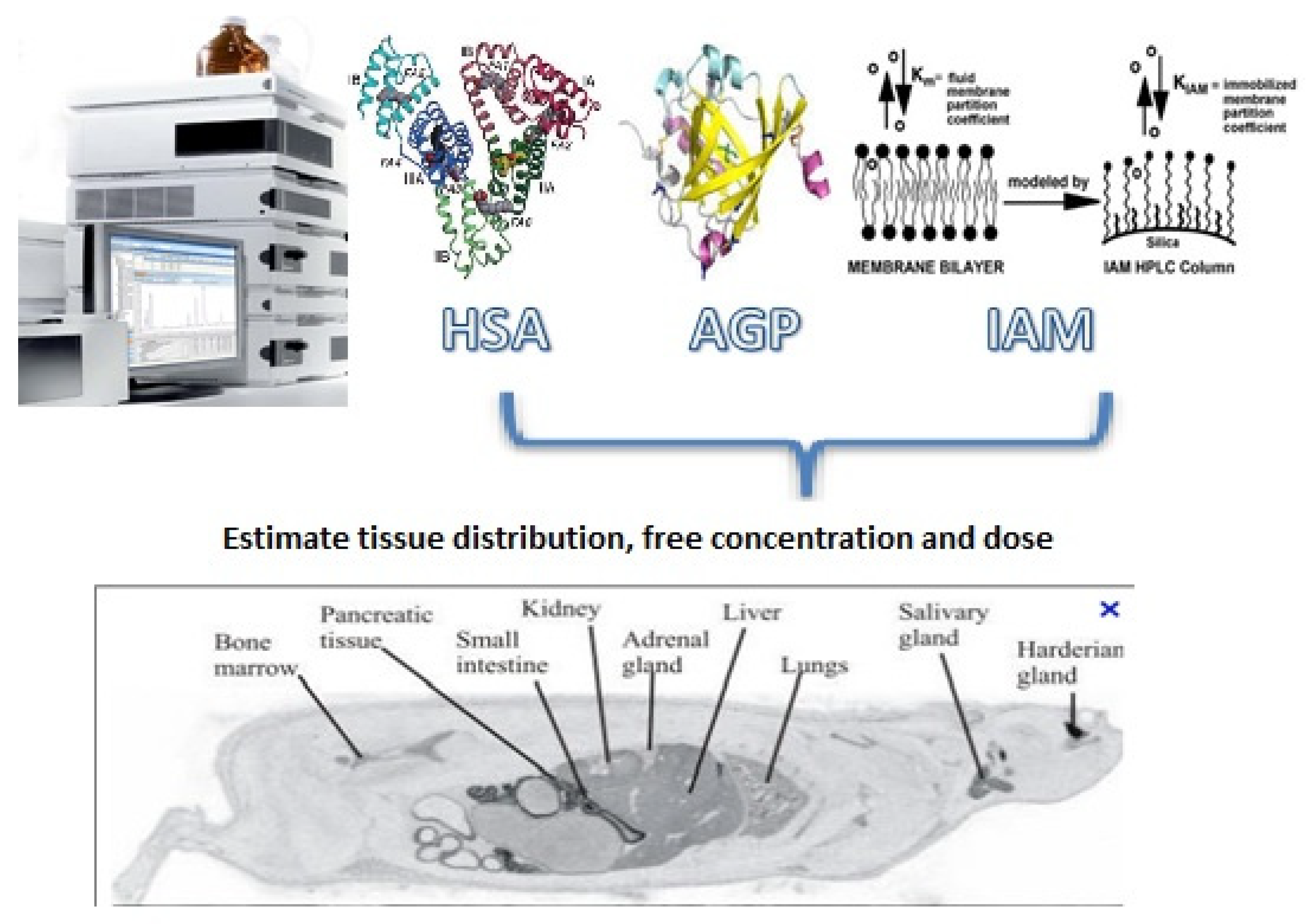
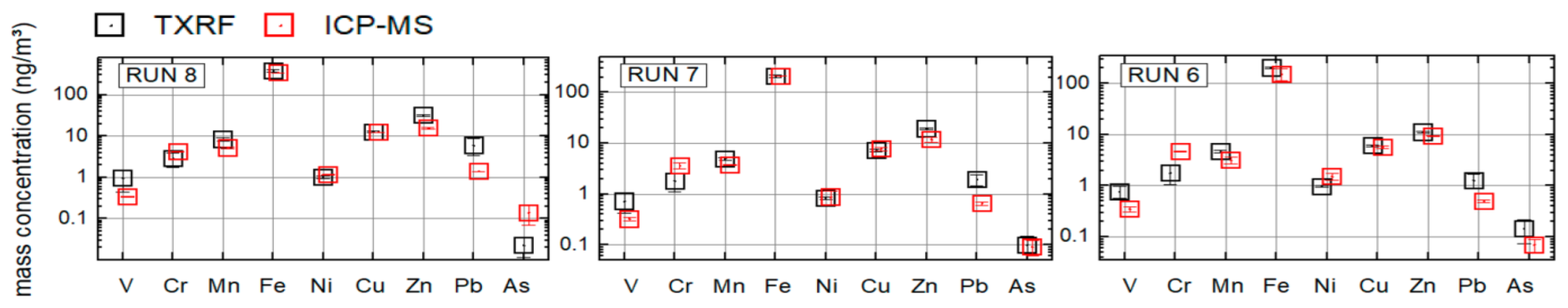
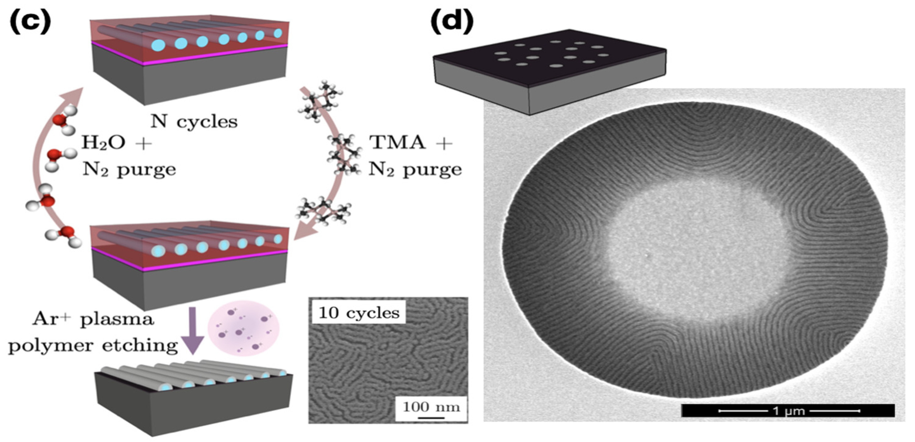
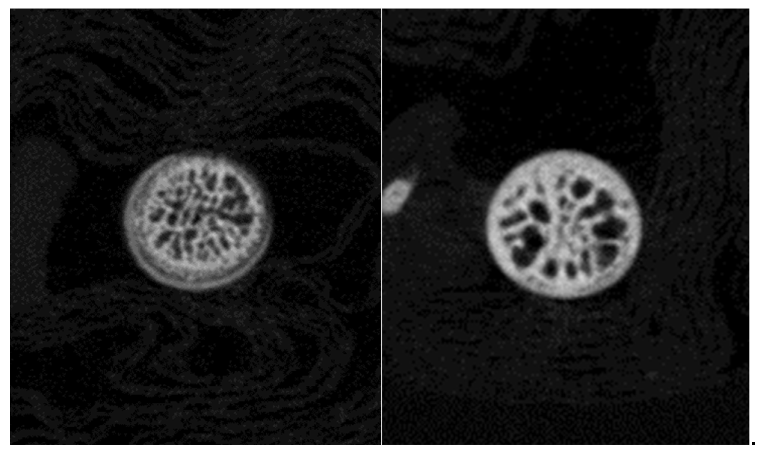
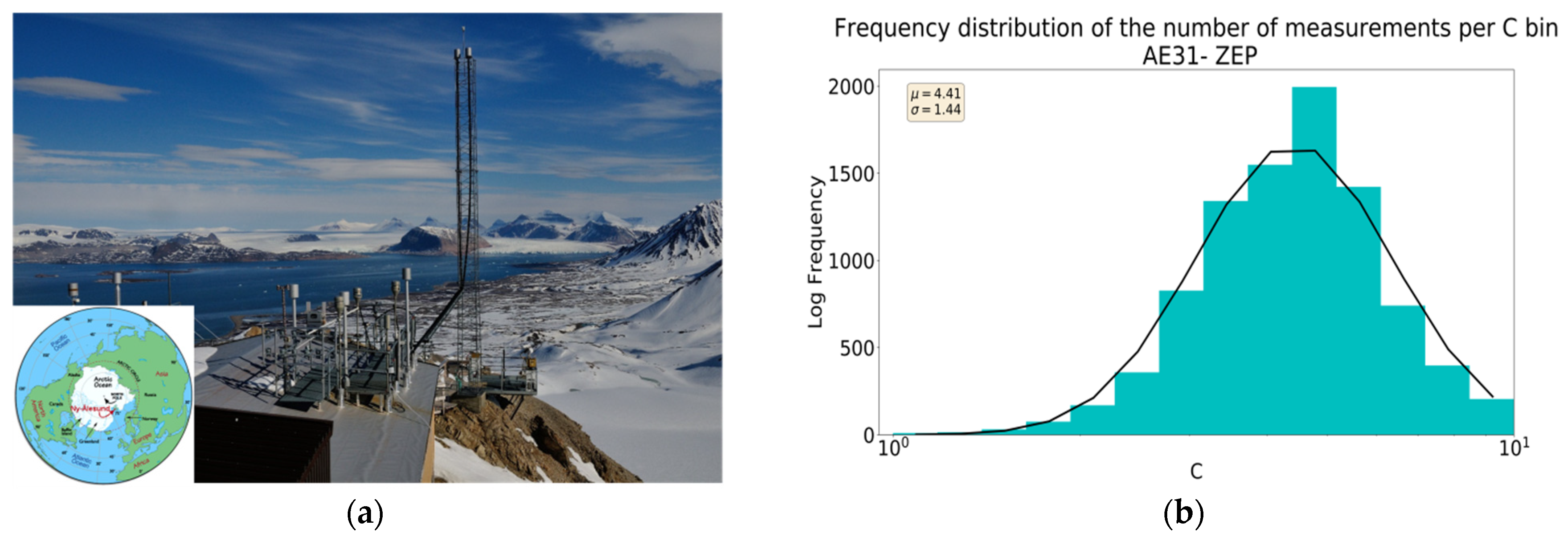
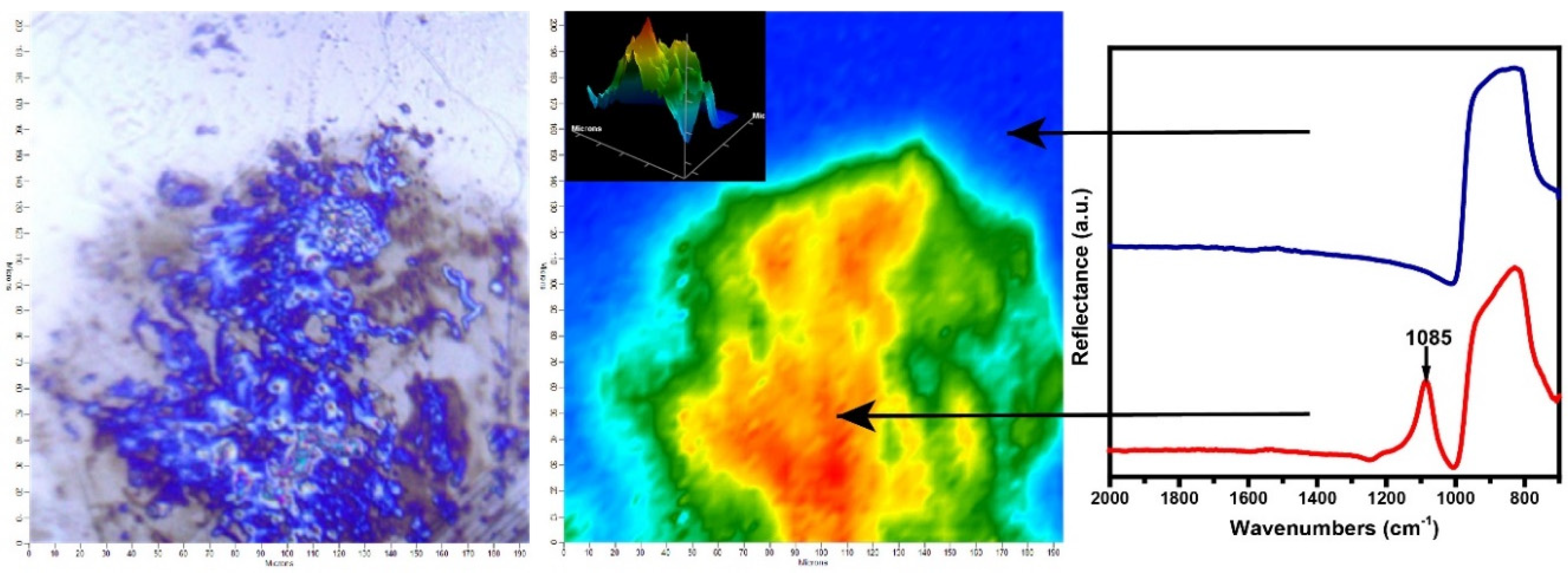
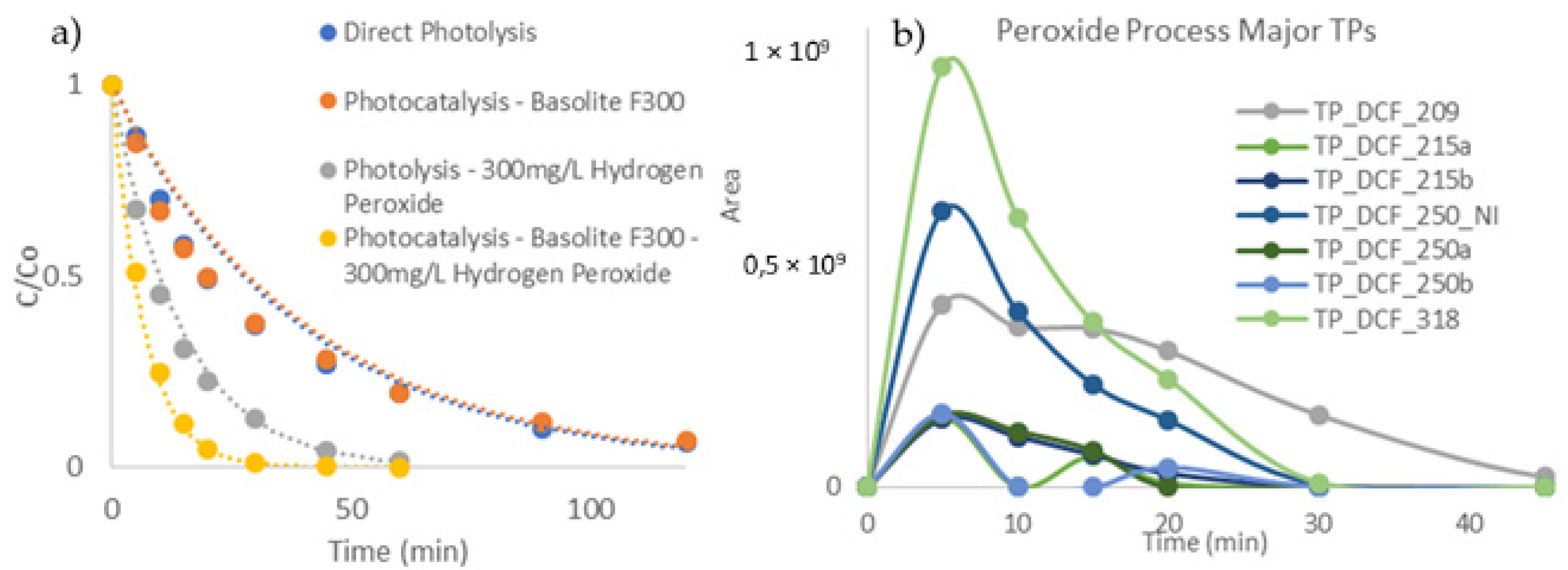
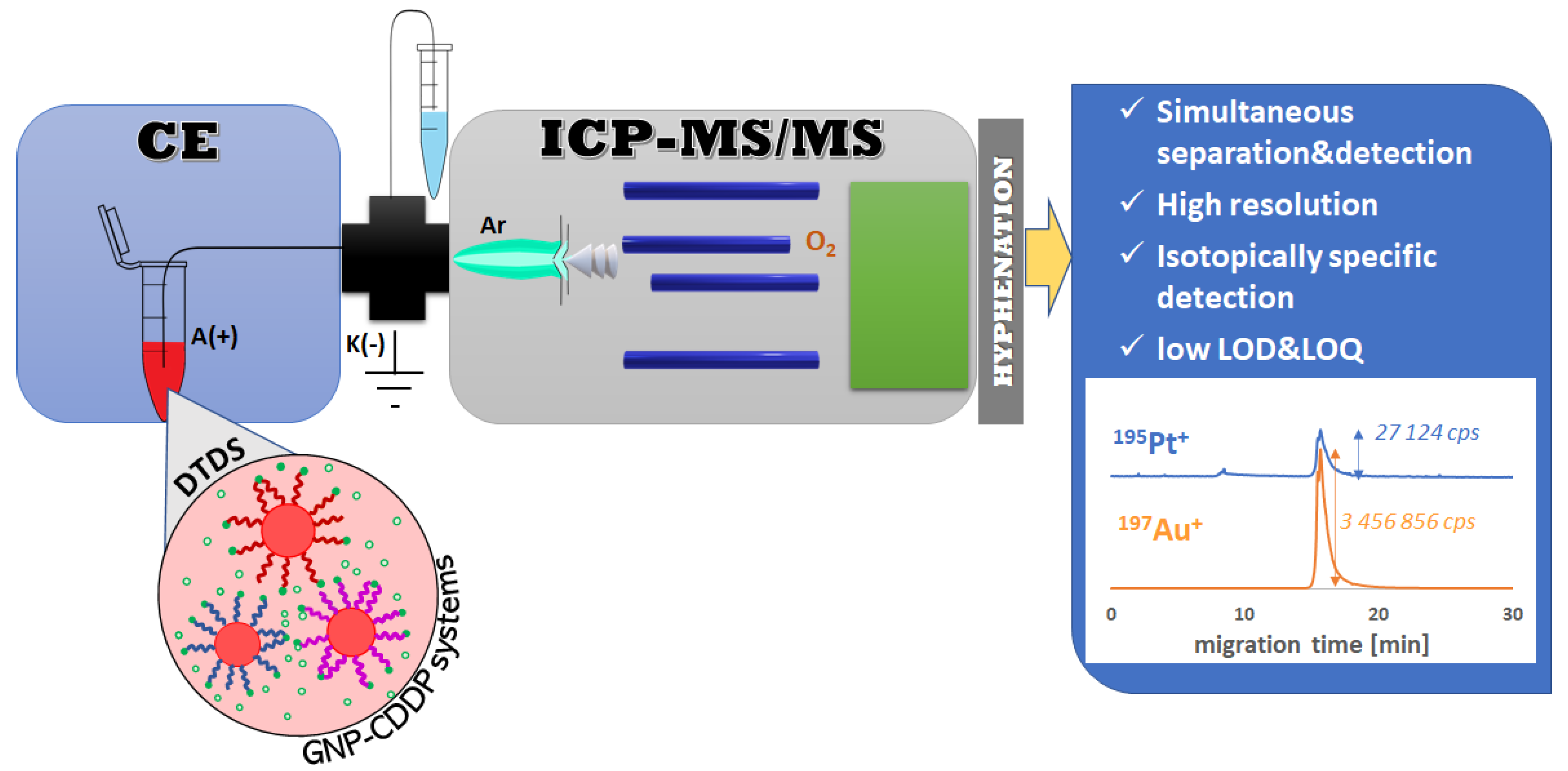
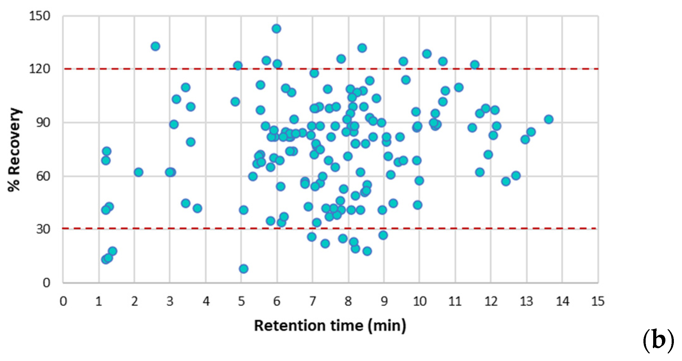
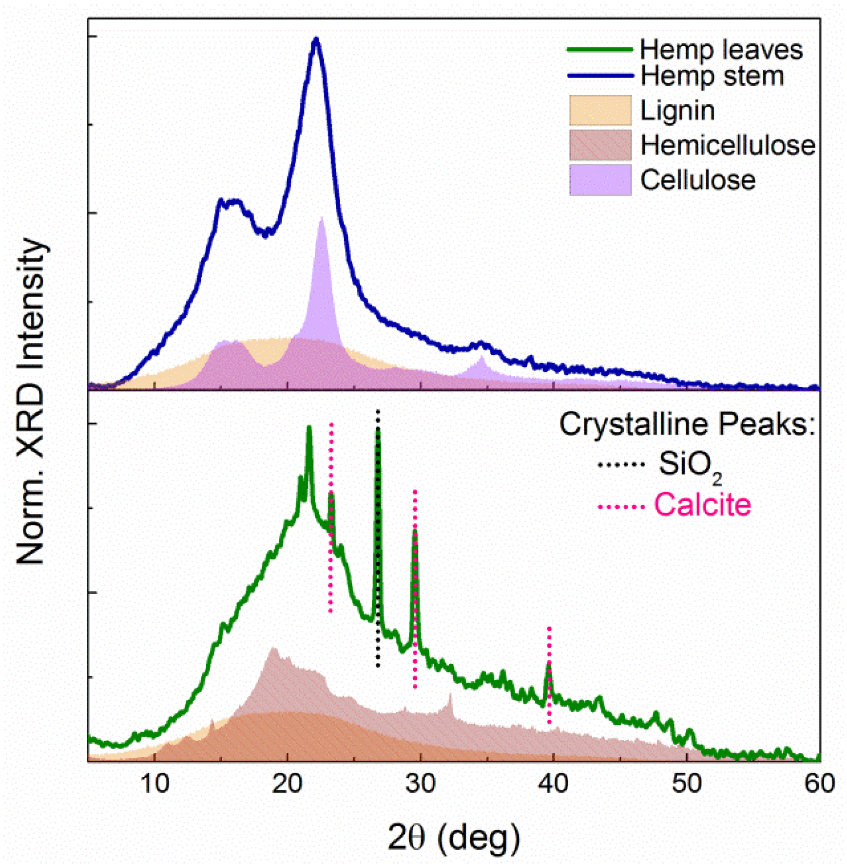
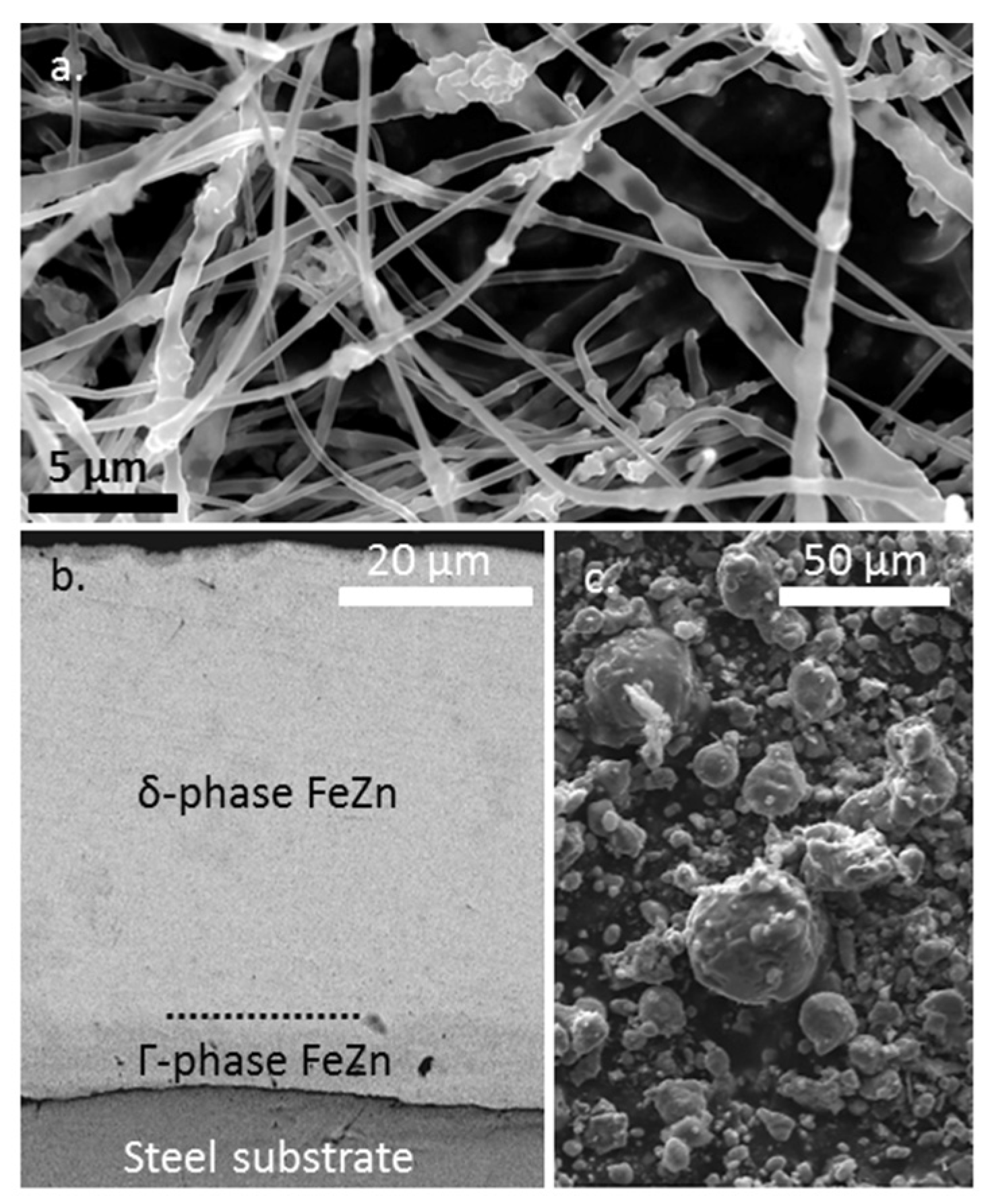
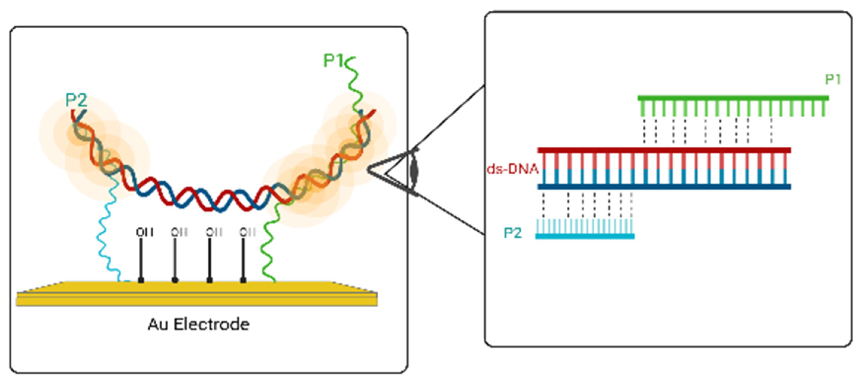
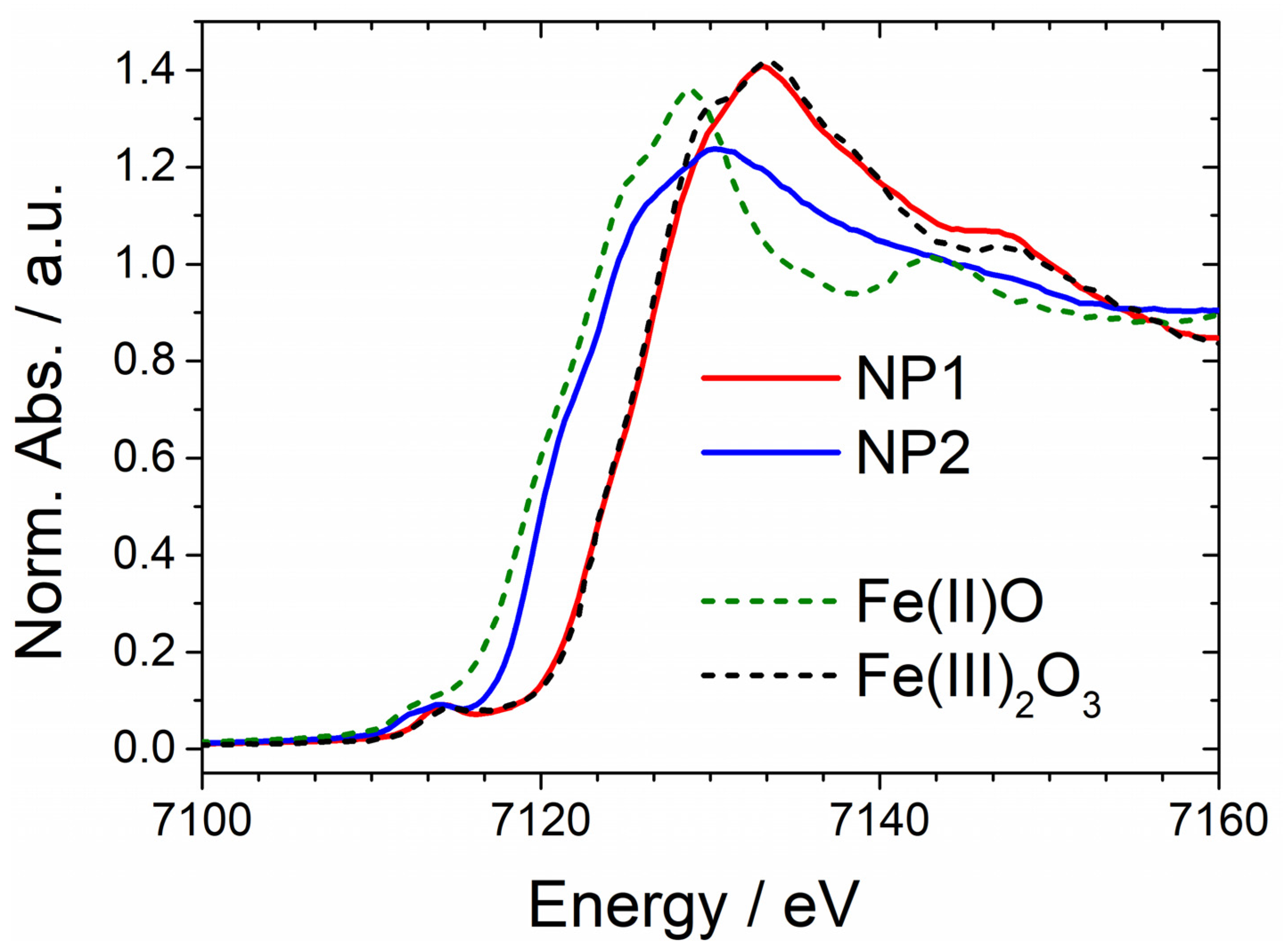
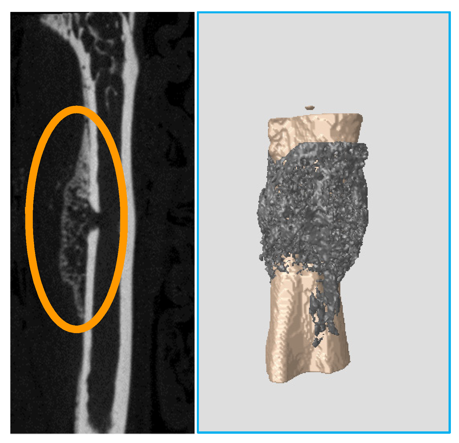
                                                                                                                                                                                                                                                                Publisher’s Note: MDPI stays neutral with regard to jurisdictional claims in published maps and institutional affiliations.

                                                                                                                                                                                                                                                                Share and Cite

                                                                                                                                                                                                                                                                MDPI and ACS Style

                                                                                                                                                                                                                                                                Mitrakas, M.; Ochsenkühn-Petropoulou, M. The 12th International Conference on Instrumental Analysis—Modern Trends and Applications (20–23 September 2021, Virtual Event). Appl. Sci. 2021, 11, 11767. https://doi.org/10.3390/app112411767

                                                                                                                                                                                                                                                                AMA Style

                                                                                                                                                                                                                                                                Mitrakas M, Ochsenkühn-Petropoulou M. The 12th International Conference on Instrumental Analysis—Modern Trends and Applications (20–23 September 2021, Virtual Event). Applied Sciences. 2021; 11(24):11767. https://doi.org/10.3390/app112411767

                                                                                                                                                                                                                                                                Chicago/Turabian Style

                                                                                                                                                                                                                                                                Mitrakas, Manassis, and Maria Ochsenkühn-Petropoulou. 2021. "The 12th International Conference on Instrumental Analysis—Modern Trends and Applications (20–23 September 2021, Virtual Event)" Applied Sciences 11, no. 24: 11767. https://doi.org/10.3390/app112411767

                                                                                                                                                                                                                                                                Note that from the first issue of 2016, this journal uses article numbers instead of page numbers. See further details here.

                                                                                                                                                                                                                                                                Article Metrics

                                                                                                                                                                                                                                                                Back to TopTop