Article

A Synergistic Anti-Diabetic Effect by Ginsenosides Rb1 and Rg3 through Adipogenic and Insulin Signaling Pathways in 3T3-L1 Cells

1
Korea Food Research Institute, Wanju 55365, Korea
2
Department of Nutrition, University of Massachusetts, Amherst, MA 01003, USA
3
Department of Food Biotechnology and Environmental Science, Kangwon National University, Chuncheon 24341, Korea
*
Authors to whom correspondence should be addressed.
These authors contributed equally to this work.
Appl. Sci. 2021, 11(4), 1725; https://doi.org/10.3390/app11041725
Submission received: 12 January 2021 / Revised: 3 February 2021 / Accepted: 10 February 2021 / Published: 15 February 2021
(This article belongs to the Special Issue Effect of Bioactive Substances Extracted from Food on Human Health)

Abstract

:
Although ginsenosides Rb1 and Rg3 have been identified as the significant ginsenosides found in red ginseng that confer anti-diabetic actions, it is unclear whether insulin-sensitizing effects are mediated by the individual compounds or by their combination. To determine the effect of ginsenosides Rb1 and Rg3 on adipocyte differentiation, 3T3-L1 preadipocytes were induced to differentiate the standard hormonal inducers in the absence or presence of ginsenosides Rb1 or Rg3. Additionally, we determined the effects of Rb1, Rg3, or their combination on the expression of genes related to adipocyte differentiation, adipogenic transcription factors, and the insulin signaling pathway in 3T3-L1 cells using semi-quantitative RT-PCR. Rb1 significantly increased the expression of CEBPα, PPARγ, and aP2 mRNAs. However, Rg3 exerted its maximal stimulatory effect on these genes at 1 μM concentration, while a high concentration (50 μM) showed inhibitory effects. Similarly, treatment with Rb1 and Rg3 (1 μM) increased the expression of IRS-1, Akt, PI3K, GLUT4, and adiponectin. Importantly, co-treatment of Rb1 and Rg3 (9:1) induced the maximal expression levels of these mRNAs. Our data indicate that the anti-diabetic activity of red ginseng is, in part, mediated by synergistic actions of Rb1 and Rg3, further supporting the significance of minor Rg3.

Graphical Abstract

1. Introduction

Enlarged adipocytes and excess adiposity play a key role in the metabolic and cardiovascular complications associated with obesity [1]. Our current understanding is that hypertrophic obesity is strongly associated with obesity-related insulin resistance and type 2 diabetes (T2D), while hyperplastic obesity is not usually linked to these metabolic consequences, which are seen in 20–30% of obese individuals characterized as “obese but metabolically normal” [2,3,4,5]. This clearly suggests that impaired formation of new adipocytes through preadipocyte differentiation (i.e., adipogenesis) is linked to the pathological expansion of adipose tissue (AT), as evidenced by decreases in insulin sensitivity and expression of mRNAs by differentiation markers such as peroxisome proliferator-activated receptor (PPARγ), glucose transporter 4 (GLUT4), and adiponectin [6,7]. Moreover, a hypertrophic environment also leads to AT dysfunction by impairing adipocyte insulin signaling pathways, thereby promoting insulin resistance [8,9]. Consequently, the hyperplasic expansion of adipocytes over hypertrophic enlargement within an individual can counteract the negative metabolic effects of obesity, and has an enormous impact on metabolic health outcomes. Accordingly, adipocytes have recently been emerging as a therapeutic target for obesity-associated metabolic disorders and thus introducing agents that promote preadipocyte differentiation and/or lipid oxidation would be a promising strategy for obesity-induced metabolic disorders.
Panax ginseng Meyer is believed to be a panacea, used in traditional oriental medicine for over 2000 years primarily to enhance physical and mental performance [10,11]. The pharmacological properties of ginseng and its constituents depend on the type and site of the attached sugar moieties, and have been largely attributed to ginsenosides [12,13,14]. Historically, raw ginseng is processed into either white ginseng by a simple drying method or red ginseng by a steaming process to enhance its shelf-life and efficacy [15,16,17]. Red ginseng is known to have a distinct ginsenoside profile compared to white and raw ginseng in the content and composition of ginsenosides [18,19,20]. In particular, red ginseng is shown to be rich in ginsenoside Rg3, which is significantly increased during the steaming process, whereas ginsenoside Rb1, the most abundant ginsenoside in ginseng root, is relatively reduced in steamed red ginseng [15,20,21,22,23]. Recent studies have shown that ginsenoside Rb1 is deglycosylated by human intestinal bacteria or digestive enzymes and is converted to a minor metabolite form, Rg3 [24,25]. It is also reported that deglycosylated ginsenoside metabolites are more readily absorbed into the blood stream and thus act as biologically active compounds [26]. Clinical and pre-clinical studies have also shown that ginseng extract and ginsenosides exert anti-diabetic effects by lowering blood glucose levels and enhancing insulin sensitivity [27,28]. Similarly, ginsenosides Rb1 and Rg3 have been reported to possess anti-diabetic effects, mainly through blood glucose control in animals and the stimulation of glucose uptake in 3T3-L1 mature adipocytes and L6 myotubes [29,30,31,32,33,34]. However, there have been contradictory results that Rb1 induces adipocyte differentiation, whereas Rg3 inhibits it, which may be due to the differences in the concentrations of ginsenosides and different cell culture models used in various studies [30,34,35,36,37,38]. In addition, no comparative studies have been reported for the potential bioactivity of Rb1 in combination with its metabolite, Rg3.
The aim of this study was to investigate the effect of the individual, as well as the combination of, ginsenosides Rb1 and Rg3 on adipocyte differentiation, glucose transport systems, adiponectin, and the insulin signaling pathway using a well-established model of adipocyte differentiation, 3T3-L1 preadipocytes. In the present study, we measured the expression levels of key molecules involved in the adipogenic program and insulin signaling pathways potentially regulated by the major ginsenosides Rb1 and Rg3. We also assessed the synergistic effects of Rb1 and Rg3, which are important for elucidating the effect of these important bioactives on T2D and its mechanism of action.

2. Materials and Methods

2.1. Cell Line and Materials

3T3-L1 cells were purchased from American Type Culture Collection (ATCC, CL-173, Manassas, VA, USA). Penicillin-streptomycin (P/S), trypsin-EDTA, phosphate-buffered saline (PBS), fetal bovine serum (FBS), bovine calf serum (BCS), and Dulbecco’s modified Eagle’s medium (DMEM) were obtained from Gibco (Gaithersburg, MD, USA). Ginsenosides Rb1 and Rg3 were purchased from Fleton Reference Substance Co., Ltd. (Chengdu, China), and dissolved in dimethyl sulfoxide (DMSO). Unless noted otherwise, all chemicals were purchased from Sigma (St. Louis, MO, USA).

2.2. Cell Culture

3T3-L1 preadipocytes were cultured using the method of Blumberg et al. [39]. Briefly, cells were plated and grown for 2 days post-confluence in DMEM with 10% BCS, 1% P/S, and 3.7 g/L sodium bicarbonate. Adipocyte differentiation was induced by treating confluent cells with MDI cocktail (0.5 mM IBMX, 1 μM Dex, and 1.67 μM insulin) and 10% FBS for 2 days. The culture medium was then replaced with DMEM supplemented with only 10% FBS and 1.67 μM insulin. Two days after the addition of 10% FBS and MDI, the cells were treated with ginsenosides Rb1 and Rg3 individually (1, 10, and 50 μM) or in combination (10 μM). The cell culture medium was changed on alternate days with DMEM containing 10% FBS and insulin (1.67 μM) with ginsenosides Rb1 and Rg3.

2.3. Determination of Lipid Accumulation by Oil Red O Staining

The extent of differentiation was determined by the amount of lipid accumulation at 0, 4, and 6 days by Oil red O staining. Briefly, cells were fixed in formaldehyde (10% v/v) for 1 h. Cells were stained with 0.5% Oil red O solution in 60% (v/v) isopropanol at room temperature for 30 min, washed with distilled water, and dried. Differentiation was monitored under a phase-contrast microscope and quantified via elution with isopropanol, and optical density was measured at 490 nm (OD490) [39].

2.4. RNA Analysis by Semi-Quantitative Reverse Transcriptase Polymerase Reaction (RT-PCR)

Total RNA was isolated from 3T3-L1 adipocyte cells using TRIzol reagent (Invitrogen, Carlsbad, CA, USA). The cDNAs were synthesized with the SuperScript First-Strand Synthesis system for RT-PCR kit (Invitrogen), performed using 30 to 35 cycles of 30 sec at 94 °C, 30 s at 55 °C, and 1 min at 72 °C, followed by 10 min extension at 72 °C. All primers used in this study are listed in Table 1. To ensure that equal amounts of reverse-transcribed cDNA were applied, β-actin was used as a housekeeping gene. Image-J (version 1.28 u, National Institutes of Health, Washington, DC, USA) was used to measure the intensity of the PCR products.

2.5. Statistical Analysis

The results are expressed as means ± standard deviations and statistically analyzed by ANOVA and Duncan’s multiple range tests. Statistical significance was set at p < 0.05 (SAS Inst., Inc., Cary, NC, USA).

3. Results

3.1. Ginsenosides Rb1 and Rg3 Enhanced the Differentiation of 3T3-L1 Preadipocytes

To determine the effect of ginsenosides Rb1 and Rg3 on adipocyte differentiation, 3T3-L1 preadipocytes were induced to differentiate with the standard hormonal inducers (MDI) in the absence or presence of ginsenosides Rb1 or Rg3 (1, 10, and 50 μM), as described in the Methods. The range of concentrations was chosen to consider physiologically achievable doses (1 and 10 μM) and a nontoxic high dose of ginsenosides (50 μM) [40,41,42]. As shown in Figure 1, both ginsenosides Rb1 and Rg3 enhanced lipid accumulated during differentiation of 3T3-L1 preadipcytes. Ginsenoside Rb1 induced a dose-dependent increase in lipid acquisition in 3T3-L1 cells. However, ginsenoside Rg3 exerted its maximal effect at 1 μM as well as a dose-dependent decrease in lipid accumulation.
PPARγ is known as a master transcription regulator of adipocyte differentiation, and orchestrates with C/EBPα to regulate downstream target genes such as adipocyte protein 2 (aP2). It is well known that PPARγ is also a nuclear receptor for anti-diabetic drugs. Consistent with the effect on adipocyte differentiation, ginsenoside Rb1 dose-dependently enhanced the expression of PPARγ, CEBPα, and aP2 mRNAs at all concentrations used (Figure 2). Moreover, ginsenoside Rg3 exerted its maximal stimulatory effect at 1 μM for the expression of PPARγ, C/EBPα, and aP2 mRNAs during the differentiation of 3T3-L1 adipocytes. In contrast, Rg3 at a high concentration (50 μM) showed inhibitory effects on the expression of C/EBPα and aP2 mRNA, but did not reach statistical significance compared to the control. These results indicate that the effects of the ginsenosides are comparable with respect to morphological changes and molecular responses.

3.2. Ginsenosides Rb1 and Rg3 Upregulate GLUT4 and Adiponectin mRNA Expression

Since PPARγ is a molecular target for anti-diabetic drugs, such as thiazolidinediones (TZDs), that enhance glucose uptake and insulin sensitivity, we next examined whether Rb1 and Rg3 regulate the PPARγ downstream genes adiponectin and GLUT4, both of which are important for insulin sensitivity and insulin-stimulated glucose uptake in adipocytes. The expression of GLUT4 and adiponectin mRNA was significantly up-regulated dose-dependently in differentiating 3T3-L1 cells exposed to 1 and 10 µM Rb1 compared to the control. In contrast, ginsenoside Rg3 showed a maximal stimulatory effect at 1 µM on the expression of GLUT4 and adiponectin mRNA (Figure 3).

3.3. Ginsenosides Rb1 and Rg3 Increase mRNA Expression of the Glucose Transport System

Glucose uptake is regulated by the glucose transport system, which involves insulin receptor substrate 1 (IRS-1) and multiple steps of the phosphoinositide 3-kinase (PI3K)/Akt signaling pathway system [43,44]. Our data showed that Rb1 treatment significantly increased the expression of IRS-1, Akt, and PI3K mRNAs at the concentrations of 1 to 50 μM (Figure 4). However, the effects of Rg3 treatment on the expression of IRS-1, Akt, and PI3K mRNA were in parallel with its regulation on the expression of PPARγ, GLUT 4, and adiponectin mRNAs (Figure 4).

3.4. Combined Effects of Ginsenosides Rb1 and Rg3 on the Expression of GLUT4 and Adiponectin mRNAs

Ginsenoside Rb1 has been shown convert into ginsenoside Rg3 by conversion processes such as steaming or contact with intestinal bacteria [18]. As shown in Figure 5, the combined treatment of Rb1 and Rg3 (9:1 ratio) markedly increased the expression of GLUT4 and adiponectin mRNAs. However, decreasing the ratio of Rb1 to Rg3 by increasing the concentrations of Rg3 showed inhibitory effects on the expression of GLUT4 and adiponectin mRNAs (Figure 4). The combined effects of Rb1 and Rg3 on gene expression were in parallel with the degree of differentiation as assessed by lipid accumulation (data not shown).

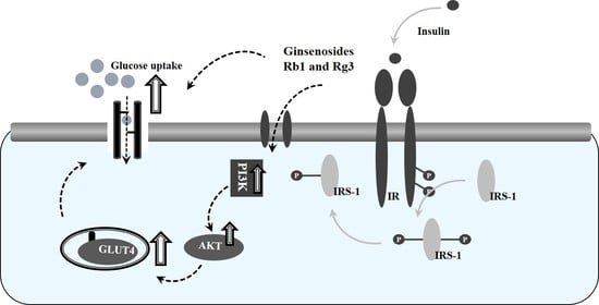
3.5. Combined Effects of Ginsenosides Rb1 and Rg3 on the Insulin Signaling Pathway

The GLUT4 gene expression level is correlated with whole-body insulin-signaling-pathway-mediated glucose homeostasis [45]. The combined treatment of Rb1 and Rg3 (9:1) significantly increased the expression of IRS-1 mRNA relative to the expression level of the control. However, decreasing the ratio of Rb1 to Rg3 by increasing the concentrations of Rg3 induced inhibitory effects on IRS-1 mRNA expression (Figure 6). The effects of Rb1 and Rg3 treatment on the expression of Akt and PI3K mRNAs followed a similar pattern as that observed with IRS expression.

4. Discussion

The biological activities of ginseng have been largely attributed to ginsenosides produced by different processing methods and/or biotransformation by human intestinal bacteria [16,17,24]. Although Rb1 is the most abundant ginsenoside in ginseng root and white ginseng, red ginseng produced by a steaming process substantially increases the concentration of Rg3, which is also known to be produced by human intestinal bacteria from Rb1 [22,23,24]. Several studies have therefore been aimed at converting ginsenoside Rb1 to Rg3 by various methods, including steaming, fermentation, or acidic and enzymatic treatments [15,46]. However, few studies have investigated the anti-diabetic potential of Rb1 in combination with Rg3, which may better reflect actual human consumption and thus biological activities.
Intervention studies in humans and animals have demonstrated that ginseng extracts and ginsenosides improve insulin sensitivity [47,48]. In vitro studies have also shown their modulatory effects on PPARγ, a nuclear receptor for anti-diabetic drugs, and the expression of its downstream target genes [36,38]. However, literature on the effect of specific ginsenosides Rb1 or Rg3 on adipocyte differentiation and function is currently inconsistent and unclear. Some studies have reported inhibitory effects of ginsenosides on PPARγ expression and adipocyte differentiation [30,34,35], while other studies have shown stimulatory effects [36,37,38]. These contradictory results from different studies might be attributable to differences in the concentrations of ginsenosides used (i.e., inhibitory with high and stimulatory with low concentrations), cell culture models (i.e., differentiating vs. mature adipocytes), and/or experimental conditions.
In the present study, we found that ginsenoside Rb1 significantly increased PPARγ expression and enhanced the differentiation of 3T3-L1 preadipocytes at concentrations between 1 and 50 μM, whereas Rg3 showed a maximal stimulatory effect at 1 μM with a dose-dependent decrease at 10 and 50 μM concentrations (Figure 1 and Figure 2). The ability of Rb1 and Rg3 to increase PPARγ mRNA suggests that these ginsenosides may have potential as a natural bioactive component for increasing insulin sensitivity. Our data also indicate that Rb1 and Rg3 may confer insulin-sensitizing effects through the upregulation of adiponectin and GLUT4 gene expression, leading to subsequent glucose uptake and utilization in adipocytes. Interestingly, we also observed similar and comparable effects of Rb1 and Rg3 co-treatment (in a ratio of 9:1) on GLUT4 and adiponectin gene expression (Figure 5). It is worth noting that the ratio of Rb1 and Rg3 (9:1) that has a maximal stimulatory effect on GLUT4 and adiponectin was similar to the profile we typically observed in steamed or fermented ginseng (in a ratio of ~8:1).
One line of research suggests that adiponectin is an insulin-sensitizing hormone that is expressed exclusively in adipocytes [49]. The mRNA expression of adiponectin and its plasma levels in adipose tissue were highly correlated with obesity-induced insulin resistance, which was reversed by the administration of adiponectin [50,51]. Further, the insulin-sensitizing actions of TZDs are also known to be mediated by targeting PPARγ and adiponectin in adipocytes [52,53]. A clear association between impaired adipocyte differentiation and insulin resistance has been explained by the observation that TZDs promote adipocyte differentiation, generating small adipocytes that are more insulin sensitive than enlarged adipocytes [54]. It cannot be determined from the present study whether ginsenoside Rb1, Rg3, or both increases the number of small adipocytes. However, the ability of Rb1 and Rg3 to upregulate the expression of PPARγ, GLUT4, and adiponectin mRNAs (Figure 2 and Figure 3) may account for their anti-hyperglycemic effects observed in vivo [55]. Although precise molecular mechanisms regulating GLUT4 and adiponectin gene expression by Rb1 and Rg3 are not clear, they are presumably due to PPARγ activation in adipocytes.
The insulin signaling pathway regulates glucose homeostasis by transducing insulin’s signal to downstream intracellular target molecules, including IRS-1 and Akt. The subsequent activation of the PI3K pathway is necessary for the recruitment of GLUT4 to the cell membrane, which will ultimately increase glucose uptake in insulin-sensitive tissues [56,57]. Our results show that upregulation of GLUT4 expression induced by individual and combined Rb1 and Rg3 correlated with the increased expression of IRS-1, Akt, and PI3K mRNAs (Figure 4 and Figure 6). Interestingly, the combined treatment of ginsenosides Rb1 and Rg3 exerted more potent effects than individual treatments, which may suggest a synergistic effect via differential chemical structures between ginsenoside Rb1 and Rg3 (Figure 5 and Figure 6). Current findings are consistent with a previous report by Shang et al. [29], where ginsenoside Rb1 stimulated glucose uptake through the insulin signaling pathway in mature adipocytes. Moreover, Kim et al. [58] reported that enhanced glucose uptake and insulin signaling pathway activity by ginsenoside Rg3 was induced, in part, by increasing IRS-1 transcripts in L6 myoblasts.

5. Conclusions

In summary, ginseng is one of the most used medicinal herbs, and both ginseng research and market demands have significantly increased in the past decades. Moreover, red ginseng is more often consumed in various processed forms (67%) than unprocessed ginseng products (33%) [59]. Given the recent findings that ginsenoside Rg3 is produced from Rb1 by the human intestine as well as steaming/fermentation processes, our results provide additional information for a possible mechanism of action of ginsenosides Rb1 and Rg3 as well as their potential synergistic effects. However, further studies are needed to identify the in vivo effects that Rb1 and Rg3 have on glucose and lipid metabolism using different preclinical and clinical models.

Author Contributions

Conceptualization, O.-H.L. and Y.-C.K.; methodology, H.-D.H.; software, S.-I.C.; validation, S.-I.C., H.-D.H. and Y.-C.K.; formal analysis, O.-H.L.; investigation, H.-D.H.; resources, Y.-C.K.; data curation, H.-D.H. and O.-H.L.; writing—original draft preparation, H.-D.H. and S.-I.C.; writing—review and editing, O.-H.L. and Y.-C.K.; supervision, Y.-C.K.; project administration, O.-H.L.; funding acquisition, Y.-C.K. All authors have read and agreed to the published version of the manuscript.

Funding

The present study was supported by Brain Pool program funded by the Ministry of Science and ICT through the National Research Foundation of Korea (NRF-2019H1D3A2A01031987) and the 2020 Research Grant from Gangneung Science & Industry Promotion Agency.

Institutional Review Board Statement

Not applicable.

Informed Consent Statement

Not applicable.

Data Availability Statement

The data presented in this study are available within the article.

Conflicts of Interest

The authors declare no conflict of interest.

References

  1. Bays, H.E.; Toth, P.P.; Kris-Etherton, P.M.; Abate, N.; Aronne, L.J.; Brown, W.V.; Gonzalez-Campoy, J.M.; Jones, S.R.; Kumar, R.; Forge, R.L.; et al. Obesity, adiposity, and dyslipidemia: A consensus statement from the National Lipid Association. J. Clin. Lipidol. 2013, 7, 304–383. [Google Scholar] [CrossRef] [PubMed] [Green Version]
  2. Stefan, N.; Kantartzis, K.; Machann, J.; Schick, F.; Thamer, C.; Rittig, K.; Balletshofer, B.; Machicao, F.; Fritsche, A.; Häring, H. Identification and characterization of metabolically benign obesity in humans. Arch. Intern. Med. 2008, 168, 1609–1616. [Google Scholar] [CrossRef] [PubMed]
  3. Marini, M.A.; Succurro, E.; Frontoni, S.; Hribal, M.L.; Andreozzi, F.; Lauro, R.; Perticone, F.; Sesti, G. Metabolically healthy but obese women have an intermediate cardiovascular risk profile between healthy nonobese women and obese insulin-resistant women. Diabetes Care 2007, 30, 2145–2147. [Google Scholar]
  4. Lafontan, M. Adipose tissue and adipocyte dysregulation. Diabetes Metab. 2014, 40, 16–28. [Google Scholar] [CrossRef]
  5. Cignarelli, A.; Genchi, V.A.; Perrini, S.; Natalicchio, A.; Laviola, L.; Giorgino, F. Insulin and Insulin Receptors in Adipose Tissue Development. Int. J. Mol. Sci. 2019, 20, 759. [Google Scholar] [CrossRef] [Green Version]
  6. Isakson, P.; Hammarstedt, A.; Gustafson, B.; Smith, U. Impaired preadipocyte differentiation in human abdominal obesity: Role of Wnt, tumor necrosis factor-alpha, and inflammation. Diabetes 2009, 58, 1550–1557. [Google Scholar] [CrossRef] [PubMed] [Green Version]
  7. Martínez-Jiménez, V.; Cortez-Espinosa, N.; Rodríguez-Varela, E.; Vega-Cárdenas, M.; Briones-Espinoza, M.; Ruíz-Rodríguez, V.M.; López-López, N.; Briseño-Medina, A.; Turiján-Espinoza, E.; Portales-Pérez, D.P. Altered levels of sirtuin genes (SIRT1, SIRT2, SIRT3 and SIRT6) and their target genes in adipose tissue from individual with obesity. Diabetes Metab. Syndr. 2019, 13, 582–589. [Google Scholar] [CrossRef] [PubMed]
  8. Hotamisligil, G.S.; Peraldi, P.; Budavari, A.; Ellis, R.; White, M.F.; Spiegelman, B.M. IRS-1-mediated inhibition of insulin receptor tyrosine kinase activity in TNF-alpha- and obesity-induced insulin resistance. Science 1996, 271, 665–670. [Google Scholar] [CrossRef]
  9. Xu, H.; Barnes, G.T.; Yang, Q.; Tan, G.; Yang, D.; Chou, C.J.; Sole, J.; Nichols, A.; Ross, J.S.; Tartaglia, L.A.; et al. Chronic inflammation in fat plays a crucial role in the development of obesity-related insulin resistance. J. Clin. Investig. 2003, 112, 1821–1830. [Google Scholar] [CrossRef] [PubMed]
  10. Arushanian, E.B. Therapeutic potential of ginseng root preparations in treating diabetes mellitus. Eksp. Klin. Farmakol. 2009, 72, 52–56. [Google Scholar]
  11. Bang, H.; Kwak, J.H.; Ahn, H.Y.; Shin, D.Y.; Lee, J.H. Korean red ginseng improves glucose control in subjects with impaired fasting glucose, impaired glucose tolerance, or newly diagnosed type 2 diabetes mellitus. J. Med. Food 2014, 17, 128–134. [Google Scholar] [CrossRef] [Green Version]
  12. Cho, C.W.; Kim, Y.C.; Rhee, Y.K.; Lee, Y.C.; Kim, K.T.; Hong, H.D. Chemical composition characteristics of Korean straight ginseng products. J. Ethn. Foods 2014, 1, 24–28. [Google Scholar] [CrossRef] [Green Version]
  13. Liang, Y.; Zhao, S. Progress in understanding of ginsenoside biosynthesis. Plant. Biol. 2008, 10, 415–421. [Google Scholar] [CrossRef]
  14. Kim, D.H. Chemical Diversity of Panax ginseng, Panax quinquifolium, and Panax notoginseng. J. Ginseng Res. 2012, 36, 1–15. [Google Scholar] [CrossRef] [Green Version]
  15. Kim, W.Y.; Kim, J.M.; Han, S.B.; Lee, S.K.; Kim, N.D.; Park, M.K.; Kim, C.K.; Park, J.H. Steaming of ginseng at high temperature enhances biological activity. J. Nat. Prod. 2000, 63, 1702–1704. [Google Scholar] [CrossRef]
  16. Wang, C.; Zhang, B.; Song, W.; Wang, A.; Ni, M.; Luo, X.; Aung, H.H.; Xie, J.; Tong, R.; He, T.; et al. Steamed American ginseng berry: Ginsenoside analyses and anticancer activities. J. Agric. Food Chem. 2006, 54, 9936–9942. [Google Scholar] [CrossRef] [PubMed]
  17. Lim, C.Y.; Moon, J.M.; Kim, B.Y.; Lim, S.H.; Lee, G.S.; Yu, H.S.; Cho, S.I. Comparative study of Korean white ginseng and Korean Red Ginseng on efficacies of ova-induced asthma model in mice. J. Ginseng Res. 2015, 39, 38–45. [Google Scholar] [CrossRef] [Green Version]
  18. Shibata, S. Chemistry and cancer preventing activities of ginseng saponins and some related triterpenoid compounds. J. Korean Med. Sci. 2001, 16, 28–37. [Google Scholar]
  19. He, M.; Huang, X.; Liu, S.; Guo, C.; Xie, Y.; Meijer, A.H.; Wang, M. The Difference between White and Red Ginseng: Variations in Ginsenosides and Immunomodulation. Planta Med. 2018, 84, 845–854. [Google Scholar] [CrossRef] [PubMed] [Green Version]
  20. Ha, Y.W.; Lim, S.S.; Ha, I.J.; Na, Y.C.; Seo, J.J.; Shin, H.; Son, S.H.; Kim, Y.S. Preparative isolation of four ginsenosides from Korean red ginseng (steam-treated Panax ginseng C. A. Meyer), by high-speed counter-current chromatography coupled with evaporative light scattering detection. J. Chromatogr. A 2007, 1151, 37–44. [Google Scholar] [CrossRef] [PubMed]
  21. Kwon, S.W.; Han, S.B.; Park, I.H.; Kim, J.M.; Park, M.K.; Park, J.H. Liquid chromatographic determination of less polar ginsenosides in processed ginseng. J. Chromatogr. A 2001, 921, 335–339. [Google Scholar] [CrossRef]
  22. Cho, H.T.; Kim, J.H.; Lee, J.H.; Kim, Y.J. Effects of Panax ginseng extracts prepared at different steaming times on thermogenesis in rats. J. Ginseng Res. 2017, 41, 347–352. [Google Scholar] [CrossRef] [PubMed] [Green Version]
  23. Li, Z.; Ji, G.E. Effects of various ginsenosides and ginseng root and ginseng berry on the activity of pancreatic lipase. Food Sci. Biotechnol. 2017, 26, 767–773. [Google Scholar] [CrossRef]
  24. Hasegawa, H. Proof of the mysterious efficacy of ginseng: Basic and clinical trials: Metabolic activation of ginsenoside: Deglycosylation by intestinal bacteria and esterification with fatty acid. J. Pharmacol. Sci. 2004, 95, 153–157. [Google Scholar] [CrossRef] [Green Version]
  25. Kato, T.; Kida, T.; Kanaoka, M.; Hattori, M.; Kobashi, K. Drug metabolism: Intestinal bacterial hydrolysis is required for the appearance of compound K in rat plasma after oral administration of ginsenoside Rb1 from Panax ginseng. J. Pharm. Pharmacol. 1998, 50, 1155–1160. [Google Scholar]
  26. Bae, E.A.; Park, S.Y.; Kim, D.H. Constitutive β-glucosidases hydrolyzing ginsenoside Rb1 and Rb2 from human intestinal bacteria. Biol. Pharm. Bull. 2000, 23, 1481–1485. [Google Scholar] [CrossRef]
  27. Yun, S.N.; Moon, S.J.; Ko, S.K. Wild ginseng prevents the onset of high fat diet induced hyperglyecemia and obesity in ICR mice. Arch. Pharm. Res. 2004, 27, 790–796. [Google Scholar] [PubMed]
  28. Chung, S.H.; Choi, C.G.; Park, S.H. Comparisons between white ginseng radix and rootlet for antidiabetic activity and mechanism in KKAy mice. Arch. Pharm. Res. 2001, 24, 214–218. [Google Scholar] [CrossRef]
  29. Shang, W.; Yang, Y.; Zhou, L.; Jiang, B.; Jin, H.; Chen, M. Ginsenoside Rb1 stimulates glucose uptake through insulin-like signaling pathway in 3T3-L1 adipocytes. J. Endocrinol. 2008, 198, 561. [Google Scholar] [CrossRef]
  30. Hwang, J.T.; Lee, M.S.; Kim, H.J.; Sung, M.J.; Kim, H.Y.; Kim, M.S.; Kwon, D.Y. Antiobesity effect of ginsenoside Rg3 involves the AMPK and PPAR-gamma signal pathways. Phytother. Res. 2009, 23, 262–266. [Google Scholar]
  31. Mohanan, P.; Subramaniyam, S.; Mathiyalagan, R.; Yang, D.C. Molecular signaling of ginsenosides Rb1, Rg1, and Rg3 and their mode of actions. J. Ginseng Res. 2018, 42, 123–132. [Google Scholar] [CrossRef] [PubMed]
  32. Park, M.W.; Ha, J.; Chung, S.H. 20(S)-ginsenoside Rg3 enhances glucose-stimulated insulin secretion and activates AMPK. Biol. Pharm. Bull. 2008, 31, 748–751. [Google Scholar] [CrossRef] [PubMed] [Green Version]
  33. Lim, S.; Yoon, J.W.; Choi, S.H.; Cho, B.J.; Kim, J.T.; Chang, H.S.; Park, H.S.; Park, K.S.; Lee, H.K.; Kim, Y.B.; et al. Effect of ginsam, a vinegar extract from Panax ginseng, on body weight and glucose homeostasis in an obese insulin-resistant rat model. Metabolism 2009, 58, 8–15. [Google Scholar] [CrossRef]
  34. Kim, S.N.; Lee, J.H.; Shin, H.; Son, S.H.; Kim, Y.S. Effects of in vitro digested ginsenosides on lipid accumulation in 3T3-L1 adipocytes. Planta. Med. 2009, 75, 596–601. [Google Scholar] [CrossRef]
  35. Hwang, J.T.; Kim, S.H.; Lee, M.S.; Kim, S.H.; Yang, H.J.; Kim, M.J.; Kim, H.S.; Ha, J.; Kim, M.S.; Kwon, D.Y. Anti-obesity effects of ginsenoside Rh2 are associated with the activation of AMPK signaling pathway in 3T3-L1 adipocyte. Biochem. Biophys. Res. Commun. 2007, 364, 1002–1008. [Google Scholar] [CrossRef] [PubMed]
  36. Shang, W.; Yang, Y.; Jiang, B.; Jin, H.; Zhou, L.; Liu, S.; Chen, M. Ginsenoside Rb1 promotes adipogenesis in 3T3-L1 cells by enhancing PPARγ2 and C/EBPα gene expression. Life Sci. 2007, 80, 618–625. [Google Scholar] [CrossRef] [PubMed]
  37. Chan, L.S.; Yue, P.Y.; Kok, T.W.; Keung, M.H.; Mak, N.K.; Wong, R.N. Ginsenoside-Rb1 promotes adipogenesis through regulation of PPARγ and microRNA-27b. Horm. Metab. Res. 2012, 44, 819–824. [Google Scholar] [CrossRef]
  38. Han, K.L.; Jung, M.H.; Sohn, H.H.; Hwang, J.K. Ginsenoside 20(S)-protopanaxatriol (PPT) activates peroxisome proliferator-activated receptor gamma (PPAR gamma) in 3T3-L1 adipocytes. Biol. Pharm. Bull. 2006, 29, 110–113. [Google Scholar] [CrossRef] [Green Version]
  39. Blumberg, J.M.; Tzameli, I.; Astapova, I.; Lam, F.S.; Flier, J.S.; Hollenberg, A.N. Complex role of the vitamin D receptor and its ligand in adipogenesis in 3T3-L1 cells. J. Biol. Chem. 2006, 28, 11205–11213. [Google Scholar] [CrossRef] [Green Version]
  40. Li, X.; Wang, G.; Sun, J.; Hao, H.; Xiong, Y.; Yan, B.; Zheng, Y.; Sheng, L. Pharmacokinetic and absolute bioavailability study of total panax notoginsenoside, a typical multiple constituent traditional Chinese medicine (TCM) in rats. Biol. Pharm. Bull. 2007, 30, 847–851. [Google Scholar]
  41. Xie, H.T.; Wang, G.J.; Sun, J.G.; Tucker, I.; Zhao, X.C.; Xie, Y.Y.; Li, H.; Jiang, X.L.; Wang, R.; Xu, M.J.; et al. High performance liquid chromatographic-mass spectrometric determination of ginsenoside Rg3 and its metabolites in rat plasma using solid-phase extraction for pharmacokinetic studies. J. Chromatogr. B 2005, 818, 167–173. [Google Scholar] [CrossRef]
  42. Kim, H.K. Pharmacokinetics of ginsenoside Rb1 and its metabolite compound K after oral administration of Korean Red Ginseng extract. J. Ginseng Res. 2013, 37, 451. [Google Scholar] [PubMed] [Green Version]
  43. Olefsky, J.M.; Ciaraldi, T.P.; Kolterman, O.G. Mechanisms ofinsulin resistance in non-insulin-dependent (type II) diabetes. Am. J. Med. 1985, 79, 12–22. [Google Scholar] [CrossRef]
  44. Pandeti, S.; Arha, D.; Mishra, A.; Reddy, S.S.; Srivastava, A.K.; Narender, T.; Tamrakar, A.K. Glucose uptake stimulatory potential and antidiabetic activity of the Arnebin-1 from Arnabia nobelis. Eur. J. Pharmacol. 2016, 789, 449–457. [Google Scholar] [CrossRef] [PubMed]
  45. Okamoto, T.; Schlegel, A.; Scherer, P.E.; Lisanti, M.P. Caveolins, a family of scaffolding proteins for organizing “preassembled signaling complexes” at the plasma membrane. J. Biol. Chem. 1998, 273, 5419–5422. [Google Scholar] [CrossRef] [PubMed] [Green Version]
  46. Cheng, L.Q.; Na, J.R.; Bang, M.H.; Kim, M.K.; Yang, D.C. Conversion of major ginsenoside Rb1 to 20(S)-ginsenoside Rg3 by Microbacterium sp. GS514. Phytochemistry 2008, 69, 218–224. [Google Scholar]
  47. Lee, W.K.; Kao, S.T.; Liu, L.M.; Cheng, J.T. Ginsenoside Rh2 is one of the active principles of Panax ginseng root to improve insulin sensitivity in fructose-rich chow-fed rats. Horm. Metab. Res. 2007, 39, 347–354. [Google Scholar] [CrossRef]
  48. Chen, W.; Balan, P.; Popovich, D.G. Review of Ginseng Anti-Diabetic Studies. Molecules 2019, 24, 4501. [Google Scholar] [CrossRef] [Green Version]
  49. Ahima, R.S. Metabolic actions of adipocyte hormones: Focus on adiponectin. Obesity 2006, 14, 9–15. [Google Scholar] [CrossRef]
  50. Maeda, N.; Shimomura, I.; Kishida, K.; Nishizawa, H.; Matsuda, M.; Nagaretani, H.; Furuyama, N.; Kondo, H.; Takahashi, M.; Arita, Y.; et al. Diet-induced insulin resistance in mice lacking adiponectin/ACRP30. Nat. Med. 2002, 8, 731–737. [Google Scholar] [CrossRef]
  51. Kadowaki, T.; Yamauchi, T. Adiponectin and adiponectin receptors. Endocr. Rev. 2005, 26, 439–451. [Google Scholar] [CrossRef] [PubMed] [Green Version]
  52. Nawrocki, A.R.; Rajala, M.W.; Tomas, E.; Pajvani, U.B.; Saha, A.K.; Trumbauer, M.E.; Pang, Z.; Chen, A.S.; Ruderman, N.B.; Chen, H.; et al. Mice lacking adiponectin show decreased hepatic insulin sensitivity and reduced responsiveness to peroxisome proliferator-activated receptor gamma agonists. J. Biol. Chem. 2006, 281, 2654–2660. [Google Scholar] [CrossRef] [Green Version]
  53. Kubota, N.; Terauchi, Y.; Kubota, T.; Kumagai, H.; Itoh, S.; Satoh, H.; Yano, W.; Ogata, H.; Tokuyama, K.; Takamoto, I.; et al. Pioglitazone ameliorates insulin resistance and diabetes by both adiponectin-dependent and -independent pathways. J. Biol. Chem. 2006, 281, 8748–8755. [Google Scholar] [CrossRef] [PubMed] [Green Version]
  54. Staels, B.; Fruchart, J.C. Therapeutic roles of peroxisome proliferator-activated receptor agonists. Diabetes 2005, 54, 2460–2470. [Google Scholar] [CrossRef] [PubMed] [Green Version]
  55. Xie, J.T.; Wang, C.Z.; Wang, A.B.; Wu, J.; Basila, D.; Yuan, C.S. Antihyperglycemic effects of total ginsenosides from leaves and stem of Panax ginseng. Acta Pharmacol. Sin. 2005, 26, 1104–1110. [Google Scholar] [CrossRef] [Green Version]
  56. Furtado, L.M.; Somwar, R.; Sweeney, G.; Niu, W.; Klip, A. Activation of the glucose transporter GLUT4 by insulin. Biochem. Cell Biol. 2002, 80, 569–578. [Google Scholar] [CrossRef] [PubMed]
  57. Wu, Z.; Xie, Y.; Morrison, R.F.; Bucher, N.L.; Farmer, S.R. PPARgamma induces the insulin-dependent glucose transporter GLUT4 in the absence of C/EBPalpha during the conversion of 3T3 fibroblasts into adipocytes. J. Clin. Investig. 1998, 101, 22–32. [Google Scholar] [CrossRef] [Green Version]
  58. Kim, M.; Ahn, B.Y.; Lee, J.S.; Chung, S.S.; Lim, S.; Park, S.G.; Jung, H.S.; Lee, H.K.; Park, K.S. The ginsenoside Rg3 has a stimulatory effect on insulin signaling in L6 myotubes. Biochem. Biophys. Res. Commun. 2009, 389, 70–73. [Google Scholar] [CrossRef]
  59. Baeg, I.H.; Seo, S.H. The world ginseng market and the ginseng (Korea). J. Ginseng Res. 2013, 37, 1–7. [Google Scholar] [CrossRef] [Green Version]
Figure 1. Effect of ginsenoside Rb1 and Rg3 on adipocyte conversion during the differentiation of 3T3-L1 preadipocytes. (a) Oil red O staining at day 0, 4, and 6 with or without ginsenoside Rb1 and Rg3. (b) Lipid accumulation determined by absorbance at 490 nm. Values are the means ± SDs of four samples. a–c Bars with different letters indicate statistically significant differences among groups at p < 0.05.
Applsci 11 01725 g001
Figure 2. Changes in the expression of PPARγ, CEBPα, and aP2 mRNAs by ginsenosides Rb1 and Rg3 during the differentiation of 3T3-L1 preadipocytes. Values are the means ± SDs of four samples. a–d Bars with different letters indicate statistically significant differences among groups at p < 0.05.
Applsci 11 01725 g002
Figure 3. Changes of GLUT4 and adiponectin mRNAs expression by ginsenosides Rb1 and Rg3 during the differentiation of 3T3-L1 preadipocytes. Values are the means ± SD of four samples. a–e Bars with different letters indicate statistically significant differences among groups at p < 0.05.
Applsci 11 01725 g003
Figure 4. Changes of IRS-1, Akt, and PI3K mRNAs expression by ginsenoside Rb1 and Rg3 during the differentiation of 3T3-L1 preadipocytes. Values are the means ± SDs of four samples. a–e Bars with different letters indicate statistically significant differences among groups at p < 0.05.
Applsci 11 01725 g004
Figure 5. Effect of combined ginsenosides Rb1 and Rg3 on the expression of GLUT4 and adiponectin mRNAs expression during the differentiation of 3T3-L1 preadipocytes. Values are the means ± SDs of four samples. a–d Bars with different letters indicate statistically significant differences among groups at p < 0.05.
Applsci 11 01725 g005
Figure 6. Effect of combined ginsenosides Rb1 and Rg3 on the expression of IRS-1, Akt, and PI3K mRNAs during the differentiation of 3T3-L1 preadipocytes. Values are the means ± SDs of four samples. a–d Bars with different letters indicate statistically significant differences among groups at p < 0.05.
Applsci 11 01725 g006
Table 1. Gene-specific primers used for RT-PCR.
Primer Sequences (5′→3′)
PPARγForwardCCA GAG TCT GCT GAT CTG CG
ReverseGCC ACC TCT TTG CTC TGA TC
C/EBP αForwardGGT GCG CAA GAG CCG AGA TAA AG
ReverseAGT TCA CGG CTC AGC TGT TCC AC
aP2ForwardGAC CTG GAA ACT CGT CTC CA
ReverseCAT GAC ACA TTC CAC CAC CA
GLUT4ForwardTAC TCA TTC TTG GAC GGT TC
ReverseTGA TGT AGA GGT ATC TGG GG
adiponectinForwardAAG GAC AAG GCCG TTC TCT
ReverseTAT GGG TAG TTG CAG TCA GTT GG
IRS-1ForwardTCG TCA ATA GCG TAA CTG GA
ReverseAGA ACG TGC AGT TCA GTC AA
PI3KForwardCAT GTT CTG GAA ACT TCA CCA
ReverseCCT GGG GAA ACA TAA ACT TG
AktForwardAGA GAT GGG GAT AGG TGT CT
ReverseACT CAC ACA AGT CTG CAT A
β-actinForwardAGG CTG TGC TGT CCC TGT ATG C
ReverseACC CAAG AAG GAA GGC TGG AAA
Publisher’s Note: MDPI stays neutral with regard to jurisdictional claims in published maps and institutional affiliations.

